Review

Obesity-Induced Brain Neuroinflammatory and Mitochondrial Changes

by
Luisa O. Schmitt
1,2 and
Joana M. Gaspar
1,2,*
1
Laboratory of Neuroimmuno-Metabolism, Federal University of Santa Catarina, Florianopolis 88040-900, SC, Brazil
2
Graduate Program in Biochemistry, Federal University of Santa Catarina, Florianopolis 88040-900, SC, Brazil
*
Author to whom correspondence should be addressed.
Metabolites 2023, 13(1), 86; https://doi.org/10.3390/metabo13010086
Submission received: 13 December 2022 / Revised: 31 December 2022 / Accepted: 2 January 2023 / Published: 5 January 2023

Abstract

:
Obesity is defined as abnormal and excessive fat accumulation, and it is a risk factor for developing metabolic and neurodegenerative diseases and cognitive deficits. Obesity is caused by an imbalance in energy homeostasis resulting from increased caloric intake associated with a sedentary lifestyle. However, the entire physiopathology linking obesity with neurodegeneration and cognitive decline has not yet been elucidated. During the progression of obesity, adipose tissue undergoes immune, metabolic, and functional changes that induce chronic low-grade inflammation. It has been proposed that inflammatory processes may participate in both the peripheral disorders and brain disorders associated with obesity, including the development of cognitive deficits. In addition, mitochondrial dysfunction is related to inflammation and oxidative stress, causing cellular oxidative damage. Preclinical and clinical studies of obesity and metabolic disorders have demonstrated mitochondrial brain dysfunction. Since neuronal cells have a high energy demand and mitochondria play an important role in maintaining a constant energy supply, impairments in mitochondrial activity lead to neuronal damage and dysfunction and, consequently, to neurotoxicity. In this review, we highlight the effect of obesity and high-fat diet consumption on brain neuroinflammation and mitochondrial changes as a link between metabolic dysfunction and cognitive decline.

1. Introduction

Obesity is a chronic disease defined as abnormal and excessive fat accumulation, and it represents an important risk factor for many diseases and premature death. Body mass index (BMI) is a weight-for-height index commonly used to classify overweight and obesity in adults. The healthy weight range is a BMI range between 18.5 and <24.9 kg/m2. If an individual’s BMI is between 25.0 and 29.9 kg/m2, they fall within the overweight range; if their BMI is 30.0 kg/m2 or higher, they are considered obese. In 2015, more than 1.9 billion adults were overweight; over 600 million were obese [1,2]. Even more alarming is that child obesity affects 107.7 million children [1,2]. Changes in lifestyle in the last century (increased consumption of hypercaloric diets and sedentary behavior) are the fundamental causes of obesity epidemics.
Obesity is associated with an increase in noncommunicable diseases, including metabolic and cardiovascular diseases, some types of cancer, musculoskeletal disease, and several brain diseases, which represent the leading causes of premature mortality and disability [2,3,4]. In addition, long-term high-fat diet (HFD) consumption has been found to induce peripheral insulin resistance and cause brain insulin resistance [5,6,7].
In recent years, increasing attention has been given to the relationship of obesity and associated insulin resistance/type 2 diabetes with the development of brain diseases, including depression, neurodegenerative diseases, dementia, and vascular dementia [8,9,10]. Epidemiological studies have shown that people with a higher BMI are at greater risk for developing Alzheimer’s disease than subjects with normal BMI [11,12,13]. Moreover, some population-based studies have identified diabetes as a risk factor for dementia and metabolic syndrome, a grouping of risk factors for type 2 diabetes mellitus [14,15,16]. However, cellular and molecular mechanisms linking these conditions have not yet been fully elucidated.
Since neuronal cells have a high energy demand, the mitochondrial machinery plays an important role that ensures a constant energy supply in order to guarantee the function of these cells. In this review, we focused on the effect of obesity on brain neuroinflammation and mitochondrial changes as a link between obesity and cognitive impairments.

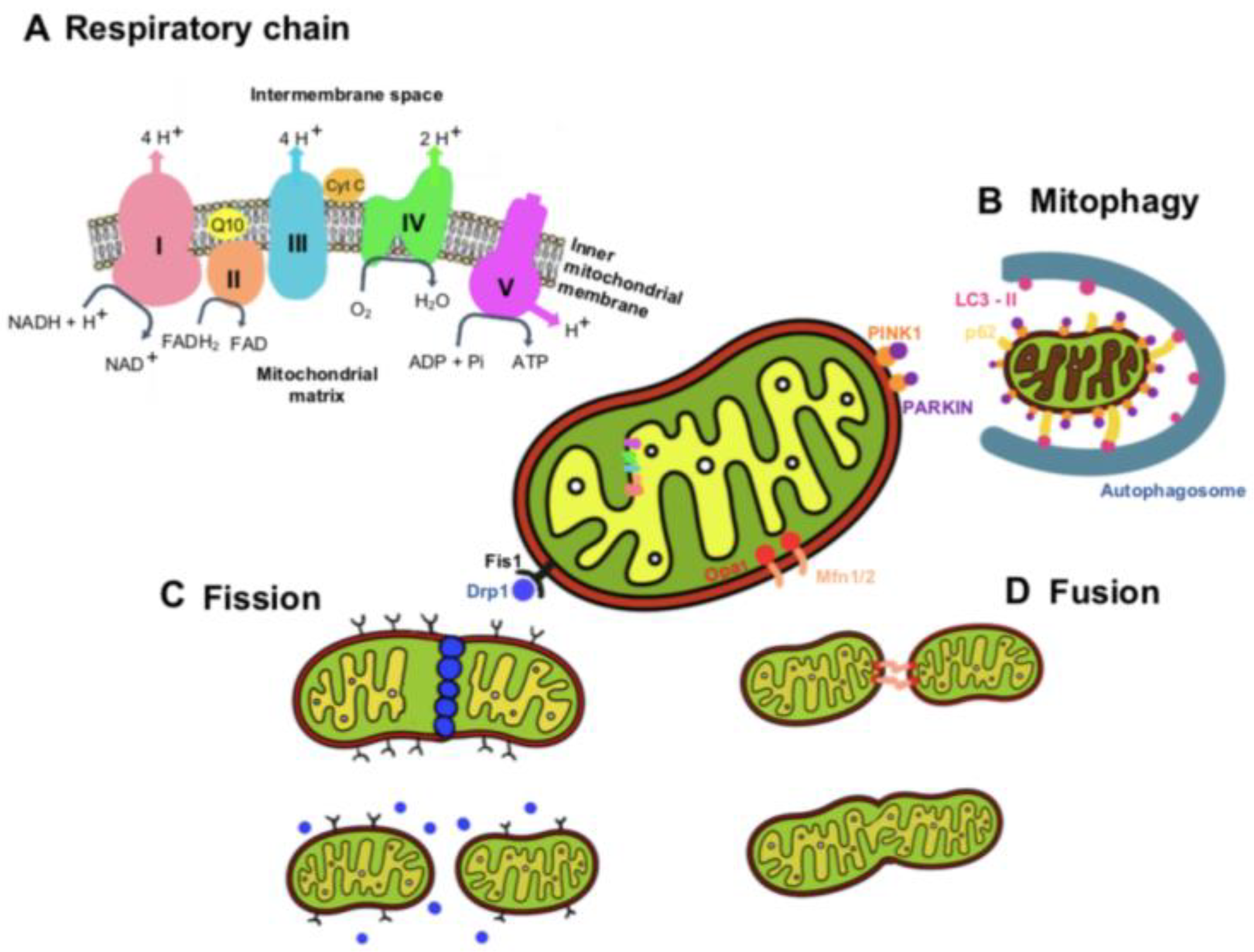
2. Mitochondria Functions and Dynamics

Mitochondria are double-membrane organelles responsible for energy production and homeostasis, the regulation of intracellular calcium levels, and the regulation of apoptosis (mainly via the intrinsic pathway) [17,18]. In addition, mitochondria are responsible for generating more than 90% of the energy for the cell through oxidative phosphorylation [19,20].
To generate ATP through oxidative phosphorylation, mitochondria use an electron transport chain inserted within the mitochondrion’s inner membrane (Figure 1) [21]. NADH and FADH2 are generated by the Krebs cycle and donate electrons to complex I (NADH: ubiquinone oxidoreductase) and complex II (succinate dehydrogenase), respectively. The electrons from NADH are passed from complex I to ubiquinone (CoQ) in order to enter the Q cycle, where CoQ is reduced to ubiquinol (QH2). This electron transfer induces the pumping of protons by complex I from the matrix into the intermembrane space. The electrons donated from FADH2 are transferred from complex II to CoQ similarly to complex I, although this process is not accompanied by proton translocation [21]. Once in the Q cycle, the electrons are transferred to complex III (coenzyme Q: cytochrome c reductase) and then to cytochrome c, releasing two protons into the intermembrane space. Then, when cytochrome c is reduced, it transports single electrons from complex III to complex IV (cytochrome c oxidase), where molecular oxygen is reduced to water. At complex IV, a total of eight protons are pumped from the matrix, of which four are used to form two water molecules, and the other four are transferred into the intermembrane space [21,22].
In response to electron transport, a total of ten protons are pumped from the matrix into the intermembrane space, where they accumulate to generate an electrochemical and concentration proton gradient that generates a proton motive force, essential for the activity of complex V (ATP synthase) to generate ATP [21]. A consequence of electron transfer is the generation of reactive oxygen species (ROS), which contributes to homeostatic signaling. However, when ROS are produced in excess, they cause oxidative stress and can lead to mitochondrial dysfunction and diseases [22]. Therefore, an efficient measurement of the electron transport chain function and ATP production, using high-resolution respirometry, such as a Seahorse XF24 Extracellular Flux Analyzer and oxygraphy, can provide insight into cellular physiology and dysfunction.
Mitochondria are highly dynamic organelles that undergo a continuous cycle of fission and fusion, processes called mitochondrial dynamics (Figure 1). Another dynamic process of mitochondria is the selective removal of dysfunctional mitochondria, a quality-control mechanism that ensures a healthy mitochondrial population. The dynamic properties of mitochondria are critical for their optimal function in energy generation [23]. Mitophagy is a mechanism of mitochondrial quality control used to eliminate damaged mitochondria and prevent excessive ROS production, thus maintaining homeostasis in mitochondria. Mitochondrial dynamics involve the plasma membrane and organelles, such as ER and lysosomes. The contact point of ER–mitochondria is referred to as mitochondria-associated ER membranes. Some studies have suggested that the integrity of mitochondria-associated ER membranes is required for insulin signaling (for a detailed description, see the revision [24]). Studies have been carried out to investigate the effect/defect of insulin signaling on different features of mitochondrial dysfunction, focusing on dynamics, biogenesis, and mitophagy and their role in pathologies in which metabolic dysmetabolism is comorbid with neurodegeneration [25,26]. Some studies have also suggested that the protective actions of leptin may be facilitated through the regulation of mitochondrial dynamics, namely, mitochondrial fission and fusion [27,28,29,30].
Dysfunctional mitochondria are recognized by the autophagy machinery, resulting in their engulfment by autophagosomes and trafficking to the lysosome for degradation. The most common mitophagy pathways are mediated by PINK1 and PARKIN proteins. Mitochondrial fission is the process where mitochondria divide into two separate mitochondrial organelles. Fission is mediated by the interaction between the mitochondrial fission factor (Mff) and dynamin-related protein-1 (Drp1). Briefly, Drp1 is recruited from a cytosolic pool onto the mitochondrial surface, where it self-assembles into spiral structures to facilitate fission, acting similarly to endocytic invaginations of the cell membrane. Several mitochondrial-bound proteins then aid in the recruitment of Drp1 to the mitochondria, including Fis1, Mff, MiD49, and MiD51 [23,31,32].
Fusion is the process of joining two adjacent mitochondria through a physical merging of the outer and then the inner mitochondrial membranes, resulting in the content mixing of the matrix components diffusing throughout the new mitochondrion. Fusion is mediated by the proteins mitofusin-1 (Mfn1) and mitofusin-2 (Mfn2) [33,34,35], located on the mitochondrial outer membrane. Mitofusins are required for outer membrane fusion. The fusion of the inner membrane is mediated by the protein optic atrophy 1 (Opa1), which is associated with the inner membrane ([36] for a detailed description of the mitochondria dynamics, please read the review manuscript [23]).
Mitochondrial dynamics is important for growth redistribution and maintenance in a healthy mitochondria network and plays a role in disease-related processes. All the cells consume energy for their homeostasis and specific activity, and they require the support of functional mitochondria that provide ATP obtained via oxidative phosphorylation. A reduction in mitochondria respiration and bioenergetics is associated with insulin resistance [24].
Therefore, the dysfunction of mitochondrial dynamics and function could lead to disorders in mitochondria, which are greatly associated with the progression of several diseases, including obesity and metabolic and neurological conditions.

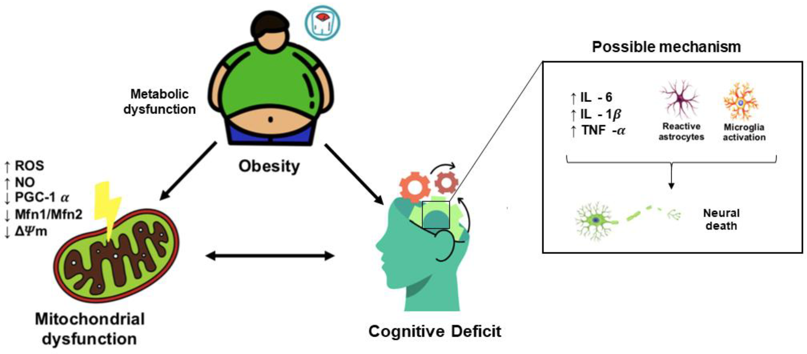
3. Obesity Induces Cognitive Decline

Obesity, as well as HFD diet consumption, and metabolic disorders, such as diabetes mellitus, are widely recognized as inducing impairments in brain structure and function in the form of memory dysfunction, as well as neurodegenerative diseases [37]. Furthermore, magnetic resonance imaging studies have demonstrated that regional brain atrophy and changes in gray and white matter are observed in patients with obesity, providing new insights into the relationship between obesity and cognitive decline from the imaging perspective [37,38,39,40]. Furthermore, a higher BMI is correlated with a lower gray matter volume in the prefrontal, temporal, insular, and occipital cortexes; thalamus; putamen; amygdala; and cerebellum, mediating the negative effects on memory performance [41].
Patients with obesity have an earlier onset of Alzheimer’s, which is considered an aging disease [42]. An 18-year follow-up longitudinal study demonstrated a higher degree of overweight in older women who developed AD. No associations were found in men [42]. In the same study, the authors concluded that Alzheimer’s disease risk increased by 36% for every 1.0 increase in BMI. In other studies, it has been shown that patients with a higher BMI present significantly lower scores in cognitive tests and a longitudinal decline in cognitive abilities in both men and women [37,43,44]. Changes in cognitive function can be potentiated since middle-aged adults with obesity may experience differentially greater brain atrophy [37]. The relationship between a higher BMI and reduced cognitive performance does not change with age [45] or race [46]. A high intake of fat and sugar is associated with impairments in hippocampal-dependent learning and memory in children [47] and adults [48,49], suggesting a negative impact on hippocampal function across the lifespan. In the community-based Framingham Offspring Cohort, it was observed that central obesity was significantly related to poorer performance in executive function and visuomotor skills, and no changes were observed for verbal memory [50]. Adults with overweight and obesity also have poorer executive function than normal-weight adults, without changes in performance on attention tests. Children and adolescents with overweight/obesity also present poor cognitive function on verbal, full-scale, and performance IQ; visual–spatial; and executive function tests [51,52]. A systemic review found that executive dysfunction is associated with obesity-related behaviors in children and adolescents, such as increased food intake, disinhibited eating, and less physical activity. In children and adolescents, obesity is associated with poorer cognitive competence and may affect their academic achievements [53].
Body weight and diet composition are modified risk factors for cognitive decline. Weight loss appears to be associated with low-order improvements in executive/attention functioning and memory in individuals with obesity. Moreover, a stable BMI predicts better cognitive trajectories [54]. Patients with severe obesity may obtain immediate verbal and delayed memory function benefits from Roux-en-Y gastric bypass [55,56].
Different animal models of obesity and metabolic disorders have also exhibited cognitive dysfunctions and worse performance in learning and memory tasks compared to non-obese animals [57,58,59,60,61,62]. In addition, based on studies of animal models and in vitro models, high levels of glucose and saturated fatty acids are responsible for neuroinflammation, microglia activation, mitochondrial dysfunction, neuronal loss, and impairments in synaptic plasticity (Figure 2) [63,64,65,66,67,68].

3.1. Obesity-Induced Cognitive Decline: Role of Neuroinflammation

Obesity is a low-grade chronic inflammatory disease that increases susceptibility to the numerous conditions associated with it. During the expansion of white adipose tissue, the recruitment and infiltration of immune cells, mainly macrophages, occur [69]. The growth of adipose tissue is also associated with an increased expression of proinflammatory cytokines, particularly interleukin-6 (IL-6), interleukin-1 beta (IL-1β), and tumor necrosis-α (TNF-α) [70,71,72]. Subjects with obesity have high circulating proinflammatory adipocytokines that trigger chronic inflammation. Systemic low-grade chronic inflammation has been reported to cause neuroinflammation and changes in different brain structures, such as the cerebellum, amygdala, cerebral cortex, and hypothalamus [57,73,74,75]. Obesity-induced inflammation has been related to changes in the integrity of blood–brain barrier permeability, inducing leukocyte extravasation, along with the potential entry of pathogens and toxins into the central nervous system, which, in turn, stimulate more inflammatory responses in a vicious cycle [57]. The decrease in tight junction protein expression and the disturbed blood–brain barrier are regulated by the NF-κB pathway, which increases the expression of proinflammatory proteins, such as IL-1β, TNFα, and IL-6. The loss of the blood–brain barrier during obesity facilitates proinflammatory cytokines to enter the brain parenchyma, thus allowing them to interact and activate glial cells (microglia and astrocytes) [76]. Activated microglia secrete more inflammatory cytokines (TNFα, IL-1β, and IL-6), perpetuating the neuroinflammation and leading to neuronal damage. NLRP3 proteins of the inflammasome secreted by visceral adipose tissue directly activate microglia through the IL1 receptor [77].
Furthermore, HFD can directly activate microglial cells, inducing morphological changes in the hypothalamus without causing microglial changes in the cerebral cortex and striatum. Moreover, HFD-induced obesity is associated with an increased entry of peripheral immune cells into the central nervous system and may contribute to the inflammatory response [78]. Fatty acid intake induces the activation of immune cells and the inflammatory response through the activation of the innate immune system through Toll-like receptors (TLRs) [63]. The binding of fatty acids to TLR4 activates nuclear factor-Κb (NF-Κb) and activator protein 1 (AP-1), which, in turn, upregulate the expression of proinflammatory cytokines and chemokines [79]. Another proposed mechanism for obesity-induced inflammation relies on the ability of an HFD to modulate the gut microbiota. Indeed, the subsequent changes in microbiota populations result in the permeabilization of the gut barrier, leading to the increased passage of bacterial endotoxins into the circulation [80,81].
Based on studies conducted on animal models of obesity, it has been proposed that inflammatory processes may participate in both the peripheral and brain disorders associated with obesity, including the development of cognitive alterations. Diet-induced obesity leads to microglia activation, which induces synaptic alterations, including impairment in hippocampal synaptic plasticity, reductions in dendritic spine density and the sites of excitatory synapses, and promoted synaptic stripping [58].

3.1.1. Hypothalamic Neuroinflammation

The hypothalamus is a master regulator of whole-body energy homeostasis and metabolism (see review [82]) through hormonal and nutrient-sensing mechanisms. The integrity of the hypothalamic nuclei that regulate appetite satiety is altered by neuroinflammation. Many studies have shown that exposure to obesity and HFD feeding strongly affects the hypothalamic mechanisms of energy homeostasis regulation [73,83,84,85].
Diet-induced obesity increases the activation of inflammation in the mediobasal hypothalamus, resulting in the production of proinflammatory cytokines (TNF-α, IL-1β, and IL-6) and impairment in insulin and leptin signaling [74]. Furthermore, the consumption of HFD triggers hypothalamic neuroinflammation by activating the Toll-like receptor 4 (TLR4) signaling pathways [86,87]. In addition to the increased levels of proinflammatory cytokines, several inflammatory and cellular stress responses, including endoplasmic reticulum (ER) stress, SOCS3, and the IKKβ/NF-κB pathways, have been shown to be upregulated [79]. Furthermore, HFD-induced obesity leads to microglia and astrocyte activation and contributes greatly to the neuroinflammatory tone [88,89,90,91].
Recent studies have also demonstrated that HFD consumption induces hypothalamic neuroinflammation, even before peripheral inflammation, insulin resistance, and obesity [64,73]. Hypothalamic inflammation was evident in rodent models of HFD within 1 to 3 days of HFD onset, before substantial weight gain, peripheral inflammation, and peripheral insulin resistance [73]. Furthermore, inhibiting microglia activation and blocking neuroinflammatory pathways in the hypothalamus prevents diet-induced obesity and the metabolic consequences associated with obesity [64,92,93,94].

3.1.2. Hippocampal Neuroinflammation

The hippocampus is a brain region primarily associated with learning and memory mechanisms. The hippocampus, located in the inner area of the temporal lobe, forms part of the limbic system, which is particularly important in regulating emotional responses. Hippocampal impairments can be found in the early phases of neurodegenerative dementias, including vascular dementia and Alzheimer’s disease. The hippocampus is very susceptible to damage by dietary factors, obesity, and metabolic disease [9,57,59,60,61,95]. Dietary fats are among the most proinflammatory components of the obesogenic diet. Saturated fatty acids induce the negative consequences of obesity, while monounsaturated fatty acids promote metabolic health.
Increased hippocampal neuroinflammation, induced by obesity or high-caloric diets, is correlated with deficits in learning and memory [95] and exacerbates cognitive decline in animal models of Alzheimer’s disease [96,97]. The cerebral cortex, also called gray matter, is the brain’s outermost layer, which plays a key role in many high-level functions, such as reasoning, emotion, problem solving, memory, language, and consciousness. Some studies have also demonstrated that HFD-induced obesity activates neuroinflammation mechanisms with harmful effects on the cerebral cortex [98,99,100].
In a genetic model of Alzheimer’s disease (APOE3 knock-in mice), the consumption of an HFD (40% fat) increased hippocampal gliosis as measured using GFAP and Iba1 immunostaining, which supports the hypothesis that the early dysregulation of inflammation could predispose to brain damage [97]. In the 3xTg-AD mice model, an HFD (60% fat) consumption elicited more severe astrogliosis, metabolic dysfunction, and weight gain than in wild-type mice [96]. Mice fed on an HFD for three months had increased hippocampal inflammatory cytokine production and a loss of synaptic protein expression. Dietary obesity impaired hippocampus-dependent memory, reduced long-term potentiation (LTP), and induced the expression of the activation marker major histocompatibility complex II (MHCII) in hippocampal microglia [58]. Rats fed an HFD had memory impairments, an effect that is augmented with a longer duration of HFD consumption and that is linked to elevated levels of IL-1β in the hippocampus [95].
In a rat model of a cafeteria diet supplemented with sucrose, animals exhibited increased hippocampal inflammation, with increased mRNA levels of TNF-α and IL-1β. In this model, the authors observed a strong negative correlation between TNF-α expression and memory performance [68]. Obesogenic western diets induced the expression of hippocampal cytokine levels (IL-1 β, TNF-α, and IL-6) and significantly impacted microglia morphology (increased microglia-shaped activation) [101]. High-fat diet consumption rapidly triggers hippocampal dysfunction associated with neuroinflammation, promoting a progressive breakdown of synaptic and metabolic functions. The consumption of a HFD for only three days induced cognitive deficits, increasing the hippocampus levels of TNF-α and IL-6. Depressive-like behavior was observed after day 5 of HFD consumption. Changes in proinflammatory cytokines accompanied changes in blood–brain barrier permeability [57]. This study highlights that hippocampus inflammation results from local cytokine signals in response to HFD, similar to hypothalamic inflammation [57,65,73].
In a genetic model of obesity, leptin-receptor-deficient db/db mice exhibited increased microglia activation (Iba1 measurements) with MHCII immunoreactivity and increased levels of IL-1β in the hippocampus. The levels of IL-1β presented a correlation between adiposity and cognitive impairment [102]. Inhibiting neuroinflammation using the intrahippocampal IL-1 receptor antibody or exercise prevented hippocampal microgliosis, synaptic dysfunction, and cognitive impairment [102,103]. Similar results were found in a rat model of high-fat-diet-induced obesity, in which treadmill exercise decreased the production of proinflammatory cytokines (IL-1 β and TNF-α) and cyclooxygenase-2 (COX-2), as well as inhibiting the TLR4 pathway (myeloid differentiation 88 and tumor necrosis factor receptor-associated factor 6 and the phosphorylation of transforming growth factor β-activated kinase 1, IkBα, and NF-Κb) [103].
In conclusion, neuroinflammation in the hippocampus is a potential mechanism for cognitive deficits induced by obesity and metabolic disorders.

3.2. Obesity-Induced Cognitive Decline: Role of Mitochondrial Dysfunction

Diabetes and obesity are modifiable risk factors for cognitive dysfunction and dementia. Several studies have demonstrated and identified overlapping neurodegenerative mechanisms observed in these disorders, including oxidative stress, mitochondrial dysfunction, and inflammation. An excessive intake of nutrients provokes the mitochondria to become overloaded with fatty acids and glucose, leading to an increase in the production of acetyl-CoA. This then causes the production of NADH through the Krebs cycle, which promotes an increase in the electron transfer chain in the mitochondria and, subsequently, increases ROS production, leading to oxidative stress. In addition, there is evidence that the brain’s energy status is decreased in obesity, although the underlying mechanisms are currently unknown. Patients with obesity have been found to have impaired cerebral energy gain upon experimentally increased blood glucose levels up to a postprandial status. This suggests that the brains of individuals with obesity cannot generate an appropriate amount of energy due to dysfunctional glucose transport or a downregulated energy synthesis in mitochondrial respiration [104].
More recent evidence highlights dietary fat’s impact on brain function and cognitive deficits, demonstrating that the long-term consumption of HFD induces impairments in mitochondrial brain function and brain insulin resistance [6,105,106,107,108]. Mitochondrial dysfunction is related to inflammation and other energy-dependent disturbances, where the generation of ROS exceeds the physiological antioxidant protective activity, causing cellular oxidative damage. Different animal models of obesity and metabolic dysfunction have demonstrated impairments in mitochondrial dysfunction and increased oxidative stress.
In a mouse model of diet-induced obesity and insulin resistance, in the brains of 16-week-old mice, the opening of the mitochondrial permeability transition pore (mPTP), the loss of mitochondrial membrane potential (ΔΨm), and apoptosis were observed, while insulin addition ameliorated these dysfunctions. An increase in fission-related proteins and the activation of mitophagy were also detected, which indicate that an alteration in the insulin pathway affects mitochondrial integrity and effective mitophagy [109]. In a type 1 diabetes rat model (streptozotocin, STZ), it was observed that oxygen consumption in basal state 4 significantly increased in the mitochondria from the hippocampus, cortex, and cerebellum. No relevant differences were observed in the activity of respiratory complexes, but hippocampal mitochondrial membrane potential was reduced [110]. Moreover, ADP-stimulated state 3 respiration and uncoupled maximal respiration (FCCP) were significantly decreased in the hippocampus of diabetic rats compared to controls [111]. Sirtuin-1 (SIRT1) is a major regulator of mitochondrial biogenesis and metabolism through the activation of proliferator-activated receptor gamma coactivator-1 α (PGC-1α). SIRT-1 and PGC-1α were also significantly decreased in the hippocampus of diabetic rats, which suggests an impairment in mitochondrial biogenesis and, consequently, a reduction in the mitochondrial respiratory capacity [111]. Sirtuin-3 (SIRT3) plays a significant role in enhancing mitochondrial protein function. The downregulation of SIRT3 is a key component of metabolic syndrome, a precondition for obesity and diabetes. In a genetic model of metabolic syndrome, Sirt3−/− mice fed a western diet had impaired brain mitochondrial respiration, lower levels of mitochondrial fission proteins Mfn1 and Mfn2, and hyperacetylated brain mitochondrial proteins [112]. The brains of Sirt3−/− mice also presented the downregulation of enzymes in several metabolic pathways, including fatty acid oxidation and the tricarboxylic acid cycle [112].
It was demonstrated that Zucker diabetic fatty (ZDF, FA/FA) rats had an increased ROS production in the brain, as well as increased nitric oxide (NO) production [113]. In addition, catalase activity in the brains of ZDF rats was significantly reduced. Compared with the lean model, glutathione metabolism and mitochondrial respiratory functions were negatively affected in ZDF rats. Specifically, complex I, II/III, and IV activities and mitochondrial ATP content were significantly decreased, indicating that the brain developed complications associated with redox homeostasis and mitochondrial dysfunction [113].
Rats fed an HFD for 12 weeks had increased brain and hippocampus ROS production (malondialdehyde, dichlorohydrofluoresceindiacetate (DCFHDA), and H2O2 levels), lipid peroxidation, brain mitochondrial depolarization (JC-1 marker), and brain mitochondrial swelling that results in cognitive decline [108,114,115,116]. It was observed that energy restriction combined with a dipeptidyl peptidase-4 inhibitor (vildagliptin) or vildagliptin alone for four weeks restored brain mitochondrial function, hippocampal synaptic plasticity, and cognitive function [115,116,117]. Vagal nerve stimulation therapy in rats fed an HFD for 12 weeks promoted an improvement in brain insulin sensitivity, decreased ROS production, attenuated mitochondrial brain dysfunction and cell apoptosis, and consequently improved cognitive function [118].
A mice model of HFD-induced obesity (50% fat for 18 weeks) demonstrated changes in brain cortex bioenergetics, with changes in mitochondrial function, efficiency, and oxidative stress [98]. Specifically, the authors showed that the brains of HFD animals used fatty acid as a preferential fuel source compared to control animals. Since dysfunctional synaptic mitochondria may lead to impaired neurotransmission and cognitive failure, using synaptosomal fractions demonstrated a decrease in basal respiration and the maximal rate of respiration and ATP production in HFD mice. The same study also revealed a significant reduction in the proton leak in the HFD mice [98]. In these animals’ cortex, glutathione (GSH) content significantly decreased compared to that in controls, and the HFD mice exhibited a lower GSH/GSSG ratio than the controls [98]. High-fat diet-induced obesity increased oxidative stress in the hippocampus with increased mitochondrial peroxide (H2O2) production. Mitochondria respiration (oxygen consumption) was significantly lower in obese mice than in lean mice [119]. Treadmill running for 12 weeks improved short-term memory and oxidative stress (decreased mitochondrial H2O2 production). The exercised animals had a higher mitochondrial O2 respiration capacity than that of a sedentary model of diet-induced obesity. Mitochondrial Ca2+ retention capacity was higher in the mice that performed exercise and, consequently, decreased mitochondrial permeability transition pore opening sensitivity and neuronal apoptosis [119].
The consumption of HFDs for only two weeks did not affect the mitochondrial activity in the hippocampus (analyzed using high-resolution respirometry). However, four weeks of HFD feeding induced mitochondrial dysfunction in hippocampal homogenates [57]. Four weeks of the consumption of HFD induced a significantly lower O2 consumption and caused a significant reduction in the electron transfer chain [57]. These animals had depressive-like behavior and changes in memory function after five days of HFD consumption [57].
In mice fed obesogenic diets (high-fat and high-glycemic diets) for 12 weeks, it was observed that changes in the brain cytoskeletal proteins, mitochondria, and metabolic proteins changed their post-translational status [120]. Specifically, proteins involved in mitochondrial functions were downregulated in mice fed obesogenic diets compared to lean mice. This suggests a reduced metabolism and a lower activity of mitochondria in obese mice [120]. In the brains of obese mice, lower expressions in nine proteins involved in mitochondrial activity were observed, namely, dehydrogenase [ubiquinone] iron-sulfur proteins 4 and 5 (NDUFS4 and NDUFS5, respectively) and cytochrome c oxidase subunit 7B, mitochondrial (COX7) (components of complex I and complex IV of the respiratory chain) [120]. Alterations in the mitochondrial respiration chain cause impairments in the functionality of the cells. The glutaredoxin (GRX) enzymes are glutathione-dependent oxidoreductases with important roles in regulating cytosolic thiol/disulfide balance and protecting proteins from oxidative damage. The hippocampus of GRX2-knockout mice fed a high-fat diet had worse oxidative stress and mitochondrial impairment than wild-type mice. These results provide evidence that preventing oxidative damage may have protective functions in HFD-elicited brain injury and cognitive deficits [121].
In the hypothalamus, mitochondrial dynamics regulate energy homeostasis and metabolism [122,123]. Although inflammation is one of the mechanisms involved in hypothalamic neuronal defects in diet-induced obesity, mitochondrial abnormalities also occur [65,124,125,126]. In the hypothalamus, mitochondrial fusion regulates neuronal firing via the modulation of intracellular ATP levels in diet-induced obesity. The deletion of Mfn2 in the anorectic pro-opiomelanocortin (POMC) neurons of the hypothalamus disrupts endoplasmic reticulum (ER)–mitochondria contacts, ER stress activation, leptin resistance, and obesity (Schneeberger et al., 2013). However, the deletion of Mfn2 in orexigenic agouti-related peptide (Agrp) neurons induced less weight gain in mice fed a HFD [126]. High-fat diet consumption for one day caused a transient reduction in Mfn2, although after seven days of the consumption of this diet, the expression of Mfn2 increased [65]. HFD diet consumption for five weeks promoted the upregulation of Mfn2 in the arcuate nucleus and downregulation after 13 weeks of HFD consumption [127]. Using an in vitro model of hypothalamic neuronal cells, the authors demonstrated that saturated palmitic acid decreased the protein levels of mitofusin-2 and activated the ER stress response, exacerbating insulin resistance in hypothalamic neuronal cells [127]. Neuronal cell treatment with a high concentration of palmitic acid increased mitochondria ROS production. Insulin resistance induced by palmitic acid was prevented by treatment with the anti-inflammatory reagent and ER stress inhibitors [127].
Under physiological conditions, the redox signaling pathway initiates the hypothalamus glucose-sensing mechanism. Obese Zücker rats had cerebral hypersensitivity to glucose, leading to abnormal vagus-induced insulin secretion [128]. The impaired hypothalamic glucose sensing in obese Zücker rats is linked to abnormal redox signaling, which originates from mitochondria dysfunction. In the hypothalamus of obese Zücker rats, abnormal glutathione redox state and increased ROS production occur. In this animal model, mitochondria activity in complexes I and IV in the hypothalamus was significantly increased. The total respiratory capacity was also increased. The expressions of complexes I, II, III, and IV were raised in the hypothalamic mitochondria of obese rats, and no changes were observed in complex V [128].
In a comparison of a diet-induced obesity-resistant mouse strain (WSB/EiJ to the obesity-prone mouse strain (C57BL/6J strain), it was possible to observe that the resistant strain displayed a lower inflammatory status, both peripherally and centrally (less activated microglia in the hypothalamus), and more reactive and responsive mitochondria in the hypothalamus. In the hypothalamus of the resistant strain, an increase occurred in ATP content after eight weeks of a high-fat diet [129]. This indicates that the reduced inflammatory responses and increased mitochondrial activity contribute to diet-induced obesity resistance. Independent of the mouse strain, eight weeks of high-fat diet feeding induces a decrease in each mitochondria area [122].
The consumption of a western diet increases the circulating levels of palmitate, which is converted into ceramide in order to accumulate in tissues in response to obesity. Pharmacological and genetic strategies that reduce tissue ceramide levels reverse the metabolic consequences of obesity. Specifically, reducing ceramide production protects mice from the metabolic effects of high-fat diet consumption by preventing the fragmentation of the mitochondrial network within the hypothalamus. Restoring mitochondrial function through the decreased accumulation of ceramide increases leptin sensitivity and, consequently, reduces food intake [130].
Targeting mitochondrial dysfunction and oxidative stress could have potential benefits for cognitive dysfunction. N-acetylcysteine (NAC) is used to treat several diseases related to oxidative stress and inflammation due to its antioxidant and anti-inflammatory properties [131]. In a rat model of high-fat diet-induced obesity, NAC treatments improve glycemia and peripheral insulin resistance and reduce the oxidative stress/neuroinflammation/inflammasome activation axis in the cerebral cortex [132]. In a mouse model of obesity, NAC prevents hippocampal alterations and memory impairment [133]. In older humans, GlyNAC (a combination of glycine and N-acetylcysteine NAC) supplementation for 24 weeks decreases oxidative stress and mitochondrial dysfunction; reduces inflammation, insulin resistance, and endothelial dysfunction; and improves cognition [134]. Although additional studies are needed to address these effects in humans, these studies suggest that treatments, such as NAC, that reduce oxidative stress and improve mitochondria could be used as a simple and viable method to promote brain health.
In summary, various studies have demonstrated that diet-induced obesity and metabolic disorders induce mitochondrial dysfunction and oxidative stress in the brain, contributing to neuronal dysfunction, the dysregulation of whole-body metabolism, and cognitive deficits.

4. Conclusions

In summary, the findings suggest that high-fat and high-caloric diet consumption, as well as obesity and associated type 2 diabetes, triggers brain dysfunction associated with neuroinflammation and mitochondrial dysfunction. Neuroinflammation and mitochondrial dysfunction could be some of the mechanisms involved in cognitive deficits and dementia associated with obesity and metabolic disorders. Treatments that ameliorate brain mitochondrial dysfunction and decrease hippocampal oxidative stress levels have beneficial effects on cognitive processes. In addition to elucidating the link between diet and cognitive function, it might be relevant to comprehend the neurodegenerative process.

Author Contributions

Writing—original draft preparation, L.O.S. and J.M.G.; writing—review and editing, L.O.S. and J.M.G. All authors have read and agreed to the published version of the manuscript.

Funding

L.O.S. was financed by the Coordenação de Aperfeiçoamento de Pessoal de Nível Superior —Brasil (CAPES)—Finance Code 001.

Conflicts of Interest

The authors declare no conflict of interest.

References

  1. Blüher, M. Obesity: Global epidemiology and pathogenesis. Nat. Rev. Endocrinol. 2019, 15, 288–298. [Google Scholar] [CrossRef]
  2. Collaborators, G.B.D.O.; Afshin, A.; Forouzanfar, M.H.; Reitsma, M.B.; Sur, P.; Estep, K.; Lee, A.; Marczak, L.; Mokdad, A.H.; Moradi-Lakeh, M.; et al. Health Effects of Overweight and Obesity in 195 Countries over 25 Years. N. Engl. J. Med. 2017, 377, 13–27. [Google Scholar] [CrossRef]
  3. Morys, F.; Dadar, M.; Dagher, A. Association Between Midlife Obesity and Its Metabolic Consequences, Cerebrovascular Disease, and Cognitive Decline. J. Clin. Endocrinol. Metab. 2021, 106, e4260–e4274. [Google Scholar] [CrossRef]
  4. De Lorenzo, A.; Gratteri, S.; Gualtieri, P. Why primary obesity is a disease? J. Transl. Med. 2019, 17, 169. [Google Scholar] [CrossRef] [Green Version]
  5. Pratchayasakul, W.; Kerdphoo, S.; Petsophonsakul, P.; Pongchaidecha, A.; Chattipakorn, N.; Chattipakorn, S. Effects of high-fat diet on insulin receptor function in rat hippocampus and the level of neuronal corticosterone. Life Sci. 2011, 88, 619–627. [Google Scholar] [CrossRef]
  6. Pipatpiboon, N.; Pratchayasakul, W.; Chattipakorn, N.; Chattipakorn, S.C. PPARγ agonist improves neuronal insulin receptor function in hippocampus and brain mitochondria function in rats with insulin resistance induced by long term high-fat diets. Endocrinology 2012, 153, 329–338. [Google Scholar] [CrossRef]
  7. Abbasnejad, Z.; Nasseri, B.; Zardooz, H.; Ghasemi, R. Time-course study of high fat diet induced alterations in spatial memory, hippocampal JNK, P38, ERK and Akt activity. Metab. Brain Dis. 2019, 34, 659–673. [Google Scholar] [CrossRef]
  8. Gómez-Apo, E.; Mondragón-Maya, A.; Ferrari-Díaz, M.; Silva-Pereyra, J. Structural Brain Changes Associated with Overweight and Obesity. J. Obes. 2021, 2021, 6613385. [Google Scholar] [CrossRef]
  9. Gaspar, J.M.; Baptista, F.I.; Macedo, M.P.; Ambrósio, A.F. Inside the Diabetic Brain: Role of Different Players Involved in Cognitive Decline. ACS Chem. Neurosci. 2016, 7, 131–142. [Google Scholar] [CrossRef]
  10. Gispen, W.H.; Biessels, G.J. Cognition and synaptic plasticity in diabetes mellitus. Trends Neurosci. 2000, 23, 542–549. [Google Scholar] [CrossRef] [PubMed]
  11. Fitzpatrick, A.L.; Kuller, L.H.; Lopez, O.L.; Diehr, P.; O’Meara, E.S.; Longstreth, W.T., Jr.; Luchsinger, J.A. Midlife and late-life obesity and the risk of dementia: Cardiovascular health study. Arch. Neurol. 2009, 66, 336–342. [Google Scholar] [CrossRef] [PubMed] [Green Version]
  12. Hughes, T.F.; Borenstein, A.R.; Schofield, E.; Wu, Y.; Larson, E.B. Association between late-life body mass index and dementia: The Kame Project. Neurology 2009, 72, 1741–1746. [Google Scholar] [CrossRef] [PubMed] [Green Version]
  13. Whitmer, R.A.; Gunderson, E.P.; Quesenberry, C.P., Jr.; Zhou, J.; Yaffe, K. Body mass index in midlife and risk of Alzheimer disease and vascular dementia. Curr. Alzheimer Res. 2007, 4, 103–109. [Google Scholar] [CrossRef] [PubMed]
  14. Biessels, G.J.; Staekenborg, S.; Brunner, E.; Brayne, C.; Scheltens, P. Risk of dementia in diabetes mellitus: A systematic review. Lancet. Neurol. 2006, 5, 64–74. [Google Scholar] [CrossRef] [PubMed]
  15. Lu, F.P.; Lin, K.P.; Kuo, H.K. Diabetes and the risk of multi-system aging phenotypes: A systematic review and meta-analysis. PLoS ONE 2009, 4, e4144. [Google Scholar] [CrossRef] [Green Version]
  16. Marseglia, A.; Darin-Mattsson, A.; Skoog, J.; Rydén, L.; Hadarsson-Bodin, T.; Kern, S.; Rydberg Sterner, T.; Shang, Y.; Zettergren, A.; Westman, E.; et al. Metabolic Syndrome Is Associated With Poor Cognition: A Population-Based Study of 70-Year-Old Adults Without Dementia. J. Gerontol. Ser. A 2021, 76, 2275–2283. [Google Scholar] [CrossRef]
  17. Giacomello, M.; Pyakurel, A.; Glytsou, C.; Scorrano, L. The cell biology of mitochondrial membrane dynamics. Nat. Rev. Mol. Cell Biol. 2020, 21, 204–224. [Google Scholar] [CrossRef]
  18. Bras, M.; Queenan, B.; Susin, S.A. Programmed cell death via mitochondria: Different modes of dying. Biochemistry 2005, 70, 231–239. [Google Scholar] [CrossRef]
  19. Mitchell, P. Coupling of Phosphorylation to Electron and Hydrogen Transfer by a Chemi-Osmotic type of Mechanism. Nature 1961, 191, 144–148. [Google Scholar] [CrossRef]
  20. Hatefi, Y. The Mitochondrial Electron Transport and Oxidative Phosphorylation System. Annu. Rev. Biochem. 1985, 54, 1015–1069. [Google Scholar] [CrossRef]
  21. Zhao, R.Z.; Jiang, S.; Zhang, L.; Yu, Z.B. Mitochondrial electron transport chain, ROS generation and uncoupling. Int. J. Mol. Med. 2019, 44, 3–15. [Google Scholar] [CrossRef] [PubMed] [Green Version]
  22. Nolfi-Donegan, D.; Braganza, A.; Shiva, S. Mitochondrial electron transport chain: Oxidative phosphorylation, oxidant production, and methods of measurement. Redox Biol. 2020, 37, 101674. [Google Scholar] [CrossRef] [PubMed]
  23. Chan, D.C. Mitochondrial Dynamics and Its Involvement in Disease. Annu. Rev. Pathol. Mech. Dis. 2020, 15, 235–259. [Google Scholar] [CrossRef] [Green Version]
  24. Galizzi, G.; Di Carlo, M. Insulin and Its Key Role for Mitochondrial Function/Dysfunction and Quality Control: A Shared Link between Dysmetabolism and Neurodegeneration. Biology 2022, 11, 943. [Google Scholar] [CrossRef] [PubMed]
  25. Cardoso, S.; López, I.P.; Piñeiro-Hermida, S.; Pichel, J.G.; Moreira, P.I. IGF1R Deficiency Modulates Brain Signaling Pathways and Disturbs Mitochondria and Redox Homeostasis. Biomedicines 2021, 9, 158. [Google Scholar] [CrossRef]
  26. Ribeiro, M.; Rosenstock, T.R.; Oliveira, A.M.; Oliveira, C.R.; Rego, A.C. Insulin and IGF-1 improve mitochondrial function in a PI-3K/Akt-dependent manner and reduce mitochondrial generation of reactive oxygen species in Huntington’s disease knock-in striatal cells. Free. Radic. Biol. Med. 2014, 74, 129–144. [Google Scholar] [CrossRef]
  27. Cheng, Y.; Buchan, M.; Vitanova, K.; Aitken, L.; Gunn-Moore, F.J.; Ramsay, R.R.; Doherty, G. Neuroprotective actions of leptin facilitated through balancing mitochondrial morphology and improving mitochondrial function. J. Neurochem. 2020, 155, 191–206. [Google Scholar] [CrossRef] [Green Version]
  28. Jong, C.J.; Yeung, J.; Tseung, E.; Karmazyn, M. Leptin-induced cardiomyocyte hypertrophy is associated with enhanced mitochondrial fission. Mol. Cell. Biochem. 2019, 454, 33–44. [Google Scholar] [CrossRef]
  29. Yang, F.; Li, B.; Yang, Y.; Huang, M.; Liu, X.; Zhang, Y.; Liu, H.; Zhang, L.; Pan, Y.; Tian, S.; et al. Leptin enhances glycolysis via OPA1-mediated mitochondrial fusion to promote mesenchymal stem cell survival. Int. J. Mol. Med. 2019, 44, 301–312. [Google Scholar] [CrossRef]
  30. Wauman, J.; Tavernier, J. The intracellular domain of the leptin receptor prevents mitochondrial depolarization and mitophagy. Biochim. Biophys. Acta. Mol. Cell Res. 2018, 1865, 1312–1325. [Google Scholar] [CrossRef]
  31. Otera, H.; Wang, C.; Cleland, M.M.; Setoguchi, K.; Yokota, S.; Youle, R.J.; Mihara, K. Mff is an essential factor for mitochondrial recruitment of Drp1 during mitochondrial fission in mammalian cells. J. Cell Biol. 2010, 191, 1141–1158. [Google Scholar] [CrossRef] [Green Version]
  32. Losón, O.C.; Song, Z.; Chen, H.; Chan, D.C. Fis1, Mff, MiD49, and MiD51 mediate Drp1 recruitment in mitochondrial fission. Mol. Biol. Cell 2013, 24, 659–667. [Google Scholar] [CrossRef]
  33. Chen, H.; Detmer, S.A.; Ewald, A.J.; Griffin, E.E.; Fraser, S.E.; Chan, D.C. Mitofusins Mfn1 and Mfn2 coordinately regulate mitochondrial fusion and are essential for embryonic development. J. Cell Biol. 2003, 160, 189–200. [Google Scholar] [CrossRef]
  34. Koshiba, T.; Detmer, S.A.; Kaiser, J.T.; Chen, H.; McCaffery, J.M.; Chan, D.C. Structural basis of mitochondrial tethering by mitofusin complexes. Science 2004, 305, 858–862. [Google Scholar] [CrossRef] [Green Version]
  35. Qi, Y.; Yan, L.; Yu, C.; Guo, X. Structures of human mitofusin 1 provide insight into mitochondrial tethering. J. Cell Biol. 2016, 215, 621–629. [Google Scholar] [CrossRef] [Green Version]
  36. Song, Z.; Ghochani, M.; McCaffery, J.M.; Frey, T.G.; Chan, D.C. Mitofusins and OPA1 mediate sequential steps in mitochondrial membrane fusion. Mol. Biol. Cell 2009, 20, 3525–3532. [Google Scholar] [CrossRef]
  37. Ward, M.A.; Carlsson, C.M.; Trivedi, M.A.; Sager, M.A.; Johnson, S.C. The effect of body mass index on global brain volume in middle-aged adults: A cross sectional study. BMC Neurol. 2005, 5, 23. [Google Scholar] [CrossRef] [Green Version]
  38. Haltia, L.T.; Viljanen, A.; Parkkola, R.; Kemppainen, N.; Rinne, J.O.; Nuutila, P.; Kaasinen, V. Brain white matter expansion in human obesity and the recovering effect of dieting. J. Clin. Endocrinol. Metab. 2007, 92, 3278–3284. [Google Scholar] [CrossRef] [Green Version]
  39. Gazdzinski, S.; Kornak, J.; Weiner, M.W.; Meyerhoff, D.J. Body mass index and magnetic resonance markers of brain integrity in adults. Ann. Neurol. 2008, 63, 652–657. [Google Scholar] [CrossRef] [Green Version]
  40. Chen, R.; Cai, G.; Xu, S.; Sun, Q.; Luo, J.; Wang, Y.; Li, M.; Lin, H.; Liu, J. Body mass index related to executive function and hippocampal subregion volume in subjective cognitive decline. Front. Aging Neurosci. 2022, 14, 905035. [Google Scholar] [CrossRef] [PubMed]
  41. Kharabian Masouleh, S.; Arélin, K.; Horstmann, A.; Lampe, L.; Kipping, J.A.; Luck, T.; Riedel-Heller, S.G.; Schroeter, M.L.; Stumvoll, M.; Villringer, A.; et al. Higher body mass index in older adults is associated with lower gray matter volume: Implications for memory performance. Neurobiol. Aging 2016, 40, 1–10. [Google Scholar] [CrossRef] [PubMed]
  42. Gustafson, D.; Rothenberg, E.; Blennow, K.; Steen, B.; Skoog, I. An 18-year follow-up of overweight and risk of Alzheimer disease. Arch. Intern. Med. 2003, 163, 1524–1528. [Google Scholar] [CrossRef] [PubMed] [Green Version]
  43. Dahl, A.; Hassing, L.B.; Fransson, E.; Berg, S.; Gatz, M.; Reynolds, C.A.; Pedersen, N.L. Being overweight in midlife is associated with lower cognitive ability and steeper cognitive decline in late life. J. Gerontol. Ser. A Biol. Sci. Med. Sci. 2010, 65, 57–62. [Google Scholar] [CrossRef] [PubMed] [Green Version]
  44. Gunstad, J.; Lhotsky, A.; Wendell, C.R.; Ferrucci, L.; Zonderman, A.B. Longitudinal examination of obesity and cognitive function: Results from the Baltimore longitudinal study of aging. Neuroepidemiology 2010, 34, 222–229. [Google Scholar] [CrossRef] [PubMed] [Green Version]
  45. Gunstad, J.; Paul, R.H.; Cohen, R.A.; Tate, D.F.; Spitznagel, M.B.; Gordon, E. Elevated body mass index is associated with executive dysfunction in otherwise healthy adults. Compr. Psychiatry 2007, 48, 57–61. [Google Scholar] [CrossRef] [PubMed]
  46. Quaye, E.; Galecki, A.T.; Tilton, N.; Whitney, R.; Briceño, E.M.; Elkind, M.S.V. Association of Obesity with Cognitive Decline in Black and White Americans. Neurology 2022. [Google Scholar] [CrossRef]
  47. Baym, C.L.; Khan, N.A.; Monti, J.M.; Raine, L.B.; Drollette, E.S.; Moore, R.D.; Scudder, M.R.; Kramer, A.F.; Hillman, C.H.; Cohen, N.J. Dietary lipids are differentially associated with hippocampal-dependent relational memory in prepubescent children. Am. J. Clin. Nutr. 2014, 99, 1026–1032. [Google Scholar] [CrossRef] [Green Version]
  48. Francis, H.M.; Stevenson, R.J. Higher reported saturated fat and refined sugar intake is associated with reduced hippocampal-dependent memory and sensitivity to interoceptive signals. Behav. Neurosci. 2011, 125, 943–955. [Google Scholar] [CrossRef]
  49. Attuquayefio, T.; Stevenson, R.J.; Oaten, M.J.; Francis, H.M. A four-day Western-style dietary intervention causes reductions in hippocampal-dependent learning and memory and interoceptive sensitivity. PLoS ONE 2017, 12, e0172645. [Google Scholar] [CrossRef] [Green Version]
  50. Wolf, P.A.; Beiser, A.; Elias, M.F.; Au, R.; Vasan, R.S.; Seshadri, S. Relation of obesity to cognitive function: Importance of central obesity and synergistic influence of concomitant hypertension. The Framingham Heart Study. Curr. Alzheimer Res. 2007, 4, 111–116. [Google Scholar] [CrossRef]
  51. Snyder, L.L.; Foland-Ross, L.C.; Cato, A.; Reiss, A.L.; Shah, C.; Hossain, J.; Elmufti, H.; Nelly, M. Impact of dysglycemia and obesity on the brain in adolescents with and without type 2 diabetes: A pilot study. Pediatr. Diabetes 2022, 23, 1674–1686. [Google Scholar] [CrossRef] [PubMed]
  52. Cserjési, R.; Molnár, D.; Luminet, O.; Lénárd, L. Is there any relationship between obesity and mental flexibility in children? Appetite 2007, 49, 675–678. [Google Scholar] [CrossRef] [PubMed]
  53. Liang, J.; Matheson, B.E.; Kaye, W.H.; Boutelle, K.N. Neurocognitive correlates of obesity and obesity-related behaviors in children and adolescents. Int. J. Obes. 2014, 38, 494–506. [Google Scholar] [CrossRef] [Green Version]
  54. Beeri, M.S.; Tirosh, A.; Lin, H.M.; Golan, S.; Boccara, E.; Sano, M.; Zhu, C.W. Stability in BMI over time is associated with a better cognitive trajectory in older adults. Alzheimer’s Dement. 2022, 18, 2131–2139. [Google Scholar] [CrossRef] [PubMed]
  55. Li, C.M.; Song, J.R.; Zhao, J.; Wang, C.F.; Zhang, C.S.; Wang, H.D.; Zhang, Q.; Liu, D.F.; Ma, Z.Y.; Yuan, J.H.; et al. The effects of bariatric surgery on cognition in patients with obesity: A systematic review and meta-analysis. Surg. Obes. Relat. Dis. 2022, 18, 1323–1338. [Google Scholar] [CrossRef]
  56. Dardano, A.; Aghakhanyan, G.; Moretto, C.; Ciccarone, A.; Bellini, R.; Ceccarini, G.; Sancho Bornez, V.; Santini, F.; Volterrani, D.; Del Prato, S.; et al. Brain effect of bariatric surgery in people with obesity. Int. J. Obes. 2022, 46, 1671–1677. [Google Scholar] [CrossRef]
  57. de Paula, G.C.; Brunetta, H.S.; Engel, D.F.; Gaspar, J.M.; Velloso, L.A.; Engblom, D.; de Oliveira, J.; de Bem, A.F. Hippocampal Function Is Impaired by a Short-Term High-Fat Diet in Mice: Increased Blood-Brain Barrier Permeability and Neuroinflammation as Triggering Events. Front. Neurosci. 2021, 15, 734158. [Google Scholar] [CrossRef]
  58. Hao, S.; Dey, A.; Yu, X.; Stranahan, A.M. Dietary obesity reversibly induces synaptic stripping by microglia and impairs hippocampal plasticity. Brain Behav. Immun. 2016, 51, 230–239. [Google Scholar] [CrossRef] [Green Version]
  59. Hsu, T.M.; Konanur, V.R.; Taing, L.; Usui, R.; Kayser, B.D.; Goran, M.I.; Kanoski, S.E. Effects of sucrose and high fructose corn syrup consumption on spatial memory function and hippocampal neuroinflammation in adolescent rats. Hippocampus 2015, 25, 227–239. [Google Scholar] [CrossRef]
  60. Stranahan, A.M.; Arumugam, T.V.; Cutler, R.G.; Lee, K.; Egan, J.M.; Mattson, M.P. Diabetes impairs hippocampal function through glucocorticoid-mediated effects on new and mature neurons. Nat. Neurosci. 2008, 11, 309–317. [Google Scholar] [CrossRef]
  61. Boitard, C.; Cavaroc, A.; Sauvant, J.; Aubert, A.; Castanon, N.; Layé, S.; Ferreira, G. Impairment of hippocampal-dependent memory induced by juvenile high-fat diet intake is associated with enhanced hippocampal inflammation in rats. Brain Behav. Immun. 2014, 40, 9–17. [Google Scholar] [CrossRef] [PubMed]
  62. Lizarbe, B.; Soares, A.F.; Larsson, S.; Duarte, J.M.N. Neurochemical Modifications in the Hippocampus, Cortex and Hypothalamus of Mice Exposed to Long-Term High-Fat Diet. Front. Neurosci. 2018, 12, 985. [Google Scholar] [CrossRef] [PubMed]
  63. Howe, A.M.; Burke, S.; O’Reilly, M.E.; McGillicuddy, F.C.; Costello, D.A. Palmitic Acid and Oleic Acid Differently Modulate TLR2-Mediated Inflammatory Responses in Microglia and Macrophages. Mol. Neurobiol. 2022, 59, 2348–2362. [Google Scholar] [CrossRef]
  64. Valdearcos, M.; Robblee, M.M.; Benjamin, D.I.; Nomura, D.K.; Xu, A.W.; Koliwad, S.K. Microglia dictate the impact of saturated fat consumption on hypothalamic inflammation and neuronal function. Cell Rep. 2014, 9, 2124–2138. [Google Scholar] [CrossRef] [PubMed] [Green Version]
  65. Carraro, R.S.; Souza, G.F.; Solon, C.; Razolli, D.S.; Chausse, B.; Barbizan, R.; Victorio, S.C.; Velloso, L.A. Hypothalamic mitochondrial abnormalities occur downstream of inflammation in diet-induced obesity. Mol. Cell. Endocrinol. 2017, 460, 238–245. [Google Scholar] [CrossRef]
  66. Dalvi, P.S.; Chalmers, J.A.; Luo, V.; Han, D.Y.; Wellhauser, L.; Liu, Y.; Tran, D.Q.; Castel, J.; Luquet, S.; Wheeler, M.B.; et al. High fat induces acute and chronic inflammation in the hypothalamus: Effect of high-fat diet, palmitate and TNF-alpha on appetite-regulating NPY neurons. Int. J. Obes. 2017, 41, 149–158. [Google Scholar] [CrossRef] [PubMed]
  67. Gaspar, J.M.; Castilho, Á.; Baptista, F.I.; Liberal, J.; Ambrósio, A.F. Long-term exposure to high glucose induces changes in the content and distribution of some exocytotic proteins in cultured hippocampal neurons. Neuroscience 2010, 171, 981–992. [Google Scholar] [CrossRef] [PubMed]
  68. Beilharz, J.E.; Maniam, J.; Morris, M.J. Short exposure to a diet rich in both fat and sugar or sugar alone impairs place, but not object recognition memory in rats. Brain Behav. Immun. 2014, 37, 134–141. [Google Scholar] [CrossRef]
  69. Xu, H.; Barnes, G.T.; Yang, Q.; Tan, G.; Yang, D.; Chou, C.J.; Sole, J.; Nichols, A.; Ross, J.S.; Tartaglia, L.A.; et al. Chronic inflammation in fat plays a crucial role in the development of obesity-related insulin resistance. J. Clin. Investig. 2003, 112, 1821–1830. [Google Scholar] [CrossRef]
  70. Hotamisligil, G.S.; Shargill, N.S.; Spiegelman, B.M. Adipose expression of tumor necrosis factor-alpha: Direct role in obesity-linked insulin resistance. Science 1993, 259, 87–91. [Google Scholar] [CrossRef]
  71. Hotamisligil, G.S.; Arner, P.; Caro, J.F.; Atkinson, R.L.; Spiegelman, B.M. Increased adipose tissue expression of tumor necrosis factor-alpha in human obesity and insulin resistance. J. Clin. Investig. 1995, 95, 2409–2415. [Google Scholar] [CrossRef] [PubMed]
  72. Fain, J.N.; Madan, A.K.; Hiler, M.L.; Cheema, P.; Bahouth, S.W. Comparison of the release of adipokines by adipose tissue, adipose tissue matrix, and adipocytes from visceral and subcutaneous abdominal adipose tissues of obese humans. Endocrinology 2004, 145, 2273–2282. [Google Scholar] [CrossRef] [PubMed] [Green Version]
  73. Thaler, J.P.; Yi, C.X.; Schur, E.A.; Guyenet, S.J.; Hwang, B.H.; Dietrich, M.O.; Zhao, X.; Sarruf, D.A.; Izgur, V.; Maravilla, K.R.; et al. Obesity is associated with hypothalamic injury in rodents and humans. J. Clin. Investig. 2012, 122, 153–162. [Google Scholar] [CrossRef] [Green Version]
  74. De Souza, C.T.; Araujo, E.P.; Bordin, S.; Ashimine, R.; Zollner, R.L.; Boschero, A.C.; Saad, M.J.; Velloso, L.A. Consumption of a fat-rich diet activates a proinflammatory response and induces insulin resistance in the hypothalamus. Endocrinology 2005, 146, 4192–4199. [Google Scholar] [CrossRef] [PubMed] [Green Version]
  75. Yu, G.; Cao, F.; Hou, T.; Cheng, Y.; Jia, B.; Yu, L.; Chen, W.; Xu, Y.; Chen, M.; Wang, Y. Astrocyte reactivation in medial prefrontal cortex contributes to obesity-promoted depressive-like behaviors. J. Neuroinflammation 2022, 19, 166. [Google Scholar] [CrossRef]
  76. Salas-Venegas, V.; Flores-Torres, R.P.; Rodríguez-Cortés, Y.M.; Rodríguez-Retana, D.; Ramírez-Carreto, R.J.; Concepción-Carrillo, L.E.; Pérez-Flores, L.J.; Alarcón-Aguilar, A.; López-Díazguerrero, N.E.; Gómez-González, B.; et al. The Obese Brain: Mechanisms of Systemic and Local Inflammation, and Interventions to Reverse the Cognitive Deficit. Front. Integr. Neurosci. 2022, 16, 798995. [Google Scholar] [CrossRef] [PubMed]
  77. Guo, D.-H.; Yamamoto, M.; Hernandez, C.M.; Khodadadi, H.; Baban, B.; Stranahan, A.M. Visceral adipose NLRP3 impairs cognition in obesity via IL-1R1 on CX3CR1+ cells. J. Clin. Investig. 2020, 130, 1961–1976. [Google Scholar] [CrossRef] [Green Version]
  78. Buckman, L.B.; Hasty, A.H.; Flaherty, D.K.; Buckman, C.T.; Thompson, M.M.; Matlock, B.K.; Weller, K.; Ellacott, K.L. Obesity induced by a high-fat diet is associated with increased immune cell entry into the central nervous system. Brain Behav. Immun. 2014, 35, 33–42. [Google Scholar] [CrossRef] [Green Version]
  79. Zhang, X.; Zhang, G.; Zhang, H.; Karin, M.; Bai, H.; Cai, D. Hypothalamic IKKbeta/NF-kappaB and ER stress link overnutrition to energy imbalance and obesity. Cell 2008, 135, 61–73. [Google Scholar] [CrossRef] [Green Version]
  80. Everard, A.; Lazarevic, V.; Derrien, M.; Girard, M.; Muccioli, G.G.; Neyrinck, A.M.; Possemiers, S.; Van Holle, A.; François, P.; de Vos, W.M.; et al. Responses of gut microbiota and glucose and lipid metabolism to prebiotics in genetic obese and diet-induced leptin-resistant mice. Diabetes 2011, 60, 2775–2786. [Google Scholar] [CrossRef]
  81. Bruce-Keller, A.J.; Salbaum, J.M.; Luo, M.; Blanchard, E.; Taylor, C.M.; Welsh, D.A.; Berthoud, H.R. Obese-type gut microbiota induce neurobehavioral changes in the absence of obesity. Biol. Psychiatry 2015, 77, 607–615. [Google Scholar] [CrossRef] [PubMed] [Green Version]
  82. Timper, K.; Bruning, J.C. Hypothalamic circuits regulating appetite and energy homeostasis: Pathways to obesity. Dis. Model. Mech. 2017, 10, 679–689. [Google Scholar] [CrossRef] [PubMed] [Green Version]
  83. Velloso, L.A.; Schwartz, M.W. Altered hypothalamic function in diet-induced obesity. Int. J. Obes. 2011, 35, 1455–1465. [Google Scholar] [CrossRef] [PubMed] [Green Version]
  84. Bergen, H.T.; Mizuno, T.; Taylor, J.; Mobbs, C.V. Resistance to diet-induced obesity is associated with increased proopiomelanocortin mRNA and decreased neuropeptide Y mRNA in the hypothalamus. Brain Res. 1999, 851, 198–203. [Google Scholar] [CrossRef]
  85. Briggs, D.I.; Lemus, M.B.; Kua, E.; Andrews, Z.B. Diet-induced obesity attenuates fasting-induced hyperphagia. J. Neuroendocrinol. 2011, 23, 620–626. [Google Scholar] [CrossRef]
  86. Kleinridders, A.; Schenten, D.; Konner, A.C.; Belgardt, B.F.; Mauer, J.; Okamura, T.; Wunderlich, F.T.; Medzhitov, R.; Bruning, J.C. MyD88 signaling in the CNS is required for development of fatty acid-induced leptin resistance and diet-induced obesity. Cell Metab. 2009, 10, 249–259. [Google Scholar] [CrossRef] [Green Version]
  87. Milanski, M.; Degasperi, G.; Coope, A.; Morari, J.; Denis, R.; Cintra, D.E.; Tsukumo, D.M.; Anhe, G.; Amaral, M.E.; Takahashi, H.K.; et al. Saturated fatty acids produce an inflammatory response predominantly through the activation of TLR4 signaling in hypothalamus: Implications for the pathogenesis of obesity. J. Neurosci. 2009, 29, 359–370. [Google Scholar] [CrossRef] [Green Version]
  88. Berkseth, K.E.; Guyenet, S.J.; Melhorn, S.J.; Lee, D.; Thaler, J.P.; Schur, E.A.; Schwartz, M.W. Hypothalamic gliosis associated with high-fat diet feeding is reversible in mice: A combined immunohistochemical and magnetic resonance imaging study. Endocrinology 2014, 155, 2858–2867. [Google Scholar] [CrossRef] [Green Version]
  89. Gupta, S.; Knight, A.G.; Gupta, S.; Keller, J.N.; Bruce-Keller, A.J. Saturated long-chain fatty acids activate inflammatory signaling in astrocytes. J. Neurochem. 2012, 120, 1060–1071. [Google Scholar] [CrossRef] [Green Version]
  90. Douglass, J.D.; Dorfman, M.D.; Fasnacht, R.; Shaffer, L.D.; Thaler, J.P. Astrocyte IKKβ/NF-κB signaling is required for diet-induced obesity and hypothalamic inflammation. Mol. Metab. 2017, 6, 366–373. [Google Scholar] [CrossRef]
  91. Jin, S.; Kim, K.K.; Park, B.S.; Kim, D.H.; Jeong, B.; Kang, D.; Lee, T.H.; Park, J.W.; Kim, J.G.; Lee, B.J. Function of astrocyte MyD88 in high-fat-diet-induced hypothalamic inflammation. J. Neuroinflammation 2020, 17, 195. [Google Scholar] [CrossRef] [PubMed]
  92. Mendes, N.F.; Gaspar, J.M.; Lima-Junior, J.C.; Donato, J., Jr.; Velloso, L.A.; Araujo, E.P. TGF-beta1 down-regulation in the mediobasal hypothalamus attenuates hypothalamic inflammation and protects against diet-induced obesity. Metabolism 2018, 85, 171–182. [Google Scholar] [CrossRef] [PubMed]
  93. Gaspar, J.M.; Mendes, N.F.; Correa-da-Silva, F.; Lima-Junior, J.C.; Gaspar, R.C.; Ropelle, E.R.; Araujo, E.P.; Carvalho, H.M.; Velloso, L.A. Downregulation of HIF complex in the hypothalamus exacerbates diet-induced obesity. Brain Behav. Immun. 2018, 73, 550–561. [Google Scholar] [CrossRef] [PubMed]
  94. Valdearcos, M.; Douglass, J.D.; Robblee, M.M.; Dorfman, M.D.; Stifler, D.R.; Bennett, M.L.; Gerritse, I.; Fasnacht, R.; Barres, B.A.; Thaler, J.P.; et al. Microglial Inflammatory Signaling Orchestrates the Hypothalamic Immune Response to Dietary Excess and Mediates Obesity Susceptibility. Cell Metab. 2017, 26, 185–197.e183. [Google Scholar] [CrossRef] [Green Version]
  95. Sobesky, J.L.; Barrientos, R.M.; De May, H.S.; Thompson, B.M.; Weber, M.D.; Watkins, L.R.; Maier, S.F. High-fat diet consumption disrupts memory and primes elevations in hippocampal IL-1β, an effect that can be prevented with dietary reversal or IL-1 receptor antagonism. Brain Behav. Immun. 2014, 42, 22–32. [Google Scholar] [CrossRef] [Green Version]
  96. Gannon, O.J.; Robison, L.S.; Salinero, A.E.; Abi-Ghanem, C.; Mansour, F.M.; Kelly, R.D.; Tyagi, A.; Brawley, R.R.; Ogg, J.D.; Zuloaga, K.L. High-fat diet exacerbates cognitive decline in mouse models of Alzheimer’s disease and mixed dementia in a sex-dependent manner. J. Neuroinflammation 2022, 19, 110. [Google Scholar] [CrossRef] [PubMed]
  97. Jones, N.S.; Watson, K.Q.; Rebeck, G.W. High-fat diet increases gliosis and immediate early gene expression in APOE3 mice, but not APOE4 mice. J. Neuroinflammation 2021, 18, 214. [Google Scholar] [CrossRef]
  98. Cavaliere, G.; Trinchese, G.; Penna, E.; Cimmino, F.; Pirozzi, C.; Lama, A.; Annunziata, C.; Catapano, A.; Mattace Raso, G.; Meli, R.; et al. High-Fat Diet Induces Neuroinflammation and Mitochondrial Impairment in Mice Cerebral Cortex and Synaptic Fraction. Front. Cell. Neurosci. 2019, 13, 509. [Google Scholar] [CrossRef]
  99. Lorena, F.B.; Nascimento, B.P.P.D.; Camargo, E.L.R.A.; Bernardi, M.M.; Fukushima, A.R.; Panizza, J.D.N.; Nogueira, P.D.B.; Brandão, M.E.S.; Ribeiro, M.O. Long-term obesity is associated with depression and neuroinflammation. Arch. Endocrinol. Metab. 2021, 65, 537–548. [Google Scholar] [CrossRef]
  100. Antunes, M.M.; Godoy, G.; Masi, L.N.; Curi, R.; Bazotte, R.B. Prefrontal Cortex and Hippocampus Inflammation in Mice Fed High-Carbohydrate or High-Fat Diets. J. Med. Food 2022, 25, 110–113. [Google Scholar] [CrossRef]
  101. Vega-Torres, J.D.; Ontiveros-Angel, P.; Stuffle, E.C.; Solak, S.; Terrones, E.; Tyner, E.; Oropeza, M.; Dela Peña, I.; Obenaus, A.; Ford, B.D.; et al. Short-term exposure to an obesogenic diet during adolescence elicits anxiety-related behavior and neuroinflammation: Modulatory effects of exogenous neuregulin-1. Transl. Psychiatry 2022, 12, 83. [Google Scholar] [CrossRef] [PubMed]
  102. Erion, J.R.; Wosiski-Kuhn, M.; Dey, A.; Hao, S.; Davis, C.L.; Pollock, N.K.; Stranahan, A.M. Obesity elicits interleukin 1-mediated deficits in hippocampal synaptic plasticity. J. Neurosci. 2014, 34, 2618–2631. [Google Scholar] [CrossRef] [PubMed] [Green Version]
  103. Kang, E.B.; Koo, J.H.; Jang, Y.C.; Yang, C.H.; Lee, Y.; Cosio-Lima, L.M.; Cho, J.Y. Neuroprotective Effects of Endurance Exercise Against High-Fat Diet-Induced Hippocampal Neuroinflammation. J. Neuroendocrinol. 2016, 28. [Google Scholar] [CrossRef]
  104. Wardzinski, E.K.; Kistenmacher, A.; Melchert, U.H.; Jauch-Chara, K.; Oltmanns, K.M. Impaired brain energy gain upon a glucose load in obesity. Metabolism 2018, 85, 90–96. [Google Scholar] [CrossRef]
  105. Davidson, T.L.; Hargrave, S.L.; Swithers, S.E.; Sample, C.H.; Fu, X.; Kinzig, K.P.; Zheng, W. Inter-relationships among diet, obesity and hippocampal-dependent cognitive function. Neuroscience 2013, 253, 110–122. [Google Scholar] [CrossRef] [PubMed]
  106. Kratz, M.; Baars, T.; Guyenet, S. The relationship between high-fat dairy consumption and obesity, cardiovascular, and metabolic disease. Eur. J. Nutr. 2013, 52, 1–24. [Google Scholar] [CrossRef]
  107. Yan, J.; Zhang, H.; Yin, Y.; Li, J.; Tang, Y.; Purkayastha, S.; Li, L.; Cai, D. Obesity- and aging-induced excess of central transforming growth factor-beta potentiates diabetic development via an RNA stress response. Nat. Med. 2014, 20, 1001–1008. [Google Scholar] [CrossRef] [Green Version]
  108. Ma, W.; Yuan, L.; Yu, H.; Xi, Y.; Xiao, R. Mitochondrial dysfunction and oxidative damage in the brain of diet-induced obese rats but not in diet-resistant rats. Life Sci. 2014, 110, 53–60. [Google Scholar] [CrossRef]
  109. Galizzi, G.; Palumbo, L.; Amato, A.; Conigliaro, A.; Nuzzo, D.; Terzo, S.; Caruana, L.; Picone, P.; Alessandro, R.; Mulè, F.; et al. Altered insulin pathway compromises mitochondrial function and quality control both in in vitro and in vivo model systems. Mitochondrion 2021, 60, 178–188. [Google Scholar] [CrossRef]
  110. Osorio-Paz, I.; Ramírez-Pérez, G.; Hernández-Ramírez, J.E.; Uribe-Carvajal, S.; Salceda, R. Mitochondrial activity in different regions of the brain at the onset of streptozotocin-induced diabetes in rats. Mol. Biol. Rep. 2018, 45, 871–879. [Google Scholar] [CrossRef]
  111. Chandrasekaran, K.; Choi, J.; Arvas, M.I.; Salimian, M.; Singh, S.; Xu, S.; Gullapalli, R.P.; Kristian, T.; Russell, J.W. Nicotinamide Mononucleotide Administration Prevents Experimental Diabetes-Induced Cognitive Impairment and Loss of Hippocampal Neurons. Int. J. Mol. Sci. 2020, 21, 3756. [Google Scholar] [CrossRef] [PubMed]
  112. Tyagi, A.; Nguyen, C.U.; Chong, T.; Michel, C.R.; Fritz, K.S.; Reisdorph, N.; Knaub, L.; Reusch, J.E.B.; Pugazhenthi, S. SIRT3 deficiency-induced mitochondrial dysfunction and inflammasome formation in the brain. Sci. Rep. 2018, 8, 17547. [Google Scholar] [CrossRef] [PubMed] [Green Version]
  113. Raza, H.; John, A.; Howarth, F.C. Increased oxidative stress and mitochondrial dysfunction in zucker diabetic rat liver and brain. Cell. Physiol. Biochem. 2015, 35, 1241–1251. [Google Scholar] [CrossRef] [PubMed]
  114. Chunchai, T.; Thunapong, W.; Yasom, S.; Wanchai, K.; Eaimworawuthikul, S.; Metzler, G.; Lungkaphin, A.; Pongchaidecha, A.; Sirilun, S.; Chaiyasut, C.; et al. Decreased microglial activation through gut-brain axis by prebiotics, probiotics, or synbiotics effectively restored cognitive function in obese-insulin resistant rats. J. Neuroinflammation 2018, 15, 11. [Google Scholar] [CrossRef] [PubMed] [Green Version]
  115. Pintana, H.; Tanajak, P.; Pratchayasakul, W.; Sa-Nguanmoo, P.; Chunchai, T.; Satjaritanun, P.; Leelarphat, L.; Chattipakorn, N.; Chattipakorn, S.C. Energy restriction combined with dipeptidyl peptidase-4 inhibitor exerts neuroprotection in obese male rats. Br. J. Nutr. 2016, 116, 1700–1708. [Google Scholar] [CrossRef] [PubMed] [Green Version]
  116. Pintana, H.; Apaijai, N.; Chattipakorn, N.; Chattipakorn, S.C. DPP-4 inhibitors improve cognition and brain mitochondrial function of insulin-resistant rats. J. Endocrinol. 2013, 218, 1–11. [Google Scholar] [CrossRef] [Green Version]
  117. Sa-Nguanmoo, P.; Tanajak, P.; Kerdphoo, S.; Jaiwongkam, T.; Wang, X.; Liang, G.; Li, X.; Jiang, C.; Pratchayasakul, W.; Chattipakorn, N.; et al. FGF21 and DPP-4 inhibitor equally prevents cognitive decline in obese rats. BioMed. Pharmacother. 2018, 97, 1663–1672. [Google Scholar] [CrossRef]
  118. Chunchai, T.; Samniang, B.; Sripetchwandee, J.; Pintana, H.; Pongkan, W.; Kumfu, S.; Shinlapawittayatorn, K.; KenKnight, B.H.; Chattipakorn, N.; Chattipakorn, S.C. Vagus Nerve Stimulation Exerts the Neuroprotective Effects in Obese-Insulin Resistant Rats, Leading to the Improvement of Cognitive Function. Sci. Rep. 2016, 6, 26866. [Google Scholar] [CrossRef] [Green Version]
  119. Park, H.S.; Cho, H.S.; Kim, T.W. Physical exercise promotes memory capability by enhancing hippocampal mitochondrial functions and inhibiting apoptosis in obesity-induced insulin resistance by high fat diet. Metab. Brain Dis. 2018, 33, 283–292. [Google Scholar] [CrossRef]
  120. Siino, V.; Jensen, P.; James, P.; Vasto, S.; Amato, A.; Mulè, F.; Accardi, G.; Larsen, M.R. Obesogenic Diets Cause Alterations on Proteins and Theirs Post-Translational Modifications in Mouse Brains. Nutr. Metab. Insights 2021, 14, 11786388211012405. [Google Scholar] [CrossRef]
  121. Wohua, Z.; Weiming, X. Glutaredoxin 2 (GRX2) deficiency exacerbates high fat diet (HFD)-induced insulin resistance, inflammation and mitochondrial dysfunction in brain injury: A mechanism involving GSK-3β. Biomed. Pharmacother. 2019, 118, 108940. [Google Scholar] [CrossRef] [PubMed]
  122. Nasrallah, C.M.; Horvath, T.L. Mitochondrial dynamics in the central regulation of metabolism. Nat. Rev. Endocrinol. 2014, 10, 650–658. [Google Scholar] [CrossRef] [PubMed]
  123. Gómez-Valadés, A.G.; Pozo, M.; Varela, L.; Boudjadja, M.B.; Ramírez, S.; Chivite, I.; Eyre, E.; Haddad-Tóvolli, R.; Obri, A.; Milà-Guasch, M.; et al. Mitochondrial cristae-remodeling protein OPA1 in POMC neurons couples Ca(2+) homeostasis with adipose tissue lipolysis. Cell Metab. 2021, 33, 1820–1835.e1829. [Google Scholar] [CrossRef] [PubMed]
  124. Thaler, J.P.; Guyenet, S.J.; Dorfman, M.D.; Wisse, B.E.; Schwartz, M.W. Hypothalamic inflammation: Marker or mechanism of obesity pathogenesis? Diabetes 2013, 62, 2629–2634. [Google Scholar] [CrossRef] [PubMed] [Green Version]
  125. Schneeberger, M.; Dietrich, M.O.; Sebastian, D.; Imbernon, M.; Castano, C.; Garcia, A.; Esteban, Y.; Gonzalez-Franquesa, A.; Rodriguez, I.C.; Bortolozzi, A.; et al. Mitofusin 2 in POMC neurons connects ER stress with leptin resistance and energy imbalance. Cell 2013, 155, 172–187. [Google Scholar] [CrossRef] [Green Version]
  126. Dietrich, M.O.; Liu, Z.W.; Horvath, T.L. Mitochondrial dynamics controlled by mitofusins regulate Agrp neuronal activity and diet-induced obesity. Cell 2013, 155, 188–199. [Google Scholar] [CrossRef] [Green Version]
  127. Diaz, B.; Fuentes-Mera, L.; Tovar, A.; Montiel, T.; Massieu, L.; Martínez-Rodríguez, H.G.; Camacho, A. Saturated lipids decrease mitofusin 2 leading to endoplasmic reticulum stress activation and insulin resistance in hypothalamic cells. Brain Res. 2015, 1627, 80–89. [Google Scholar] [CrossRef]
  128. Colombani, A.L.; Carneiro, L.; Benani, A.; Galinier, A.; Jaillard, T.; Duparc, T.; Offer, G.; Lorsignol, A.; Magnan, C.; Casteilla, L.; et al. Enhanced hypothalamic glucose sensing in obesity: Alteration of redox signaling. Diabetes 2009, 58, 2189–2197. [Google Scholar] [CrossRef] [Green Version]
  129. Terrien, J.; Seugnet, I.; Seffou, B.; Herrero, M.J.; Bowers, J.; Chamas, L.; Decherf, S.; Duvernois-Berthet, E.; Djediat, C.; Ducos, B.; et al. Reduced central and peripheral inflammatory responses and increased mitochondrial activity contribute to diet-induced obesity resistance in WSB/EiJ mice. Sci. Rep. 2019, 9, 19696. [Google Scholar] [CrossRef] [Green Version]
  130. Jayashankar, V.; Selwan, E.; Hancock, S.E.; Verlande, A.; Goodson, M.O.; Eckenstein, K.H.; Milinkeviciute, G.; Hoover, B.M.; Chen, B.; Fleischman, A.G.; et al. Drug-like sphingolipid SH-BC-893 opposes ceramide-induced mitochondrial fission and corrects diet-induced obesity. EMBO Mol. Med. 2021, 13, e13086. [Google Scholar] [CrossRef]
  131. Tenório, M.C.D.S.; Graciliano, N.G.; Moura, F.A.; Oliveira, A.C.M.D.; Goulart, M.O.F. N-Acetylcysteine (NAC): Impacts on Human Health. Antioxidants 2021, 10, 967. [Google Scholar] [CrossRef] [PubMed]
  132. Keshk, W.A.; Ibrahim, M.A.; Shalaby, S.M.; Zalat, Z.A.; Elseady, W.S. Redox status, inflammation, necroptosis and inflammasome as indispensable contributors to high fat diet (HFD)-induced neurodegeneration; Effect of N-acetylcysteine (NAC). Arch. Biochem. Biophys. 2020, 680, 108227. [Google Scholar] [CrossRef] [PubMed]
  133. Garcia-Serrano, A.M.; Vieira, J.P.P.; Fleischhart, V.; Duarte, J.M.N. Taurine and N-acetylcysteine treatments prevent memory impairment and metabolite profile alterations in the hippocampus of high-fat diet-fed female mice. Nutr. Neurosci. 2022, 1–13. [Google Scholar] [CrossRef] [PubMed]
  134. Kumar, P.; Liu, C.; Hsu, J.W.; Chacko, S.; Minard, C.; Jahoor, F.; Sekhar, R.V. Glycine and N-acetylcysteine (GlyNAC) supplementation in older adults improves glutathione deficiency, oxidative stress, mitochondrial dysfunction, inflammation, insulin resistance, endothelial dysfunction, genotoxicity, muscle strength, and cognition: Results of a pilot clinical trial. Clin. Transl. Med. 2021, 11, e372. [Google Scholar] [PubMed]
Figure 1. Respiratory chain and mitochondrial dynamics. (A) Respiratory chain. Electrons and protons flow through complexes of the respiratory chain in oxidative phosphorylation. (B) Mitophagy is mediated by PINK1, PARKIN, and P-62 proteins, which recruit the protein LC3-II and form the autophagosome for cell degradation. (C) Mitochondrial fission is mediated by the protein Drp1, which is recruited from the cytosol to interact with the protein fis1 in the mitochondrial outer membrane, forming constriction points that lead to mitochondrial fission. (D) Mitochondrial fusion requires the action of the Opa1 protein on the inner mitochondrial membrane and the action of Mfn1 and Mfn2 proteins in the outer mitochondrial membrane, promoting the fusion of juxtaposed mitochondrial membranes.
Metabolites 13 00086 g001
Figure 2. Obesity, neuroinflammation, and mitochondrial dysfunction. Excessive food consumption in obesity can lead to mitochondrial dysfunction characterized by increased reactive oxygen species (ROS) levels, increased nitric oxide (NO) levels, decreased protein content of PGC-1α and Mfn1/Mfn2, and decreased mitochondrial membrane potential (ΔΨm). Obesity is associated with increased levels of inflammatory cytokines in the brain and compromises neural viability.
Metabolites 13 00086 g002
Disclaimer/Publisher’s Note: The statements, opinions and data contained in all publications are solely those of the individual author(s) and contributor(s) and not of MDPI and/or the editor(s). MDPI and/or the editor(s) disclaim responsibility for any injury to people or property resulting from any ideas, methods, instructions or products referred to in the content.

