Article

Penetration Estimation in SEM, EDAX Dental Imaging Systems for Desensitization Application

by
Prawin Angel Michael
1,
Pamela Dharmaraj
2,
Rajasekaran Meenal
3,
Francisxavier Thomas Josh
3,
Jeyaraj Jency Joseph
3,
Kulandaisamy Gerard Joe Nigel
4 and
Jude Hemanth
5,*
1
Department of Aerospace Engineering, Karunya Institute of Technology and Sciences, Coimbatore 641114, India
2
Department of Biomedical Engineering, Karunya Institute of Technology and Sciences, Coimbatore 641114, India
3
Department of Electrical and Electronics Engineering, Karunya Institute of Technology and Sciences, Coimbatore 641114, India
4
Department of Robotics Engineering, Karunya Institute of Technology and Sciences, Coimbatore 641114, India
5
Department of Electronics and Communication Engineering, Karunya Institute of Technology and Sciences, Coimbatore 641114, India
*
Author to whom correspondence should be addressed.
Electronics 2022, 11(19), 3234; https://doi.org/10.3390/electronics11193234
Submission received: 6 September 2022 / Revised: 27 September 2022 / Accepted: 4 October 2022 / Published: 8 October 2022
(This article belongs to the Section Industrial Electronics)

Abstract

:
Background: In the dental field, many people undergo an extreme fear of injections, which is referred to as trypanophobia. The medical procedures that involve injections in the dental field to create numbness raises a certain level of discomfort to all of the patients to an extent that the patients avoid treating their teeth or show an anxious or avoidance behavior. Hence, needle phobia is one of the more common phobias amongst people but was not officially recognized as a phobia in dentistry for a long time. In rural areas, some patients, mainly elderly people, might go away without treating their damaged tooth due to fear of injections. Aim: Thus, setting this as the major point of consideration, the researchers have put forth a new concept of dental treatment of creating desensitization without injections rather by adopting a new concept as “iontophoresis”, which causes the ions of specific charges to penetrate the semipermeable membrane, which helps in performing surgeries in the dental field. In the present manuscript, the ‘iontophoresis’ method, along with the imaging systems, was adopted and 45 tooth samples were taken and tested with four different ionic gels that are used in the dental field, and the results were analyzed using the imaging systems of SEM and EDAX for clear analysis. Results: The results through these imaging systems show that the ions have penetrated the tooth, which causes a desensitizing effect in the tooth and makes it numb, so that dental operations can be performed easier and with more perfection. The process of performing dental surgery with a needless process is that the patient to be treated by the dentist is exposed to a gel with electrodes wherein the ions penetrate the tooth, which causes numbness. Conclusion: The incorporation of needle-free injection through the concept of iontophoresis and imaging systems in the dental field introduces a new era in the field of dentistry, making the process simple.

1. Introduction

Fear of needles is called needle phobia and the incidence is about 20% and 4 to 10% can faint due to injections. Since humanity has started its research into medicine, anesthesia has played a vital role in all forms, mostly by doctors and rarely in the dentist field. This anesthesia was not in use until 1846, when founded by an American dentist William Morton [1], who performed the first surgery in 1846. Since its origin, the method of injecting anesthesia has been conducted through syringes. However, this has sometimes been very tedious in old patients, children, and patients who are phobic to needles, also causing a loss of time for surgical and medical caregivers [2]. Later, there were advancements [3] in microneedles that were used for the same. However, the need was not met for the elderly and people with allergies and phobias who are from rural communities. The development of the iontophoric method in dermatology has made significant improvements, and researchers have continued to extend its application in various fields. Zavattiniand Charalambous noticed the availability of methods and devices that may replace needle anesthesia for dental applications and also stated the failures of local anesthesia in dentistry [4].
Recent research has been based on the new concept of “iontophoresis”, the movement of ions that uses current for its activation [5], thus numbness is achieved with the ions of specific charges to penetrate into the semipermeable membrane, helping in performing operations and are used in dermatological research [6]. Various studies have been provided by enhancing the efficiency of local anesthetics and the precautionary measures during treatments [7]. Dentinal hypersensitivity [8] involving diagnosis, etiology, and its clinical management using hydrogel forming microneedles for transdermal drug delivery [9,10] have also been discussed in the literature. The Tuttle Numb, a one-step localized method for anesthesia [11], and the transdermal drug delivery [12] were used in previous studies, but all the above stated that the research was conducted using needles. The overall inference is that although many researchers have worked in the improvements over the anesthetic procedures in surgery [13,14], many authors in their research have proposed only working with microneedles. Other researchers have put forth their views on iontophoresis [15,16,17], but for general applications and not for tooth or oral related problems. Recently, few studies have explored the commercially available desensitizing agents with or without iontophoresis and panoramic images have also been widely used in the diagnosis of dental diseases [18,19].
This paper intends on highlighting (i) the fact that needleless anesthesia procedures are the need of the hour in the density field to be used for the desensitization of teeth and (ii) to contribute to presenting the results and limitations of the research papers published during the last three decades, paving the way for future researchers to work more in the field of anesthesia by imaging systems such as SEM and EDAX analyses. From an extensive literature survey, in this present work, the iontophoric method was attempted in the dental field with various gels for various aspects of its applications, thus proving that desensitization in a particular area is possible. SEM and EDAX were imaging systems [20] adopted in this process. The rest of the paper is organized as follows. Section 2 presents the materials and methods, Section 3 discusses the implementation of the proposed prototype, Section 4 is the results and discussion, and Section 5 presents our conclusions.

2. Materials and Methods
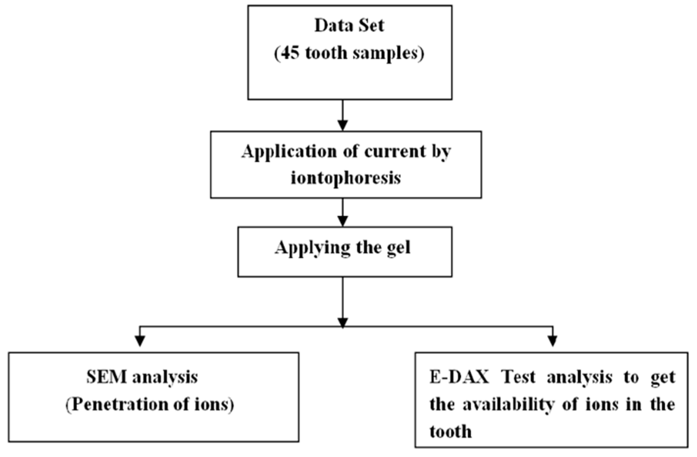
The overall methodology performed in using iontophoresis is a process of transdermal drug delivery by use of a voltage gradient on the skin. In a manner of speaking, it is an injection without a needle, and may be described as non-invasive, [21] which does not rely on an electric field. Thus, the teeth to be treated is first exposed to the current by an iontophoresis circuit, which has two electrodes. One electrode is positioned over the teeth wherein the ionic gel is applied and the other electrode to the ground through the body. Thus, when a current is passed through the electrode, ions of the same polarity repel and move through the tooth surface, reaching the dentin layer, which has soft nerve endings and hence desensitization in a particular area is achieved. Once the gel is passed and the current is induced, then the ions in the gel may combine with other ions and can indicate a porous nature. This enhances the possibility of the penetration of ions and can be analyzed using SEM images. The primary study for the method of iontophoresis was conducted by using SEM analysis for two sets of samples that were divided into two categories: the control group and the second sample with lidocaine gel treated with iontophoresis. From the results, Figure 1a shows the SEM image for the controlled group sample and Figure 1b is the sample with gel applied and treated with iontophoresis. From this, it can be concluded that the gel is passed and the current is induced, then the ions in the gel may combine with other ions and can give a porous nature. This enhances the possibility of the penetration of ions. The tooth has a porous nature, thus citing the proof that this can allow the ionic gel to pass through the tooth surface with or without the application of current, although the application of a current makes it even easier for the ions to penetrate through this medium.
The tooth samples were dissected into two equal halves and subjected to the EDAX analysis test to obtain the availability of ions in the tooth. Through the iontophoric principle, the ions are rippled due to the DC current given by the circuit, in which a particular ion will penetrate the surface of the tooth and reach the dentin layer, which has the soft nerve endings. Thus, the sensitivity is cut-off and helps in performing the operations.
The studies by Krishnaprasad et al. and Shashikanth et al. [22] have also reported on a comparison of the efficacy of two different commercially available desensitizing agents and concluded that acidulated sodium fluoride (NaF) gel 0.33% with the iontophoretic procedure obtained better results. Thus, this paper also justifies the desensitization of teeth with the method of iontophoresis, which has been proven by testing various tooth samples. The overall process of the studies taken up in this research paper is pictorially shown in Figure 2.

2.1. Dataset

To perform this above described process, 45 tooth samples were tested using imaging systems [23,24], namely, the SEM and EDAX methods, and the results were analyzed. The tooth samples were collected from SEESHA (Samiti for Education, Environment, Social and Health Action) Karunya Community Hospital. Samples of 45 teeth were taken and separated into different groups based on the structure of the tooth, in which two samples belonged to the control group as a reference for a comparison with other samples. The concept of iontophoresis has found significance at this stage for further excellence as iontophoresis is a painless and needleless way of delivering local anesthesia to the desired areas. Samples of 45 teeth were taken and separated into different groups based on the structure of the tooth, in which two samples belong to the control group for reference as to compare it with other samples. Initially, an iontophoric circuit was used to pass a current over the ionic gel applied to the surface of the patient’s tooth for a particular time.

2.2. Iontophoresis Method for Treatment in Dentistry

Iontophoresis [25] is a process of transdermal drug delivery by use of a voltage gradient on the skin. Molecules are transported across the stratum corneum by electrophoresis and electro osmosis and the electric field can also increase the permeability of the skin. These phenomena, directly and indirectly, constitute the active transport of matter due to an applied electric current. Iontophoresis has experimental, therapeutic, and diagnostic applications. In a manner of speaking, it is an injection without a needle, and may be described as non-invasive. It is different from dermal patches, which do not rely on an electric field. It drives a charged substance, usually a medication or bioactive agent, trans dermally by repulsive electromotive force, through the skin. Thus, the tooth to be treated is first given the current by the iontophoresis circuit, which has two electrodes. One electrode is passed over the teeth where the ionic gel is applied and the other electrode to the ground through the body. Thus, when the current is passed through the electrode, ions of the same polarity repel and move through the tooth surface, reaching the dentin layer, which has soft nerve endings, and hence desensitization in a particular area is achieved. Thus, the major significance of the proposed method is iontophoresis, which can be used as an alternate to treat dental surgeries, mainly for elderly people in the dental field. It can be further extended to all fields of research and applications because of its simplicity and economical aspects as it sharply reduces the time constraints faced in performing surgeries. The most significant aspect is that it is environmentally safe, replacing a large number of needles.

2.3. Ionic Gel Application
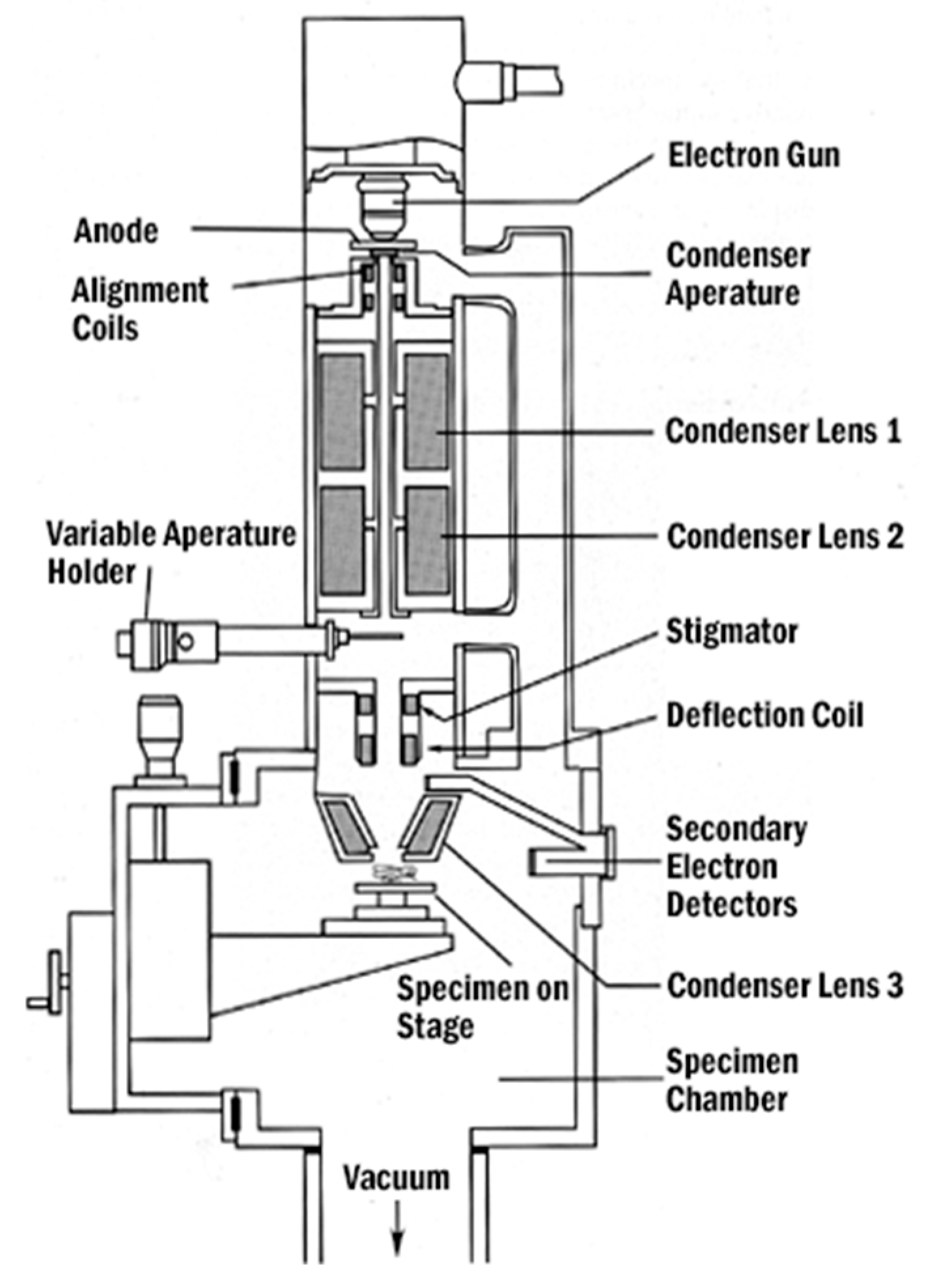
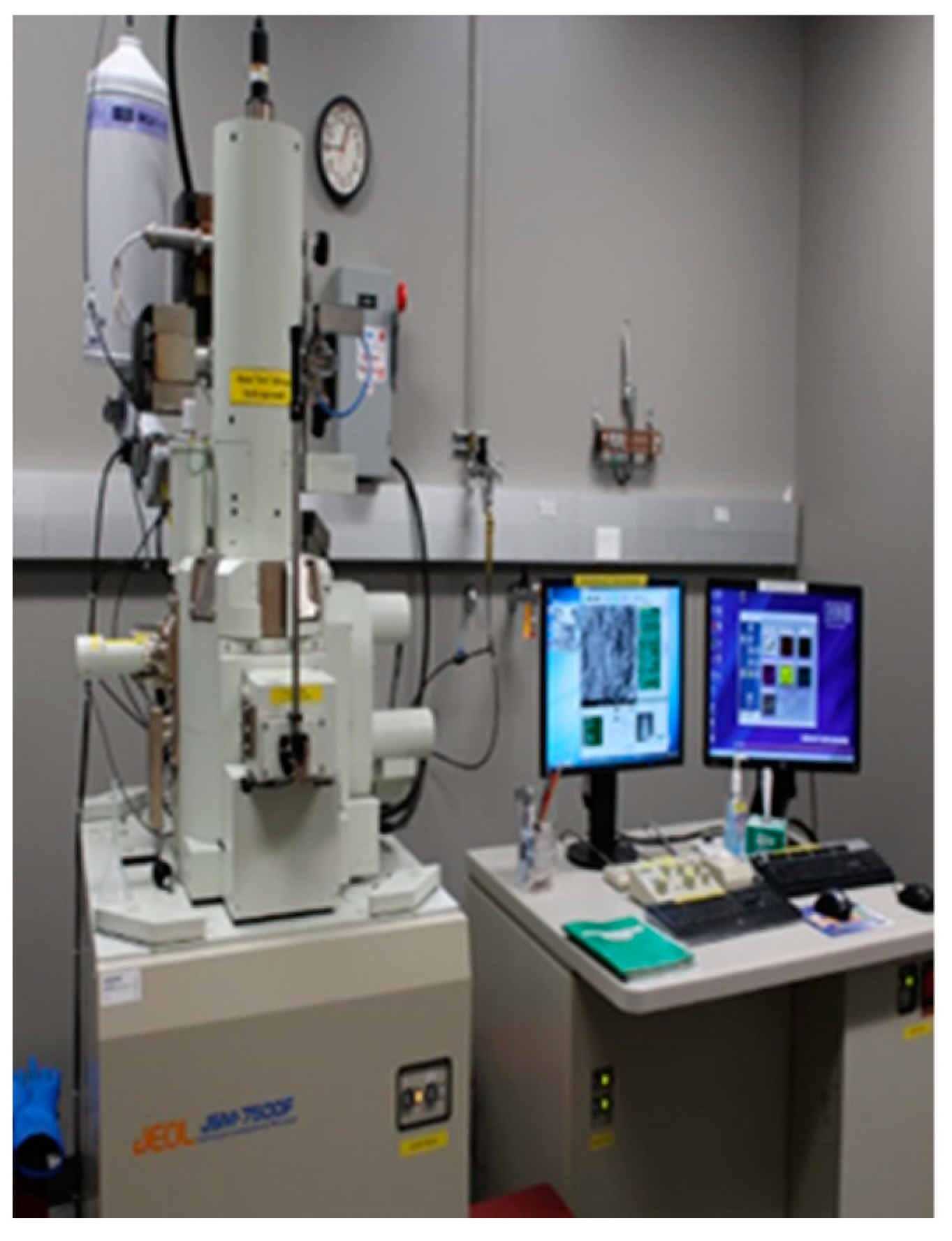
The tooth samples were collected from SEESHA (Samiti for Education, Environment, Social and Health Action) Karunya Community Hospital. Samples of 45 teeth were taken and separated into different groups based on the structure of the tooth, in which two samples belonged to the control group as a reference for comparison with the other samples. The concept of iontophoresis has found significance at this stage for further excellence as iontophoresis is a painless and needleless [26] way of delivering local anesthesia to the desired areas.
Samples of 45 teeth were taken and separated into different groups based on the structure of the tooth, in which two samples belong to the control group for reference as to compare it with other samples. Initially, an iontophoric circuit is used to pass current over the ionic gel applied to the surface of the patient’s tooth for a particular time. Table 1 presents the samples with different ionic gels such as potassium nitrate (KNO3), lidocaine, varnish, and strontium chloride (SrCl) used in the research.
An ion gel (or ionogel) is a composite material consisting of an ionic liquid immobilized by an inorganic or a polymer matrix. The material has the quality of maintaining high ionic conductivity while in the solid state. To create an ion gel, the solid matrix is mixed or synthesized in situ with an ionic liquid. The varnish used here is a clear transparent hard protective coating or film. It usually has a yellowish shade from the manufacturing process and materials used, but it can also be pigmented as desired and is sold commercially in various shades. Potassium nitrate is an ionic salt of potassium ions and nitrate ions and is therefore an alkali metal nitrate. Lidocaine stabilizes the neuronal membrane by binding to and inhibiting the voltage gated sodium channels, thereby commonly used for pain control during minor surgery and the conduction of impulses, effecting the local anesthesia. Strontium chloride is described as a salt of strontium and chloride. The strontium chloride formula is given as SrCl2 and is defined as a typical strontium chloride salt by forming neutral aqueous solutions.

2.4. SEM Analysis

A scanning electron microscope (SEM) is a type of electron microscope that produces images of a sample by scanning the surface with a focused beam of electrons. The electrons interact with atoms in the sample, producing various signals that contain information about the surface topography and composition of the sample. The electron beam is scanned in a raster scan pattern, and the position of the beam is combined with the detected signal to produce an image. The SEM architecture is shown in Figure 3 and the SEM instrument through which the results were taken is shown in Figure 4. SEM can achieve a resolution better than 1 nanometer. Specimens are observed in high vacuum in conventional SEM, or in low vacuum or wet conditions in variable pressure or environmental SEM, and at a wide range of cryogenic or elevated temperatures with specialized instruments.
The signals used by a scanning electron microscope to produce an image result from interactions of the electron beam with atoms at various depths within the sample. Various types of signals are produced including secondary electrons (SE), reflected or back-scattered electrons (BSE), characteristic X-rays and light (cathodoluminescence) (CL), absorbed current (specimen current), and transmitted electrons. The tooth samples after the application of ionic gels were subjected to the process of iontophoresis and then underwent SEM analysis, the images of which are discussed in the next section.

2.5. EDAX Analysis

EDAX analysis—energy dispersive XRAY spectroscopy (EDS, EDX, EDXS or XEDS), sometimes-called energy dispersive X-ray analysis (EDXA) or energy dispersive X-ray microanalysis (EDXMA)—is an analytical technique used for the elemental analysis or chemical characterization of a sample. A high-energy beam of charged particles such as electrons or protons or a beam of X-rays is focused into the sample being studied. The incident beam may excite an electron in an inner shell, ejecting it from the shell while creating an electron hole where the electron was. An electron from an outer, higher-energy shell then fills the hole, and the difference in energy between the higher-energy shell and the lower energy shell may be released in the form of an X-ray. The number and energy of the X-rays emitted from a specimen can be measured by an energy dispersive spectrometer.
Samples of 45 teeth were taken and were separated into different groups based on the structure of the tooth, in which two samples belonged to the control group as aa reference for comparison with other samples. Initially, an iontophoric circuit was used to pass the current over the ionic gel applied to the surface of the patient’s tooth for a particular time.
The tooth samples were dissected into two equal halves for the EDAX analysis test to obtain the availability of ions in the tooth. By the iontophoric principle, the ions are rippled due to the DC current given by the circuit in which a particular ion will penetrate the surface of the tooth and reach the dentin layer, which contains the soft nerve endings. Thus, the sensitivity is cut-off and helps in performing the operations

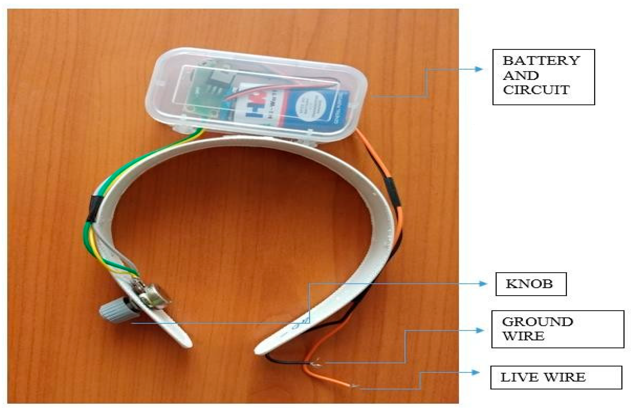
3. Implementation of the Proposed Prototype

This setup was designed in a way that provides a high degree of comfort to the patients while treating them. Thus, the current controlled circuit embedded inside the mechanical structure even acts as an insulator, so that total protection is ensured. The current circuit consists of a simple combination of resistors and a thyristor to control the current in the device. A potentiometer is also used to vary the current output at the end, which is connected between the voltage dividers. This circuit helps in delivering the controlled current in the electrode [27] to enhance the iontophoresis technique in a unique way. A knob is placed at one end of the device to control the current in the circuit to limit the current to be given for treatment. At the other end is an electrode with a micro tip, which makes the current pass through the gel when touched on the surface of the ionic gel on the surface of the tooth. A milli ammeter is placed at other end to measure the current flow, or it can be monitored through a sophisticated channel (i.e., is a screen placed right above the patient where the data are visually fed and monitored).
The hardware consists of a device with a current controlled circuit where a suitable current is given for a particular time. The device is similar to a wireless earphone module that incorporates the features of controllability and feasibility by providing a sophisticated treatment module. It has an electrode at one end and a controlling knob at the other end, so that the person who performs the operation can also monitor the current level and time, which are the major factors for the treatment using this device. The device as shown in Figure 5 performs the operation where a direct current is applied onto the tooth. The mechanical structure resembles like that of a wireless earphone that is worn by the patients around their neck so that the operation is performed quickly and with high accuracy.

4. Results and Discussion

In this, 45 tooth samples were tested using SEM and EDAX methods and the results were analyzed, which are discussed below. The iontophoric method used for treatment on the patient’s teeth is discussed with the SEM and EDAX analyses to obtain the availability of ions in the tooth.

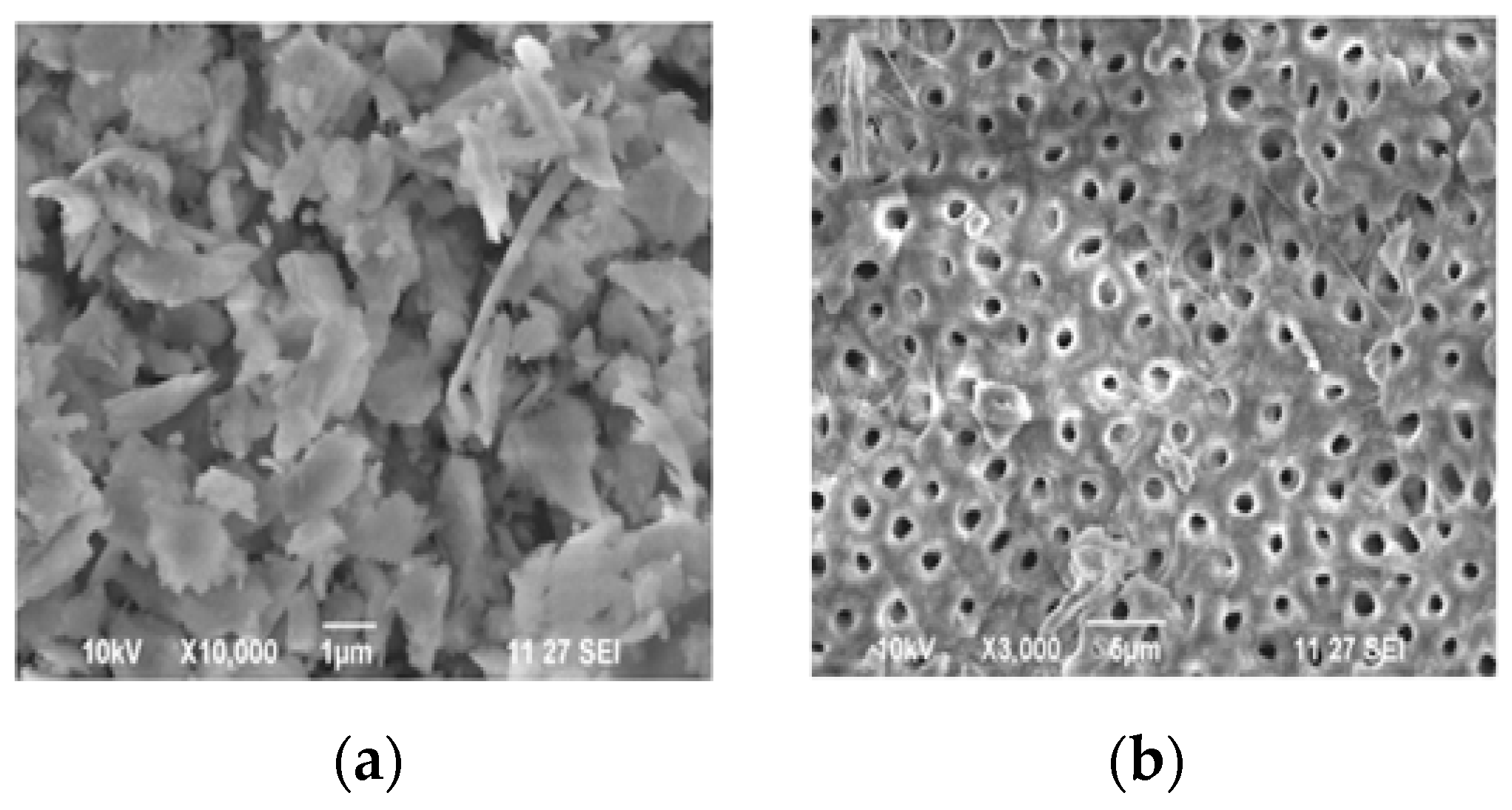
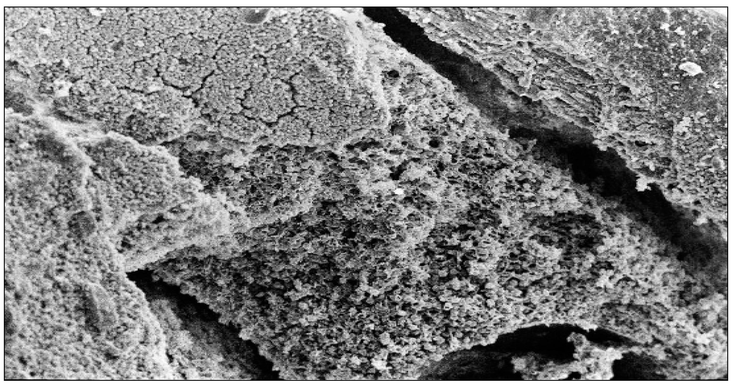
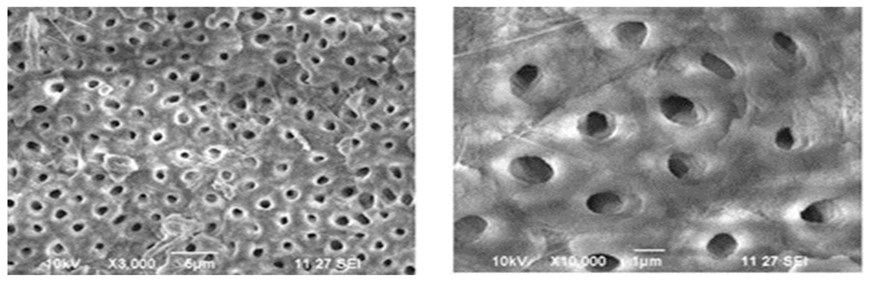
4.1. Phase 1—SEM Test Results

4.1.1. Tooth Samples before Iontophoric Application

Figure 6 shows the SEM images of the pure tooth, proving that the tooth had a porous nature, thus citing proof that this can allow the ionic gel to pass through the tooth surface with or without the application of current. Furthermore, the application of the current makes it even easier for the ions to penetrate this medium.

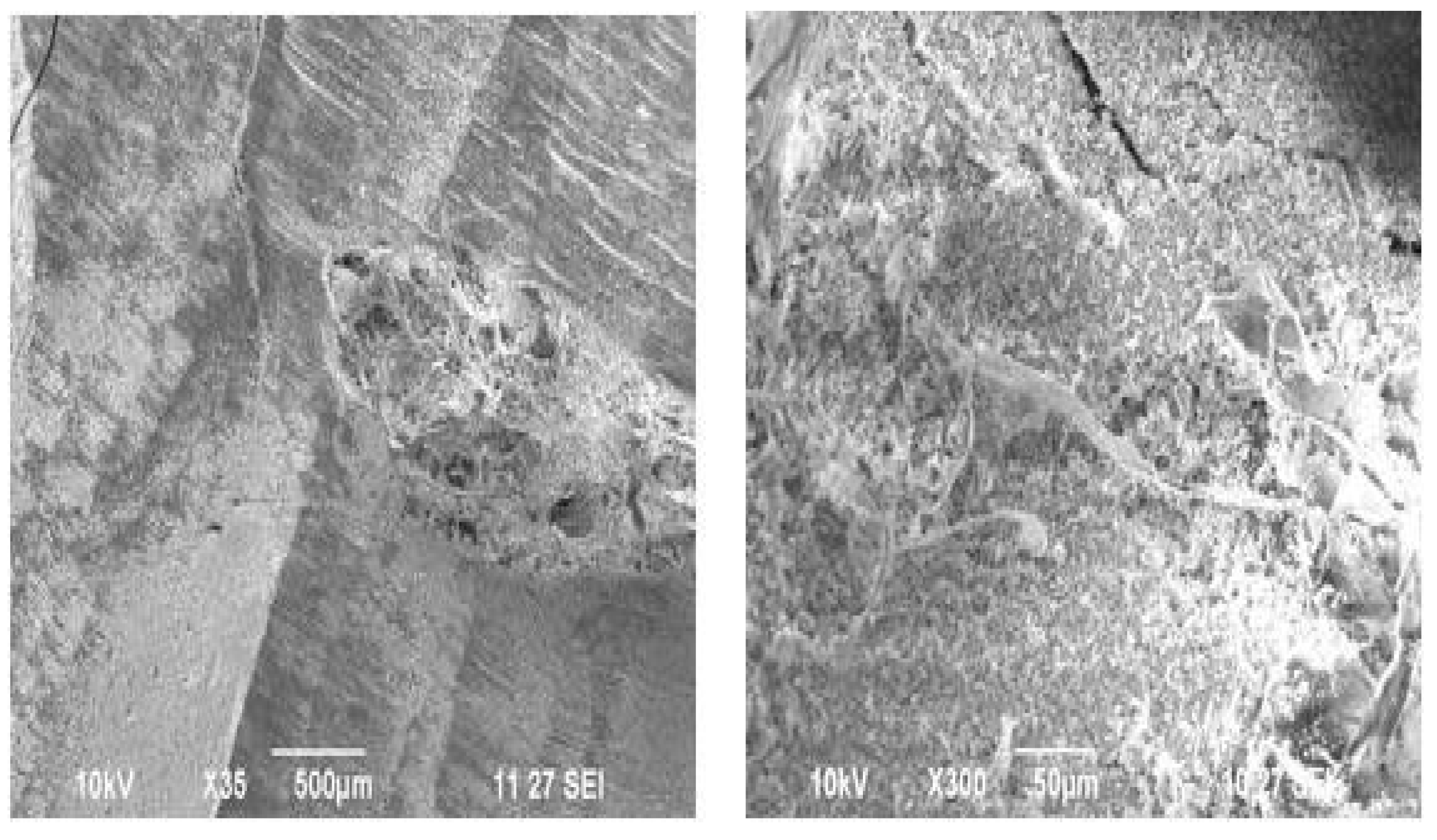
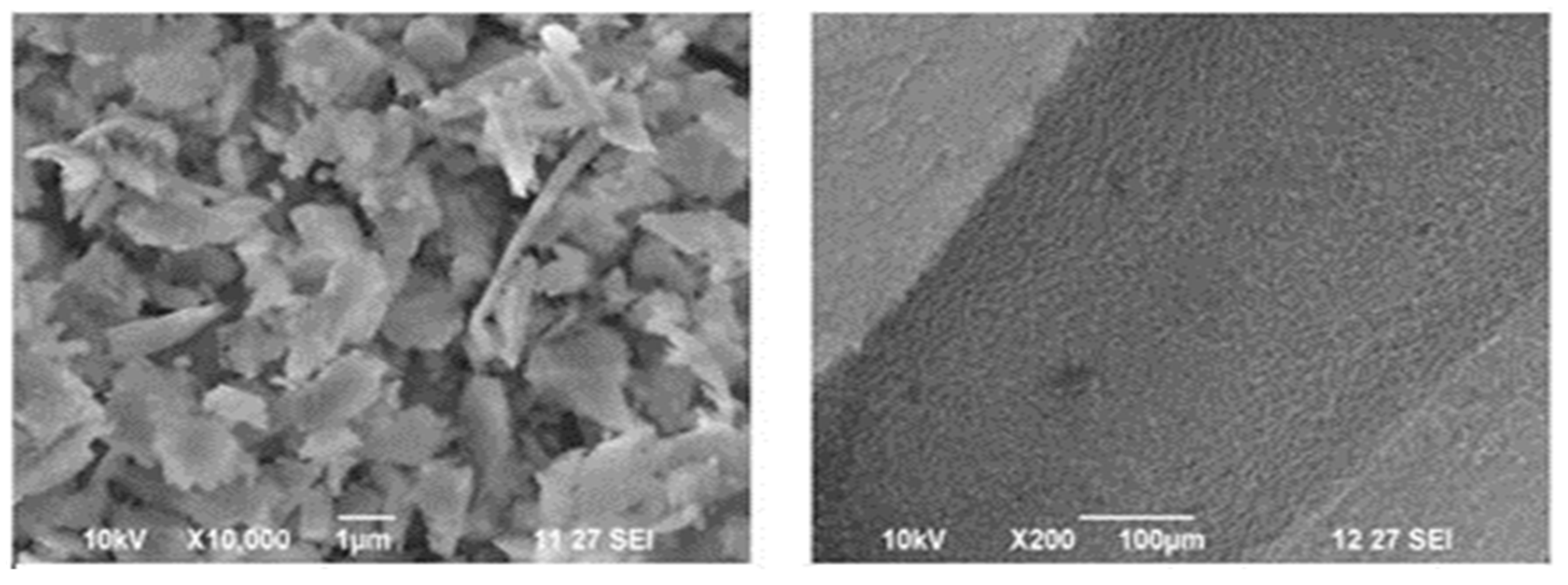
4.1.2. SEM Images with Only Ionic Gel

In an ion gel is a type of semi-solid compound that retains an electrical charge through the embedding of an ionic liquid. Samples with different ionic gels such as potassium nitrate (KNO3), lidocaine, varnish, and strontium chloride (SrCl2) were used in the research. Figure 7 and Figure 8 depicts the SEM image of the tooth sample—lateral view at 500 µm and 50 µm and the SEM image of the tooth sample—sectional view at 5 µm and 500 µm, respectively.

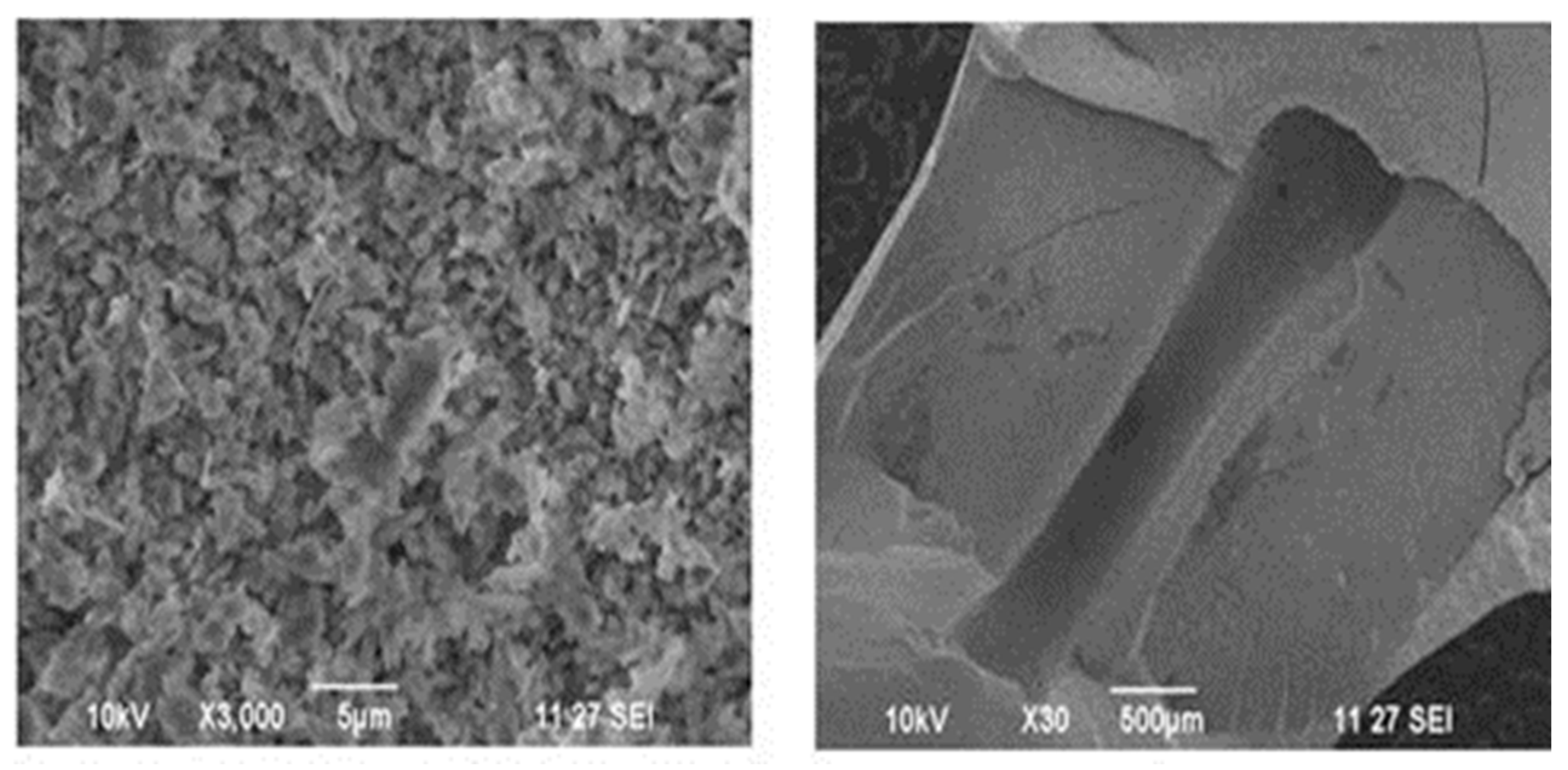
4.1.3. SEM Images with Ionic Gel and Iontophoric Application

To confirm the fact that the tooth had a porous nature, thus proving that this can allow the ionic gel to pass through the tooth surface, the tooth was exposed to the iontophoric application. Iontophoresis is a process of transdermal drug delivery by use of a voltage gradient on the skin. Thus, the teeth to be treated are first given the current by an iontophoresis circuit.
Figure 9 and Figure 10 show the SEM images of the tooth sample after iontophoresis—sectional view at 1 µm and 100 µm and the SEM image of the tooth sample after iontophoresis—with pores formed in the gel at 5 µm and 1 µm, respectively.
Thus, Figure 7, Figure 8, Figure 9 and Figure 10 are the SEM images before and after the application of the iontophoric methods. This shows that the tooth structure has the penetrability capacity of ions in the gel, which when applied, penetrated through the surface of the tooth, hence forming a layer over the tooth when dissected. Thus, the formation of holes proves that there is a combinational reaction of different ions present in the gel, leading to these holes.

4.2. Phase 2—EDAX Test Results

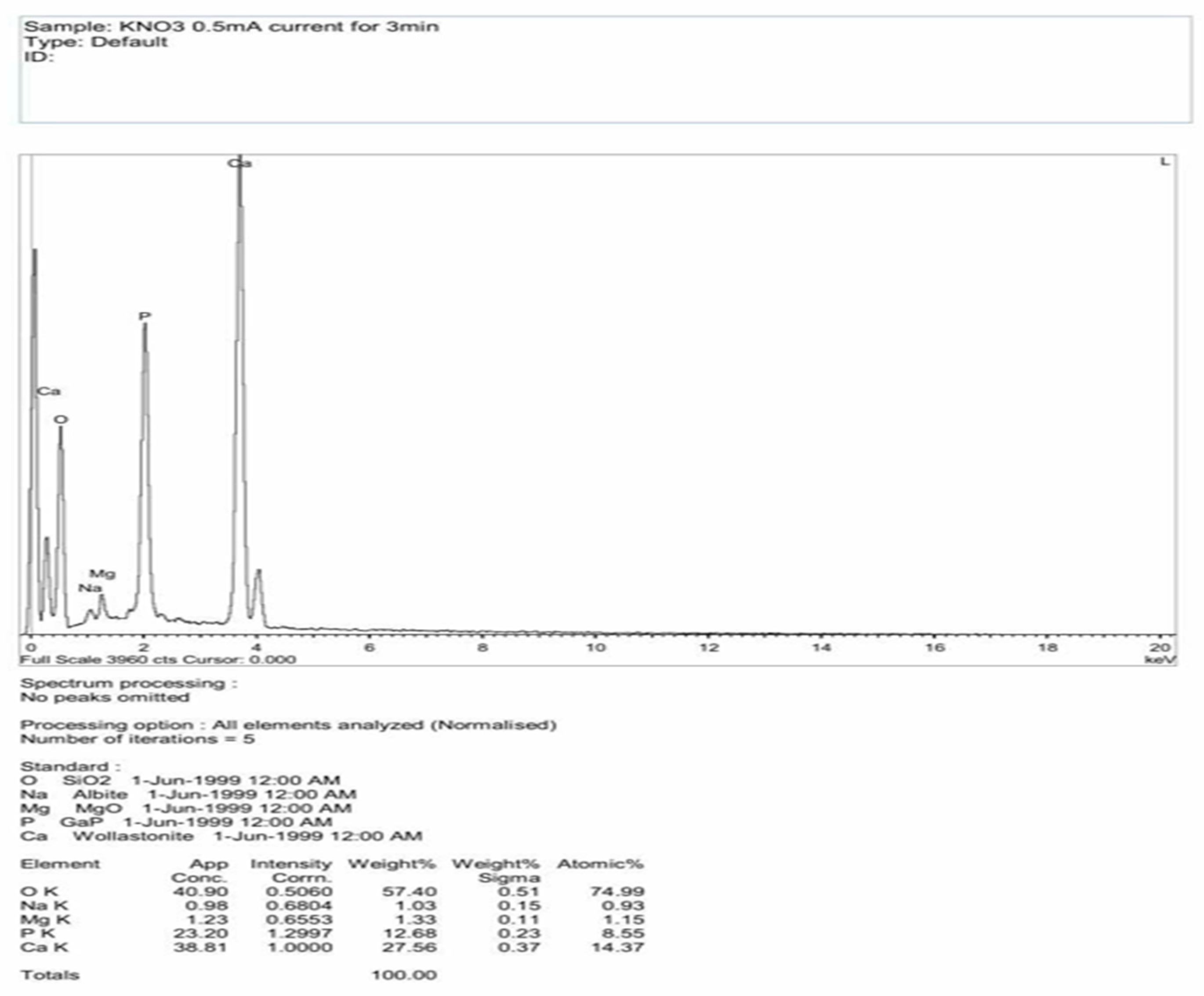
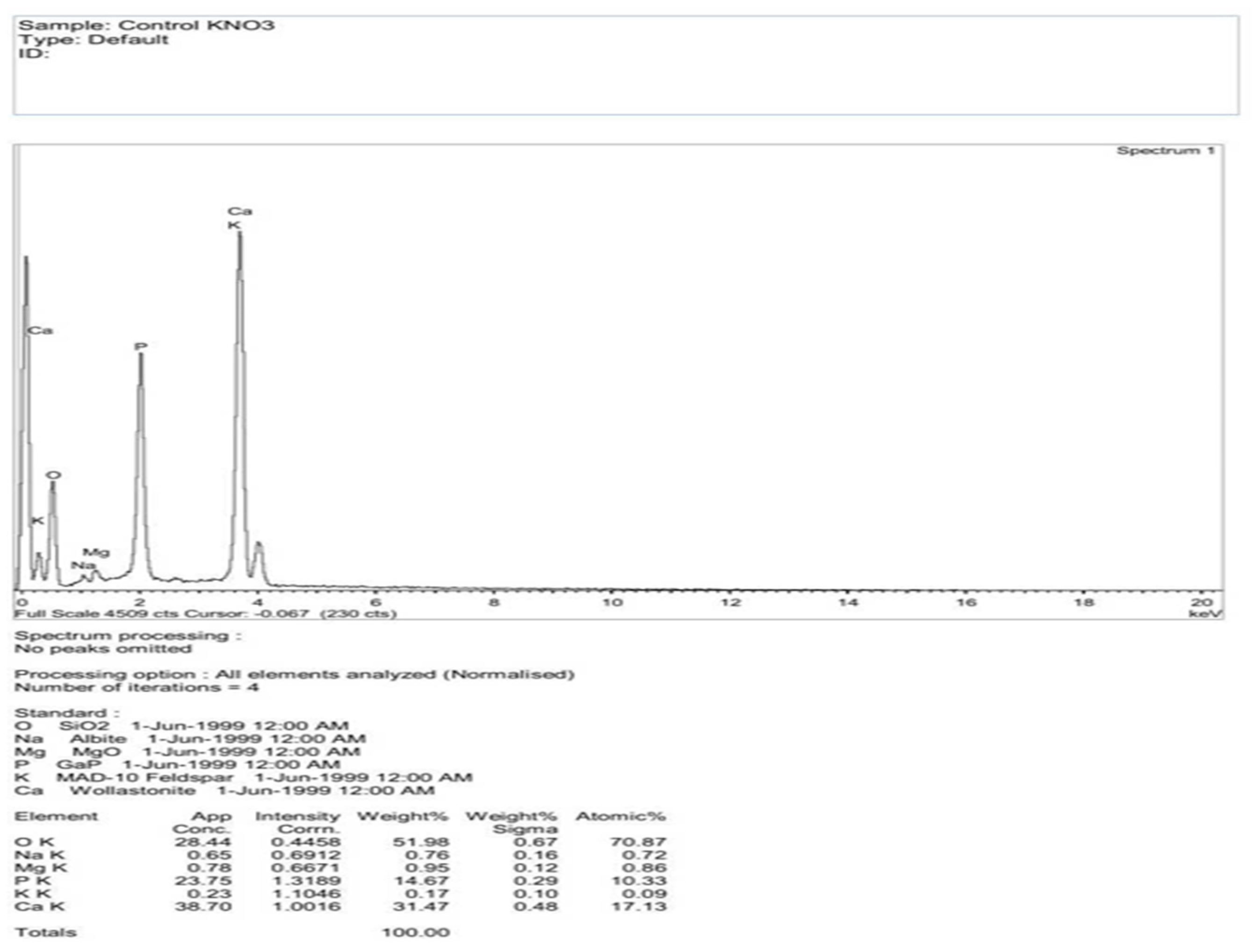
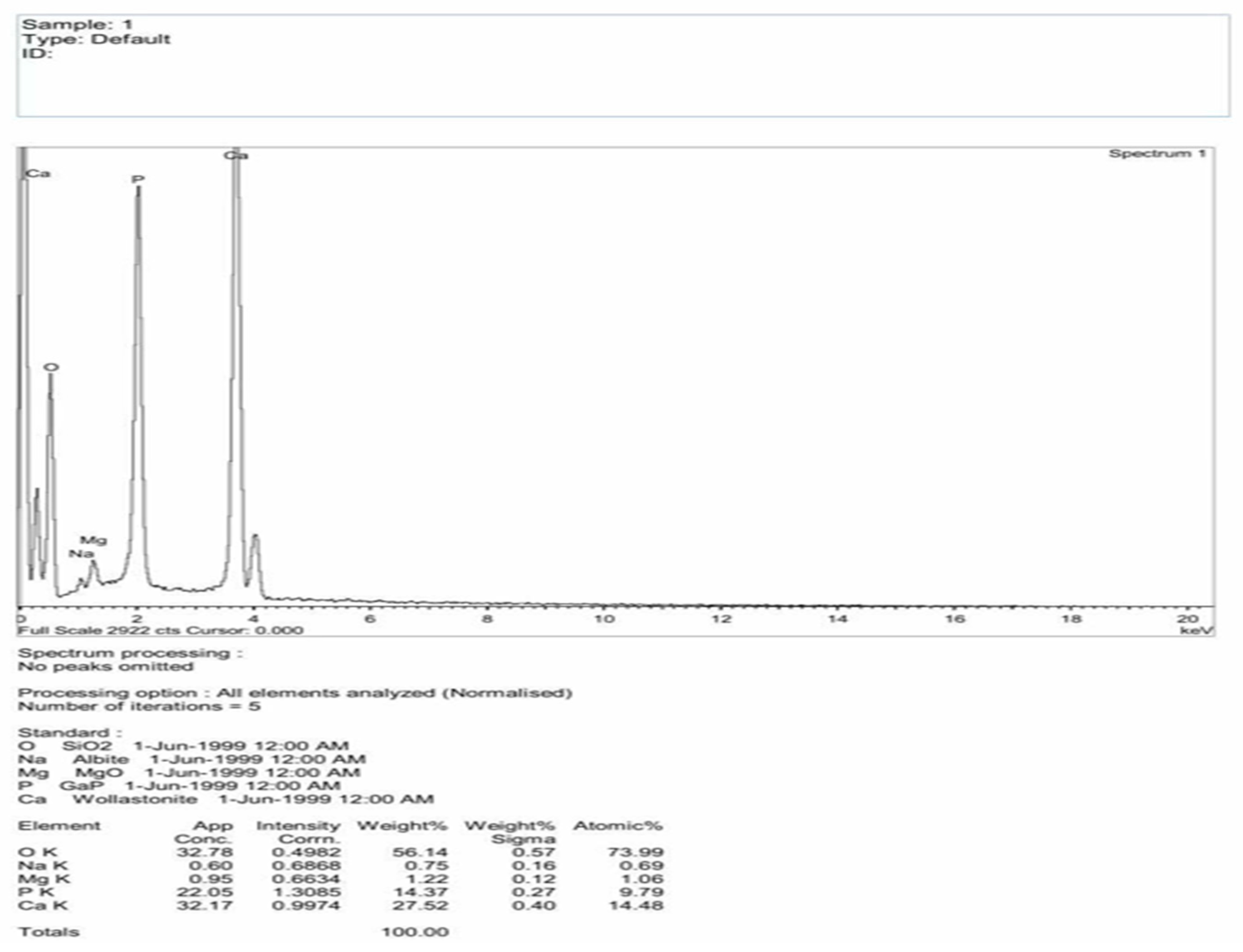
4.2.1. EDAX Results for KNO3 Ionic Gel

The tooth samples were divided into five groups: potassium nitrate, varnish, lidocaine, and strontium chloride, which are commonly used in the dental process, and one for the control group. Figure 11 and Figure 12 are the EDAX results for the tooth samples with the iontophoresis method and the control group type with the application of KNO3, respectively.
The EDAX results for the tooth samples treated for 0.3 mA current for 5 min and the tooth samples with 0.5 mA current for 3 min were observed. The results of the 0.5 mA current for a duration of 3 min gave promising results in which the traces of potassium ions (K) were nearly 60%. The presence of potassium (K) was 0.05 in the samples where a potassium nitrate based ionic gel was applied with a 0.5 mA current for 5 min by the iontophoric method, and that of the control group without iontophoresis was 0.09 in atomic size. This proves that nearly 60% of the K ions was traced after iontophoresis in a short time.

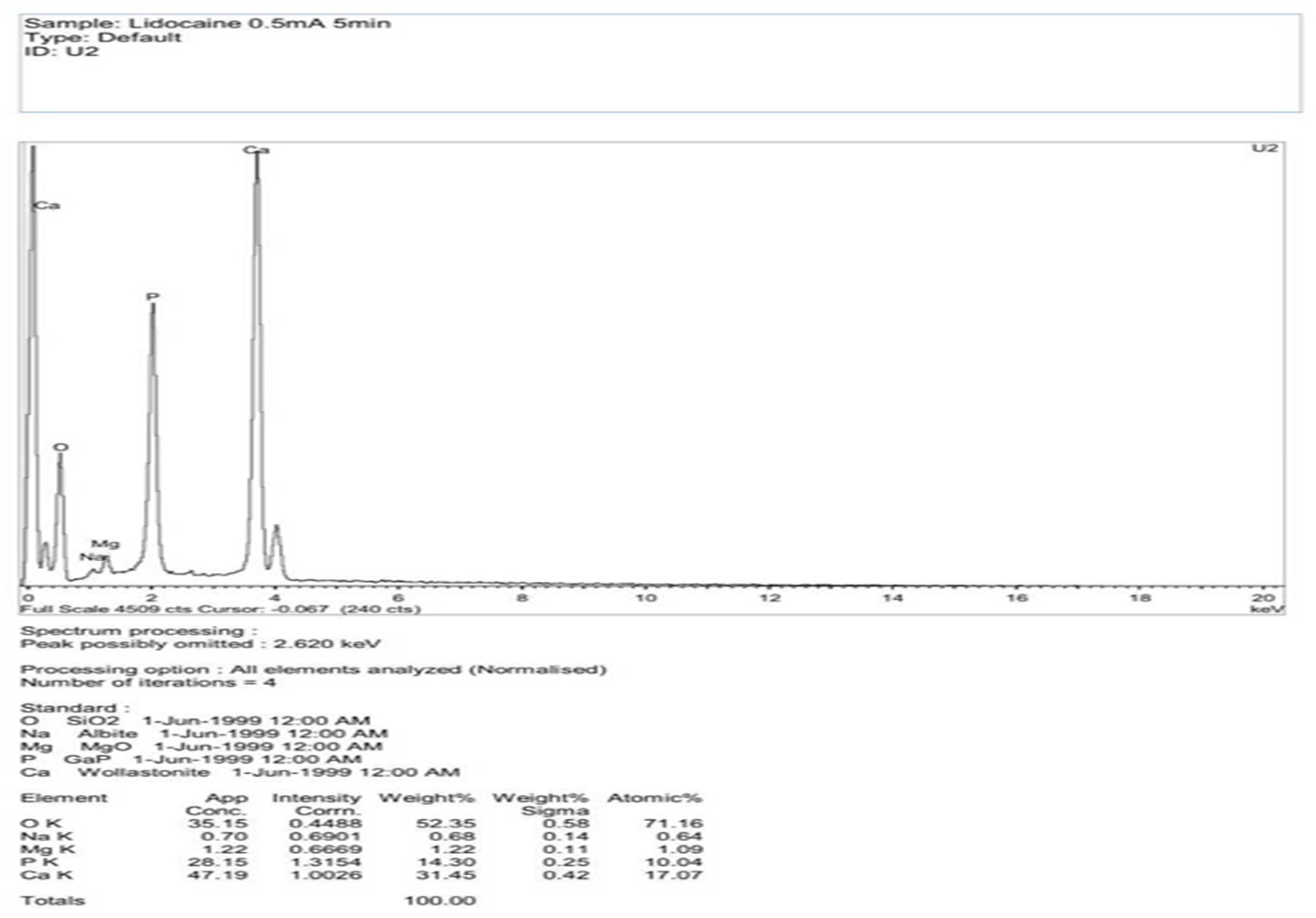
4.2.2. EDAX Results for Lidocaine

Figure 13 and Figure 14 show the EDAX results for the tooth samples with lidocaine ionic gel for the controlled group and the samples tested with the iontophoric method, respectively.
EDAX analysis was conducted for the 0.3 mA current for a duration of 3 min and showed the same results for the 0.5 mA current for 5 min. The presence of sodium ions was 0.87 for the tooth sample treated by iontophoresis, which were not traced for the control group samples with lidocaine. This is because sodium ions (Na) are used in lidocaine gel to balance the pH in the gel. During the iontophoresis method, the Na ion used is exited, which is traced. Thus, Na can be used for desensitization of the tooth.

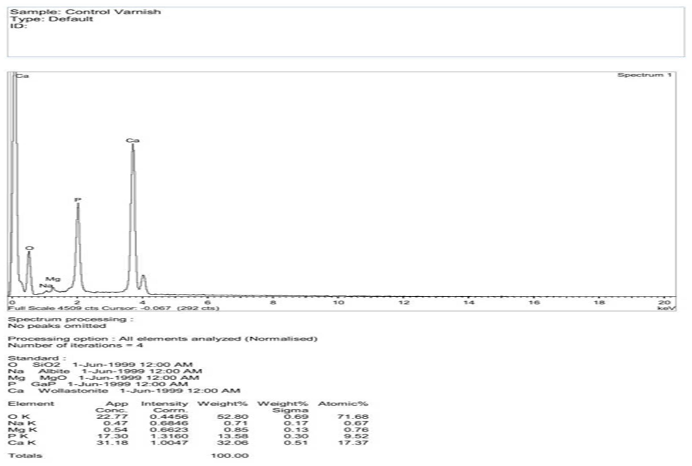
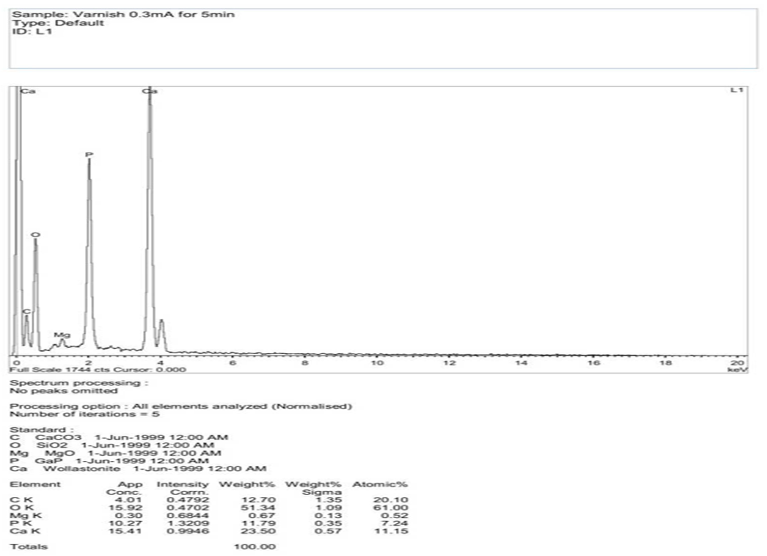
4.2.3. EDAX Results for Varnish

Figure 15 and Figure 16 show the EDAX results for tooth samples with varnish for the control group and the samples tested with iontophoresis, respectively. For the varnish samples, apart from other compounds given in the table, the presence of sodium (Na) ions was 0.67 for the control group and 0.61 for that of the samples treated by iontophoresis for a duration of 5 min with the 0.5 mA current (i.e., 75% of the ions were present in the tooth after the above method).
The EDAX results for the tooth samples with the varnish gel, which was the control group (GP), showed that they contained sodium (Na) when compared to the pure tooth. This sodium (Na) helps in desensitizing the dentin layer. The process of iontophoresis was then conducted on eight tooth samples. The above process was carried out with a 0.3 mA current for 5 min and the same with the 0.5 mA current for 3 min. The results were better when treated for the 0.5 mA current for 3 min. The presence of sodium (Na) was found to be 0.61 in atomic weight.

4.2.4. EDAX Results for Strontium Chloride

The inference from Figure 17 shows that the samples treated by iontophoresis with strontium chloride contained Sr-0.11 (strontium), Na-0.61 (sodium), and for Cl-0.07 (chloride), which were present in the tooth samples.
The samples were treated by iontophoresis with strontium chloride (SrCl2)for a duration of 5 min with 0.3 mA of current and same with 0.5 mA of current for 5 min. Thus, the presence of strontium −0.11 and chloride −0.07 gives us the scope that these ions help in desensitizing the nerves in the dentin layer.

4.2.5. Comparative Analysis of the Gels Applied

Table 2 shows the EDAX results of the tooth samples. Thus, from the overall inference from the 45 samples, the presence of sodium (Na) ions and chloride ions (Cl) can be concluded.
After the process of iontophoresis in the tooth samples, it was proven that these ions have the ability to desensitize a particular area effectively in a more efficient way in the new treatment method. It can be concluded that iontophoresis can be used as an alternate to treat dental surgeries, mainly for elderly people in the dental field. It can be further extended to all fields of research and applications because of its simplicity and economical aspects. It sharply reduces the time constraints faced in performing surgeries. The most significant aspect is that it is environmentally safe, replacing a large number of needles. The comparative analysis of the various gels applied, along with their penetration level, is consolidated and shown in Table 3.
  • The presence of potassium (K) was 0.05 in the samples where a potassium nitrate based ionic gel was applied with a 0.5 mA current for 5 min by the iontophoric method. This proves that nearly 60% of the K ions were traced after iontophoresis in a short time.
  • The presence of sodium ions was 0.87 for the tooth sample treated by iontophoresis, which were not traced for the control group samples with lidocaine. During the iontophoresis method, the Na ion used is exited, which is traced. Thus, Na can be used for tooth desensitization.
  • For the varnish samples, the presence of sodium (Na) ions was 0.67 for the control group and 0.61 for the samples treated by iontophoresis (i.e., 75% of the ions were present in the tooth after the above method).
Thus, after the process of iontophoresis in the tooth samples, it was proven that these ions have the ability to desensitize a particular area effectively in a more efficient way in the new treatment method.

5. Conclusions

The overall inference from the 45 samples leads us to the conclusion that the presence of sodium (Na) ions and chloride ions (Cl) after the process of strontium chloride with iontophoresis in the tooth samples proves that these ions have the ability to desensitize a particular area effectively in a more efficient way in the new treatment method. Therefore, this treatment of desensitization will help elderly people to go in for dental treatments without any fear.
The results obtained from SEM and EDAX were analyzed and the results prove that the ions in the ionic gel reached a certain level through the activation of a current of 0.5 mA to 0.5 mA, which was very efficient when the 0.5 mA current was applied for 3 min. This current rating enables the higher penetrability of the ions present in the gels.
Therefore, the concept of iontophoresis can be used in the dental field, which replaces traditional methods, thus making it patient friendly, economical, and environmentally safe when treating mostly, elderly patients with the utmost care in performing dental treatment. This makes the entire dental operation to be performed easier, thus eliminating the term “phobia”. The future scope of this method has a wider scope in treating cancer patients and others who suffer a high degree of pain in day-to-day life. Thus, this method can be used for people who have chronic pain that is intolerable. As aa result, this research work based on iontophoresis in the dental field will help future researchers to impart deep knowledge in this area.

Author Contributions

Conceptualization: P.A.M.; Methodology: P.A.M.; Software: P.D.; Validation: R.M., J.J.J. and F.T.J.; Investigation: K.G.J.N.; Supervision: J.H., P.A.M. All authors have read and agreed to the published version of the manuscript.

Funding

This research received no external funding.

Institutional Review Board Statement

Not Applicable.

Informed Consent Statement

Not Applicable.

Data Availability Statement

The study did not report any separate data as all the details are presented in the manuscript itself.

Conflicts of Interest

The authors declare no conflict of interest.

References

  1. History of Anesthesia Society. Available online: http://www.histansoc.org.uk/timeline.html (accessed on 20 February 2021).
  2. Universitat Leiden. Available online: http://www.universiteitleiden.nl/en/research/research (accessed on 20 February 2021).
  3. Angelo, Z.; Charalambous, P. Alternative practices of achieving anaesthesia fordental procedures: A review. Dent. Anaesth. Pain. Med. 2018, 18, 79–88. [Google Scholar] [CrossRef] [PubMed] [Green Version]
  4. Hironori, T. Dental anesthesia in the presence of inflammation: Pharmacological mechanisms for the reduced efficacy of local anesthetics. Int. J. Clin. Anesthesiol. 2016, 4, 1059. [Google Scholar]
  5. Krishnaprasad Shetty, S.S.V.; Kilaru, K.R.; Azeez, F.A.; Patil, S.; gouda Sangam, B.; Luke, A.M.; Prithviraj, D.R.; Saraswat, S.; Sounderraj, K.; Patel, A. Comparative evaluation of efficacy of commercially available desensitizing toothpastes (Shy-Nm and Thermoseal Ra) with 0.33% acidulated NaF gel used with and without iontophoresis in patients with dentinal hypersensitivity. J. Appl. Dent. Med. Sci. 2017, 3, 1–7. [Google Scholar]
  6. Lee, J.; Kwon, K.; Kim, M.; Min, J.; Hwang, N.S.; Kim, W. Transdermal iontophoresis patch with reverse electrodialysis. Drug Deliv. 2017, 24, 701–706. [Google Scholar] [CrossRef] [PubMed] [Green Version]
  7. Subramaniam, S.; Neelakantan, P. Local anesthesia in dentistry-Clinical considerations. Int. J. Drug Dev. Res 2013, 5, 30–36. [Google Scholar]
  8. Bubteina, N.; Garoushi, S. Dentine Hypersensitivity: A Review. Dentistry 2015, 5, 9. [Google Scholar]
  9. Ita, K. Transdermal Delivery of Drugs with Microneedles—Potential and Challenges. Pharmaceutics 2015, 7, 90–105. [Google Scholar] [CrossRef] [PubMed] [Green Version]
  10. Sharma, S.; Parvez, N.; Sharma, P.K. Iontophoresis-Models and applications: A review. Afr. J. Basic Appl. Sci. 2015, 7, 1–7. [Google Scholar]
  11. Tuttlenumbnow. Available online: https://tuttlenumbnow.com/ (accessed on 5 September 2022).
  12. Zaid Alkilani, A.; McCrudden, M.T.; Donnelly, R.F. Transdermal Drug Delivery:Innovative Pharmaceutical Developments Based on Disruption of the Barrier Properties of the stratum corneum. Pharmaceutics 2015, 7, 438–470. [Google Scholar] [CrossRef] [Green Version]
  13. Wanasathop, A.; Li, S.K. Iontophoretic Drug Delivery in the Oral Cavity. Pharmaceutics 2018, 10, 121. [Google Scholar] [CrossRef] [Green Version]
  14. Tan, G.Z.; Orndorff, P.E. The ion delivery manner influences the antimicrobial efficacy of silver oligodynamic iontophoresis. J. Med. Biol. Eng. 2019, 39, 622–631. [Google Scholar] [CrossRef] [Green Version]
  15. Li, Y.; Yang, J.; Zheng, Y.; Ye, R.; Liu, B.; Huang, Y.; Zhou, W.; Jiang, L. Iontophoresis–driven porous microneedle array patch for active transdermal drug delivery. Acta Biomaterilia 2021, 121, 349–358. [Google Scholar] [CrossRef]
  16. Kaur, T. Transdermal drug delivery by iontophoresis: Mechanistic aspects. Innov. Pharm. Pharmacother. 2018, 6, 13–16. [Google Scholar] [CrossRef]
  17. Bozkurt, M.H.; Karagol, S. Jaw and Teeth Segmentation on the Panoramic X-Ray Images for Dental Human Identification. J. Digit. Imaging 2020, 33, 1410–1427. [Google Scholar] [CrossRef]
  18. Zhang, J.; Jiang, Y.; Gao, F.; Zhao, S.; Yang, F.; Song, L. A Fast Automatic Reconstruction Method for Panoramic Images Based on Cone Beam Computed Tomography. Electronics 2022, 11, 2404. [Google Scholar] [CrossRef]
  19. Dao-Ngoc, L.; Liu, C.; Du, Y. A Segmentation Enhancement Method for the Low-Contrast and Narrow-Banded Substances in CBCT Images. Electronics 2020, 9, 974. [Google Scholar] [CrossRef]
  20. Moratin, J.; Berger, M.; Rückschloss, T.; Metzger, K.; Berger, H.; Gottsauner, M.; Head motion Engel, M.; Hoffmann, J.; Freudisperger, C.; Ristow, O. Head motion during cone-beam computed tomography: Analysis of frequency and influence on image quality. Imaging Sci. Dent. 2020, 50, 227–236. [Google Scholar] [CrossRef] [PubMed]
  21. Canjau, S.; Todea, C. Minimally-invasive diagnostic approaches in periodontics: Laser Doppler imaging and optical coherence tomography. Clin. Dent. 2021, 5, 1–14. [Google Scholar] [CrossRef]
  22. Hegde, S.; Mankude, S.; Kashyap, R.; Kumar, A. Comparative assessment of efficacy of iontophoresis with acidulated phosphate fluoride gel and a commercially available desensitizing agent-bifluoride varnish in the treatment of hypersensitive teeth. IOSR J. Dent. Med. Sci. 2015, 14, 34–41. [Google Scholar]
  23. Stember, J.N.; Moonis, G.; Silva, C. Panoramic Dental Reconstruction for Faster Detection of Dental Pathology on Medical Non-dental CT Scans: A Proof of Concept from CT Neck Soft Tissue. J. Digit. Imaging 2021, 34, 959–966. [Google Scholar] [CrossRef] [PubMed]
  24. Villoria, E.M.; Rodrigues, R.C.; Peraira, C.H.M.; Conceicao, G.S.A. The importance of digital radiographic systems in dental schools and oral radiology centers as part of reopening during the COVID-19 pandemic. Imaging Sci. Dent. 2021, 51, 91–92. [Google Scholar] [CrossRef]
  25. Vernon, J.A.; Kaufman, M.R.; Brummett, R.E.; Bender, H.G. Iontophoresis Apparatus for Applying Local Anesthetics. U.S. Patent No.US3991755A, January 1975. [Google Scholar]
  26. Cubayachi, C.; do Couto, R.O.; de Gaitani, C.M.; Pedrazzi, V.; de Freitas, O.; Lopez, R.F.V. Needle-free buccal anesthesia using iontophoresis and amino amide salts combined in a mucoadhesive formulation. Colloids Surf. B Biointerfaces 2015, 136, 1193–1201. [Google Scholar] [CrossRef] [PubMed]
  27. Paterson, A.; Franco, V.; Patel, S.; Foschi, F. Use of preoperative cone-beam computed tomography to aid in establishment of endodontic working length: A systematic review and meta-analysis. Imaging Sci. Dent. 2020, 50, 183–192. [Google Scholar] [CrossRef]
Figure 1. (a) Control group. (b) Sample with lidocaine gel with iontophoresis.
Electronics 11 03234 g001
Figure 2. Process Flow of the Methodology used.
Electronics 11 03234 g002
Figure 3. SEM architecture.
Electronics 11 03234 g003
Figure 4. SEM instrument.
Electronics 11 03234 g004
Figure 5. Prototype model for the iontophoric device.
Electronics 11 03234 g005
Figure 6. SEM images of the pure tooth.
Electronics 11 03234 g006
Figure 7. SEM image of the tooth sample—lateral view at 500 µm and 50 µm.
Electronics 11 03234 g007
Figure 8. SEM image of the tooth sample—sectional view at 5 µm and 500 µm.
Electronics 11 03234 g008
Figure 9. SEM image of the tooth sample after iontophoresis—sectional view at 1 µm and 100 µm.
Electronics 11 03234 g009
Figure 10. SEM image of the tooth sample after iontophoresis—with pores formed in the gel at 5 µm and 1 µm.
Electronics 11 03234 g010
Figure 11. Tooth sample—KNO3 with iontophoresis (0.5 mA for 3 min).
Electronics 11 03234 g011
Figure 12. Control group with KNO3.
Electronics 11 03234 g012
Figure 13. Control group with lidocaine.
Electronics 11 03234 g013
Figure 14. Tooth sample—lidocaine with iontophoresis (0.5 mA for 5 min).
Electronics 11 03234 g014
Figure 15. Control group with varnish.
Electronics 11 03234 g015
Figure 16. Tooth sample—varnish with iontophoresis (0.3 mA for 5 min).
Electronics 11 03234 g016
Figure 17. Tooth sample—strontium chloride with iontophoresis.
Electronics 11 03234 g017
Table 1. List of the test samples with different ionic gels.
Sl. No.Nature of the SampleSample TypeNo. of Samples
1Pure teethControl group1
2Tooth with varnish Control group1
0.3 mA for 5 min4
0.5 mA for 3 min3
3Tooth with KNO3Control group1
0.3 mA for 5 min4
0.5 mA for 3 min3
4Tooth with lidocaineControl group1
0.3 mA for 3 min4
0.5 mA for 5 min4
5Tooth with strontium chloride 0.3 mA for 5 min4
0.5 mA for 5 min4
Table 2. The EDAX results of the tooth samples.
ElementO
(wt.%)
Na
(wt.%)
Mg
(wt.%)
P
(wt.%)
K
(wt.%)
Ca
(wt.%)
Ci
(wt.%)
Sr
(wt.%)
Tooth sample—KNO3 with iontophoresis
(0.5 mA for 5 min)
57.52-1.0413.960.0927.39--
Control group with KNO3 51.980.760.9514.670.1731.47--
Control group with lidocaine54.62-1.3114.6329.43---
Tooth sample—lidocaine with iontophoresis
(0.5 mA for 5 min)
51.180.911.0713.9732.86---
Control group with varnish52.800.710.8513.5832.06---
Tooth sample—varnish with iontophoresis
(0.5 mA for 5 min)
49.870.630.8215.0233.65---
Tooth sample—strontium chloride with iontophoresis53.630.651.1914.35-29.610.120.44
Table 3. The comparative analysis of the various gels applied along with the penetration level.
ElementPenetration Level
Tooth sample—varnish with iontophoresis (0.5 mA for 5 min)75% of ions traced after iontophoresis in a short time.
Tooth sample—KNO3 with iontophoresis60% of ions traced after iontophoresis in a short time.
Tooth sample—lidocaine with iontophoresis
(0.5 mA for 5 min)
87% of ions traced after iontophoresis in a short time.
Tooth sample—strontium chloride with iontophoresis70% of ions traced after iontophoresis in a short time.
Publisher’s Note: MDPI stays neutral with regard to jurisdictional claims in published maps and institutional affiliations.

