Review

Evolution of Machine Learning in Tuberculosis Diagnosis: A Review of Deep Learning-Based Medical Applications

by
Manisha Singh
1,
Gurubasavaraj Veeranna Pujar
1,*,
Sethu Arun Kumar
1,
Meduri Bhagyalalitha
1,
Handattu Shankaranarayana Akshatha
1,
Belal Abuhaija
2,*,
Anas Ratib Alsoud
3,
Laith Abualigah
3,4,
Narasimha M. Beeraka
5,6 and
Amir H. Gandomi
7,*
1
Department of Pharmaceutical Chemistry, JSS College of Pharmacy, JSS Academy of Higher Education and Research, Sri Shivarathreeshwara Nagara, Mysuru 570015, India
2
Department of Computer Science, Wenzhou—Kean University, Wenzhou 325015, China
3
Hourani Center for Applied Scientific Research, Al-Ahliyya Amman University, Amman 19328, Jordan
4
Faculty of Information Technology, Middle East University, Amman 11831, Jordan
5
Department of Human Anatomy, I.M. Sechenov First Moscow State Medical University (Sechenov University), 8/2 Trubetskaya Street, 119991 Moscow, Russia
6
Center of Excellence in Molecular Biology and Regenerative Medicine (CEMR), Department of Biochemistry, JSS Academy of Higher Education and Research (JSS AHER), Mysuru 570015, India
7
Faculty of Engineering and Information Technology, University of Technology Sydney, Ultimo, NSW 2007, Australia
*
Authors to whom correspondence should be addressed.
Electronics 2022, 11(17), 2634; https://doi.org/10.3390/electronics11172634
Submission received: 7 July 2022 / Revised: 9 August 2022 / Accepted: 12 August 2022 / Published: 23 August 2022
(This article belongs to the Special Issue Big Data Analytics Using Artificial Intelligence)

Abstract

:
Tuberculosis (TB) is an infectious disease that has been a major menace to human health globally, causing millions of deaths yearly. Well-timed diagnosis and treatment are an arch to full recovery of the patient. Computer-aided diagnosis (CAD) has been a hopeful choice for TB diagnosis. Many CAD approaches using machine learning have been applied for TB diagnosis, specific to the artificial intelligence (AI) domain, which has led to the resurgence of AI in the medical field. Deep learning (DL), a major branch of AI, provides bigger room for diagnosing deadly TB disease. This review is focused on the limitations of conventional TB diagnostics and a broad description of various machine learning algorithms and their applications in TB diagnosis. Furthermore, various deep learning methods integrated with other systems such as neuro-fuzzy logic, genetic algorithm, and artificial immune systems are discussed. Finally, multiple state-of-the-art tools such as CAD4TB, Lunit INSIGHT, qXR, and InferRead DR Chest are summarized to view AI-assisted future aspects in TB diagnosis.

1. Introduction

Tuberculosis is a complex and chronic disease caused by a widely spread microbe, Mycobacterium tuberculosis (MTB). It is a slow-growing microbe that can ride out in extracellular and intracellular conditions [1]. It can also go into the latency phase and reverts to the exponential growth phase when the host gets into an immune-compromised condition [2]. In 2019, Word Health Organization (WHO) reported that around 10.0 million people had been infected, and 1.4 million individuals died from TB infection [1]. Furthermore, tuberculosis is a leading cause of death globally, forming a worldwide health crisis, particularly for HIV and immune-deficient patients [3]. TB also disproportionately affects developing countries, which suffer from a high TB burden due to a lack of expert radiologists and medical equipment [4].
Further, multi- and extensively drug-resistant mycobacterium strains have made it challenging to control tuberculosis [5]. In many cases, it is suspected to worsen by turning into totally drug-resistant TB, making it even more challenging for treatment [2]. Identifying such resistant strains of MTB and early diagnosis of patients will be a significant challenge in the coming decades. Artificial intelligence has been a stowed solution for fighting against TB.
Computer-aided diagnostics (CAD) tools have been a boon in interpreting medical imaging and promising assistance to radiologists in TB diagnosis. Many recent works have been conducted to build a high-performance diagnostic system. For example, a CAD model was built to diagnose the TB cavity, which could identify areas of interest in the chest X-ray image. It overcame the drawbacks of the existing CAD systems that failed to identify the TB cavities due to superimposed anatomic parts in the lung field [6]. Similarly, a CAD algorithm was developed that could directly detect TB. The features of this algorithm could identify and extract the images of ribs from the chest radiograph. This development led to a clear picture of the lung surface for detecting lesions or opaque mass, leading to a more focused TB diagnosis [7]. Recent advancements in AI programming have led to the development of algorithms that could detect more TB features. A TB detecting channel was built on similar grounds that sequentially combined techniques such as texture analysis, masking, and chest radiograph analysis. The algorithm focused on lesions and cavitary features and enveloped the diverse presentation of TB [8].
A more advanced CAD system was built in which the model applied image pre-processing to the chest X-ray images, thereby enhancing the image quality. The developed algorithm also firstly segmented the lung field and extracted the required features that were analyzed using a classifier to predict the presence of TB. The algorithm used the Shenzhen and the Montgomery databases [9] (which have additionally been used in various deep learning algorithms) and achieved an accuracy of 95.6% [10]. The development of more advanced algorithms, i.e., deep learning models, has helped clinicians to deliver high precision in their work. Deep learning in integration with fuzzy logic, genetic algorithms, and the artificial immune system has come up with several simple processes, leading to an increased scope in TB diagnosis specificity and efficiency. A mobile health technology was developed using deep learning to improve TB diagnosis in marginalized and developing countries. The work aimed to reduce the diagnosis delay of the deadly disease by developing a tech-socio system that could classify chest X-ray images into different types of TB manifestation [11]. Similarly, a novel method for TB screening was built using deep learning, which used a lesion-specific filtering system. In this method, automated features were extracted by the model depending on the target from the provided data.
The model was pre-trained using a transfer learning process, which helped to overcome the problem of handling high-resolution X-ray images and training many parameters using limited images, which led to the model’s high performance [12]. Furthermore, deep learning has also been used to develop state-of-the-art tools such as CAD4TB, Lunit INSIGHT, qXR, etc., for early and efficient TB diagnosis. This review focused on aspects that have already successfully demonstrated its usefulness as a promising sub-field and will reflect refinement and promise for the new AI phase in TB diagnosis.

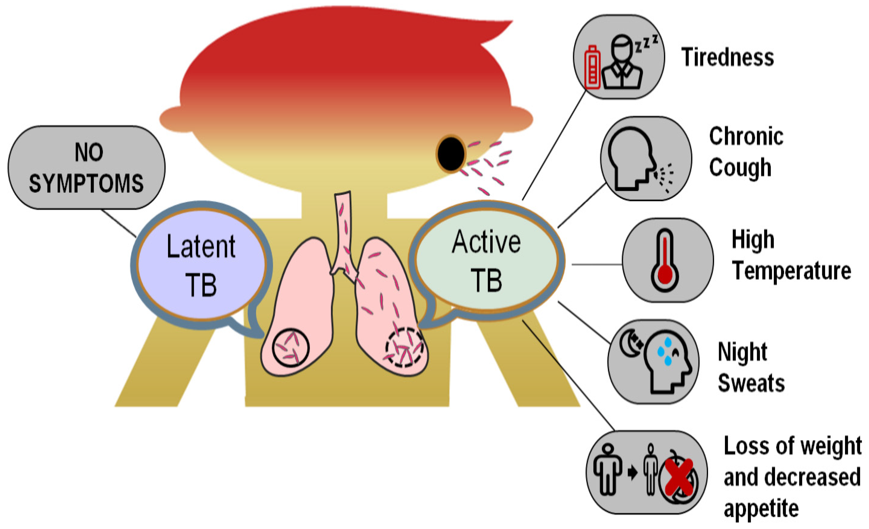
2. Tuberculosis (TB) and Its Occurrence

TB is an airborne bacterial infection caused by Mycobacterium tuberculosis (MTB), which mainly attacks the lungs [13]. However, the microbe may also spread to other body parts, such as the guts, skeleton, brain, and gland, from the lungs via different routes. When an infected TB person sneezes, spits, or coughs, the bacteria are expelled from the body. If inhaled by a healthy person, even in minimal quantities, these bacteria can cause TB [14]. People who exhibit symptoms are called active TB (ATB) patients [15], whereas TB patients without any signs are called latent TB (LTB) patients [16]. LTB patients cannot spread the disease to other people but have a high risk of developing TB if they do not maintain a healthy lifestyle (Figure 1). Moreover, people with weak immune systems due to infections such as diabetes and HIV; undernourishment; or people prone to using tobacco products are at high risk of catching TB if they come into contact with a TB-infected person [17]. When the bacteria can invade other body organs such as the bone, spine, and brain, it is called extrapulmonary tuberculosis (EPT). Miliary tuberculosis is a rare type of active TB in which the mycobacterium enters the other body organs via blood vessels. It infects various organs at once, including the lungs, spinal cord, and heart, making this kind of TB disease highly deadly [18]. Active TB is further classified into multidrug-resistant TB (MDR-TB) and extensively drug-resistant TB (XDR-TB). MDR-TB shows resistance to first-line antitubercular drugs due to the patient’s irregular treatment or insufficient and inadequate quality supply of drugs for the treatment. XDR-TB is resistant to both first-line and second-line antitubercular drugs (capreomycin, kanamycin, and amikacin) [19].

3. Conventional Diagnostic Techniques for Pulmonary TB

Early diagnosis of pulmonary tuberculosis is a preference for disease management. It is beneficial for both the patient and the public well-being, reducing the chances of further disease transmission in the community [20]. The diagnosis of TB disease is a combination of related symptoms and an investigation of the patient’s history for important information. The problem involved with early TB diagnosis is the slow bacterial reproduction inside the patient’s lungs that takes quite some time before showing the disease symptoms [21]. Presently several tests exist for the TB diagnosis and a few of which are chest X-ray [22], conventional light microscopy [23], light-emitting diode (LED) fluorescence smear microscopy [23], [24], liquid culture with drug susceptibility testing (DST) [25], lipoarabinomannan (LAM) lateral flow assay [26], Xpert MTB/RIF [27], first-line (FL) line probe assay (LPA) [28], second-line (SL) line probe assay (LPA) [29] and Loopamp Mycobacterium tuberculosis complex assay [30]. Early evaluation of pulmonary TB is performed by microscopic examination, sputum cultures, and chest X-rays. In contrast, the diagnosis of drug-resistant strains is made via a drug susceptibility test (DST) [21]. The traditional methods are tedious to perform, time-consuming, and require more time to interpret reports for diagnosing disease and drug resistance.
Moreover, this process gets delayed in detecting the disease early, leading to the patient’s suffering and further transmission of the disease to the surrounding healthy human [31]. Therefore, many immunoassay techniques have also been developed for the rapid diagnosis of TB, which have high sensitivity and require less time [32]. However, these tests’ major disadvantage is their cost in laboratory establishments, and the requirement of highly skilled staff is given in Table 1 [33].

4. History of AI Applications in TB Diagnosis

Many researchers have applied AI to TB diagnosis, contending the challenge of predicting and evaluating tuberculosis. The introduction of neural networks (Perceptron’s [34] and their improved versions) bought the idea of the pattern recognition method, which could recognize the structural patterns [35,36] in chest X-ray images and help in diagnosis. Artificial neural networks (ANN) started to impact TB diagnosis around early 1990 due to programs for pattern recognition. In 1990, a neural network was built to distinguish between the different kinds of interstitial lung diseases, including tuberculosis. The training dataset was prepared using ten cases for each of the nine types of lung disease. The model showed good performance and results, suggesting that ANN has high potential in computer-aided diagnosis of lung diseases [37]. In 1998, the first automated neural technique was built to identify TB bacillus in sputum smears stained with auramine. The model’s sensitivity was 93.5%, aiming to diagnose TB rapidly and accurately and reduce health risks for staff processing smear slides [38].
Similarly, in 1999, the first neural network was developed to diagnose active pulmonary tuberculosis using GRNN (general regression neural network). The model used 21 different parameters to form the input patterns and achieved sensitivity and specificity of 100% and 72%, respectively, in diagnosing active TB [39]. These machine-learning systems possess the capability to solve problems, learn from the given data, and deal with new problems. In addition to the systems mentioned above, several machine learning programs were also developed, including support vector machine (SVM) [40], decision tree (DT) [41], and random forest (RF) [42] that were applied for TB diagnosis.
The major challenge faced by all these models was diagnostic features to comply with making an accurate prediction. Furthermore, more efficient work was carried out to uphold AI advancement, leading to deep learning (DL). The DL turned up to be a useful concept in early 2010, when the integration of neural networks with algorithms (genetic algorithm [43] and fuzzy logic [44]) and single hidden layer feed-forward neural networks came into use for TB diagnosis. The capability of some deep learning approaches to predict and evaluate complex and diverse data has provided a new ray of hope for solving TB-related problems, such as drug resistance. In 2017, a deep learning model was developed to predict multidrug-resistant tuberculosis from drug-sensitive TB images obtained from computed tomography (CT). The model achieved good accuracy of 91.11% as the CT images have high resolution, are cost-effective, and help in the speedy diagnosis of TB [45]. In addition, deep learning in TB diagnosis also found its footsteps in predicting the severity of TB from CT pulmonary images, fast screening, and evaluation of chest radiography, and it builds DL tools (e.g., CAD4TB, Lunit INSIGHT, qXR, InferRead DR Chest, etc.) for easy and fast predictions.

5. Overview of AI Techniques Used in TB Diagnosis

AI or machine learning algorithms encircle a diverse variety of techniques. To assist readers with a better understanding of AI-assisted TB diagnosis, we will provide a short overview of the various AI learning techniques. Most machine learning techniques used for TB diagnosis are supervised learning, unsupervised learning, and semi-supervised learning. The choice of ML technique depends on the nature of the requirement, and each of the learning methods has its advantage and disadvantage (Table 2). Supervised learning is a process where the learning involves a set of input data (X) and output data (Y), and the training mode aims to search for a pattern in the provided data that would correlate with the coveted output data [46]. A fully automated CAD system effective for TB diagnosis was proposed to combine deep features with hand-crafted features. The authors used supervised learning and pre-trained CNN frameworks to detect TB in chest X-ray images that have minimized the pre-processing time and diagnosis performance, leading to an early screening of the disease [47].
Unsupervised learning is when only the input (X), but no comparable output data, is present [48]. The data are unlabeled in an unsupervised system. The process aims to train the model by extracting features from the data that could be used to cluster the input data into different units. In such a case, the algorithm learns from an elementary structure in the data to identify and give an interesting pattern [49]. The main difference between supervised and unsupervised learning is that unsupervised learning does not use a feedback signal to examine the standard solutions, making it less accurate and computationally complex [50]. In recent years, supervised and unsupervised learning was used in Bogota, DC, Colombia, to build two models using an artificial neural network to diagnose the disease and cluster data. The dataset was extracted based on sex, age, AIDS status, and other medical conditions of the subjects. The models probed supervised learning for disease diagnosis and unsupervised learning for data clustering from available information. The models showed a specificity of 71% and a sensitivity of 97% for TB diagnosis, giving an advantage of fast and low execution cost over traditional methods [51]. Similarly, in Rio de Janeiro, Brazil, two ANN models were developed and evaluated for diagnosing PTB in hospitalized patients. The first model used supervised learning for classification purposes, and the second used unsupervised learning to create risk groups. These predictive models were proposed to be promising tools for the early diagnosis of patients having radiological and clinical doubt of PTB [52].
An intercede of supervised and unsupervised learning [53] is known as semi-supervised learning. This AI technique utilizes a vast number of data in which only a few labeled data are available. This makes it widely applicable to real-world problems where it can increase the application of unlabeled data either by modification or reprioritization of hypotheses attained from limited tagged data [54]. For example, one of the studies used semi-supervised learning to arbitrate whether an unlabeled database could be used to train and enhance the accuracy of the machine learning model used to diagnose TB on chest X-ray images. The trained ML model showed good accuracy and could identify relevant features for TB diagnosis that the radiologists falsified. This learning technique could also significantly decrease the need for large, organized data even when the database labels were absent or noisy [55]. A specialized form of semi-supervised learning is active learning (AL), in which the model can contend with the problem of inadequate tagged training data in various ways [56]. For example, the model can request the user or any other information source to find the tags for the untagged data, for which the input data is least sure. A CAD system was developed using active learning for the detection of TB. This described learning method improved the algorithm to build a highly accurate system and helped narrow down the intrinsic uncertainty of the model while reducing the efforts for labeling [57].
Transfer learning has been one of the most popular machine learning techniques in recent years. This learning process provides relaxation from the common concept that the test and the training data should have, i.e., the same pattern and features [58]. In this way, the model learns valuable information from the old data source and transfers it to the new one by fine-tuning some parameters specific to the required output. This leads to an increase in the predictive efficiency of the target source. This learning technique was used to diagnose TB from chest X-ray images reliably. A dataset of 3500 TB infected and 3500 non-TB normal chest X-ray images were created. This database was used to transfer learning of already existing neural network models trained, authenticated, and tested to categorize TB and normal chest X-ray images. The best-performing model achieved high accuracy with 96.62% sensitivity and 96.51% specificity in diagnosing TB using chest X-ray images [59]. A CAD system was developed to detect TB with a hybrid approach, i.e., an ensemble of a deep model with a classifier. The approach increased the system’s performance, but retraining the deep model was time-consuming. This limitation was overcome by using a transfer learning approach where the existing models were fine-tuned with a few epochs of data, and the training time was reduced by a large extent [60].
Table 2. Merits and limitations of the various machine learning techniques used in TB diagnosis.
S.No.Learning TechniqueMeritsLimitationsRefs.
1.Supervised Learning
  • High accuracy.
  • Helps solve problems based on previous data training.
  • Labeled training data are required.
  • High quality and enough data needed.
[46]
2.Unsupervised Learning
  • It is ideal for unknown or raw data.
  • Less accurate.
  • Unlike the supervised approach, cannot specify the output data.
[48]
3.Semi-supervised Learning
  • It can use both labeled and unlabeled data.
  • Cannot manage unseen data.
[54]
4.Transfer Learning
  • Reduces the time required to build a model.
  • The model works only when the input and target models’ problems are the same.
[58]

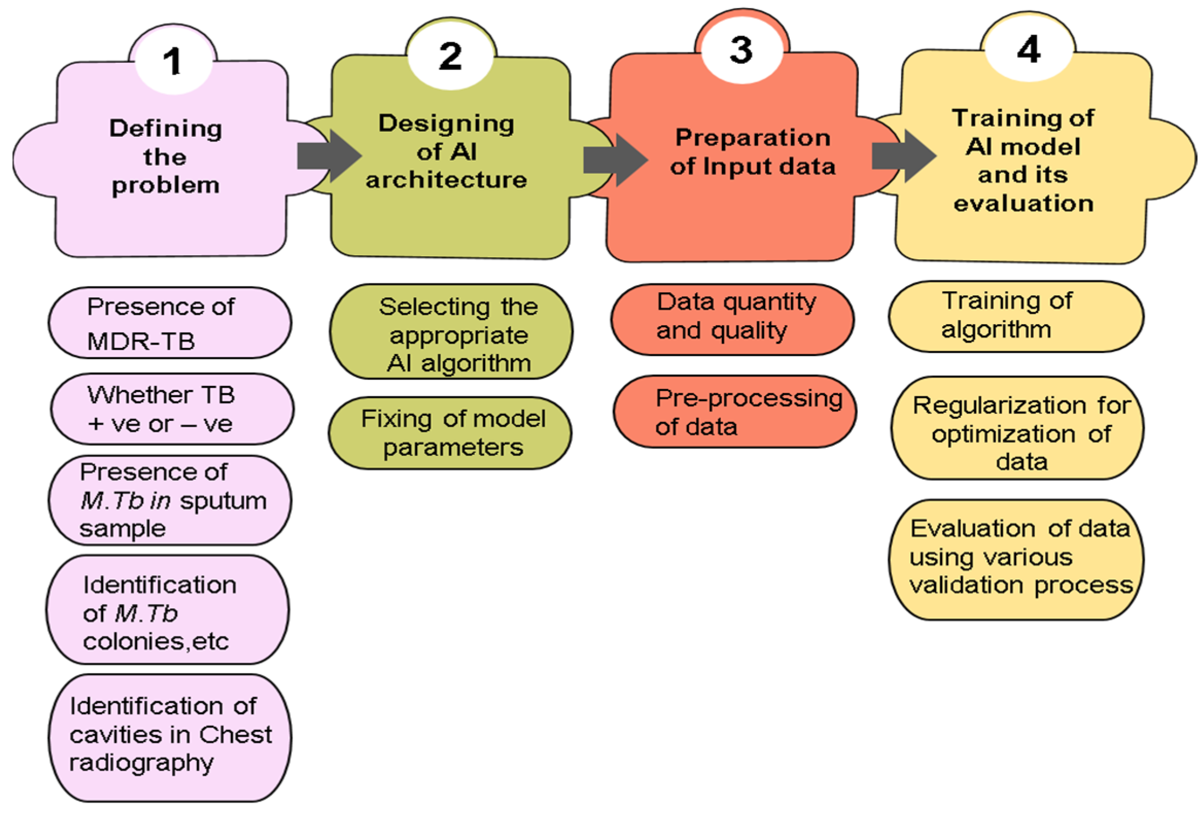
6. Construction of AI Model in TB Diagnosis

The application of a specific AI algorithm in TB diagnosis is a process that needs a proper understanding of the problems involved. The necessary steps in model construction include defining the problem, collecting adequate and related data, designing the AI algorithm’s architecture, training and evaluating the model, and correctly interpreting the output [61] (Figure 2). The first step involves problem identification, selecting the appropriate machine learning process, and an algorithm depending on the investigation problem [62]. The problem data can be divided into generative and discriminative inputs. The next step is to prepare a suitable model structure with appropriate algorithms such as SVM, RF, DT, ANN, etc., and fix model parameters with a clear idea about the problem. The parameters of the algorithms vary from one to another [63]. For example, in a neural network, the number of hidden layers and neurons can be the criterion, and the number of kernels can be a criterion in SVM. After the model structure is resolved, the dataset must be prepared. The quality and the quantity of the input data play a crucial role in determining the quality of the model under development. Once the model’s structure and input dataset are ready, the model’s training and evaluation are done. This training step aims to reduce the prediction error and increase the model’s efficiency. The final developed model should express the relation between the parameters and the developer’s purpose of model building. If the essential goal is not met, the model can be fine-tuned using pertinent data to accomplish the objective [63,64].

6.1. Preparation of Input Data

The input data/dataset used for the model’s training plays an important part, as it determines the model’s overall performance. Nevertheless, preparing the correct data is laborious and time-demanding work [65]. Before data preparation, one should understand the needs of the training set, i.e., representation of the complexity and type of data required, the quantity of the domain-specific input data, and its distribution in the internal space [66,67]. The quality of the training set also depends on the occupying space with possible input data required for making the prediction. The model can be pre-processed if inadequate data are available [65,68]. Here, in this article, some guidance for these problems has been provided.

6.2. Input Databases Used in TB Diagnosis

The most used input datasets for TB diagnosis are the Shenzhen Dataset and Montgomery County chest X-ray dataset. Nonetheless, there are limitations constitutive of these datasets. Compared to the training model size, these datasets are small, leading to more computational memory and time. For example, Lakhani et al. [69] used AlexNet (60 million parameters) and GoogleNet (7 million parameters) as input training models for TB diagnosis. These are the most frequently used deep learning models, pre-trained on many images (besides TB radiography). Table 3 summarizes the various datasets used for developing AI models in TB diagnosis. As such, many parameters for sparse data result in more memory usage.
Similarly, evaluation using many parameters is also error-prone (overfitting). Various improved dataset models have been introduced to overcome such problems, such as the shufflenetV2 [70] model for TB diagnosis. This dataset model is more specific, accurate, has fast prediction, and uses a lightweight neural network. The other limitations include early diagnosis of multidrug-resistant TB and differentiating it from different types of TB. This calls for an AI model requirement trained to diagnose multidrug-resistant TB in its early stage. A ResNeXt 50 CNN classifier was developed using transfer learning to identify whether the person has MDR-TB or DS-TB. ResNeXt 50 is a CNN model with 50 hidden deep layers that are easy to use and have a highly modular structure for image classification [71].
Table 3. Various datasets used in TB diagnosis.
S. No.Name of the DatabaseDeveloped byFeatures of the DatabaseRef.
Chest X-ray Dataset
1.Shenzhen datasetPartnership with Shenzhen No.3 People’s Hospital, Guangdong Medical College, Shenzhen, China
  • Consists of a total of 662 anterior chest X-ray images.
  • In total, 336 images are TB cases (including pediatric chest x-rays) and 326 images are normal cases.
[72]
2.Montgomery County chest X-ray dataset (MC)Partnership with the Department of Health and Human Services, Montgomery County, Maryland, USA
  • Consists of a total of 138 anterior chest X-rays from the Montgomery County TB screening program.
  • In total, 58 images are TB cases and 80 are normal cases.
[72]
3.PadChestRadiologist at San Juan Hospital, Spain
  • Consists of a total of 160,000 images and their associated reports.
  • It contains a total of 152 images of TB cases.
[73]
4.ChestX-ray8 datasetRadiologist at NIH Clinic center,
Bethesda, Maryland, USA, as a part of routine care
  • Consist of a total of 112,120 anterior chest X-ray images.
  • In total, there are 51,760 abnormal images (18,898 TB related) and 60,360 normal chest X-rays.
[9]
5. Belarus TB Portal dataset TB specialist at Minsk city, capital of Belarus, Europe
  • Consists of a total of 304 images of confirmed TB cases.
[74]
6. TBX11K dataset Media Computing Lab, Nankai University, China
  • Consists of a total of 11,200 images.
  • In total, 924 images are of TB cases.Comprises of four classes: active TB, latent TB, healthy and unhealthy but non-TB.
[75]
7.8-Bit dataset-ARadiologist at National Institute of Tuberculosis and Respiratory Diseases, New Delhi, India
  • Consists of a total of 156 images, where 78 images are TB cases, and the other 78 images are non-TB cases.
[76]
8.14-Bit dataset-BRadiologist at National Institute of Tuberculosis and Respiratory Diseases, New Delhi, India
  • Consists of a total of 150 images, where 75 images are TB cases, and the other 75 images are non-TB cases.
[76]
Sputum Smear Microscopy Image Dataset
9.ZNSM iDBJaypee University of Information Technology, Solan, India
  • Consists of image data compiled from seven different datasets by using three different light field microscopes.
  • TB diagnosis is made on three main domains: autofocusing, auto stitching, and auto-grading of TB bacillus.
[77]

6.3. Quantity and Quality of Input Data

Input data are one of the essential parts of model construction that need close attention. For example, suppose the dataset’s size is insufficient for the learning process; the training task leads to overfitting and errors and increases the chances of model incapability to generalize the new data efficiently. In such a case, the training model’s performance should be checked as to whether it is acceptable. In an unacceptable case, the model normalization [78] can be carried out, and even still the model is not benefited, so some reconstruction or tuning of the model is required. Generally, learning the training data should be a possible task even with a less optimized structure, so it is better to focus on the model’s architecture rather than collecting more data. However, even if the reconstruction of the model or use of another algorithm does not improve, it is time to check the quality of training data for errors, logical correspondence between input and output data, and balanced training data distribution [79].
Moreover, it is better to retrain the model on unlabeled as it gives an inefficient performance, which will conclude that the model is forming a meaningful link with training data. However, even if the model performance is still unacceptable, it is recommended to collect more data. The primary consideration here includes the high cost of collecting data, which can be overcome by optimizing the model [80]. This can be done by selecting suitable parameters, advanced architectures, regular normalization of the structure, and a preference for high-quality data at an affordable cost [78,80].

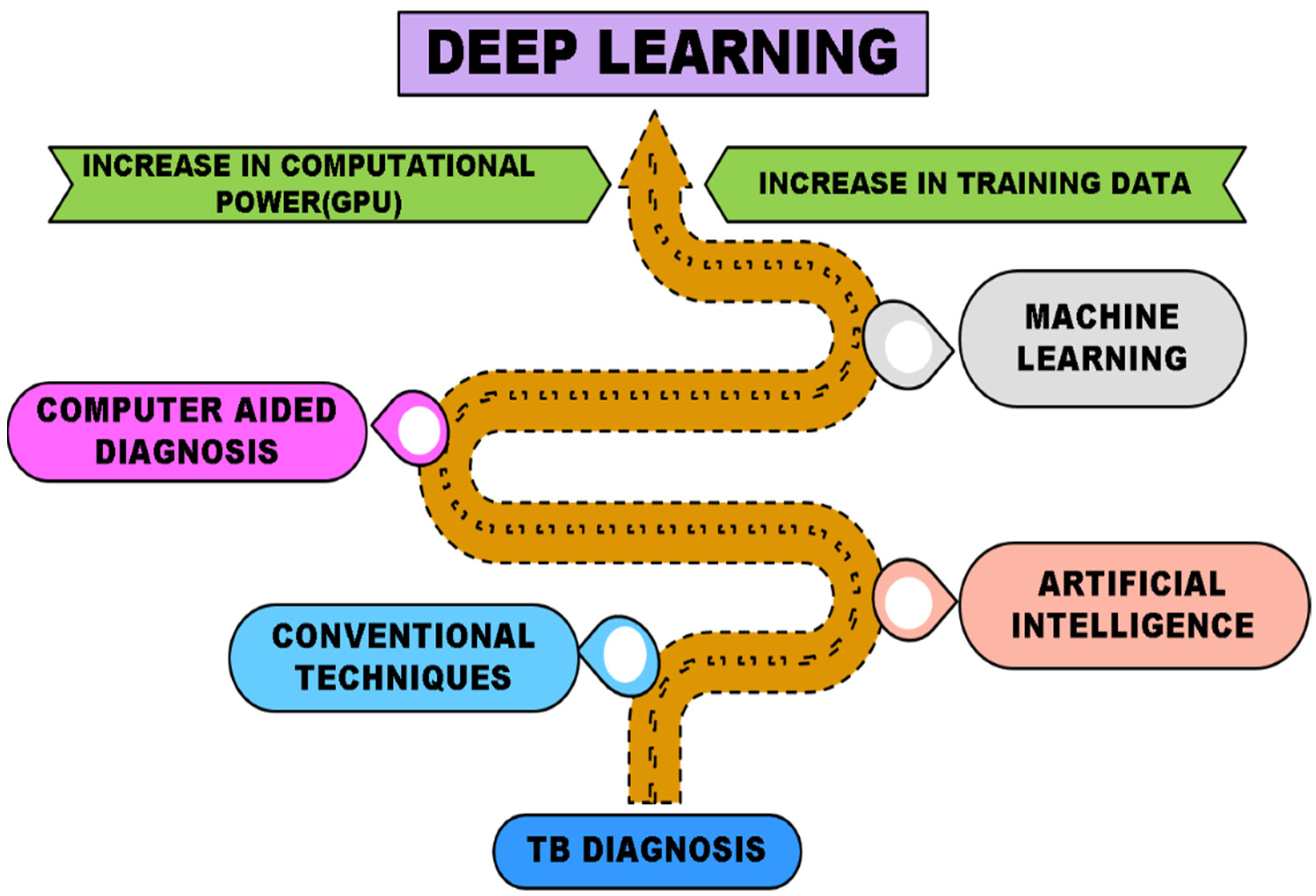
7. Advancing with Deep Learning

Deep learning is a machine learning technique in which machines learn to perform classification/prediction tasks directly from the input data [81]. Building deep learning models with high accuracy requires a combination of signals with different weights (where the dataset can be adjusted according to weights to give an expected response for a given input), which passes the results successively deeper in the multi-layer neural network framework until an output layer [79]. Multiple learnable stages make this approach more useful in tackling complex problems. “Deep” here refers to the number of hidden layers in the network, i.e., it can have numerous hidden layers compared to traditional neural networks. Deep learning has been widely used for bioinformatics and computational biology [82]. With increased training data and powerful computational capacity, conventional machine learning methods have transformed into advanced machine learning models (Figure 3).

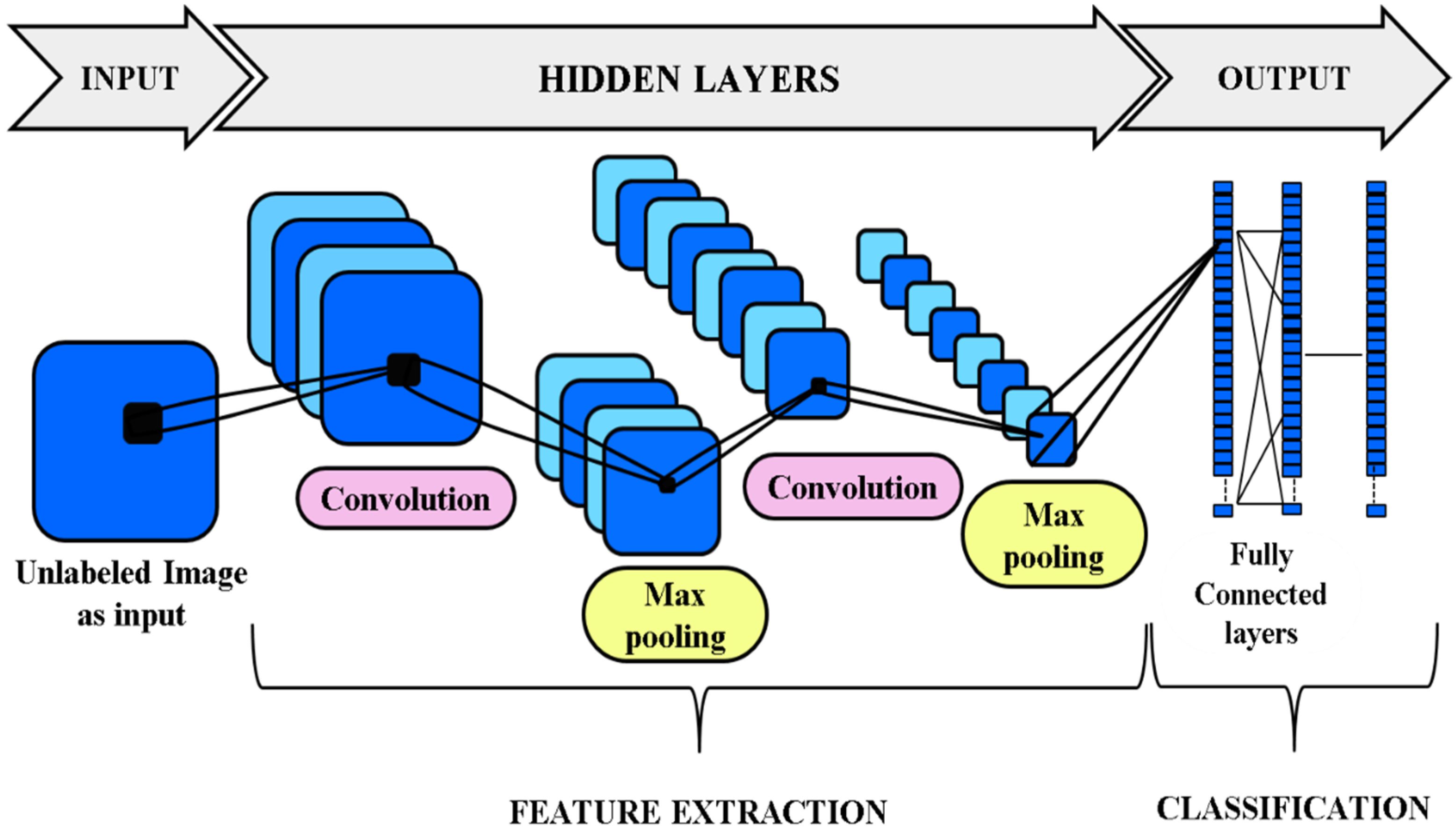
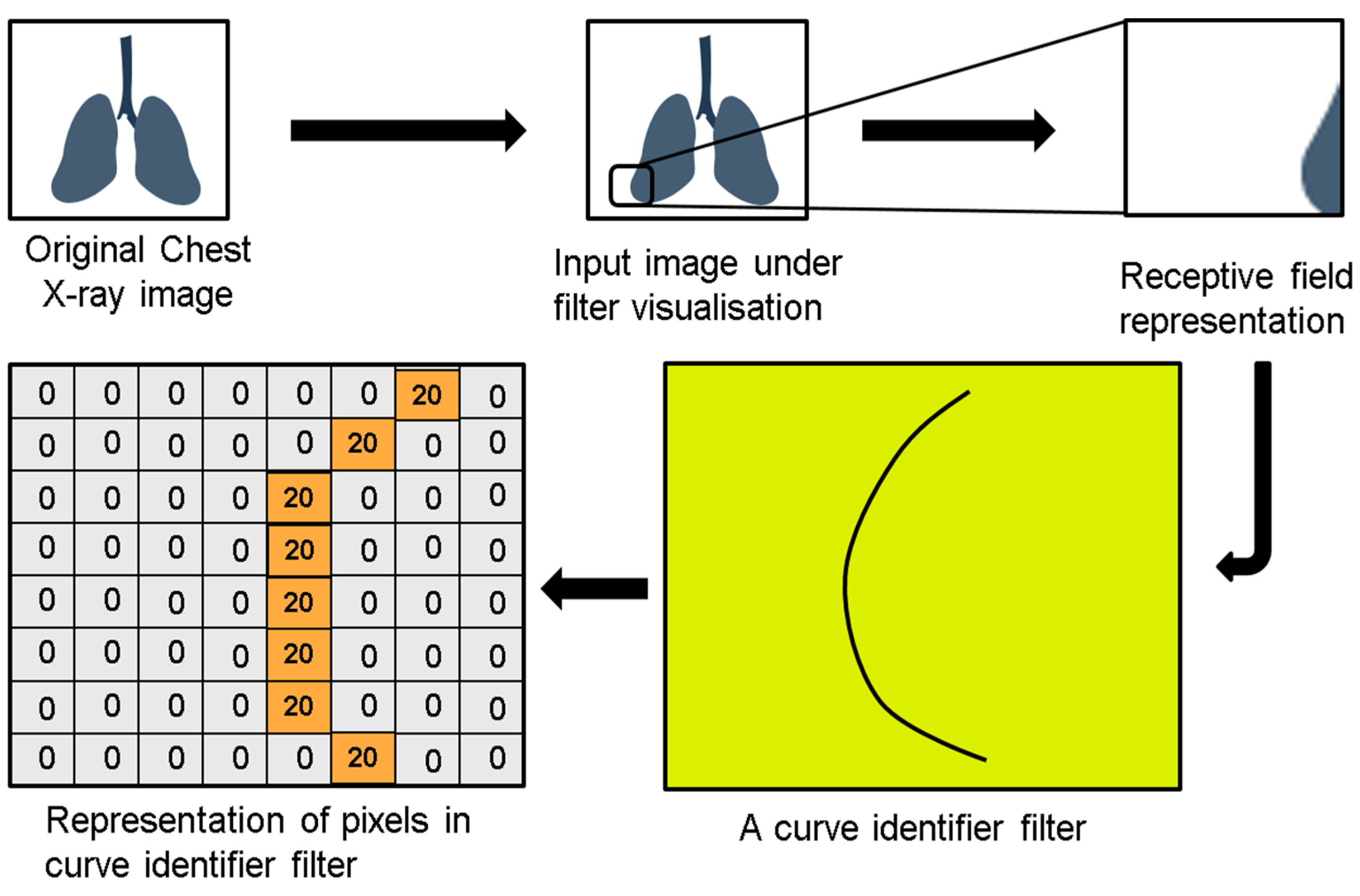
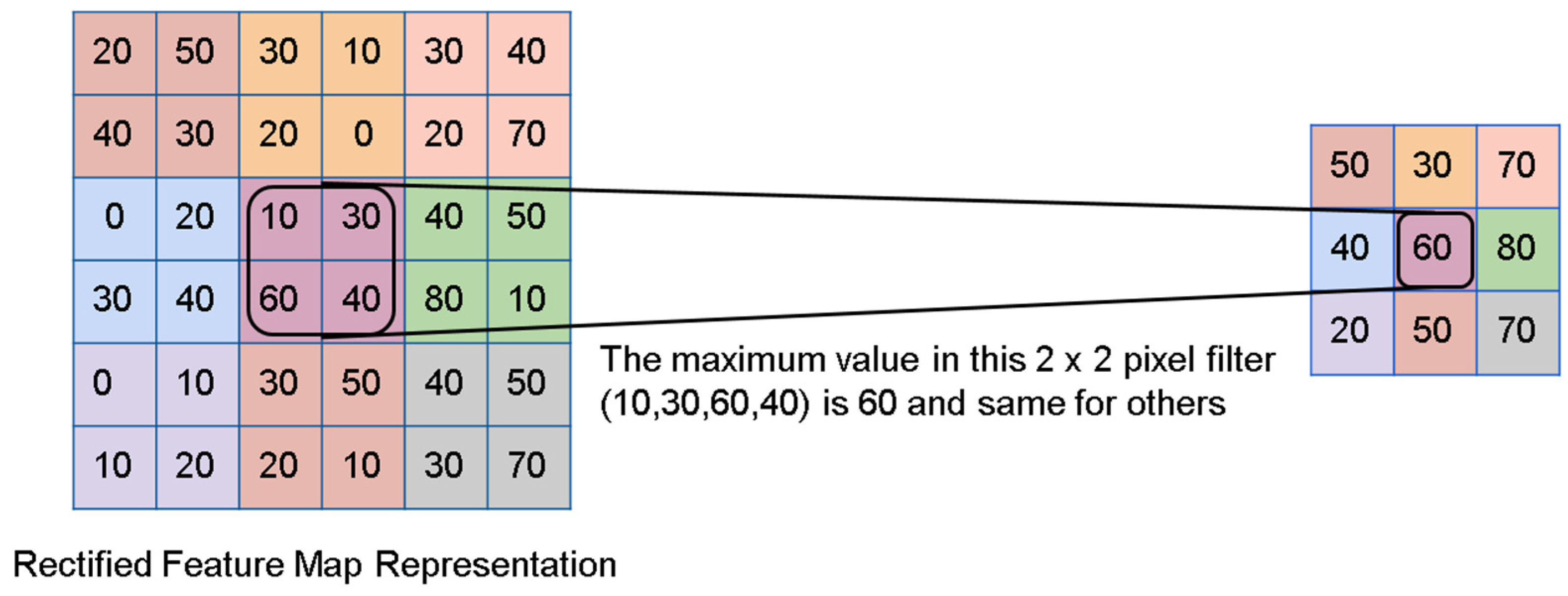
7.1. Convolutional Neural Networks (CNN)

CNN is a deep learning class that uses feed-forward artificial neural networks to analyze images [83]. It is considered state of the art for image classification due to the connectivity pattern between its neurons, which are arranged so that they respond to overlapping regions building the visual field. It consists of many hidden layers, out of which the important ones are the pooling layer and fully connected (FC) layers. The pooling layer reduces the image’s spatial size without changing the depth, and the FC layers are generally placed before the final output layer, where the image classification process starts [84]. CNN is motivated by biological processes and variations of multi-layer neural networks designed to use minimal amounts of pre-processing [85]. Moreover, due to its non-requirement of domain knowledge, it can extract and learn meaningful features by segregating the target classes during the training phase [83] (Figure 4).

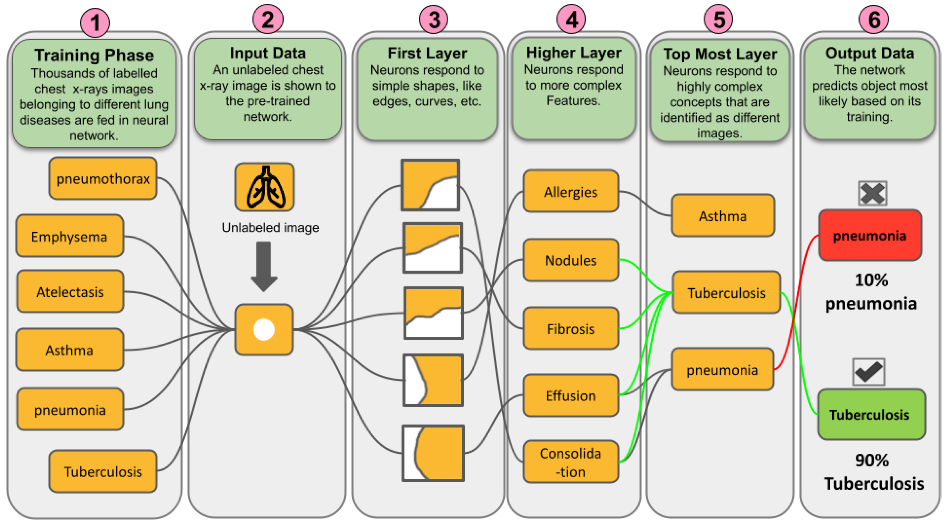
7.2. Does CNN Make Our Job Easier in TB Diagnosis?

CNN is a remarkable combination of math and biology with a bit of a mist of computer science. It has been a significant invention in image processing and machine vision. The conception of CNN depicts the probability of the data (images) into certain classes on the same basis as the human brain works [84]. For example, when a human looks at an image, they classify it into different types based on its characteristic features. Similarly, the computer algorithm can organize the image by identifying the low-level features such as curves and edges at initial levels and gradually generating more intense concepts with high-level features through a series of hidden layers (convolutional layer, pooling layer, fully connected layer) [83]. Similarly, a CNN model learns distinguishing features from the TB dataset and tries to classify a new input image as a TB or non-TB case (Figure 5). Details of steps required to build a CNN model for TB diagnosis using chest X-ray images are as follows.
Input for a convolutional layer: The input for a CNN model is in the form of images. These images are resized into an optimal size and transferred to the hidden layers. Each hidden layer consists of kernels or filters or neurons put over some portion of the input image subjected to the neurons’ size [86]. The CNN model compares images fraction by fraction, and each fraction is known as a feature. Each feature is like a mini-image, and each neuron is a feature identifier [68]. For example, one of the filters identifies the curves in the chest X-ray image as a feature. When the input image passes through this filter, it will locate the curves present in the image. Similarly, there will be other filters for identifying features such as a straight line, curves bending to the right, curves bending to the left, or straight edges in the input image. Furthermore, the greater the number of filters, the greater the identification depth, leading to more information about the input image [87].
Activation of the convolutional layer: Each filter can identify a specific feature in the input image. When the image passes through the filter, it searches for that feature in the image. Once the filter spots the feature, it can identify the features that lead to activation of the filter [87]. The filter further moves to spot the specific feature in the input image. This leads to forming an activation map of that filter for the feature in the image. The value generated by the activation map of the filter gives an idea about the percentage of that feature present in the input image [88]. For example, we will consider a filter of dimensions (8 × 8) as a curve identifier (Figure 6). When a chest X-ray image passes through this filter, it will try to spot the image’s curves.
When the filter identifies a feature, it will try to match the pixels of the input image and the pixels of the filter. Furthermore, suppose an image section responds to the matching process, leading to the generation of a value (the sum of the multiplication of filter pixels with the input image) and an activation map [89]. The higher the value generated by the activation map, the greater the presence of that feature in the image, i.e., it shows some type of curve in the input image that caused the activation of the filter and vice versa. This is only for one filter; similarly, many filters are present for each feature. Furthermore, the activation map of one filter acts as the input for the next filter, identifying another feature and forming a new activation map. This series of feature identification carries on, creating a more complex activation map [88].
Pooling layers: The pooling layer is another essential layer (the max-pooling layer (Figure 7)) in the CNN model that shrinks down a large image while preserving the essential data. It means that it keeps the maximum value of the pixel window, i.e., the best fit of the feature within the pixel window, and makes the output have few pixels but with the same number of features [88]. For example, in a chest radiograph, the max-pooling layer will be done for each feature, such as the shape of the cavity, opacity area, density of the cavity, etc. This layer helps in reducing the computation load and solves the problem of the system being hyper-literal [90].
Extracting the output from fully connected layers (FCL): The fully connected layers in the CNN model convert the highly filtered images into votes. FCL is the primary building unit of the traditional neural network, which treats the input images as a single unit instead of two-dimensional blocks [90,91]. An FCL looks for high-value features that strongly complement a particular category and have respective weights. When the model is analyzed, it gives the correct probabilities for various categories [92]. For example, suppose the algorithm must decide that the input images are TB manifested. In that case, it will show high values in the activation maps that represent high-level features, like the opacity of the cavity, size of the cavity, lesions in lung space, etc. This will be followed by studying and interpreting the probability values for confirmation of TB in the chest X-ray image [93].
Data interpretation and explainable AI (XAI) for decision making of the CNN model: The output values obtained from the CNN model now need to be interpreted for the prediction of TB in the chest X-ray image [94]. This can be done by using explainable AI (XAI), where explainability is the ability of the AI model to explain decision making in an accessible form for medical experts in a broader range, thus making it easy to understand how a decision has been reached. There are two types of explainable AI techniques, i.e., post-hoc and intrinsic [95]. In the post-hoc method, we can approximately understand the behavior of the black box based on the decision set and perturbation-based methods, in which the relationship between prediction and feature values is extracted. This method includes class activation mapping (CAM), gradient-weighted class activation mapping (Grad-CAM), score class activation mapping (Score-CAM), Shapley Additive explanation, and principal component analysis (PCA) [96]. On the other hand, in intrinsic methods, we can understand the decision-making procedure as it accounts for which part of the input data is responsible for classification in any type of classifier. This method includes rule-based learner, logistic regression, decision tree, and Bayesian model [97].
Many research groups have reported the use of the DL models for the detection of TB by including transfer learning in the CNN model for diagnosis of TB using pre-trained models and also by varying the deep-layer CNN model parameters. A CNN model was developed using a chest X-ray dataset to diagnose TB by replacing the complexity of the pre-processing seen in DT analysis with a generalized model. It used transfer learning to train the model and the hidden layers for detection to help achieve high accuracy, i.e., with or without augmentation. The work concluded that applying CNN models could help bypass the need to build segmented algorithms, which can be time-consuming and require expertise [87]. A CNN-based LIRA (Lesion Image Recognition and Analysis) model was built to evaluate lesions obtained from pulmonary TB tissues. The study was to overcome shortcomings such as reproducibility and efficiency of histopathology studies. The LIRA model used seven pathology features, including three different lesion types from pulmonary tissue. The proposed model gave good results [98]. The FC-SVNN (fractional crow search-based deep convolutional neural network) model was proposed, which was used to classify and detect TB in a patient. The analysis for severity was conducted by extraction of features, which is detected using an adaptive fractional crow deep convolutional neural network (AFC-CNN), the modified version of the FC algorithm. The detection of TB severity using the model helped determine the level of infection [99]. Using the CNN model, a two-stage classification method (TSCM) was built to classify tuberculosis culture. The researchers used transfer learning for the small and imbalanced dataset, balanced using SMOTE (synthetic minority over-sampling technique). The model could boost 98% recall and 99% precision on non-negative class, which indicated successful detection of anomalies in the culture [100].
A deep CNN model was presented that used transfer learning to detect tuberculosis from the X-ray images. It used many pre-train CNN models i.e., MobileNet, ResNet, Xception, EfficientNet, and Inception for extracting features from the input image. It was concluded that datasets having extracted ROI gave high accuracy and better results in the detection of TB. The researchers also reported the use of two visualization techniques, i.e., grad-CAM, which helps in consolidating the medical expert diagnosis, and T-SNE, which helps in explaining the training efficiency of the trained model [101]. A simple, faster, and high-accuracy CNN model was proposed to overcome the overfitting problem and can be easily installed in mobiles. The model showed an accuracy of 86% and used a grad-CAM visualization technique for the detection of tuberculosis [102]. Similarly, a deep learning model was proposed to reliably detect TB from CXR images based on image preposing, image data augmentation, and segmentation followed by DL classification methods. It used transfer learning to classify TB and non-TB cases from the pre-trained deep CNN models. Further, it also used the Score-CAM visualization method to show that the model learns from the regions of segmented lung areas and produces the results for the diagnosis of TB with high precision, accuracy, and sensitivity, i.e., 97.34%, 97.07%, and 97.07%, respectively [59].

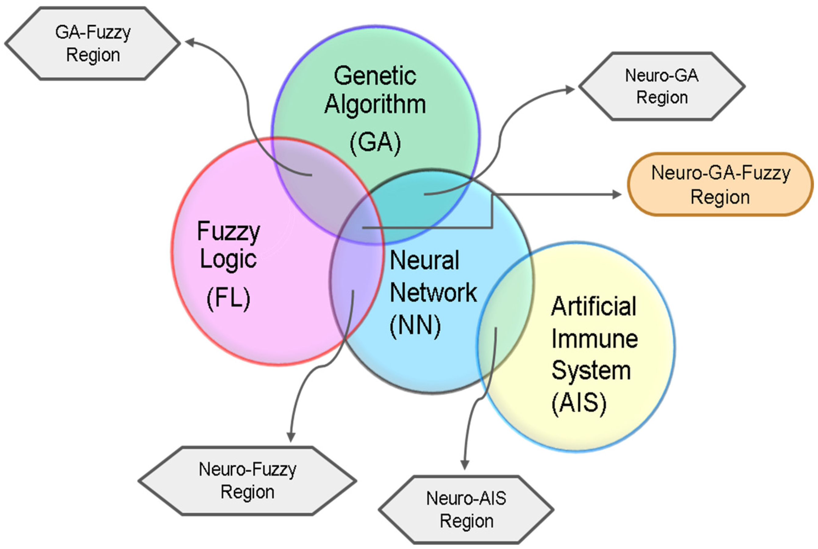
8. Integration of Deep Learning with Advanced Algorithms in TB Diagnosis

The power of neural networks can be amplified by integrating it with other algorithms such as fuzzy logic, genetic algorithm, and artificial immune system. These integrated systems have been applied for TB diagnosis and have proven to be useful (Figure 8).

8.1. Adaptive Neuro-Fuzzy Inference System

Fuzzy logic (FL) is a technique that handles numeric data and linguistic knowledge simultaneously; it is the rule-based mapping of a set of input values to a single output value. It maps the numeric value to numeric values [103]. Fuzzy logic presents an inference morphology that incorporates human reasoning capabilities in knowledge-based systems. Neural networks can recognize patterns, classify data, and predict events effectively, but the models cannot explain how patterns are classified or recognized. On the other hand, fuzzy systems are good at computing and explaining decisions, but they fail to adapt to new environmental conditions [104,105]. An adaptive neuro-fuzzy inference system (ANFIS) was used to diagnose tuberculosis. The model used input parameters such as sputum, chest X-ray, weight loss, etc., as TB symptoms for a rule-based fuzzy system to make output decisions. The work is under consideration for rural areas where the availability of doctors is difficult [106]. The work was extended by building an ANFIS model having 159 distinct rules and a backpropagation algorithm that would help in minimizing errors in the output. It also used a triangular fuzzifier for the fuzzification and MATLAB software results [107]. A neuro-fuzzy methodology was developed to diagnose TB in which eleven TB symptoms were used as input in MATLAB 7.0 software for the ANFIS experiment. The evaluation of the system was done by using the Trapezoidal membership function and backpropagation algorithm. The model was efficient in learning in a shorter time and gave good results [108].

8.2. Genetic Algorithm with Deep Learning

Genetic algorithms (GAs) are commonly used to build high-quality solutions to search for problems by relying on bio-inspired operators such as mutation, crossover, and selection [109]. GAs were mainly used to simulate the environments and behaviors of entities in a population. A genetic algorithm creates a sample from the population at its very core by a random selection process. Then, it scores each member of the population based on some goal called a fitness function. Next, the mutation process selects the sample closest to the fitness function, breeds the best population members to produce more like them, and kills off the rest (survival of the fittest and all). This process is repeated multiple times, and each iteration through these steps is called a generation [110]. Genetic algorithms were trendy before Neural Networks (NNs) [111]. Since NNs require big data while GAs do not, genetic algorithms are used in artificial intelligence, like other search algorithms, to search a subset of potential solutions [109].
The genetic-neuro approach was used to study its application in diagnosing tuberculosis. A backpropagation algorithm classifier with the Levenberg—Marquardt algorithm was used to train the model, where nine features were selected for developing the dataset. GA was used for the extraction of individual features having importance. The GA-NN model could classify 96.3% correctly from the test data [112]. Similarly, a hybrid algorithm was built to diagnose chest diseases, including tuberculosis. An ANN having backpropagation algorithms was used to evaluate the output error and calculate the error gradient. It conducted a comparative study of chest diseases using the genetic-neuro method and concluded it to be a valuable tool for early TB diagnosis [113].

Genetic-Neuro-Fuzzy Inference System (GENFIS)

An inference system integrates a genetic algorithm, neural network, and fuzzy logic to develop an adaptive model that can carry out self-training for imprecise and uncertain data. A genetic-neuro-fuzzy inferential method was used to diagnose TB. The model was studied with a case study of 10 patients with the help of the MATLAB 7.9 version. The evaluation of the model was done using the medical records of 100 patients. GNFIS was optimized using a decision support engine, giving sensitivity and accuracy of 60% and 70%, respectively [114].
Similarly, a GENFIS model was applied for the early diagnosis of tuberculosis. Again, they used a decision support system to evaluate 100 patients’ medical records. In this, 70% of data were used as training data, 15% for validation, and the rest for measuring the model’s performance. The proposed model gave sensitivity and accuracy of 72% and 82%, respectively [115].

8.3. Artificial Immune System (AIS) with Deep Learning

AIS is a new AI technique that is starting to grow through many interdisciplinary researchers’ collaborative efforts. AIS is developed using the core ideas, theories, and components of the immune system. It aims at solving problems such as pattern recognition, classification, elimination, and optimization, which are complex and require high computational power [116]. The natural immune system principles are found in the human body, forming the basis of the AIS tools. The powerful information processing capabilities of the biological immune system (BIS) are an area of significant research for multiple reasons, mainly the distributed nature of its memory and decentralized control mechanisms from an informational perspective. Such computational models are believed to solve many science and engineering problems [117]. Diagnosis for chest diseases such as tuberculosis via proper interpretation of the disease data is a significant classification problem.
Furthermore, AIS has the potential to provide an efficient way of solving chest disease diagnosis problems. Using AIS in Turkey, a study was conducted to diagnose tuberculosis. The dataset was prepared from a chest disease hospital using patients’ epicrisis reports. The classification accuracy obtained was 90% with AIS and MLNN with LM (Levenberg—Marquardt) algorithm [118]. An AIRS (artificial immune recognition system) incorporated with a fuzzy logic controller was used to create an unsupervised hybrid machine learning model. This hybrid system showed bioinspired ways to help doctors in their diagnostic conclusions [119].
A new hybrid system, SAIRS2, was built to diagnose TB by incorporating AIRS with the SVM classifier. The study’s primary purpose was to reduce the loss of diversity and overcome the existing selection pressure. It used the dataset to build the SAIRS2 model, which was analyzed using WEKA (Waikato Environment for Knowledge Analysis), a machine learning program [120]. The author also integrated AIRS with real-world tournament selection (RWTS), forming a new hybrid system, RAIRS2, to diagnose TB. RWTS is one of the favored and applicable tournament selection mechanisms in genetic algorithms that help control the population size and overcome the selection pressure [121]. Using a computational model UISS (universal immune system simulator), an agent-based modeling method with AIS was used to diagnose TB. The model helped understand the underlying TB pathogenesis and its interaction with the patient’s immune system [122].

9. Tools Built Using Deep Learning Techniques

For many years, TB management has been approached according to a paradigm of symptomatic active infection and asymptomatic latent disease. However, in 2015, the end TB strategy specifically outlined some policies for increasing the TB diagnosis capacity across the high tuberculosis burden countries [123]. These policies prioritize rapid diagnosis using various advanced tools, mainly drug resistance. In the past few years, many computer-aided diagnostic tools using deep learning have been built for fast screening and triaging a quick and efficient screen of a large population, helping identify people needing further treatment. These CNN-based tools provide a labor-saving and structured interpretation of TB-related images [124,125] In addition, an online resource has recently been launched [https://www.ai4hlth.org/ (accessed on 3 July 2022)] by Foundation for Innovative New Diagnostics (FIND) and the Stop TB Partnership. This resource center will provide relevant data for several computer-assisted products in areas with limited access to radiologists and low-resource countries with a high TB burden (Table 4).
Amongst all the tools mentioned, a few tools such as CAD4TB, Lunit INSIGHT CXR, and qXR have been validated with real-time parameters. In August 2018 in Korea, for the first time Lunit Insight CXR nodule, version 1, Lunit Inc., was approved by the government for analyzing the CXR of adults. Again in October 2019, Lunit Insight CXR MCA, version 2, based on ResNet34, was approved in Korea, and in March 2020 it was applied for all patient chest radiograph detection. From then until February 2021, around 56,192 CXR of adults have been analyzed considering the posteroanterior and anteroposterior views. This software can diagnose three forms of lesion, i.e., pneumothorax, nodule, and consolidation, and also lesions with an abnormality score of more than 15%. The image analysis and upload of results by AI happened in less than one minute, thus reducing the scan time and giving a board interaction. Lunit Insight CXR, version 3, was approved in October 2020 and installed in March 2021 in the hospital in Korea. It could detect eight types of lesions, i.e., fibrosis, pleural effusion, pneumoperitoneum, nodule, atelectasis, consolidation, cardiomegaly, and pneumothorax, and had an additional visualization method, i.e., grayscale contour maps. It could analyze around 106,230 CXR until February 2022. These AI CAD could not only diagnose the disease but also optimize the workflow by reducing the mean time to report critical and urgent cases [137].
In Dhaka, Bangladesh, a study was conducted to evaluate five commercially available AI CAD, i.e., Lunit INSIGHT CXR (version 4.9.0), CAD4TB (version 7), qXR (version 3), InferRead DR (version 2), and JF CXR-1 (version 2) between May 15, 2014 to October 4, 2016. People aged 15 years or above were referred to three TB diagnostic centers in Dhaka. All the participants were screened for symptoms and received an Xpert MTB/RIF test and digital anterior-posterior chest X-ray. These were evaluated by the five commercially available AI CAD and three registered radiologists. The performances of all the AI algorithms were compared with each other and with that of the radiologist, and also with the WHO Target Product Profile (TPP) of triage tests. Around 23954 people were analyzed for tuberculosis, and all AI algorithms outperformed all radiologists. All the AI algorithms maintained 90% sensitivity and helped in reducing of the number of Xpert tests required in TB diagnosis. The major disadvantage of all the AI algorithms was that they gave worse performance for the older age group, i.e., >60 years, and also for people having history of tuberculosis [138].
In Kharachi, Pakistan, a study was conducted at the Indus Hospital where two AI algorithms CAD4TB version 6.0 (CAD4TBv6) and qXR version 2.0 (qXRv2) were compared with a mycobacterial culture of two sputa as reference. The study included 2198 participants, out of which 2198 were HIV-negative and 272 were pulmonary tuberculosis confirmed by culture examination. Both software showed minimum value (CAD4TBv6 sensitivity 0.93, non-inferiority p < 0.0001; qXR sensitivity 0.93, non-inferiority p = 0.0002; CAD4TBv6 specificity 0.69, p = 0.0003; qXR specificity 0.75, p < 0.0001) for the test for non-inferiority as recommended by WHO, having a non-inferiority limit of 0.05. It was also seen that for both the software, the sensitivity was low in the case of smear-negative pulmonary TB and for women in CAD4TBv6. Furthermore, the specificity was low in men, those having a previous history of tuberculosis, older age groups, and those with low body mass index [139].

10. Conclusions and Future Aspect

Advances in AI techniques, particularly in deep learning (DL), have been supported by improved hardware storage such as LSPC (large-scale parallel computing), GPU (graphics processing unit), and TPU (tensor processing unit) [140], as well as big data. Accomplishment in the fields such as image and voice recognition has brought AI more public acceptance and new hopes for diagnosing disease. Machine learning has exceeded human expertise in several areas when seen in terms of performance. Therefore, it is no surprise but more promising when applied to TB diagnosis. AI approaches in TB diagnosis are mainly integrated for early disease diagnosis, focusing on TB symptoms and drug resistance. Successful DL model examples for TB diagnosis are at the fore. For instance, ImageCLEF is a forum established in 2003 that evaluates images and cross-languages retrieval by organizing conferences [141]. It is a platform that sees participation from both industrial and academic researchers in machine vision and pattern recognition, computer—human interaction, and medical informatics as a few classes in the ImageCLEF campaign [141]. In 2018, a classifier was developed to identify whether the patient has MDR tuberculosis or drug-sensitive tuberculosis based on the patient’s CT scan. It secured the best accuracy and second position in AUC evaluation at ImageCLEF 2018 tuberculosis conference [71]. In Peru, a deep learning model was developed on similar grounds to identify TB based on its types. The developed model was promising and later planned to be implemented into mobile devices for TB diagnosis [142].
Regardless of significant success in the past, AI implementation in TB diagnosis is still challenging due to the acquirement of quality; thus, adequate and problem-solving data remain a challenge. Data compilation is easy when we see it in the computer vision field, as the chosen data are highly reliable and form a large dataset. However, a horse will always be a horse and cannot be an elephant. Unfortunately, it is not the same in disease diagnosis as many reasons will hinder producing good quality and sufficient data to form a dataset. On the one hand, the data collected from different experimental sources highly depends on the variant experimental conditions applied, which might give different or opposite results. This is due to our complicated biological system, which shows various symptoms and drug resistance for the same disease and a drug. On the other hand, a vast range of data is available, depending on the nature of the requirement and the necessity to build a high-quality dataset.
One solution to the kettle of fish is developing an algorithm that can deal with different or insufficient datasets. In 2019, a constructed DL model was trained using only the chest radiograph to detect TB. This model was trained using NIH and Shenzhen datasets [72] and the Belarus Tuberculosis Portal dataset [143]. However, this dataset was not of high quality since it still needed further chest image upgrading, and its distinctive quality focused solely on TB diagnosis [102]. Another critical challenge is to utilize and stick to the principle of removing unwanted data to achieve more refinement and a task-oriented standards set. In 2017, a DL model was pre-trained on millions of images using AlexNet and GoogLeNet datasets, containing many images, including chest radiographs. This model required significant computer storage space to work efficiently, even for a small job of TB detection using chest X-ray images [69]. At the same time, a dataset was proposed that required lesser storage memory and a cheaper processor as it required storage only in mega-FLOPs in contrast to AlexNet and GoogLeNet datasets that required storage in Giga–FLOPs [102].
The deep learning methods integrated with other neuro-fuzzy, genetic algorithms and artificial immune systems are the most promising subdomain for TB diagnosis. These systems increase the system’s sensitivity, specificity, and accuracy since they take biological information. Therefore, we can expect to see these integrated algorithms for a much improved and increased success rate in TB diagnosis in the coming years. However, overfitting (a modeling error in which a function fits too closely to a set of limited data points) and understanding the internal mechanism of the CNN model to make specific decisions are the limitations. This overfitting is mainly because of a limited dataset for accurate TB diagnosis and nevertheless, much research related to DL and the integrated system has been taking place, which might help overcome this limitation. Furthermore, the advancement in technology has led to the development of visualization methods. These techniques help in the better interpretation of CNN model decision making by visual representation. These techniques also provide transparency to the model by visualizing the reason behind interference, thus making it more interpretable and understandable for humans. Therefore, this increases the confidence in the output data of the CNN model [59].
In brief, AI has shown its potential in various diagnostic fields. However, its application will increase research as the adoption of AI-specific domains is still in its initial stage. AI can be proven to be an elixir for many deadly diseases and its early diagnosis. Nonetheless, acceptance of new technology is a slow process as it involves duplicating work and overcoming many setbacks involved with the process. However, AI will bring specific changes in TB diagnosis, and hence there will be an advancement in the research field.

Author Contributions

M.S., G.V.P., S.A.K., M.B., H.S.A. and L.A. conceptualized and designed the study. M.S. performed the literature analysis and wrote the original manuscript draft. G.V.P., L.A., M.B., H.S.A., S.A.K. and N.M.B. revised the manuscript. B.A., A.R.A., A.R.A., A.H.G. and N.M.B. edited, and extended the final draft. All authors have read and agreed to the published version of the manuscript.

Funding

Funding was received from Indian Council of Medical Research (RFC No. ECD/Adhoc/27/2019-20 dt 27.08.2019).

Institutional Review Board Statement

Not applicable.

Informed Consent Statement

Not applicable.

Data Availability Statement

Not applicable.

Acknowledgments

The authors are thankful to Indian Council of Medical Research (ICMR), New Delhi for funding and the Principal, JSS College of Pharmacy, JSS Academy of Higher Education and Research, Mysore, India, for providing necessary facilities.

Conflicts of Interest

The authors declare no conflict of interest.

References

  1. WHO. World Health Organization Global Tuberculosis Report 2020. 2020. Available online: http://apps.who.int/iris (accessed on 3 July 2022).
  2. Cole, S.T.; Riccardi, G. New tuberculosis drugs on the horizon. Curr. Opin. Microbiol. 2011, 14, 570–576. [Google Scholar] [CrossRef] [PubMed]
  3. Reid, M.J.A.; Arinaminpathy, N.; Bloom, A.; Bloom, B.R.; Boehme, C.; Chaisson, R.; Chin, D.P.; Churchyard, G.; Cox, H.; Ditiu, L.; et al. Building a tuberculosis-free world: The Lancet Commission on tuberculosis. Lancet 2019, 393, 1331–1384. [Google Scholar] [CrossRef] [Green Version]
  4. Melendez, J.; Sánchez, C.I.; Philipsen, R.H.H.M.; Maduskar, P.; Dawson, R.; Theron, G.; Dheda, K.; Van Ginneken, B. An automated tuberculosis screening strategy combining X-ray-based computer-aided detection and clinical information. Sci. Rep. 2016, 6, 25265. [Google Scholar] [CrossRef]
  5. Dye, C.; Williams, B.G. Criteria for the control of drug-resistant tuberculosis. Proc. Natl. Acad. Sci. USA 2000, 97, 8180–8185. [Google Scholar] [CrossRef] [PubMed] [Green Version]
  6. Xu, T.; Cheng, I.; Long, R.; Mandal, M. Novel coarse-to-fine dual scale technique for tuberculosis cavity detection in chest radiographs. Eurasip J. Image Video Process. 2013, 2013, 3. [Google Scholar] [CrossRef]
  7. Song, Y.L.; Yang, Y. Localization algorithm and implementation for focal of pulmonary tuberculosis chest image. In Proceedings of the 2010 International Conference on Machine Vision and Human-machine Interface, Kaifeng, China, 24–25 April 2010; pp. 361–364. [Google Scholar] [CrossRef]
  8. Jaeger, S.; Karargyris, A.; Antani, S.; Thoma, G. Detecting tuberculosis in radiographs using combined lung masks. In Proceedings of the Annual International Conference of the IEEE Engineering in Medicine and Biology Society, San Diego, CA, USA, 28 August–1 September 2012; pp. 4978–4981. [Google Scholar] [CrossRef] [Green Version]
  9. Sathitratanacheewin, S.; Sunanta, P.; Pongpirul, K. Deep learning for automated classification of tuberculosis-related chest X-Ray: Dataset distribution shift limits diagnostic performance generalizability. Heliyon 2020, 6, e04614. [Google Scholar] [CrossRef]
  10. Vajda, S.; Karargyris, A.; Jaeger, S.; Santosh, K.C.; Candemir, S.; Xue, Z.; Antani, S.K.; Thoma, G.R. Feature Selection for Automatic Tuberculosis Screening in Frontal Chest Radiographs. J. Med. Syst. 2018, 42, 146. [Google Scholar] [CrossRef]
  11. Cao, Y.; Liu, C.; Liu, B.; Brunette, M.J.; Zhang, N.; Sun, T.; Zhang, P.; Peinado, J.; Garavito, E.S.; Garcia, L.L.; et al. Improving Tuberculosis Diagnostics Using Deep Learning and Mobile Health Technologies among Resource-Poor and Marginalized Communities. In Proceedings of the 2016 IEEE First International Conference on Connected Health: Applications, Systems and Engineering Technologies (CHASE), Washington, DC, USA, 27–29 June 2016; pp. 274–281. [Google Scholar] [CrossRef]
  12. Hwang, S.; Kim, H.-E.; Jeong, J.; Kim, H.-J. A novel approach for tuberculosis screening based on deep convolutional neural networks. In Proceedings of the Medical Imaging 2016: Computer-Aided Diagnosis, San Diego, CA, USA, 27 February–3 March 2016; Volume 9785, p. 97852W. [Google Scholar] [CrossRef]
  13. Berthel, S.J.; Cooper, C.B.; Fotouhi, N. Chapter One—Tuberculosis. In Medicinal Chemistry Approaches to Tuberculosis and Trypanosomiasis; Annual Reports in Medicinal Chemistry Series; Elsevier: Amsterdam, The Netherlands, 2019; Volume 52, pp. 1–25. [Google Scholar]
  14. Richeldi, L. An Update on the Diagnosis of Tuberculosis Infection. Am. J. Respir. Crit. Care Med. 2006, 174, 736–742. [Google Scholar] [CrossRef] [Green Version]
  15. Subbaraman, R.; Nathavitharana, R.R.; Mayer, K.H.; Satyanarayana, S.; Chadha, V.K.; Arinaminpathy, N.; Pai, M. Constructing care cascades for active tuberculosis: A strategy for program monitoring and identifying gaps in quality of care. PLoS Med. 2019, 16, e1002754. [Google Scholar] [CrossRef]
  16. Jasmer, R.M.; Nahid, P.; Hopewell, P.C. Latent Tuberculosis Infection. J. Gastroenterol. Hepatol. 2015, 30, 13–26. [Google Scholar] [CrossRef]
  17. Noubissi, E.C.; Katte, J.-C.; Sobngwi, E. Diabetes and HIV. Curr. Diabetes Rep. 2018, 18, 125. [Google Scholar] [CrossRef] [PubMed]
  18. Sharma, S.K.; Mohan, A. Miliary Tuberculosis. ASM J. Microbiol. Spectr. 2017, 5, 491–513. [Google Scholar] [CrossRef]
  19. Mbuagbaw, L.; Guglielmetti, L.; Hewison, C.; Bakare, N.; Bastard, M.; Caumes, E.; Jachym, M.F.; Robert, J.; Veziris, N.; Khachatryan, N.; et al. Outcomes of bedaquiline treatment in patients with multidrug-resistant tuberculosis. Emerg. Infect. Dis. 2019, 25, 936–943. [Google Scholar] [CrossRef] [Green Version]
  20. Bhirud, P.; Joshi, A.; Hirani, N.; Chowdhary, A. Rapid Laboratory Diagnosis of Pulmonary Tuberculosis. Int. J. Mycobacteriol. 2017, 6, 296–301. [Google Scholar] [CrossRef] [PubMed]
  21. Miotto, P.; Zhang, Y.; Cirillo, D.M.; Yam, W.C. Drug resistance mechanisms and drug susceptibility testing for tuberculosis. Respirology 2018, 23, 1098–1113. [Google Scholar] [CrossRef] [PubMed]
  22. World Health Organisation. Chest Radiography in Tuberculosis. 2016. Available online: http://www.who.int (accessed on 3 July 2022).
  23. Steingart, K.R.; Steingart, M.; Ng, V.; Hopewell, P.C.; Ramsay, A.; Cunningham, J.; Urbanczik, R.; Perkins, M.; Aziz, M.A.; Pai, M. Fluorescence versus conventional sputum smear microscopy for tuberculosis: A systematic review. Lancet Infect. Dis. 2006, 6, 570–581. [Google Scholar] [CrossRef]
  24. Ojha, A.; Banik, S.; Melanthota, S.K.; Mazumder, N. Light emitting diode (LED) based fluorescence microscopy for tuberculosis detection: A review. Lasers Med. Sci. 2020, 35, 1431–1437. [Google Scholar] [CrossRef]
  25. Cruciani, M.; Scarparo, C.; Malena, M.; Bosco, O.; Serpelloni, G.; Mengoli, C. Meta-Analysis of BACTEC MGIT 960 and BACTEC 460 TB, with or without Solid Media, for Detection of Mycobacteria. J. Clin. Microbiol. 2004, 42, 2321–2325. [Google Scholar] [CrossRef] [Green Version]
  26. Uplekar, M.; Weil, D.; Lonnroth, K.; Jaramillo, E.; Lienhardt, C.; Dias, H.M.; Falzon, D.; Floyd, K.; Gargioni, G.; Getahun, H.; et al. WHO’s new End TB Strategy. Lancet 2015, 385, 1799–1801. [Google Scholar] [CrossRef]
  27. Steingart, K.R.; Sohn, H.; Schiller, I.; Kloda, L.A.; Boehme, C.C.; Pai, M.; Dendukuri, N. Xpert® MTB/RIF assay for pulmonary tuberculosis and rifampicin resistance in adults. Cochrane Database Syst. Rev. 2013, 1, CD009593. [Google Scholar] [CrossRef] [Green Version]
  28. Ling, D.I.; Zwerling, A.A.; Pai, M. GenoType MTBDR assays for the diagnosis of multidrug-resistant tuberculosis: A meta-analysis. Eur. Respir. J. 2008, 32, 1165–1174. [Google Scholar] [CrossRef] [PubMed] [Green Version]
  29. World Health Organisation. The Use of Molecular Line Probe Assays for the Detection of Resistance to Second-Line Anti-Tuberculosis Drugs: Policy Guidance. 2019. Available online: https://apps.who.int/iris/handle/10665/246131 (accessed on 3 July 2022).
  30. World Health Organisation. The Use of Loop-Mediated Isothermal Amplification (TB-LAMP) for the Diagnosis of Pulmonary Tuberculosis: Policy Guidance. 2016. Available online: https://apps.who.int/iris/handle/10665/249154 (accessed on 3 July 2022).
  31. Virenfeldt, J.; Rudolf, F.; Camara, C.; Furtado, A.; Gomes, V.; Aaby, P.; Petersen, E.; Wejse, C. Treatment delay affects clinical severity of tuberculosis: A longitudinal cohort study. BMJ Open 2014, 4, e004818. [Google Scholar] [CrossRef] [PubMed] [Green Version]
  32. Zhou, L.; He, X.; He, D.; Wang, K.; Qin, D. Biosensing Technologies for Mycobacterium tuberculosis Detection: Status and New Developments. Clin. Dev. Immunol. 2011, 2011, 1–9. [Google Scholar] [CrossRef] [PubMed] [Green Version]
  33. Gupta, S.; Kakkar, V. Recent technological advancements in tuberculosis diagnostics—A review. Biosens. Bioelectron. 2018, 115, 14–29. [Google Scholar] [CrossRef] [PubMed]
  34. Rosenblatt, F. The perceptron: A probabilistic model for information storage and organization in the brain. Psychol. Rev. 1958, 65, 386–408. [Google Scholar] [CrossRef] [Green Version]
  35. Abe, H.; Kumazawa, S.; Taji, T.; Sasaki, S.I. Applications of computerized pattern recognition: A survey of correlations between pharmacological activities and mass spectra. Biol. Mass Spectrom. 1976, 3, 151–154. [Google Scholar] [CrossRef]
  36. Maliwan, N.; Reid, R.W.; Pliska, S.R.; Bird, T.J.; Zvetina, J.R. Direct diagnosis of tuberculosis by computer assisted pattern recognition gas chromatographic analysis of sputum. Biomed. Chromatogr. 1991, 5, 165–170. [Google Scholar] [CrossRef]
  37. Asada, N.; Doi, K.; MacMahon, H.; Montner, S.M.; Giger, M.L.; Abé, C.; Wu, Y. Potential usefulness of an artificial neural network for differential diagnosis of interstitial lung diseases: Pilot study. Radiology 1990, 177, 857–860. [Google Scholar] [CrossRef]
  38. Veropoulos, K.; Campbell, C.; Learmonth, G.; Knight, B.; Simpson, J. The Automated Identification of Tubercle Bacilli using Image Processing and Neural Computing Techniques. In ICANN 1998. Perspectives in Neural Computing; Niklasson, L., Bodén, M., Ziemke, T., Eds.; Springer: London, UK, 1998; pp. 797–802. [Google Scholar] [CrossRef]
  39. El-Solh, A.A.; Hsiao, C.-B.; Goodnough, S.; Serghani, J.; Grant, B.J. Predicting Active Pulmonary Tuberculosis Using an Artificial Neural Network. Chest 1999, 116, 968–973. [Google Scholar] [CrossRef] [Green Version]
  40. Hearst, M.A.; Dumais, S.T.; Osuna, E.; Platt, J.; Scholkopf, B. Support Vector Machines. IEEE Intell. Syst. Their Appl. 1998, 13, 18–28. [Google Scholar] [CrossRef] [Green Version]
  41. El-Solh, A.; Mylotte, J.; Sherif, S.; Serghani, J.; Grant, B.J. Validity of a decision tree for predicting active pulmonary tuberculosis. Am. J. Respir. Crit. Care Med. 1997, 155, 1711–1716. [Google Scholar] [CrossRef] [PubMed]
  42. Ho, T.K. Random Decision Forests. In Proceedings of the 3rd International Conference on Document Analysis and Recognition, Montreal, QC, Canada, 14–16 August 1995; pp. 278–282. [Google Scholar] [CrossRef]
  43. Osman, M.K.; Ahmad, F.; Saad, Z.; Mashor, M.Y.; Jaafar, H. A genetic algorithm-neural network approach for Mycobacterium tuberculosis detection in Ziehl-Neelsen stained tissue slide images. In Proceedings of the 2010 10th International Conference on Intelligent Systems Design and Applications, Cairo, Egypt, 29 November–1 December 2010; pp. 1229–1234. [Google Scholar] [CrossRef]
  44. Semogan, A.R.C.; Gerardo, B.D.; Tanguilig, B.T.; De Castro, J.T.; Cervantes, L.F. A rule-based fuzzy diagnostics decision support system for tuberculosis. In Proceedings of the 2011 Ninth International Conference on Software Engineering Research, Management and Applications, Baltimore, MD, USA, 10–12 August 2011; pp. 60–63. [Google Scholar] [CrossRef]
  45. Gao, X.W.; Qian, Y. Prediction of Multidrug-Resistant TB from CT Pulmonary Images Based on Deep Learning Techniques. Mol. Pharm. 2018, 15, 4326–4335. [Google Scholar] [CrossRef] [PubMed] [Green Version]
  46. Raymond, J.L.; Medina, J.F. Computational Principles of Supervised Learning in the Cerebellum. Annu. Rev. Neurosci. 2018, 41, 233–253. [Google Scholar] [CrossRef] [PubMed]
  47. Ayaz, M.; Shaukat, F.; Raja, G. Ensemble learning based automatic detection of tuberculosis in chest X-ray images using hybrid feature descriptors. Phys. Eng. Sci. Med. 2021, 44, 183–194. [Google Scholar] [CrossRef]
  48. Weber, M.; Welling, M.; Perona, P. Unsupervised learning of models for recognition. In Computer Vision—ECCV 2000. ECCV 2000. Lecture Notes in Computer Science; Springer: Berlin, Heidelberg, 2000; Volume 1842, pp. 18–32. [Google Scholar] [CrossRef]
  49. Meier, N.R.; Sutter, T.M.; Jacobsen, M.; Ottenhoff, T.H.M.; Vogt, J.E.; Ritz, N. Machine Learning Algorithms Evaluate Immune Response to Novel Mycobacterium tuberculosis Antigens for Diagnosis of Tuberculosis. Front. Cell. Infect. Microbiol. 2021, 10, 594030. [Google Scholar] [CrossRef]
  50. Karmani, P.; Chandio, A.A.; Karmani, V.; Soomro, J.A.; Korejo, I.A.; Chandio, M.S. Taxonomy on Healthcare System Based on Machine Learning Approaches: Tuberculosis Disease Diagnosis. Int. J. Comput. Digit. Syst. 2020, 9, 1199–1212. [Google Scholar] [CrossRef]
  51. Orjuela-Cañón, A.D.; Mendoza, J.E.C.; García, C.E.A.; Vela, E.P.V. Tuberculosis diagnosis support analysis for precarious health information systems. Comput. Methods Programs Biomed. 2018, 157, 11–17. [Google Scholar] [CrossRef]
  52. Aguiar, F.S.; Torres, R.C.; Pinto, J.V.F.; Kritski, A.L.; Seixas, J.M.; Mello, F.C.Q. Development of two artificial neural network models to support the diagnosis of pulmonary tuberculosis in hospitalized patients in Rio de Janeiro, Brazil. Med. Biol. Eng. Comput. 2016, 54, 1751–1759. [Google Scholar] [CrossRef]
  53. Kumar, A.; Padhy, S.K.; Takkar, B.; Chawla, R. Artificial intelligence in diabetic retinopathy: A natural step to the future. Indian J. Ophthalmol. 2019, 67, 1004–1009. [Google Scholar] [CrossRef]
  54. Van Engelen, J.E.; Hoos, H.H. A survey on semi-supervised learning. Mach. Learn. 2019, 109, 373–440. [Google Scholar] [CrossRef] [Green Version]
  55. Kim, T.K.; Yi, P.H.; Hager, G.D.; Lin, C.T. Refining dataset curation methods for deep learning-based automated tuberculosis screening. J. Thorac. Dis. 2020, 12, 5078–5085. [Google Scholar] [CrossRef] [PubMed]
  56. Reker, D.; Schneider, P.; Schneider, G.; Brown, J.B. Active learning for computational chemogenomics. Future Med. Chem. 2017, 9, 381–402. [Google Scholar] [CrossRef] [PubMed] [Green Version]
  57. Melendez, J.; van Ginneken, B.; Maduskar, P.; Philipsen, R.H.H.M.; Ayles, H.; Sanchez, C.I. On Combining Multiple-Instance Learning and Active Learning for Computer-Aided Detection of Tuberculosis. IEEE Trans. Med. Imaging 2015, 35, 1013–1024. [Google Scholar] [CrossRef] [PubMed]
  58. Buchanan, B.G. Expert systems: Working systems and the research literature. Expert Syst. 1986, 3, 32–50. [Google Scholar] [CrossRef]
  59. Rahman, T.; Khandakar, A.; Kadir, M.A.; Islam, K.R.; Islam, K.F.; Mazhar, R.; Hamid, T.; Islam, M.T.; Kashem, S.; Bin Mahbub, Z.; et al. Reliable Tuberculosis Detection Using Chest X-ray with Deep Learning, Segmentation and Visualization. IEEE Access 2020, 8, 191586–191601. [Google Scholar] [CrossRef]
  60. Rashid, R.; Khawaja, S.G.; Akram, M.U.; Khan, A.M. Hybrid RID Network for Efficient Diagnosis of Tuberculosis from Chest X-rays. In Proceedings of the 2018 9th Cairo International Biomedical Engineering Conference (CIBEC), Cairo, Egypt, 20–22 December 2018; pp. 167–170. [Google Scholar] [CrossRef]
  61. Kuddus, A.; Meehan, M.T.; White, L.J.; McBryde, E.S.; Adekunle, A.I. Modeling drug-resistant tuberculosis amplification rates and intervention strategies in Bangladesh. PLoS ONE 2020, 15, e0236112. [Google Scholar] [CrossRef]
  62. Xu, J.; Xue, K.; Zhang, K. Current status and future trends of clinical diagnoses via image-based deep learning. Theranostics 2019, 9, 7556–7565. [Google Scholar] [CrossRef]
  63. Ko, C.-H.; Cheng, M.-Y. Hybrid use of AI techniques in developing construction management tools. Autom. Constr. 2003, 12, 271–281. [Google Scholar] [CrossRef]
  64. Riad, N.; Arditi, D.; Mohammadi, J. A conceptual model for claim management in construction: An ai approach. Comput. Struct. 1991, 40, 67–74. [Google Scholar] [CrossRef]
  65. Miller, D.D. The medical AI insurgency: What physicians must know about data to practice with intelligent machines. NPJ Digit. Med. 2019, 2, 62. [Google Scholar] [CrossRef] [Green Version]
  66. Mittelstadt, B.; Russell, C.; Wachter, S. Explaining explanations in AI. In Proceedings of the Conference on Fairness, Accountability, and Transparency (FAT* ’19), Atlanta, GA, USA, 29–31 January 2019; pp. 279–288. [Google Scholar] [CrossRef]
  67. Hase, P.; Bansal, M. Evaluating Explainable AI: Which Algorithmic Explanations Help Users Predict Model Behavior? In Proceedings of the 58th Annual Meeting of the Association for Computational Linguistics, Online, 4 May 2020; pp. 5540–5552. [Google Scholar] [CrossRef]
  68. Liu, S.; Deng, W. Very deep convolutional neural network based image classification using small training sample size. In Proceedings of the 2015 3rd IAPR Asian Conference on Pattern Recognition (ACPR), Kuala Lumpur, Malaysia, 3–6 November 2015; pp. 730–734. [Google Scholar] [CrossRef]
  69. Lakhani, P.; Sundaram, B. Deep Learning at Chest Radiography: Automated Classification of Pulmonary Tuberculosis by Using Convolutional Neural Networks. Radiology 2017, 284, 574–582. [Google Scholar] [CrossRef] [PubMed]
  70. Che, J.; Ding, H.; Zhou, X. Chejiao at ImageCLEFmed Tuberculosis 2020: CT Report Generation Based on Transfer learning. In Proceedings of the CLEF2020 Working Notes. CEUR Workshop Proceedings, Thessaloniki, Greece, 22–25 September 2020; pp. 22–25. [Google Scholar]
  71. Gentili, A. ImageCLEF2018: Transfer learning for deep learning with CNN for tuberculosis classification. CEUR Workshop Proc. 2018, 2125, 6–12. [Google Scholar]
  72. Jaeger, S.; Candemir, S.; Antani, S.; Wáng, Y.-X.J.; Lu, P.-X.; Thoma, G. Two public chest X-ray datasets for computer-aided screening of pulmonary diseases. Quant. Imaging Med. Surg. 2014, 4, 475–477. [Google Scholar] [CrossRef] [PubMed]
  73. Filho, M.E.C.; Galliez, R.M.; Bernardi, F.A.; de Oliveira, L.L.; Kritski, A.; Santos, M.K.; Alves, D. Preliminary Results on Pulmonary Tuberculosis Detection in Chest X-Ray Using Convolutional Neural Networks. In Computational Science—ICCS 2020. ICCS 2020; Lecture Notes in Computer Science Series; Springer: Cham, Switzerland, 2020; Volume 12140, pp. 563–576. [Google Scholar] [CrossRef]
  74. Belarus Tuberculosis Database and TB Portal. Available online: http://tuberculosis.by/ (accessed on 3 July 2022).
  75. Liu, Y.; Wu, Y.-H.; Ban, Y.; Wang, H.; Cheng, M.-M. Rethinking computer-aided tuberculosis diagnosis. In Proceedings of the IEEE Computer Society Conference on Computer Vision and Pattern Recognition, Seattle, WA, USA, 13–19 June 2020; pp. 2643–2652. [Google Scholar] [CrossRef]
  76. Chauhan, A.; Chauhan, D.; Rout, C. Role of Gist and PHOG Features in Computer-Aided Diagnosis of Tuberculosis without Segmentation. PLoS ONE 2014, 9, e112980. [Google Scholar] [CrossRef] [PubMed]
  77. Shah, M.I.; Mishra, S.; Yadav, V.K.; Chauhan, A.; Sarkar, M.; Sharma, S.K.; Rout, C. Ziehl–Neelsen sputum smear microscopy image database: A resource to facilitate automated bacilli detection for tuberculosis diagnosis. J. Med. Imaging 2017, 4, 027503. [Google Scholar] [CrossRef]
  78. Srivastava, N.; Hinton, G.; Krizhevsky, A.; Sutskever, I.; Salakhutdinov, R. Dropout: A simple way to prevent neural networks from overfitting. J. Mach. Learn. Res. 2014, 15, 1929–1958. [Google Scholar]
  79. Denil, M.; Shakibi, B.; Dinh, L.; Ranzato, M.; de Freitas, N. Predicting parameters in deep learning. Adv. Neural Inf. Process. Syst. 2013, 26, 1–9. [Google Scholar]
  80. Gregory, R.W.; Henfridsson, O.; Kaganer, E.; Kyriakou, S.H. The Role of Artificial Intelligence and Data Network Effects for Creating User Value. Acad. Manag. Rev. 2021, 46, 534–551. [Google Scholar] [CrossRef]
  81. LeCun, Y.; Bengio, Y.; Hinton, G. Deep learning. Nature 2015, 521, 436–444. [Google Scholar] [CrossRef]
  82. Schmidhuber, J. Deep Learning in Neural Networks: An Overview. Neural Netw. 2015, 61, 85–117. [Google Scholar] [CrossRef] [Green Version]
  83. Zhang, Q.; Zhang, M.; Chen, T.; Sun, Z.; Ma, Y.; Yu, B. Recent advances in convolutional neural network acceleration. Neurocomputing 2018, 323, 37–51. [Google Scholar] [CrossRef] [Green Version]
  84. Shin, H.-C.; Roth, H.R.; Gao, M.; Lu, L.; Xu, Z.; Nogues, I.; Yao, J.; Mollura, D.; Summers, R.M. Deep Convolutional Neural Networks for Computer-Aided Detection: CNN Architectures, Dataset Characteristics and Transfer Learning. IEEE Trans. Med. Imaging 2016, 35, 1285–1298. [Google Scholar] [CrossRef] [PubMed] [Green Version]
  85. Chauhan, R.; Ghanshala, K.K.; Joshi, R.C. Convolutional Neural Network (CNN) for Image Detection and Recognition. In Proceedings of the 2018 First International Conference on Secure Cyber Computing and Communication (ICSCCC), Jalandhar, India, 15–17 December 2018; IEEE: Piscataway, NJ, USA; pp. 278–282. [Google Scholar] [CrossRef]
  86. Abbas, A.; Abdelsamea, M.M. Learning Transformation for Automated classification of manifestation of Tuberculosis using Convolutional Neural Network. In Proceedings of the 2018 13th International Conference on Computer Engineering and Systems (ICCES), Cairo, Egypt, 18–19 December 2018; pp. 122–126. [Google Scholar] [CrossRef]
  87. Ahsan, M.; Gomes, R.; Denton, A. Application of a convolutional neural network using transfer learning for tuberculosis detection. In Proceedings of the 2019 IEEE International Conference on Electro Information Technology (EIT), Brookings, SD, USA, 20–22 May 2019; pp. 427–433. [Google Scholar] [CrossRef]
  88. Andika, L.A.; Pratiwi, H.; Handajani, S.S. Convolutional neural network modeling for classification of pulmonary tuberculosis disease. J. Physics: Conf. Ser. 2020, 1490, 012020. [Google Scholar] [CrossRef]
  89. Lopez-Garnier, S.; Sheen, P.; Zimic, M. Automatic diagnostics of tuberculosis using convolutional neural networks analysis of MODS digital images. PLoS ONE 2019, 14, e0212094. [Google Scholar] [CrossRef]
  90. Msonda, P.; Uymaz, S.A.; Karaağaç, S.S. Spatial Pyramid Pooling in Deep Convolutional Networks for Automatic Tuberculosis Diagnosis. Trait. Du Signal 2020, 37, 1075–1084. [Google Scholar] [CrossRef]
  91. Zhang, Y.-D.; Nayak, D.R.; Zhang, X.; Wang, S.-H. Diagnosis of secondary pulmonary tuberculosis by an eight-layer improved convolutional neural network with stochastic pooling and hyperparameter optimization. J. Ambient Intell. Humaniz. Comput. 2020, 1–18. [Google Scholar] [CrossRef]
  92. Liu, C.; Cao, Y.; Alcantara, M.; Liu, B.; Brunette, M.; Peinado, J.; Curioso, W. TX-CNN: Detecting tuberculosis in chest X-ray images using convolutional neural network. In Proceedings of the 2017 IEEE International Conference on Image Processing (ICIP), Beijing, China, 17–20 September 2017; pp. 2314–2318. [Google Scholar] [CrossRef]
  93. Rohilla, A.; Hooda, R.; Mittal, A. TB Detection in Chest Radiograph Using Deep Learning Architecture. Int. J. Adv. Res. Sci. Eng. 2017, 6, 1073–1084. [Google Scholar]
  94. Vilone, G.; Luca, L. Notions of explainability and evaluation approaches for explainable artificial intelligence. Inf. Fusion 2021, 76, 89–106. [Google Scholar] [CrossRef]
  95. Adadi, A.; Berrada, M. Explainable AI for Healthcare: From Black Box to Interpretable Models. In Advances in Intelligent Systems and Computing; Springer: Singapore, 2020; Volume 1076, pp. 327–337. [Google Scholar] [CrossRef]
  96. Barredo Arrieta, A.; Díaz-Rodríguez, N.; Del Ser, J.; Bennetot, A.; Tabik, S.; Barbado, A.; Garcia, S.; Gil-Lopez, S.; Molina, D.; Benjamins, R.; et al. Explainable Explainable Artificial Intelligence (XAI): Concepts, taxonomies, opportunities and chal-lenges toward responsible AI. Inf. Fusion 2020, 58, 82–115. [Google Scholar] [CrossRef] [Green Version]
  97. Doshi-Velez, F.; Been, K. Towards a rigorous science of interpretable machine learning. arXiv 2017, arXiv:1702.08608. [Google Scholar]
  98. Asay, B.C.; Edwards, B.B.; Andrews, J.; Ramey, M.E.; Richard, J.D.; Podell, B.K.; Gutiérrez, J.F.M.; Frank, C.B.; Magunda, F.; Robertson, G.T.; et al. Digital Image Analysis of Heterogeneous Tuberculosis Pulmonary Pathology in Non-Clinical Animal Models using Deep Convolutional Neural Networks. Sci. Rep. 2020, 10, 6047. [Google Scholar] [CrossRef] [PubMed] [Green Version]
  99. Chithra, R.S.; Jagatheeswari, P. Severity detection and infection level identification of tuberculosis using deep learning. Int. J. Imaging Syst. Technol. 2020, 30, 994–1011. [Google Scholar] [CrossRef]
  100. Chang, R.I.; Chiu, Y.H.; Lin, J.W. Two-stage classification of tuberculosis culture diagnosis using convolutional neural network with transfer learning. J. Supercomput. 2020, 76, 8641–8656. [Google Scholar] [CrossRef]
  101. Nafisah, S.I.; Ghula, M. Tuberculosis detection in chest radiograph using convolutional neural network architecture and explainable artificial intelligence. Neural Comput. Appl. 2022, 1–21. [Google Scholar] [CrossRef]
  102. Pasa, F.; Golkov, V.; Pfeiffer, F.; Cremers, D.; Pfeiffer, D. Efficient deep network architectures for fast chest X-ray tuberculosis screening and visualization. Sci. Rep. 2019, 9, 6268. [Google Scholar] [CrossRef] [Green Version]
  103. Ahmadi, H.; Gholamzadeh, M.; Shahmoradi, L.; Nilashi, M.; Rashvand, P. Diseases diagnosis using fuzzy logic methods: A systematic and meta-analysis review. Comput. Methods Programs Biomed. 2018, 161, 145–172. [Google Scholar] [CrossRef]
  104. Sharma, T.; Singh, V.; Sudhakaran, S.; Verma, N. Fuzzy based Pooling in Convolutional Neural Network for Image Classification. In Proceedings of the IEEE International Conference on Fuzzy Systems, New Orleans, LA, USA, 23–26 June 2019; pp. 1–6. [Google Scholar] [CrossRef]
  105. Kang, C.; Yu, X.; Wang, S.H.; Guttery, D.S.; Pandey, H.M.; Tian, Y.; Zhang, Y. A Heuristic Neural Network Structure Relying on Fuzzy Logic for Images Scoring. IEEE Trans. Fuzzy Syst. 2021, 29, 34–45. [Google Scholar] [CrossRef] [Green Version]
  106. Shrivastava, A.K.; Rajak, A. Modeling Pulmonary Tuberculosis using Adaptive Neuro Fuzzy Inference System. Int. J. Innov. Res. Comput. Sci. Technol. 2016, 4, 24–27. [Google Scholar] [CrossRef]
  107. Shrivastava, A.K.; Rajak, A.; Bhardwaj, S. Detection of tuberculosis based on multiple parameters using ANFIS. In Proceedings of the 3rd International Conference on Innovative Applications of Computational Intelligence on Power, Energy and Controls with their Impact on Humanity, CIPECH 2018, Ghaziabad, India, 1–2 November 2018; pp. 120–124. [Google Scholar] [CrossRef]
  108. Goni, I.; Ngene, C.U.; Manga, I.; Nata’ala, A. Intelligent System for Diagnosing Tuberculosis Using Adaptive Neuro-Fuzzy. Asian J. Res. Comput. Sci. 2018, 2, 1–9. [Google Scholar] [CrossRef]
  109. Oreski, S.; Oreski, G. Genetic algorithm-based heuristic for feature selection in credit risk assessment. Expert Syst. Appl. 2014, 41, 2052–2064. [Google Scholar] [CrossRef]
  110. Brabazon, A.; O’Neill, M.; McGarraghy, S. Genetic algorithm. Nat. Comput. Algorithms 2015, 28, 21–42. [Google Scholar] [CrossRef]
  111. Maulik, U.; Bandyopadhyay, S. Genetic algorithm-based clustering technique. Pattern Recognit. 2000, 33, 1455–1465. [Google Scholar] [CrossRef]
  112. Geetha, P.V.; Lukshmi, R.A.; Venkatesan, P. Tuberculosis Disease Classification using Genetic-neuro Expert System. Indian J. Sci. Technol. 2014, 7, 421–425. [Google Scholar] [CrossRef]
  113. Vally, D.; Sarma, C.H.V. Diagnosis Chest Diseases Using Neural Network and Genetic Hybrid Algorithm. Int. J. Eng. Res. Appl. 2015, 5, 20–26. [Google Scholar]
  114. Omisore, M.O.; Samuel, O.W.; Atajeromavwo, E.J. A Genetic-Neuro-Fuzzy inferential model for diagnosis of tuberculosis. Appl. Comput. Informatics. 2017, 13, 27–37. [Google Scholar] [CrossRef] [Green Version]
  115. Vathana, R.B.; Balasubramanian, R. Genetic-Neuro-Fuzzy Inferential Model for Tuberculosis Detection. Int. J. Appl. Eng. Res. 2018, 13, 13308–13312. [Google Scholar]
  116. Greensmith, J.; Whitbrook, A. Aickelin Artificial Immune Systems. Int. J. Recent Res. Appl. Stud. 2010, 3, 21–448. [Google Scholar] [CrossRef] [Green Version]
  117. Dasgupta, D.; Yu, S.; Nino, F. Recent advances in artificial immune systems: Models and applications. Appl. Soft Comput. J. 2011, 11, 1574–1587. [Google Scholar] [CrossRef]
  118. Er, O.; Yumusak, N.; Temurtas, F. Diagnosis of chest diseases using artificial immune system. Expert Syst. Appl. 2012, 39, 1862–1868. [Google Scholar] [CrossRef]
  119. Shamshirband, S.; Hessam, S.; Javidnia, H.; Amiribesheli, M.; Vahdat, S.; Petković, D.; Gani, A.; Kiah, L. Tuberculosis disease diagnosis using artificial immune recognition system. Int. J. Med. Sci. 2014, 11, 508–514. [Google Scholar] [CrossRef] [Green Version]
  120. Saybani, M.R.; Shamshirband, S.; Hormozi, S.G.; Wah, T.Y.; Aghabozorgi, S.; Pourhoseingholi, M.A.; Olariu, T. Diagnosing tuberculosis with a novel support vector machine-based artificial immune recognition system, Iran. Red Crescent Med. J. 2015, 17, e24557. [Google Scholar] [CrossRef] [Green Version]
  121. Saybani, M.R.; Shamshirband, S.; Golzari, S.; Wah, T.Y.; Saeed, A.; Kiah, L.M.; Balas, V.E. RAIRS2 a new expert system for diagnosing tuberculosis with real-world tournament selection mechanism inside artificial immune recognition system. Med. Biol. Eng. Comput. 2016, 54, 385–399. [Google Scholar] [CrossRef] [PubMed]
  122. Pappalardo, F.; Russo, G.; Pennisi, M.; Sgroi, G.; Palumbo, G.A.P.; Motta, S.; Fichera, E. An agent based modeling approach for the analysis of tuberculosis-Immune system dynamics. In Proceedings of the 2018 IEEE International Conference on Bioinformatics and Biomedicine, BIBM, Madrid, Spain, 3–6 December 2019; pp. 1386–1392. [Google Scholar] [CrossRef]
  123. World Health Organization (WHO). The End TB Strategy. 2013. Available online: http://www.who.int (accessed on 3 July 2022).
  124. World Health Organization (WHO). Operational Handbook on Tuberculosis. 2020. Available online: http://www.who.int (accessed on 3 July 2022).
  125. Qin, Z.Z.; Naheyan, T.; Ruhwald, M.; Denkinger, C.M.; Gelaw, S.; Nash, M.; Creswell, J.; Kik, S.V. A new resource on artificial intelligence powered computer automated detection software products for tuberculosis programmes and implementers. Tuberculosis 2021, 127, 102049. [Google Scholar] [CrossRef]
  126. AI4HLTH Resource Database. Product Profile: Delft Imaging. 2020. Available online: http://www.delft.care (accessed on 3 July 2022).
  127. AI4HLTH Resource Database. Product Profile: Infervision. 2020. Available online: http://www.infervision.com (accessed on 3 July 2022).
  128. AI4HLTH Resource Database. Product Profile: JLK. 2020. Available online: http://www.jlkgroup.com (accessed on 3 July 2022).
  129. AI4HLTH Resource Database. Product Profile: Lunit. 2020. Available online: http://www.lunit.io (accessed on 3 July 2022).
  130. AI4HLTH Resource Database. Product Profile: Qure. ai. 2020. Available online: http://www.qure.ai (accessed on 3 July 2022).
  131. AI4HLTH Resource Database. Product Profile: Radisen. 2020. Available online: http://www.radisentech.com (accessed on 3 July 2022).
  132. AI4HLTH Resource Database. Product Profile: Artelus. 2020. Available online: https://www.artelus.com/ (accessed on 3 July 2022).
  133. AI4HLTH Resource Database. Product Profile: DeepTek Inc. 2020. Available online: https://www.deeptek.ai (accessed on 3 July 2022).
  134. AI4HLTH Resource Database. Product Profile: Dr CADx. 2020. Available online: https://www.drcadx.com (accessed on 3 July 2022).
  135. AI4HLTH Resource Database. Product Profile: EPCON. 2021. Available online: https://www.epcon.ai (accessed on 3 July 2022).
  136. AI4HLTH Resource Database. Product Profile: JF Healthcare. 2021. Available online: http://www.jfhealthcare.com/ (accessed on 3 July 2022).
  137. Lee, S.; Hyun, J.S.; Sungwon, K.; Eun-Kyung, K. Successful Implementation of an Artificial Intelligence-Based Computer-Aided Detection System for Chest Radiography in Daily Clinical Practice. Korean J. Radiol. 2022, 23, e52. [Google Scholar] [CrossRef] [PubMed]
  138. Qin, Z.Z.; Shahriar, A.; Mohammad, S.S.; Kishor, P.; Ahammad SS, A.; Tasneem, N.; Rachael, B.; Sayera, B.; Jacob, C. Tuberculosis detection from chest x-rays for triaging in a high tuberculosis-burden setting: An evaluation of five artificial intelligence algorithms. Lancet Digit. Health 2021, 3, e543–e554. [Google Scholar] [CrossRef]
  139. Khan, F.A.; Arman, M.; Gamuchirai, T.; Ahsana, N.; Syed, K.A.; Andrea, B.; Dick, M.; James, C.J.; Aamir, J.K.; Saima, S. Chest x-ray analysis with deep learning-based software as a triage test for pulmonary tuberculosis: A prospective study of diagnostic accuracy for culture-confirmed disease. Lancet Digit. Health 2020, 2, e573–e581. [Google Scholar] [CrossRef]
  140. Gawehn, E.; Hiss, J.A.; Brown, J.B.; Schneider, G. Advancing drug discovery via GPU-based deep learning. Expert Opinion on Drug Discovery. Expert Opin. Drug Discov. 2018, 13, 579–582. [Google Scholar] [CrossRef] [Green Version]
  141. ImageCLEF-The CLEF Cross Language Image Retrieval Track | ImageCLEF/LifeCLEF-Multimedia Retrieval in CLEF. 2003. Available online: https://www.imageclef.org/ (accessed on 3 July 2022).
  142. Alcantara, M.F.; Cao, Y.; Liu, C.; Liu, B.; Brunette, M.; Zhang, N.; Sun, T.; Zhang, P.; Chen, Q.; Li, Y.; et al. Improving Tuberculosis Diagnostics using Deep Learning and Mobile Health Technologies among Resource-poor Communities in Perú. Smart Health 2017, 1, 66–76. [Google Scholar] [CrossRef] [Green Version]
  143. Hartigan, M.A.; Wong, J.A. Algorithm AS 136: A k-Means Clustering Algorithm. J. R. Stat. Soc. C Appl. 2017, 28, 100–108. [Google Scholar] [CrossRef]
Figure 1. Diagrammatic representation of symptoms of active and latent TB.
Electronics 11 02634 g001
Figure 2. Steps involved in the construction of AI model for TB diagnosis.
Electronics 11 02634 g002
Figure 3. Evolution of artificial intelligence in TB diagnosis.
Electronics 11 02634 g003
Figure 4. Working on a basic CNN model.
Electronics 11 02634 g004
Figure 5. Representation of working of a CNN model for TB diagnosis.
Electronics 11 02634 g005
Figure 6. Curve identifier filter and its activation for a chest X-ray image.
Electronics 11 02634 g006
Figure 7. Working of max pooling layer from rectified feature map.
Electronics 11 02634 g007
Figure 8. Integration of deep learning with fuzzy logic, genetic algorithm, and artificial immune system.
Electronics 11 02634 g008
Table 1. Limitations of conventional methods used in TB diagnosis.
S.NoTestPrincipleDetectsDrawbacksRefs.
1Chest X-rayImaging of inflammations in the lungsActive tuberculosis
  • Low sensitivity and specificity.
  • Cannot examine EPT.
[22]
2Conventional light microscopyLight microscopy is used to visualize the Mycobacterium in the sputum smearActive tuberculosis
  • Low sensitivity in cases of HIV/TB co-infection.
[23]
3Fluorescent LED microscopyFluorescence
microscopy is used to visualize the Mycobacterium in the sputum sample
Active tuberculosis
  • Tedious and time-consuming.
  • High cost.
[23,24]
4Liquid culturing with drug susceptibility testingLiquid media is used to culture MycobacteriumActive tuberculosis and drug resistance
  • Tedious and time-consuming.
[25]
5Lipoarabinomannan lateral flow assayDetects antigenActive tuberculosis in
HIV-positive patients
  • High cost of laboratory establishment.
  • Requires skilled staff.
[26]
6Xpert MTB/RIFNucleic acid amplification test using quantitative PCRActive tuberculosis and
drug resistance mainly for rifampicin
  • High cost of laboratory establishment.
  • Requires skilled staff.
[27]
7Line probe assay for drug resistance to first-line anti-TB drugs
(FL-LPA)
Nucleic acid amplification test using the line probe assayActive tuberculosis and drug resistance to first-line anti-TB drugs
  • High cost of laboratory establishment.
  • Requires skilled staff.
[28]
8Line probe
assay for drug
resistance to second-line anti-TB drugs
(SL-LPA)
Nucleic acid amplification test using the line probe assayActive tuberculosis and drug resistance
to second-line anti-TB drugs including injectable
  • High cost of laboratory establishment.
  • Requires skilled staff.
[29]
9Loopamp M. tuberculosis complex assayNucleic acid amplification test using loop-mediated isothermal amplificationActive tuberculosis
  • High cost of laboratory establishment.
  • Requires skilled staff.
  • Not able to detect drug resistance.
[30]
Table 4. Available and upcoming TB diagnostic tools built using the deep learning techniques.
Sl.NoName of the ToolDesign StageAdvised Age GroupProcess TimeProduct Development MethodRefs.
Tools with CE-marked Certification *
1.CAD4TB
(Delft Imaging, the Netherlands)
Available for sale4+ yearsLess than 20 s
  • Supervised learning
  • CNN
  • RNN
[126]
2.Infer Read
DR Chest
(InferVISION, Beijing, China)
Available for sale16+ years (approved), 12–18 years
recommended
Less than 5 s
  • Supervised learning
  • CNN
  • RNN
[127]
3.JLD02K
(JVIEWER-X)
(JLK, Seoul, South Korea)
Available for sale10+ years15–20 s
  • Supervised learning
  • CNN
  • DBN
[128]
4.Lunit INSIGHT CXR
(Lunit, Seoul, South Korea)
Available for sale14+ years≈20 s
per on X-ray
  • Supervised learning
  • CNN
[129]
5.qXR
(Qure.ai, Mumbai, India)
Available for sale6+ years (approved),
2+ years recommended
Less than a minute
  • Uses DL for analyzing chest X-ray
[130]
Tools with pending Certification
1.AXIR
(Radisen, Seoul, South Korea)
Validation16+ yearsLess than 20 s
  • Supervised learning
  • CNN
[131]
2.T-Xnet
(Artelus, Bangalore, India)
Validation 18+ yearsMax. 10 s
  • Supervised learning
  • CNN
  • RNN
[132]
3.DxTB
(DeepTek, Delaware, USA)
Available for sale14+ years≈2 s
  • Supervised and unsupervised learning
  • CNN
  • RNN
[133]
4.Dr. CADx
(Dr CADx, Bulawayo, Zimbabwe)
Validation16+ yearsLess than a minute
  • Supervised learning
  • CNN
  • RNN
[134]
5.XrayAME
(Epcon, Antwerp, Belgium)
Available for sale18+ years20 s
  • Supervised learning
  • CNN
  • RNN
[135]
6.JF CXR-1
(JF Healthcare, Nanchang, China)
Available for sale15+ years≈1–5 s
  • Supervised learning
  • CNN
  • RNN
[136]
* CE-marked certification ensures that the product complies with the specific standards of safety, efficacy, quality, and performance set by the European Union Directives.
Publisher’s Note: MDPI stays neutral with regard to jurisdictional claims in published maps and institutional affiliations.

