Article

Physicochemical Properties, Antioxidant and Anti-Tyrosinase Activities of Durio zibethinus Murray and Value Added for Cosmetic Product Formulation

by
Lapatrada Mungmai
1,2,*,
Chanapa Kanokwattananon
1,
Supawadee Thakang
1,
Arkhanut Nakkrathok
1,
Pawalee Srisuksomwong
3 and
Patcharawan Tanamatayarat
4
1
Department of Cosmetic Science, School of Pharmaceutical Sciences, University of Phayao, Phayao 56000, Thailand
2
Research and Innovation Center in Cosmetic Sciences and Natural Products (RCCN), School of Pharmaceutical Sciences, University of Phayao, Phayao 56000, Thailand
3
Program of Science and Mathematics, Faculty of Science and Technology, Phuket Rajabhat University, Phuket 83000, Thailand
4
Department of Pharmaceutical Sciences, School of Pharmaceutical Sciences, University of Phayao, Phayao 56000, Thailand
*
Author to whom correspondence should be addressed.
Cosmetics 2023, 10(3), 87; https://doi.org/10.3390/cosmetics10030087
Submission received: 19 April 2023 / Revised: 18 May 2023 / Accepted: 26 May 2023 / Published: 5 June 2023
(This article belongs to the Special Issue Active Substances and Bioavailability in Cosmetics)

Abstract

:
Durio zibethinus Murray, or durian fruit, is considered the “King of fruit” in various parts of Southeast Asia. It is classified in the Durio genus, which has been reported to be beneficial in the medical and pharmaceutical industry. The objective of this study was to investigate the physicochemical properties, biological activities, and cosmetic applications of the polysaccharide extractions from the green (GH) and white (WH) fruit-hulls and the ethanolic extracts from the pulp (P) and seeds (S) of D. Zibethinus. The extracts were developed as cosmetic products, and skin irritation was assessed by volunteers. P, S, GH, and WH extracts gave percentage yields of 12.13, 4.01, 1.60, and 1.18, respectively. The morphological structure of GH was highly porous, while the surface of WH was flattened, with no porosity observed. The seed extract showed the highest total phenolic content and antioxidant activity (DPPH) of 0.33 ± 0.01 mg GAE/g and IC50 0.08 ± 0.00 mg/mL, respectively, while no anti-tyrosinase activity was detected in any individual extract. The total phenolic content, DPPH assay, and anti-tyrosinase activity after blending the extracts were significantly (p < 0.05) higher than in the individual extracts. Combining extracts such as SGH and SGWH gave IC50 values of anti-tyrosinase activity at 8.69 ± 1.82 mg/mL and 0.067 ± 0.00 mg/mL, respectively. No durian extracts exhibited growth inhibition against Staphylococcus aureus, Streptococcus pyogenes, and Cutibacterium acnes. A gel formulation containing durian extract was prepared by a cold process. It had good stability, with no skin irritation reported by the volunteers. Both crude durian and polysaccharide extracts showed promise as active ingredients in cosmetic products.

1. Introduction

Durian fruit (Durio zibethinus Murray) has economic value and belongs to the Bombacaceae family. The Durio genus contains 29 species, and D. zibethinus is one of six that are edible [1]. Durian has been used in the medical industry to develop a mucoadhesive film-forming agent showing antibacterial activity [2,3] as well as antidiabetic, anticholesterol, antioxidant, digestiva, anti-inflammatory, and neuroprotective effects [4]. The fruit-hulls of the durian contain polysaccharide gel [5], mainly composed of pectin. Durian polysaccharide, containing galacturonic acid, glucose, rhamnose, arabinose, and fructose [6], is viscous and water-soluble and is used as a natural viscosity enhancer in cosmeceutical products. A previous study showed that an ethanolic extract of the inner and outer peel of the durian fruit had antioxidant activity [7], while durian fruit contained lipophilic antioxidants such as lutein, zeaxanthin, lycopene, carotene, and tocopherol [8], with antioxidant activity from phenolic compounds including caffeic acid, p-coumaric, cinnamic acid, vanillic acid, and flavonoids such as quercetin, myricetin, apigenin, and kaempferol [9,10]. Caffeic acid and quercetin were the dominant antioxidant substances in ripe durian [9]. Leverett et al. [11] reported that the seed, peel, and fruit of the durian inhibited melanogenesis. In a previous study, phytochemical screening showed that the durian leaf contains chemical compounds of flavonoids, steroids, and glycosides and that the water extract has the potential for antidiabetic activity [12]. Charoenphun and Klangbud [13] reported that extracts of the pulp, inner peel, and seeds of the durian contained total phenolic contents with anti-inflammatory and antioxidant capacities, including ABTS scavenging, nitric oxide (NO) scavenging, superoxide radical scavenging, hydroxyl radical scavenging, and metal ion chelating. Huang et al. [14] reviewed nanomaterials incorporating biopolymers with increasingly antibacterial preservatives in products. Moreover, Nosrati et al. [15] studies the delivery of antibacterial agents via polysaccharide-based scaffolds for wound healing applications; they reported that incorporating the nanomaterials into polysaccharides could prevent their aggregation, providing a sustained release system that can reduce their potential toxicity. Polysaccharides are highly biocompatible and could be used to produce a wide variety of scaffolds to deliver antibacterial agents for wound healing applications. Bioactive plant polysaccharides have various functions such as antioxidant, antidiabetic, anti-inflammatory, anticoagulant, antibacterial, and anticancer activities that are reported as beneficial to biomedical and pharmaceutical applications, such as drug delivery, wound healing, and tissue engineering [16].
Fatty acids from durian seed oil include unsaturated fatty acids such as oleic acid 14.95% and linoleic acid 12.46%, while the saturated fatty acids are stearic acid 45.84% and palmitic acid 26.75%. Casillas-Vargas et al. [17] reported that stearic acid and palmitic acid exhibited growth inhibition against Pseudomonas aeruginosa and Staphylococcus aureus, while oleic acid exhibited growth inhibition against P. Aeruginosa, and Thunyakipisal et al. [18] reported the bactericidal activity of the polysaccharide gel extract from durian fruit rinds against oral bacteria, namely Streptococcus mutans and Aggregatibacter actinomycetemcomitans. However, the physicochemical and biological properties of D. zibethinus in relation to cosmeceutical applications have not been reported.
Therefore, here, the physicochemical properties and biological activities of durian extracts were examined for cosmetic and cosmeceutical applications, while gel formulations containing durian extracts were tested for irritation effects on human volunteers to determine the possible cosmetic value of durian wastes.

2. Materials and Methods

2.1. Chemicals and Equipment

Ethanol from Labscan (Bangkok, Thailand) was commercial grade and was redistilled before use. Citric acid, triethanolamine, dimethyl sulfoxide (DMSO), acetic anhydride (C4H6O3), ferric chloride (FeCl3), sodium carbonate (Na2CO3), 2, 2′-diphenyl-1-picrylhydrazyl (DPPH•), mushroom tyrosinase, Folin–Ciocalteu reagent, butylated hydroxytoluene, Muller–Hinton agar (MHA), Brain Heart Infusion agar (BHIA), Fluid Thioglycolate Medium (FTM), Molisch’s reagent, bromine water, potassium mercuric iodide, and potassium bismuth iodide were purchased from Sigma-Aldrich (St. Louis, MO, USA). L-DOPA was obtained from Loba Chemie (Mumbai, India). Propylene glycol, polysorbate 20, caprylhydroxamic acid, 1,2-hexanediol, and butylene glycol were purchased from NSG Namsiang group (Chiangmai, Thailand). Acrylates/C10-30 and alkyl acrylate crosspolymer were purchased from Cheme cosmetics (Bangkok, Thailand). The standard compounds, including gallic acid, L-ascorbic, and kojic acid, were obtained from Sigma-Aldrich (St. Louis, MO, USA). The equipment used included a scanning electron microscope (SEM) (Model: Quanta 450 FEI Company, Eindhoven, The Netherlands), a pH meter (Mettler toledo, Greifensee, Switzerland), a UV-Vis spectrophotometer, and a microplate reader (BioTek Synergy H1, San Leandro, CA, USA).

2.2. Sample Preparation

Fresh durian fruits were obtained from garden areas in Uttaradit Province, Thailand. The plant was identified by experts from the School of Pharmaceutical Sciences, University of Phayao, Phayao, Thailand, and a voucher specimen was retained. The durian peel was washed with water. Pulp (P), seeds (S), white fruit-hulls (WH), and green fruit-hulls (GH) were separated, dried at 60 °C for 30 min, and then powdered. Pulp and seed powders were extracted with 95% ethanol on an incubator shaker for 24 h, then filtered through Whatman filter paper No.1 and evaporated under reduced pressure until dryness. The percentage yield of each extract was calculated.
Polysaccharide gel extraction following the method described by Hokputsa et al. [6] with slight modifications. The GH and WH powders were boiled with distilled water, and the pH was adjusted with citric acid (pH 4–4.5) at the ratio 1:10 v/v for 20 min. The extracts were then filtered and centrifuged at 3000 rpm for 10 min, and the supernatants were precipitated with acid ethanol (4% citric acid in 95% ethanol) (ratio 1:2 v/v) for 5 min and then filtrated. The residue was washed with 95% ethanol and neutralized with base ethanol (0.7% triethanolamine in 75% ethanol) (ratio 1:1 v/v) and then washed again with 95% ethanol before dissolution in distilled water at 55–60 °C. The polysaccharide extracts were freeze-dried and frozen at −20 °C until required for use. The percentage yield of each extract was calculated.

2.3. Physical and Chemical Properties of the Extracts

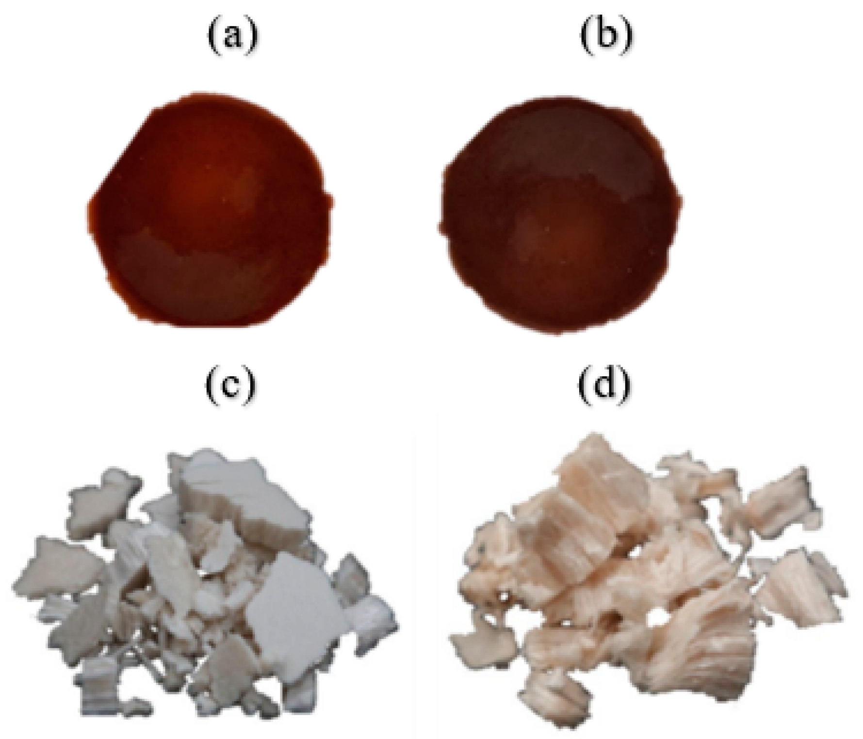
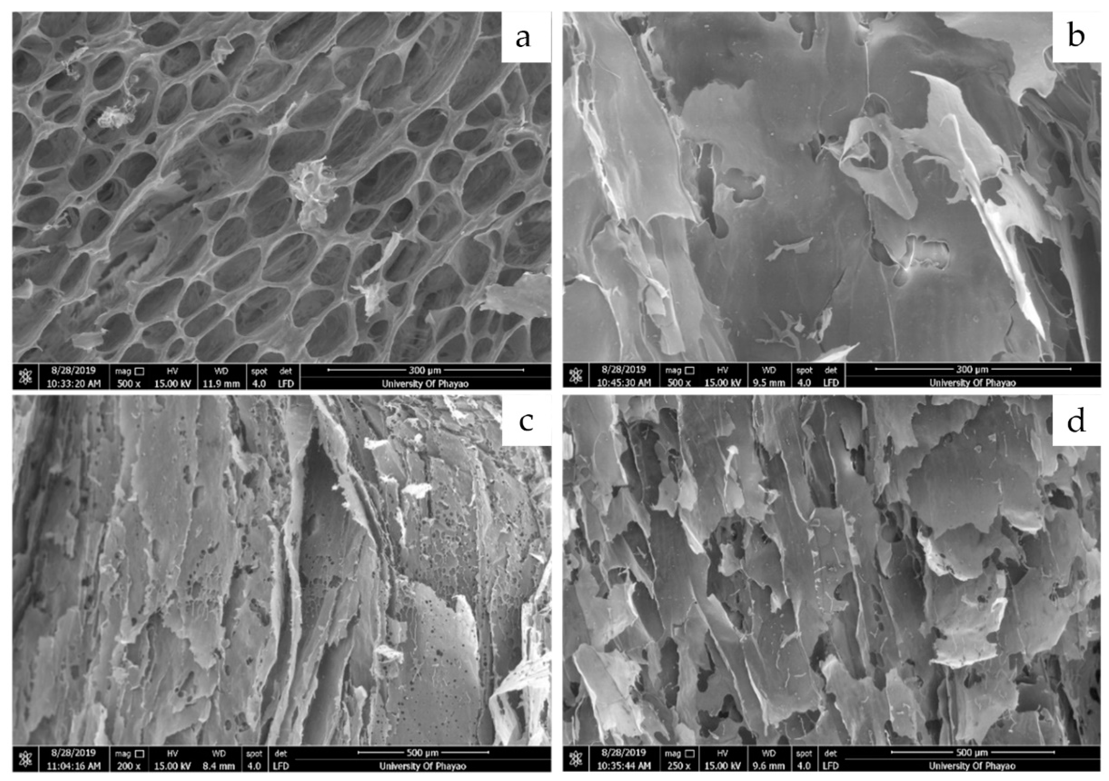
2.3.1. Morphological Properties

Surface morphologies of the polysaccharide extracts (GH, WH) were examined using scanning electron microscopy (SEM) at an acceleration voltage of 15.00 kV. The samples were spread directly on the surface of the stub and coated with gold (20 nm) using a sputter coater [19].

2.3.2. Absorption Properties

To determine their absorption properties, 10 mg of GH and WH extracts were dissolved in 3 mL of distilled water in a beaker and stirred with a magnetic stirrer. The time required for complete swelling of the extracts was recorded for measuring the swelling rate. The profile of swelling capacity versus time of a sample is obtained by performing free-absorbency capacity measurements at consecutive time intervals. The absorption properties test was performed at 25 ± 2 °C. All experiments were conducted in triplicate.

2.3.3. Relative Concentrations of Extract Physical Properties

Relative concentrations of the physical properties of the ethanolic (P, S) and polysaccharide (GH, WH) extracts were determined by dissolving them in distilled water and propylene glycol (ratio 3:1 w/w) to concentrations of 20, 10, 5, 2.5, and 1.25 mg/g. The pH values of all concentrations of test samples were measured by pH meter, while the color, odor, and appearance characteristics of all concentrations of test samples were also recorded. All experiments were conducted in triplicate.

2.3.4. Relative Acid–Base Values for Extract Physical Properties

Relative acid–base values for the physical properties of the ethanolic (P, S) and polysaccharide (GH, WH) extracts were determined by dissolving them in distilled water and propylene glycol (ratio 3:1 w/w) to a concentration of 20 mg/g. The pH was adjusted to between 4 and 8 using HCl and NaOH solutions. Color, viscosity, and odor were recorded. All experiments were conducted in triplicate.

2.3.5. Stability of the Extracts

The stability of the ethanolic (P, S) and polysaccharide (GH, WH) extracts at varying temperatures was determined by dissolving them in distilled water and propylene glycol (ratio 3:1 w/w) to concentrations of 20, 10, 5, 2.5, and 1.25 mg/g. The stability test was performed at 4 °C, room temperature, and 45 °C for 30 days. Acid–base value, color, precipitation, viscosity, and odor were recorded at 0, 1, 4, 9, 16, and 30 days. All experiments were conducted in triplicate.

2.4. Phytochemical Screening

Bioactive compounds such as flavonoids, tannins, triterpenoids, steroids, alkaloids, and carbohydrates were chemically tested using the following standard procedures used in [20,21]. All experiments were conducted in triplicate.

2.4.1. Test for Carbohydrates

Molisch’s test was performed according to the previous method with some modifications [20,21]. The crude extracts (10 mg) were mixed with 2–3 drops of Molisch’s reagent in a test tube. Two milliliters of concentrated H2SO4 were then added down the sides of the test tube to facilitate the formation of a layer and avoid mixing. The development of a purple ring at the interface layer formed by the concentrated acid is a positive indicator for Molisch’s test. If no purple or reddish-purple ring arises, the given analyte does not contain any carbohydrates.

2.4.2. Test for Flavonoids

The Shinoda test was used to determine the presence of flavonoids, which was performed according to the previous method with some modifications [20,21]. The crude extracts (10 mg) were dissolved in distilled water and 5% DMSO. Then, a few fragments of magnesium ribbon and concentrated hydrochloric acid in 2–3 drops were added to the ethanolic extracts. A red to pink color appearing after a few minutes indicated the presence of flavonoids.

2.4.3. Test for Steroids

The Liebermann–Burchard test was performed according to the previous method with some modifications [20,21]. The crude extracts (10 mg) were mixed with 2 mL of acetic anhydride. The mixture was then boiled and cooled before adding 1 mL of concentrated H2SO4 down the sides of the test tube. The appearance of a green ring indicated the existence of steroids, while a deep red ring indicated the presence of triterpenoids.

2.4.4. Test for Tannins

The test for tannins was performed according to the previous method with some modifications [20,21]. The crude extracts (5 mg) were dissolved in 2 mL of distilled water and 1 mL of 5% DMSO before mixing with 1% gelatin solution containing 10% sodium chloride. The formation of a white precipitate indicated the presence of tannins.
In the ferric chloride test, the crude extracts (5 mg) were dissolved in distilled water and 5% DMSO (1 mL) before mixing with 1% ferric chloride (1 mL). The appearance of a blue-green color suggested the presence of tannins.
In the bromine water test, the crude extracts (5 mg) were dissolved in 5 mL of 95% ethanol and filtered. The filtrate was then added with 3–4 drops of bromine water. The appearance of a buff color indicated the existence of condensed tannins whereas hydrolyzable tannins gave no color indication.
For the lime water test, the crude extracts (5 mg) were dissolved in 5 mL of 95% ethanol and filtered. The filtrate was then added with 3–4 drops of lime water. The appearance of a brown precipitation indicated the existence of condensed tannins.

2.4.5. Test for Alkaloids

Mayer’s test was performed according to the previous method with some modifications [20,21]. The crude extracts (5 mg) were mixed with 2–3 drops of Mayer’s reagent (potassium mercuric iodide solution). The formation of a white color precipitate suggested the presence of alkaloids.
Dragendorff’s test was performed according to the previous method with some modifications [20,21]. The crude extracts (5 mg) were mixed with 2–3 drops of Dragendorff’s reagent (potassium bismuth iodide solution). The formation of a reddish-brown precipitate indicated the presence of alkaloids.

2.5. Antibacterial Activity

The antibacterial activity of the extracts was tested by the disc diffusion method using S. aureus, S. pyogenes, and C. acnes. Bacterial inoculums of 1.0 × 10⁸ CFU/mL were swabbed on cultures of S. aureus in Muller–Hinton agar (MHA) at 37 °C for 18–24 h, S. pyogenes in Brain Heart Infusion agar (BHIA) at 37 °C, 5% CO2 for 48–72 h, and C. acnes in Fluid Thioglycolate Medium (FTM) at 37 °C for 48–72 h under anaerobic conditions. The extracts (P and S extracts 500 mg/mL; GH and WH extracts 100 mg/mL) were diluted in ethanol and water and impregnated into 6 mm sterile filter paper disks at 15 µL for each sample. Clindamycin (2 µg/disc) and distilled water were used as the positive and negative controls, respectively. Antibacterial activity was indicated by the inhibition zone that was measured after treatment [22,23]. All samples were performed in triplicate.

2.6. Total Phenolic Content

The total phenolic content of the extracts was determined by the Folin–Ciocalteu assay following the method described by Mungmai et al. [24] with slight modifications. Briefly, 40 µL (1 mg/mL) of the extracts were mixed with dimethyl sulfoxide (DMSO) (40 µL) and deionized water (200 µL) then mixed thoroughly with Folin–Ciocalteu reagent (250 µL) and shaken for 5 min at room temperature. Next, 250 µL of 5% (w/v) sodium carbonate was added, and the mixture was left to stand for 1 h in the dark at room temperature. Absorbance was measured at 750 nm using a UV-Vis spectrophotometer, with results reported as mg gallic acid equivalent per g dry weight (GAE; mg gallic acid/g extract). All samples were assayed in triplicate.

2.7. Free Radical Scavenging Assay

Free radical scavenging of the extracts was evaluated using 2, 2′-diphenyl-1-picrylhydrazyl (DPPH•), with the method adjusted slightly from an existing protocol [24]. Briefly, 100 µL of each extract (20–100 µg/mL) were mixed with 67 µL of 1 M Tris-HCl (pH 7.9) and 100 µL of 130 µM DPPH• solution in a 96-well plate. The mixture was then left to stand for 30 min in the dark at room temperature, and the absorbance was measured at 517 nm using L-ascorbic acid as the positive control. All samples were assayed in triplicate. The percentage of DPPH radical scavenging activity was calculated using the following equation:
% DPPH radical scavenging activity = [(Acontrol − Asample)/Acontrol] × 100
where Acontrol is the absorbance of the control, and Asample is the absorbance of the sample containing all reagents including the test compound.
The ability of the sample to scavenge DPPH• was expressed in terms of the IC50 value (mg/mL). The IC50 value was the dose–response curve plotted between the percentage DPPH radical scavenging and the various concentrations of sample extracts.

2.8. Tyrosinase Inhibition Assay

The tyrosinase inhibition activity of the extracts was evaluated using L-DOPA and mushroom tyrosinase [25]. The samples were dissolved in 10% DMSO, 20 µL of the samples (20–100 µg/mL), 40 µL of 0.1 M phosphate buffer at pH 6.5, and 20 µL of tyrosinase solution in phosphate buffer (47 units/mL) were added in a 96-well plate. The plate was left to react at 25 ± 2 °C for 10 min and then added with 20 µL of 2.5 mM L-DOPA in phosphate buffer. Kojic acid was used as the positive control. Absorbance of the mixtures was measured at 480 nm using a microplate reader. All samples were assayed in triplicate. The percentage of tyrosinase inhibition activity was obtained using the following equation:
% tyrosinase inhibition = [(Acontrol − Asample)/Acontrol] × 100
where Acontrol is the absorbance of the control, and Asample is the absorbance of the sample. The IC50 value (mg/mL) of tyrosinase inhibition was obtained from the sample concentration at 50 % inhibition activity.

2.9. Preparation of Gel Formulations Containing Durian Extract

Gel formulations were evaluated for physicochemical properties and stability, with the most suitable base incorporated into the durian extract. The formulations consisted of distilled water, propylene glycol, polysorbate 20, butylated hydroxytoluene, caprylhydroxamic acid, 1,2-hexanediol, butylene glycol, caprylhydroxamic acid, 1,2-hexanediol, butylene glycol, triethanolamine, acrylates/C10-30, and alkyl acrylate crosspolymer. The gel base was continuously homogenized and then incorporated with the extracts (0.65%).

2.10. Stability Test

The gel formulations were evaluated at room temperature, 4 °C, and 45 °C for 3 months and a heating/cooling process for 6 cycles (45 °C, 48 h alternated with 4 °C, 48 h for 1 cycle). Changes in physicochemical appearance were observed, including color, pH, odor, and spreadability [26]. All samples were assayed in triplicate.

2.11. Study Design of the Skin Irritation Test

Measurement methods were approved by the University of Phayao Human Ethics Committee, Thailand (Approval number 3/013/62). The skin irritation study was carried out using a Finn Chamber® with 30 normal healthy volunteers. The Draize scoring system was employed to calculate the primary dermal irritation index (PDII); the backs of the volunteers were covered with a Finn Chamber® that contained the test samples, consisting of deionized water (negative control), S, GH and WH extracts, gel base, SGWH gel, and 5% w/v sodium lauryl sulfate solution (positive control) for 48 h. Then, they were observed for any irritating reaction such as erythema and edema at 24 h, 48 h, and 1 week after removal of the patch. These methods were slightly adjusted from an existing protocol [27].

2.12. Statistical Analysis

Results, summarized from three independent experiments, were presented as means ± SD and analyzed by one-way ANOVA following Tukey’s multiple comparisons using the Statistical Package for the Social Sciences (SPSS), version 17.0 for Windows (SPSS Inc., Chicago, IL, USA).

3. Results

3.1. Physicochemical Properties of the Extracts

Percentage yield of the P crude extract from 95% ethanol at 12.13% was higher than S (4.01%), while polysaccharide extracts showed percentage yields of GH (1.60%) higher than WH (1.18%), as shown in Table 1. Extract characteristics are shown in Figure 1. Crude extracts of P and S were dark brown; polysaccharide extracts of GH were grayish-white; while WH extracts were orange to pink in color.
Top-view scanning electron micrograph (SEM) images of (a) GH and (b) WH at a magnification of 500× and side-views of (c) GH and (d) WH at a magnification of 200× are depicted in Figure 2. The morphological structure of GH was highly porous, while the surface of WH was flattened with no porosity observed. This result concurred with the absorption property of GH, presenting a significantly different (p < 0.05) absorption time of 23.53 ± 1.03 min compared to WH at 31.26 ± 2.15 min. The porosity impacted the ability of durian polysaccharide extract to absorb water. Increasing the porosity showed greater absorbency, with less time taken to absorb water.
When relative concentrations of polysaccharide extracts (GH and WH) increased, the viscosity also increased. GH and WH extracts can be used to increase the viscosity and consistency of cosmetic products. This did not occur in the crude ethanolic P and S extracts. All the extracts (P, S, GH, and WH) underwent color and odor changes with changing extract concentrations. The acid–base values of P, S, GH, and WH were 5–5.5, 4.5, 5.5, and 5.5, respectively. Acid–base values (pH 4–8) gave darker colored crude extracts (P and S) at pH 8 than at pH 4–7, while the viscosity did not change. Polysaccharide extracts (GH and WH) showed color changes from grayish-white of GH and orange to pink of WH to brown at pH 7 and 8, while viscosity decreased when pH values increased to pH 6–8. Odor stability for P and S extracts changed at pH 8, while GH and WH extracts did not change. Physical properties of the extracts at varying temperatures did not change.

3.2. Phytochemical Screening

Phytochemical analyses of the different durian extracts were conducted to determine the active constituents as carbohydrates, flavonoids, steroids, triterpenoids, tannins, or alkaloids. Results revealed that the P extract contained bioactive compound groups of flavonoids and triterpenoids, the S extract had groups of flavonoids, steroids and condensed tannins, while the GH and WH extracts presented carbohydrates and triterpenoids. Chang & Shen [28] and Liang et al. [29] reported that acetone extracts of durian shells contained condensed tannins. Alkaloids were not present in all extracts. However, alkaloids, flavonoids, tannins and triterpenoids were found in methanolic extracts of durian pulp [30], while durian seed and fruit-hulls presented flavonoids and triterpenoids [31], and durian leaf presented flavonoids and alkaloids [12]. Diverse testing results were due to different solvent extraction methods, planting sites, and harvest times, with results shown in Table 1.

3.3. Antibacterial Activity

Antibacterial screening revealed that no durian extracts exhibited growth inhibition against S. aureus, S. pyogenes, and C. acnes, from previous reports. Thunyakipisal et al. [18] reported that polysaccharide gel extract from durian fruit rinds showed inhibitory activity against bacteria such as S. mutan and A. actinomycetemcomitans, while polysaccharide extracts from Cordyceps cicadae displayed broad spectrum activity against Escherichia coli, Pseudomonas aeruginosa, and S. aureus. [32]. Kusumaningrum and Yuliana [33] reported that durian-rind pectin showed a weak antibacterial activity, while the sterilized durian-rind pectin showed strong antibacterial activity towards S. aureus and E. coli. They concluded that depolymerization and de-esterification of durian-rind pectin by heat sterilization was able to improve the antibacterial activity. Previous studies reported that flavonoids, steroids, and tannins showed inhibitory activity against a wide range of pathogenic microorganisms [34,35,36]. Crude P and S extracts contained flavonoids, steroids, and tannins with no inhibition activity against bacteria. Results differed depending on the extraction, extraction process, and planting sites.

3.4. Total Phenolic Content

Phenolic compounds are more soluble in polar organic solvents due to the presence of hydroxyl groups that facilitate free radical scavenging [37] and control the antioxidant properties of plants [38]. Total phenolic content results are shown in Table 2. The P, S, GH, and WH extracts showed significantly (p < 0.05) different values at 0.09 ± 0.01, 0.33 ± 0.01, 0.21 ± 0.02, and 0.26 ± 0.00 mg gallic acid/g extract, respectively. Total phenolic content was dependent on the extraction solvent used [26] and the sample components. The P extract presented the lowest total phenolic content and was not selected for the next experiment. Total phenolic contents of the combined extracts for SGH (S + GH), SWH (S + WH), GWH (GH + WH) (ratio 1:1 mg/mg), and SGWH (S + GH + WH) (ratio 1:1:1 mg/mg) were 0.85 ± 0.05, 0.83 ± 0.01, 0.47 ± 0.01, and 0.88 ± 0.02 mg gallic acid/g extract, respectively. High synergism was observed for mixtures of the S extract, with significantly (p < 0.05) higher total phenolic contents than the individual extracts. Previous studies reported total polyphenols were found in durian extracts (seeds and peels) [13]. Therefore, durian wastes are potential sources of phenolic content.

3.5. Free Radical Scavenging Activity

DPPH radical scavenging ability of the S extract (IC50 = 0.08 ± 0.00 mg/mL) was higher than the other extracts (GH; 0.31 ± 0.08 mg/mL, WH; 0.18 ± 0.08 mg/mL) and also related to the GAE values of 0.33 ± 0.01, 0.21 ± 0.02, and 0.26 ± 0.00 mg gallic acid/g extract. DPPH radical scavenging activity was not detected for the P extract. Shi et al. [39] reported that polysaccharides of Camellia sinensis cultivars showed potent scavenging ability against superoxide and hydroxyl radicals and increased activities of superoxide dismutase, glutathione peroxidase, and peroxidase. Moreover, Shang et al. [40] reported that polysaccharides from Silphium perfoliatum extract showed significantly increased free radical scavenging activity against ABTS and DPPH radicals. The combined extracts presented IC50 values higher than the individual extracts, except for the GWH sample. The combined extracts SGH, SWH, and SGWH indicate a synergistic effect, while GWH showed an antagonistic effect. DPPH radical scavenging activity was related to the GAE values of the combined samples and confirmed their synergistic effects. The phenolic compounds greatly influenced the antioxidant activity of the extracts [41]. A positive correlation (p < 0.05) was observed between total phenolic content and free radical DPPH• scavenging efficacies, except for the GWH sample. Results also indicated that the combined extracts showed significantly higher (p < 0.05) total phenolic contents than the individual extracts, except for the GWH sample (Table 2). Skroza et al. [42] concluded that mixed phenolic compound samples presented both synergistic and antagonistic effects, with antioxidant activity dependent on compound structure, number, distribution of substituents, and concentration.

3.6. Tyrosinase Inhibition Activity

Mushroom tyrosinase was used to determine the role of the extracts in the process of melanogenesis. The inhibitory effects of the extracts on tyrosinase enzyme activity were determined to evaluate their potential as skin-whitening agents, with results shown in Table 2. The samples (P, S, GH, WH, SWH, and GWH) did not show tyrosinase inhibition activity. Tyrosinase inhibition activity was recorded at IC50 values of SGWH and SGH samples was 0.067 ± 0.00 mg/mL and 8.69 ± 1.82 mg/mL, respectively, whereas kojic acid as the positive control showed IC50 of 0.07 ± 0.00 mg/mL. The results were significantly different (p < 0.05). The tyrosinase inhibition activity of SGWH and SGH indicated correlation between total phenolic content and DPPH radical scavenging activity. Cha et al. [43] reported that phenolic compounds contribute directly to antioxidant activity, while antioxidants exhibit potent inhibitory activities toward tyrosinase. Condensed tannin was also reported to inhibit tyrosinase activity. Huang et al. [44] and Liang et al. [29] suggested that acetone extracts from durian shells showed potential as antioxidants and tyrosinase inhibitors.

3.7. Stability of Gel Formulations Containing Durian Extract

For biological activities, the SGWH extracts exhibited relatively high DPPH scavenging activity and the highest anti-tyrosinase activity. Therefore, the SGWH sample as a by-product from durian was selected for incorporation in gel formulations and investigated for stability.
The gel was stored under various conditions including a heating-cooling cycle and different temperatures: room temperature, 4 °C, and 45 °C. The color remained the same at orange to brown, and the odor did not change. Moreover, pH changes among the conditions were 5.96 ± 0.47 and not statistically significant (p < 0.05), with spreadability changes 11.50 ± 0.50 mm that were also not statistically significantly different (p < 0.05).

3.8. Skin Irritation Patch Testing

Patch testing at 1 h, 24 h, and 48 h showed that for deionized water, S, GH and WH extracts, gel base, and SGWH gel did not irritate the skin or induce the formation of erythema or edema (PDII value = 0.00). A 5% w/v sodium lauryl sulfate solution showed slight formation of erythema (PDII value = 0.42) but was non-irritating with low PDII of <0.5. Therefore, the ingredients of the formula were considered safe to apply on the skin.

4. Conclusions

This work reveals that D. zibethinus extract can be appropriated as an ingredient for the cosmetic industry. Durian extracts showed promising physicochemical properties and biological, antioxidant, and anti-tyrosinase activities, with interactions between combined extracts having a synergistic effect. The extracts are used as antioxidants and whitening agents in cosmetic products. Crude durian extract can be used as an active ingredient, while the polysaccharide extract can be used as an active ingredient and as a raw material to improve cosmetic formulation stability and increase viscosity. The polysaccharide extract showed rheological and sensorial properties, with a moisturizing effect on the skin [45]. Formulations containing the extract did not irritate the skin, and no allergic reactions were reported by the volunteers. The product was considered safe to use as an active ingredient in cosmetic formulations. Utilization as an active ingredient and raw material in cosmetic products adds value to durian by-products.

Author Contributions

Conceptualization, L.M.; methodology, L.M. and P.T.; software, P.T. and P.S.; validation, L.M. and P.T.; formal analysis, L.M.; investigation, L.M., C.K., S.T. and A.N.; writing—original draft preparation, L.M.; writing—review and editing, L.M., P.T. and P.S. All authors have read and agreed to the published version of the manuscript.

Funding

This research received no external funding.

Institutional Review Board Statement

The study was conducted in accordance with the Declaration of Helsinki and approved by the Human Ethics Committee of University of Phayao (Phayao, Thailand). The approval number was 3/013/62.

Informed Consent Statement

Informed consent was obtained from all the study participants.

Data Availability Statement

The authors confirm that data supporting the findings are presented in this paper.

Acknowledgments

The authors acknowledge the Department of Cosmetic Sciences, School of Pharmaceutical Sciences, University of Phayao, Research and Innovation Center in Cosmetic Sciences and Natural Products (RCCN), School of Pharmaceutical Sciences, University of Phayao, and the Faculty of Science and Technology, Phuket Rajabhat University for providing instruments and research facilities.

Conflicts of Interest

The authors declare no conflict of interest.

References

  1. Siriphanich, J. Postharvest biology and technology of tropical and subtropical fruits: Durian (Durio zibethinus Merr.). In Woodhead Publishing Series in Food Science, Technology and Nutrition; Yahia, E.M., Ed.; Woodhead Publishing Limited: Cambridge, UK, 2011; Volume 3, pp. 80–114. [Google Scholar]
  2. Gorinstein, S.; Haruenkit, R.; Poovarodom, S.; Vearasilp, S.; Ruamsuke, P.; Namiesnik, J.; Leontowicz, M.; Leontowicz, H.; Suhaj, M.; Sheng, G.P. Some analytical assays for the determination of bioactivity of exotic fruits. Phytochem. Anal. 2010, 21, 355–362. [Google Scholar] [CrossRef] [PubMed]
  3. Suksaeree, J.; Karnsopa, P.; Wannaphruek, N.; Prasomkij, J.; Panrat, K.; Pichayakorn, W. Transdermal Delivery of Nicotine Using Pectin Isolated from Durian Fruit-Hulls-Based Polymer Blends as a Matrix Layer. J. Polym. Environ. 2018, 26, 3216–3225. [Google Scholar] [CrossRef]
  4. Alkandahri, M.Y.; Patala, R.; Pratiwi, M.I.; Agustina, L.S.; Farhamzah; Kusumawati, A.H.; Hidayah, H.; Amal, S.; Frianto, D. Pharmacological studies of Durio Zibethinus: A medicinal plant review. Ann. Rom. Soc. Cell Biol. 2021, 25, 640–646. [Google Scholar]
  5. Chansiripornchai, N.; Chansiripornchai, P.; Pongsamart, S. A preliminary study of polysaccharide gel extracted from the fruit hulls of durian (Durio zibethinus) on immune responses and cholesterol reduction in chicken. Acta Hortic. 2008, 786, 57–60. [Google Scholar] [CrossRef]
  6. Hokputsa, S.; Gerddit, W.; Pongsamart, S.; Inngjerdingen, K.; Heinze, T.; Koschella, A.; Harding, S.E.; Paulsen, B.S. Water-soluble polysaccharides with pharmaceutical importance from durian rinds (Durio zibethinus Murr.): Isolation, fractionation, characterization and bioactivity. Carbohydr. Polym. 2004, 56, 471–481. [Google Scholar] [CrossRef]
  7. Muhtadi, H.; Sujono, T.A.; Suhendi, A.; Yen, K.H. Antioxidant and chemical constituents of some Indonesian fruit peels. Med. Plants-Int. J. Phytomed. Relat. Ind. 2014, 6, 43–46. [Google Scholar] [CrossRef]
  8. Isabelle, M.; Lee, B.L.; Lim, M.T.; Koh, W.P.; Huang, D.; Ong, C.N. Antioxidant activity and profiles of common fruits in Singapore. Food Chem. 2010, 123, 77–84. [Google Scholar] [CrossRef]
  9. Gorinstein, S.; Poovarodom, S.; Leontowicz, H.; Leontowicz, M.; Namiesnik, J.; Vearasilp, S.; Haruenkit, R.; Ruamsuke, P.; Katrich, E.; Tashma, Z. Antioxidant properties and bioactive constituents of some rare exotic Thai fruits and comparison with conventional fruits in vitro and in vivo studies. Food Res. Int. 2011, 44, 2222–2232. [Google Scholar] [CrossRef]
  10. Arancibia-Avila, P.; Toledo, F.; Park, Y.S.; Jung, S.T.; Kang, S.G.; Heo, B.G.; Lee, S.H.; Sajewicz, M.; Kowalska, T.; Gorinstein, S. Antioxidant properties of durian fruit as influenced by ripening. LWT-Food Sci. Technol. 2008, 41, 2118–2125. [Google Scholar] [CrossRef]
  11. Leverett, C.; Chandra, A.; Rana, J.; Fast, D.J.; Missler, S.R.; Flower, D.M. Extracts of Durian Fruit for Use in Skin Care Compositions, Access Business Group International LLC. U.S. Patent 20070116789A1, 22 February 2007. [Google Scholar]
  12. Aruan, D.G.R.; Tonel Barus, T.; Haro, G.; Siburian, R.; Simanjuntak, P. Phytochemical screening and antidiabetic activity of N-hexane, ethyl acetate and water extract from durian leaves (Durio zibethinus L.). Orient. J. Chem. 2019, 35, 487–490. [Google Scholar] [CrossRef]
  13. Charoenphun, N.; Klangbud, W.K. Antioxidant and anti-inflammatory activities of durian (Durio zibethinus Murr.) pulp, seed and peel flour. PeerJ 2022, 10, e12933. [Google Scholar] [CrossRef] [PubMed]
  14. Huang, W.; Tao, F.; Li, F.; Mortimer, M.; Guo, L.H. Antibacterial nanomaterials for environmental and consumer product applications. NanoImpact 2020, 20, 100268. [Google Scholar] [CrossRef]
  15. Nosrati, H.; Heydari, M.; Tootiaei, Z.; Ganjbar, S.; Khodaei, M. Delivery of antibacterial agents for wound healing applications using polysaccharide-based scaffolds. J. Drug Deliv. Sci. Technol. 2023, 84, 104516. [Google Scholar] [CrossRef]
  16. Govindarajan, S.; Noor, A. Biological activities of plant polysaccharides, mechanism of action and biomedical applications. Res. J. Biotech. 2021, 16, 255–272. [Google Scholar] [CrossRef]
  17. Casillas-Varga, G.; Ocasio-Malavé, C.; Medina, S.; Morales-Guzmán, C.; García Del Valle, R.; Carballeira, N.M.; Sanabria-Ríos, D.J. Antibacterial fatty acids: An update of possible mechanisms of action and implications in the development of the next-generation of antibacterial agents. Prog. Lipid Res. 2021, 82, 101093. [Google Scholar] [CrossRef]
  18. Thunyakipisal, P.; Saladyanant, T.; Hongprasong, N.; Pongsamart, S.; Apinhasmit, W. Antibacterial activity of polysaccharide gel extract from fruit rinds of Durio zibethinus Murr. against oral pathogenic bacteria. J. Investig. Clin. Dent. 2010, 1, 120–125. [Google Scholar] [CrossRef] [PubMed]
  19. Ramos, A.H.; Rockenbach, B.A.; Ferreira, C.D.; de Oliveira, M. Characteristics of flour and starch isolated from red rice subjected to different drying conditions. Starch-Stärke 2019, 71, 1800257. [Google Scholar] [CrossRef]
  20. Trease, G.E.; Evans, W.C. Pharmacognosy, 15th ed.; Saunders: London, UK, 2002. [Google Scholar]
  21. Yadav, P.; Kumar, A.; Mahour, K.; Vihan, V.S. Phytochemical analysis of some Indigenous plants potent against endoparasite. J. Adv. Lab. Res. Biol. 2010, 1, 56–59. [Google Scholar]
  22. Gera, K.; McIver, K.S. Laboratory growth and maintenance of Streptococcus pyogenes (The Group A Streptococcus, GAS). Curr. Protoc. Microbiol. 2013, 30, 9D.2.1–9D.2.13. [Google Scholar] [CrossRef] [PubMed] [Green Version]
  23. Luangnarumitchai, S.; Lamlertthon, S.; Tiyaboonchai, W. Antimicrobial activity of essential oils against five strains of Propionibacterium acnes. Mahidol Univ. J. Pharm. Sci. 2007, 34, 60–64. [Google Scholar]
  24. Mungmai, L.; Preedalikit, W.; Aunsri, N.; Peerakam, N. Bioactivity test and GC–MS analysis of different solvent extracts from Perilla frutescens (Linn.) Britton and cosmetic product application for sensitive skin. Sci. Technol. RMUTT J. 2019, 9, 78–93. [Google Scholar]
  25. Sainakham, M.; Mungmai, L. In vitro Anti-oxidative activity and tyrosinase inhibition of Inca Peanut (Plukenetia volubilis L.) shell extracts from different preparation methods. TJST Thai J. Sci. Technol. 2020, 9, 407–417. [Google Scholar] [CrossRef]
  26. Srisuksomwong, P.; Kaenhin, L.; Mungmai, L. Collagenase and Tyrosinase inhibitory activities and stability of facial cream formulation containing cashew leaf extract. Cosmetics 2023, 10, 17. [Google Scholar] [CrossRef]
  27. Mungmai, L.; Preedalikit, W.; Pintha, K.; Tantipaiboonwong, P.; Aunsri, N. Collagenase and melanogenesis inhibitory effects of Perilla Frutescens pomace extract and its efficacy in topical cosmetic formulations. Cosmetics 2020, 7, 69. [Google Scholar] [CrossRef]
  28. Cheng, H.T.; Shen, X.S. Optimization of the extraction of proanthocyanidins from durian skin ultrasonic cavitation-mechanical grinding. Food Ind. 2020, 41, 129–131. [Google Scholar]
  29. Liang, D.; Liu, L.; Qin, Z.; Li, G.; Ding, B.; Chen, H.; Li, Z.; Wei, S.; Wang, Z. Antioxidant and antityrosinase activity of extractable condensed tannins from durian shells with antibrowning effect in fresh-cut asparagus lettuce model. LWT-Food Sci. Technol. 2022, 162, 113423. [Google Scholar] [CrossRef]
  30. Ho, L.H.; Bhat, R. Exploring the potential nutraceutical values of durian (Durio zibethinus L.)—An exotic tropical fruit. Food Chem. 2015, 168, 80–89. [Google Scholar] [CrossRef]
  31. Ang, A.M.G.; Nalda, C.M.D.R.; Sabejon, S.E. Brine shrimp lethality and antioxidant activity of the leaf, rind and seed ethanolic extracts of Durio zibethinus L. Asian J. Biol. Sci. 2018, 7, 105–111. [Google Scholar] [CrossRef] [Green Version]
  32. Liang, J.; Huang, X.; Ma, G. Antimicrobial activities and mechanisms of extract and components of herbs in East Asia. RSC Adv. 2022, 12, 29197–29213. [Google Scholar] [CrossRef]
  33. Kusumaningrum, H.D.; Yuliana, N.D. Effect of sterilization on the degree of esterification, FTIR analysis, and antibacterial activity of durian-rind pectin. Sains Malays. 2021, 51, 3677–3688. [Google Scholar] [CrossRef]
  34. Xie, Y.; Yang, W.; Tang, F.; Chen, X.; Ren, L. Antibacterial activities of flavonoids: Structure-activity relationship and mechanism. Curr. Med. Chem. 2015, 22, 132–149. [Google Scholar] [CrossRef] [PubMed]
  35. Vollaro, A.; Esposito, A.; Antonaki, E.; Dora Iula, V.; Alonzo, D.; Guaragna, A.; Gregorio, E. Steroid derivatives as potential antimicrobial agents against Staphylococcus aureus planktonic cells. Microorganisms 2020, 8, 468. [Google Scholar] [CrossRef] [PubMed] [Green Version]
  36. Kaczmarek, B. Tannic acid with antiviral and antibacterial activity as a promising component of biomaterials—A minireview. Materials 2020, 13, 3224. [Google Scholar] [CrossRef] [PubMed]
  37. Aryal, S.; Baniya, M.K.; Danekhu, K.; Kunwar, P.; Gurung, R.; Koirala, N. Total phenolic content, flavonoid content and antioxidant potential of wild vegetables from Western Nepal. Plants 2019, 8, 96. [Google Scholar] [CrossRef] [PubMed] [Green Version]
  38. Boeing, J.S.; Barizão, É.O.; e Silva, B.C.; Montanher, P.F.; de Cinque Almeida, V.; Visentainer, J.V. Evaluation of solvent effect on the extraction of phenolic compounds and antioxidant capacities from the berries: Application of principal component analysis. Chem. Cent. J. 2014, 8, 48. [Google Scholar]
  39. Shi, Y.; Ai, Y.; Chen, Y.; Ni, D.; Yu, Z. The antioxidant and hepatoprotective activities of two tea polysaccharides. J. Antioxid. Act. 2017, 1, 23–36. [Google Scholar] [CrossRef] [Green Version]
  40. Shang, H.M.; Zhou, H.Z.; Li, R.; Duan, M.Y.; Wu, H.X.; Lou, Y.J. Extraction optimization and influences of drying methods on antioxidant activities of polysaccharide from cup plant (Silphium perfoliatum L.). PLoS ONE 2017, 12, e0183001. [Google Scholar] [CrossRef] [Green Version]
  41. Hervert-Hernández, D.; García, O.P.; Rosado, J.L.; Goñi, I. The contribution of fruits and vegetables to dietary intake of poly-phenols and antioxidant capacity in a Mexican rural diet: Importance of fruit and vegetable variety. Int. Food Res. J. 2011, 44, 1182–1189. [Google Scholar]
  42. Skroza, D.; Šimat, V.; Vrdoljak, L.; Jolić, N.; Skelin, A.; Cagalj, M.; Frleta, R.; Mekinić, I.G. Investigation of antioxidant synergisms and antagonisms among phenolic acids in the model matrices using FRAP and ORAC methods. Antioxidants 2022, 11, 1784. [Google Scholar] [CrossRef]
  43. Cha, J.Y.; Yang, H.J.; Moon, H.I.; Cho, Y.S. Inhibitory effect and mechanism on melanogenesis from fermented herbalcomposition for medical or food uses. Food Res. Int. 2012, 45, 225–231. [Google Scholar] [CrossRef]
  44. Huang, Q.; Lin, M.Z.; Chai, W.M.; Ou-Yang, C.; Huang, W.Y.; Wang, Y.X.; XU, K.L.; Feng, H.L. Condensed tannins from longan bark as inhibitor of tyrosinase: Structure, activity, and mechanism. J. Agric. Food Chem. 2018, 66, 908–917. [Google Scholar]
  45. Campos, P.M.B.G.M.; Melo, M.O.; Camargo Junior, F.B. Effects of polysaccharide-based formulations on human skin. In Polysaccharides; Springer: Cham, Switzerland, 2014; pp. 1–8. [Google Scholar] [CrossRef]
Figure 1. Durian extracts: (a) pulp (P); (b) seed (S); (c) green fruit-hulls (GH); (d) white fruit-hulls (WH).
Cosmetics 10 00087 g001
Figure 2. Top-view SEM image of polysaccharide extracts at 500× magnification: (a) green fruit-hulls (GH) and (b) white fruit-hulls (WH). Side-view of polysaccharide extracts at 200× magnification (c) green fruit-hulls (GH) and 250× magnification (d) white fruit-hulls (WH).
Cosmetics 10 00087 g002
Table 1. Chemical constituents of different durian extracts.
Crude Extract% Yield Chemical Constituents
CarbohydratesFlavonoidsSteroidsTriterpenoidsTanninsAlkaloids
GelatinFerric ChlorideBromine WaterLime WaterMayerDragendorff
Pulp (P)12.13-+-+------
Seed (S)4.01-++-++++--
Green fruit-hulls (GH)1.60+--+------
White fruit-hulls (WH)1.18+--+------
(+) = positive test, (-) = negative test.
Table 2. Total phenolic content, IC50 standard equivalent of antioxidant activity, and tyrosinase inhibition activity of the durian extracts.
Crude ExtractTotal Phenolic Content
(mg GAE/g)
DPPH Radical-Scavenging Activity (IC50, mg/mL)Tyrosinase Inhibition Activity
(IC50, mg/mL)
Pulp (P)0.09 ± 0.01 FNDND
Seed (S)0.33 ± 0.01 D0.08 ± 0.00 AND
Green fruit-hulls (GH)0.21 ± 0.02 E0.31 ± 0.08 B,C ND
White fruit-hulls (WH)0.26 ± 0.00 E0.18 ± 0.08 A,BND
S + GH (SGH)0.85 ± 0.05 B0.04 ± 0.00 A8.69 ± 1.82 B
S + WH (SWH)0.83 ± 0.01 A,B0.04 ± 0.00 AND
GH + WH (GWH)0.47 ± 0.01 C0.46 ± 0.15 CND
S + GH + WH (SGWH)0.88 ± 0.02 A0.04 ± 0.02 A0.067 ± 0.00 A
L—ascorbic-0.002 ± 0.00-
Kojic acid--0.07 ± 0.00
- = not determined; ND = not detected. Data are mean ± standard error (SD) of three replicates (Letters A, B, C, D, E and F represent statistical differences between groups).
Disclaimer/Publisher’s Note: The statements, opinions and data contained in all publications are solely those of the individual author(s) and contributor(s) and not of MDPI and/or the editor(s). MDPI and/or the editor(s) disclaim responsibility for any injury to people or property resulting from any ideas, methods, instructions or products referred to in the content.

