Article

Beneficial Bio-Extract of Camellia sinensis var. assamica Fermented with a Combination of Probiotics as a Potential Ingredient for Skin Care

by
Netnapa Makhamrueang
1,
Araya Raiwa
1,
Jutamas Jiaranaikulwanitch
1,2,
Ekkachai Kaewarsar
1,
Widawal Butrungrod
1 and
Sasithorn Sirilun
1,3,*
1
Department of Pharmaceutical Sciences, Faculty of Pharmacy, Chiang Mai University, Chiang Mai 50200, Thailand
2
Center of Excellence for Innovation in Analytical Science and Technology for Biodiversity-Based Economic and Society (I–ANALY–S–T_B.BES–CMU), Chiang Mai University, Chiang Mai 50200, Thailand
3
Innovation Center for Holistic Health, Nutraceuticals, and Cosmeceuticals, Faculty of Pharmacy, Chiang Mai University, Chiang Mai 50200, Thailand
*
Author to whom correspondence should be addressed.
Cosmetics 2023, 10(3), 85; https://doi.org/10.3390/cosmetics10030085
Submission received: 3 May 2023 / Revised: 25 May 2023 / Accepted: 29 May 2023 / Published: 1 June 2023
(This article belongs to the Special Issue Novel Trends in Skin Care Products)

Abstract

:
Biotechnology, cosmetics, and aesthetic remedies are now inextricably intertwined due to the production of alternative, more effective, and safer active ingredients. Additionally, there has been an increase in demand for natural cosmetic ingredients across the globe. Camellia sinensis var. assamica (Miang tea) is a good alternative because of several biological activities, and is commercially cultivated as a resource in northern Thailand. The process of fermentation mediated by probiotics can enhance the bioavailability of compounds, transform bioactive compounds, and decrease chemical solvent use for sustainability. This study aims to apply the functional evaluation of Miang tea bio-extracts to promote skin health. On the basis of their bioactive enzymes, β-glucosidase, and antioxidant properties, the strains Lacticaseibacillus rhamnosus (previously Lactobacillus rhamnosus), Lactiplantibacillus plantarum (previously Lactobacillus plantarum), and Saccharomyces cerevisiae were used as mixed probiotic starter cultures. The activities of white, green, and black Miang tea bio-extracts, including ferric reducing antioxidant power, lipid peroxidation, nitric oxide inhibition, tyrosinase inhibition, collagenase inhibition (MMP-1 and MMP-2), and antimicrobial activity, were all considerable after 7 days of fermentation time. Additionally, phenolic antioxidant compounds (gallic acid, epigallocatechin gallate, caffeic acid, caffeine, and p-coumaric acid) were identified. The current study’s findings can determine the most effective fermentation time and dose of bio-extract, as well as suggest improvements in bioactive compounds for use in skin care formulations. These results will be used for testing on human participants in further work.

1. Introduction

Currently, cosmetic products with a natural background are gaining popularity in the global cosmetics industry. Researchers are exploring in search of novel substances that produce a positive impact on skin care with safe, especially active, and high-quality ingredients. Miang tea (Camellia sinensis var. assamica) and its active components are extensively used in skin care and skin treatment products because of their abundance of bioactive compounds. White, green, and black teas are all produced from leaves and shoots; the only difference between them in terms of their use is how they are collected and processed. Polyphenols, especially catechins, are the primary category of chemical substances found in tea. The most significant catechins, belonging to the group of flavanols, account for 20 to 30% of the dry matter of tea samples, such as (-)-epicatechin (EC), (-)-epicatechin gallate (ECG), (-)-epigallocatechin (EGC), and (-)-epigallocatechin gallate (EGCG), with the latter having the highest concentration [1,2]. The tea plant and its active components demonstrate a broad range of biological activities. Koch et al. [3] reviewed the principal applications of tea extracts in the cosmetic industry in terms of antioxidant activity, anti-aging properties, anti-cellulite (slimming), photoprotective properties, skin condition improvement, and skin microcirculation. The biological activities can influence the absorption of ultraviolet B (UV B) radiation by the skin. EGCG can block the action of an enzyme that degrades urocanic acid, which is the skin’s natural defense against UV B rays [1]. Cosmetic products containing tea extracts, especially polyphenols, affect skin appearance and ameliorate skin damage, erythema and lipid peroxidation following UV exposure [3]. The skin is routinely exposed to stressful factors, producing a large amount of free radicals and inflammatory markers of cell metabolism, damaging the biological skin membrane and causing visible signs of advancing age and skin degeneration [4]. Thus, the formulation of anti-aging cosmetic products has gained importance in the last few years due to the desire to limit the appearance of skin damage and aging [4].
The benefits of using tea extract in the cosmetics and food sectors are well recognized in general publications. Recently, advanced techniques have been employed to modify the chemical and biological substances of raw materials to improve their active compounds. One of the most common methods for producing biologically active compounds is biotransformation. This process can transform, using microorganisms such as bacteria or fungi, the active compounds in tea leaves into modified compounds that may have new or enhanced cosmetic properties possessing antioxidant, anti-inflammatory, or antimicrobial effects on the skin [5]. Some studies report that biotransformation can also produce compounds that are more stable or more easily absorbed by the skin than the original tea compounds. A common microorganism used in starter cultures is lactic acid bacteria (LAB), including Lactobacillus, Bifidobacterium, and Streptococcus [6]. The substances produced by LAB during the fermentation of tea produce a variety of positive effects on the skin, including regulating pH, protecting against UV damage, reducing inflammation, protecting against harmful bacteria, and supporting healthy skin cells. Lactic acid is the most well-known compound produced by lactic acid bacteria. It can help regulate the skin’s pH, exfoliate dead skin cells, and promote the growth of healthy skin cells [7,8]. Bacteriocins are antimicrobial compounds that can help protect the skin from harmful bacteria and other pathogens. Some lactic acid bacteria produce hydrogen peroxide during the fermentation process. This compound has antibacterial properties and can help reduce inflammation on the skin [8]. Lactic acid bacteria can produce short-chain fatty acids (SCFAs), such as butyric acid and propionic acid, producing anti-inflammatory effects on the skin [8,9]. In addition, Saccharomyces cerevisiae, also known as baker’s yeast, is a variety of fungus extensively employed in the manufacturing of bread, beer, and other fermented products. S. cerevisiae can be rapidly and easily grown from diverse plant-derived sources at quite a low production cost, and its whole genome is already known. Furthermore, S. cerevisiae is a good natural choice for health-functional β-glucan production [10]. Due to its capacity to moisturize, nourish, and protect the skin, it has also been used in several cosmetics and personal care products, such as shampoos, conditioners, and facial cleansers. Several research studies have revealed that S. cerevisiae may possess anti-inflammatory and antioxidant characteristics, making it potentially beneficial in skin care products. To completely comprehend the possible advantages and hazards of employing this probiotic strain in cosmetics, further study is required. β-glucosidase is an enzyme that plays a key role in the fermentation process. During fermentation, microorganisms such as yeast and bacteria produce β-glucosidase, breaking down complex carbohydrates into smaller, more readily available molecules, including simple sugars (such as glucose and fructose) and organic acids. β-glucosidases, especially those derived from microorganisms, have the potential to be utilized in a variety of biotechnological processes. They are common glycoside hydrolases, which are reported to be able to finish the enzymatic de-glycosylation of many natural flavone glycosides [11], thereby enhancing their positive effect on human health. Additionally, β-glucosidases can be used to detoxify some plant toxins. β-glucosidase is used in the biosynthesis of oligosaccharides and alkyl glycosides on the basis of its synthetic activity. β-glucosidase is utilized in the biosynthesis of oligosaccharides and alkyl glycosides. These compounds have a wide range of uses in medical science as therapeutics agents, diagnostics tools, and growth promoters for probiotics bacteria. Alkyl glycosides have anionic surfactant properties and can be used as antimicrobial agents, and in the pharmaceutical, cosmetic, detergent, and food industries [12,13].
Consequently, the aim of this study is to develop a bioactive extract for use as an alternative ingredient in skin care and cosmeceutical products derived from fermented Miang tea and selected β-glucosidases to aid in the production of probiotics that enhance skin health via antioxidants, in vitro nitric oxidation, and anti-aging skin-related enzymes. The activities of antioxidants, anti-inflammatories, anti-microbials, enzyme inhibitors (tyrosinase and collagenase), and other phenolic antioxidant compounds (gallic acid, EGCG, caffeic acid, caffeine, and p-coumaric acid) correlated to biological activities and potential skin cosmetic ingredients were evaluated using Miang tea extract and Miang tea bio-extract at multiple sampling points during fermentation.

2. Materials and Methods

2.1. Materials Collection and Maceration Method

Three kinds of dried tea leaves, white, green, and black Miang teas, were provided from Chiang Dao District, Chiang Mai Province, Thailand. Using a blender (Blend-Xtract 3-in-1 Blender, BL237WG, Irving, TX, USA) for 2 min, each dried tea leaf was reduced to powder and then subjected to an extraction procedure. The tea powder was combined with 95% ethanol (RCl Labscan, Bangkok, Thailand) in the following appropriate proportion of solid-to-solvent for maceration: 1:4. In a magnetic stirrer (Drawell, DGT-G135, Shanghai, China), the mixtures were triple-extracted for 12 h at 180 rpm. The extract was filtered using Whatman filter paper No. 1 (Millipore, Bedford, MA, USA) and stored at 4 °C before analysis.

2.2. Characterization of Selected Microbial Starter Culture for Fermentation

Lactic acid bacteria (LAB) and yeast strains were isolated from 43 fermented tea leaf (pickled tea leaves) products and various traditional Thai fermented plant-based dietary products (such as soybean, green lettuce, leek, and allium) collected from a local market in Chiang Mai, Thailand. The microbial strains were screened and isolated using the serial dilution and pour plate culture method [14]. Some key properties of isolated strains were investigated. β-glucosidase activity was used to indicate a starter culture selection. The selected strain was characterized by morphological observation and physiological properties and identified using 16S rDNA sequence analysis. The selected strains were inoculated in culture broth overnight under aerobic conditions at 37 °C. The genomic DNA of strains was extracted and purified using an extraction kit (Nucleospin® Microbial DNA isolation and clean up) (Takara Bio USA, Inc., San Jose, CA, USA). For sequencing, the microbial genomic DNA extracts were sent to Macrogen, Inc., Seoul, South Korea, which used the BLAST Database. The partial 16S and 18S ribosomal DNA sequences of bacterial and yeast strains, respectively, were performed using the BLAST Program obtained from the National Center for Biotechnology Information (NCBI) GenBank (https://blast.ncbi.nlm.nih.gov/Blast.cgi) (accessed on 31 January 2020).

2.3. Fermentation Process with Selected Strains of Microbial Starter Culture

The fermentation or biotransformation process was referenced by the process of fermenting plant beverages [15]. Tea infusion was prepared by tea leaves (2% w/v) which were added into sterilized hot water (95 °C) and brewed for 20 min. The tea leaves were filtered through a cloth sheet to obtain the infusions. Then, the tea infusions supplemented with 2% (w/v) cane sugar were sterilized at 121 °C for 15 min. Before inoculating the mixed strains, one yeast (S. cerevisiae) and two bacterial strains (Lacticaseibacillus rhamnosus (previously Lactobacillus rhamnosus), Lactiplantibacillus plantarum (previously Lactobacillus plantarum) [16]) of the starter culture were tested for antagonistic effect by cross-streaking assay on the same media with Plate Count Agar (PCA), MRS agar, and Potato Dextrose Agar (PDA). The three strains, activated in YM and MRS broths, respectively, were centrifuged and then washed three times in 0.85% (w/v) NaCl solutions. The pellet cells were inoculated in the tea infusions to achieve total initial cells, approximately 6 log CFU/mL. Fermentation of each white, green, and black Miang tea infusion was conducted by inoculating tea infusion with mixed starter culture strains and incubating them at 30 °C. The samples were collected on 0, 1, 3, 5, 7, 10, 14, 21, and 30 day(s).

2.4. β-Glucosidase Activity Assay

The β-glucosidase activity was determined by measuring the hydrolysis of p-nitrophenyl β-D-glucopyranoside (pNPG) [17]. The sample was mixed with 2.5 mM pNPG (Sigma-Aldrich, St. Louis, MO, USA) in acetate buffer (pH 5.0) and incubated at 37 °C for 30 min. The reaction was stopped by adding 2 M sodium carbonate (QreC, Kuala Lumpur, Selangor, Malaysia). The released p-nitrophenol (Sigma-Aldrich, St. Louis, MO, USA) in each sample was determined by measuring the absorbance at 450 nm and the result was calculated by comparison with the standard curve of p-nitrophenol in acetate buffer.

2.5. Total Phenolic Content (TPC) Assay

TPC was performed using the Folin–Ciocalteu assay [18]. Then, 80 µL of diluted sample was mixed with 0.2 mol/L of Folin–Ciocalteu reagent (Merck, Darmstadt, Germany) and 75 g/L of sodium carbonate (Fisher Scientific, Hampton, NH, USA) solution in a 96-well plate. The mixture was continued in a dark at room temperature for 30 min. The reaction was measured at a wavelength of 765 nm by a multi-mode microplate reader (model SpectraMax M3, San Jose, CA, USA). The amount of TPC was calculated as gallic acid equivalent (GAE) from the standard calibration curve of gallic acid (Sigma-Aldrich, St. Louis, MO, USA) and expressed as µg GAE/mL of sample.

2.6. Antioxidant Activity Assay

The assay of ABTS (2,2′-azino-bis (3-ethylbenzthiazoline-6-sulphonic acid)) free radical scavenging activity was a modified method of Farooq and Sehgal [18]. The ABTS•+ cation radical was prepared by reacting a 7 mM aqueous ABTS (Merck, Darmstadt, Germany) stock solution and 2.45 mM potassium persulfate (RCl Labscan, Bangkok, Thailand) in distilled water and then kept in a dark at room temperature for 16 h. The working solution was set at an absorbance of 0.70 ± 0.02 by diluting with distilled water. In a 96-well plate, 40 µL of sample and 160 µL of working ABTS•+ were mixed and incubated in the dark at room temperature for 30 min. The absorbance was measured at 734 nm using a multi-mode microplate reader. The percentage of inhibition was calculated and compared with a standard curve of Trolox (Merck, Darmstadt, Germany).
The FRAP (ferric reducing antioxidant power) assay was carried out according to a related study [19] with modification. The samples were mixed with FRAP reagent, which was prepared by combining 300 mM sodium acetate (QreC, Kuala Lumpur, Selangor, Malaysia) acetic acid (RCl Labscan, Bangkok, Thailand) buffer, 10 mM TPTZ (2,4,6-tripyridyl-s-triazine (Sigma-Aldrich, St. Louis, MO, USA)) solution, and 20 mM ferric chloride (QreC, Kuala Lumpur, Selangor, Malaysia) solution at a ratio of 10:1:1. The reaction was conducted at room temperature for 4 min, and the absorbance was measured at 593 nm. Ferrous sulfate (QreC, Kuala Lumpur, Selangor, Malaysia) was used as a standard.
The TBARS (2-thiobarbituric acid reactive substances) method was used to determine the lipid peroxidation activity in the samples, which comprised a modified method of Jayabalan et al. [16] and Abeyrathne et al. [20]. The sample was mixed with the thiobarbituric acid reagent, containing 20% trichloroacetic acid (RCl Labscan, Bangkok, Thailand), 0.5% thiobarbituric acid (Merck, Darmstadt, Germany), and 2.5 N hydrochloric acid (RCl Labscan, Bangkok, Thailand), and then the reaction was heated for 20 min until a pink solution was observed. The absorbance of supernatant was measured at 532 nm using a multi-mode microplate reader. Malondialdehyde (Sigma-Aldrich, St. Louis, MO, USA) was used as lipid peroxidation marker and expressed as the percentage of inhibition.

2.7. Nitric Oxide Assay

The nitric oxide assay was an adaptation of the method from a related study [21,22]. Briefly, human keratinocyte (HaCaT) cell lines were cultured in Dulbecco’s Modified Eagle’s Medium (DMEM) supplemented with fetal bovine serum (FBS), antibiotic–antimycotic (AA) (Gibco, Grand Island, NY, USA), non-essential amino acid (NeAA) (Gibco, Grand Island, NY, USA), and L-glutamine (Capricorn Scientific, Ebsdorfergrund, Germany) (37 °C, 5% carbon dioxide, 100% relative humidity). HaCaT cells were transferred to 96-well plates in a density of 5 × 10⁴ cells/well and cultured until 80–85% confluence. After that, cells were stimulated with 1 µg/mL lipopolysaccharides from Escherichia coli O111:B4 (LPS) (Sigma-Aldrich, St. Louis, MO, USA). The sample was added to each well and incubated in medium for 24 h. After that, 50 µL of cell culture medium was mixed with 50 µL of Griess reagent which was freshly prepared. The reactions were measured at the absorbance at 540 nm after 15 min of incubation. The culture medium was used as a blank and the percentage inhibition of the sample was calculated.

2.8. Phenolic Antioxidant Compounds Assay Using High-Performance Liquid Chromatography (HPLC)

The HPLC analysis detected gallic acid, EGCG, caffeic acid, caffeine, and p-coumaric acid on a C18 reversed-phase column (4.6 × 250 mm, ACE Generix5 C18, Advanced Chromatography Technologies, Aberdeen, UK) with a system controller (model SPD-20A, Shimadzu, Tokyo, Japan). The mobile phases were phases A and B: acetonitrile (RCl Labscan, Bangkok, Thailand) and 0.05% trifluoroacetic acid (Sigma-Aldrich, St. Louis, MO, USA), respectively. The flow rate was 0.8 mL/min. The phenolic antioxidant compounds were detected using an ultraviolet-visible (UV/VIS) detector at 210 nm [23].

2.9. Tyrosinase Inhibition Assay

The tyrosinase inhibition activity was modified following the method of Uchida et al. [24] and Wu et al. [25]. The sample was dissolved in dimethyl sulfoxide (Sigma-Aldrich, St. Louis, MO, USA) in a 96-well plate. An amount of 50 mM of phosphate buffer (pH 6.8), 0.9 mg/mL 3,4-Dihydroxy-L-phenylalanine (Sigma-Aldrich, St. Louis, MO, USA) and 500 U/mL tyrosinase (Sigma-Aldrich, St. Louis, MO, USA) were subsequently added and then the sample was incubated at 27 °C for 10 min. The reaction was measured at 490 nm using a multi-mode microplate reader. The positive control was Kojic acid (Sigma-Aldrich, St. Louis, MO, USA) in 50 mM phosphate buffer. The tyrosinase inhibition of the samples was expressed as mg KAE/mL of sample.

2.10. MMP-1 and MMP-2 Inhibitory Assay

The inhibition of matrix metalloproteinase (MMP), MMP-1 and MMP-2, was determined to represent collagenase inhibition activity [26,27,28]. The collagenase enzymes (0.2 mg/mL) were dissolved in a 96-well plate with buffer comprising the combination of 0.1 M tris-HCL (Himedia, Mumbai, India) (pH 7.8), 10 mM calcium chloride (RCl Labscan, Bangkok, Thailand), and 150 mM sodium chloride (RCl Labscan, Bangkok, Thailand). Subsequently, samples and collagenase substrate (4-phenylazo benzyloxycarbon-yl-Pro-Leu-Gly-Pro-D-Arg) (Sigma-Aldrich, St. Louis, MO, USA) were sequentially added. The solution was incubated at 37 °C for 30 min. After adding 6% citric acid (RCl Labscan, Bangkok, Thailand) and ethyl acetate (RCl Labscan, Bangkok, Thailand), the reaction was left to stand at room temperature for 10 min and centrifuged at 8000× g for 5 min. The supernatant was measured at the absorbance of 320 nm using a multi-mode microplate reader. The MMP-1 and MMP-2 inhibitory effects were calculated in terms of the percentage inhibition.

2.11. Antimicrobial Activity by Broth Dilution Assay

The antimicrobial activity was performed with a minimal inhibitory concentration (MIC) test [29] against Staphylococcus aureus ATCC 25923, S. epidermidis ATCC 12228, Propionibacterium acnes ATCC 6919, and Pseudomonas aeruginosa ATCC 27853, provided by the Faculty of Pharmacy, Chiang Mai University, Chiang Mai, Thailand. Samples were serially diluted with Mueller Hinton broth (MHB) (Himedia, Mumbai, India) in a 96-well plate. The strains were prepared at an optical density of 0.50 (600 nm) using a spectrophotometer, and the final concentration of bacteria and yeast were approximately 105 and 104 CFU/mL, respectively. The diluted cultures were mixed in each well of the sample and then the plate was incubated in MHB at 37 °C for 18 h. The MIC was established as the lowest concentration at which no visible growth could be observed.

2.12. Miang Tea Bio-Extracts Preparation and Stability Test

After fermentation, the solutions of white, green, and black Miang tea, with selected fermentation time from the above experiments, were initially centrifuged at 8000× g for 10 min, and then filtered through the filter membrane to obtain the bio-extract filtrate (Filtrate). For another part, after centrifugation at 8000× g for 10 min, the supernatants were separated and the 200 mg/L potassium metabisulfite (KMS) was added and it was left overnight (KMS). The bio-extract infusions were kept in amber glass bottles and incubated at 30 °C for 30 days. The bio-extracts were evaluated for microbial content (using the plate count technique), TPC, and antioxidant activity (ABTS free radical scavenging activity) at 0 and 30 days of incubation time.

2.13. Statistical Analysis

The information was given with mean values and standard deviations. The data were analyzed using a one-way analysis of variance (ANOVA) followed by Duncan’s test (p < 0.05) to determine differences in mean values; values were accepted with a 95% level of confidence. SPSS was used for all statistical analyses (version 17, SPSS Inc., Chicago, IL, USA). All experiments were conducted in triplicate unless otherwise specified.

3. Results

3.1. Phytochemicals of Ethanolic Crude Extracts of Different Miang Teas

Ethanol was applied to extract three dried leaves. White, green, and black Miang tea extract yield percentages averaged 17.99 ± 0.38, 13.16 ± 0.15, and 2.89 ± 0.08, respectively. TPC, ABTS•+ activity, ferric reducing antioxidant activity, lipid peroxidation activity, and nitric oxidation activity are all shown in Table 1. The results revealed that green tea extract had the highest level of these activities, whereas black tea extract had the lowest amount. In addition, gallic acid, EGCG, caffeic acid, caffeine, and p-coumaric acid concentrations are displayed in Table 1.

3.2. Selection of Microbial Starter Culture for Fermentation

A total of 53 individual microorganisms were isolated from diverse fermented plant samples. The activity of β-glucosidase was implemented as markers to select a suitable starter culture in the fermentation process. In total, 13 of 53 isolates exhibited the β-glucosidase activity (Table 2). The activity of β-glucosidase was in the range of 0.10 to 0.34 µmol/mL; GBW53 exhibited the greatest activity, followed by GBW47 and GBW36. The 16S rDNA sequences of bacteria (GBW36 and GBW47) expressed that the GBW36 strain had 100% homology with L. rhamnosus and were submitted to the NCBI GenBank database with the accession number NR_113332.1. The GBW47 had 99% homology with L. plantarum, and the strain was submitted to the GenBank database with the accession number NR_115605.1. The 18S rDNA sequences of GBW53 yeast had 100% homology with S. cerevisiae and was submitted with the accession number CP046092.1.
For additional information, the antioxidant potential of selected strains was tested using the ABTS and FRAP assays (Table 3). Of 53 isolates, 8 displayed active antioxidant activities. The highest ABTS•+ inhibition percentages were found in GBW36 and GBW47, which were 30.12 ± 1.04 and 33.05 ± 1.07, respectively. Moreover, the ferrous sulfate equivalents of GBW36 and GBW47 were 937.23 ± 7.33 and 1008.60 ± 8.26 mg/g of the samples, respectively, which was substantially higher.
The cross-streaking assay of two lactobacilli (L. rhamnosus (GBW36) and L. plantarum (GBW47)) and one yeast (S. cerevisiae (GBW53)) exhibited no inhibitory zone within close contact points of the streaked lines. Thus, this result indicated no antagonist activities among all strains of starter cultures (data not shown).

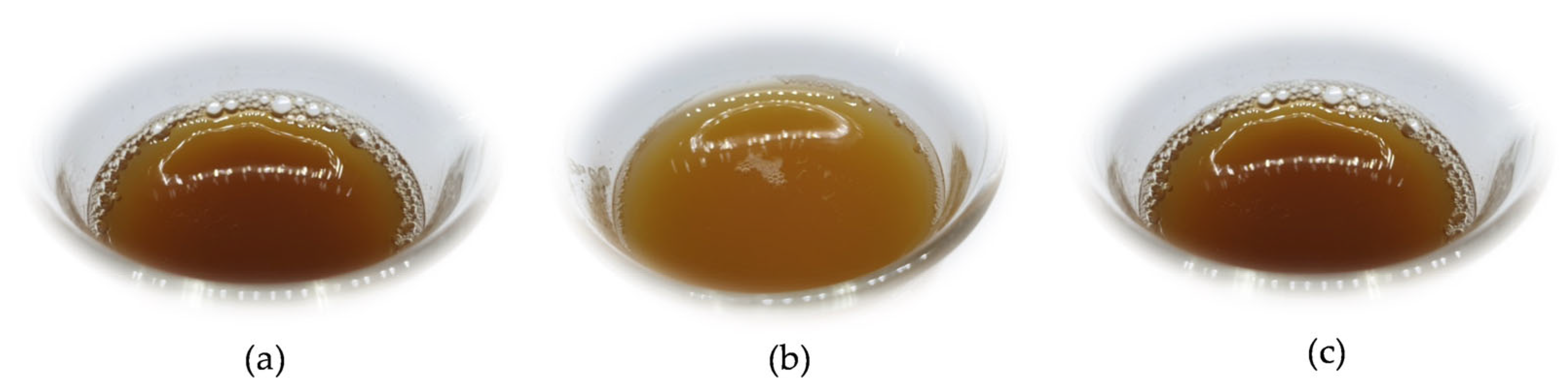
3.3. Physical Characteristics of Miang Tea Bio-Extracts

White, green, and black Miang tea bio-extracts all exhibited physical properties that were black-red, dark brown, and reddish-brown, respectively (Figure 1). Additionally, all bio-extracts featured the unique aroma of tea leaves blended with a sour scent from the fermentation process, as well as somewhat murky sediment characteristics. Moreover, the pH of green, white, and black tea leaves’ bio-extracts were 4.92, 5.08, and 4.87, respectively, before fermentation (0 day of the fermentation), and at 30 days, the pH had fallen by 3.26, 3.38, and 3.50, respectively. At 30 days, all of the fermented tea leaves had the same amount of alcohol, which was 0.8%.

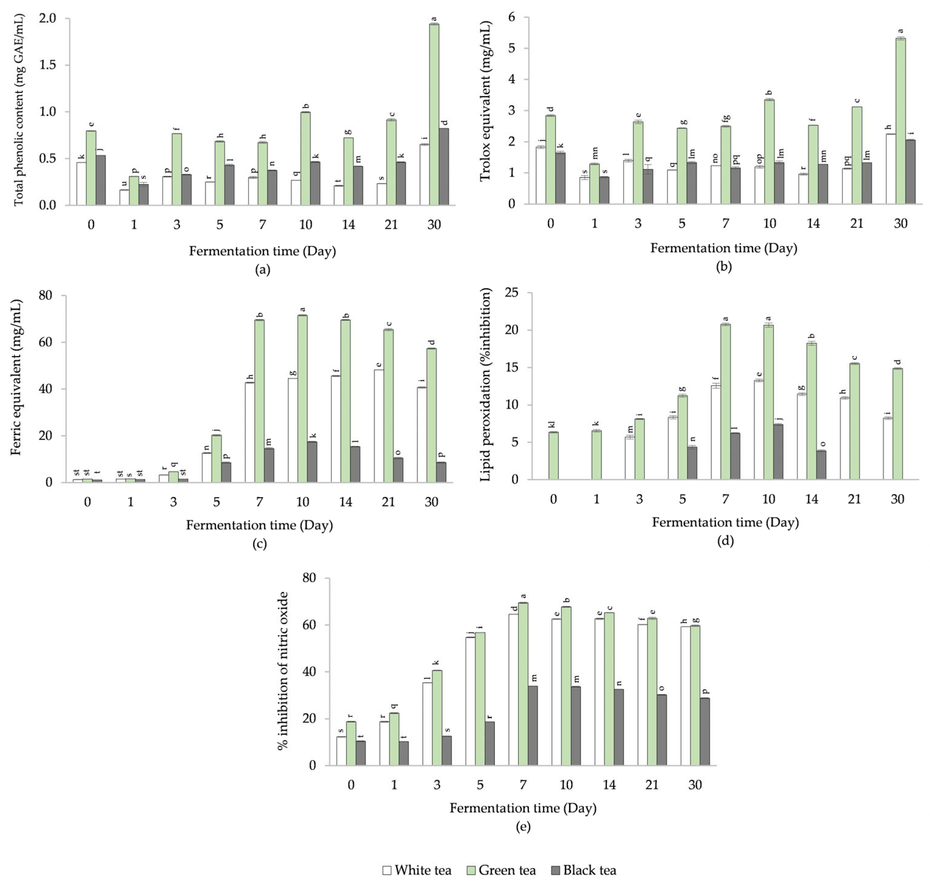
3.4. Total Phenolic Content of Miang Tea Bio-Extracts

Figure 2a represents TPC levels in Miang tea bio-extracts during a 30-day fermentation process. Green Miang tea bio-extract obviously had the greatest amount of TPC, followed by black Miang tea and white Miang tea, consecutively, according to data collected at the same fermentation time. Based on the maximum TPC level among the fermentation times of each Miang tea bio-extract, the GAE concentrations at 30 days of white, green, and black Miang tea bio-extracts were 0.65 ± 0.01, 1.94 ± 0.01, and 0.82 ± 0.00 mg GAE/mL, respectively. This was determined by taking the highest TPC level into account. The lowest TPC levels were discovered on day 1 of the bio-extract fermentation period. The contents of white, green, and black Miang tea were 0.16 ± 0.00, 0.31 ± 0.00, and 0.22 ± 0.02 mg GAE/mL, respectively.

3.5. ABTS Free Radical Scavenging Activity of Miang Tea Bio-Extracts

According to data obtained at the same fermentation period, green Miang tea bio-extract clearly had the highest quantity of ABTS•+ activity compared with white and black Miang tea bio-extract (Figure 2b). There were in the range of 1.29 ± 0.03 to 5.31 ± 0.05 mg Trolox equivalents/mL. The Trolox equivalent activities of white tea and black Miang tea bio-extracts ranged from 0.85 ± 0.06 to 2.25 ± 0.01 and 0.86 ± 0.02 to 2.06 ± 0.02 mg/mL, respectively.

3.6. Ferric Reducing Antioxidant Activity of Miang Tea Bio-Extracts

The ferric equivalent activity of green Miang tea bio-extract was shown to be at a high level at same fermentation time, followed by white tea bio-extract and black Miang tea bio-extract (Figure 2c). The activity trend of all tea bio-extracts was slightly increased during 0–10 days. The ferric equivalent activities of white, green, and black tea bio-extracts ranged from 1.27 ± 0.05 to 48.24 ± 0.08, 1.52 ± 0.02 to 71.62 ± 0.28, and 1.13 ± 0.03 to 17.39 ± 0.28 mg/mL, respectively.

3.7. Lipid Peroxidation Activity of Miang Tea Bio-Extracts

Green Miang tea bio-extract demonstrated lipid peroxidation activity from 0 to 30 days as shown in Figure 2d. The inhibitory activity trend grew gradually from 0 to 7 days, with the maximum inhibition activity observed on day 7 (20.78 ± 0.16%). Following that, the lipid peroxidation activity slightly dropped. At the same time of fermentation, green Miang tea bio-extract showed more inhibitory action than white and black Miang tea bio-extracts. From 3 to 30 days, a bio-extract from white tea displayed lipid peroxidation activity. A progressive increase in inhibitory activity was found between 3 and 10 days, with the highest level being recorded at 10 days at 13.26 ± 0.18%. After that, a small decrease was observed in lipid peroxidation activity. On the other hand, bio-extract from black Miang tea inhibited lipid peroxidation for at least 5 to 14 days. Overall, inhibitory activity increased from 5 to 10 days, with the highest level being recorded at 10 days (7.37 ± 0.14%). Subsequently, a slight drop was noted, to 3.87 ± 0.14%, in the activity of lipid peroxidation.

3.8. Nitric Oxide Inhibition of Miang Tea Bio-Extracts

During the 30-day fermentation period, all tea bio-extracts inhibited nitric oxide (Figure 2e). At the beginning of the fermentation period, the activities were enhanced. The highest percentages of green, white, and black Miang tea bio-extracts were 69.48 ± 0.25, 64.56 ± 0.04, and 33.88 ± 0.09, respectively, at 7 days. The inhibitory activities remained very high at the end of the fermentation time. When comparing by type of Miang tea bio-extract, white and green Miang tea bio-extract exhibited significantly greater inhibition quantities than black Miang tea bio-extract.

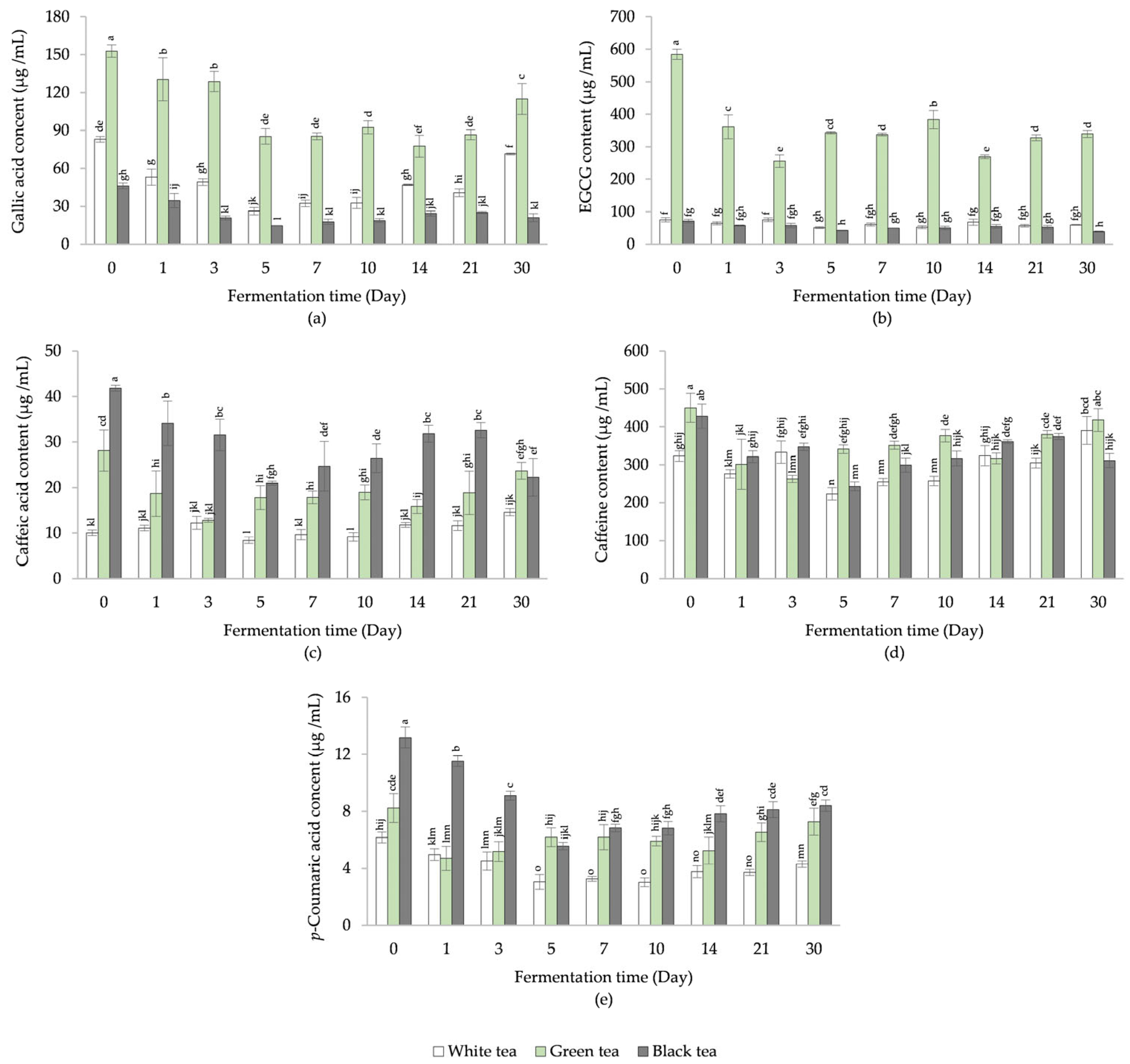
3.9. Phenolic Antioxidant Compounds of Miang Tea Bio-Extracts

According to Figure 3a, the content of gallic acid for each tea bio-extract was greatest on day 0: 83.03 ± 2.29 µg/mL for white Miang tea, 152.77 ± 4.90 µg/mL for green Miang tea, and 46.20 ± 2.20 µg/mL for black Miang tea. Based on data obtained at the same fermentation time, green Miang tea bio-extract had the highest concentration of gallic acid, followed by white and black Miang teas in that order.
Considering the data collected at the same fermentation time, green Miang tea bio-extract obviously had a significantly greater amount of EGCG compared with white and black Miang tea bio-extracts (Figure 3b). The maximum EGCG content (584.50 ± 16.02 µg/mL) was found on day 0 in green Miang tea bio-extracts.
The maximum caffeic acid concentration (41.84 ± 0.65 µg/mL) was found in black Miang tea bio-extracts at the beginning of the fermentation process (0 days). Overall, black Miang tea bio-extract had the highest concentration of caffeic acid at the same fermentation time, followed by green and white Miang tea in that order (Figure 3c).
The caffeine content of white, green, and black Miang tea bio-extract was in the range of 223.16 ± 16.26–390.65 ± 37.06, 262.67 ± 9.80–449.84 ± 38.21, and 243.26 ± 11.67–427.90 ± 32.29 µg/mL, respectively, as shown in Figure 3d. The maximum caffeine content of white, green, and black Miang tea bio-extracts was found at 30, 0, and 0 days of fermentation time, respectively.
Figure 3e shows the concentration of p-coumaric acid. During 30 days of fermentation, white, green, and black Miang tea bio-extracts had the maximum p-coumaric acid concentration at the beginning of the fermentation process (day 0). The values for these bio-extracts were as follows: 6.16 ± 0.38, 8.22 ± 1.01, and 13.17 ± 0.73 µg/mL, respectively.

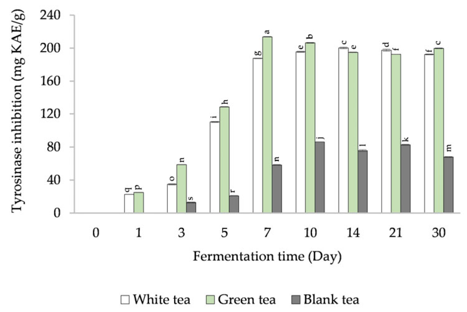
3.10. Tyrosinase Inhibition Activity of Miang Tea Bio-Extracts

From 1 to 7 days, the tyrosinase inhibition of white and green Miang tea bio-extracts gradually increased (Figure 4). This wasn’t seen on day 0. At the end of the fermentation period (10–30 days), the level of activity remained high. For the bio-extract of black Miang tea, the tyrosinase activity was observed between 3 and 30 days. The inhibition ranged from 12.64 ± 0.48 to 86.24 ± 0.12 mg KAE/g, with the greatest level occurring on day 7.

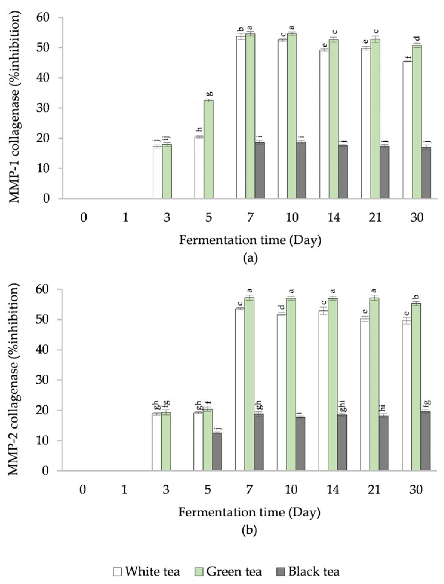
3.11. MMP-1 and MMP-2 Inhibition Activity of Miang Tea Bio-Extracts

MMP-1 and MMP-2 collagenase/gelatinase activities were reported as inhibition percentages (Figure 5a,b). Similar patterns were seen for both enzyme inhibitions. No observations were made in the early fermentation period (Day 0 and 1). After 7 days, the inhibitory action of white and green Miang tea bio-extracts reached its peak, whereas black Miang tea bio-extract revealed a significantly lower level of inhibition than other tea bio-extracts.

3.12. Antimicrobial Activity of Miang Tea Bio-Extracts

According to Table 4, the antimicrobial activity was reported as the dilution of MIC against S. aureus, S. epidermidis, P. acnes, and Ps. aeruginosa. At day 0, the bio-extract of black Miang tea was inactive against all microorganisms. White and green Miang tea bio-extracts exhibited the greatest antimicrobial activity against tested microorganisms after 7 days. In contrast, black Miang tea bio-extracts showed the greatest antibacterial activity against S. aureus and S. epidermidis after 10 days.

3.13. Elimination of Microbial Starter in Miang Tea Bio-Extracts at the End of Fermentation

White, green, and black Miang tea bio-extracts were filtered (filtrate) and KMS addition (KMS) was used to eliminate the microbial culture starter after 7 days of fermentation. The maximum quantity of microbial starter in white, green, and black Miang tea bio-extracts at the finish of fermentation time at 7 days (final) were 9.76 ± 0.58, 9.82 ± 0.11, and 9.57 ± 1.49 log cfu/mL, respectively. After microbial elimination, the microbial starter was not detected, as shown in Table 5.
The TPC of white, green, and black Miang tea bio-extracts in finished products were 493.77 ± 9.24, 912.23 ± 7.70, and 267.13 ± 10.82 µg GAE/mL, respectively. The number of microorganisms eliminated by either method did not significantly differ from the maximum values in the final products.
The antioxidant activity, compared with the Trolox equivalent content, in the final products exhibited the highest level in green Miang tea bio-extract (1094.72 ± 3.88 mg/mL), followed by white Miang tea bio-extract (912.25 ± 3.88 mg/mL) and black Miang tea bio-extract (421.50 ± 5.96 mg/mL). The contents of white and black Miang tea bio-extracts after both elimination procedures did not significantly differ compared with the final products. However, the activity of green Miang tea bio-extract obtained through a filter procedure proved significantly lower than the final filtrate and that obtained through the KMS addition method (Table 5).

4. Discussion

The beneficial properties of tea and tea extract are very well known, and have been extensively described in several research articles. The most popular and effective approach for isolating useful compounds from plant materials is solvent extraction [30,31,32]. In the present study, white, green, and black Miang tea samples were selected to represent different types of tea processed using buds and young leaves. Table 1 displayed some selected antioxidant and anti-inflammation activity data on different types of Miang tea leaf extracts using ethanolic maceration. The findings demonstrated that green Miang tea extract showed the most of these activities, while black tea extract exhibited the least. Moreover, polyphenol compounds were found in the Miang tea extracts: white Miang tea extract had the highest level of gallic acid and EGCG, green Miang tea extract had the highest level of caffeic acid, and black Miang tea extract had the highest level of caffeine and p-coumaric acid. In tea leaf extracts, major active compounds with related biological activities were detected. The future production of fermented Miang tea bio-extracts may utilize it as an indicator for standardization of tea leaf raw materials. The quantity of active compounds and, thus, the biological functions might be influenced by the harvested and/or processing procedures of tea. Tea plants have often been classified into green, albino, yellow, and ‘Zijuan’, based on the content of chlorophyll and anthocyanin present. Moreover, six types of tea (black tea (BT), green tea (GT), oolong tea (OT), white tea (WT), dark tea (DT), and yellow tea (YT)) with different flavor and aroma profiles have been created through different processing techniques [33,34,35,36].
Tea and its extracts have historically been used as essential ingredients in cosmetic manufacturing. Many benefits are available, owing to their functional components such as flavonoids, amino acids, caffeine, theaflavins, and catechins [2,35,37]. These substances, however, can not readily be absorbed or immediately used by the human body. Research involving microorganisms has been conducted on biotransformation during fermentation, which has the potential to be used in skin treatment and prevention of disease [3,5]. β-glucosidase is an enzyme playing an important role in helping to break down these complex molecules into simpler sizes or to hydrolyze plant β-glucosidase, which can then be metabolized by microorganisms during fermentation and release a variety of plant secondary metabolites from their glycosylated precursors, leading to enhanced bioavailability in health-promoting applications and the synthesis of new active metabolites [35,38]. This enzyme can promote in the release of natural humectants, such as amino acids and sugars, that can help to hydrate the skin and improve its moisture barrier function. It also helps to break down the cell walls of natural ingredients, making them more readily available for absorption by the skin. Based on this information about benefits, β-glucosidase activity was considered as the main factor in selecting the starter culture in this study. A total of 53 strains were identified from 43 fermented tea products and categorized. As demonstrated in Table 1, β-glucosidase activity was detected in 13 strains. GBW36, GBW47, and GBW53 had the most potential of β-glucosidase activity when compared to other isolates. According to molecular identification by gene sequencing compared to the GenBank database using the BLAST algorithm, GBW36, GBW47, and GBW53 were identified as L. rhamnosus, L. plantarum, and S. cerevisiae, respectively. β-glucosidase activity may vary between microbial strains based on variables such as their genetic makeup and growth conditions. A previous study reported that L. plantarum FSO1 strains showed an ability to produce a high level of β-glucosidase enzyme useful for application in the biological processing of fermented green olive [39]. Similarly, increased β-glucosidase activity of soymilk mediated with LAB (L. acidophilus, L. bulgaricus, L. casei, L. fermentum, L. plantarum, and B. lactis) was reported, which improved the product’s biological functionality [40,41]. Some yeast species were able to develop β-glucosidase activity as well as antioxidant activity. S. cerevisiae, Hanseniapora uvarum, Wicker-hamomyces anomalus, and Trichosporon asahii are all examples of common yeasts that have been shown to exhibit significant levels of β-glucosidase activity throughout the fermentation process [42]. Moreover, antioxidant activities (through ABTS•+ and FRAP assay) were found in 8 of 53 isolates. The highest activities were clearly presented in GBW47 and GBW36. The above examples establish a clear foundation for employing LAB and yeast in the fermentation process, demonstrating safety and a wide range of bioactive compounds. For this study, the combined strains were a good choice to use as a starter culture in Miang tea bio-extract.
Currently, fermentation mediated by microorganisms is often applied to obtain bioavailable extracts. Some reports have suggested that the addition of microorganisms in the fermentation process could improve the efficacy of the bioactive compounds, produce a new substance, and also limit solvent usage in an environmentally responsible manner. Wang et al. [6] investigated biotransformation using L. paracasei NTU 101 on fermented green tea extract in connection to anti-obesity effects. According to the findings, the levels of EGCG, EGC, and chlorogenic acid were higher in the fermented green tea extract than in the unfermented green tea extract. Jin et al. [43] examined the lactic acid fermentation of green tea with the Levilactobacillus brevis strain to produce γ-aminobutyric acid (GABA). The results revealed that boosting the GABA content, lactic acid content, acetic acid content, and antioxidant activity by 232.52, 552.20, 238.72, and 94.38%, respectively, was highly effective and could be useful in the food and health industries. In this study, the selected mixed strains (L. rhamnosus, L. plantarum, and S. cerevisiae) were used as starter cultures in the fermentation process of white, green, and black Miang tea. Overall, TPC, ABTS•+, FRAP, lipid peroxidation, and nitric oxide inhibition of the green Miang tea bio-extract were significantly greater than those of the white and black Miang tea bio-extracts, which related to the highest level of activities in the green Miang tea extract by ethanol. The antioxidant activity of the bio-extracts exhibited a rising trend after fermentation time. The high level of FRAP, lipid peroxidation, and nitric oxide inhibition showed up after 7 days of fermentation time, whereas the high level of TPC and ABTS•+ activities appeared at 30 days. It might be that fermentation was a form of oxidation that alters the composition of bioactive substances depending on the degree of fermentation [36]. During the fermentation process, enzymes in the tea leaves are activated, leading to a range of chemical reactions that can affect the concentration of phenolic compounds in the tea. The activity of β-glucosidase enzymes is able to break down the chemical components of plant cell walls and release phenolic compounds from their β-D-glucoside linkages, leading to an increase in TPC during the fermentation process [44]. Additionally, some microorganisms involved in fermentation, such as lactic acid bacteria, can produce β-glucosidase enzymes as part of their metabolic pathways, which can also lead to an increase in TPC [44]. It has been demonstrated that phenolic compounds possess antioxidant properties due to their ability to scavenge free radicals and prevent oxidative damage. However, the correlation between TPC and antioxidant activity is not always straightforward; it is affected by a wide range of variables, such as the type and concentration of the phenolic compounds present, the microorganisms used in fermentation, the conditions of fermentation (i.e., temperature, pH), and the specificity of the assay. A specific phenolic compound may or may not exhibit the same level of antioxidant activity as another phenolic compound. Considering this, the phenolic antioxidant compounds (gallic acid, EGCG, caffeic acid, caffeine, and p-coumaric acid) in different Miang tea bio-extracts might be changed due to the metabolic activity of microorganisms during fermentation. Green Miang tea bio-extract contained significantly more gallic acid and EGCG than white and black Miang tea bio-extract at the same time of fermentation. During the processing of tea leaves, gallic acid can be degraded or converted into other compounds. Steaming, one of the processes used in the production of green tea, can help preserve gallic acid and EGCG, whereas the longer oxidation process used in black tea production can lead to a reduction in the contents. These polyphenols that are found in white, green, and black tea are known for their antioxidant and anti-inflammatory properties, which can protect the skin from damage caused by free radicals and UV radiation. They have been shown to help reduce oxidative stress, which can contribute to skin aging and damage, and may also help to protect against skin cancer. For example, EGCG has been shown to improve skin hydration, elasticity, and barrier function, which can help to keep skin healthy and prevent dryness and wrinkles [45]. Caffeic acid has been shown to have anti-inflammatory effects that may help to reduce skin redness and irritation and relate to dermal wound healing in mice skin [46].
According to Figure 4 and Figure 5, the tyrosinase and collagenase enzymes were inhibited more in white and green bio-extracts than in black bio-extracts. Notably, the enzyme inhibition was at its highest level after 7 days of fermentation. The results were related to high antioxidant and anti-inflammation activities after 7 days of fermentation time. Antioxidants are compounds that protect the skin from free radicals, which are unstable molecules that may cause oxidative stress and cell damage. Multiple variables, such as exposure to ultraviolet radiation, air pollution, and smoking, can create free radicals. Collagen and elastin fibers, which provide the skin its structure and support, can be damaged by free radicals when these cells are attacked. This can cause a loss of firmness as well as the development of fine lines and wrinkles. These antioxidants prevent free radicals from causing harm to the skin by neutralizing them. There is some suggestion that some antioxidants, e.g., vitamin C and vitamin E, could have a role in the production of the pigment melanin in the skin. In particular, it has been demonstrated that vitamin C can block the activity of the enzyme tyrosinase, contributing to a decrease in the synthesis of the pigment melanin. This can result in skin whitening; nevertheless, the impact may be limited. Overall, tyrosinase was shown to be a key enzyme in forming melanin and other pigments associated with skin brightness [47] whereas collagenase was shown to be a key enzyme in collagen turnover [48].
Many skin care products contain antimicrobials to help stop the spread of microbes and other skin-infecting microorganisms. Antimicrobials in skin care products can originate from both synthetic compounds (namely Triclosan and Benzoyl peroxide) and natural ingredients. Due to its antibacterial and antifungal properties, tea is a popular ingredient in skin care products for a wide range of applications, including treating acne and dandruff. In this study, MICs of S. aureus, S. epidermidis, P. acnes, and Ps. aeruginosa strains were determined using a dilution technique. Results show that, on day 7, white and green Miang tea bio-extracts proved effective against all tested bacteria.
In brief, fermentation of Miang tea leaves can increase bioactivity levels compared to non-fermentation (bio-extract on day 0), especially FRAP, lipid peroxidation, and nitric oxide inhibition of bio-extract at 7 days. In addition, the active compounds, which indicate the main efficiency of the ingredients in cosmetic products, were observed during the fermentation process. Furthermore, after 7 days of fermentation, regulating the activity of enzymes with tyrosinase and collagenase inhibitors provided outstanding outcomes. Therefore, bio-extracts fermented with mixed starter cultures for 7 days are suggested as active ingredients for cosmetics.
In this study, the bio-extracts were derived from microorganisms and may contain viable cells of microorganisms that can cause alterations to the final product and/or other contaminants that can be harmful to humans. The microbial elimination procedures of finished bio-extract products are therefore necessary to ensure that the bio-extracts are safe and qualify for use. The microbial elimination can be achieved using a variety of methods, depending on the type of products and the requirements of the cosmetic formulation. The filter separation method and the addition of sanitizer such as KMS and sodium metabisulfite can be effective in decontamination Miang tea bio-extracts’ final products (at day 7 of fermentation). The extracts had no detectable microbial content after both sterilization tests. Moreover, the amounts of bioactivity, such as total phenolic and antioxidant activity, can show high retention after both sterilization tests. However, the filter method can effectively remove most bacteria and fungi from the bio-extract, but it may not be effective in removing viruses or other small microorganisms. In addition, the filter membrane may become clogged with particulates, which can reduce its effectiveness over time. The addition of KMS can effectively eliminate any remaining microorganisms in the bio-extract, is simple to implement, and has no effect on the extracts’ quality and stability or storage time. Furthermore, KMS is widely utilized in the beverage industry.

5. Conclusions

Information on the ingredients providing a beneficial effect on skin care was acquired from the study of white, green, and black Miang tea. Green Miang tea extract was shown to have the highest antioxidant and anti-inflammatory properties, whereas black Miang tea extract had the least. The mixed strains L. rhamnosus (GBW36), L. plantarum (GBW47), and S. cerevisiae (GBW53), were selected as starter cultures and had the potential to be starter cultures in this fermentation process. The activities, including FRAP, lipid peroxidation, nitric oxide inhibition, tyrosinase inhibition, collagenase inhibition (MMP-1 and MMP-2), and antimicrobial activity showed a high level after 7 days of fermentation time. In addition, the contents of phenolic antioxidant compounds (gallic acid, EGCG, caffeic acid, caffeine, and p-coumaric acid) were presented. The addition of KMS to the bio-extracts to eliminate microorganisms ensured that the bio-extracts were safe, stable, and of high quality for human applications. The quantity of microbial starter and/or contaminants was not detected, and the TPC levels and antioxidant activity remained unchanged. These properties, especially the antioxidant activities and enzyme inhibition, are beneficial to skin health. Therefore, Miang tea bio-extract from probiotic fermentation is a potential active ingredient for skin care products.

Author Contributions

Conceptualization, N.M. and S.S.; methodology, N.M., A.R., J.J. and S.S.; formal analysis, N.M., A.R., J.J., E.K., W.B. and S.S.; investigation, N.M., A.R., J.J., E.K., W.B. and S.S.; resources, S.S.; data curation, N.M., A.R., J.J., E.K., W.B. and S.S.; writing—original draft preparation, N.M., A.R., J.J., E.K., W.B. and S.S.; writing—review and editing, N.M. and S.S.; visualization, S.S.; supervision, S.S.; project administration, S.S.; funding acquisition, S.S. All authors have read and agreed to the published version of the manuscript.

Funding

This research was partially funded by Northern Science Park Thailand, Industrial Research and Technology Capacity Development Program: IRTC 2559 in “Prototype of bio-extract from tea leaves by microbial fermentation as a skin care cosmetic ingredient”.

Institutional Review Board Statement

Not applicable.

Informed Consent Statement

Not applicable.

Data Availability Statement

The data presented in the manuscript is available on request from the corresponding author.

Acknowledgments

Authors gratefully acknowledge the Faculty of Pharmacy, Chiang Mai University, Chiang Mai, Thailand and a postdoctoral fellowship granted by Chiang Mai University, Chiang Mai, Thailand (N.M.).

Conflicts of Interest

The authors declare no conflict of interest. The funder had no role in the design of the study; in the collection, analyses, or interpretation of data; in the writing of the manuscript; or in the decision to publish the results.

References

  1. Arct, J.; Bielenda, B.; Oborska, A.; Pytkowska, K. The tea and its cosmetic application. J. Appl. Cosmetol. 2003, 21, 117–127. [Google Scholar]
  2. Rajbhar, K.; Dawda, H.; Mukundan, U. Tea polyphenols for skin care. Res. J. Top. Cosmet. Sci. 2015, 6, 1–6. [Google Scholar]
  3. Koch, W.; Zagórska, J.; Marzec, Z.; Kukula-Koch, W. Applications of tea (Camellia sinensis) and its active constituents in cosmetics. Molecules 2019, 24, 4277. [Google Scholar] [CrossRef]
  4. Binic, I.; Lazarevic, V.; Ljubenovic, M.; Mojsa, J.; Sokolovic, D. Skin ageing: Natural weapons and strategies. Evid. Based Complement. Altern. Med. 2013, 2013, 827248. [Google Scholar] [CrossRef]
  5. Ziemlewska, A.; Nizioł-Łukaszewska, Z.; Bujak, T.; Zagórska-Dziok, M.; Wójciak, M.; Sowa, I. Effect of fermentation time on the content of bioactive compounds with cosmetic and dermatological properties in Kombucha Yerba Mate extracts. Sci. Rep. 2021, 11, 18792. [Google Scholar] [CrossRef]
  6. Wang, L.C.; Pan, T.M.; Tsai, T.Y. Lactic acid bacteria-fermented product of green tea and Houttuynia cordata leaves exerts anti-adipogenic and anti-obesity effects. J. Food Drug Anal. 2018, 26, 973–984. [Google Scholar] [CrossRef]
  7. Tang, S.C.; Yang, J.H. Dual effects of alpha-hydroxy acids on the skin. Molecules 2018, 23, 863. [Google Scholar] [CrossRef]
  8. Manguntungi, B.; Mustopa, A.Z.; Meilina, L.; Nurfatwa, M.; Vanggy, L.R.; Irawan, S.; Tamzil, M.S.; Aprilian, T.; Fidduha, A.S.; Wersian, I.N. The profile analysis of lactic acid bacteria (LAB) from Sumbawa white honey and its potential producing antibacterial compounds. Walailak J. Sci. Technol. 2021, 18, 22204–22212. [Google Scholar] [CrossRef]
  9. Xiao, X.; Hu, X.; Yao, J.; Cao, W.; Zou, Z.; Wang, L.; Qin, H.; Zhong, D.; Li, Y.; Xue, P.; et al. The role of short-chain fatty acids in inflammatory skin diseases. Front. Microbiol. 2022, 13, 5417. [Google Scholar] [CrossRef]
  10. Zhu, F.; Du, B.; Xu, B. A critical review on production and industrial applications of beta-glucans. Food Hydrocoll. 2016, 52, 275–288. [Google Scholar] [CrossRef]
  11. Fang, X.; Dong, Y.; Xie, Y.; Wang, L.; Wang, J.; Liu, Y.; Zhao, L.; Cao, F. Effects of β-glucosidase and α-rhamnosidase on the Contents of Flavonoids, Ginkgolides, and Aroma Components in Ginkgo Tea Drink. Molecules 2019, 24, 2009. [Google Scholar] [CrossRef]
  12. Ahmed, A.; Batool, K.; Bibi, A. Microbial β-glucosidase: Sources, production and applications. Appl. Environ. Microbiol. 2017, 5, 31–46. [Google Scholar]
  13. Stradwick, L.; Inglis, D.; Kelly, J.; Pickering, G. Development and application of assay for determining β-glucosidase activity in human saliva. Flavour 2017, 6, 1. [Google Scholar] [CrossRef]
  14. Liu, N.; Miao, S.; Qin, L. Screening and application of lactic acid bacteria and yeasts with L-lactic acid-producing and anti-oxidant capacity in traditional fermented rice acid. Food Sci. Nutr. 2020, 8, 6095–6111. [Google Scholar] [CrossRef]
  15. Chaiyasut, C.; Jantavong, S.; Kruatama, C.; Peerajan, S.; Sirilun, S.; Shank, L. Factors affecting methanol content of fermented plant beverage containing Morinda citrifolia. Afr. J. Biotechnol. 2013, 12, 4356–4363. [Google Scholar]
  16. Zheng, J.; Wittouck, S.; Salvetti, E.; Franz, C.M.; Harris, H.M.; Mattarelli, P.; Toole, P.W.O.; Pot, B.; Vandamme, P.; Walter, J.; et al. A taxonomic note on the genus Lactobacillus: Description of 23 novel genera, emended description of the genus Lactobacillus Beijerinck 1901, and union of Lactobacillaceae and Leuconostocaceae. Int. J. Syst. Evol. Microbiol. 2020, 70, 2782–2858. [Google Scholar]
  17. Otieno, D.O.; Ashton, J.F.; Shah, N.P. Evaluation of enzymic potential for biotransformation of isoflavone phytoestrogen in soymilk by Bifidobacterium animalis, Lactobacillus acidophilus and Lactobacillus casei. Food Res. Int. 2006, 39, 394–407. [Google Scholar] [CrossRef]
  18. Zhao, C.N.; Tang, G.Y.; Cao, S.Y.; Xu, X.Y.; Gan, R.Y.; Liu, Q.; Mao, Q.Q.; Shang, A.; Li, H.B. Phenolic profiles and antioxidant activities of 30 tea infusions from green, black, oolong, white, yellow and dark teas. Antioxidants 2019, 8, 215. [Google Scholar] [CrossRef]
  19. Farooq, S.; Sehgal, A. Antioxidant activity of different forms of green Tea: Loose leaf, bagged and matcha. Curr. Res. Nutr. Food Sci. 2018, 6, 35–40. [Google Scholar] [CrossRef]
  20. Jayabalan, R.; Subathradevi, P.; Marimuthu, S.; Sathishkumar, M.; Swaminathan, K. Changes in free-radical scavenging ability of kombucha tea during fermentation. Food Chem. 2008, 109, 227–234. [Google Scholar] [CrossRef]
  21. Piktel, E.; Wnorowska, U.; Cieśluk, M.; Deptula, P.; Pogoda, K.; Misztalewska-Turkowicz, I.; Paprocka., P.; Niemirowicz-Laskowska, K.; Wilczewska, A.Z.; Janmey, P.A.; et al. Inhibition of inflammatory response in human keratinocytes by magnetic nanoparticles functionalized with PBP10 peptide derived from the PIP2-binding site of human plasma gelsolin. J. Nanobiotechnol. 2019, 17, 22. [Google Scholar] [CrossRef]
  22. Yoon, W.J.; Kim, S.S.; Oh, T.H.; Lee, N.H.; Hyun, C.G. Abies koreana essential oil inhibits drug-resistant skin pathogen growth and LPS-induced inflammatory effects of murine macrophage. Lipids 2009, 44, 471–476. [Google Scholar] [CrossRef]
  23. Theppakorn, T.; Wongsakul, S. Optimization and validation of the HPLC-based method for the analysis of gallic acid, caffeine and 5 catechins in green tea. Naresuan Univ. J. Sci. Technol. 2013, 20, 1–11. [Google Scholar]
  24. Uchida, R.; Ishikawa, S.; Tomoda, H. Inhibition of tyrosinase activity and melanin pigmentation by 2-hydroxytyrosol. Acta Pharm. Sin. B 2014, 4, 141–145. [Google Scholar] [CrossRef]
  25. Wu, L.; Chen, C.; Cheng, C.; Dai, H.; Ai, Y.; Lin, C.; Chung, Y. Evaluation of tyrosinase inhibitory, antioxidant, antimicrobial, and antiaging activities of Magnolia officinalis extracts after Aspergillus niger fermentation. Biomed Res. Int. 2018, 2018, 5201786. [Google Scholar] [CrossRef]
  26. Eun Lee, K.; Bharadwaj, S.; Yadava, U.; Gu Kang, S. Evaluation of caffeine as inhibitor against collagenase, elastase and tyrosinase using in silico and in vitro approach. J. Enzyme Inhib. Med. Chem. 2019, 34, 927–936. [Google Scholar] [CrossRef]
  27. Thring, T.S.; Hili, P.; Naughton, D.P. Anti-collagenase, anti-elastase and anti-oxidant activities of extracts from 21 plants. BMC Complement. Altern. Med. 2009, 9, 27. [Google Scholar] [CrossRef]
  28. Ghimeray, A.; Jung, U.; Lee, H.; Kim, Y.; Ryu, E.; Chang, M. In vitro antioxidant, collagenase inhibition, and in vivo anti-wrinkle effects of combined formulation containing Punica granatum, Ginkgo biloba, Ficus carica, and Morus alba fruits extract. Clin. Cosmet. Investig. Dermatol. 2015, 389, 389–396. [Google Scholar] [CrossRef]
  29. Blaskovich, M.A.T.; Elliott, A.G.; Kavanagh, A.M.; Ramu, S.; Cooper, M.A. In vitro antimicrobial activity of acne drugs against skin-associated bacteria. Sci. Rep. 2019, 9, 14658. [Google Scholar] [CrossRef]
  30. Perva-Uzunalić, A.; Škerget, M.; Knez, Ž.; Weinreich, B.; Otto, F.; Grüner, S. Extraction of active ingredients from green tea (Camellia sinensis): Extraction efficiency of major catechins and caffeine. Food Chem. 2006, 96, 597–605. [Google Scholar] [CrossRef]
  31. Sarwa, K.K.; Rudrapal, M.; Debnath, M. Extraction of green tea leaves: The use of different methods, their optimization and comparative evaluation. Biosci. Biotechnol. Res. Asia 2013, 10, 383–386. [Google Scholar] [CrossRef]
  32. Banerjee, S.; Chatterjee, J. Efficient extraction strategies of tea (Camellia sinensis) biomolecules. J. Food Sci. Technol. 2015, 52, 3158–3168. [Google Scholar] [CrossRef]
  33. Wang, C.; Han, J.; Pu, Y.; Wang, X. Tea (Camellia sinensis): A Review of Nutritional Composition, Potential Applications, and Omics Research. Appl. Sci. 2022, 12, 5874. [Google Scholar] [CrossRef]
  34. Li, H.; Guo, H.; Luo, Q.; Wu, D.T.; Zou, L.; Liu, Y.; Gan, R.Y. Current Extraction, Purification, and Identification Techniques of Tea Polyphenols: An Updated Review; Taylor Francis Group: Abingdon, UK, 2021. [Google Scholar] [CrossRef]
  35. Wang, Y.; Kan, Z.; Thompson, H.J.; Ling, T.; Ho, C.T.; Li, D.; Wan, X. Impact of six typical processing methods on the chemical composition of tea leaves using a single Camellia sinensis cultivar, Longjing 43. J. Agric. Food Chem. 2018, 67, 5423–5436. [Google Scholar] [CrossRef]
  36. Wu, D.; Chen, R.; Zhang, W.; Lai, X.; Sun, L.; Li, Q.; Zhang, Z.; Cao, J.; Wen, S.; Lai, Z.; et al. Tea and its components reduce the production of uric acid by inhibiting xanthine oxidase. Food Nutr. Res. 2022, 66, 8239. [Google Scholar] [CrossRef]
  37. Jiang, H.; Engelhardt, U.H.; Thräne, C.; Maiwald, B.; Stark, J. Determination of flavonol glycosides in green tea, oolong tea and black tea by UHPLC compared to HPLC. Food Chem. 2015, 183, 30–35. [Google Scholar] [CrossRef]
  38. Michlmayr, H.; Kneifel, W. β-Glucosidase activities of lactic acid bacteria: Mechanisms, impact on fermented food and human health. FEMS Microbiol. Lett. 2014, 352, 1–10. [Google Scholar] [CrossRef]
  39. Rokni, Y.; Abouloifa, H.; Bellaouchi, R.; Hasnaoui, I.; Gaamouche, S.; Lamzira, Z.; Salah, R.B.E.N.; Saalaoui, E.; Ghabbour, N.; Asehraou, A. Characterization of β-glucosidase of Lactobacillus plantarum FSO1 and Candida pelliculosa L18 isolated from traditional fermented green olive. J. Genet. Eng. Biotechnol. 2021, 19, 117. [Google Scholar] [CrossRef]
  40. Donkor, O.N.; Shah, N.P. Production of β-Glucosidase and Hydrolysis of Isoflavone Phytoestrogens by Lactobacillus acidophilus, Bifidobacterium lactis, and Lactobacillus casei in Soymilk. J. Food Sci. 2008, 73, M15–M20. [Google Scholar] [CrossRef]
  41. Rekha, C.R.; Vijayalakshmi, G. Isoflavone phytoestrogens in soymilk fermented with β-glucosidase producing probiotic lactic acid bacteria. Int. J. Food Sci. Nutr. 2011, 62, 111–120. [Google Scholar] [CrossRef]
  42. Zhang, P.; Zhang, R.; Sirisena, S.; Gan, R.; Fang, Z. Beta-glucosidase activity of wine yeasts and its impacts on wine volatiles and phenolics: A mini-review. Food Microbiol. 2021, 100, 103859. [Google Scholar] [CrossRef]
  43. Jin, Y.H.; Hong, J.H.; Lee, J.H.; Yoon, H.; Pawluk, A.M.; Yun, S.J.; Mah, J.H. Lactic acid fermented green tea with Levilactobacillus brevis capable of producing γ-aminobutyric acid. Fermentation 2021, 7, 110. [Google Scholar] [CrossRef]
  44. Nguyen Thai, H.; Van Camp, J.; Smagghe, G.; Raes, K. Improved release and metabolism of flavonoids by steered fermentation processes: A review. Int. J. Mol. Sci. 2014, 15, 19369–19388. [Google Scholar] [CrossRef]
  45. Kim, E.; Hwang, K.; Lee, J.; Han, S.Y.; Kim, E.M.; Park, J.; Cho, J.Y. Skin protective effect of epigallocatechin gallate. Int. J. Mol. Sci. 2018, 19, 173. [Google Scholar] [CrossRef]
  46. Song, H.S.; Park, T.W.; Sohn, U.D.; Shin, Y.K.; Choi, B.C.; Kim, C.J.; Sim, S.S. The effect of caffeic acid on wound healing in skin-incised mice. Korean J. Physiol. Pharmacol. 2008, 12, 343–347. [Google Scholar]
  47. Pillaiyar, T.; Manickam, M.; Namasivayam, V. Skin whitening agents: Medicinal chemistry perspective of tyrosinase inhibitors. J. Enzyme Inhib. Med. Chem. 2017, 32, 403–425. [Google Scholar] [CrossRef]
  48. Shingleton, W.D.; Cawston, T.E.; Hodges, D.J.; Brick, P. Collagenase: A key enzyme in collagen turnover. Biochem. Cell Biol. 1996, 74, 759–775. [Google Scholar]
Figure 1. Physical characteristic of (a) white, (b) green, and (c) black Miang tea leaf bio-extracts after 30 days of fermentation time.
Cosmetics 10 00085 g001
Figure 2. (a) Total phenolic content, (b) ABTS free radical scavenging activity, (c) ferric reducing antioxidant activity, (d) lipid peroxidation activity, and (e) nitric oxide inhibition of the white, green, and black Miang tea bio-extracts at sampling fermentation times. Different letters above the bars indicate statistically significant differences (p < 0.05) among 27 samples within each graph.
Cosmetics 10 00085 g002
Figure 3. Phenolic antioxidant compounds of the white, green, and black Miang tea bio-extracts at sampling fermentation times: (a) gallic acid; (b) EGCG; (c) caffeic acid; (d) caffeine; and (e) p-coumaric acid. Different letters above the bars indicate statistically significant differences (p < 0.05) among 27 samples within each graph.
Cosmetics 10 00085 g003
Figure 4. Tyrosinase enzyme activity inhibition of the white, green, and black Miang tea bio-extracts at sampling fermentation times. Different letters above the bars indicate statistically significant differences (p < 0.05) among 27 samples within each graph.
Cosmetics 10 00085 g004
Figure 5. Collagenase enzyme inhibition activity of the white, green, and black tea bio-extracts at sampling fermentation times: (a) MMP-1 collagenase inhibition; (b) MMP-2 collagenase inhibition. Different letters above the bars indicate statistically significant differences (p < 0.05) among 27 samples within each graph.
Cosmetics 10 00085 g005
Table 1. Phytochemical of crude ethanolic Miang tea extracts.
PhytochemicalTypes of Miang Tea Leaf Extract
White TeaGreen TeaBlack Tea
Yield
(%)
17.99 ± 0.38 a13.16 ± 0.15 b 2.89 ± 0.08 c
Total phenolic content
(mg GAE/g of sample)
241.38 ± 3.50 b391.48 ± 1.16 a137.79 ± 2.30 c
Trolox equivalent
(mg/g of sample)
840.03 ± 11.18 b998.21 ± 3.63 a403.89 ± 17.77 c
FeSO4 equivalent
(mg/g of sample)
41.62 ± 6.15 b68.77 ± 7.58 a11.54 ± 2.26 c
Lipid peroxidation inhibition
(%)
14.22 ± 0.19 b21.12 ± 0.44 a7.28 ± 0.23 c
Nitric oxide inhibition
(%)
37.76 ± 0.29 b 54.27 ± 0.38 a 18.33 ± 0.52 c
Gallic acid
(mg/g of sample)
14.32 ± 0.02 a9.80 ± 0.01 b7.52 ± 0.03 c
EGCG
(mg/g of sample)
171.30 ± 2.71 a154.30 ± 5.87 b17.22 ± 0.79 c
Caffeic acid
(mg/g of sample)
2.63 ± 0.02 c7.82 ± 0.11 a4.74 ± 0.30 b
Caffeine
(mg/g of sample)
134.43 ± 1.20 b127.86 ± 0.35 c178.80 ± 3.95 a
p-Coumaric acid
(mg/g of sample)
2.12 ± 0.19 b2.02 ± 0.01 b3.42 ± 0.14 a
Significant differences (p < 0.05) are indicated by the mean value ± SD with different superscript letters (a, b, and c) in the same row.
Table 2. β-glucosidase activity of microbial strains isolated from various traditional Thai fermented plant-based dietary products.
Code of Isolate
Microorganism
β-Glucosidase Activity (µmol/mL)
GBW070.12 ± 0.00 de
GBW110.16 ± 0.02 cd
GBW230.14 ± 0.02 cd
GBW270.15 ± 0.00 cd
GBW290.17 ± 0.02 c
GBW360.24 ± 0.03 b
GBW380.18 ± 0.03 c
GBW410.16 ± 0.03 cd
GBW440.10 ± 0.00 e
GBW470.26 ± 0.02 b
GBW480.12 ± 0.03 de
GBW500.14 ± 0.01 cd
GBW530.34 ± 0.04 a
Significant differences (p < 0.05) are indicated by the mean value ± standard deviation (SD) with different superscript letters (a, b, c, d and e) in the same column.
Table 3. Antioxidant activities of microbial strains isolated from various traditional Thai fermented plant-based dietary products.
Code of Isolate
Microorganism
Antioxidant Activity
ABTS•+ Inhibition
(%)
FeSO4 Equivalent
(mg/g of Sample)
GBW0716.61 ± 0.42 f484.37 ± 5.06 e
GBW1121.55 ± 0.70 c522.27 ± 7.12 c
GBW2318.22 ± 0.38 e491.53 ± 6.08 e
GBW27NDND
GBW29NDND
GBW3630.12 ± 1.04 b937.23 ± 7.43 b
GBW3820.24 ± 0.54 d512.18 ± 5.77 d
GBW4119.42 ± 1.12 d466.18 ± 4.67 f
GBW44NDND
GBW4733.05 ± 1.07 a1008.60 ± 8.26 a
GBW48NDND
GBW5011.67 ± 0.83 g375.25 ± 4.23 g
GBW53ND ND
Significant differences (p < 0.05) are indicated by the mean value ± standard deviation (SD) with different superscript letters (a, b, c, d, e, f and g) in the same column. ND means not detected.
Table 4. Antimicrobial activity of Miang tea bio-extracts during fermentation against microorganisms causing skin disease.
Bio-ExtractFermentation Time (Day)Minimal Inhibitory Concentration (MIC) (Titer)
S. aureusS. epidermidisP. acnesPs. aeruginosa
White tea01:21:21:21:2
11:21:21:21:2
31:41:41:41:2
51:81:81:81:4
71:81:81:81:8
101:81:81:81:8
141:81:81:81:8
211:81:81:81:8
301:81:81:81:8
Green tea01:21:21:21:2
11:21:21:21:2
31:41:41:41:2
51:81:81:81:4
71:81:81:81:8
101:81:81:81:8
141:81:81:81:8
211:81:81:81:8
301:81:81:81:8
Black tea0NDNDNDND
11:21:2NDND
31:21:21:21:2
51:21:21:21:2
71:21:21:21:2
101:41:41:21:2
141:41:41:21:2
211:41:41:21:2
301:41:41:21:2
Data were displayed as a dilution titer between the volume of Miang tea bio-extract and the total series of dilutions. ND means not detected.
Table 5. Microbial numbers, total phenolic content, and antioxidant activity of Miang tea bio-extracts through elimination procedures at 0 and 30 day(s) of incubation time.
Bio-ExtractConditionIncubation Time (Day)Analysis
Microbial Number
(log cfu/mL)
Total Phenolic Content
(µg GAE/mL)
Trolox Equivalent
(mg/mL)
White teaFinal09.76 ± 0.58493.77 ± 9.24912.25 ± 3.88
Filtrate0ND491.33 ± 8.63910.33 ± 8.61
30ND490.11 ± 10.03910.57 ± 6.23
KMS0ND491.95 ± 5.72910.58 ± 5.96
30ND491.22 ± 8.57909.77 ± 11.46
Green teaFinal09.82 ± 0.11912.23 ± 7.701094.72 ± 3.97
Filtrate0ND908.58 ± 9.861028.56 ± 6.63 *
30ND906.84 ± 4.711019.64 ± 9.92 *
KMS0ND911.67 ± 7.551088.68 ± 5.56
30ND910.58 ± 9.581087.23 ± 10.74
White teaFinal09.57 ± 1.49267.13 ± 10.82421.50 ± 5.96
Filtrate0ND264.28 ± 11.76419.87 ± 7.82
30ND264.12 ± 9.42418.94 ± 7.58
KMS0ND265.83 ± 4.28420.13 ± 7.05
30ND264.33 ± 7.44420.67 ± 10.96
* Indicates statistically significant differences (p < 0.05) between treatments and final conditions within each type of bio-extract and the same column. ND means not detected.
Disclaimer/Publisher’s Note: The statements, opinions and data contained in all publications are solely those of the individual author(s) and contributor(s) and not of MDPI and/or the editor(s). MDPI and/or the editor(s) disclaim responsibility for any injury to people or property resulting from any ideas, methods, instructions or products referred to in the content.

