Review

Vitiligo: A Review of Aetiology, Pathogenesis, Treatment, and Psychosocial Impact

by
Khadeejeh AL-smadi
1,
Mohammad Imran
1,
Vania R. Leite-Silva
1,2 and
Yousuf Mohammed
1,3,*
1
Frazer Institute, The University of Queensland, Brisbane, QLD 4102, Australia
2
Instituto de Ciências Ambientais, Químicas e Farmacêuticas, Departamento de Ciências Farmacêuticas, Universidade Federal de São Paulo, UNIFESP, Diadema 09913-030, Brazil
3
School of Pharmacy, The University of Queensland, Brisbane, QLD 4102, Australia
*
Author to whom correspondence should be addressed.
Cosmetics 2023, 10(3), 84; https://doi.org/10.3390/cosmetics10030084
Submission received: 2 May 2023 / Revised: 22 May 2023 / Accepted: 23 May 2023 / Published: 27 May 2023
(This article belongs to the Collection Feature Papers in Cosmetics in 2023)

Abstract

:
Vitiligo is an acquired, chronic condition characterised by depigmentation of the epidermis or by destruction/loss of melanin. Skin cells (melanocytes) are responsible for producing melanin, the substance that gives pigmentation to the skin. This review aims to provide a comprehensive overview of the current state of knowledge about vitiligo. Although there is no specific ethnic group, gender, or skin type that is more prone to vitiligo than others, it can affect anyone. The most commonly prescribed treatments for vitiligo are systemic and topical phototherapy and immunomodulators such as corticosteroids, calcineurin inhibitors, and vitamin D analogues, in addition to cosmetics that can camouflage and improve the quality of life. Even though vitiligo is typically thought of as a cosmetic disorder, its effects on the physical and psychosocial health of sufferers cannot be ignored.

1. Introduction

The skin loses colour due to a disorder called vitiligo (pronounced vit-il-EYE-go) [1]. In the Aushooryan era, roughly 2200 B.C., vitiligo was first mentioned in writing under the name Kilăsa. Further, the Egyptian Ebers Papyrus also has information on vitiligo that dates back to 1550 B.C. [2]. It is described as an autoimmune disease characterised by depigmented macules as well as patches of various shapes that are driven by the destruction of melanocytes or loss of their functioning in the skin [3]. As a result, several areas of the body, including the skin, hair, and mucous membranes, develop discoloured white marks. The lesion is known as a macule if the area of the skin losing colour is less than 1 centimetre wide and as a patch if it is greater than that [4]. Like all other skin disorders, patients with vitiligo are considered social outcasts in various societies, which has psychological and physical impacts [5]. Vitiligo has received very little investigation since most epidemiological studies either focus on highly chosen contexts, such as clinical populations, or on the prevalence of comorbidity in vitiligo patients without discussing the general population [6,7]. One study included more than 50 studies using a variety of methodologies and general demographic subgroups. Kruger et al. [8] found that the prevalence of vitiligo in the world’s population overall ranges between 0.06% and 2.28% and between 0.0 and 2.16% in children and adolescent populations.
Geographically, prevalence rates vary and are frequently greater in Africa and India [9]. The incidence of vitiligo in the Indian subcontinent has the highest proportion at 9.98% [10], followed by Nigeria at 2.8% [11], and Romania at 2.28% [8]. According to various studies from India, vitiligo prevalence among dermatology outpatients ranges between 0.25 and 4%, with the states of Gujarat and Rajasthan having a maximum frequency of 8.8% [12]. These variations in the results might point to the existence of a single skin institute in India, or they might demonstrate the inclusion of cases involving toxic and chemical depigmentation, which could explain the high value in some areas [8,13].
Males and females are equally affected; however, few studies have indicated a female predominance, which may be related to women’s higher tendency for autoimmune disorders or because women tend to be more concerned with their appearance when seeking advice and treatment [14,15]. Vitiligo typically begins before the age of 30, and most studies show that half of the patients begin to experience symptoms by age 20. When the disease has an early onset in children, it may be related to a family history [16]. Segmental and non-segmental vitiligo are both types of vitiligo. While non-segmental vitiligo can appear at any age, young people between the ages of 10 and 30 are the most frequently affected, and about 25% of vitiligo sufferers develop the illness before becoming 10 years old. On the other hand, segmental vitiligo develops earlier than non-segmental vitiligo and can occur in 41.3% of patients before the age of 10 years [17].
Thomas B. Fitzpatrick created Fitzpatrick skin phototypes in 1975 based on a person’s skin tone and how they react to exposure to the sun in terms of burning and tanning [18]. The Fitzpatrick skin type has been most frequently employed in a prospective population-based and case-control research study to analyse sun sensitivity and the causes of skin cancer, including exposure to UV radiation, tanning, and protective activities [19]. Although many recent studies have revealed that people of all ethnicities and skin types (Fitzpatrick) are affected by vitiligo equally [20]. An online panel was used to recruit 35,694 people over the age of 18 from Europe, Japan, and the USA to participate in a survey about any skin conditions, including vitiligo, that they may have had in the past. The estimated prevalence of vitiligo overall was 1.3%. Europe has the highest prevalence (1.6%), followed by the USA (1.4%), and Japan came in third (0.5%). According to the Fitzpatrick scale, the prevalence of vitiligo was highest among people with type III (light brown; 0.5%) and type IV (moderate brown; 0.4%) skin phototypes [21]. This article reviews the present understanding of vitiligo, including its aetiology, classification, pathogenesis, diagnoses, and available treatments.

2. Psychosocial Aspects of Vitiligo and Associated Disorders

2.1. Psychological Disorders

Currently, appearance has a big impact on how people see and judge others. Those with skin conditions that impact their appearance may be negatively affected. In the 1950s, the World Health Organisation expanded its definition of health to include more than just the absence of disease but also having a fulfilling life. Therefore, it has already been proven that they have an impact on the patient’s everyday activities, psychological well-being, and social and personal life [22]. Negative emotions like guilt, shame, worry, insecurity, and even psychological symptoms like depression can also result from skin diseases. Sufferers may be struggling with significant depressive symptoms and low self-esteem. In addition to feeling unpleasant in social environments, they could also feel inferior and become objects of discrimination [23]. Vitiligo is linked to significant declines in quality of life in routine activities, employment, and psychosocial health. According to studies, vitiligo lesions on sensitive or visible body parts, such as the genitalia, face, and hands, have a more negative effect on a patient’s well-being [24,25]. Many vitiligo patients exhibit signs of severe distress that are connected to particular social situations and emotional disturbances. More than 50% of vitiligo patients indicate they have experienced staring, 16% have overheard rude comments, and 13% have experienced job discrimination and stigmatisation as a result of their vitiligo condition. A combination of both quantitative and qualitative techniques is used to evaluate the type and severity of vitiligo-related social and psychological problems. In a survey with 600 patients, 59% of respondents stated their vitiligo had caused them to have a difficult time [26,27].

2.2. Systemic Disorders

2.2.1. Thyroid Disorders

Thyroid functional problems and autoimmune thyroid diseases have been linked to vitiligo, and it appears that vitiligo patients are more likely than healthy people to experience clinical and subclinical thyroid disease [26,27]. The two most significant and prevalent autoimmune thyroid conditions connected to vitiligo are Hashimoto thyroiditis and Graves’ disease [28]. In 1994, a study by Hegedus et al. on 35 vitiligo patients to determine the frequency and type of thyroid disease showed that 43% of patients had one or more signs of thyroid disease, and 22.8% had thyroid dysfunction (17.1% hyperthyroidism and 5.7% hypothyroidism) [27]. Therefore, people with autoimmune thyroid disease have a much higher prevalence of vitiligo than patients with non-autoimmune thyroid disease. Individuals with generalised vitiligo (non-segmental), particularly those inherited, are more likely than those with segmental vitiligo to have autoimmune problems [29].

2.2.2. Other Disorders

Several studies have hypothesised a connection between vitiligo and other autoimmune disorders such as pernicious anaemia, diabetes, systemic lupus erythematosus, rheumatoid arthritis, psoriasis, alopecia areata, and Addison’s disease [14,30]. A questionnaire survey between 2001 and 2006 for Birlea in a Romanian isolated population with a high rate of familial connections revealed that, of the 51 total patients with vitiligo, 22 had one or more autoimmune disorders. Additionally, about 82% of the vitiligo and other autoimmune disease patients had generalised vitiligo. They had autoimmune thyroid disease in 31% of cases, rheumatoid arthritis in 14%, and adult-onset type 1 diabetes in 12% of cases [31]. Moreover, there is a higher prevalence of these conditions among first-degree relatives of vitiligo patients, indicating that some families may be predisposed genetically to this set of autoimmune and autoinflammatory conditions [32].

3. Aetiology and Pathogenesis

The destruction of melanocytes and the development of white patches in vitiligo have been linked to a variety of different mechanisms. They include neural, genetic, autoimmune, oxidative stress, production of inflammatory mediators, and other mechanisms for melanocyte separation [33].

3.1. Autoimmune Theory

Autoimmune mediation is the most common and well-established theory that suggests a disruption in the response causes melanocytes to be destroyed by autoimmune effector mechanisms, either memory cytotoxic T cells or autoantibodies targeted to melanocyte surface antigens. It is well recognised that vitiligo and autoimmune disorders are related; for example, vitiligo is usually associated with thyroid disorders including Hashimoto’s thyroiditis and Graves’ disease, as well as other endocrinopathies like Addison’s disease and diabetes mellitus. There has to be additional research on the relevance of some of these conditions, including autoimmune polyglandular syndrome, alopecia areata, systemic lupus erythematosus, inflammatory bowel disease, rheumatoid arthritis, psoriasis, and pernicious anaemia [34,35].

3.2. Genetic Theory

Familial clustering is observed in vitiligo. According to numerous studies, the prevalence of vitiligo among first-degree relatives ranges from 0.14% to 20%. The information clearly shows a genetic component; however, just 23% of monozygotic twins showed concordance, indicating that there may be a considerable non-genetic factor in the pathophysiology of vitiligo. On the other hand, since vitiligo is a polygenic condition, several candidate genes have been identified, including the major histocompatibility complex (MHC), angiotensin-converting enzyme (ACE), catalase (CAT), cytotoxic T lymphocyte antigen-4 (CTLA-4), protein tyrosine phosphatase, human leukocyte antigen (HLA), and interleukin-2 receptor A (IL2RA). All of these involved in the control of immunity have been tested for genetic association with generalised vitiligo [3,34,36].

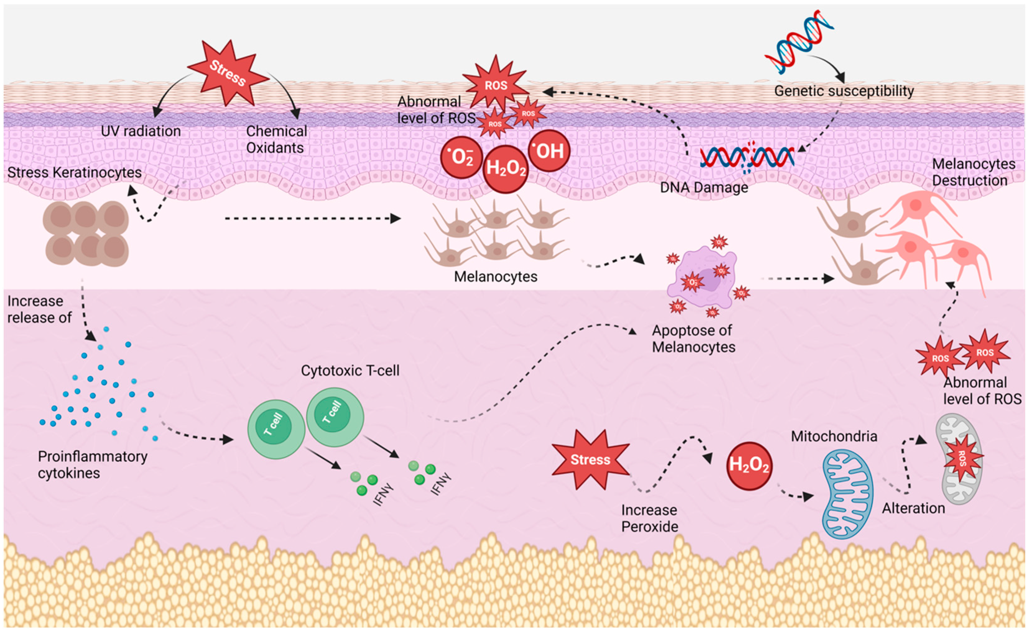
3.3. Oxidative Stress Theory

According to the oxidative stress theory, the intra-epidermal buildup of reactive oxygen species, the most well-known of which is hydrogen peroxide (H2O2), whose concentration can reach up to one millimole, is the primary factor in the pathogenesis of vitiligo. H2O2 causes alterations in the mitochondria at this concentration, which causes the melanocytes to apoptose and die. Patients with vitiligo frequently exhibit changes in redox status indicators (Figure 1). Malondialdehyde (MDA), selenium, vitamins C and E, glutathione peroxidase (GPx), superoxide dismutase (SOD), and catalase (CAT) are significant markers of interest. MDA is a by-product of lipid peroxidation and a sign of oxidative stress. Selenium, a key antioxidant found in erythrocytes, is necessary for GPx action. Superoxide radicals are neutralised by SOD, which lessens their toxicity, and are converted to oxygen (O2) and water (H2O) by CAT. Patients with vitiligo have significantly greater amounts of SOD, decreased erythrocyte GPx activity, low levels of the enzyme CAT, and low levels of the vitamins C and E in both their epidermis and serum [34,35,36].

3.4. Neural Theory

The neural theory postulates that nerve endings release neurochemical substances that can reduce melanin production or damage melanocytes. It also proposes that the pathogenesis of vitiligo is connected to the catalase gene. The peroxisome enzyme catalase is present in almost all living things. It stimulates the hydrolysis of hydrogen peroxide into water and oxygen, which protects cells from highly reactive oxygen radicals. Catalase enzyme activity is decreased in both lesional and nonlesional skin of vitiligo patients [33,38].

3.5. Biochemical Theory

The biochemical theory suggests that the accumulation of toxic intermediate metabolites of melanin synthesis and inadequate free radical defence lead to excessive amounts of hydrogen peroxide (H2O2), which is a cause of melanocyte destruction. Some theories include that the genetic factor, flaws in melanocyte structure and function, and a lack of melanocyte development factors all contribute to the depigmentation process [33,38,39]. The overall contribution of each of these mechanisms is still subject to debate, even though there is now general agreement that vitiligo is an autoimmune disease. None of these suggested hypotheses is sufficient to explain the many vitiligo phenotypes. However, the development of manifest diseases depends on environmental factors [40].

4. Classification

Vitiligo can appear clinically in three different ways (according to the evaluation carried out between 2011 and 2012 by the Vitiligo Global Issues Consensus Conference (1)) segmental, non-segmental, and mixed/unclassified (Table 1). In addition to clinical symptoms (the appearance of the first skin lesions, their location and extent, the coexistence of accompanying autoimmune disorders, and the natural progression of the dermatosis), these subtypes differ in their aetiologies as well [41]. Sites that are often hyperpigmented are usually affected by vitiligo, such as the face (periorificial), hands’ dorsal surfaces, nipples, axillae, umbilicus, sacrum, and inguinal/anogenital regions. It favours the elbows, knees, digits, and flexor wrists on the extremities [42].

4.1. Segmental Vitiligo

An acquired chronic pigmentation condition called segmental vitiligo is identified by white patches that have a unilateral distribution and may completely or partially resemble a dermatome. It has an earlier onset and is less prevalent than non-segmental vitiligo, and it quickly affects the follicular melanocyte reservoir, which causes hair bleaching [43]. The most frequently impacted area is the face, followed by the trunk and extremities. Leukotrichia is frequently observed and manifests early in the disease’s course. For a period of six months to two years, the disease progresses before stabilising without intervention. Even after years of stability, rare recurrences have been seen, manifesting as new lesions in the same segment or different body locations, leading to mixed vitiligo. It has a poor response to medical treatment when compared to other subtypes of the illness, which could be explained by the frequent occurrence of leukotrichia [17,33,39].

4.2. Non-Segmental Vitiligo

The most common type of vitiligo, which represents 80–90% of all cases, is non-segmental vitiligo (NSV). It is a chronic acquired pigmentation disorder marked by white patches, bilateral, frequently symmetrical, that enlarge over time and typically reflect a considerable loss of functioning melanocytes in the epidermis and some in the hair follicles. NSV includes focal, mucosal (when affecting more than one mucosal location), acrofacial, generalised, universal, mixed, and others. Several depigmented patches involving the oral, vaginal, and buccal mucosa are referred to as mucosal vitiligo. A single macule or patch without a segmental distribution that is stable over two years is referred to as focal vitiligo, which may be a precursor to generalised vitiligo. Patches that are largely confined to the face and distal extremities are the features of acrofacial vitiligo. Fingers and the perioral and periorbital regions of the face are both involved distinctively. Vitiligo that is distributed widely in scattered patches is known as generalised vitiligo. When more than 80% of the body’s surface is depigmented, the disorder is considered universal vitiligo [33,38,40].

4.3. Unclassified/Mixed Vitiligo

Unclassifiable forms or undetermined vitiligo include focal, for isolated white macules without segmental distribution, and mucosal, when only one mucosa is affected. Mixed vitiligo (MV) occurs when SV and NSV coexist. The association can be seen as an indication of a generalised polygenic disorder’s overlapped segmental expression, in which segmental involvement occurs before disease generalisation and is more resistant to treatment. Halo nevi and leukotrichia at the time of onset may be risk factors for the development of MV in SV patients. The loss of pigmentation surrounding the pre-existing nevus that creates a halo is known as a halo nevus (Sutton nevus). Many halo nevi are a sign of nested pigment-producing cell autoimmunity, which increases the risk of developing vitiligo [33,44,45].

5. Diagnosis and Assessment of Severity

The majority of diagnostic criteria are based on clinical findings of acquired, clearly defined white lesions on the skin that do not have any associated inflammation and tend to expand centrifugally. On a Wood’s light inspection, vitiligo lesions are more noticeable [42,46], which is a diagnostic procedure used to examine the skin or hair while it is exposed to black light that is generated by the Wood’s lamp.
VASI Index and VIDA Score
For the evaluation of disease severity and activation, the vitiligo area severity index (VASI) and vitiligo disease activity score (VIDA) can be used. The proportion of vitiligo involvement is determined in terms of hand units. One hand unit, which includes the palm and the volar surfaces of all the digits, is about equal to 1% of the surface area of the entire body. The product of the area of vitiligo in hand units and the degree of depigmentation within each hand unit-measured patch, yields the VASI for each area of the body. One hundred percent (total depigmentation), 90% of the pigment spots are present, 75% of the area is depigmented, 50% (of the area either pigmented/depigmented), 25% (more pigmented area than unpigmented area), and 10% (a few spots of depigmentation). The VIDA is a six-point scale used to rate the severity of vitiligo. The individual’s perception of the current illness activity over time is used to determine the score. Growing lesions as well as the development of new lesions are both symptoms of active vitiligo. An activity of +4 being fewer than six weeks, +3 activity of between six weeks and three months, +2 activity of between three and six months, activity of +1 during 6 to 12 months, 0 indicates stability for at least a year, while −1 indicates stability with spontaneous repigmentation for at least a year [44,47].

6. Vitiligo and Skin Cancer Connection

Numerous studies have shown that UV exposure is one of the main risk factors for the development of skin cancer (nonmelanoma skin carcinoma (NMSC)). Melanin serves as one of the defences for cellular DNA [48]. Melanoma is listed as the most frequent cancer among adults between the ages of 15 and 44 in Australia, where 2 in 3 people will have been diagnosed with it by the time they are 70 years old [49]. Darker skin provides an intrinsic UV protection factor for a lower incidence of cutaneous malignancies due to increased epidermal melanin [50]. In contrast to the smaller, less melanised melanosomes in the epidermis of white skin, the larger, more melanised melanosomes in the epidermis of darker skin absorb and scatter more light energy [51]. It is believed that skin with vitiligo is more susceptible to sun exposure and has a higher risk of developing skin cancer than pigmented skin since the melanin barrier is completely missing [48]. According to many studies, the genetic and immunological profiles of people with vitiligo may offer a significant degree of protection against melanoma and non-melanoma skin malignancies [52,53]. Tyrosinase (TYR) gene polymorphisms were discovered in vitiligo patients through genetic investigations. Tyrosinase, which is involved in the production of melanin, is expressed by this gene. The TYR mutation that increases the risk of vitiligo also reduces the likelihood of melanoma [54]. In contrast, patients with melanoma have been reported to have improved 5 year survival after developing vitiligo. Moreover, it has been noted that therapies used to treat metastatic melanoma can cause vitiligo, and if skin cancer does occur, it is typically seen in nonlesional locations [51]. Comprehensive analysis revealed that 5% of patients with stage III–IV melanoma who received immunotherapy developed vitiligo, suggesting that melanocytes are a target for the immune response [55]. Hypersensitivity to ultraviolet (UV) light causes vitiligo lesions to burn easily when exposed to the sun. It is common to observe the development of vitiligo following a severe sunburn. Sunblock use on vitiligous areas is crucial because it prevents sunburn, which can diminish photodamage as well as tanning of healthy skin, and may decrease the disparity with the affected lesions. In amelanotic areas where melanin pigment is absent, there is a higher risk of both sunburn and skin cancer [42,56,57].

7. Treatment Options for Vitiligo

Phototherapy and topical and oral immunomodulators such as corticosteroids and calcineurin inhibitors are common vitiligo therapies [58]. Psychosocial therapies, depigmentation therapy, non-traditional therapy, and surgical therapy are also some of the other therapeutic options for vitiligo [59]. Although the lesions are generally resistant to therapies, spontaneous repigmentation happens in more than 1–25% of cases [56,60].

7.1. Pharmacotherapy

7.1.1. Immunomodulators

Phototherapy and topical and oral immunomodulators such as corticosteroids and calcineurin inhibitors are common vitiligo therapies. Topical corticosteroids (TCS) of moderate to high potency and topical calcineurin inhibitors (TCI), both of which reduce the cellular immune response, are the first-line treatments for vitiligo [58]. In a randomised controlled trial the topical steroid combination (betamethasone) with a narrow-band UVB (NB-UVB) and topical calcipotriol therapy significantly increased repigmentation at six months compared to NB-UVB alone and NB-UVB with topical calcipotriol [61]. Topical steroids, such as mometasone 0.1% or clobetasol 0.05% daily, have been found in recent studies to be equally effective as calcineurin inhibitors, like tacrolimus 0.1% or pimecrolimus 1.0% daily, and to have manageable rates of adverse drug reactions [62,63]. Another area of concern for certain prescribers is the continuous use of topical tacrolimus. When tacrolimus was originally made accessible, its long-term safety was unknown, yet animal studies suggested that systemic exposure could lead to immune-mediated cancers [64]. Few studies investigated the possibility of administering 5-fluorouracil (5-FU), a cytotoxic chemotherapy drug used to cure cancer, intradermally to treat vitiligo after discovering the adverse effect of localised hyperpigmentation during cancer treatment [65]. They found that the combination of 5-FU with microneedling was more efficient than microneedles or tacrolimus alone [66,67]. Corticosteroids are typically administered orally in the form of a micropulse to treat vitiligo. In comparison with conventional corticosteroid treatment, an oral mini-pulse has far fewer negative effects and uses cyclical pulse doses or substantially lower levels of corticosteroids (megadoses) in order to increase effectiveness and reduce the need for long-term therapy [68]. To enable the adrenal glands to start producing cortisol again, systemic corticosteroids should be progressively cut back [69]. TCS was more successful than TCI in obtaining 50% repigmentation, although TCI was comparable to TCS in achieving 75% repigmentation [70]. The most common side effect of topical corticosteroids is skin atrophy, which manifests as tiny degenerative alterations within two weeks. These side effects can be reduced by using low-potency corticosteroids, reducing the dosage of high-potency corticosteroids, and stopping treatment when the patient has fully recovered. Corticosteroids should only be used for a maximum of three months, but because they must be used for longer periods for treating vitiligo, taking several weeks off from their use is advised [71,72]. Methotrexate and JAK inhibitors are two additional, uncommon immunosuppressants used to treat vitiligo. Although results were limited due to small sample sizes, low-dose oral methotrexate was reported to be comparable to oral corticosteroids and suggested when steroid drugs are contraindicated [73]. Janus kinase inhibitors (JAK) have been proven to decrease IFN-g signaling, which aids in repigmentation in vitiligo patients. Tofacitinib, ruxolitinib, and baricitinib are the three most commonly prescribed JAK inhibitors used to treat vitiligo [74].

7.1.2. Vitamin D Analogues

A small study examined the relationship between vitiligo and polymorphisms in the vitamin D receptor (VDR) gene, and they found that the Apa-I polymorphism of the VDR gene is related to vitiligo. Additionally, their findings show that families with vitiligo have higher rates of thyroidopathies, diabetes, and rheumatoid arthritis. This shows that the main processes of skin pigmentation may be influenced by vitamin D or its receptors [75,76]. Parsad et al. were the first to report the use of topical calcipotriol and psoralen-UVA (PUVA) as a combination for the treatment of vitiligo. Their research reveals that it is possible to shorten the therapy duration with PUVA in the treatment of vitiligo by combining it with calcipotriol since it is more effective and acts quicker [77]. Several studies have since been published on the use of vitamin D analogues alone or combined with corticosteroids or UV radiation to treat vitiligo by stimulating repigmentation [78]. Cholecalciferol and ergocalciferol stimulate melanogenesis by elevating the level of tyrosinase activity in a cultured cell system [79]. Following six days of vitamin D3 culture, human melanocytes released more immunoreactive tyrosinase [80]. In another study, it was found that vitamin D and UVB irradiation encouraged melanocyte development and accelerated the repigmentation rate, suggesting that the combination may work well together to treat vitiligo [61,81].
The first in vivo clinical trial, conducted by Oh et al. [82], examines the enhancement of repigmentation in non-segmental vitiligo (NSV) by combining high-concentration tacalcitol (HT) with a 308 nm xenon chloride excimer laser. They claim that using HT alone or in combination with excimer laser therapy has only a limited impact on the treatment of vitiligo. One study showed a synergistic effect of a low dosage of UVB light with the topical application of 100 μg of cholecalciferol to the pinnal epidermis of mice for 5 or 10 days, resulting in more melanocytes [83]. In placebo-controlled double-blind research, Ermis et al. found that using topical calcipotriol and PUVA together to treat vitiligo was safe and significantly more successful in developing and achieving complete repigmentation than using PUVA alone as a placebo [84]. In a randomised controlled trial in which each medication was administered either alone or in combination to patients with localised vitiligo, Kumaran et al. investigated the efficacy of the combination therapy of calcipotriol and betamethasone dipropionate. A total of 6.7% of patients in the calcipotriol, 13.3% in the betamethasone, and 26.7% in the combination groups, respectively, experienced substantial repigmentation (50–75%) [85]. This means that combination therapy is effective and accelerates repigmentation in all patients. Ibrahim et al. recently found that calcipotriol with betamethasone is more effective than its combination with tacrolimus in the treatment of vitiligo, although both of these are safe, affordable, and well-tolerated procedures with few side effects [86]. A fixed dose of calcipotriol 0.005% and betamethasone 0.05% on 30% of the body surface area is advised in many studies for the use of calcipotriol and corticosteroids to cure vitiligo, with a maximum application time of 4 weeks for the ointment and 8 weeks for the cream [69].

7.2. Phototherapy

UVB irradiation, with a wavelength of 280–320 nm, is more significant than UVA (with a wavelength of 320–400 nm) [15]. Today, narrow-band UVB is often used in combination with topical corticosteroids (TCS) or calcineurin inhibitors (TCI) to treat a variety of subtypes of vitiligo [13]. Vitiligo can be treated with PUVA and UVB (narrow-band UVB (NB-UVB), excimer laser, or lamp) phototherapy. The first successful phototherapy protocol was PUVA, but it has several drawbacks, including the inability to be used on children or pregnant women and phototoxic side effects like nausea, headache, dizziness, and skin cancer risk. Compared to PUVA, NB-UVB has shown higher treatment efficacy and is also linked to fewer, milder side effects [87]. Few studies examine the ideal length of phototherapy for vitiligo because of variable study designs, outcome measures, and a lack of prospective trials. According to Yones et al., NB-UVB treatment should last about 6 to 12 months to achieve 75% or more repigmentation [88].

7.3. Surgery

Surgery is only an option for vitiligo that is segmental or stable. Skin grafting and micropigmentation are the most common surgical procedures. Before performing the definitive graft on hypopigmented patches that have been stable for at least 2 years, it is strongly advised to perform a mini-grafting test in order to evaluate the patient’s positive response and the unfavourable occurrence of Koebner’s phenomenon at the donor site after 2–3 months of follow-up. The Koebner phenomenon at the donor site, keloids, hyperpigmentation, “cobblestoning”, scarring, and infections are some of the side effects of vitiligo surgery. In comparison to the control, suction blister, and combination split-thickness suction grafts, split-thickness suction grafting appears to be superior [89,90,91,92]. Hyaluronic acid is growing in popularity as it is frequently used in grafting procedures due to its improved biocompatibility [93]. In a double-blind trial, the administration of a hyaluronic acid-enriched cellular graft resulted in a repigmentation rate above 70% in the vitiligous areas in 77% of patients after 12 months versus placebo [94].

7.4. Cosmetic Camouflage and Aesthetic Therapy

Recently, for localised, stable vitiligo, particularly of the mucosal variety, tattoos or camouflage have been widely used. Many studies suggest that cosmetic concealment can enhance the quality of life for vitiligo patients. Additionally, patients may be assisted in choosing and utilising the camouflage preparation that best suits their needs. The appearance of patients is improved by camouflage, which could be permanent or temporary.

7.4.1. Temporary Camouflage

Liquid dyes, traditional remedies, foundation-based cosmetic camouflage, and self-tanning products are examples of temporary camouflage [95]. Sufferers frequently adopt various techniques of disguise; at least temporarily, corrective cosmetic cover creams can hide the vitiligo imperfection. A high concentration of pigment is added to water-free or anhydrous foundations to develop a colour that is similar to patients’ skin [96,97]. According to one study on Indian traditional remedies, Swarna Karani, a form of clay blended with oils and henna, works well as a disguise for vitiligo [98].
The average score for stigmatisation worldwide is 38%, and important factors of the Dermatological Life Quality Index (DLQI) include illness severity and disease extent. The DLQI is significantly impacted by vitiligo on the face, head, and neck, regardless of the severity of involvement. To assess the effectiveness of camouflage such as dihydroxyacetone (DHA) cream in hiding vitiligo on exposed Asian shins, research reported that the cosmetic effects of the 6% DHA cream are moderate to significantly satisfactory for 88.9% of vitiligo patients. Another study revealed that using a higher dosage of DHA in patients with darker skin allowed for colour matching [99]. Therefore, subjects with darker skin require more DHA cream than those with fairer skin. Moreover, camouflage not only hides the white areas but also enhances the overall well-being of vitiligo patients.
With one or more applications, makeup concealer like shade cream base, some fruit juices such as Roselle, henna, and dyes like potassium permanganate can also be used to cover vitiligo lesions. These covers produce an instant, natural colour that is easily removed by washing and can last for several weeks or even months [95,100].

7.4.2. Permanent Camouflage

With a cosmetic tattoo, mostly consisting of inert iron oxides that come in a variety of colours, one can achieve permanent camouflage. When only tiny sections of the body are treated, excellent results are still obtained since the shade is implanted into the dermal layer using specialised procedures and cannot be removed [101].

8. Conclusions

Vitiligo can affect anyone, regardless of gender, ethnicity, age, or skin colour. It usually first manifests before the third decade, although when it has an early onset in children, it could be related to a family history of the disease. Most vitiligo patients desire to hide their visible lesions by using clothing, camouflage, shade cream bases, and other methods that can help them improve their quality of life and social functioning. Since it results in cosmetic skin problems and is not contagious or threatening the patient’s life, the available treatment is sufficient. However, given the long duration of the disease, which could last months or even years, and the psychological problems associated with the condition, a substantial treatment that can help sufferers feel more comfortable and reduce social discrimination and stigmatisation is still needed.

Author Contributions

Conceptualization, K.A.-s.; writing—original draft preparation and draw the figure, M.I., V.R.L.-S. and Y.M.; writing—review and editing and supervision, Y.M. All authors have read and agreed to the published version of the manuscript.

Funding

This research received no external funding.

Institutional Review Board Statement

Not applicable.

Informed Consent Statement

Not applicable.

Data Availability Statement

Not applicable.

Conflicts of Interest

The authors declare no conflict of interest.

References

  1. Arthi, G.; Lakshmanakumar, V.; Rathinam, J.; Sivaraman, D. Molecular docking analysis to validate the efficacy of phytocompounds of traditional siddha herb Glycyrrhiza glabra against enzyme tyrosinase for the treatment of vitiligo. TMR Integr. Med. 2022, 6, e22032. [Google Scholar] [CrossRef]
  2. Prasad, P.V.; Bhatnagar, V.K. Medico-historical study of “Kilasa”(vitiligo/leucoderma) a common skin disorder. Bull. Indian Inst. Hist. Med. 2003, 33, 113–127. [Google Scholar]
  3. Czajkowski, R.; Męcińska-Jundziłł, K. Current aspects of vitiligo genetics. Adv. Dermatol. Allergol. Postępy Dermatol. I Alergol. 2014, 31, 247–255. [Google Scholar] [CrossRef] [PubMed]
  4. Medicine, S.P.C.C.J.o. Vitiligo. Clevel. Clin. J. Med. 2022. Available online: https://my.clevelandclinic.org/health/diseases/12419-vitiligo (accessed on 24 March 2023).
  5. Hartmann, A.; Lurz, C.; Hamm, H.; Bröcker, E.B.; Hofmann, U.B. Narrow-band UVB311 nm vs. broad-band UVB therapy in combination with topical calcipotriol vs. placebo in vitiligo. Int. J. Dermatol. 2005, 44, 736–742. [Google Scholar] [CrossRef]
  6. Mohr, N.; Petersen, J.; Kirsten, N.; Augustin, M. Epidemiology of Vitiligo–A Dual Population-Based Approach. Clin. Epidemiol. 2021, 13, 373–382. [Google Scholar] [CrossRef] [PubMed]
  7. Marinho, F.d.S.; Cirino, P.V.; Fernandes, N.C. Clinical epidemiological profile of vitiligo in children and adolescents. An. Bras. Dermatol. 2013, 88, 1026–1028. [Google Scholar] [CrossRef] [PubMed]
  8. Krüger, C.; Schallreuter, K.U. A review of the worldwide prevalence of vitiligo in children/adolescents and adults. Int. J. Dermatol. 2012, 51, 1206–1212. [Google Scholar] [CrossRef]
  9. Zhang, Y.; Cai, Y.; Shi, M.; Jiang, S.; Cui, S.; Wu, Y.; Gao, X.-H.; Chen, H.-D. The prevalence of vitiligo: A meta-analysis. PLoS ONE 2016, 11, e0163806. [Google Scholar] [CrossRef]
  10. Kumar, S.; Nayak, C.S.; Padhi, T.; Rao, G.; Rao, A.; Sharma, V.K.; Srinivas, C.R. Epidemiological pattern of psoriasis, vitiligo and atopic dermatitis in India: Hospital-based point prevalence. Indian Dermatol. Online J. 2014, 5, S6. [Google Scholar] [CrossRef]
  11. Ayanlowo, O.; Olumide, Y.M.; Akinkugbe, A.; Ahamneze, N.; Otike-Odibi, B.I.; Ekpudu, V.I.; Nnaji, T.; Akolawole, N.A. Characteristics of vitiligo in Lagos, Nigeria. West Afr. J. Med. 2009, 28, 118–121. [Google Scholar]
  12. Behl, P.N.; Bhatia, R.K. 400 cases of vitiligo. A clinico-therapeutic analysis. Indian J. Dermatol. 1972, 17, 51–56. [Google Scholar]
  13. Sehgal, V.; Srivastava, G. Vitiligo: Compendium of clinico-epidemiological features. Indian J. Dermatol. Venereol. Leprol. 2007, 73, 149–156. [Google Scholar] [CrossRef]
  14. Alkhateeb, A.; Fain, P.R.; Thody, A.; Bennett, D.C.; Spritz, R.A. Epidemiology of vitiligo and associated autoimmune diseases in Caucasian probands and their families. Pigment Cell Res. 2003, 16, 208–214. [Google Scholar] [CrossRef]
  15. Alikhan, A.; Felsten, L.M.; Daly, M.; Petronic-Rosic, V. Vitiligo: A comprehensive overview: Part I. Introduction, epidemiology, quality of life, diagnosis, differential diagnosis, associations, histopathology, etiology, and work-up. J. Am. Acad. Dermatol. 2011, 65, 473–491. [Google Scholar] [CrossRef]
  16. Nejad, S.B.; Qadim, H.H.; Nazeman, L.; Fadaii, R.; Goldust, M. Frequency of autoimmune diseases in those suffering from vitiligo in comparison with normal population. Pak. J. Biol. Sci. PJBS 2013, 16, 570–574. [Google Scholar] [CrossRef]
  17. Hann, S.K.; Lee, H.J. Segmental vitiligo: Clinical findings in 208 patients. J. Am. Acad. Dermatol. 1996, 35, 671–674. [Google Scholar] [CrossRef]
  18. Fitzpatrick, T.B. The validity and practicality of sun-reactive skin types I through VI. Arch. Dermatol. 1988, 124, 869–871. [Google Scholar] [CrossRef]
  19. Weinstock, M.A. Assessment of sun sensitivity by questionnaire: Validity of items and formulation of a prediction rule. J. Clin. Epidemiol. 1992, 45, 547–552. [Google Scholar] [CrossRef]
  20. Silverberg, J.I.; Silverberg, A.I.; Malka, E.; Silverberg, N.B. A pilot study assessing the role of 25 hydroxy vitamin D levels in patients with vitiligo vulgaris. J. Am. Acad. Dermatol. 2010, 62, 937–941. [Google Scholar] [CrossRef]
  21. Bibeau, K.; Pandya, A.G.; Ezzedine, K.; Jones, H.; Gao, J.; Lindley, A.; Harris, J.E. Vitiligo prevalence and quality of life among adults in Europe, Japan and the USA. J. Eur. Acad. Dermatol. Venereol. 2022, 36, 1831–1844. [Google Scholar] [CrossRef]
  22. Jayaprakasam, A.; Darvay, A.; Osborne, G.; McGibbon, D. Comparison of assessments of severity and quality of life in cutaneous disease. Clin. Exp. Dermatol. 2002, 27, 306–308. [Google Scholar] [CrossRef]
  23. Jowett, S.; Ryan, T. Skin disease and handicap: An analysis of the impact of skin conditions. Soc. Sci. Med. 1985, 20, 425–429. [Google Scholar] [CrossRef] [PubMed]
  24. Silverberg, J.I.; Silverberg, N.B. Association between vitiligo extent and distribution and quality-of-life impairment. JAMA Dermatol. 2013, 149, 159–164. [Google Scholar] [CrossRef] [PubMed]
  25. Morrison, B.; Burden-Teh, E.; Batchelor, J.M.; Mead, E.; Grindlay, D.; Ratib, S. Quality of life in people with vitiligo: A systematic review and meta-analysis. Br. J. Dermatol. 2017, 177, e338–e339. [Google Scholar] [CrossRef] [PubMed]
  26. Korkij, W.; Solatani, K.; Simjee, S.; Marcincin, P.G.; Chuang, T.Y. Tissue-specific autoantibodies and autoimmune disorders in vitiligo and alopecia areata: A retrospective study. J. Cutan. Pathol. 1984, 11, 522–530. [Google Scholar] [CrossRef]
  27. Hegedüs, L.; Heidenheim, M.; Gervil, M.; Hjalgrim, H.; Høier-Madsen, M. High frequency of thyroid dysfunction in patients with vitiligo. Acta Derm. Venereol. 1994, 74, 120–123. [Google Scholar]
  28. Daneshpazhooh, M.; Mostofizadeh, G.M.; Behjati, J.; Akhyani, M.; Robati, R.M. Anti-thyroid peroxidase antibody and vitiligo: A controlled study. BMC Dermatol. 2006, 6, 3. [Google Scholar] [CrossRef]
  29. Zhang, Z.; Xu, S.-X.; Zhang, F.-Y.; Yin, X.-Y.; Yang, S.; Xiao, F.-L.; Du, W.-H.; Wang, J.-F.; Lv, Y.-M.; Tang, H.-Y. The analysis of genetics and associated autoimmune diseases in Chinese vitiligo patients. Arch. Dermatol. Res. 2009, 301, 167–173. [Google Scholar] [CrossRef]
  30. Laberge, G.; Mailloux, C.M.; Gowan, K.; Holland, P.; Bennett, D.C.; Fain, P.R.; Spritz, R.A. Early disease onset and increased risk of other autoimmune diseases in familial generalized vitiligo. Pigment Cell Res. 2005, 18, 300–305. [Google Scholar] [CrossRef]
  31. Birlea, S.A.; Fain, P.R.; Spritz, R.A. A Romanian population isolate with high frequency of vitiligo and associated autoimmune diseases. Arch. Dermatol. 2008, 144, 310–316. [Google Scholar] [CrossRef]
  32. Jin, Y.; Mailloux, C.M.; Gowan, K.; Riccardi, S.L.; LaBerge, G.; Bennett, D.C.; Fain, P.R.; Spritz, R.A. NALP1 in vitiligo-associated multiple autoimmune disease. N. Engl. J. Med. 2007, 356, 1216–1225. [Google Scholar] [CrossRef]
  33. Ezzedine, K.; Lim, H.W.; Suzuki, T.; Katayama, I.; Hamzavi, I.; Lan, C.C.E.; Goh, B.K.; Anbar, T.; Silva de Castro, C.; Lee, A.Y. Revised classification/nomenclature of vitiligo and related issues: The Vitiligo Global Issues Consensus Conference. Pigment Cell Melanoma Res. 2012, 25, E1–E13. [Google Scholar] [CrossRef]
  34. Kemp, E.H.; Waterman, E.A.; Weetman, A.P. Autoimmune aspects of vitiligo. Autoimmunity 2001, 34, 65–77. [Google Scholar] [CrossRef]
  35. Dwivedi, M.; Kemp, E.H.; Laddha, N.C.; Mansuri, M.S.; Weetman, A.P.; Begum, R. Regulatory T cells in vitiligo: Implications for pathogenesis and therapeutics. Autoimmun. Rev. 2015, 14, 49–56. [Google Scholar] [CrossRef]
  36. Arora, A.K.; Kumaran, M.S. Pathogenesis of vitiligo: An update. Pigment Int. 2017, 4, 65. [Google Scholar]
  37. He, S.; Xu, J.; Wu, J. The promising role of chemokines in vitiligo: From oxidative stress to the autoimmune response. Oxidative Med. Cell. Longev. 2022, 2022, 8796735. [Google Scholar] [CrossRef]
  38. Ezzedine, K.; Le Thuaut, A.; Jouary, T.; Ballanger, F.; Taieb, A.; Bastuji-Garin, S. Latent class analysis of a series of 717 patients with vitiligo allows the identification of two clinical subtypes. Pigment Cell Melanoma Res. 2014, 27, 134–139. [Google Scholar] [CrossRef]
  39. Reghu, R.; James, E. Epidemiological profile and treatment pattern of vitiligo in a tertiary care teaching hospital. Children 2011, 2, 2–5. [Google Scholar]
  40. Boniface, K.; Seneschal, J.; Picardo, M.; Taïeb, A. Vitiligo: Focus on clinical aspects, immunopathogenesis, and therapy. Clin. Rev. Allergy Immunol. 2018, 54, 52–67. [Google Scholar] [CrossRef]
  41. Passeron, T.; Ortonne, J.-P. Physiopathology and genetics of vitiligo. J. Autoimmun. 2005, 25, 63–68. [Google Scholar] [CrossRef]
  42. Wolff, K.; Goldsmith, L.A.; Katz, S.I.; Gilchrest, B.A.; Paller, A.S.; Leffell, D.J. Fitzpatrick’s Dermatology in General Medicine; McGraw-Hill: New York, NY, USA, 2008. [Google Scholar]
  43. Picardo, M.; Dell’Anna, M.L.; Ezzedine, K.; Hamzavi, I.; Harris, J.E.; Parsad, D.; Taieb, A. Vitiligo (Primer). Nat. Rev. Dis. Prim. 2015, 1, 1–16. [Google Scholar] [CrossRef]
  44. Ezzedine, K.; Diallo, A.; Léauté-Labrèze, C.; Séneschal, J.; Prey, S.; Ballanger, F.; Alghamdi, K.; Cario-André, M.; Jouary, T.; Gauthier, Y. Halo naevi and leukotrichia are strong predictors of the passage to mixed vitiligo in a subgroup of segmental vitiligo. Br. J. Dermatol. 2012, 166, 539–544. [Google Scholar] [CrossRef] [PubMed]
  45. Ezzedine, K.; Gauthier, Y.; Léauté-Labrèze, C.; Marquez, S.; Bouchtnei, S.; Jouary, T.; Taieb, A. Segmental vitiligo associated with generalized vitiligo (mixed vitiligo): A retrospective case series of 19 patients. J. Am. Acad. Dermatol. 2011, 65, 965–971. [Google Scholar] [CrossRef] [PubMed]
  46. Lotti, T.; Gori, A.; Zanieri, F.; Colucci, R.; Moretti, S. Vitiligo: New and emerging treatments. Dermatol. Ther. 2008, 21, 110–117. [Google Scholar] [CrossRef]
  47. Lakhani, R.; Prakash, C.; Tiwari, S.; Purohit, S.; Paliwal, V.; Mathur, D.K.; Bhargava, P. Scoring system in dermatology: A review. IOSR 2016, 15, 89–99. [Google Scholar] [CrossRef]
  48. Oulehri, A.; Elloudi, S.; Baybay, H.; Douhi, Z.; Mernissi, F.Z. Vitiligo and skin cancer: What’s the link? Our Derm. Online 2020, 11, e144. [Google Scholar] [CrossRef]
  49. Staples, M.P.; Elwood, M.; Burton, R.C.; Williams, J.L.; Marks, R.; Giles, G.G. Non-melanoma skin cancer in Australia: The 2002 national survey and trends since 1985. Med. J. Aust. 2006, 184, 6–10. [Google Scholar] [CrossRef]
  50. Gloster, H.M., Jr.; Neal, K. Skin cancer in skin of color. J. Am. Acad. Dermatol. 2006, 55, 741–760. [Google Scholar] [CrossRef]
  51. Rodrigues, M. Skin cancer risk (nonmelanoma skin cancers/melanoma) in vitiligo patients. Dermatol. Clin. 2017, 35, 129–134. [Google Scholar] [CrossRef]
  52. Schallreuter, K.U.; Tobin, D.J.; Panske, A. Decreased photodamage and low incidence of non-melanoma skin cancer in 136 sun-exposed caucasian patients with vitiligo. Dermatology 2002, 204, 194–201. [Google Scholar] [CrossRef]
  53. Teulings, H.E.; Overkamp, M.; Ceylan, E.; Nieuweboer-Krobotova, L.; Bos, J.D.; Nijsten, T.; Wolkerstorfer, A.W.; Luiten, R.M.; Van der Veen, J.P.W. Decreased risk of melanoma and nonmelanoma skin cancer in patients with vitiligo: A survey among 1307 patients and their partners. Br. J. Dermatol. 2013, 168, 162–171. [Google Scholar] [CrossRef]
  54. Jin, Y.; Birlea, S.A.; Fain, P.R.; Gowan, K.; Riccardi, S.L.; Holland, P.J.; Mailloux, C.M.; Sufit, A.J.D.; Hutton, S.M.; Amadi-Myers, A. Variant of TYR and autoimmunity susceptibility loci in generalized vitiligo. New Engl. J. Med. 2010, 362, 1686–1697. [Google Scholar] [CrossRef]
  55. Teulings, H.-E.; Limpens, J.; Jansen, S.N.; Zwinderman, A.H.; Reitsma, J.B.; Spuls, P.I.; Luiten, R.M. Vitiligo-like depigmentation in patients with stage III-IV melanoma receiving immunotherapy and its association with survival: A systematic review and meta-analysis. J. Clin. Oncol. 2015, 33, 773–781. [Google Scholar] [CrossRef]
  56. James, W.D.; Berger, T.G.; Elston, D.M. Andrew’s Diseases of the Skin: Clinical Dermatol; Saunders Elsevier: Philadelphia, PA, USA, 2006; Volume 10. [Google Scholar]
  57. López, M. Treatment of skin disease. Comprehensive therapeutic strategies. Dermatol. Venez. 2006, 44, 76–1398. [Google Scholar]
  58. Garg, B.J.; Saraswat, A.; Bhatia, A.; Katare, O.P. Topical treatment in vitiligo and the potential uses of new drug delivery systems. Indian J. Dermatol. Venereol. Leprol. 2010, 76, 231. [Google Scholar]
  59. Saputra, M.A.R.; Purwoko, I.H.; Toruan, T.L. The Role of Topical Vitamin D in Vitiligo: A Narrative Literature Review. Biosci. Med. J. Biomed. Transl. Res. 2022, 6, 2516–2526. [Google Scholar]
  60. Jha, A.K.; Karki, S. Pigmentary disorders; vitiligo and melasma in context of south asian countries: A psychosocio-cosmetic challenge. Int. J. Dermatol. Clin. Res. 2015, 1, 24–30. [Google Scholar] [CrossRef]
  61. Akdeniz, N.; Yavuz, I.H.; Gunes Bilgili, S.; Ozaydın Yavuz, G.; Calka, O. Comparison of efficacy of narrow band UVB therapies with UVB alone, in combination with calcipotriol, and with betamethasone and calcipotriol in vitiligo. J. Dermatol. Treat. 2014, 25, 196–199. [Google Scholar] [CrossRef]
  62. Köse, O.; Arca, E.; Kurumlu, Z. Mometasone cream versus pimecrolimus cream for the treatment of childhood localized vitiligo. J. Dermatol. Treat. 2010, 21, 133–139. [Google Scholar] [CrossRef]
  63. Ho, N.; Pope, E.; Weinstein, M.; Greenberg, S.; Webster, C.; Krafchik, B. A double-blind, randomized, placebo-controlled trial of topical tacrolimus 0.1% vs. clobetasol propionate 0.05% in childhood vitiligo. Br. J. Dermatol. 2011, 165, 626–632. [Google Scholar] [CrossRef]
  64. Carr, W.W. Topical calcineurin inhibitors for atopic dermatitis: Review and treatment recommendations. Pediatr. Drugs 2013, 15, 303–310. [Google Scholar] [CrossRef] [PubMed]
  65. Mohamed, H.A.; Mohammed, G.F.; Gomaa, A.H.A.; Eyada, M.M.K. Carbon dioxide laser plus topical 5-fluorouracil: A new combination therapeutic modality for acral vitiligo. J. Cosmet. Laser Ther. 2015, 17, 216–223. [Google Scholar] [CrossRef] [PubMed]
  66. Attwa, E.M.; Khashaba, S.A.; Ezzat, N.A. Evaluation of the additional effect of topical 5-fluorouracil to needling in the treatment of localized vitiligo. J. Cosmet. Dermatol. 2020, 19, 1473–1478. [Google Scholar] [CrossRef] [PubMed]
  67. Pazyar, N.; Hatami, M.; Yaghoobi, R.; Parvar, S.Y.; Radmanesh, M.; Hadibarhaghtalab, M. The efficacy of adding topical 5-fluorouracil to micro-needling in the treatment of vitiligo: A randomized controlled trial. J. Cosmet. Dermatol. 2023, 22, 1513–1520. [Google Scholar] [CrossRef]
  68. Pasricha, J.S.; Khaitan, B.K. Oral mini-pulse therapy with betamethasone in vitiligo patients having extensive or fast-spreading disease. Int. J. Dermatol. 1993, 32, 753–757. [Google Scholar] [CrossRef]
  69. Lotti, T.; Agarwal, K.; Podder, I.; Satolli, F.; Kassir, M.; Schwartz, R.A.; Wollina, U.; Grabbe, S.; Navarini, A.A.; Mueller, S.M. Safety of the current drug treatments for vitiligo. Expert Opin. Drug Saf. 2020, 19, 499–511. [Google Scholar] [CrossRef]
  70. Chang, H.-C.; Hsu, Y.-P.; Huang, Y.-C. The effectiveness of topical calcineurin inhibitors compared with topical corticosteroids in the treatment of vitiligo: A systematic review and meta-analysis. J. Am. Acad. Dermatol. 2020, 82, 243–245. [Google Scholar] [CrossRef]
  71. Dillon, A.B.; Sideris, A.; Hadi, A.; Elbuluk, N. Advances in vitiligo: An update on medical and surgical treatments. J. Clin. Aesthetic Dermatol. 2017, 10, 15. [Google Scholar]
  72. Daniel, B.S.; Wittal, R. Vitiligo treatment update. Australas. J. Dermatol. 2015, 56, 85–92. [Google Scholar] [CrossRef]
  73. Singh, H.; Kumaran, M.S.; Bains, A.; Parsad, D. A randomized comparative study of oral corticosteroid minipulse and low-dose oral methotrexate in the treatment of unstable vitiligo. Dermatology 2015, 231, 286–290. [Google Scholar] [CrossRef]
  74. Qi, F.; Liu, F.; Gao, L. Janus kinase inhibitors in the treatment of vitiligo: A review. Front. Immunol. 2021, 12, 790125. [Google Scholar] [CrossRef]
  75. Birlea, S.; Birlea, M.; Cimponeriu, D.; Apostol, P.; Cosgarea, R.; Gavrila, L.; Tigan, S.; Costin, G.; Das, P. Autoimmune diseases and vitamin D receptor Apa-I polymorphism are associated with vitiligo in a small inbred Romanian community. Acta Derm. Venereol. 2006, 86, 209–214. [Google Scholar] [CrossRef]
  76. Imran, M.; Jin, X.; Ali, M.; Tapfumaneyi, P.; Lelasseur, P.; Carlo, L.; Jude, A.; Bourg, A.L.; Panchal, B.; Dick, A. The Pandemic and Your Skin—Direct and Indirect Impact of COVID-19. Cosmetics 2023, 10, 34. [Google Scholar] [CrossRef]
  77. Parsad, D.; Saini, R.; Verma, N. Combination of PUVAsol and topical calcipotriol in vitiligo. Dermatology 1998, 197, 167–170. [Google Scholar] [CrossRef]
  78. Parsad, D.; Kanwar, A. Topical vitamin D3 analogues in the treatment of vitiligo. Pigment Cell Melanoma Res. 2009, 22, 487–488. [Google Scholar] [CrossRef]
  79. Oikawa, A.; Nakayasu, M. Stimulation of melanogenesis in cultured melanoma cells by calciferols. FEBS Lett. 1974, 42, 32–35. [Google Scholar] [CrossRef]
  80. Tomita, Y.; Torinuki, W.; Tagami, H. Stimulation of human melanocytes by vitamin D 3 possibly mediates skin pigmentation after sun exposure. J. Investig. Dermatol. 1988, 90, 882–884. [Google Scholar] [CrossRef]
  81. Xu, Q.; Du, J.; He, P.; Zhang, J.; Zhu, T. Effects of 1alpha, 25-dihydroxyvitamin D (3) and UVB on cell proliferation and melanin synthesis of cultured human melanocyte. Beijing Da Xue Xue Bao Yi Xue Ban J. Peking Univ. Health Sci. 2004, 36, 483–486. [Google Scholar]
  82. Oh, S.H.; Kim, T.; Jee, H.; Do, J.E.; Lee, J.H. Combination treatment of non-segmental vitiligo with a 308-nm xenon chloride excimer laser and topical high-concentration tacalcitol: A prospective, single-blinded, paired, comparative study. J. Am. Acad. Derm. 2011, 65, 428–430. [Google Scholar] [CrossRef]
  83. Abdel-Malek, Z.A.; Ross, R.; Trinkle, L.; Swope, V.; Pike, J.W.; Nordlund, J.J. Hormonal effects of vitamin D3 on epidermal melanocytes. J. Cell. Physiol. 1988, 136, 273–280. [Google Scholar] [CrossRef]
  84. Ermis, O.; Alpsoy, E.; Cetin, L.; Yilmaz, E. Is the efficacy of psoralen plus ultraviolet A therapy for vitiligo enhanced by concurrent topical calcipotriol? A placebo-controlled double-blind study. Br. J. Dermatol. 2001, 145, 472–475. [Google Scholar] [CrossRef] [PubMed]
  85. Kumaran, M.; Kaur, I.; Kumar, B. Effect of topical calcipotriol, betamethasone dipropionate and their combination in the treatment of localized vitiligo. J. Eur. Acad. Dermatol. Venereol. 2006, 20, 269–273. [Google Scholar] [CrossRef] [PubMed]
  86. Ibrahim, Z.A.; Hassan, G.F.; Elgendy, H.Y.; Al-shenawy, H.A. Evaluation of the efficacy of transdermal drug delivery of calcipotriol plus betamethasone versus tacrolimus in the treatment of vitiligo. J. Cosmet. Dermatol. 2019, 18, 581–588. [Google Scholar] [CrossRef] [PubMed]
  87. Bouceiro Mendes, R.; Alpalhão, M.; Filipe, P. UVB phototherapy in the treatment of vitiligo: State of the art and clinical perspectives. Photodermatol. Photoimmunol. Photomed. 2022, 38, 215–223. [Google Scholar] [CrossRef] [PubMed]
  88. Yones, S.S.; Palmer, R.A.; Garibaldinos, T.M.; Hawk, J.L. Randomized double-blind trial of treatment of vitiligo: Efficacy of psoralen–UV-A therapy vs narrowband–UV-B therapy. Arch. Dermatol. 2007, 143, 578–584. [Google Scholar] [CrossRef]
  89. Lotti, T.; Berti, S.; Moretti, S. Vitiligo therapy. Expert Opin. Pharmacother. 2009, 10, 2779–2785. [Google Scholar] [CrossRef]
  90. Savin, J.A. The hidden face of dermatology. Clin. Exp. Dermatol. 1993, 18, 393–395. [Google Scholar] [CrossRef]
  91. Mulekar, S.V.; Isedeh, P. Surgical interventions for vitiligo: An evidence-based review. Br. J. Dermatol. 2013, 169, 57–66. [Google Scholar] [CrossRef]
  92. Anbar, T.S.; Hegazy, R.A.; Picardo, M.; Taieb, A. Beyond vitiligo guidelines: Combined stratified/personalized approaches for the vitiligo patient. Exp. Dermatol. 2014, 23, 219–223. [Google Scholar] [CrossRef]
  93. Tapfumaneyi, P.; Imran, M.; Mohammed, Y.H.; Roberts, M. Recent advances and future prospective of topical and transdermal delivery systems. Front. Drug Deliv. 2022, 25. [Google Scholar] [CrossRef]
  94. Van Geel, N.; Ongenae, K.; De Mil, M.; Vander Haeghen, Y.; Vervaet, C.; Naeyaert, J.M. Double-blind placebo-controlled study of autologous transplanted epidermal cell suspensions for repigmenting vitiligo. Arch. Dermatol. 2004, 140, 1203–1208. [Google Scholar] [CrossRef]
  95. Sarveswari, K.N. Cosmetic camouflage in vitiligo. Indian J. Dermatol. 2010, 55, 211. [Google Scholar] [CrossRef]
  96. Tanioka, M.; Yamamoto, Y.; Kato, M.; Miyachi, Y. Camouflage for patients with vitiligo vulgaris improved their quality of life. J. Cosmet. Dermatol. 2010, 9, 72–75. [Google Scholar] [CrossRef]
  97. Kaliyadan, F.; Kumar, A. Camouflage for patients with vitiligo. Indian J. Dermatol. Venereol. Leprol. 2012, 78, 8. [Google Scholar] [CrossRef]
  98. Sugathan, P.; Nejeeba, R. A new comouflage for vitiligo. Indian J. Dermatol. Venereol. Leprol. 1991, 57, 45. [Google Scholar]
  99. Rajatanavin, N.; Suwanachote, S.; Kulkollakarn, S. Dihydroxyacetone: A safe camouflaging option in vitiligo. Int. J. Dermatol. 2008, 47, 402–406. [Google Scholar] [CrossRef]
  100. Yoon, J.; Sun, Y.-W.; Kim, T.-H. Complementary and alternative medicine for vitiligo. In Vitiligo: Manage Therapy; BoD–Books on Demand: Kyungnam, Republic of Korea, 2011; pp. 143–158. [Google Scholar]
  101. Holme, S.A.; Beattie, P.E.; Fleming, C.J. Cosmetic camouflage advice improves quality of life. Br. J. Dermatol. 2002, 147, 946–949. [Google Scholar] [CrossRef]
Figure 1. The effects of oxidative stress and genetic susceptibility in vitiligo. Oxidative stress plays a key role in initiating the onset of vitiligo with melanocyte damage. It induces a high level of H2O2, which alters the mitochondria and causes melanocyte apoptosis. Additionally, it can trigger DNA damage, and increase the production of proinflammatory cytokines and cytotoxic T cells. Additionally, an increase in the production of reactive oxygen species (ROS), which is stimulated by exogenous factors like ultraviolet irradiation and endogenous factors such as abnormal energy metabolism in mitochondria, leads to further dysfunction of molecules and organelles, a subsequent immune response, and ultimately the death of melanocytes. This figure is modified from Shan et al. [37].
Cosmetics 10 00084 g001
Table 1. Classification of vitiligo.
Types of VitiligoSubtypes
SegmentalBisegmental/Unisegemntal/Plurisegmental
Non-segmentalGeneralised/Acrofacial/Universal/Mucosal/Mixed
Unclassified/UndeterminedFocal/Mucosal (one site)
Disclaimer/Publisher’s Note: The statements, opinions and data contained in all publications are solely those of the individual author(s) and contributor(s) and not of MDPI and/or the editor(s). MDPI and/or the editor(s) disclaim responsibility for any injury to people or property resulting from any ideas, methods, instructions or products referred to in the content.

