Next Issue
Volume 8, September
Previous Issue
Volume 8, March


Biology, Volume 8, Issue 2 (June 2019) – 31 articles

Cover Story (view full-size image): 
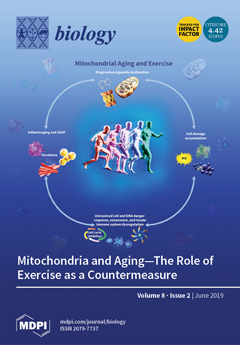
Ever since ancient times, humans have been fascinated by the roots of aging and the fountain of youth. Unifying features of the aging process, lysosomal storage diseases and neurodegenerative conditions include the cumulative buildup of oxidative damage, protein aggregates, and toxic waste. Organelles that regulate reactive oxygen species (ROS) production, quality control/repair, and recycling comprise the major defense systems against aging. Arguably, mitochondria are ‘the hubs of aerobic life’ given their role in eukaryotic evolution and the rise of complex life. Mitochondria are related to biological aging by regulating energy production, ROS generation, Ca2+ handling, inflammation, and apoptosis. Exercise therapeutics rejuvenates mitochondria and decelerates the ‘vicious cycle’ of cell aging, thereby extending mammalian life- and health- span. View this paper
  • Issues are regarded as officially published after their release is announced to the table of contents alert mailing list.
  • You may sign up for e-mail alerts to receive table of contents of newly released issues.
  • PDF is the official format for papers published in both, html and pdf forms. To view the papers in pdf format, click on the "PDF Full-text" link, and use the free Adobe Reader to open them.
Order results
Result details
Section
Select all
Export citation of selected articles as:
11 pages, 5137 KiB  
Article
Intergeneric Hybrid from Jatropha curcas L. and Ricinus communis L.: Characterization and Polyploid Induction
by Duangporn Premjet, Abraham Kusi Obeng, Anupan Kongbangkerd and Siripong Premjet
Biology 2019, 8(2), 50; https://doi.org/10.3390/biology8020050 - 22 Jun 2019
Cited by 10 | Viewed by 8686
Abstract
Jatropha curcas L. (2n = 2× = 22) is increasingly attracting attention in the biodiesel industry for its oil. However, the cultivation of J. curcas L. is faced with numerous challenges unlike the cultivation of Ricinus communis L. (2n = 2× = 20), [...] Read more.
Jatropha curcas L. (2n = 2× = 22) is increasingly attracting attention in the biodiesel industry for its oil. However, the cultivation of J. curcas L. is faced with numerous challenges unlike the cultivation of Ricinus communis L. (2n = 2× = 20), a closely related species. The generation of an intergeneric hybrid between J. curcas L. and R. communis L. was investigated. Intergeneric hybrids were produced by hand crossing. Immature embryos were rescued, in vitro, from the hybrid seeds and cultured on an enriched Murashige and Skoog (MS) medium for a month. The plantlets produced were grown in sterile peat moss in plastic pots and covered with polyethylene for 30 days, after which they were transferred into cement potted soil. The hybridity and the genuineness of the hybrids were successfully confirmed using randomly amplified polymorphic DNA (RAPD) markers. The number of branches, stem diameter, and leaf size of the F1 hybrids were similar to those of J. curcas L. while the plant height was similar to that of R. communis L. Young hybrids were treated with various concentrations (0%, 0.3%, 0.4%, and 0.5%) of colchicine to induce polyploids. The calli (JR6) treated with 0.3% colchicine recorded the highest tetraploid cell percentage (38.89%). A high tetraploid cell percentage (>50%) is significant in overcoming the problem of sterility after hybridization. Full article
Show Figures

Figure 1

18 pages, 973 KiB  
Article
Affinity Isolation and Mass Spectrometry Identification of Prostacyclin Synthase (PTGIS) Subinteractome
by Pavel V. Ershov, Yuri V. Mezentsev, Arthur T. Kopylov, Evgeniy O. Yablokov, Andrey V. Svirid, Aliaksandr Ya. Lushchyk, Leonid A. Kaluzhskiy, Andrei A. Gilep, Sergey A. Usanov, Alexey E. Medvedev and Alexis S. Ivanov
Biology 2019, 8(2), 49; https://doi.org/10.3390/biology8020049 - 20 Jun 2019
Cited by 9 | Viewed by 4048
Abstract
Prostacyclin synthase (PTGIS; EC 5.3.99.4) catalyzes isomerization of prostaglandin H2 to prostacyclin, a potent vasodilator and inhibitor of platelet aggregation. At present, limited data exist on functional coupling and possible ways of regulating PTGIS due to insufficient information about protein–protein interactions in [...] Read more.
Prostacyclin synthase (PTGIS; EC 5.3.99.4) catalyzes isomerization of prostaglandin H2 to prostacyclin, a potent vasodilator and inhibitor of platelet aggregation. At present, limited data exist on functional coupling and possible ways of regulating PTGIS due to insufficient information about protein–protein interactions in which this crucial enzyme is involved. The aim of this study is to isolate protein partners for PTGIS from rat tissue lysates. Using CNBr-activated Sepharose 4B with covalently immobilized PTGIS as an affinity sorbent, we confidently identified 58 unique proteins by mass spectrometry (LC-MS/MS). The participation of these proteins in lysate complex formation was characterized by SEC lysate profiling. Several potential members of the PTGIS subinteractome have been validated by surface plasmon resonance (SPR) analysis. SPR revealed that PTGIS interacted with full-length cytochrome P450 2J2 and glutathione S-transferase (GST). In addition, PTGIS was shown to bind synthetic peptides corresponding to sequences of for GSTA1, GSTM1, aldo-keto reductase (AKR1A1), glutaredoxin 3 (GLRX3) and histidine triad nucleotide binding protein 2 (HINT2). Prostacyclin synthase could potentially be involved in functional interactions with identified novel protein partners participating in iron and heme metabolism, oxidative stress, xenobiotic and drugs metabolism, glutathione and prostaglandin metabolism. The possible biological role of the recognized interaction is discussed in the context of PTGIS functioning. Full article
(This article belongs to the Section Biochemistry and Molecular Biology)
Show Figures

Figure 1

5 pages, 169 KiB  
Editorial
Mitochondrial Dysfunction in Aging and Diseases of Aging
by Richard H. Haas
Biology 2019, 8(2), 48; https://doi.org/10.3390/biology8020048 - 17 Jun 2019
Cited by 98 | Viewed by 11212
Abstract
Mitochondria have been increasingly recognized as the important players in the aging process [...] Full article
(This article belongs to the Special Issue Mitochondrial Dysfunction in Aging and Diseases of Aging)
15 pages, 2804 KiB  
Article
Identification and Characterization of microRNAs and Their Predicted Functions in Biomineralization in the Pearl Oyster (Pinctada fucata)
by Songqian Huang, Yuki Ichikawa, Kazutoshi Yoshitake, Shigeharu Kinoshita, Yoji Igarashi, Fumito Omori, Kaoru Maeyama, Kiyohito Nagai, Shugo Watabe and Shuichi Asakawa
Biology 2019, 8(2), 47; https://doi.org/10.3390/biology8020047 - 17 Jun 2019
Cited by 12 | Viewed by 3833
Abstract
The biological process of pearl formation is an ongoing research topic, and a number of genes associated with this process have been identified. However, the involvement of microRNAs (miRNAs) in biomineralization in the pearl oyster, Pinctada fucata, is not well understood. In [...] Read more.
The biological process of pearl formation is an ongoing research topic, and a number of genes associated with this process have been identified. However, the involvement of microRNAs (miRNAs) in biomineralization in the pearl oyster, Pinctada fucata, is not well understood. In order to investigate the divergence and function of miRNAs in P. fucata, we performed a transcriptome analysis of small RNA libraries prepared from adductor muscle, gill, ovary, and mantle tissues. We identified 186 known and 42 novel miRNAs in these tissues. Clustering analysis showed that the expression patterns of miRNAs were similar among the somatic tissues, but they differed significantly between the somatic and ovary tissues. To validate the existence of the identified miRNAs, nine known and three novel miRNAs were verified by stem-loop qRT-PCR using U6 snRNA as an internal reference. The expression abundance and target prediction between miRNAs and biomineralization-related genes indicated that miR-1990c-3p, miR-876, miR-9a-3p, and novel-3 may be key factors in the regulatory network that act by controlling the formation of matrix proteins or the differentiation of mineralogenic cells during shell formation in mantle tissue. Our findings serve to further clarify the processes underlying biomineralization in P. fucata. Full article
(This article belongs to the Section Marine Biology)
Show Figures

Figure 1

14 pages, 1742 KiB  
Article
Plasticity in Standard and Maximum Aerobic Metabolic Rates in Two Populations of an Estuarine Dependent Teleost, Spotted Seatrout (Cynoscion nebulosus)
by Jingwei Song, Richard W. Brill and Jan R. McDowell
Biology 2019, 8(2), 46; https://doi.org/10.3390/biology8020046 - 14 Jun 2019
Cited by 7 | Viewed by 4377
Abstract
We studied the effects of metabolic cold adaptation (MCA) in two populations of a eurythermal species, spotted seatrout (Cynoscion nebulosus) along the U.S. East Coast. Fish were captured from their natural environment and acclimated at control temperatures 15 °C or 20 [...] Read more.
We studied the effects of metabolic cold adaptation (MCA) in two populations of a eurythermal species, spotted seatrout (Cynoscion nebulosus) along the U.S. East Coast. Fish were captured from their natural environment and acclimated at control temperatures 15 °C or 20 °C. Their oxygen consumption rates, a proxy for metabolic rates, were measured using intermittent flow respirometry during acute temperature decrease or increase (2.5 °C per hour). Mass-specific standard metabolic rates (SMR) were higher in fish from the northern population across an ecologically relevant temperature gradient (5 °C to 30 °C). SMR were up to 37% higher in the northern population at 25 °C and maximum metabolic rates (MMR) were up to 20% higher at 20 °C. We found evidence of active metabolic compensation in the southern population from 5 °C to 15 °C (Q10 < 2), but not in the northern population. Taken together, our results indicate differences in metabolic plasticity between the northern and southern populations of spotted seatrout and provide a mechanistic basis for predicting population-specific responses to climate change. Full article
(This article belongs to the Special Issue Fish Metabolic Physiology in Response to Stress)
Show Figures

Figure 1

9 pages, 1132 KiB  
Article
Estrogenic Compounds or Adiponectin Inhibit Cyclic AMP Response to Human Luteinizing Hormone in Mouse Leydig Tumor Cells
by Thi Mong Diep Nguyen, Danièle Klett and Yves Combarnous
Biology 2019, 8(2), 45; https://doi.org/10.3390/biology8020045 - 11 Jun 2019
Cited by 4 | Viewed by 3620
Abstract
Mouse Leydig Tumor cells (mLTC), transiently expressing cAMP-dependent luciferase, were used to study the influence of sexual steroids and of adiponectin (ADPN) on the cAMP response to luteinizing hormones (LH). While testosterone and progesterone had no significant effect, several molecules with estrogenic activity [...] Read more.
Mouse Leydig Tumor cells (mLTC), transiently expressing cAMP-dependent luciferase, were used to study the influence of sexual steroids and of adiponectin (ADPN) on the cAMP response to luteinizing hormones (LH). While testosterone and progesterone had no significant effect, several molecules with estrogenic activity (17β-estradiol, ethynylestradiol, and bisphenol A) provoked a decrease in intracellular cyclic AMP accumulation under 0.7 nM human LH stimulation. Adiponectin exhibited a bimodal dose-effect on LH response: synergistic between 2–125 ng/mL and inhibitory between 0.5–5 µg/mL. In brief, our data indicate that estrogens and ADPN separately exert rapid (<1 h) inhibitory and/or synergistic effects on cAMP response to LH in mLTC-1 cells. As the inhibitory effect of each estrogenic molecule was observed after only 1-h preincubation, it might be mediated through the G protein-coupled estrogen receptor (GPER) membrane receptor, but this remains to be demonstrated. The synergistic effect with low concentrations of ADPN with human Luteinizing Hormone (hLH) was observed with both fresh and frozen/thawed ADPN. In contrast, the inhibitory effect with high concentrations of ADPN was lost with frozen/thawed ADPN, suggesting deterioration of its polymeric structure. Full article
(This article belongs to the Section Cell Biology)
Show Figures

Figure 1

8 pages, 1330 KiB  
Article
N-acetyl-L-cysteine Prevents Lactate-Mediated PGC1-alpha Expression in C2C12 Myotubes
by Minas Nalbandian, Zsolt Radak and Masaki Takeda
Biology 2019, 8(2), 44; https://doi.org/10.3390/biology8020044 - 10 Jun 2019
Cited by 10 | Viewed by 4169
Abstract
Background: Exercise induces many physiological adaptations. Recently, it has been proposed that some of these adaptations are induced by exercise-mediated lactate production. In this study, we aimed to investigate in vitro the effect of lactate in cultured myotubes and whether antioxidants could inhibit [...] Read more.
Background: Exercise induces many physiological adaptations. Recently, it has been proposed that some of these adaptations are induced by exercise-mediated lactate production. In this study, we aimed to investigate in vitro the effect of lactate in cultured myotubes and whether antioxidants could inhibit the effect. Methods: Differentiated myotubes were cultured at different concentrations of L-lactate (0, 10, 30, 50 mM) in the absence or presence of an antioxidant, N-acetyl-L-cysteine (Nac). The temporal effect of lactate exposure in myotubes was also explored. Results: Two hours of exposure to 50 mM L-lactate and six hours of exposure to 30 or 50 mM L-lactate caused a significant increase in PGC1-alpha (peroxisome proliferator-activated receptor γ coactivator-1α) expression in the myotubes. This up-regulation was suppressed by 2 mM Nac. Intermittent and continuous lactate exposure caused similar PGC1-alpha up-regulation. These results suggest that the increase in PGC1-alpha expression is mediated by reactive oxygen species (ROS) production from lactate metabolism and that both continuous and intermittent exposure to L-lactate can cause the up-regulation. Full article
(This article belongs to the Section Cell Biology)
Show Figures

Figure 1

14 pages, 2175 KiB  
Opinion
Amyloidosis and Longevity: A Lesson from Plants
by Andrei Surguchov, Fatemeh Nouri Emamzadeh and Alexei A. Surguchev
Biology 2019, 8(2), 43; https://doi.org/10.3390/biology8020043 - 24 May 2019
Cited by 11 | Viewed by 5128
Abstract
The variety of lifespans of different organisms in nature is amazing. Although it is acknowledged that the longevity is determined by a complex interaction between hereditary and environmental factors, many questions about factors defining lifespan remain open. One of them concerns a wide [...] Read more.
The variety of lifespans of different organisms in nature is amazing. Although it is acknowledged that the longevity is determined by a complex interaction between hereditary and environmental factors, many questions about factors defining lifespan remain open. One of them concerns a wide range of lifespans of different organisms. The reason for the longevity of certain trees, which reaches a thousand years and exceeds the lifespan of most long living vertebrates by a huge margin is also not completely understood. Here we have discussed some distinguishing characteristics of plants, which may explain their remarkable longevity. Among them are the absence (or very low abundance) of intracellular inclusions composed of amyloidogenic proteins, the lack of certain groups of proteins prone to aggregate and form amyloids in animals, and the high level of compounds which inhibit protein aggregation and possess antiaging properties. Full article
(This article belongs to the Section Biochemistry and Molecular Biology)
Show Figures

Figure 1

19 pages, 3843 KiB  
Article
Which Species Should We Focus On? Umbrella Species Assessment in Southwest China
by Xuewei Shi, Cheng Gong, Lu Zhang, Jian Hu, Zhiyun Ouyang and Yi Xiao
Biology 2019, 8(2), 42; https://doi.org/10.3390/biology8020042 - 23 May 2019
Cited by 10 | Viewed by 4501
Abstract
In conservation biology, umbrella species are often used as agents for a broader set of species, or as representatives of an ecosystem, and their conservation is expected to benefit a large number of naturally co-occurring species. Southwest China is home to not only [...] Read more.
In conservation biology, umbrella species are often used as agents for a broader set of species, or as representatives of an ecosystem, and their conservation is expected to benefit a large number of naturally co-occurring species. Southwest China is home to not only global biodiversity hotspots, but also rapid economic and population growth and extensive changes in land use. However, because of the large regional span, the diverse species distributions, and the difficulty of field investigations, traditional methods used to assess umbrella species are not suitable for implementation in Southwest China. In the current study, we assessed 810 key protected species from seven taxa by indicator value analysis, correlation analysis, and factor analysis. We selected 32 species as umbrella species, whose habitats overlapped the habitats of 97% of the total species. Furthermore, the selected species were significantly correlated with 70% of all species in the study area. A total of 16 out of 19 selected animal species have been previously mentioned as umbrella species, compared with only 3 out of 13 plants species; this is despite plants accounting for a large proportion of the total species in Southwest China. We discuss the roles of indicator species and co-occurring species, and provide suggestions for species protection in Southwest China based on the current results. Our research provides valuable scientific information for research on umbrella conservation species over large geographical scales, and related fields of biodiversity conservation. Full article
(This article belongs to the Section Conservation Biology and Biodiversity)
Show Figures

Figure 1

19 pages, 2431 KiB  
Review
Are There Circadian Clocks in Non-Photosynthetic Bacteria?
by Francesca Sartor, Zheng Eelderink-Chen, Ben Aronson, Jasper Bosman, Lauren E. Hibbert, Antony N. Dodd, Ákos T. Kovács and Martha Merrow
Biology 2019, 8(2), 41; https://doi.org/10.3390/biology8020041 - 22 May 2019
Cited by 30 | Viewed by 8887
Abstract
Circadian clocks in plants, animals, fungi, and in photosynthetic bacteria have been well-described. Observations of circadian rhythms in non-photosynthetic Eubacteria have been sporadic, and the molecular basis for these potential rhythms remains unclear. Here, we present the published experimental and bioinformatical evidence for [...] Read more.
Circadian clocks in plants, animals, fungi, and in photosynthetic bacteria have been well-described. Observations of circadian rhythms in non-photosynthetic Eubacteria have been sporadic, and the molecular basis for these potential rhythms remains unclear. Here, we present the published experimental and bioinformatical evidence for circadian rhythms in these non-photosynthetic Eubacteria. From this, we suggest that the timekeeping functions of these organisms will be best observed and studied in their appropriate complex environments. Given the rich temporal changes that exist in these environments, it is proposed that microorganisms both adapt to and contribute to these daily dynamics through the process of temporal mutualism. Understanding the timekeeping and temporal interactions within these systems will enable a deeper understanding of circadian clocks and temporal programs and provide valuable insights for medicine and agriculture. Full article
(This article belongs to the Special Issue Biological Clocks)
Show Figures

Figure 1

18 pages, 1335 KiB  
Review
Mitochondria and Aging—The Role of Exercise as a Countermeasure
by Mats I Nilsson and Mark A Tarnopolsky
Biology 2019, 8(2), 40; https://doi.org/10.3390/biology8020040 - 11 May 2019
Cited by 54 | Viewed by 21860
Abstract
Mitochondria orchestrate the life and death of most eukaryotic cells by virtue of their ability to supply adenosine triphosphate from aerobic respiration for growth, development, and maintenance of the ‘physiologic reserve’. Although their double-membrane structure and primary role as ‘powerhouses of the cell’ [...] Read more.
Mitochondria orchestrate the life and death of most eukaryotic cells by virtue of their ability to supply adenosine triphosphate from aerobic respiration for growth, development, and maintenance of the ‘physiologic reserve’. Although their double-membrane structure and primary role as ‘powerhouses of the cell’ have essentially remained the same for ~2 billion years, they have evolved to regulate other cell functions that contribute to the aging process, such as reactive oxygen species generation, inflammation, senescence, and apoptosis. Biological aging is characterized by buildup of intracellular debris (e.g., oxidative damage, protein aggregates, and lipofuscin), which fuels a ‘vicious cycle’ of cell/DNA danger response activation (CDR and DDR, respectively), chronic inflammation (‘inflammaging’), and progressive cell deterioration. Therapeutic options that coordinately mitigate age-related declines in mitochondria and organelles involved in quality control, repair, and recycling are therefore highly desirable. Rejuvenation by exercise is a non-pharmacological approach that targets all the major hallmarks of aging and extends both health- and lifespan in modern humans. Full article
(This article belongs to the Special Issue Mitochondrial Dysfunction in Aging and Diseases of Aging)
Show Figures

Figure 1

18 pages, 903 KiB  
Review
Mitochondrial Dysfunction and Stress Responses in Alzheimer’s Disease
by Ian Weidling and Russell H. Swerdlow
Biology 2019, 8(2), 39; https://doi.org/10.3390/biology8020039 - 11 May 2019
Cited by 46 | Viewed by 14032
Abstract
Alzheimer’s disease (AD) patients display widespread mitochondrial defects. Brain hypometabolism occurs alongside mitochondrial defects, and correlates well with cognitive decline. Numerous theories attempt to explain AD mitochondrial dysfunction. Groups propose AD mitochondrial defects stem from: (1) mitochondrial-nuclear DNA interactions/variations; (2) amyloid and neurofibrillary [...] Read more.
Alzheimer’s disease (AD) patients display widespread mitochondrial defects. Brain hypometabolism occurs alongside mitochondrial defects, and correlates well with cognitive decline. Numerous theories attempt to explain AD mitochondrial dysfunction. Groups propose AD mitochondrial defects stem from: (1) mitochondrial-nuclear DNA interactions/variations; (2) amyloid and neurofibrillary tangle interactions with mitochondria, and (3) mitochondrial quality control defects and oxidative damage. Cells respond to mitochondrial dysfunction through numerous retrograde responses including the Integrated Stress Response (ISR) involving eukaryotic initiation factor 2α (eIF2α), activating transcription factor 4 (ATF4) and C/EBP homologous protein (CHOP). AD brains activate the ISR and we hypothesize mitochondrial defects may contribute to ISR activation. Here we review current recognized contributions of the mitochondria to AD, with an emphasis on their potential contribution to brain stress responses. Full article
(This article belongs to the Special Issue Mitochondrial Dysfunction in Aging and Diseases of Aging)
Show Figures

Graphical abstract

26 pages, 10469 KiB  
Review
Mitochondrial Dysfunction in Parkinson’s Disease—Cause or Consequence?
by Chun Chen, Doug M. Turnbull and Amy K. Reeve
Biology 2019, 8(2), 38; https://doi.org/10.3390/biology8020038 - 11 May 2019
Cited by 165 | Viewed by 21013
Abstract
James Parkinson first described the motor symptoms of the disease that took his name over 200 years ago. While our knowledge of many of the changes that occur in this condition has increased, it is still unknown what causes this neurodegeneration and why [...] Read more.
James Parkinson first described the motor symptoms of the disease that took his name over 200 years ago. While our knowledge of many of the changes that occur in this condition has increased, it is still unknown what causes this neurodegeneration and why it only affects some individuals with advancing age. Here we review current literature to discuss whether the mitochondrial dysfunction we have detected in Parkinson’s disease is a pathogenic cause of neuronal loss or whether it is itself a consequence of dysfunction in other pathways. We examine research data from cases of idiopathic Parkinson’s with that from model systems and individuals with familial forms of the disease. Furthermore, we include data from healthy aged individuals to highlight that many of the changes described are also present with advancing age, though not normally in the presence of severe neurodegeneration. While a definitive answer to this question may still be just out of reach, it is clear that mitochondrial dysfunction sits prominently at the centre of the disease pathway that leads to catastrophic neuronal loss in those affected by this disease. Full article
(This article belongs to the Special Issue Mitochondrial Dysfunction in Aging and Diseases of Aging)
Show Figures

Figure 1

17 pages, 1506 KiB  
Review
Mitochondrial Dysfunction and Multiple Sclerosis
by Isabella Peixoto de Barcelos, Regina M. Troxell and Jennifer S. Graves
Biology 2019, 8(2), 37; https://doi.org/10.3390/biology8020037 - 11 May 2019
Cited by 142 | Viewed by 10477
Abstract
In recent years, several studies have examined the potential associations between mitochondrial dysfunction and neurodegenerative diseases such as multiple sclerosis (MS), Parkinson’s disease and Alzheimer’s disease. In MS, neurological disability results from inflammation, demyelination, and ultimately, axonal damage within the central nervous system. [...] Read more.
In recent years, several studies have examined the potential associations between mitochondrial dysfunction and neurodegenerative diseases such as multiple sclerosis (MS), Parkinson’s disease and Alzheimer’s disease. In MS, neurological disability results from inflammation, demyelination, and ultimately, axonal damage within the central nervous system. The sustained inflammatory phase of the disease leads to ion channel changes and chronic oxidative stress. Several independent investigations have demonstrated mitochondrial respiratory chain deficiency in MS, as well as abnormalities in mitochondrial transport. These processes create an energy imbalance and contribute to a parallel process of progressive neurodegeneration and irreversible disability. The potential roles of mitochondria in neurodegeneration are reviewed. An overview of mitochondrial diseases that may overlap with MS are also discussed, as well as possible therapeutic targets for the treatment of MS and other neurodegenerative conditions. Full article
(This article belongs to the Special Issue Mitochondrial Dysfunction in Aging and Diseases of Aging)
Show Figures

Figure 1

14 pages, 648 KiB  
Review
Mitochondrial Transport and Turnover in the Pathogenesis of Amyotrophic Lateral Sclerosis
by Veronica Granatiero and Giovanni Manfredi
Biology 2019, 8(2), 36; https://doi.org/10.3390/biology8020036 - 11 May 2019
Cited by 28 | Viewed by 6276
Abstract
Neurons are high-energy consuming cells, heavily dependent on mitochondria for ATP generation and calcium buffering. These mitochondrial functions are particularly critical at specific cellular sites, where ionic currents impose a large energetic burden, such as at synapses. The highly polarized nature of neurons, [...] Read more.
Neurons are high-energy consuming cells, heavily dependent on mitochondria for ATP generation and calcium buffering. These mitochondrial functions are particularly critical at specific cellular sites, where ionic currents impose a large energetic burden, such as at synapses. The highly polarized nature of neurons, with extremely large axoplasm relative to the cell body, requires mitochondria to be efficiently transported along microtubules to reach distant sites. Furthermore, neurons are post-mitotic cells that need to maintain pools of healthy mitochondria throughout their lifespan. Hence, mitochondrial transport and turnover are essential processes for neuronal survival and function. In neurodegenerative diseases, the maintenance of a healthy mitochondrial network is often compromised. Numerous lines of evidence indicate that mitochondrial impairment contributes to neuronal demise in a variety of neurodegenerative diseases, including amyotrophic lateral sclerosis (ALS), where degeneration of motor neurons causes a fatal muscle paralysis. Dysfunctional mitochondria accumulate in motor neurons affected by genetic or sporadic forms of ALS, strongly suggesting that the inability to maintain a healthy pool of mitochondria plays a pathophysiological role in the disease. This article critically reviews current hypotheses on mitochondrial involvement in the pathogenesis of ALS, focusing on the alterations of mitochondrial axonal transport and turnover in motor neurons. Full article
(This article belongs to the Special Issue Mitochondrial Dysfunction in Aging and Diseases of Aging)
Show Figures

Figure 1

15 pages, 1249 KiB  
Review
From Powerhouse to Perpetrator—Mitochondria in Health and Disease
by Nima B. Fakouri, Thomas Lau Hansen, Claus Desler, Sharath Anugula and Lene Juel Rasmussen
Biology 2019, 8(2), 35; https://doi.org/10.3390/biology8020035 - 11 May 2019
Cited by 14 | Viewed by 6437
Abstract
In this review we discuss the interaction between metabolic stress, mitochondrial dysfunction, and genomic instability. Unrepaired DNA damage in the nucleus resulting from excess accumulation of DNA damages and stalled replication can initiate cellular signaling responses that negatively affect metabolism and mitochondrial function. [...] Read more.
In this review we discuss the interaction between metabolic stress, mitochondrial dysfunction, and genomic instability. Unrepaired DNA damage in the nucleus resulting from excess accumulation of DNA damages and stalled replication can initiate cellular signaling responses that negatively affect metabolism and mitochondrial function. On the other hand, mitochondrial pathologies can also lead to stress in the nucleus, and cause sensitivity to DNA-damaging agents. These are examples of how hallmarks of cancer and aging are connected and influenced by each other to protect humans from disease. Full article
(This article belongs to the Special Issue Mitochondrial Dysfunction in Aging and Diseases of Aging)
Show Figures

Figure 1

27 pages, 470 KiB  
Review
Cardiovascular Manifestations of Mitochondrial Disease
by Jason Duran, Armando Martinez and Eric Adler
Biology 2019, 8(2), 34; https://doi.org/10.3390/biology8020034 - 11 May 2019
Cited by 27 | Viewed by 5715
Abstract
Genetic mitochondrial cardiomyopathies are uncommon causes of heart failure that may not be seen by most physicians. However, the prevalence of mitochondrial DNA mutations and somatic mutations affecting mitochondrial function are more common than previously thought. In this review, the pathogenesis of genetic [...] Read more.
Genetic mitochondrial cardiomyopathies are uncommon causes of heart failure that may not be seen by most physicians. However, the prevalence of mitochondrial DNA mutations and somatic mutations affecting mitochondrial function are more common than previously thought. In this review, the pathogenesis of genetic mitochondrial disorders causing cardiovascular disease is reviewed. Treatment options are presently limited to mostly symptomatic support, but preclinical research is starting to reveal novel approaches that may lead to better and more targeted therapies in the future. With better understanding and clinician education, we hope to improve clinician recognition and diagnosis of these rare disorders in order to improve ongoing care of patients with these diseases and advance research towards discovering new therapeutic strategies to help treat these diseases. Full article
(This article belongs to the Special Issue Mitochondrial Dysfunction in Aging and Diseases of Aging)
17 pages, 881 KiB  
Review
Mitochondrial Dysfunction and Diabetes: Is Mitochondrial Transfer a Friend or Foe?
by Magdalene K Montgomery
Biology 2019, 8(2), 33; https://doi.org/10.3390/biology8020033 - 11 May 2019
Cited by 32 | Viewed by 9411
Abstract
Obesity, insulin resistance and type 2 diabetes are accompanied by a variety of systemic and tissue-specific metabolic defects, including inflammation, oxidative and endoplasmic reticulum stress, lipotoxicity, and mitochondrial dysfunction. Over the past 30 years, association studies and genetic manipulations, as well as lifestyle [...] Read more.
Obesity, insulin resistance and type 2 diabetes are accompanied by a variety of systemic and tissue-specific metabolic defects, including inflammation, oxidative and endoplasmic reticulum stress, lipotoxicity, and mitochondrial dysfunction. Over the past 30 years, association studies and genetic manipulations, as well as lifestyle and pharmacological invention studies, have reported contrasting findings on the presence or physiological importance of mitochondrial dysfunction in the context of obesity and insulin resistance. It is still unclear if targeting mitochondrial function is a feasible therapeutic approach for the treatment of insulin resistance and glucose homeostasis. Interestingly, recent studies suggest that intact mitochondria, mitochondrial DNA, or other mitochondrial factors (proteins, lipids, miRNA) are found in the circulation, and that metabolic tissues secrete exosomes containing mitochondrial cargo. While this phenomenon has been investigated primarily in the context of cancer and a variety of inflammatory states, little is known about the importance of exosomal mitochondrial transfer in obesity and diabetes. We will discuss recent evidence suggesting that (1) tissues with mitochondrial dysfunction shed their mitochondria within exosomes, and that these exosomes impair the recipient’s cell metabolic status, and that on the other hand, (2) physiologically healthy tissues can shed mitochondria to improve the metabolic status of recipient cells. In this context the determination of whether mitochondrial transfer in obesity and diabetes is a friend or foe requires further studies. Full article
(This article belongs to the Special Issue Mitochondrial Dysfunction in Aging and Diseases of Aging)
Show Figures

Figure 1

14 pages, 681 KiB  
Review
Drug-Induced Mitochondrial Toxicity in the Geriatric Population: Challenges and Future Directions
by Yvonne Will, Jefry E. Shields and Kendall B. Wallace
Biology 2019, 8(2), 32; https://doi.org/10.3390/biology8020032 - 11 May 2019
Cited by 47 | Viewed by 10829
Abstract
Mitochondrial function declines with age, leading to a variety of age-related diseases (metabolic, central nervous system-related, cancer, etc.) and medication usage increases with age due to the increase in diseases. Drug-induced mitochondrial toxicity has been described for many different drug classes and can [...] Read more.
Mitochondrial function declines with age, leading to a variety of age-related diseases (metabolic, central nervous system-related, cancer, etc.) and medication usage increases with age due to the increase in diseases. Drug-induced mitochondrial toxicity has been described for many different drug classes and can lead to liver, muscle, kidney and central nervous system injury and, in rare cases, to death. Many of the most prescribed medications in the geriatric population carry mitochondrial liabilities. We have demonstrated that, over the past decade, each class of drugs that demonstrated mitochondrial toxicity contained drugs with both more and less adverse effects on mitochondria. As patient treatment is often essential, we suggest using medication(s) with the best safety profile and the avoidance of concurrent usage of multiple medications that carry mitochondrial liabilities. In addition, we also recommend lifestyle changes to further improve one’s mitochondrial function, such as weight loss, exercise and nutrition. Full article
(This article belongs to the Special Issue Mitochondrial Dysfunction in Aging and Diseases of Aging)
Show Figures

Figure 1

9 pages, 1264 KiB  
Review
Mitochondrial Dysfunction in the Aging Retina
by Janis T. Eells
Biology 2019, 8(2), 31; https://doi.org/10.3390/biology8020031 - 11 May 2019
Cited by 85 | Viewed by 8669
Abstract
Mitochondria are central in retinal cell function and survival and they perform functions that are critical to cell function. Retinal neurons have high energy requirements, since large amounts of ATP are needed to generate membrane potentials and power membrane pumps. Mitochondria over the [...] Read more.
Mitochondria are central in retinal cell function and survival and they perform functions that are critical to cell function. Retinal neurons have high energy requirements, since large amounts of ATP are needed to generate membrane potentials and power membrane pumps. Mitochondria over the course of aging undergo a number of changes. Aged mitochondria exhibit decreased rates of oxidative phosphorylation, increased reactive oxygen species (ROS) generation and increased numbers of mtDNA mutations. Mitochondria in the neural retina and the retinal pigment epithelium are particularly susceptible to oxidative damage with aging. Many age-related retinal diseases, including glaucoma and age-related macular degeneration, have been associated with mitochondrial dysfunction. Therefore, mitochondria are a promising therapeutic target for the treatment of retinal disease. Full article
(This article belongs to the Special Issue Mitochondrial Dysfunction in Aging and Diseases of Aging)
Show Figures

Figure 1

15 pages, 1080 KiB  
Review
Vitamin D Deficiency: Effects on Oxidative Stress, Epigenetics, Gene Regulation, and Aging
by Sunil J. Wimalawansa
Biology 2019, 8(2), 30; https://doi.org/10.3390/biology8020030 - 11 May 2019
Cited by 246 | Viewed by 21970
Abstract
Recent advances in vitamin D research indicate that this vitamin, a secosteroid hormone, has beneficial effects on several body systems other than the musculoskeletal system. Both 25 dihydroxy vitamin D [25(OH)2D] and its active hormonal form, 1,25-dihydroxyvitamin D [1,25(OH)2D] [...] Read more.
Recent advances in vitamin D research indicate that this vitamin, a secosteroid hormone, has beneficial effects on several body systems other than the musculoskeletal system. Both 25 dihydroxy vitamin D [25(OH)2D] and its active hormonal form, 1,25-dihydroxyvitamin D [1,25(OH)2D] are essential for human physiological functions, including damping down inflammation and the excessive intracellular oxidative stresses. Vitamin D is one of the key controllers of systemic inflammation, oxidative stress and mitochondrial respiratory function, and thus, the aging process in humans. In turn, molecular and cellular actions form 1,25(OH)2D slow down oxidative stress, cell and tissue damage, and the aging process. On the other hand, hypovitaminosis D impairs mitochondrial functions, and enhances oxidative stress and systemic inflammation. The interaction of 1,25(OH)2D with its intracellular receptors modulates vitamin D–dependent gene transcription and activation of vitamin D-responsive elements, which triggers multiple second messenger systems. Thus, it is not surprising that hypovitaminosis D increases the incidence and severity of several age-related common diseases, such as metabolic disorders that are linked to oxidative stress. These include obesity, insulin resistance, type 2 diabetes, hypertension, pregnancy complications, memory disorders, osteoporosis, autoimmune diseases, certain cancers, and systemic inflammatory diseases. Vitamin D adequacy leads to less oxidative stress and improves mitochondrial and endocrine functions, reducing the risks of disorders, such as autoimmunity, infections, metabolic derangements, and impairment of DNA repair; all of this aids a healthy, graceful aging process. Vitamin D is also a potent anti-oxidant that facilitates balanced mitochondrial activities, preventing oxidative stress-related protein oxidation, lipid peroxidation, and DNA damage. New understandings of vitamin D-related advances in metabolomics, transcriptomics, epigenetics, in relation to its ability to control oxidative stress in conjunction with micronutrients, vitamins, and antioxidants, following normalization of serum 25(OH)D and tissue 1,25(OH)2D concentrations, likely to promise cost-effective better clinical outcomes in humans. Full article
(This article belongs to the Special Issue Mitochondrial Dysfunction in Aging and Diseases of Aging)
Show Figures

Figure 1

12 pages, 530 KiB  
Review
Mitochondria’s Role in Skin Ageing
by Roisin Stout and Mark Birch-Machin
Biology 2019, 8(2), 29; https://doi.org/10.3390/biology8020029 - 11 May 2019
Cited by 53 | Viewed by 16072
Abstract
Skin ageing is the result of a loss of cellular function, which can be further accelerated by external factors. Mitochondria have important roles in skin function, and mitochondrial damage has been found to accumulate with age in skin cells, but also in response [...] Read more.
Skin ageing is the result of a loss of cellular function, which can be further accelerated by external factors. Mitochondria have important roles in skin function, and mitochondrial damage has been found to accumulate with age in skin cells, but also in response to solar light and pollution. There is increasing evidence that mitochondrial dysfunction and oxidative stress are key features in all ageing tissues, including skin. This is directly linked to skin ageing phenotypes: wrinkle formation, hair greying and loss, uneven pigmentation and decreased wound healing. The loss of barrier function during skin ageing increases susceptibility to infection and affects wound healing. Therefore, an understanding of the mechanisms involved is important clinically and also for the development of antiageing skin care products. Full article
(This article belongs to the Special Issue Mitochondrial Dysfunction in Aging and Diseases of Aging)
Show Figures

Figure 1

22 pages, 1316 KiB  
Review
CoQ10 and Aging
by Isabella Peixoto de Barcelos and Richard H. Haas
Biology 2019, 8(2), 28; https://doi.org/10.3390/biology8020028 - 11 May 2019
Cited by 79 | Viewed by 12793
Abstract
The aging process includes impairment in mitochondrial function, a reduction in anti-oxidant activity, and an increase in oxidative stress, marked by an increase in reactive oxygen species (ROS) production. Oxidative damage to macromolecules including DNA and electron transport proteins likely increases ROS production [...] Read more.
The aging process includes impairment in mitochondrial function, a reduction in anti-oxidant activity, and an increase in oxidative stress, marked by an increase in reactive oxygen species (ROS) production. Oxidative damage to macromolecules including DNA and electron transport proteins likely increases ROS production resulting in further damage. This oxidative theory of cell aging is supported by the fact that diseases associated with the aging process are marked by increased oxidative stress. Coenzyme Q10 (CoQ10) levels fall with aging in the human but this is not seen in all species or all tissues. It is unknown whether lower CoQ10 levels have a part to play in aging and disease or whether it is an inconsequential cellular response to aging. Despite the current lay public interest in supplementing with CoQ10, there is currently not enough evidence to recommend CoQ10 supplementation as an anti-aging anti-oxidant therapy. Full article
(This article belongs to the Special Issue Mitochondrial Dysfunction in Aging and Diseases of Aging)
Show Figures

Figure 1

18 pages, 2228 KiB  
Review
Incomplete Healing as a Cause of Aging: The Role of Mitochondria and the Cell Danger Response
by Robert K. Naviaux
Biology 2019, 8(2), 27; https://doi.org/10.3390/biology8020027 - 11 May 2019
Cited by 14 | Viewed by 12884
Abstract
The rate of biological aging varies cyclically and episodically in response to changing environmental conditions and the developmentally-controlled biological systems that sense and respond to those changes. Mitochondria and metabolism are fundamental regulators, and the cell is the fundamental unit of aging. However, [...] Read more.
The rate of biological aging varies cyclically and episodically in response to changing environmental conditions and the developmentally-controlled biological systems that sense and respond to those changes. Mitochondria and metabolism are fundamental regulators, and the cell is the fundamental unit of aging. However, aging occurs at all anatomical levels. At levels above the cell, aging in different tissues is qualitatively, quantitatively, and chronologically distinct. For example, the heart can age faster and differently than the kidney and vice versa. Two multicellular features of aging that are universal are: (1) a decrease in physiologic reserve capacity, and (2) a decline in the functional communication between cells and organ systems, leading to death. Decreases in reserve capacity and communication impose kinetic limits on the rate of healing after new injuries, resulting in dyssynchronous and incomplete healing. Exercise mitigates against these losses, but recovery times continue to increase with age. Reinjury before complete healing results in the stacking of incomplete cycles of healing. Developmentally delayed and arrested cells accumulate in the three stages of the cell danger response (CDR1, 2, and 3) that make up the healing cycle. Cells stuck in the CDR create physical and metabolic separation—buffer zones of reduced communication—between previously adjoining, synergistic, and metabolically interdependent cells. Mis-repairs and senescent cells accumulate, and repeated iterations of incomplete cycles of healing lead to progressively dysfunctional cellular mosaics in aging tissues. Metabolic cross-talk between mitochondria and the nucleus, and between neighboring and distant cells via signaling molecules called metabokines regulates the completeness of healing. Purinergic signaling and sphingolipids play key roles in this process. When viewed against the backdrop of the molecular features of the healing cycle, the incomplete healing model provides a new framework for understanding the hallmarks of aging and generates a number of testable hypotheses for new treatments. Full article
(This article belongs to the Special Issue Mitochondrial Dysfunction in Aging and Diseases of Aging)
Show Figures

Figure 1

10 pages, 1190 KiB  
Review
Mitochondrial Dysfunction and the Aging Immune System
by Peter J. McGuire
Biology 2019, 8(2), 26; https://doi.org/10.3390/biology8020026 - 11 May 2019
Cited by 67 | Viewed by 8356
Abstract
Mitochondria are ancient organelles that have co-evolved with their cellular hosts, developing a mutually beneficial arrangement. In addition to making energy, mitochondria are multifaceted, being involved in heat production, calcium storage, apoptosis, cell signaling, biosynthesis, and aging. Many of these mitochondrial functions decline [...] Read more.
Mitochondria are ancient organelles that have co-evolved with their cellular hosts, developing a mutually beneficial arrangement. In addition to making energy, mitochondria are multifaceted, being involved in heat production, calcium storage, apoptosis, cell signaling, biosynthesis, and aging. Many of these mitochondrial functions decline with age, and are the basis for many diseases of aging. Despite the vast amount of research dedicated to this subject, the relationship between aging mitochondria and immune function is largely absent from the literature. In this review, three main issues facing the aging immune system are discussed: (1) inflamm-aging; (2) susceptibility to infection and (3) declining T-cell function. These issues are re-evaluated using the lens of mitochondrial dysfunction with aging. With the recent expansion of numerous profiling technologies, there has been a resurgence of interest in the role of metabolism in immunity, with mitochondria taking center stage. Building upon this recent accumulation of knowledge in immunometabolism, this review will advance the hypothesis that the decline in immunity and associated pathologies are partially related to the natural progression of mitochondrial dysfunction with aging. Full article
(This article belongs to the Special Issue Mitochondrial Dysfunction in Aging and Diseases of Aging)
Show Figures

Figure 1

16 pages, 3104 KiB  
Article
Alcohol Modulates the Biogenesis and Composition of Microglia-Derived Exosomes
by Brennetta J. Crenshaw, Sanjay Kumar, Courtnee’ R. Bell, Leandra B. Jones, Sparkle D. Williams, Sabita N. Saldanha, Sameer Joshi, Rajnish Sahu, Brian Sims and Qiana L. Matthews
Biology 2019, 8(2), 25; https://doi.org/10.3390/biology8020025 - 27 Apr 2019
Cited by 26 | Viewed by 4184
Abstract
Exosomes are small extracellular vesicles that have emerged as an important tool for intercellular communication. In the central nervous system, exosomes can mediate glia and neuronal communication. Once released from the donor cell, exosomes can act as discrete vesicles and travel to distant [...] Read more.
Exosomes are small extracellular vesicles that have emerged as an important tool for intercellular communication. In the central nervous system, exosomes can mediate glia and neuronal communication. Once released from the donor cell, exosomes can act as discrete vesicles and travel to distant and proximal recipient cells to alter cellular function. Microglia cells secrete exosomes due to stress stimuli of alcohol abuse. The goal of this study was to investigate the effects of alcohol exposure on the biogenesis and composition of exosomes derived from microglia cell line BV-2. The BV-2 cells were cultured in exosome-free media and were either mock treated (control) or treated with 50 mM or 100 mM of alcohol for 48 and 72 h. Our results demonstrated that alcohol significantly impacted BV-2 cell morphology, viability, and protein content. Most importantly, our studies revealed that exosome biogenesis and composition was affected by alcohol treatment. Full article
Show Figures

Figure 1

10 pages, 943 KiB  
Article
The Expression of Adipose Tissue-Derived Cardiotrophin-1 in Humans with Obesity
by Jacqueline Stephens, Eric Ravussin and Ursula White
Biology 2019, 8(2), 24; https://doi.org/10.3390/biology8020024 - 13 Apr 2019
Cited by 8 | Viewed by 3463
Abstract
Cardiotrophin-1 (CT-1) is a gp130 cytokine that was previously characterized for its effects on cardiomyocytes and identified as a marker of heart failure. More recent studies reported elevated circulating levels of CT-1 in humans with obesity and metabolic syndrome (MetS). However, a subsequent [...] Read more.
Cardiotrophin-1 (CT-1) is a gp130 cytokine that was previously characterized for its effects on cardiomyocytes and identified as a marker of heart failure. More recent studies reported elevated circulating levels of CT-1 in humans with obesity and metabolic syndrome (MetS). However, a subsequent rodent study implicated CT-1 as a potential therapeutic target for obesity and MetS. Adipose tissue (AT) is broadly acknowledged as an endocrine organ and is a substantial source of CT-1. However, no study has examined the expression of adipose-derived CT-1 in humans. We present the first analysis of CT-1 mRNA expression in subcutaneous AT and its association with clinical variables in 22 women with obesity and 15 men who were 40% overfed for 8-weeks. We observed that CT-1 expression was higher in the subcutaneous abdominal (scABD) than the femoral (scFEM) depot. Importantly, we reveal that scFEM but not scABD, CT-1 expression was negatively associated with visceral adiposity and intrahepatic lipid, while positively correlated with insulin sensitivity in obese women. Also, men with higher CT-1 levels at baseline had less of a decline in insulin sensitivity in response to overfeeding. Our data provide new knowledge on the regulation of adipose-derived CT-1 in obesity and during weight gain in response to overfeeding in humans and suggest that CT-1 may play a protective role in obesity and related disorders. Full article
(This article belongs to the Special Issue New Players in Adipocyte Biology)
Show Figures

Figure 1

14 pages, 887 KiB  
Review
Deciphering White Adipose Tissue Heterogeneity
by Quyen Luong, Jun Huang and Kevin Y. Lee
Biology 2019, 8(2), 23; https://doi.org/10.3390/biology8020023 - 11 Apr 2019
Cited by 65 | Viewed by 15736
Abstract
Adipose tissue not only stores energy, but also controls metabolism through secretion of hormones, cytokines, proteins, and microRNAs that affect the function of cells and tissues throughout the body. Adipose tissue is organized into discrete depots throughout the body, and these depots are [...] Read more.
Adipose tissue not only stores energy, but also controls metabolism through secretion of hormones, cytokines, proteins, and microRNAs that affect the function of cells and tissues throughout the body. Adipose tissue is organized into discrete depots throughout the body, and these depots are differentially associated with insulin resistance and increased risk of metabolic disease. In addition to energy-dissipating brown and beige adipocytes, recent lineage tracing studies have demonstrated that individual adipose depots are composed of white adipocytes that are derived from distinct precursor populations, giving rise to distinct subpopulations of energy-storing white adipocytes. In this review, we discuss this developmental and functional heterogeneity of white adipocytes both between and within adipose depots. In particular, we will highlight findings from our recent manuscript in which we find and characterize three major subtypes of white adipocytes. We will discuss these data relating to the differences between subcutaneous and visceral white adipose tissue and in relationship to previous work deciphering adipocyte heterogeneity within adipose tissue depots. Finally, we will discuss the possible implications of adipocyte heterogeneity may have for the understanding of lipodystrophies. Full article
(This article belongs to the Special Issue New Players in Adipocyte Biology)
Show Figures

Figure 1

13 pages, 1877 KiB  
Article
Factors that May Protect the Native Hibernator Syrian Hamster Renal Tubular Epithelial Cells from Ferroptosis Due to Warm Anoxia-Reoxygenation
by Theodoros Eleftheriadis, Georgios Pissas, Vassilios Liakopoulos and Ioannis Stefanidis
Biology 2019, 8(2), 22; https://doi.org/10.3390/biology8020022 - 31 Mar 2019
Cited by 11 | Viewed by 4407
Abstract
Warm anoxia-reoxygenation induces ferroptotic cell death in mouse proximal renal tubular epithelial cells (RPTECs), whereas RPTECs of the native hibernator Syrian hamster resist cell death. Clarifying how hamster cells escape ferroptosis may reveal new molecular targets for preventing or ameliorating ischemia-reperfusion-induced human diseases [...] Read more.
Warm anoxia-reoxygenation induces ferroptotic cell death in mouse proximal renal tubular epithelial cells (RPTECs), whereas RPTECs of the native hibernator Syrian hamster resist cell death. Clarifying how hamster cells escape ferroptosis may reveal new molecular targets for preventing or ameliorating ischemia-reperfusion-induced human diseases or expanding the survival of organ transplants. Mouse or hamster RPTECs were subjected to anoxia and subsequent reoxygenation. Cell death was assessed with the lactated dehydrogenase (LDH) release assay and lipid peroxidation by measuring cellular malondialdehyde (MDA) fluorometrically. The effect of the ferroptosis inhibitor α-tocopherol on cell survival was assessed by the 2,3-bis (2-methoxy-4-nitro-5-sulfophenyl)-5-[(phenylamino) carbonyl]-2H-tetrazolium hydroxide (XTT) assay. The expression of the critical ferroptotic elements cystine-glutamate antiporter (xCT), ferritin, and glutathione peroxidase 4 (GPX4) was assessed by Western blot. Contrary to mouse RPTECs, hamster RPTECs resisted anoxia-reoxygenation-induced cell death and lipid peroxidation. In mouse RPTECs, α-tocopherol increased cell survival. Anoxia increased the levels of xCT, ferritin, and GPX4 in both cell types. During reoxygenation, at which reactive oxygen species overproduction occurs, xCT and ferritin decreased, whereas GPX4 increased in mouse RPTECs. In hamster RPTECs, reoxygenation raised xCT and ferritin, but lowered GPX4. Hamster RPTECs resist lipid peroxidation-induced cell death. From the three main evaluated components of the ferroptotic pathway, the increased expression of xCT and ferritin may contribute to the resistance of the hamster RPTECs to warm anoxia-reoxygenation. Full article
Show Figures

Figure 1

15 pages, 2437 KiB  
Article
Transcriptome Analysis of Yamame (Oncorhynchus masou) in Normal Conditions after Heat Stress
by Waraporn Kraitavin, Kazutoshi Yoshitake, Yoji Igarashi, Susumu Mitsuyama, Shigeharu Kinoshita, Daisuke Kambayashi, Shugo Watabe and Shuichi Asakawa
Biology 2019, 8(2), 21; https://doi.org/10.3390/biology8020021 - 29 Mar 2019
Cited by 2 | Viewed by 4189
Abstract
Understanding the mechanism of high-temperature tolerance in cold-freshwater fish is crucial for predicting how certain species will cope with global warming. In this study, we investigated temperature tolerance in masu salmon (Oncorhynchus masou, known in Japan as ‘yamame’), an important aquaculture [...] Read more.
Understanding the mechanism of high-temperature tolerance in cold-freshwater fish is crucial for predicting how certain species will cope with global warming. In this study, we investigated temperature tolerance in masu salmon (Oncorhynchus masou, known in Japan as ‘yamame’), an important aquaculture species. By selective breeding, we developed a group of yamame (F2) with high-temperature tolerance. This group was subjected to a high-temperature tolerance test and divided into two groups: High-temperature tolerant (HT) and non-high-temperature tolerant (NT). RNA was extracted from the gill and adipose fin tissues of each group, and the mRNA expression profiles were analyzed using RNA sequencing. A total of 2893 differentially expressed genes (DEGs) from the gill and 836 from the adipose fin were identified by comparing the HT and NT groups. Functional analyses were then performed to identify associated gene ontology (GO) terms and the Kyoto Encyclopedia of Genes and Genomes (KEGG) pathways. The HT group showed a high expression of heat shock protein 70 (HSP70) gene and enriched gene expression in the extracellular matrix (ECM), cell junction, and adhesion pathways in gill tissues compared to the NT group. The HT group also exhibited highly expressed genes in glycolysis and showed lower expression of the genes in the p53 signaling pathway in adipose fin tissues. Taken together, the difference of expression of some genes in the normal condition may be responsible for the difference in heat tolerance between the HT and NT yamame in the heat stress condition. Full article
(This article belongs to the Special Issue Fish Metabolic Physiology in Response to Stress)
Show Figures

Figure 1

Previous Issue
Next Issue
Back to TopTop