Article

The Relationship between Lifespan of Marine Bivalves and Their Fatty Acids of Mitochondria Lipids

by
Aleksandra Anatolyevna Istomina
1,
Avianna Fayazovna Zhukovskaya
1,
Andrey Nikolaevich Mazeika
2,
Ekaterina Andreevna Barsova
2,
Victor Pavlovich Chelomin
1,
Marina Alexandrovna Mazur
3,
Olesya Alexandrovna Elovskaya
1,
Andrey Alexandrovich Mazur
1,*,
Nadezda Vladimirovna Dovzhenko
1,
Yuliya Vladimirovna Fedorets
1 and
Alexander Alexandrovich Karpenko
3
1
Il’ichev Pacific Oceanological Institute, Far Eastern Branch, Russian Academy of Sciences, 690041 Vladivostok, Russia
2
Institute of the World Ocean, Far Eastern Federal University, 690922 Vladivostok, Russia
3
A.V. Zhirmunsky National Scientific Center of Marine Biology, Far Eastern Branch, Russian Academy of Sciences, 690041 Vladivostok, Russia
*
Author to whom correspondence should be addressed.
Biology 2023, 12(6), 837; https://doi.org/10.3390/biology12060837
Submission received: 5 May 2023 / Revised: 4 June 2023 / Accepted: 7 June 2023 / Published: 9 June 2023

Abstract

:

Simple Summary

Determining the physiological and biochemical causes of aging in animals is important both because of the potential health utility for humans and because aging is related to growth, reproduction processes, and the response of organisms to environmental conditions and stress. It is assumed that the peculiarities of the fatty acid composition of mitochondrial membranes (“membrane-pacemaker” theory of aging) can influence the rate of oxidative damage in cells, as well as the rate of the aging process. This property, in turn, may be fundamental for all living organisms. In this study, the fatty acid composition of gill tissues’ mitochondrial membranes, in marine bivalves, was determined and analyzed. The observed features in the lipid composition of mollusk mitochondria correlate well with the longevity of these animals.

Abstract

Marine bivalves belonging to the Mytilidae and Pectinidae Families were used in this research. The specific objectives of this study were: to determine the Fatty Acids (FAs) of mitochondrial gill membranes in bivalves with different lifespans, belonging to the same family, and to calculate their peroxidation index; to compare the levels of ROS generation, malondialdehyde (MDA), and protein carbonyls in the mitochondria of gills, in vitro, during the initiation of free-radical oxation; to investigate whether the FAs of mitochondria gill membranes affect the degree of their oxidative damage and the maximum lifespan of species (MLS). The qualitative membrane lipid composition was uniform in the studied marine bivalves, regardless of their MLS. In terms of the quantitative content of individual FAs, the mitochondrial lipids differed significantly. It is shown that lipid matrix membranes of the mitochondria of long-lived species are less sensitive to in vitro-initiated peroxidation compared with the medium and short-lived species. The differences in MLS are related to the peculiarities of FAs of mitochondrial membrane lipids.

1. Introduction

The process of biological aging is characterized by a progressive decline in the efficiency of physiological functions. The ability to maintain the homeostasis of basic cellular processes weakens with age, which ultimately leads to an increased risk of many diseases and increases the probability of death [1].
Currently, the most widely accepted explanation of the mechanisms of aging is the free radical theory proposed by Harman in 1956. According to this theory, reactive oxygen species (ROS) formed during metabolism exhibit high reactivity and inevitably damage important biological structures (including lipids, proteins, and nucleic acids). The accumulation of damages is accompanied by a decrease in physiological functions, and it ultimately leads to the aging and death of the organism [2]. However, the status of this theory is far from certain, as some studies have found a lack of correlation between oxidative damage and lifespan, and the genetic manipulation of antioxidant pathways in invertebrate models (e.g., the nematode worm Caenorhabditis elegans and the fruit fly Drosophila melanogaster) have yielded variable results on life span, whereas studies in higher animals (e.g., the naked mole-rat Heterocephalus glaber) have not, generally, supported a role for oxidative stress in modulating longevity [3,4,5,6].
Later, it became evident that there is a close link between ROS generation (mainly in the respiratory chain of mitochondria) and the aging process [7,8]. Most of the oxygen consumed by the cell is involved in mitochondrial oxidative phosphorylation. During this process, a stepwise one-electron reduction in an oxygen molecule occurs, with the generation of its active forms (O2•−, OH, H2O2) as intermediate products [9]. It has been shown that the rate of oxidative attacks, of ROS, on mitochondrial DNA is higher than on nuclear cell DNA [10]. At the same time, it turned out that the oxidative damage of mitochondrial DNA was inversely correlated with the lifespan of some birds and mammals since mutations, caused by damage in mitochondrial DNA, increased the aging process [10].
When explaining the mechanisms of aging, an attempt was made to combine the theory of oxidative stress (“free radical” theory) with the intensity of metabolism (“rate of living” theory). According to this viewpoint, organisms with a high metabolic rate are characterized by an increased production of oxyradicals that promote the rapid generation and accumulation of oxidative damage in the cell. On examples of certain representatives of mammals, birds, cephalopod mollusks, and the housefly (Musca domestica), by direct and indirect methods, it has been shown that ROS generation negatively correlates with lifespan [11,12,13].
However, as the experimental data were accumulated, this popular concept was considered to be erroneous. For example, it has been shown that spontaneous physical exercise and the associated increase in metabolic rate do not decrease the lifespans of mammals [1]. Another example is that birds and mammals have similar metabolic rates, but birds tend to live much longer than similarly sized mammals [14].
A relatively recent viewpoint has emerged, according to which the processes of aging and maximum lifespan (MLS) are closely connected to the susceptibility of a membrane lipid matrix to peroxidation, the key role in which is assigned to the nature of Fatty Acids (FAs). The composition of the FAs of membrane lipids subjected to oxidation correlates with the MLS of some birds and mammals, varies with body size [15,16], and is related to their MLS [17,18]. These studies played an important role in the development of “homeoviscous-longevity” theory and, later, the “membrane-pacemaker” theory of aging [19,20]. These theories assume that the characteristics of the FAs of mitochondrial membranes may influence the rate of oxidative damage in cells and the MLS of species.
It is known that the susceptibility of the same FAs to peroxidation increases exponentially with the number of double bonds of the carbon chain. Therefore, a single average value of susceptibility to peroxidation for any biological membrane, which has been named as peroxidation index (PI) [1,21], can be calculated from the profile of membrane FAs. The higher the value of the index, the more sensitive the lipid matrix is to oxidation.
The first indications of the relationship between the membrane composition and maximal lifespan were given by Pamplona and colleagues [22], who showed that the oxidation index PI of rat liver, guinea pig, and human mitochondria membranes correlated with their respective lifespan values. Later, it was shown that such a pattern is also typical for other tissues of humans and animals, including mammals, birds, and crustaceans [1].
Nevertheless, there are very few papers describing the applicability of this hypothesis to various invertebrate species. Despite the fact that there are extensive literature data on the FAs of lipids in invertebrate membranes, there are almost no papers linking it to the aging processes and lifespan of a particular species. For example, among invertebrates, the membrane lipid oxidation index has been calculated for individual representatives of bivalves [21,23]. The authors claim that there is a significant negative correlation between PI and the maximum lifespans (MLS) of these species. MLS is equivalent to the lifespan of the oldest observed specimen of a particular animal species, and it remains a frequently used trait in comparative biology [24].
Taking into account the fact that similar studies on phylogenetically similar species of marine mollusks have not been performed, we aimed to fill this gap and make some contribution to the development of this theory (“membrane-pacemaker” theory of aging). In addition, the specific objectives of this study were:
-
to determine the FAs of mitochondrial gill membranes, in bivalves with different lifespans belonging to the same family, and to calculate their peroxidation index;
-
to compare the levels of ROS generation, products of oxidative damage to lipids—malondialdehyde (MDA)—and protein carbonyls in the mitochondria of mollusk gills, in vitro, during the initiation of free-radical oxidation in the Fe-ascorbic acid model;
-
to investigate whether the FAs of gill mitochondrial membranes affect the degree of their oxidative damage and the MLS of species.
Marine bivalves belonging to the Mytilidae Families (Mytilus trossulus Gould, 1850, Modiolus kurilensis Bernard, 1983, Crenomytilus grayanus Dunker, 1853) and Pectinidae Families (Chlamys farreri Jones and Preston, 1904, Swiftopecten swiftii Bernardi, 1858, Mizuhopecten yessoensis Jay, 1857) were used in this research. Bivalves are genetically intermediate to classical invertebrate models of aging (e.g., worms and flies) and mammals. This provides a better opportunity to understand the evolution of stress-response pathways and organismic aging [7]. Recent studies have shown that bivalves are excellent models for aging research [25,26]. First, some individuals can reach a significant age: for example, Arctica islandica (507 years) or Crenomytilus grayanus (150 years). At the same time, among them, there are also short-lived species, such as surf clams (Family Donacidae), with species of no more than a 1 year lifespan, as well as Mytilus trossulus (6 years). Second, it is possible to study the different-aged species living in the same environmental conditions and, accordingly, experiencing similar fluctuations in the environmental temperature during the year. It is likely that such species should have an approximately constant FA composition of membranes. Third, some bivalves are capable of maintaining their metabolism, at a basic level, under stressful conditions in the shelf zone. Among the mechanisms for maintaining such a state, one is the low susceptibility to membrane lipid peroxidation. In addition, the composition of mollusk membranes is very different from that of endothermic animals. Plasmalogens and non-methylene-interrupted FAs are found in significant amounts in the membrane lipids of all molluskan organs. It is assumed that they significantly affect the liquid crystal structure of the lipid matrix and act as retarders of the peroxidation processes in the membrane. Their presence also increases the antioxidant activity of lipids [8,27]. The variations in membrane FA composition may be an important missing link in the problem of explaining aging and the mechanisms that determine the maximum lifespan specific to each species. This is a testable hypothesis that requires further experiments.

2. Materials and Methods

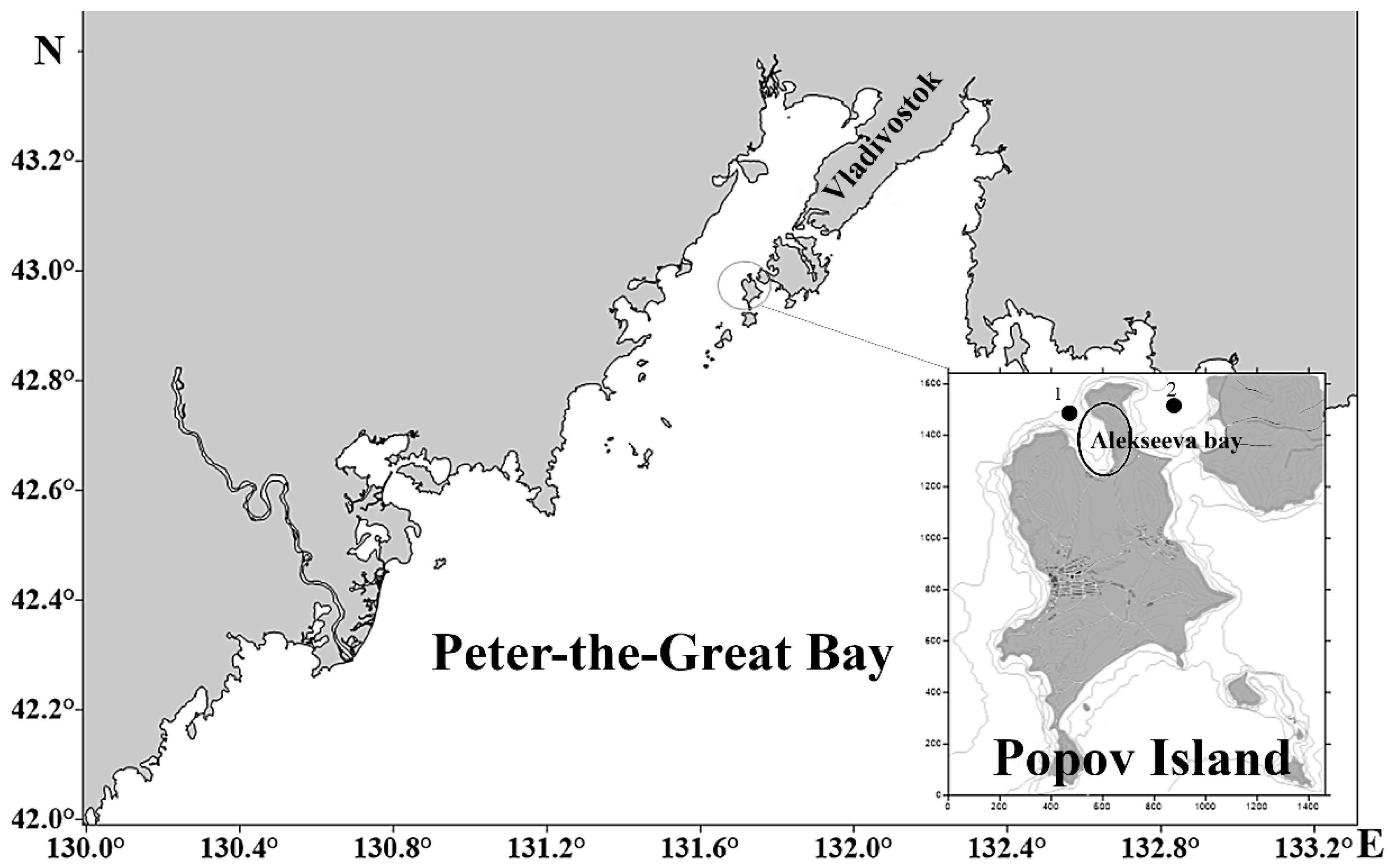
2.1. Site of Bivalves Collection and Material

Mature mollusks were collected during the post-spawning period, in November 2021, in the waters of the Alekseev Bay and Stark Strait in the Sea of Japan (Figure 1).
The biological characteristics of bivalves are shown in Table 1.
Mollusks were transported to the aquarium of the A.V. Zhirmunsky National Scientific Center of Marine Biology, where they were maintained at a constant temperature of 16 °C for 3 days to relieve the stress of transportation.
For FA analysis, 2 g of gills were obtained from one individual, for a total of 5 individuals for each species; for M. trossulus, tissue from 16 individuals was pooled for a total of 80 individuals. The mitochondria obtained for each mollusk species were separated into three samples (n = 3). For ROS, MDA, and carbonyl analysis, mitochondria were obtained from gills weighing 0.6 g. For most mollusks, 1 sample was 1 individual, for a total of 6–8 samples (n = 6–8). For M. trossulus, 1 sample was an assemblage of 3 individuals, for a total of 6–8 samples (n = 6–8). The isolated gills were frozen in liquid nitrogen and stored for not more than 1 month before analysis. All procedures in the present work, as well as the mollusk disposal methods, were approved by the Commission on Bioethics at the V.I. Il’ichev Pacific Oceanological Institute, Far Eastern Branch of Russian Academy of Science (protocol №16 and date of approval 15 April 2021), Vladivostok, Russia.
The individual age of scallops and M. trossulus was estimated by growth retardation rings on the surface of the shell. The data were comparable with the growth curves obtained for these species by other authors (Table 1). The age of C. grayanus and M. kurilensis was determined by the curves of group linear growth (Table 1).

2.2. Mitochondria Isolation

Gills were homogenized on ice (1:5, weight/volume). Mitochondria were isolated in 0.5 M NaCl in a 0.05 M Tris-HCl (pH 7.5) medium containing 0.25 M sucrose, 1 mM EDTA, and 0.1 mM PMSF. The medium for homogenization was pre-blown with argon. The homogenate was centrifuged at 1000× g for 12 min to remove large residual cells and nuclei. The resulting supernatant was centrifuged at 12,000× g for 30 min. The mitochondria were washed from sucrose 3 times in 0.5 M NaCl in 0.05 M Tris-HCl (pH 7.5).

2.3. Biochemical Analysis

ROS levels were determined by the oxidation of DHR 123 (dihydrorhodamine 123) to fluorescent rhodamine 123 [35]. MDA content was determined by a color reaction with 2-thiobarbituric acid [36]. Protein carbonyl groups were determined by the alkaline method [37], and protein concentration was determined by the modified Lowry method [38].

2.4. Oxidative Stress In Vitro

The oxidative stress reaction was triggered by adding Fe2+ and ascorbic acid (50 μM and 100 μM in the incubation medium, respectively) to mitochondria at 20 °C for one hour for MDA and carbonyl determination, as well as 15 min for ROS determination.

2.5. Determination of FAs

Lipid FAs were analyzed in the form of methyl esters using an Agilent 3700 chromatograph with a flame ionization detector. We used a Carbowax-20 M capillary column 25 m × 0.2 mm, a helium carrier gas, and a thermostat temperature of 200 °C [39]. FAs were identified by comparing the relative retention times of their methyl esters with the FA methyl esters of the standard mixture and the “carbon numbers” values [40]. The percentage of acids was calculated according to the method of Carrol [41]. FA methyl esters were obtained according to the method of Carreau and Dubacq [42].

2.6. Statistical Analysis

Statistical processing of the results was performed using Statistica 7. Breakdown and one-way ANOVA, as well as Statistics by Groups, Post-hoc were used to assess the reliability of parameter changes. Significance was established at p < 0.05.

3. Results

3.1. FAs in Mitochondrial Membranes of Mollusk Gill Cells

According to the results of the analysis presented in Table 2, the qualitative composition of the FAs of gill cell mitochondria lipids is uniform in all the studied marine bivalves, regardless of their MLS. However, in terms of the quantitative content of individual FAs, the mitochondrial lipids of mollusks differed significantly.
Despite significant variations (from 21.06 to 47.8%) in the content of total saturated fatty acids (SFAs), in all representatives of the Mytilidae Families and the Pectinidae Families, the palmitic and stearic acids (16:0 and 18:0) dominated. At the same time, the lowest content of SFAs was found in the mitochondrial lipids of the scallop S. swiftii, and the maximum was in the Pacific mussel M. trossulus. In general, it turned out that the SFAs in mitochondrial lipids in short-lived species was higher than in medium-lived and long-lived bivalves (Table 2).
In most mollusks, oleic acid (18:1 n−7) dominated among monounsaturated fatty acids (MUFAs), except for C. grayanus and M. yessoensis, in which eicosenoic acid (20:1 n−9) MUFAs predominated. The total content (MUFAs) in lipids also varied widely (from 11.4 to 20.5%) in the mollusks studied: the minimum amount was observed in the mitochondria of M. trossulus, and the maximum was in C. grayanus and C. farreri.
Non-methylene-interrupted fatty acids (NMI FAs), represented mainly by docosadienoic acid (22:2), were found in the FA composition of the mitochondria of marine mollusks. The greatest variation in the content of this acid was observed in pectinids: from 1% in the scallop C. farreri to 11.7% in the scallop M. yessoensis. The 22:2 level in representatives of the Mytilidae Family increased in the series: M. trossulusM. kurilensisC. grayanus; in representatives of the Pectinidae Family—in the series: C. farreriS. swiftiiM. yessoensis. Regarding NMI FAs, both by the content of individual 22:2 and by the total level of NMI FAs, the total content of which varies from 2.76% (C. farreri) to 13.7% (C. grayanus), a direct connection with MLS is observed in species of the relevant family.
In representatives of the Pectinidae Family, polyunsaturated fatty acids (PUFAs) dominated in the FAs of gill mitochondrial cell membranes; their amount was greater than the total sum of SFAs and MUFAs. The Mytilidae Family showed a different pattern: the level of PUFAs did not exceed, and in some cases, it was lower than the total sum of saturated and monounsaturated acids (Table 2). Docosahexaenoic acid (22:6 n−3) significantly prevailed in the PUFA of mitochondria lipids, especially in pectinids. Among PUFAs, the ratio of n−3/n−6 acids varied from 2.10 to 4.92, with the minimum values observed in the C. grayanus and the M. yessoensis, and the maximum values were characteristic of the Pacific mussel M. trossulus and the Zhikong scallop C. farreri (Table 2).

3.2. PI of Mitochondrial Membranes

On the basis of the composition of FAs, according to the formula given in [21], the lipid peroxidation index was calculated, the values of which are shown in Table 2. From the analysis of these values, it follows that the propensity to oxidation of the FA lipids of mitochondrial membranes, in representatives of the Pectinidae Family, is higher than that in representatives of Mytilidae Family. At the same time, no correlation between the obtained values of PI and MLS of bivalves was revealed.

3.3. Constitutive Levels of ROS, MDA and Carbonyls

The basal levels of ROS generation and MDA content in mitochondria were highest in the long-lived Gray’s mussel C. grayanus and the coastal scallop M. yessoensis, as compared with the medium and short-lived representatives of the respective families (Figure 2). In general, representatives of the Mytilidae Family differed from those of the Pectinidae Family (S. swiftii, C. farreri) in higher MDA content in the mitochondria of gill cells. At the same time, C. grayanus and M. kurilensis had a lower level of ROS generation compared to M. yessoensis and C. farreri, respectively. No interspecific differences in the content of protein carbonyls were found in any of the bivalves studied.

3.4. Induction of Oxidative Stress In Vitro

The results of this series of experiments showed that, when free-radical processes were initiated using the Fenton reaction, the lowest level of oxygen radical generation was registered in the mitochondria of the long-lived mussel Gray’s C. grayanus and the scallop M. yessoensis in contrast to the short-lived mussel M. trossulus and the scallop C. farreri (Figure 3). A similar pattern was observed in the formation of the main product of lipid oxidation—MDA.
Under these conditions of ROS generation initiation, the least amount of MDA accumulated in the mitochondrial lipids of long-lived mollusks (C. grayanus mussel and M. yessoensis scallop) compared with short-lived ones (M. trossulus and C. farreri). Mitochondrial membranes did not differ in protein carbonyl levels in bivalves from both families (Figure 3).

4. Discussion

4.1. Specific Features of FAs in Gill Mitochondria Lipids

Unsaturated fatty acids are easily subjected to oxidative damage in the cell, and the rate of oxidation increases with the number of double bonds. Therefore, unlike SFAs and MUFAs, which are relatively resistant to oxidation, PUFAs are easily and rapidly oxidized [8,43]. According to homeoviscous theory, the liquid crystalline state of the lipid matrix, necessary for the function of biological membranes, is maintained by regulating the degree of unsaturation of the acyl chains of phospholipids. In this respect, using mammalian and avian representatives as an example, it has been shown that the high unsaturation of membrane lipid FAs is associated with an increased level of oxidative lipid damage, but it negatively correlates with MLS [17,44]. Thus, in representatives of long-lived mammals, as compared to species with shorter lifespans (short-lived ones), a decrease in the ratio of acids with 4 or 6 double bonds and an increase in the level of FAs with 2 and 3 double bonds were found. At the same time, as noted by the authors, not only was a significant increase in lipid resistance to peroxidation observed but the corresponding fluidity of the lipid matrix was also maintained, and all the most important functions of membranes (receptor, ion transport, metabolite transport, etc.) were performed [8,44].
In fact, the results of this study of the FAs of mitochondrial lipids, in representatives of two families of marine bivalves, confirm this theory.
On the basis of the SFAs/PUFAs ratio, the authors showed that the amount of SFAs in gill membrane lipids was higher in representatives of the Mytilidae Family compared with the Pectinidae Family. It turned out that the long-lived C. grayanus and M. yessoensis had a lower SFAs/PUFAs ratio compared to the short-lived M. trossulus and C. farreri from the respective families. In addition, as in mammalian representatives, the proportion of FAs with 2 and 3 double bonds, in relation to FAs with 4 and 6 double bonds, was higher in long-lived species vs. short-lived species (Table 2).
It is known that the tendency of oxidation of acyl chains of lipids is determined not only by the degree of unsaturation but, also, by the position of double bonds. It was found that n−3 PUFAs are oxidized faster than n−6 PUFAs. Accordingly, membranes enriched with phospholipids with n−6 FAs are more stable in response to unfavorable environmental factors [45]. In addition, the ratio of these PUFAs (Σn−3/Σn−6) is an index characterizing the viscosity/liquidity of the lipid matrix of biological membranes. The lower is the ratio of n−3/n−6, the lower is the viscosity of the lipid matrix, but the higher is the resistance of lipids to oxidation, which is beneficial for the stability of membrane processes. In this respect, the paper of Valencak and Ruf [46] should be particularly noted. The authors revealed a negative correlation between the increase in the n−3/n−6 ratio in the skeletal muscle lipids of mammalian representatives and their lifespan. This interesting tendency is also clearly seen in marine bivalves. In this study, it was found that, in the mitochondrial lipids of long-lived mussel C. grayanus and scallop M. yessoensis, the ratio of Σn−3/Σn−6 is lower in comparison with the medium-lived and short-lived representatives of the respective families (Table 2). Based on this, there is every reason to believe that the lipid matrix of mitochondrial membranes of long-lived species is more stable in response to the effects of unfavorable environmental factors.
In addition to the above characteristics of mitochondrial membrane lipids, the presence of NMI FAs draws attention, which can also have a significant influence on the structure and function of biological membranes. These unusual FAs can act as “structural antioxidants”, slowing down the lipid matrix peroxidation processes [47]. The obtained results showed that the acids [Σ20:2 (5,11); 20:2 (5,13); 22:2] were present in far greater amounts in the lipids of long-lived C. grayanus and M. yessoensis mollusks than in the lipids of medium-lived and short-lived species from the respective families (Table 2). Therefore, it is logical to assume that the lifespans of the studied mollusks are related to the presence of these FAs in the lipids, which protect mitochondrial membranes from oxidative damage to a certain extent.
In general, the observed features in the lipid composition of mollusk mitochondria, through the presumed effect on lipid matrix oxidability, correlate well with the lifespans of these animals. Although the integral index (PI), calculated based on FA composition, demonstrated an increased sensitivity to the oxidative degradation of lipid membranes of representatives of the Pectinidae Family vs. representatives of the Mytilidae Family, it showed no relationship with the lifespans of bivalves. In this respect, the results of these studies and reasoning are consistent with those of Valencak and Ruf [46], who also found no correlation between the skeletal muscle lipid oxidation index (PI) and lifespan in 42 mammalian species.
The absence of such correlation calls into question the correctness of the calculation of this index, which does not consider additional factors influencing lipid oxidability. Among them, it should be emphasized that the high content of etheric lipids with alkyl and alkenyl fat radicals is characteristic of bivalves, whose contribution to lipid matrix oxidation of membranes is practically unstudied [48].

4.2. Constitutive Levels of ROS, MDA and Carbonyls in Gill Mitochondria

The “membrane-pacemaker” theory of aging suggests that lifespan can be related not only to lipid matrix oxidizability (based on PI) but, also, to the rate generation of ROS in the cell. The main source of ROS generation in the cell is the electron-transport chain localized in mitochondrial membranes. Taking into account their high reactivity, these ROS can initiate free-radical processes and cause the destruction of membrane lipids, proteins, and damage to mitochondrial DNA. In the latter case, there is strong evidence that the rate of aging is closely related to the frequency of mutations occurring in mitochondrial DNA [10]. In the lipid matrix of membranes, unsaturated fatty acids, especially PUFAs, become the preferred target for ROS. After the initiation of free-radical processes through a cascade of reactions, these FAs decompose to form highly reactive carbonyl compounds, such as malondialdehyde (MDA) and 4- hydroxynonenal (4-HN), which exhibit various cytotoxic and genotoxic properties [49]. It is likely that, through the regulation of ROS generation, mitochondria play a key role in preventing the formation and accumulation of various destructive damages affecting aging processes.
This opinion is based on the results of mammalian and bird studies in which it has been shown that, regardless of oxygen uptake rate, long-lived species show low rates of mitochondrial radical generation and contain lower constitutive levels of antioxidant activity [7,8,12,14]. Nevertheless, it has been shown that, in the long-lived (naked mole rat) Heterocephalus glaber, the endothelial and smooth muscle cells of carotid arteries and aorta produce comparable—or even higher—levels of ROS compared to short-lived mice [50].
The results of these studies also do not fit the general hypothesis. In long-lived C. grayanus and M. yesonensis, a relatively high baseline level of ROS generation and elevated MDA content in mitochondria were observed compared with other representatives of their families. Moreover, all this is realized against the background of a relatively low baseline level of antioxidant potential, including the activity of antioxidant enzymes and low molecular weight antioxidants [51].
Previously, in comparative studies of bivalves belonging to different families, it was shown that the isolated gill and heart mitochondria of long-lived Arctica islandica generated less ROS compared to short-lived Mya arenaria, Spisula solidissima, and Mercenaria mercenaria [27,51]. At the same time, the short-lived scallop Argopecten irradians and the long-lived Tridacna derasa did not significantly differ in ROS generation in gills, adductor muscles, and heart cells [52]. There was also no difference in the carbonyl content of the gills and adductor muscle in these species.
Comparing these results with the above examples, it is logical to assume that, in long-lived mollusks, against the background of low antioxidant protection, the hydrophobic component probably plays an important role in the mechanisms maintaining the oxidative stability of the lipid membrane matrix. These ideas, to a certain extent, were confirmed in the authors’ experiments with the induction of mitochondrial lipid peroxidation initiated by the Fenton reaction. This approach makes it possible to not only estimate the potential ability of mitochondria to generate ROS but, also, to reveal the integral vulnerability of the hydrophobic matrix to oxidative degradation in case of oxidative stress.

4.3. Response to Induced Oxidative Stress In Vitro

The results showed that the mitochondria of long-lived C. grayanus and M. yessoensis produced lower levels of ROS and less MDA as compared to medium and short-lived representatives of the respective families, indicating greater resistance of their lipid matrix to in vitro-induced oxidative damage. As far as one can estimate from the published data, the presented results are not only characteristic for the study species. It was also found that exposure of the scallop Argopecten irradians to paraquat, rotenone, or organic hydroperoxide causing oxidative damage of mitochondria was accompanied by a faster death of these short-lived mollusks compared to the long-lived Mercenaria mercenaria, Arctica islandica, and Tridacna derasa [53,54]. Moreover, there is evidence that experiments in vitro, fibroblasts, and lymphocytes of long-lived vertebrate species show increased resistance to induced oxidative stress [55].
When analyzing the results of mollusk mitochondrial resistance to the in vitro-induced oxidative damage of membrane lipids, the authors found two opposite trends. In representatives of the Mytilidae Family, in response to Fenton’s reagents, mitochondria generated high levels of ROS, which nevertheless led to an insignificant accumulation of MDA. In similar experiments on representatives of the Pectinidae Family, a different picture was observed: against the background of an insignificant level of ROS generation, we detected a significant increase in the MDA content of mitochondrial membrane lipids. Although it is beyond the scope of this study to investigate the reasons for these peculiarities, the authors should admit that the mitochondrial membranes of these two families differ significantly not only in lipid matrix accessibility to peroxidation but, also, in the function of ROS generation centers.
As the experimental data accumulate, it becomes more and more evident that different mechanisms of stabilization of not only the lipid matrix but proteins can make a certain contribution to the processes ensuring cell resistance to stress and lifespan [51,56]. Nevertheless, according to the experimental data, the authors found no changes in the content of protein carbonyls at neither the baseline nor after ROS generation in bivalves with different lifespans. Apparently, the reparation processes of the damaged mitochondrial membrane proteins, with the participation of proteosomal and autophagic mechanisms, are stable and exhibit resistance to short-term exposure to oxidative stress in these representatives of marine mollusks. Regarding the poor study of this issue, the authors consider it necessary to perform further studies to identify the mechanisms maintaining the stability and integrity of membrane protein components with the involvement of representatives of other taxonomic groups with different lifespans.

5. Conclusions

The common features of the relationship between the FA composition of gill mitochondrial membranes and the MLS of species are revealed only in a comparative analysis of mollusks having a common origin within a family. The response to in vitro-induced oxidative stress also has a relationship with the MLS of species belonging to the same family.
The basal levels of ROS and MDA formation in gill mitochondria are higher in the long-lived C. grayanus and M. yessoensis vs. medium and short-lived representatives of the respective families, and the gill mitochondrial membranes of these species are more resistant to in vitro-induced oxidative stress (low levels of ROS and MDA).
It is likely that an important mechanism of lifespan maintenance in C. grayanus and M. yessoensis is a specific FA composition of mitochondrial membranes. It is characterized by a lower ratio of SFAs/PUFAs and n−3/n−6, a higher ratio of the sum of FAs with 2 and 3 bonds and the sum of FAs with 4 and 6 bonds, and higher content of the sum of NMI FAs vs. medium and short-lived species of the respective family.

Author Contributions

Conceptualization, A.A.I. and V.P.C.; methodology, A.F.Z., E.A.B. and N.V.D.; software, O.A.E. and A.A.K.; validation, A.A.K.; formal analysis, A.F.Z., A.N.M.; investigation, A.A.I., M.A.M. and Y.V.F.; resources, A.N.M., E.A.B., M.A.M., A.A.M. and N.V.D.; data curation, O.A.E.; writing—original draft preparation, A.A.I.; writing—review and editing, V.P.C.; visualization, A.A.M. and Y.V.F.; funding acquisition, V.P.C. All authors have read and agreed to the published version of the manuscript.

Funding

The research was supported by the State assignment for research work of V.I. Il’ichev Pacific Oceanological Institute, FEB RAS (no. 121021500052-9) at the marine experimental station “Popov Island” Il’ichev Pacific Oceanological Institute.

Institutional Review Board Statement

Not applicable.

Informed Consent Statement

Not applicable.

Data Availability Statement

Not applicable.

Acknowledgments

The authors are grateful to I.V. Popeko and A.A. Khudoleyev (NSCMB FEB RAS), who provided equipment for the aquarium and maintained the necessary conditions for animal housing.

Conflicts of Interest

The authors declare no conflict of interest.

References

  1. Hulbert, A.J.; Pamplona, R.; Buffenstein, R.; Buttemer, W.A. Life and death: Metabolic rate, membrane composition, and life span of animals. Physiol. Rev. 2007, 87, 1175–1213. [Google Scholar] [CrossRef] [PubMed]
  2. Harman, D. Aging: A theory based on free radical and radiation chemistry. J. Gerontol. 1956, 11, 298–300. [Google Scholar] [CrossRef] [PubMed] [Green Version]
  3. Blagosklonny, M.V. Aging: ROS or TOR. Cell Cycle 2008, 7, 3344–3354. [Google Scholar] [CrossRef] [Green Version]
  4. Pérez, V.I.; Bokov, A.; Van Remmen, H.; Mele, J.; Ran, Q.; Ikeno, Y.; Richardson, A. Is the oxidative stress theory of aging dead? Biochim. Biophys. Acta 2009, 1790, 1005–1014. [Google Scholar] [CrossRef] [PubMed] [Green Version]
  5. Salmon, A.B.; Richardson, A.; Pérez, V.I. Update on the oxidative stress theory of aging: Does oxidative stress play a role in aging or healthy aging? Free Radic. Biol. Med. 2010, 48, 642–655. [Google Scholar] [CrossRef] [Green Version]
  6. Du, C.; Anderson, A.; Lortie, M.; Parsons, R.; Bodnar, A. Oxidative damage and cellular defense mechanisms in sea urchin models of aging. Free Radic. Biol. Med. 2013, 63, 254–263. [Google Scholar] [CrossRef] [Green Version]
  7. Buttemer, W.A.; Abele, D.; Costantini, D. From bivalves to birds: Oxidative stress and longevity. Funct. Ecol. 2010, 24, 971–983. [Google Scholar] [CrossRef] [Green Version]
  8. Pamplona, R.; Costantini, D. Molecular and structural antioxidant defenses against oxidative stress in animals. Am. J. Physiol. Regul. Integr. Comp. Physiol. 2011, 301, 843–863. [Google Scholar] [CrossRef] [Green Version]
  9. Balaban, R.S.; Nemoto, S.; Finkel, T. Mitochondria, oxidants, and aging. Cell 2005, 120, 483–495. [Google Scholar] [CrossRef] [Green Version]
  10. Skrha, J. Caloric restriction and oxidative stress. In Oxidative Stress in Vertebrates and Invertebrates: Molecular Aspects of Cell Signaling, 1st ed.; Blackwell: Hoboken, NJ, USA, 2012; p. 424. [Google Scholar]
  11. Sohal, R.S.; Donato, H.; Biehl, E.R. Effect of age and metabolic rate on lipid peroxidation in the housefly, Musca domestica L. Mech. Ageing. Dev. 1981, 16, 159–167. [Google Scholar] [CrossRef]
  12. Ku, H.H.; Sohal, R.S. Comparison of mitochondrial pro-oxidant generation and anti-oxidant defenses between rat and pigeon: Possible basis of variation in longevity and metabolic potential. Mech. Ageing. Dev. 1993, 72, 67–76. [Google Scholar] [CrossRef]
  13. Zielinski, S.; Pörtner, H.O. Oxidative stress and antioxidative defense in cephalopods: A function of metabolic rate or age? Comp. Biochem. Physiol. Part B Biochem. Mol. Biol. 2000, 125, 147–160. [Google Scholar] [CrossRef] [Green Version]
  14. Barja, G. Aging in vertebrates, and the effect of caloric restriction: A mitochondrial free radical production DNA damage mechanism? Biol. Rev. Camb. Philos. Soc. 2004, 79, 235–251. [Google Scholar] [CrossRef] [PubMed]
  15. Couture, P.; Hulbert, A.J. Membrane fatty acid composition is related to body mass in mammals. J. Membr. Biol. 1995, 148, 27–39. [Google Scholar] [CrossRef] [PubMed]
  16. Brand, M.D.; Turner, N.; Ocloo, A.; Else, P.L.; Hulbert, A.J. Proton conductance and fatty acyl composition of liver mitochondria correlates with body mass in birds. Biochem. J. 2003, 376, 741–748. [Google Scholar] [CrossRef] [PubMed] [Green Version]
  17. Pamplona, R.; Portero-Otin, M.; Riba, D. Mitochondrial membrane peroxidizability index is inversely related to maximum lifespan in mammals. J. Lipid. Res. 1998, 39, 1989–1994. [Google Scholar] [CrossRef] [PubMed]
  18. Barja, G. Rate of generation of oxidative stress-related damage and animal longevity. Free Radic. Biol. Med. 2002, 33, 1167–1172. [Google Scholar] [CrossRef]
  19. Pamplona, R.; Barja, G.; Portero-Otin, M. Membrane fatty acid unsaturation, protection against oxidative stress, maximum life span: A homeoviscous-longevity adaptation? Ann. NY Acad. Sci. 2002, 959, 475–490. [Google Scholar] [CrossRef]
  20. Hulbert, A.J.; Else, P.L. Membranes as possible pacemakers of metabolism. J. Theor. Biol. 1999, 199, 257–274. [Google Scholar] [CrossRef]
  21. Munro, D.; Blier, P.U. The extreme longevity of Arctica Islandica is associated with increased peroxidation resistance in mitochondrial membranes. Aging Cell 2012, 11, 845–855. [Google Scholar] [CrossRef]
  22. Pamplona, R. Low fatty acid unsaturation protects against lipid peroxidation in liver mitochondria from long-lived species: The pigeon and human case. Mech. Ageing. Dev. 1996, 86, 53–66. [Google Scholar] [CrossRef]
  23. Rodríguez, E.; Dégletagne, C.; Hagen, T.M.; Abele, D.; Blier, P.U. Mitochondrial Traits Previously Associated with Species Maximum Lifespan Do Not Correlate with Longevity Across Populations of the Bivalve Arctica islandica. Front. Physiol. 2019, 10, 946. [Google Scholar] [CrossRef] [PubMed] [Green Version]
  24. Speakman, J.R. Correlations between physiology and lifespan—Two widely ignored problems with comparative studies. Aging Cell 2005, 4, 167–175. [Google Scholar] [CrossRef] [PubMed]
  25. Abele, D.; Brey, T.; Philipp, E. Bivalve models of aging and the determination of molluscan lifespans. Exp. Gerontol. 2009, 44, 307–315. [Google Scholar] [CrossRef] [PubMed] [Green Version]
  26. Philipp, E.E.; Abele, D. Masters of longevity: Lessons from long-lived bivalves--a mini-review. Gerontology 2010, 56, 55–65. [Google Scholar] [CrossRef] [Green Version]
  27. Zakhartsev, M.V.; Naumenko, N.V.; Chelomin, V.P. Non-methylene-interrupted fatty acids in phospholipids of the membranes of the mussel Crenomytilus grayanus. Russ. J. Mar. Biol. 1998, 24, 183–186. [Google Scholar]
  28. Sadykhova, I.A. Gray mussel growth in the Peter-the-Great Bay (Sea of Japan). Biology of Gray mussel. Nauka 1983, 1, 62–68. [Google Scholar]
  29. Zolotarev, V.N. Sklerohronologiya morskih dvustvorchatyh mollyuskov. Kiev. Nauk. Dumka 1989, 112. [Google Scholar]
  30. Selin, N.I. The composition and structure of a mixed population of Crenomytilus grayanus (Dunker, 1853) and Modiolus kurilensis (Bernard, 1983) (bivalvia: Mytilidae) in Peter the Great bay, Sea of Japan. Russ. J. Mar. Bio. 2018, 449, 363–372. [Google Scholar] [CrossRef]
  31. Leskova, S.E. The growth of the Pacific mussel in the conditions of the Voevoda Bay (O. Russian). Proc. Dalrybvtuz. 2021, 56, 15–19. [Google Scholar]
  32. Motavkin, P.A. Primorskiy Grebeshok. 1986. Available online: https://waves-vagues.dfo-mpo.gc.ca/library-bibliotheque/116222.pdf (accessed on 4 May 2023).
  33. Ponurovsky, S.K.; Silina, A.V. Determination of the age and rate of linear growth of the Swift scallop. Biol. Sea 1983, 1, 20–24. [Google Scholar]
  34. Karpenko, D.T. Growth rates of the Japanese scallop (Chlamys farreri) in the bays of the coastal zone of Russian Island (Peter-the-Great Bay, Sea of Japan). Complex research in the fisheries industry. In Proceedings of the V International Scientific and Technical Conference of Students, Postgraduates and Young Scientists Vladivostok, Vladivostok, Russia, 29 November 2020; pp. 14–18. [Google Scholar]
  35. Viarengo, B.; Burlando, M.; Cavaletto, B.; Marchi, E.; Ponzano, J.; Blasco, J. Role of metallothionein against oxidative stress in the mussel Mytilus galloprovincialis. Am. J. Physiol. 1999, 277, 1612–1619. [Google Scholar] [CrossRef]
  36. Buege, J.A.; Aust, S.D. Microsomal lipid peroxidation. Meth. Enzymol. 1978, 52, 302–310. [Google Scholar] [CrossRef]
  37. Mesquita, C.S.; Oliveira, R.; Bento, F.; Geraldo, D.; Rodrigues, J.V.; Marcos, J.C. Simplified 2,4-dinitrophenylhydrazine spectrophotometric assay for quantification of carbonyls in oxidized proteins. Anal. Biochem. 2014, 458, 69–71. [Google Scholar] [CrossRef] [PubMed]
  38. Markwell, M.; Haas, S.; Bieber, L.; Tolbert, N. A modification of the Lowry procedure to simplify protein determination in membrane and lipoprotein samples. Anal. Biochem. 1978, 82, 206–210. [Google Scholar] [CrossRef]
  39. Kramer, J.K.G.; Fouchard, R.C.; Jenkins, K.J. Differences in chromatographic properties of fused silica capillary columns, coated, crosslnked, bonded, or crosslincked and bonded with polyethylene glycols (Carbowax 20M) using complex fatty acid methyl ester mixtures. J. Chromatogr. Sci. 1985, 23, 54–56. [Google Scholar] [CrossRef]
  40. Christie, W.W. Equivalent chain length of methyl ester derivatives of fatty acids on gas chromatography. J. Cromatogr. 1988, 447, 305–314. [Google Scholar] [CrossRef]
  41. Carrol, K.K. Quantitative estimation of peak areasin gas-liquid chromatography. Nature 1961, 191, 377–378. [Google Scholar] [CrossRef]
  42. Carreau, J.P.; Dubacq, J.P. Adaptation of macro-scale method to the microscale for faty acid methyl trans esterification of biologicall lipid extracts. J. Chromatogr. Sci. 1985, 23, 54–56. [Google Scholar] [CrossRef]
  43. Crockett, E.L. The cold but not hard fats in ectotherms: Consequences of lipid restructuring on susceptibility of biological membranes to peroxidation, a review. J. Comp. Physiol. B 2008, 178, 795–809. [Google Scholar] [CrossRef]
  44. Naudí, A.; Jové, M.; Ayala, V.; Portero-Otín, M.; Barja, G.; Pamplona, R. Membrane lipid unsaturation as physiological adaptation to animal longevity. Front. Physiol. 2013, 17, 372. [Google Scholar] [CrossRef] [Green Version]
  45. Hulbert, A.J. On the importance of fatty acid composition of membranes for aging. J. Theor. Biol. 2005, 234, 277–288. [Google Scholar] [CrossRef]
  46. Valencak, T.G.; Ruf, T. N-3 polyunsaturated fatty acids impair lifespan but have no role for metabolism. Aging Cell 2007, 6, 15–25. [Google Scholar] [CrossRef]
  47. Hanuš, L.O.; Levitsky, D.O.; Shkrob, I.; Dembitsky, V.M. Plasmalogens, fatty acids and alkyl glyceryl ethers of marine and freshwater clams and mussels. Food Chem. 2009, 116, 491–498. [Google Scholar] [CrossRef]
  48. Yin, D. Biochemical basis of lipofuscin, ceroid, and age pigment-like fluorophores. Free Radic. Biol. Med. 1996, 21, 871–888. [Google Scholar] [CrossRef] [PubMed]
  49. Labinskyy, N.; Csiszar, A.; Orosz, Z. Comparison of endothelial function, O2−·and H2O2 production, and vascular oxidative stress resistance between the longest-living rodent, the naked mole rat, and mice. Am. J. Physiol. Heart Circ. Physiol. 2006, 291, 2698–2704. [Google Scholar] [CrossRef] [PubMed] [Green Version]
  50. Istomina, A.; Yelovskaya, O.; Chelomin, V.; Karpenko, A.; Zvyagintsev, A. Antioxidant activity of Far Eastern bivalves in their natural habitat. Mar. Environ. Res. 2021, 169, 105383. [Google Scholar] [CrossRef]
  51. Ungvari, Z.; Ridgway, I.; Philipp, E.E.R. Extreme Longevity Is Associated with Increased Resistance to Oxidative Stress in Arctica islandica, the Longest-Living Non-Colonial Animal. J. Gerontol. Ser. A Biomed. Sci. Med. Sci. 2011, 66, 741–750. [Google Scholar] [CrossRef] [Green Version]
  52. Munro, D.; Pichaud, N.; Paquin, F.; Kemeid, V.; Blier, P.U. Low hydrogen peroxide production in mitochondria of the long-lived Arctica islandica: Underlying mechanisms for slow aging. Aging Cell 2013, 12, 584–592. [Google Scholar] [CrossRef] [PubMed]
  53. Ungvari, Z.; Csiszar, A.; Sosnowska, D. Testing Predictions of the Oxidative Stress Hypothesis of Aging Using a Novel Invertebrate Model of Longevity: The Giant Clam (Tridacna derasa). J. Gerontol. Ser. A Biomed. Sci. Med. Sci. 2012, 68, 359–367. [Google Scholar] [CrossRef] [Green Version]
  54. Ungvari, Z.; Sosnowska, D.; Mason, J.B. Resistance to Genotoxic Stresses in Arcticai slandica, the Longest Living Noncolonial Animal: Is Extreme Longevity Associated with a Multistress Resistance Phenotype? J. Gerontol. Ser. A Biomed. Sci. Med. Sci. 2013, 68, 521–529. [Google Scholar] [CrossRef] [PubMed] [Green Version]
  55. Kapahi, P.; Boulton, M.E.; Kirkwood, T.B. Positive correlation between mammalian life span and cellular resistance to stress. Free Radic. Biol. Med. 1999, 26, 495–500. [Google Scholar] [CrossRef] [PubMed]
  56. Perez, V.I.; Buffenstein, R.; Masamsetti, V. Protein stability and resistance to oxidative stress are determinants of longevity in the longest-living rodent, the naked mole rat. Proc. Natl. Acad. Sci. USA 2009, 106, 3059–3064. [Google Scholar] [CrossRef] [PubMed] [Green Version]
Figure 1. The location of the sampling sites in the Peter-the-Great Bay of the Japan Sea (Russia) (1—the collection site of mussels, 2—scallops).
Biology 12 00837 g001
Figure 2. Constitutive levels of ROS (reactive oxygen species), MDA (malondialdehyde), and carbonyls in gill mitochondria. Significance of differences between: a—C. grayanus vs. M. kurilensis and M. trossulus; b—C. grayanus vs. M. kurilensis; c—M. yessoensis vs. S. swiftii and C. farreri; d—M. yessoensis vs. S. swiftii and C. farreri; #—C. grayanus vs. M. yessoensis; * C. grayanus, M. kurilensis and M. trossulus vs. S. swiftii and C. farreri; ##—M. kurilensis vs. C. farreri (n = 6–8; Post-hoc, p < 0.05).
Biology 12 00837 g002
Figure 3. Response to induced oxidative stress in vitro. Significance of differences between: a—C. grayanus vs. M. trossulus; b—C. grayanus vs. M. kurilensis and M. trossulus; c—M. yessoensis vs. S. swiftii and C. farreri; d—M. yessoensis vs. C. farreri; *—C. grayanus, M. kurilensis and M. trossulus vs. M. yessoensis, S. swiftii and C. farreri; #—C. grayanus vs. M. yessoensis, S. swiftii and C. farreri; ##—M. kurilensis and M. trossulus vs. C. farreri (n = 6–8; Post-hoc, p < 0.05).
Biology 12 00837 g003
Table 1. Biological characteristics of bivalves.
SpeciesLength, mmApproximate Age, YearsMLS, YearsReference
Mytilidae
Crenomytilus grayanus116.9 ± 5.024150[28,29]
Modiolus kurilensis111.7 ± 4.92061[29,30]
Mytilus trossulus42.2 ± 3.946[31]
Pectinidae
Mizuhopecten yessoensis132.0 ± 11.2522[32]
Swiftopecten swiftii83.9 ± 5.2415[29,33]
Chlamys farreri92.3 ± 5.349[34]
Note: MLS—maximum lifespan.
Table 2. Fatty acids (%) from gill mitochondria lipids. Values are mean ± SD, n = 3.
Fatty AcidMytilidaePectinidae
C. grayanusM. kurilensisM. trossulusM. yessoensisS. swiftiiC. farreri
12:01.0 ± 0.00.7 ± 0.21.1 ± 0.10.7 ± 0.00.7 ± 0.21.0 ± 0.7
14:0 ai0.2 ± 0.00.7 ± 0.10.8 ± 0.00.5 ± 0.00.3 ± 0.10.8 ± 0.2
14:00.6 ± 0.00.3 ± 0.10.7 ± 0.00.1 ± 0.00.4 ± 0.10.1 ± 0.0
15:1 n−71.0 ± 0.01.8 ± 0.01.6 ± 0.30.4 ± 0.00.4 ± 0.10.6 ± 0.0
16:017.9 ± 0.915.9 ± 0.319.0 ± 0.112.5 ± 0.612.7 ± 0.813.7 ± 0.7
16:1 n−92.1 ± 0.12.0 ± 0.52.7 ± 0.31.3 ± 0.0-2.2 ± 0.2
16:1 n−72.3 ± 0.12.3 ± 0.11.9 ± 0.01.4 ± 0.02.3 ± 0.51.2 ± 0.0
17:0 i0.6 ± 0.00.5 ± 0.00.5 ± 0.00.3 ± 0.00.9 ± 0.10.1 ± 0.0
17:0 ai1.1 ± 0.11.8 ± 0.11.8 ± 0.01.2 ± 0.00.3 ± 0.12.3 ± 0.0
17:00.8 ± 0.01.9 ± 0.11.7 ± 0.01.0 ± 0.00.9 ± 0.2-
18:0 i2.8 ± 0.12.5 ± 0.24.0 ± 0.10.9 ± 0.01.2 ± 0.31.3 ± 0.1
18:09.6 ± 0.510.9 ± 0.319.8 ± 3.14.8 ± 0.23.3 ± 0.69.7 ± 0.1
18:1 n−90.7 ± 0.0---0.1 ± 0.11.0 ± 0.0
18:1 n−74.3 ± 0.25.1 ± 0.32.3 ± 1.64.3 ± 0.24.5 ± 0.45.4 ± 0.4
18:2 n−61.0 ± 0.02.4 ± 0.51.6 ± 0.32.0 ± 0.12.2 ± 0.22.5 ± 0.1
18:2 n−40.4 ± 0.01.5 ± 1.50.5 ± 0.20.6 ± 0.00.1 ± 0.00.3 ± 0.1
18:3 n−6--0.2 ± 0.00.2 ± 0.00.2 ± 0.0-
18:3 n−30.8 ± 0.01.2 ± 0.00.3 ± 0.00.3 ± 0.00.2 ± 0.00.1 ± 0.0
20:0-i1.8 ± 0.11.7 ± 0.10.4 ± 0.1--0.8 ± 0.1
18:4 n−30.2 ± 0.0-0.4 ± 0.0-0.1 ± 0.0-
20:1 n−132.2 ± 0.10.6 ± 0.10.8 ± 0.22.0 ± 0.11.8 ± 0.02.9 ± 0.1
20:1 n−95.8 ± 0.33.4 ± 0.11.9 ± 0.35.5 ± 0.34.2 ± 0.54.2 ± 0.2
20:1 n−72.2 ± 0.13.9 ± 0.01.3 ± 0.21.0 ± 0.10.6 ± 0.32.5± 0.1
20:2 (5,11)3.9 ± 0.21.9 ± 0.01.8 ± 0.15.4 ± 0.35.5 ± 0.60.9 ± 0.0
20:2 (5,13)1.6 ± 0.10.6 ± 0.11.1 ± 0.21.7 ± 0.11.0 ± 0.10.7 ± 0.1
20:4 n−65.5 ± 0.35.7 ± 0.53.5 ± 0.36.7 ± 0.35.8 ± 1.23.9 ± 0.2
20:5 n−34.2 ± 0.26.2 ± 0.56.0 ± 0.43.9 ± 0.24.7 ± 0.69.7 ± 0.3
22:28.2 ± 0.45.2 ± 0.14.1 ± 0.211.7 ± 0.69.0 ± 1.21.0 ± 0.0
22:6 n−38.6 ± 0.49.7 ± 0.39.3 ± 0.422.1 ± 1.125.8 ± 1.421.7 ± 1.3
Total91.1 ± 4.690.4 ± 0.987.4 ± 4.093.6 ± 4.789.5 ± 7.292.0 ± 1.3
SFAs36.3 ± 1.837.0 ± 0.047.8 ± 0.522.36 ± 1.121.0 ± 1.930.3 ± 1.4
MUFAs20.5 ± 1.019.0 ± 1.011.4 ± 3.116.12 ± 0.813.4 ± 0.420.4 ± 0.6
PUFAs34.3 ± 1.734.4 ± 0.128.2 ± 1.455.13 ± 2.855.0 ± 4.943.6 ± 5.7
n−313.6 ± 0.717.1 ± 0.815.4 ± 1.026.55 ± 1.330.8 ± 1.931.6 ± 1.6
n−66.5 ± 0.38.1 ± 1.05.2 ± 0.49.03 ± 0.58.3 ± 1.56.4 ± 0.4
n−3/n−62.102.112.982.943.694.92
SFAs/
PUFAs
1.051.081.700.410.380.70
∑NMI FAs13.7 ± 0.017.75 ± 0.246.93 ± 0.1518.9 ± 0.115.68 ± 1.632.76 ± 0.22
∑2n, 3n/
∑4n, 6n
1.110.840.730.770.580.22
PI124.5 ± 6.2146.7 ± 6.6129.7 ± 7.9238.2 ± 11.2267.3 ± 19.5252.7 ± 13.6
MLS15061622159
Note: MLS—maximum lifespan; PI—peroxidation index; SFAs—saturated fatty acids; MUFAs—monounsaturated fatty acids; PUFAs—polyunsaturated fatty acids; NMI FAs—non-methylene-interrupted fatty acids.
Disclaimer/Publisher’s Note: The statements, opinions and data contained in all publications are solely those of the individual author(s) and contributor(s) and not of MDPI and/or the editor(s). MDPI and/or the editor(s) disclaim responsibility for any injury to people or property resulting from any ideas, methods, instructions or products referred to in the content.

