Article

Apoptotic Changes, Oxidative Stress and Immunomodulatory Effects in the Liver of Japanese Seabass (Lateolabrax japonicus) Induced by Ammonia-Nitrogen Stress during Keep-Live Transport

1
College of Food Science and Technology, Shanghai Ocean University, Shanghai 201306, China
2
National Experimental Teaching Demonstration Center for Food Science and Engineering, Shanghai Ocean University, Shanghai 201306, China
3
Shanghai Engineering Research Center of Aquatic Product Processing and Preservation, Shanghai 201306, China
4
Shanghai Professional Technology Service Platform on Cold Chain Equipment Performance and Energy Saving Evaluation, Shanghai 201306, China
*
Authors to whom correspondence should be addressed.
Biology 2023, 12(6), 769; https://doi.org/10.3390/biology12060769
Submission received: 28 April 2023 / Revised: 22 May 2023 / Accepted: 23 May 2023 / Published: 25 May 2023

Abstract

:

Simple Summary

Excreta produced by fish during preservation transport can increase ammonia-nitrogen (NH3-N) levels in transport water and reduce survival rates. However, studies on the effects of NH3-N stress on Japanese seabass during transport are limited. The objective of this study was to investigate the effects of NH3-N stress on tissue damage, oxidative stress, apoptosis and immunity in Japanese seabass during transport, and to determine tolerance and response to NH3-N toxicity. The results of this study provide insight into the impact of environmental factors on Japanese seabass during maintenance of live transport, which can be considered during transport in terms of improving the quality of transport water, among other things, to develop optimal procedures for live fish transport and improve survival rates during transport.

Abstract

This study investigated the effects of NH3-N on antioxidant responses, histoarchitecture, and immunity of Japanese seabass (Lateolabrax japonicus) during keep-live transport. The findings suggest that NH3-N stress transport alters the transcription of P53, Caspase 9, Bcl2, Caspase 3 and Bax genes, demonstrating that NH3-N stress can trigger the apoptotic pathway of P53-Bax-Bcl2 and Caspase and induce apoptosis. NH3-N stress transport also evoked transcriptional upregulation of inflammatory cytokines (tumor necrosis factor α (TNF-α), Toll-like receptor 3 (TLR-3), nuclear factor kappa β (NF-κB), interleukin 6 (IL-6) and interleukin 1β (IL-1β)) and increased complement C3, C4, lysozyme (LZM) and immunoglobulin (IgM) levels, activating the innate immunological system during keep-live transport. In addition, NH3-N stress transport altered changes in the levels of superoxide dismutase (SOD), catalase (CAT), glutathione-related enzymes, and heat shock proteins 70 and 90 in the liver, indicating that the antioxidant system and Hsp protected the cells from NH3-N-induced oxidative stress. When excess ROS were not removed, they caused the body to respond with immunological and inflammatory responses, as well as apoptosis and tissue damage. This helps towards understanding the effect of NH3-N levels on sea bass during keep-live transport.

1. Introduction

In China, Japanese seabass (Lateolabrax japonicus) is an economically important cultured fish. Nevertheless, high-density seabass keep-live transport causes ammonia-nitrogen (NH3-N) concentration to rise to dangerous levels in a short period, endangering the life of the sea bass. Higher NH3-N levels can disturb the metabolism of organisms, cause tissue damage, lead to a decrease in immunity and the inflammatory reaction, and even kill fish [1]. At the same time, when fish have been in water with a high NH3-N content, this will lead to the continuous accumulation of reactive oxygen species (ROS), causing oxidative stress and metabolic disorders in the biological system of fish. To combat this issue, cells form antioxidant defense systems such as the glutathione system [2,3]. The dynamic balance of GSH/GSSG is critical to the organism’s defense and proper physiological condition [4]. Systematic analysis of the changes and functional regulation of glutathione in antioxidant defense can provide further insight into its role in NH3-N stress transport. In addition, heat shock protein (Hsp) is crucial in defending the organism from stress-related damage. Hsp protects cells from stressor-induced protein dysfunction, such as ROS and hypoxia [5]. The organism’s ability to tolerate NH3-N is tightly correlated with the expression pattern and levels of Hsp [6]. Hsp levels are raised as a result of NH3-N stress, according to earlier research [7].
Currently, there are many studies on NH3-N stress, transport stress and combined stress of various stressors. According to Tao et al. [8], hybrid yellow catfish (Tachysurus fulvidraco ♀ × Pseudobagrus vachellii ♂) activates tlr5 in the TLR signaling pathway during transport, which affects immunological and inflammatory responses. It was also found that heat stress transport reduced the content of antioxidant enzymes including CAT, T-AOC and meat quality in rainbow trout (Oncorhynchus mykiss) [9]. Dual stress tends to affect organisms more adversely than a single stressor. Molayemraftar et al. [10] discovered that the combined impact of NH3-N and nitrite is harmful to Common carp (Cyprinus carpio) antioxidant defenses and has negative effects on the blood. Esam et al. [11] also found that the combined stress of NH3-N and heat affect the Nile tilapia (Oreochromis niloticus) more in terms of immunosuppression, oxidative stress and inflammation.
However, there is little research on how NH3-N stress affects seabass during keep-live transport. The goal of this study was to look into the impact of NH3-N stress on tissue damage, oxidative stress, apoptosis and immunity of Japanese seabass during keep-live transport, as well as to identify the tolerance and response to NH3-N toxicity in terms of gene expression associated with the glutathione system and the NF-κB signaling pathway.

2. Materials and Methods

2.1. Experimental Fish

Japanese seabass (mean weight 400 ± 3.5 g, mean length 35 ± 1.0 cm) were obtained from the seafood market in Shanghai. The seabass were temporarily incubated in a 300 L food-grade tank in the laboratory for 36 h before transport experiments. Temporary incubation conditions were as follows: the water temperature was 20 °C, salinity was 16‰, pH was 7.5 and dissolved oxygen (DO) concentration was above 7.0 mg/L. To maintain high water quality, the water was replaced every day. After the transient period, the water was cooled from 20 to 12 °C at a rate of 3 °C/h.

2.2. Experiment to Determine LC50 and Experimental Procedure:

A NH4Cl master mix was used during the experiment to adjust the NH3-N concentration to reach the required value for each experimental group. The range of NH3-N concentration was obtained from the lowest concentration where no fish died in 48 h (2 mg/L) to the highest concentration where all fish died (40 mg/L) by the pre-experiment and divided logarithmically. Six concentration gradients were established—2 mg/L, 3.6411 mg/L, 6.6289 mg/L, 12.0684 mg/L, 21.9712 mg/L, and 40 mg/L. Nine fish were used in each experimental group and this was repeated three times. The number of dead Japanese seabass was recorded on the 12th, 24th, 36th and 48th h. Dead fish were removed promptly (considered dead if they were stationary and did not respond to the surrounding stimuli) and water quality management was the same as during the transient period. The LC50 = 21.32 mg/L at 48 h was calculated based on the survival rate.
LC50 = 21.32 mg/L was obtained for sea bass by pre-experiment. Four groups were set up in the formal experiment: a control group (CK), a low NH3-N group (LA, 6.4 mg/L), a medium NH3-N group (MA, 12.8 mg/L), and a high NH3-N group (HA, 19.2 mg/L), which were equivalent to 30%, 60% and 90% of the 48 h LC50, respectively (Table 1). NH3-N concentration was measured using an NH3-N detector (DWS-296, Shanghai Insa Scientific Instruments Co., Ltd., Shanghai, China) to achieve the specified NH3-N levels. The fish were reared in sealed tanks with a fish-to-water ratio of 1:2. The dissolved oxygen (DO) concentration was guaranteed to exceed 7.0 mg/L, the water temperature was 20 °C, salinity was 16‰ and pH was 7.5 during transport. Live fish were simulated transport in a vibration conveyor in terms of 1 h on a class B road, followed by 5 h on a class A road. The cycles were repeated 8 times [12]. The numbers of dead fish were recorded at 0, 12th, 24th, 36th and 48th h, respectively. After 48 h transport, the surviving fish recovered for 24 h in seawater under transient conditions.

2.3. Sample Collection and Processing

Samples were taken once at the 0, 12th, 24th, 36th, and 48th h of NH3-N exposure and at the 12th and 24th h of recovery, respectively. Eighteen fish were sampled per experimental group at a time, for a total of three replicate groups, using nine fish per replicate. The livers of sea bass were immediately removed, washed with saline and divided into two parts. One part of the liver tissue samples were immersed for 24 h and fixed for histological and TUNEL assays, the remaining samples were stored at −80 °C qPCR index determination and enzyme index measurement.

2.4. Biochemical, Immunological and Oxidation Parameters Analysis

Liver tissue samples were diluted 10 times with 0.1 M PBS and homogenized, then centrifuged at 5000× g for 10 min at 4 °C. The supernatant was extracted and used to determine the following indicators using commercial kits (Jiancheng Bioengineering Institute, Nanjing, China) measuring the following enzymes: SOD (A001-3-2), CAT (A001-3-2), alanine aminotransferase (ALT, C009-2-1), GST, aspartate aminotransferase (AST, C010-2-1), GSH-PX (A005-1-2), GR (A001-3-2), GSH/GSSG (A061-2-1), LZM (A050-1-1), total protein (TP, A045-2-2), immunoglobulin (IgM, H109-1-1), complement C3 (E032-1-1), complement C4 (E033-1-1), Hsp70 (H264-2-2), and Hsp90 (H264-3).
Malondialdehyde (MDA) in the liver was measured using the TBA method according to the kit (A003-1-2, Jiancheng Bioengineering Institute, Nanjing, China) instructions and in nmol/mg protein. MDA is a peroxidized lipid degradation product that condenses with thiobarbituric acid (TBA) to form a red product with a maximum absorption peak of 532 nm.
The level of ROS in the liver was detected with fluorescent probe 2′,7′-dichlorofluorescein diacetate (DCFH-DA) (E004-1-1, Jiancheng Bioengineering Institute, Nanjing, China). DCFH-DA can be oxidized by esterase into strong green fluorescent substance DCF (2′,7′-dichlorofluorescein) in the presence of reactive oxygen species. The liver tissue was homogenized and made into a single cell suspension. 2′,7′-dichlorofluorescein yellow diacetate was added to the suspension and then incubated for 1 h at 37 °C, centrifuged for 10 min, and single cells were collected and washed in PBS solution. The excitation wavelength of 500 nm and the fluorescence wavelength of 525 nm were used to determine the amount of ROS. These values were expressed as the following equation:
R O S   c o n t e n t   % = f l u o r e s c e n c e   i n t e n s i t y   o f   t r e a t e d   c e l l s f l u o r e s c e n c e   i n t e n s i t y   o f   c o n t r o l × 100

2.5. Apoptosis Detection

Samples were processed according to the method of He et al. [13]. Paraffin sections were made from liver tissue preserved in 4% formaldehyde solution. After dewaxing, hydration and cell permeation, the sections were assayed using a kit (Roche, Basel, Switzerland). TUNEL-positive cells were observed using a fluorescence microscope (Nikon, Tokyo, Japan). Positive apoptotic nuclei are green.

2.6. Histological Examination

The liver was dried, translucent, and embedded after being fixed for 24 h, and it was then sliced into 6 m slices using a slicer (Leica, Wetzlar Germany). The liver was examined under a light microscope (Leica, Wetzlar, Germany) after being stained with hematoxylin and eosin (HE). Then, the severity score is assigned to the degree and range of change: (−) no histopathology; (−) no histopathology; (+) <20% histopathology in the visual field; (++) 20–60% histopathology in the visual field; (+++) >60% histopathology in visual field [14].

2.7. Real-Time PCR

Total RNA was extracted from the liver sample using an RNA extraction solution (Xavier Biotechnology Co., Ltd., Wuhan, China). Ultramicro spectrophotometer (NanoDrop2000, Thermo Fisher Scientific, Waltham, MA, USA) was used to measure the quantity and quality of RNA. Using 10 μL of total RNA (200 ng/μL), the single-stranded cDNA was created by the directions of Servicebio®RT cDNA Synthesis Kit (G3330) (Xavier Biotechnology Co., Wuhan, China). The mRNA expression level of inflammatory factors and apoptotic genes was determined using RT-PCR. The total volume of the amplification reaction, which contained 2.0 μL of retrotranscription product (cDNA), 1.5 μL of 2.5 μM gene primer, 4.0 μL of water nuclease-free, and 7.5 μL of 2× qPCR Mix, was 15 μL. The reaction parameters were shown in Table 2. Primer sequences were designed according to the previously reported sequences (Table 3), and the 2−ΔΔct technique was used to assess the degree of gene expression [15]. All samples were set in triplicate.

3. Results

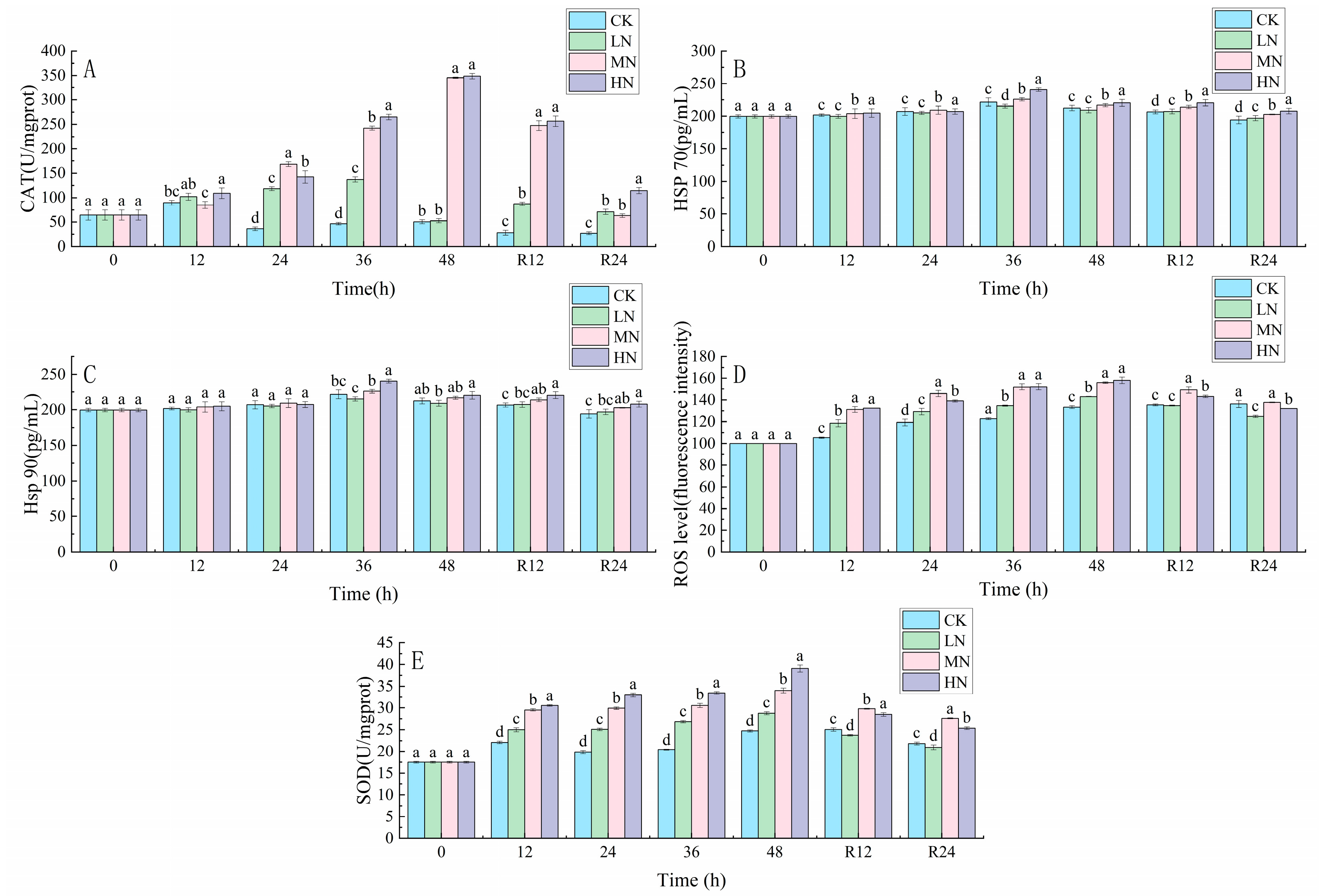
3.1. Oxidative Stress Response

The CAT, SOD, and ROS contents of each sample increased continuously with the transportation time, and the MN and HN groups had higher CAT, SOD, and ROS contents after 48 h of transportation (Figure 1). The CAT, SOD, and ROS contents of all groups decreased to the initial levels. The Hsp 70 and Hsp 90 contents of each group increased with the transport time and reached the peak after 36 h of transport, and the Hsp 70 and Hsp 90 contents of the MN and HN groups were significantly higher than those of the CK group (p < 0.05).

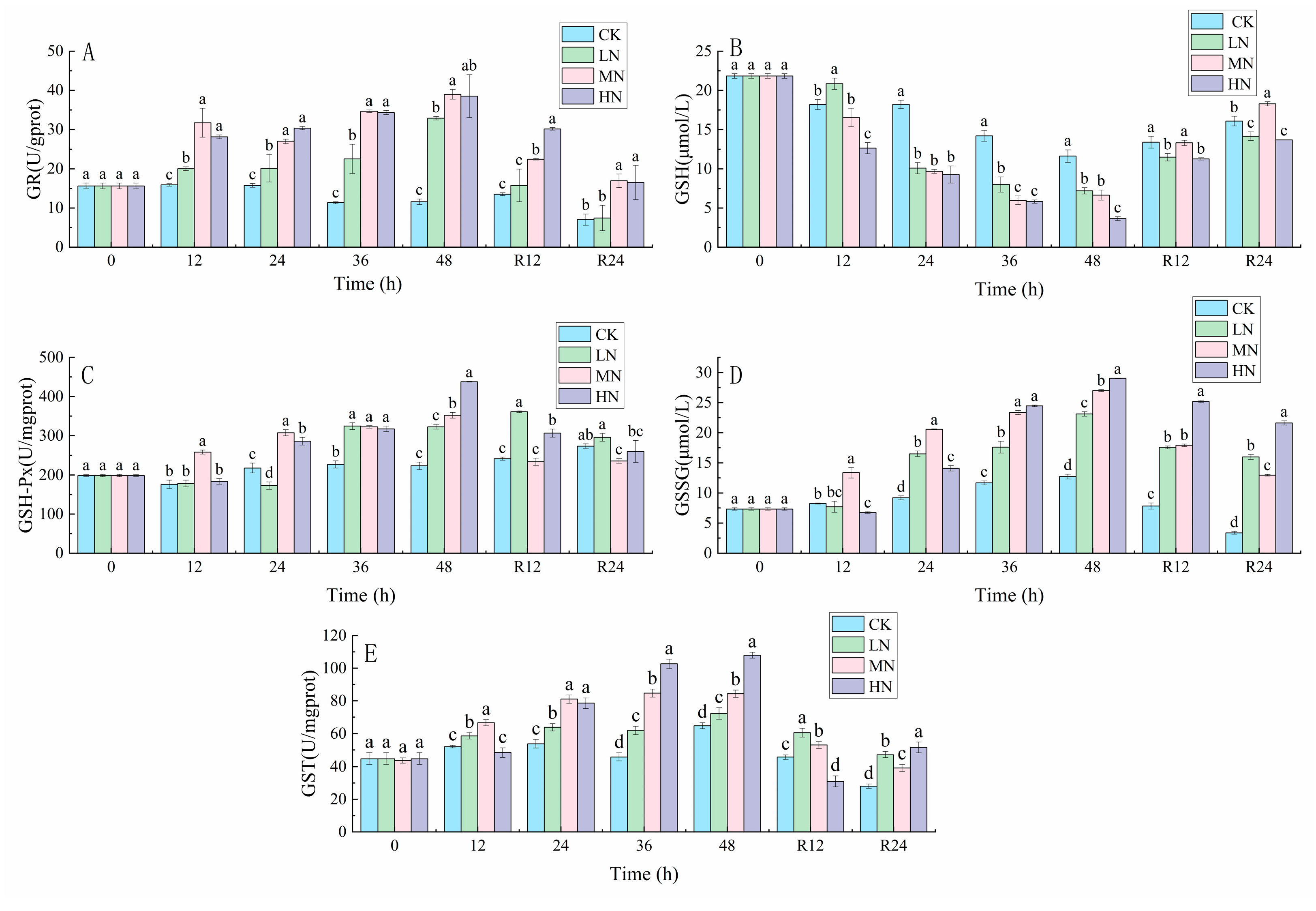
3.2. Glutathione-Related Reactions

During the recovery process, GR and GSSG contents in ammonia stress-treated samples showed a decreasing trend, while GSH contents showed an increasing trend. The contents of GSH-Px and GST in the liver of the NH3-N stress group increased with the transport time and reached the peak at 48th h. The contents of GSH-Px and GST decreased with the recovery time (Figure 2).

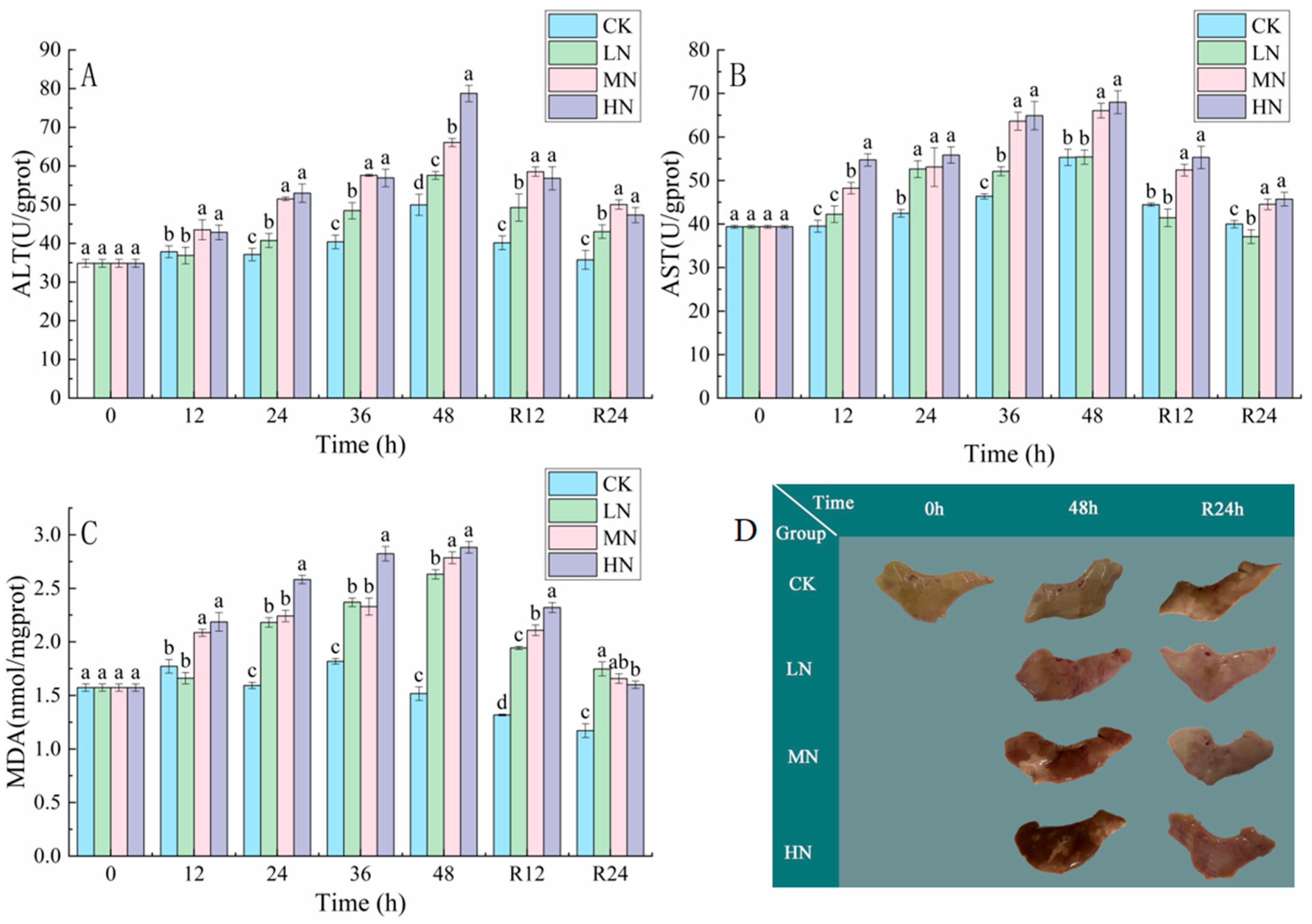
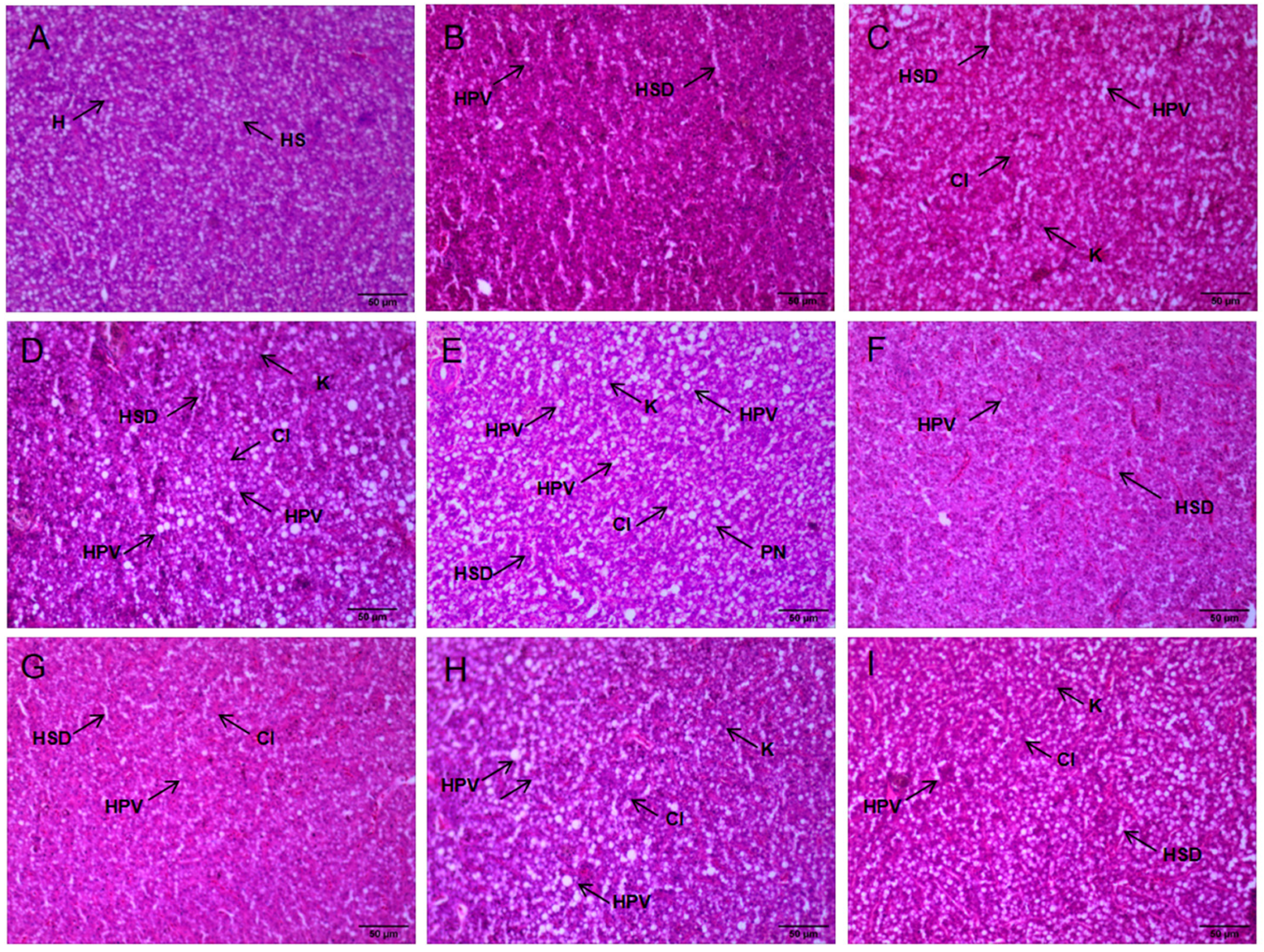
3.3. Tissue Damage and Liver Pathology

The results in Figure 3 show that the ALT, AST, and MDA contents of all groups increased continuously with the transportation time, and the ALT, AST, and MDA contents of the HN group were the highest after 48 h of transportation (p < 0.05), and the ALT, AST, and MDA contents of all groups showed a decreasing trend in the recovery period.
The liver color of the LN, MN, and LN groups was significantly darker than that of the CK after 48 h of NH3-N stress transport, especially the LN group. After 24 h of recovery, the livers of the LN, MN, and LN groups were significantly lighter compared to the 48 h of transport, but still darker compared to the CK (Figure 3).
In addition, histopathological damage of the seabass liver occurred to different degrees after 48 h of NH3-N stress transport (Table 4). The CK liver showed normal hepatocytes with the neat arrangement, normal morphology, clear and intact boundaries, and nuclei located in the center of the cells (Figure 4). Hepatic tissues in the LN and MN groups showed hepatocyte vacuolization and hepatic sinusoidal dilatation compared with the CK (Table 4). Liver tissues in the HN group were more severely damaged, showing more hepatocyte vacuolization and hepatic sinusoidal dilatation, and the nuclei shifted to the periphery and the cell boundaries blurred.

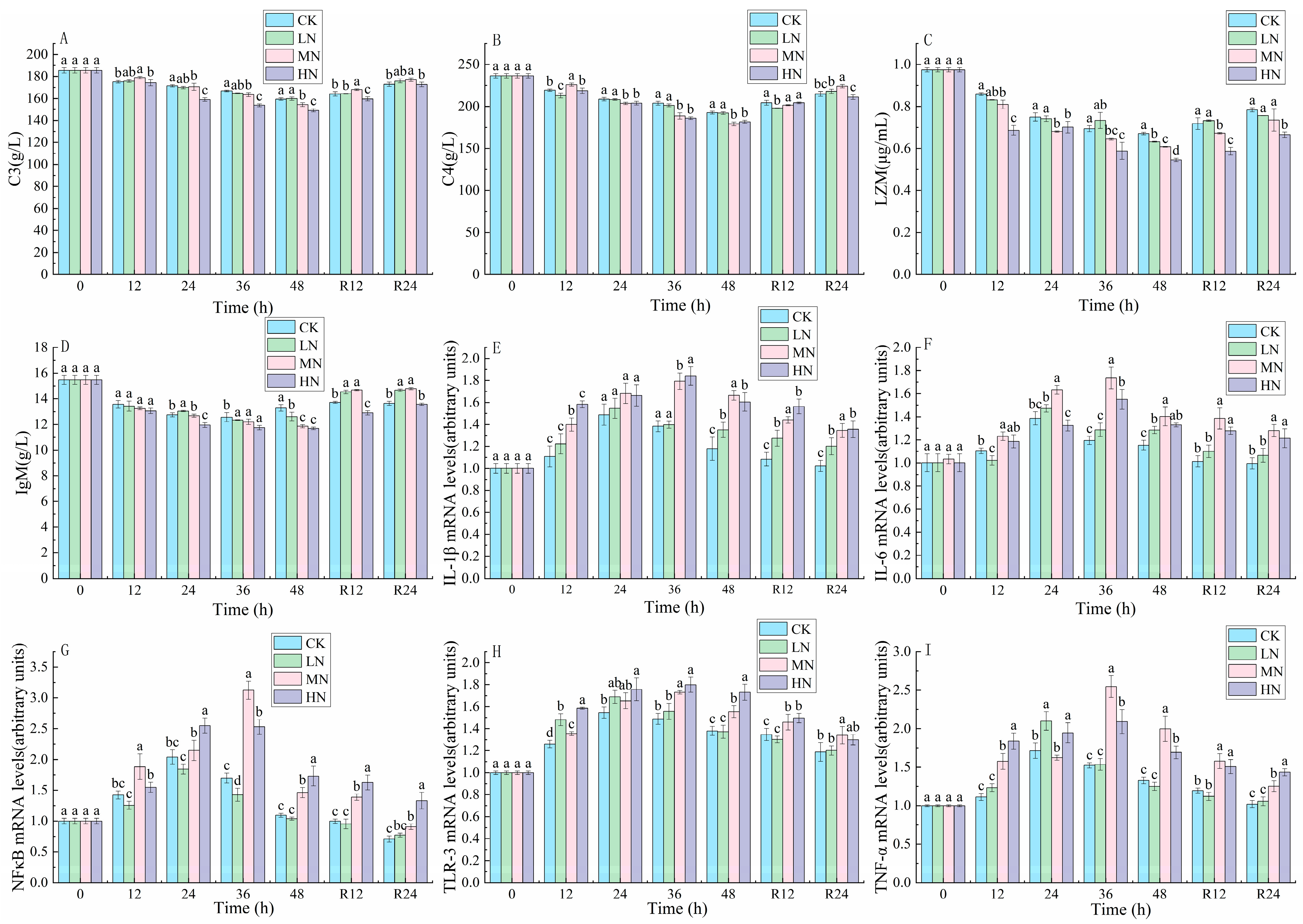
3.4. Immune Response and Inflammatory Factors
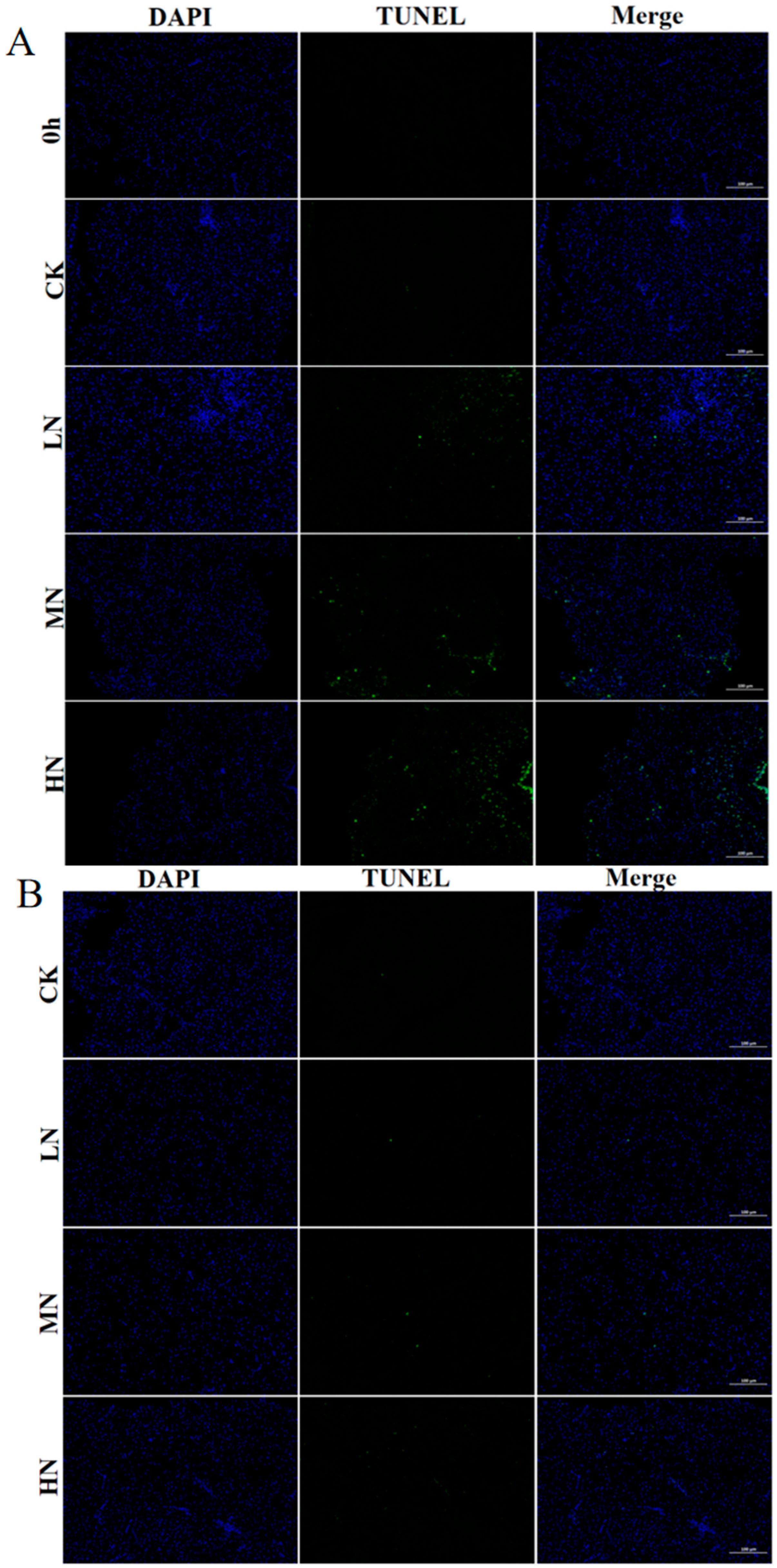
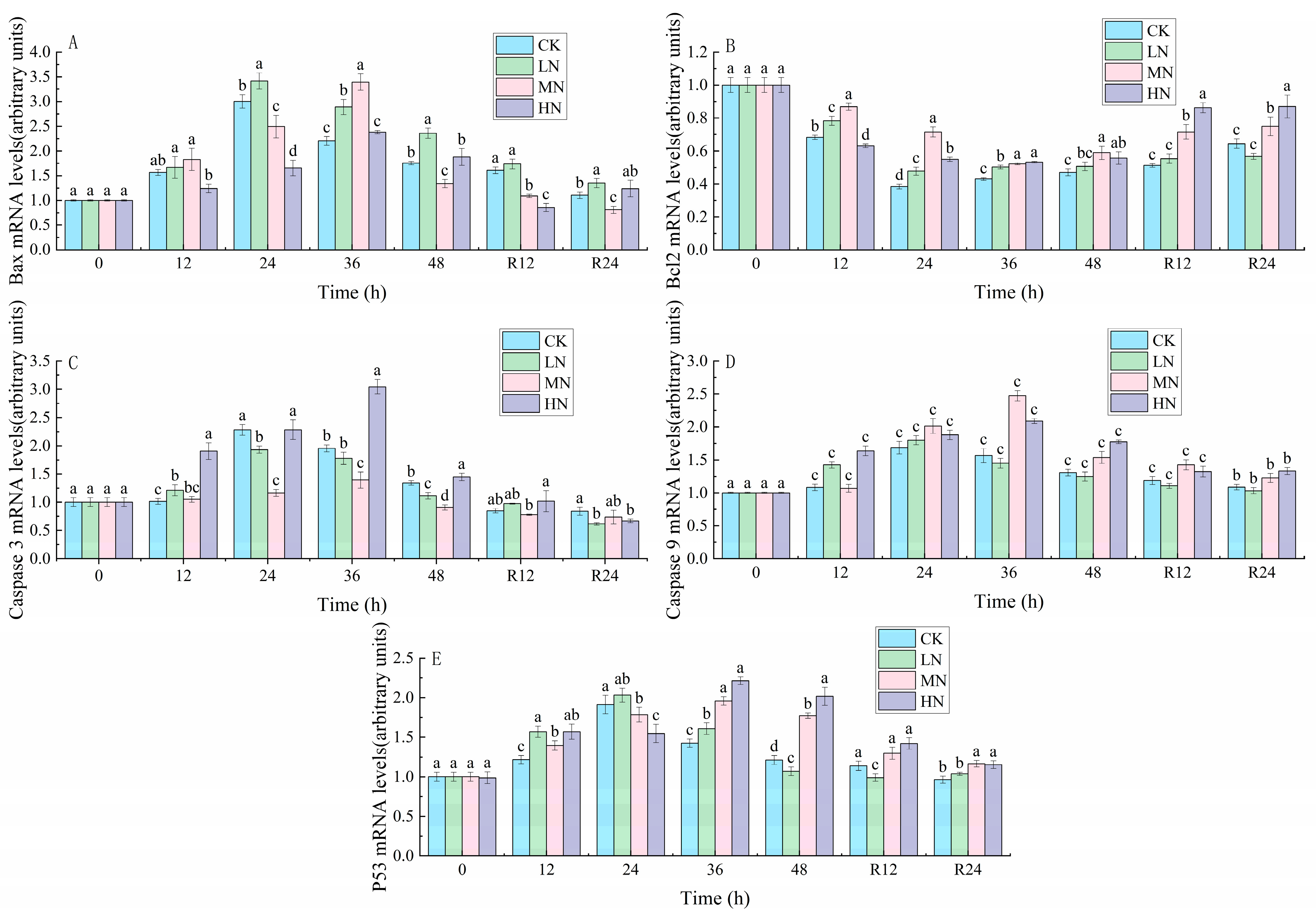
The results in Figure 5 show that the contents of C3, C4, IgM, and LZM decreased continuously with the transport time. The contents of C3, C4, IgM, and LZM increased during the recovery process. After 24 h of recovery, these parameters were still lower in the HN group than in the CK.
The expressions of TLR-3, NFκB, TNF-α, IL-6 and 1L-1β were first increased and then decreased in all groups. With the increase in NH3-N transport stress time, the expression of TLR-3, NFκB, TNF-α, IL-6 and 1L-1β in the CK and LN groups peaked at 24th h, while the gene expression of each pro-inflammatory factor in the MN and HN groups peaked at 36th h (Figure 5).

3.5. Apoptosis

As shown in Figure 6, positive TUNEL signals were detected in all groups after 48 h of NH3-N stress transport. The number of apoptotic cells was HN < MN < LN < CK. After 24 h of recovery, the number of positive cells declined in each group.
The peak expression levels of P53, Caspase 3, Caspase 9 and Bax genes in the CK and LN groups appeared at 24th h, while the peak expression levels of P53, Caspase 3, Caspase 9 and Bax genes in the CK and LN groups appeared at 36th h (Figure 7).

4. Discussion

4.1. The impact of NH3-N Stress Transport on the Antioxidant Defense System of the Liver of Japanese Seabass

ROS are increased in concentration by ammonia, which causes oxidative stress in living things [1]. Organisms reduce ROS by increasing the levels of antioxidant enzymes (e.g., CAT, GST and SOD) to reduce ROS-induced oxidative damage. The initial line of defense in preventing the harm that oxidative stress causes is thought to be SOD-CAT and SOD-GPX [22]. Superoxide is converted to H2O2 by SOD, which is then reduced to water by CAT or GPX [23]. Because it maintains metabolic homeostasis and removes toxins, the liver is the ideal organ for assessing oxidative stress [24]. ROS produced by Japanese seabass under NH3-N stress transport could activate its antioxidant defense system, which prevented the damage caused by NH3-N and transport stress to the organism. A similar finding was made by Cheng et al. [1], where excessive ROS production caused by NH3-N production resulted in increased antioxidant enzyme activity in red puffer fish. Liu et al. [14] discovered that during the start of NH3-N stress, the juvenile golden pompano (Trachinotus ovatus) had an increase in antioxidant enzyme activity, and the antioxidant enzyme activity decreased when it returned to the normal environment. ROS induced by higher NH3-N concentrations were not eliminated by the organisms after 48 h. Chen et al. [25] found that snapping turtle (Chelydra serpentina) exposed to high ambient NH3-N had a high level of basic antioxidant defense, that most likely relied mostly on the GSH system to eliminate extra ROS. GSH/GSSG is also considered one of the essential antioxidant systems in cells, and its stable state is crucial for maintaining the normal physiological functions of cells. NH3-N stress transport resulted in a decline in GSH content and a gradual rise in GSSG content in the liver of seabass in the current study, which is due to GSH being converted to GSSG by GSH-Px and GST in order to scavenge intracellular free radicals and peroxides, thus maintaining normal cell and tissue function and metabolism [23]. After 48 h of NH3-N stress transport, GR activity increased significantly and subsequently decreased. However, the increase in GSSG content was still higher than the decrease in GSH content, indicating that the increase in GR did not offset the GSH produced by GST and GSH-Px. After a period of recovery, GSH and GSSG did not reach their initial states and the glutathione system had not recovered for short-time equilibrium.
Molecular chaperones, such as Hsp, play an essential role in preventing cellular damage from oxidative stress by ensuring the correct folding of proteins to regulate cellular homeostasis [26]. High NH3-N promotes the production of Hsp90 and Hsp70, according to studies [27], which suggests that the buildup of too much ROS and denatured proteins causes the elevation of Hsp levels during NH3-N stress. According to the study’s findings, Hsp70 and Hsp 90 levels were significantly elevated during NH3-N stress transport compared to CK. These findings are in line with the patterns of Hsp70 and Hsp90 gene expression after NH3-N exposure in kuruma shrimp (Marsupenaeus japonicus) and European seabass (Dicentrarchus labrax) [27,28]. Hsp has a key function in the prevention of protein synthesis, repair and degradation as well as apoptosis in Japanese seabass exposed to NH3-N stress. Hsp upregulation may be an effective measure to protect cells from oxidative stress [29].

4.2. The Impact of NH3-N Stress Transport on Liver Tissue Damage in Japanese Seabass

Excess ROS produced by NH3-N stress are not scavenged in time and are responsible for lipid peroxidation, inflammation and cellular damage [30]. The accumulation of ROS generates lipid peroxides (e.g., MDA) that cause cellular damage [31]. MDA levels increase after NH3-N stress transport and reflect the NH3-N generated in the liver of Japanese seabass oxidative stress, which can lead to liver damage. Peacock fish (Poecilia reticulate) were shown to exhibit elevated levels of ROS and MDA as well as altered antioxidant enzyme activity during NH3-N stress in a study by Zhang et al. [32]. However, the MDA content started to decrease after 24 h of recovery, showing that the harm caused by NH3-N stress transport was being gradually counteracted. After a period of recovery, the transport-induced tissue damage caused by NH3-N stress can revert to its original state.
The toxicity of a substance can be determined by measuring elevated AST and ALT activity, which are important markers of tissue damage [22,33]. Ali Taheri Mirghaed et al. [34] found the AST and ALT levels increased in common carp (Cyprinus carpio) after 24 h of NH3-N stress. Zhao et al. [35] found that NH3-N stress exposure resulted in elevated ALT and AST levels, indicating NH3-N stress caused tissue damage in juvenile yellow catfish (Pelteobagrus fulvidraco). In comparison to the CK group, the HN group’s liver was much darker and more congested. NH3-N poisoning can cause changes in the color and appearance of the liver during NH3-N stress [36,37]. NH3-N stress transport resulted in hepatotoxicity, which was characterized by histopathological lesions, such as vacuolation of hepatocytes, dilated hepatic sinusoids, indistinguishable cell contours, deviated nuclei, and nuclei disintegration. The damage to liver tissue was significantly reduced through photograms and histopathological optical microscopy, but it was not enough to recover to the initial level within 48 h. This indicates that the damage caused by NH3-N toxicity is reversible. Furthermore, Yan et al. [23] found that tissue sections of bighead carp (Aristichthys nobilis) after nitrite stress also revealed that liver damage caused by nitrite toxicity could be alleviated to some extent after a recovery period.

4.3. The Impact of NH3-N Stress Transport on Immunological and Inflammatory Responses in Japanese Seabass

The immunological response is a self-protective mechanism for fish against various infections. To combat infectious challenges, fish have evolved different immune systems, including lysozyme and complement. LZM and IgM maintain a healthy state in fish by eliminating invading bacteria and other pathogens while activating the complement system [38]. Studies have shown that a variety of environmental pollutants can inhibit complement C3, complement C4, IgM levels and LZM activities, thus affecting the immunological parameters of scleractinian fish [14,39]. Esam et al. [11] found the mRNA levels of complement C3 and LZM decreased, resulting in immunotoxicity when the combined ammonia- nitrogen and heat stress was applied to Nile tilapia (Oreochromis niloticus). In our study, complement C3, complement C4, IgM levels and LZM activities of seabass decreased after 48 h NH3-N stress transport, indicating a continued decline in immunological function. Gao et al. [40] also found that Takifugu rubripes (T. rubripes) showed a considerable decline in complement C3, complement C4, IgM levels and LZM activities after 12 h of NH3-N stress at different concentrations. After 24 h of recovery, the immunological parameters increased due to the weakening of oxidative stress and the gradual recovery of the immunological system from the toxic environment.
Fish that are under oxidative stress develop an inflammatory reaction. To control inflammation, TLR activation triggers the expression and release of cytokines. The results of this research concur with those of Gao et al. [41], who discovered that NH3-N exposure raised the gene level of TLR-3 and activated the TLR-3 signaling pathway. TNF-α is regarded as a crucial component of the TNF family, which causes inflammation and stimulates the immune system. The activated TNF-α could activate NF-κB channels. NF-κB, as a signaling factor, can activate several pro-inflammatory factors, including IL-6 and IL-1β. In addition, The NF-κB pathway can also be activated by an excessive buildup of ROS, which can worsen the inflammatory response. Our experimental results suggest that both NH3-N stress and transport stress activated the NF-κB inflammatory pathway to exert anti-inflammatory effects. This was in line with previous studies on gibel carp (Carassius gibelio) and guppies (Poecilia reticulate), where it was discovered that NH3-N stress induced the expression of genes linked to inflammation, resulting in an inflammatory response [32,42]. Furthermore, we found that the different timing of peak gene expression of anti-inflammatory factors may vary depending on the NH3-N concentration.

4.4. The Impact of NH3-N Stress Transport on Apoptosis in Japanese Seabass

In addition to inducing inflammation, oxidative stress also leads to apoptosis. The oncogene P53 is a crucial regulator of apoptosis, and ROS production brought on by NH3-N stress can modulate the activity of P53. In response to stress, P53 alters the expression of Bcl2 and Bax to induce apoptosis. The increase in the ratio of Bax to Bcl2 can be used to determine the apoptosis of the organism [1]. In the current research, the Bax/Bcl2 ratio increased in all experimental groups during NH3-N stress transport, suggesting that NH3-N stress transport could cause cell apoptosis via the P53-Bax-Bcl2 pathway. To identify stress-induced apoptosis, Caspase expression is a crucial marker. According to Li et al. [43], that NH3-N exposure upregulated genes Caspase 3 and Caspase 9 and induced cell apoptosis. The current work showed that the expression of Caspase 9 and Caspase 3 was raised by both NH3-N exposure and transport stress, which also suggested that Caspase-dependent apoptotic pathways might be implicated in the cell death generated by transport stress of NH3-N.
The results of TUNEL staining showed green fluorescent spots, which verified the hypothesis that NH3-N stress transport leads to apoptosis. The P53-Bax/Bcl2 apoptotic route and the Caspase-dependent apoptotic pathway may both be affected by NH3-N stress in Yellow catfish (Pelteobagrus fulvidraco) [36]. This is in line with what the current experimental study’s findings show. These findings also imply that oxidative stress and inflammation may be intimately related to cell apoptosis brought on by simultaneous exposure to these two stressors.

5. Conclusions

This research was the first to investigate the influence of NH3-N on oxidative stress, apoptosis, tissue damage and immunological response in Japanese seabass during keep-live transport. The histopathological and TUNEL results confirmed that ammonia and nitrogen stress during transport could produce ROS, resulting in oxidative stress, and that eliminated ROS through the glutathione system, but this system failed to provide adequate protection when exposed to highly oxidizing conditions. Excess ROS was involved in NH3-N stress-induced apoptosis through the P53-Bax-Bcl2 pathway and the cysteine-dependent apoptosis pathway and induced an inflammatory response via the NF-κB pathway. These defenses declined during the recovery period, but did not return to the normal levels compared to CK. The findings of this research help towards a thorough understanding of how environmental factors effect seabass during keep-live transport, which helps towards developing optimal procedures for transport and improving the survival rate during transport.

Author Contributions

Conceptualization, M.G. and Q.Y.; formal analysis, M.G., Q.Y., Y.D. and J.M.; funding acquisition, J.X.; investigation, Q.Y.; methodology, M.G., Y.D. and Z.D.; project administration, J.X.; resources, J.M.; software, M.G. and Z.D.; supervision, J.X.; writing—original draft, M.G.; writing—review and editing, J.M. and J.X. All authors have read and agreed to the published version of the manuscript.

Funding

The research was supported by the Agriculture Research System of China (CARS-47) and National Key R&D Program of China (2019YFD0901601).

Institutional Review Board Statement

The experimental procedures were approved by the Animal Care and Use Committee of Shanghai Ocean University (SHOU-DW-2022-059).

Informed Consent Statement

Not applicable.

Data Availability Statement

The data presented in this study are available upon request from the corresponding author.

Conflicts of Interest

The authors attest that there are no conflict of interest concerning this paper.

References

  1. Cheng, C.-H.; Yang, F.-F.; Ling, R.-Z.; Liao, S.-A.; Miao, Y.-T.; Ye, C.-X.; Wang, A.-L. Effects of ammonia exposure on apoptosis, oxidative stress and immune response in pufferfish (Takifugu obscurus). Aquat. Toxicol. 2015, 164, 61–71. [Google Scholar] [CrossRef] [PubMed]
  2. Wang, Y.; Wang, B.; Liu, M.; Jiang, K.; Wang, M.; Wang, L. Aflatoxin B1 (AFB1) induced dysregulation of intestinal microbiota and damage of antioxidant system in pacific white shrimp (Litopenaeus vannamei). Aquaculture 2018, 495, 940–947. [Google Scholar] [CrossRef]
  3. Hodkovicova, N.; Chmelova, L.; Sehonova, P.; Blahova, J.; Doubkova, V.; Plhalova, L.; Fiorino, E.; Vojtek, L.; Vicenova, M.; Siroka, Z.; et al. The effects of a therapeutic formalin bath on selected immunological and oxidative stress parameters in common carp (Cyprinus carpio). Sci. Total Environ. 2019, 653, 1120–1127. [Google Scholar] [CrossRef] [PubMed]
  4. Schafer, F.Q.; Buettner, G.R. Redox environment of the cell as viewed through the redox state of the glutathione disulfide/glutathione couple. Free Radic. Biol. Med. 2001, 30, 1191–1212. [Google Scholar] [CrossRef] [PubMed]
  5. Kim, J.-H.; Kim, S.K.; Hur, Y.B. Temperature-mediated changes in stress responses, acetylcholinesterase, and immune responses of juvenile olive flounder Paralichthys olivaceus in a bio-floc environment. Aquaculture 2019, 506, 453–458. [Google Scholar] [CrossRef]
  6. Liu, B.; Jia, R.; Huang, B.; Lei, J. Interactive effect of ammonia and crowding stress on ion-regulation and expression of immune-related genes in juvenile turbot (Scophthalmus maximus). Mar. Freshw. Behav. Physiol. 2017, 50, 179–194. [Google Scholar] [CrossRef]
  7. Xu, Z.; Cao, J.; Qin, X.; Qiu, W.; Mei, J.; Xie, J. Toxic Effects on Bioaccumulation, Hematological Parameters, Oxidative Stress, Immune Responses and Tissue Structure in Fish Exposed to Ammonia Nitrogen: A Review. Animals 2021, 11, 3304. [Google Scholar] [CrossRef]
  8. Zheng, T.; Song, Z.; Tao, Y.; Qiang, J.; Ma, J.; Lu, S.; Xu, P. Transport stress induces innate immunity responses through TLR and NLR signaling pathways and increases mucus cell number in gills of hybrid yellow catfish (Tachysurus fulvidraco ♀ × Pseudobagrus vachellii ♂). Fish Shellfish Immunol. 2022, 127, 166–175. [Google Scholar] [CrossRef]
  9. Wu, Y.; You, X.; Sun, W.; Xiong, G.; Shi, L.; Qiao, Y.; Wu, W.; Li, X.; Wang, J.; Ding, A.; et al. Insight into acute heat stress on meat qualities of rainbow trout (Oncorhynchus mykiss) during short-time transportation. Aquaculture 2021, 543, 737013. [Google Scholar] [CrossRef]
  10. Molayemraftar, T.; Peyghan, R.; Razi Jalali, M.; Shahriari, A. Single and combined effects of ammonia and nitrite on common carp, Cyprinus carpio: Toxicity, hematological parameters, antioxidant defenses, acetylcholinesterase, and acid phosphatase activities. Aquaculture 2022, 548, 737676. [Google Scholar] [CrossRef]
  11. Esam, F.; Khalafalla, M.M.; Gewaily, M.S.; Abdo, S.; Hassan, A.M.; Dawood, M.A.O. Acute ammonia exposure combined with heat stress impaired the histological features of gills and liver tissues and the expression responses of immune and antioxidative related genes in Nile tilapia. Ecotoxicol. Environ. Saf. 2022, 231, 113187. [Google Scholar] [CrossRef] [PubMed]
  12. Wang, Q.; Mei, J.; Cao, J.; Xie, J. Effects of Melissa officinalis L. Essential Oil in Comparison with Anaesthetics on Gill Tissue Damage, Liver Metabolism and Immune Parameters in Sea Bass (Lateolabrax maculatus) during Simulated Live Transport. Biology 2022, 11, 11. [Google Scholar] [CrossRef] [PubMed]
  13. He, K.; Luo, X.; Wen, M.; Wang, C.; Qin, C.; Shao, J.; Gan, L.; Dong, R.; Jiang, H. Effect of acute ammonia toxicity on inflammation, oxidative stress and apoptosis in head kidney macrophage of Pelteobagrus fulvidraco and the alleviation of curcumin. Comp. Biochem. Physiol. Part C Toxicol. Pharmacol. 2021, 248, 109098. [Google Scholar] [CrossRef] [PubMed]
  14. Liu, M.-J.; Guo, H.-Y.; Zhu, K.-C.; Liu, B.-S.; Liu, B.; Guo, L.; Zhang, N.; Yang, J.-W.; Jiang, S.-G.; Zhang, D.-C. Effects of acute ammonia exposure and recovery on the antioxidant response and expression of genes in the Nrf2-Keap1 signaling pathway in the juvenile golden pompano (Trachinotus ovatus). Aquat. Toxicol. 2021, 240, 105969. [Google Scholar] [CrossRef]
  15. Livak, K.J.; Schmittgen, T.D. Analysis of Relative Gene Expression Data Using Real-Time Quantitative PCR and the 2−ΔΔCT Method. Methods 2001, 25, 402–408. [Google Scholar] [CrossRef]
  16. Tan, P.; Dong, X.; Xu, H.; Mai, K.; Ai, Q. Dietary vegetable oil suppressed non-specific immunity and liver antioxidant capacity but induced inflammatory response in Japanese sea bass (Lateolabrax japonicus). Fish Shellfish Immunol. 2017, 63, 139–146. [Google Scholar] [CrossRef]
  17. Huang, H.; Zhang, X.; Liang, X.; Wu, X.; Gu, X.; Han, J.; Xue, M. N-carbamoylglutamate improves lipid metabolism, inflammation, and apoptosis responses in visceral adipocytes of Japanese seabass (Lateolabrax japonicus), in vivo and in vitro. Anim. Nutr. 2021, 7, 707–715. [Google Scholar] [CrossRef]
  18. Li, X.; Sun, J.; Wang, L.; Song, K.; Lu, K.; Zhang, L.; Ma, X.; Zhang, C. Effects of dietary vitamin E levels on growth, antioxidant capacity and immune response of spotted seabass (Lateolabrax maculatus) reared at different water temperatures. Aquaculture 2023, 565, 739141. [Google Scholar] [CrossRef]
  19. Fan, H.; Wang, L.; Wen, H.; Wang, K.; Qi, X.; Li, J.; He, F.; Li, Y. Genome-wide identification and characterization of toll-like receptor genes in spotted sea bass (Lateolabrax maculatus) and their involvement in the host immune response to Vibrio harveyi infection. Fish Shellfish Immunol. 2019, 92, 782–791. [Google Scholar] [CrossRef]
  20. Xiang, Y.; Jia, P.; Liu, W.; Yi, M.; Jia, K. Comparative transcriptome analysis reveals the role of p53 signalling pathway during red-spotted grouper nervous necrosis virus infection in Lateolabrax japonicus brain cells. J. Fish Dis. 2019, 42, 585–595. [Google Scholar] [CrossRef]
  21. Le, Y.; Jia, P.; Jin, Y.; Liu, W.; Jia, K.; Yi, M. The antiviral role of heat shock protein 27 against red spotted grouper nervous necrosis virus infection in sea perch. Fish Shellfish Immunol. 2017, 70, 185–194. [Google Scholar] [CrossRef] [PubMed]
  22. Zhang, H.; Wang, Q.; Dong, Y.; Mei, J.; Xie, J. Effects of tricaine methanesulphonate (MS-222) on physiological stress and fresh quality of sea bass (Lateolabrax maculatus) under simulated high-density and long-distance transport stress. Biology 2023, 12, 223. [Google Scholar] [CrossRef] [PubMed]
  23. Lin, Y.; Miao, L.-H.; Pan, W.-J.; Huang, X.; Dengu, J.M.; Zhang, W.-X.; Ge, X.-P.; Liu, B.; Ren, M.-C.; Zhou, Q.-L.; et al. Effect of nitrite exposure on the antioxidant enzymes and glutathione system in the liver of bighead carp, Aristichthys nobilis. Fish Shellfish Immunol. 2018, 76, 126–132. [Google Scholar] [CrossRef] [PubMed]
  24. Abdel-Latif, H.M.R.; Shukry, M.; El Euony, O.I.; Mohamed Soliman, M.; Noreldin, A.E.; Ghetas, H.A.; Dawood, M.A.O.; Khallaf, M.A. Hazardous Effects of SiO2 Nanoparticles on Liver and Kidney Functions, Histopathology Characteristics, and Transcriptomic Responses in Nile Tilapia (Oreochromis niloticus) Juveniles. Biology 2021, 10, 183. [Google Scholar] [CrossRef]
  25. Chen, X.; Li, M.; Niu, C. Diverse defense responses to ammonia stress in three freshwater turtles. Aquaculture 2022, 546, 737302. [Google Scholar] [CrossRef]
  26. Cui, Y.; Zhao, N.; Wang, C.; Long, J.; Chen, Y.; Deng, Z.; Zhang, Z.; Zhao, R.; Sun, J.; Wang, Z.; et al. Acute ammonia stress-induced oxidative and heat shock responses modulated by transcription factors in Litopenaeus vannamei. Fish Shellfish Immunol. 2022, 128, 181–187. [Google Scholar] [CrossRef]
  27. Sinha, A.K.; Zinta, G.; AbdElgawad, H.; Asard, H.; Blust, R.; De Boeck, G. High environmental ammonia elicits differential oxidative stress and antioxidant responses in five different organs of a model estuarine teleost (Dicentrarchus labrax). Comp. Biochem. Physiol. Part C Toxicol. Pharmacol. 2015, 174, 21–31. [Google Scholar] [CrossRef]
  28. Ou, H.; Liang, J.; Liu, J. Effects of acute ammonia exposure on oxidative stress, endoplasmic reticulum stress and apoptosis in the kuruma shrimp (Marsupenaeus japonicus). Aquac. Rep. 2022, 27, 101383. [Google Scholar] [CrossRef]
  29. Li, X.; Liu, S.; Wang, Y.; Lu, W.; Zhang, Q.; Cheng, J. Genomic and Transcriptomic Landscape and Evolutionary Dynamics of Heat Shock Proteins in Spotted Sea Bass (Lateolabrax maculatus) under Salinity Change and Alkalinity Stress. Biology 2022, 11, 353. [Google Scholar] [CrossRef]
  30. Elshopakey, G.E.; Mahboub, H.H.; Sheraiba, N.I.; Abduljabbar, M.H.; Mahmoud, Y.K.; Abomughaid, M.M.; Ismail, A.K. Ammonia toxicity in Nile tilapia: Potential role of dietary baicalin on biochemical profile, antioxidant status and inflammatory gene expression. Aquac. Rep. 2023, 28, 101434. [Google Scholar] [CrossRef]
  31. Bian, C.; Yu, H.; Yang, K.; Mei, J.; Xie, J. Effects of single-, dual-, and multi-frequency ultrasound-assisted freezing on the muscle quality and myofibrillar protein structure in large yellow croaker (Larimichthys crocea). Food Chem. X 2022, 15, 100362. [Google Scholar] [CrossRef] [PubMed]
  32. Zhang, C.; Ma, J.; Qi, Q.; Xu, M.; Xu, R. Effects of ammonia exposure on anxiety behavior, oxidative stress and inflammation in guppy (Poecilia reticulate). Comp. Biochem. Physiol. Part C Toxicol. Pharmacol. 2023, 265, 109539. [Google Scholar] [CrossRef] [PubMed]
  33. Shi, A.; Ma, H.; Shi, X.; Zhou, W.; Pan, W.; Song, Y.; Chen, Q.; Yu, X.; Niu, C.; Yang, Y.; et al. Effects of microbe-derived antioxidants on growth, digestive and aminotransferase activities, and antioxidant capacities in the hepatopancreas of Eriocheir sinensis under ammonia nitrogen stress. Aquac. Fish. 2023, in press. [Google Scholar] [CrossRef]
  34. Taheri Mirghaed, A.; Paknejad, H.; Mirzargar, S.S. Hepatoprotective effects of dietary Artemisia (Artemisia annua) leaf extract on common carp (Cyprinus carpio) exposed to ambient ammonia. Aquaculture 2020, 527, 735443. [Google Scholar] [CrossRef]
  35. Zhao, H.; Peng, K.; Wang, G.; Mo, W.; Huang, Y.; Cao, J. Metabolic changes, antioxidant status, immune response and resistance to ammonia stress in juvenile yellow catfish (Pelteobagrus fulvidraco) fed diet supplemented with sodium butyrate. Aquaculture 2021, 536, 736441. [Google Scholar] [CrossRef]
  36. Sun, Z.; Wang, S.; Zhang, M.; Jiang, H.; Li, M. Chronic toxicity study of ammonia exposure in juvenile yellow catfish Pelteobagrus fulvidraco. Aquaculture 2023, 567, 739266. [Google Scholar] [CrossRef]
  37. Zhang, M.; Wang, S.; Sun, Z.; Jiang, H.; Qian, Y.; Wang, R.; Li, M. The effects of acute and chronic ammonia exposure on growth, survival, and free amino acid abundance in juvenile Japanese sea perch Lateolabrax japonicus. Aquaculture 2022, 560, 738512. [Google Scholar] [CrossRef]
  38. Chen, W.; Gao, S.; Huang, Y.; Chang, K.; Zhao, X. Addition of Chlorella sorokiniana meal in the diet of juvenile rainbow trout (Oncorhynchus mykiss): Influence on fish growth, gut histology, oxidative stress, immune response, and disease resistance against Aeromonas salmonicida. Fish Shellfish Immunol. 2022, 129, 243–250. [Google Scholar] [CrossRef]
  39. Gao, X.-Q.; Fei, F.; Huo, H.H.; Huang, B.; Meng, X.S.; Zhang, T.; Liu, B.-L. Impact of nitrite exposure on plasma biochemical parameters and immune-related responses in Takifugu rubripes. Aquat. Toxicol. 2020, 218, 105362. [Google Scholar] [CrossRef]
  40. Gao, X.; Wang, X.; Wang, X.; Fang, Y.; Cao, S.; Huang, B.; Chen, H.; Xing, R.; Liu, B. Toxicity in Takifugu rubripes exposed to acute ammonia: Effects on immune responses, brain neurotransmitter levels, and thyroid endocrine hormones. Ecotoxicol. Environ. Saf. 2022, 244, 114050. [Google Scholar] [CrossRef]
  41. Gao, X.-Q.; Fei, F.; Huang, B.; Meng, X.S.; Zhang, T.; Zhao, K.-F.; Chen, H.-B.; Xing, R.; Liu, B.-L. Alterations in hematological and biochemical parameters, oxidative stress, and immune response in Takifugu rubripes under acute ammonia exposure. Comp. Biochem. Physiol. Part C Toxicol. Pharmacol. 2021, 243, 108978. [Google Scholar] [CrossRef] [PubMed]
  42. Wu, L.; Chen, Q.; Dong, B.; Han, D.; Zhu, X.; Liu, H.; Yang, Y.; Xie, S.; Jin, J. Resveratrol attenuated oxidative stress and inflammatory and mitochondrial dysfunction induced by acute ammonia exposure in gibel carp (Carassius gibelio). Ecotoxicol. Environ. Saf. 2023, 251, 114544. [Google Scholar] [CrossRef] [PubMed]
  43. Li, M.; Zhang, M.; Qian, Y.; Shi, G.; Wang, R. Ammonia toxicity in the yellow catfish (Pelteobagrus fulvidraco): The mechanistic insight from physiological detoxification to poisoning. Fish Shellfish Immunol. 2020, 102, 195–202. [Google Scholar] [CrossRef] [PubMed]
Figure 1. Changes in catalase (CAT) (A), heat stress protein 70 (Hsp 70) (B), heat stress protein 90 (Hsp90) (C), reactive oxygen species (ROS) (D) and superoxide dismutase (SOD) (E) concentrations in the liver of Japanese seabass exposed to NH3-N of 6.4 mg/L (LA), 12.8 mg/L (MA), 19.2 mg/L (HA), and 0 mg/L (CK) for 48 h and recovered for 24 h, respectively. Values are expressed as the mean ± S.D. Different lowercase letters indicate significant differences (p < 0.05) among groups. The CK NH3-N group served as the control. (p < 0.05, N = 3).
Biology 12 00769 g001
Figure 2. Changes in glutathione reductase (GR) (A), glutathione (GSH) (B), glutathione peroxidase (GSH-Px) (C), oxidized glutathione (GSSG) (D) and glutathione S-transferase (GST) (E) contents in the liver of Japanese seabass exposed to NH3-N of 6.4 mg/L (LA), 12.8 mg/L (MA), 19.2 mg/L (HA), and 0 mg/L (CK) for 48 h and recovered for 24 h, respectively. Values are expressed as the mean ± S.D. Different lowercase letters indicate significant differences (p < 0.05) among groups. The CK NH3-N group served as the control. (p < 0.05, N = 3).
Biology 12 00769 g002
Figure 3. Changes in alanine aminotransferase (ALT) (A), aspartate aminotransferase (AST) (B), malondialdehyde (MDA) (C) and morphological of the liver (D) contents in the liver of Japanese seabass exposed to NH3-N of 6.4 mg/L (LA), 12.8 mg/L (MA), 19.2 mg/L (HA), and 0 mg/L (CK) for 48 h and recovered for 24 h, respectively. Values are expressed as the mean ± S.D. Different lowercase letters indicate significant differences (p < 0.05) among groups. The CK NH3-N group served as the control. (p < 0.05. N = 3).
Biology 12 00769 g003
Figure 4. Paraffin sections of liver tissue were stained with HE to assess the pathological changes in different NH3-N concentrations on Japanese seabass. Micrographs (×400) and scale bars (50 μm). (A) represents tissue sections of the normal liver at 0 h; (BE) represents liver tissue sections of 6.4 mg/L (LA), 12.8 mg/L (MA), 19.2 mg/L (HA), and 0 mg/L (CK) at 48th h after NH3-N stress transport; (FI) represents liver tissue sections of 6.4 mg/L (LA), 12.8 mg/L (MA), 19.2 mg/L (HA), and 0 mg/L (CK) at 24th h after recovery from NH3-N stress transport. The CK NH3-N group served as the control. (H, hepatocyte; VD, vacuolar degeneration; HS, hepatic sinusoid; HSD, hepatic sinusoid dilatation; K. karyolysis; HPV, hepatocellular vacuolation; PN, cellular peripheral nucleus; CI, cellular outline indistinguishable).
Biology 12 00769 g004
Figure 5. Changes in complement C3 (A), complement C4 (B), lysozyme (LZM) (C), immunoglobulin (IgM) (D), interleukin 1β (IL-1β) (E), interleukin 6 (IL-6) (F), nuclear factor kappa β (NF-κB) (G), Toll-like receptor 3 (TLR-3) (H) and tumor necrosis factor α (TNF-α) (I) contents in the liver of Japanese seabass exposed to NH3-N of 6.4 mg/L (LA), 12.8 mg/L (MA), 19.2 mg/L (HA), and 0 mg/L (CK) for 48 h and recovered for 24 h, respectively. Values are expressed as the mean ± S.D. Different lowercase letters indicate significant differences (p < 0.05) among groups. The CK NH3-N group served as the control (p < 0.05, N = 3).
Biology 12 00769 g005
Figure 6. The TUNEL staining results of liver tissues of Japanese marine fish exposed to 6.4 mg/L (LA), 12.8 mg/L (MA), 19.2 mg/L (HA) and 0 mg/L (CK) NH3-N concentrations in transport water at 0th h and 48th h (A) and after 24 h of recovery (B). The CK NH3-N group served as the control.
Biology 12 00769 g006
Figure 7. Changes in Bax (A), Bcl2 (B), Caspase 3 (C), Caspase 9 (D) and P53 (E) contents in the liver of Japanese seabass exposed to NH3-N of 6.4 mg/L (LA), 12.8 mg/L (MA), 19.2 mg/L (HA), and 0 mg/L (CK) for 48 h and recovered for 24 h, respectively. Values are expressed as the mean ± S.D. Different lowercase letters indicate significant differences (p < 0.05) among groups. The CK NH3-N group served as the control. (p < 0.05, N = 3).
Biology 12 00769 g007
Table 1. Experimental groups and actual NH3-N concentration.
Experimental GroupActual NH3-N Concentration (mg/L)
Control group (CK)0.3
Low NH3-N group (LA, 6.4 mg/L)6.5
Medium NH3-N group (MA, 12.8 mg/L)11.5
High NH3-N group (HA, 19.2 mg/L)19.0
Table 2. The reaction parameters for quantitative PCR.
StageTemperature and TimeProcess
Stage 195 °C, 30 spredenaturation
Stage 295 °C, 15 sdenature
60 °C, 30 s
(40 cycles)
annealing/extension
Stage 365 °C → 95 °Cmelting curve: every time the temperature rises by 0.5 °C, the fluorescence signal is collected
Table 3. Primer sequence for quantitative PCR.
GenePrimer Sequence (5′–3′)Product Length (bp)Temperature (°C)Efficiency (%)References
Apoptosis gene
ActinF: CAACTGGGATGACATGGAGAAG20060101[16]
R: TTGGCTTTGGGGTTCAGG
TNF-αF: GACTCCATAGGCAGCAAAGC20560103.2[17]
R: AGAAAGTCTTGCCCTCGTCA
IL-1βF: CTGAACATCAAGGGCACAGA1926092.8[17]
R: GTTGAAGGGGACAGACCTGA
IL-6F: TACAATGTCCTCCTCAAGCACG1296098.4[18]
R: GCCTTTGACCTCCTCCATCAG
TLR-3F: GGCCTGGATCAAATTCAAGA13960103[19]
R: GACAGGGGACTGAATGGAGA
NF-κBF: GAAGGTATGGGAGGAGGAGTTT18960102[20]
R: AACCACAGGGTCCAGAGGAAA
Pro-inflammatory factor
P53F: ACCATCCTGCTGAGCTTCAT1786095.3[20]
R: GCCCAAAACAAGTCCCTCTG
Caspase 3F: ATCACAGCAACTACGCCTCATTCG1766098.9[17]
R: GCCTCTGCAAGCCTGGATGAAG
Caspase 9F: TGCGGAGGAGGTGAACGAGAC1386090.5[17]
R: CGGTTCGTCGGACATGCTCAG
BaxF: GCTCCAAAGGATGATAAACGAC1826096.7[20]
R: AACAGTGCAACCACCCGAC
Bcl2F: GTGGGGCTCTTCGCTTTTG1916095.1[21]
R: CCATCCTCCTTGGCTCTGGA
Table 4. The damage of liver records of Japanese bass transported under different NH3-N stress was evaluated semi-quantitatively.
TimeGroupsHPVHSDPNKCI
0 hCK
48 hCK+++++
LN+++++++
MN++++++++
HN++++++++++++
R24 hCK+++
LN+++
MN++++++
HN+++++++
(−) no histopathology; histopathology of (+) <20% visual field; (++) 20–60% visual field histopathology; histopathology in (+++) >60% visual field. R means recovery.
Disclaimer/Publisher’s Note: The statements, opinions and data contained in all publications are solely those of the individual author(s) and contributor(s) and not of MDPI and/or the editor(s). MDPI and/or the editor(s) disclaim responsibility for any injury to people or property resulting from any ideas, methods, instructions or products referred to in the content.

