Review

The Gut Microbiota in Kidney Transplantation: A Target for Personalized Therapy?

by
Yuselys García-Martínez
1,*,
Margherita Borriello
2,
Giovanna Capolongo
1,
Diego Ingrosso
2 and
Alessandra F. Perna
1
1
Department of Translational Medical Science, University of Campania “Luigi Vanvitelli”, Via Pansini, Bldg 17, 80131 Naples, Italy
2
Department of Precision Medicine, University of Campania “Luigi Vanvitelli”, Via L. De Crecchio 7, 80138 Naples, Italy
*
Author to whom correspondence should be addressed.
Biology 2023, 12(2), 163; https://doi.org/10.3390/biology12020163
Submission received: 19 December 2022 / Revised: 15 January 2023 / Accepted: 17 January 2023 / Published: 20 January 2023
(This article belongs to the Special Issue Gut Microbiome in Health and Disease)

Abstract

:

Simple Summary

Kidney function is compromised by several post-transplant complications associated with immunosuppressive therapy, infections, gastrointestinal toxicity, and graft rejection in kidney transplant patients. Given the ability of the gut microbiota to influence alloimmunity, drug metabolism, infections, and gastrointestinal diseases, we studied the therapeutic potential of the gut microbiota in kidney transplantation. Specific microbial signatures have been associated with graft rejection, mycophenolate mofetil and tacrolimus metabolism, and the development of new-onset diabetes after transplantation. In addition, the abundance of gut enterobacteria has been linked to the development of urinary tract infections, while other microbial populations have a protective role in urinary and respiratory tract infections. The application of microbiota-based therapies such as fecal microbiota transplantation has successfully resolved infection and refractory diarrhea events in these patients. Current data suggest that modulating the gut microbiota could potentially contribute to personalizing immunosuppressive and post-transplant complication therapies to improve graft survival and patients’ quality of life.

Abstract

Kidney transplantation improves quality of life, morbidity, and mortality of patients with kidney failure. However, integrated immunosuppressive therapy required to preserve graft function is associated with the development of post-transplant complications, including infections, altered immunosuppressive metabolism, gastrointestinal toxicity, and diarrhea. The gut microbiota has emerged as a potential therapeutic target for personalizing immunosuppressive therapy and managing post-transplant complications. This review reports current evidence on gut microbial dysbiosis in kidney transplant recipients, alterations in their gut microbiota associated with kidney transplantation outcomes, and the application of gut microbiota intervention therapies in treating post-transplant complications.

1. Introduction

Kidney transplantation (KT) is the treatment of choice for patients with end-stage kidney disease (ESKD). While successful KT improves patients’ quality of life, morbidity, and mortality compared to those on maintenance dialysis, integrated immunosuppressive and antimicrobial therapies increase the risk of post-transplant complications [1]. Immunosuppressants possess a narrow therapeutic index and large interpatient variability regarding dose requirements. Supratherapeutic levels can promote the development of post-transplant malignancy, nephrotoxicity, gastrointestinal toxicity, and opportunistic infections, whereas subtherapeutic levels can trigger immune rejection [2]. Clinical guidelines suggest personalizing immunosuppressive therapies according to each patient’s risk for acute rejection and adverse effects, but the accurate implementation of these therapies remains to be perfected [3].
Recently, the gut microbiota has emerged as a potential target to personalize immunosuppressive therapy and manage post-transplant complications. Gut microbial dysbiosis has been reported following KT. It is generally characterized by a loss of microbial diversity and an increase in the relative abundance of Proteobacteria compared to healthy individuals [4,5,6,7,8,9]. The impact appears to be bidirectional, as the gut microbiota is increasingly recognized to influence alloimmunity, drug metabolism, and post-transplant complications in kidney transplant recipients (KTRs) [10,11,12,13,14,15,16,17,18,19].
The balance between allograft rejection and regulation is crucial to determine long-term graft survival. The gut microbiota modulates the differentiation of regulatory T cells (Tregs), helper Th1, and Th17 cells. Furthermore, it is involved in the generation of memory alloreactive T cells and the maturation of NKT cells [20]. Gut bacteria can also metabolize immunosuppressive medications commonly used in KT, such as MMF [21]. Other studies suggest the impact of the gut microbiota on the development of post-transplant infections and diarrhea, as well as the successful treatment of these complications using microbiome-based therapies [22,23,24,25].
This review provides evidence supporting the gut microbiota as a potential therapeutic target to personalize immunosuppressive therapy and the treatment of post-transplant complications in KTRs. This paper is organized into three sections describing (i) gut microbial dysbiosis in KTRs compared to chronic kidney disease (CKD) patients and healthy individuals, (ii) alterations in the gut microbiota associated with KT outcome, and (iii) gut microbiome-based therapies to treat post-transplant complications.

2. Review Strategy

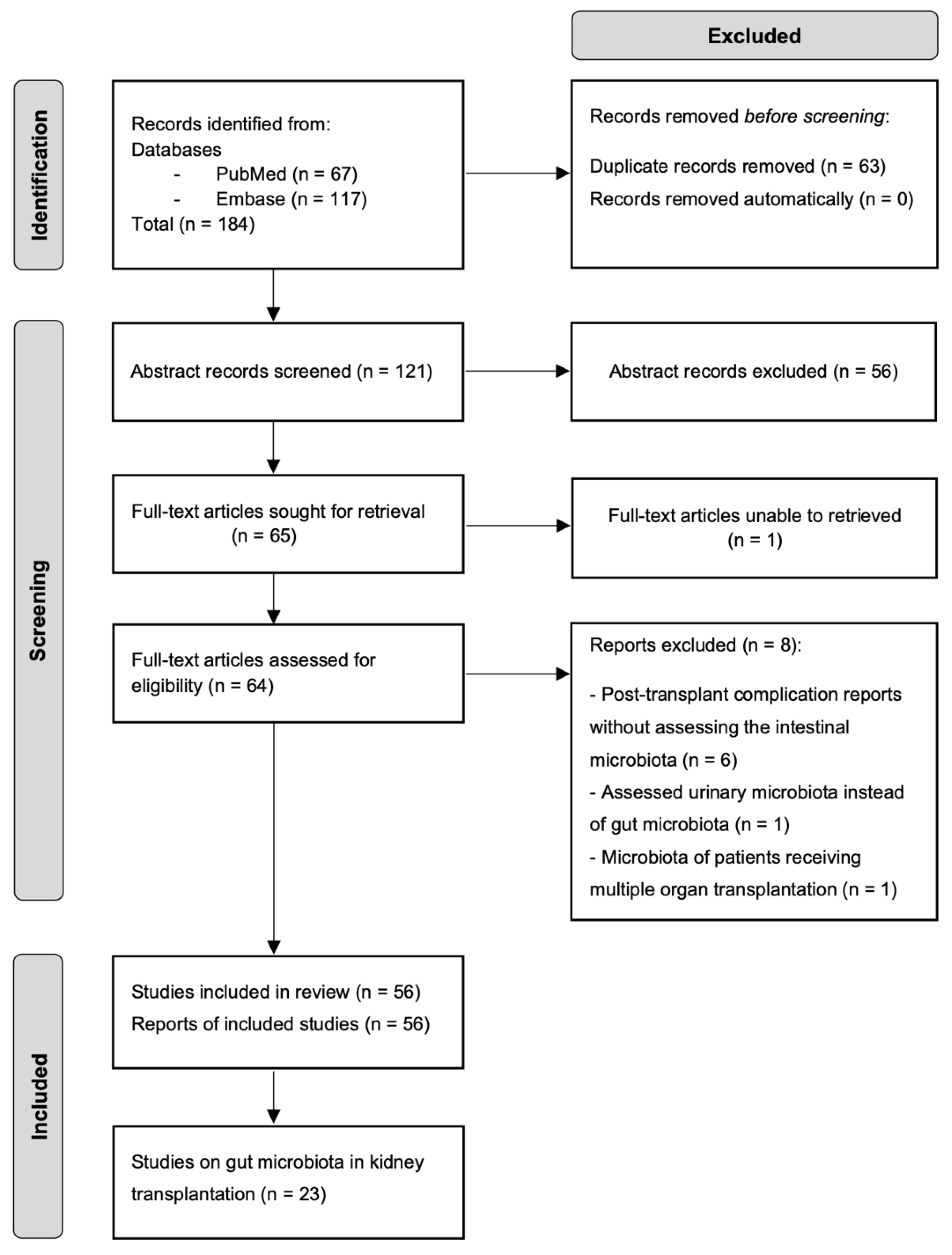
2.1. Literature Search

PubMed and Embase were searched for studies addressing the role of gut microbiota in KT outcomes up to December 2021. We used the keywords gut microbiota AND kidney transplantation, gut microbiome AND renal transplantation, gut dysbiosis AND kidney transplantation, intestinal microbiota AND kidney post-transplant complications, probiotics/prebiotics AND kidney/renal transplantation, microbiome therapy AND kidney/renal transplantation, uremic toxins and kidney transplantation, immune system AND gut microbiota”. In addition, a backward reference search on selected articles was conducted when a paper of interest was identified.

2.2. Eligible Criteria

Cohort studies, single-case reports, and reviews published in English studying the gut microbiota alterations in KTRs older than 18 years, post-transplant complications, immunosuppressants metabolism, uremic toxicity, and the gut microbiota as a therapeutic target were selected. Studies involving cancer patients were excluded.

2.3. Data Extraction

Relevant abstracts were selected for full-text review. Details regarding the study population, design, microbiome composition in KTRs, and transplantation outcome were extracted per article chosen. Out of 184 records identified, 121 abstracts were screened, 64 full texts were retrieved and assessed for eligibility, and 56 were included in the review. Of those, 23 studies reported a potential role of gut microbiota in KT. The selection process is illustrated in Figure 1.

3. Gut Microbial Dysbiosis in Kidney Transplant Recipients

Several transplantation-related factors can alter the gut microbiota of KTRs, such as discontinued dialysis and drugs used in ESKD, dietary changes, induction therapy, surgery, maintenance immunosuppressive therapy, and antimicrobial prophylaxis following transplantation [26]. The resulting alterations in the gut microbiome may create a signature in the microbial community that, in turn, impacts transplantation outcomes. Whether a distinctive profile can be attributed to KTRs has been gradually explored over the last decade (Table 1).
Guirong et al. showed an increased abundance of Bacteroides and Enterobacteriaceae in the gut microbiota of KTRs compared with healthy individuals. Higher Enterobacteriaceae abundance in KTRs was identified as a microbial signature distinctive from ESKD patients undergoing hemodialysis. Enterobacteriaceae is the most common cause of urinary tract infection (UTI), and higher intestinal abundance may increase the risk of post-transplant infections in KTRs. Post-transplant microbiota was also characterized by a decreased abundance of the short-chain-fatty-acid-producing bacteria Ruminococcaceae, Lachnospira, and Faecalibacterium. The most common SCFAs include acetate, propionate, and butyrate. SCFAs are essential in immune regulation and blood pressure control—both critical functions in KTRs [4]. Other butyrate-producing bacteria, such as Eubacterium rectale and Roseburia, have been identified to decrease in KTRs after six years post-transplantation compared with healthy individuals [5] (Table 1).
A gut microbiota analysis of KTRs at different graft periods between 3 months and 22 years post-transplantation showed decreased microbial diversity, richness, and compositional changes over time compared to healthy subjects. Proteobacteria increased shortly after transplantation and continued to increase up to 22 years post-transplantation (Table 1). At the genus level, a higher abundance of Asteroleplasma, Roseburia, Faecalibacterium, and Bacteroides characterized the first-year post-transplantation, while the gut microbiota in the long post-graft period was characterized by a higher abundance of Rikenellaceae RC9, Dialister, Parabacteroides, Sutterella, Escherichia/Shigella, and Succinivibrio. These results pointed out differences in the gut microbiota according to graft time, which may be a future target to predict graft function and stability over time [6].
To better evaluate whether a specific microbial signature can be identified for KTRs, few studies have compared changes in the gut microbiota pre- and post-transplantation. A pilot study analyzed the gut microbiota of five KTRs before and two weeks after KT. Although changes in richness and evenness were not detected post-transplantation, the authors observed an increase in the relative abundance of Proteobacteria and a decrease in Bacteroidetes [7] (Table 1). This imbalance could contribute to the high rate of post-transplant infections in KTRs, as Proteobacteria include numerous infectious pathogens. Moreover, decreased Bacteroidetes abundance could negatively impact immune regulation. Bacteroides spp. have immunomodulatory functions, such as CD4+ T-cell development, Th1/Th2 immune balance regulation, and IL-10 activation [27]. It has been described that Bacteroides fragilis mediates the conversion of CD4+ T cells into IL10-producing Foxp3+ Treg cells through the capsular polysaccharide A in a toll-like receptor-2-dependent mechanism [28]. Foxp3+ Treg cells and IL-10 are involved in mechanisms of allograft immune tolerance [29], and a decreased colonic abundance of B. fragilis could potentially contribute to allograft rejection.
Yu et al. reported a reduction in microbial richness within the first-month post-transplantation compared to the fecal microbiome one-week pre-transplantation. An alteration in the abundance of specific taxa was associated with KT, but these results should be validated in a larger population [8] (Table 1). When comparing the gut microbiota of KTRs before and up to eight weeks after transplantation, Chan et al. reported an increase in Roseburia intestinalis and F. prausnitzii following transplantation. These observations contrast with other reports suggesting a decreased abundance of these SCFA-producing bacteria [4,5] (Table 1), which may be related to the application of different sequencing and data analysis methods and the use of KTRs’ pre-transplant specimens as the control group. The study also indicated that nephrectomy had no impact on gut microbial richness or diversity because no significant changes in microbial composition were observed in kidney donors after the nephrectomy. Instead, other factors might be accounting for the alterations in KTRs, such as reduced kidney function, induction and immunosuppressive therapies, and antimicrobial prophylaxis [9].
These studies generally revealed compositional changes following transplantation, including lower richness and diversity, increased abundance of Proteobacteria, and depletion in several SCFA-producing species. However, identifying a unique profile associated with KT remains challenging due to the confounding effect of immunosuppressants, antimicrobial treatment, and dietary habits on microbiome analyses. In addition, KTRs represent a heterogeneous population diagnosed with different pathologies and comorbidities that might shape the gut microbiome differently and add complexity to post-transplant comparative analysis. Moreover, the comparison across studies is limited by the need for standard methodologies in collecting and assessing microbiome data.
It is worth noting that lower diversity and dysbiosis already characterize the gut microbiota of CKD patients before transplantation. CKD is associated with lower intestinal Bifidobacteriaceae, Lactobacillaceae, Bacteroidaceae, and Prevotellaceae and with higher intestinal levels of Enterobacteriaceae [30]. Studies on KTRs have reported similar results showing a decreased abundance of Bifidobacteriaceae and an increased abundance of Enterobacteriaceae [5]. This suggests that part of the dysbiosis observed in KTRs is present pre-transplantation and may not recover afterward. Moreover, as previously reported for KTRs [4,5,6], ESKD patients have a higher abundance of Proteobacteria compared with healthy individuals [31] and depletion in several SCFA-producing species, such as Roseburia spp., Faecalibacterium spp., and E. rectale, consistent with a reduction in SCFAs in the fecal metabolome of the patients [32]. Taking these similarities into account, it can be challenging to discern the gut microbial community of CKD and ESKD patients from that in KTRs.

4. The Impact of Gut Microbiota on Kidney Transplantation

Despite advances in surgical techniques, immunosuppressive therapy, and post-transplant surveillance protocols, long-term allograft survival remains challenged by several complications. KTRs can develop T-cell-mediated rejection (TCMR) and/or antibody-mediated rejection (ABMR), potentially leading to allograft failure [33]. Episodes of allograft rejection, immunosuppressive medications, and the accumulation of uremic retention solutes increase the risk of cardiovascular diseases. Moreover, immunosuppressives such as calcineurin inhibitors and corticosteroids induce metabolic complications that increase the risk of developing post-transplant diabetes. KTRs may also suffer from infectious complications, diarrhea, and cancer [34]. Hence the search for strategies that improve these patients’ quality of life, graft survival, and life expectancy. Recent studies have suggested the potential influence of gut microbiota on KT outcomes (Table 2).

4.1. Uremic Retention Solutes

Gut-derived uremic toxins trimethylamine N-oxide (TMAO), p-cresyl sulfate (pCS), p-cresyl glucuronide (pCG), indoxyl sulfate (IxS), and indole-3-acetic acid (IAA) have been identified to play a role in inflammation, metabolic function, cardiovascular disease, and fibrosis in CKD patients [35]. Several gut bacteria that have been found to increase after KT (Table 1) and are associated with post-transplant complications (Table 2) produce gut-derived uremic toxins precursors. Among these bacteria, Enterobacteriaceae, Enterococcaceae, Clostridiaceae, and Bacteroides sp. produce IAA, E. coli produces both IAA and indole, and Clostridium tertium and Bacteroides sp. produce p-cresol [36]. Studying these uremic toxins and their association with kidney function and graft outcome is an open area in KTRs.
TMAO plasma concentrations have been associated with an increased risk of graft failure and proposed as a potential biomarker of graft function in KTRs [37]. Other studies have reported increased serum pCS and IxS in KTRs with advanced CKD stages [38]. The treatment with a synbiotic (Probinul Neutro, CadiGroup, Rome, Italy), mainly containing Lactobacillus and Bifidobacterium spp., decreased plasma p-cresol in KTRs after 15 and 30 days of administration. Although the study included a small population (n = 36), the results indicate that gut microbiota interventions could reduce uremic toxicity in KTRs [39].
Liabeuf et al. reported no association between serum IxS levels and adverse outcomes post-transplantation, including graft loss, cardiovascular events, and mortality in a cohort of 311 KTRs [40]. Similarly, a study on the same cohort reported no association between serum-free and total IAA with adverse outcomes of graft loss, cardiovascular events, and death following KT [41].

4.2. Allograft Function

Delayed graft function is a common post-transplant event that can expose the patient to a longer uremic period and increase the risk of gut dysfunction, systemic inflammation, and allograft rejection [42]. Kim et al. showed that six-month allograft function was associated with a similar pre-transplantation microbial structure between donor and KTR, especially in genetically unrelated pairs such as spousal donor and recipient. The microbial distance in unrelated pairs was a better predictor of the eGFR than the number of human leukocytes antigen mismatches and incompatibility. This may indicate that microbial similarity between donors and KTRs could, to some extent, compensate for the disadvantages of genetic disparity and influence the criteria for living donor selection. Furthermore, a similar microbial structure between donor–KTR could potentially reduce the risk of infections, as microbial dissimilarity was associated with an increased post-transplant infection rate [43].

4.3. Allograft Rejection

Immune tolerance is crucial to ensure graft function and survival of transplant recipients. T-lymphocyte peripheral tolerance is the primary mode of tolerance to transplanted organs. It eliminates activated T-cell clones in the periphery via apoptosis, develops T-lymphocyte anergy, and suppresses alloreactive T lymphocytes by Tregs [44].
Members of the gut microbiota, such as Clostridia, Bacteroides fragilis, and Bacteroides thetaiotaomicron, have been found to modulate Tregs differentiation [45]. CD4+ Tregs, regulatory B cells, and dendritic cells (DCs) secrete IL-10 that inhibits APC activity and promotes the conversion of T cells into T regulatory type 1 cells (TR1s). TR1s are able to suppress the pro-inflammatory activities of both APC and effector T cells. Furthermore, in the presence of IL-10, naive CD8+ T cells can be converted into CD8+ Tregs that inhibit effector T cells. CD4CD8 Tregs induce effector T-cell apoptosis via the CD95-CD95L pathway and by downregulating the expression of DCs molecules CD80 and CD86, consequently inhibiting the ability of DCs to stimulate pro-inflammatory responses. Tolerogenic DCs inhibit effector T-cell proliferation and differentiation into Th1 and Th17 cells [29].
The gut microbiota and derived metabolites can also modulate host inflammatory response through microbiota–cytokine interactions [46]. Schirmer et al. observed that interindividual variation in cytokine response to various microbial stimulations was linked to specific commensal bacteria and microbial functions in healthy individuals. For instance, a higher abundance of Roseburia was associated with lower IL-6 levels, and a higher abundance of Bilophila and Odoribacter was associated with lower TNFα levels. Furthermore, lower IL-17 production was linked to a higher abundance of Faecalibacterium and Atopobium, whereas Escherichia, Anaerotruncus, Coprobacillus, and Clostridium higher abundance was linked to increased production of IL-17. These results suggest that modulating cytokine expression by microbial interventions may have therapeutic value in KTRs. However, microbiota–cytokine interactions should be evaluated in KT, given the stimulus-specific and/or cytokine-specific nature of these interactions [47].
To date, few studies have addressed the potential association between gut microbiota and allograft rejection in KTRs. Wang et al. analyzed the gut microbial community of KTRs with ABMR compared to KTRs who did not develop graft rejection. ABMR was related to lower microbial richness and decreased relative abundance of Clostridia, Paraprevotellaceae, and Faecalibacterium, as well as increased abundance of Enterococcaceae, Coprobacillus, and Enterobacter, among other taxa (Table 2). These microbial changes are likely associated with ABMR because recipients with a recent history of infection, antibiotic usage, gastric/colon resection, and non-infectious diarrhea were excluded from the study. However, immunosuppressive therapy could have contributed to the alterations observed. Clostridiales was proposed as a potential biomarker of ABMR after KT, but further studies are needed to validate its use as a diagnostic tool [10].
Lee et al. also reported alterations in the gut microbiota of KTRs associated with acute rejection (AR). During the first three months post-transplantation, three patients of the studied cohort were diagnosed with ABMR, TCMR, and mixed ABMR-TCMR, respectively. A lower abundance of Clostridiales, Bacteroidales, Eubacterium dolichum, and Ruminococcus, and a higher abundance of Lactobacillales, Enterococcus, Anaerofilum, and Clostridium tertium characterized the gut microbiota of AR patients compared to those who did not develop AR (Table 2). It should be noted that AR patients received antibiotic therapy to treat Clostridioides difficile infection (CDI), Enterococcus UTI, Escherichia coli UTI, and Klebsiella/Serratia UTI before AR. It is then difficult to determine whether the observed microbial alterations are related to AR or the result of antibiotic therapy [7].
Another study analyzed the pre-and post-transplant rectal microbiota of four KTRs who experienced rejection compared to KTRs who did not experience rejection or other adverse events. A decreased abundance of Anaerotruncus, Coprobacillus, Coprococcus, and an unknown member of Peptostreptococcaceae correlated with the development of future rejection events [13] (Table 2). However, these results require further validation in a larger cohort to assess the potential of gut microbial taxa as biomarkers of rejection events.

4.4. Immunosuppressants Metabolism

Current clinical guidelines recommend combining immunosuppressive medications as maintenance therapy, including a calcineurin inhibitor and an antiproliferative agent with or without corticosteroids [3]. Recent data suggest the impact of the gut microbiota on the most prescribed immunosuppressive medications: MMF and TAC.
The use of MMF has been correlated to a lower diversity of the gut microbiome [5], post-transplant diarrhea, and impaired quality of life in KTRs [48]. The gut microbiome can also metabolize MMF, thereby influencing drug dosage. MMF is converted to its active form, mycophenolic acid (MPA), by plasma and tissue esterases and inactivated by hepatic glucuronidation to MPA glucuronide. MPA glucuronide is then excreted in the urine and bile. If secreted into the gastrointestinal tract through the ATP-binding cassette subfamily C member 2 protein, bacteria expressing beta-glucuronidase enzymes can cleave the glucuronic acid (GA) of MPA glucuronide to produce free MPA and GA. The resulting GA is a carbon source for bacterial metabolism, and MPA undergoes enterohepatic recirculation, which has been related to gastrointestinal toxicity [21,26]. Hence, the management strategies for gastrointestinal complications include MMF dose reduction or discontinuation, which can lead to an increased risk of allograft rejection [49].
TAC is a macrolide of the calcineurin inhibitor family that binds to the FK506-binding protein, forming a complex that inhibits calcineurin phosphatase, ultimately blocking T-cell activation. Despite its efficacy in avoiding TCMR and ABMR, TAC decreases eGFR and promotes glucose intolerance, new-onset diabetes, and hypertension [2,3]. TAC has also been associated with gastrointestinal symptoms in KTRs, including diarrhea, nausea, and vomiting [48]. Its antimicrobial activity can disrupt the gut microbial community, and recent data indicate a bidirectional relationship as gut bacteria metabolize TAC.
Lee et al. observed that the fecal abundance of F. prausnitzii was positively correlated with the one-month TAC dose required to maintain therapeutic levels in KTRs. This suggested a possible role of F. prausnitzii influencing TAC levels, but the underlying mechanisms were not identified [2]. A follow-up study found that F. prausnitzii and other commensal bacteria, mainly belonging to Clostridiales, metabolize TAC into a less effective immunosuppressive metabolite (M1). M1 is a C9 keto-reduction product, uniquely synthesized by gut bacteria and 15-fold less immunosuppressive than its parent in inhibiting T-lymphocyte proliferation in vitro. However, the authors observed no correlation between Clostridiales abundance (including F. prausnitzii) and M1 production in stool samples from KTRs undergoing oral TAC therapy [11]. In a subsequent study, the group showed active metabolism of TAC in KTRs by evaluating the pharmacokinetics of M1 after oral administration of TAC. M1 was detected within the first four hours of administration, with concentrations reduced by at least five-fold compared to parent TAC. These results could explain the interpatient variability in TAC therapeutic level requirements and suggest that changes in the gut microbiota might impact TAC trough variability [12] (Table 2).

4.5. Post-Transplant Infection

Urinary tract infections remain among the most frequent complications affecting post-transplant patients [50]. A pilot study reported an increased fecal abundance of Enterococcus correlated with the development of Enterococcus UTI in KTRs (Table 2). However, this observation was limited by the confounding effect of immunosuppressives, antimicrobial therapy, and the small sample size studied: 3 KTRs Enterococcus UTI compared with 23 KTRs who did not develop Enterococcus UTI [7].
Fricke et al. described alterations in the rectal microbiota of KTRs associated with urinary and upper respiratory tract infections during the first six months post-transplantation (Table 2). A potential role in predicting post-transplant infection events was attributed to Anaerotruncus as it significantly decreased in 4 KTRs with infection compared with 14 KTRs without post-transplant adverse events. Nonetheless, the value of these microbial alterations as diagnosis markers requires further evaluation in a larger population [13].
Another study serially profiled fecal specimens from KTRs within the first three months after transplantation (Table 2). The authors observed 1% Escherichia gut abundance associated with the future development of Escherichia bacteriuria. Female gender was also a predictor of future Escherichia bacteriuria. Likewise, 1% Enterococcus gut abundance was associated with the future development of Enterococcus bacteriuria, independent from other factors such as gender, antimicrobial treatment, and immune maintenance therapy. The phylogenetic analysis showed a close relationship between the same subject’s urine and fecal strains of E. coli, Enterococcus faecalis, and Enterococcus faecium. The similarity of E. coli strains was supported by detecting uropathogenic genes and beta-lactams, sulfonamides, and trimethoprim resistance genes in paired urine and fecal specimens associated with E. coli bacteriuria. These results suggest that an overgrowth of enteropathogenic bacteria could influence UTI development in KTRs [14].
In a follow-up study, the group found that a high abundance of Faecalibacterium and Romboutsia was significantly associated with a lower risk of developing Enterobacteriaceae bacteriuria and Enterobacteriaceae UTI. On the other hand, increased Lactobacillus abundance was associated with an increased risk of developing Enterobacteriaceae bacteriuria and Enterobacteriaceae UTI [15] (Table 2). These results are promising for developing personalized UTI treatments such as Enterobacteriaceae-reducing probiotics.
Analyzing the previous cohort, the authors suggested a possible protective role of butyrate-producing bacteria in developing respiratory viral infections. A fecal abundance of butyrate-producing bacteria higher than 1% was associated with a decreased risk of developing respiratory viral infections in the first two years post-transplantation and CMV viremia in the first-year post-transplantation [16] (Table 2). Butyrate has important roles in immunomodulation, maintenance of the intestinal barrier, and protection against bacterial and viral infections. Another study in allogeneic hematopoietic stem cell patients indicated that a high intestinal abundance of butyrate-producing bacteria at the time of engraftment conferred protection against viral lower respiratory tract infections [51]. However, these studies could not determine a causal relationship between butyrate-producing bacteria and protection against respiratory viral infections.

4.6. Post-Transplant Diarrhea

Post-transplant diarrhea increases the risk of graft failure, death-censored graft survival, and patient death. Despite being a common complication, the etiology of post-transplant diarrhea is not identified in most cases, and it is frequently associated with intestinal drug toxicity if infections are excluded. A retrospective study reported that over 80% of KTRs were diagnosed with unspecified non-infectious diarrhea, with greater incidence in patients following TAC and MMF combined therapy [52]. Recent data suggest alterations in the gut microbiome of KTRs as a potential non-infectious etiology of post-transplant diarrhea.
In a pilot study, Lee et al. observed a lower gut microbial diversity and a decrease in the relative abundance of Bacteroides, Ruminococcus, Coprococcus, and Dorea to be associated with the development of diarrhea within the first-month post-transplantation [7]. Analyzing a different cohort, the group found that the development of post-transplant diarrhea was associated with gut dysbiosis instead of infectious etiologies. The gut microbiota of KTRs with diarrhea was characterized by a decreased abundance of Ruminococcus, Coprococcus, Dorea, Faecalibacterium, and Bifidobacterium, as well as an increased abundance of Escherichia and Enterococcus. The potential role of these taxa on post-transplant diarrhea was also suggested by the gut microbial analysis of two KTRs with a history of CDI and who underwent fecal microbial transplantation (FMT). The patients had persistent diarrhea despite testing negative for C. difficile before FMT. After FMT, the resolution of diarrhea correlated with an overall increase in the abundance of the taxa previously identified as significantly lower in diarrheal specimens and an overall decrease in the abundance of the taxa identified as significantly higher in diarrheal specimens [17] (Table 2). These results indicate that restoring the gut microbial community imbalance could successfully manage post-transplant diarrhea.
A further study by the group reported a lower abundance of Ruminococcus, Anaerostipes, Fusicatenibacter, Eubacterium, Ruminiclostridium, Dorea, and Bifidobacterium associated with non-infectious diarrhea episodes in KTRs. Moreover, prolonged diarrhea was associated with higher beta-glucuronidase activity, indicating that the toxicity from the free MPA in the colon could contribute to the diarrhea episodes, though these results require further validation. Four genera were positively correlated with beta-glucuronidase activity: Subdoligranulum, Coprococcus, Tyzzerella, and an unspecified Erysipelotrichaceae [18] (Table 2). These studies denote a potential relationship between the gut microbiota and the development of post-transplant diarrhea up to three months after KT.

4.7. New Onset Diabetes (NODAT)

NODAT develops in approximately 20% of KTRs in the first year after transplantation and has been identified as an adverse effect of immunosuppressive treatment, including corticosteroids, cyclosporin, TAC, and sirolimus [2,19]. Lecronier et al. observed alterations in the gut microbiota associated with the development of NODAT after KT by comparing pre- and post-transplant fecal samples from KTRs. An increase in Lactobacillus sp. relative abundance and a decrease in Akkermansia muciniphila were associated with NODAT presentation (Table 2). The same microbial changes were observed in patients with pre-transplant diabetes but not in patients without diabetes either before or after KT, suggesting a potential role for these taxa in the future development of NODAT. It should be noted that the results were obtained by qPCR of targeted bacterial species. Other taxonomic changes may be revealed through metagenomic analyses. Moreover, other factors could contribute to NODAT presentation, such as immunosuppressive medications and increased body mass index of the patients following KT, because obesity is a known risk factor for diabetes. Additional studies should validate the possible role of gut microbiota in NODAT development in KTRs [19].

5. Gut-Microbiota-Based Therapies in Kidney Transplantation

The plasticity of the gut microbiome allows the development of therapeutic interventions to prevent and treat health disorders. Diet intervention and FMT are approaches to reshape the entire gut microbiome, while prebiotics, probiotics, and bacteriophages are more targeted manipulations [53]. FMT and probiotics have been used to decrease the risk of recurrent CDI. A year of prophylaxis with the probiotic Lactobacillus plantarum 299v (LP299v) decreased CDI incidence in immunosuppressed patients receiving antibiotics therapy. Furthermore, LP299v prophylaxis was associated with reduced diarrhea events and lower serum C-reactive protein concentrations. A year of follow-up upon stopping LP299v use showed increased CDI incidence, suggesting that this probiotic may prevent CDI in immunosuppressed patients [54].
The administration of probiotics has also shown immunomodulatory effects. A mixture containing Lactobacillus acidophilus, Lactobacillus casei, Lactobacillus reuteri, Bifidobacterium bifidium, and Streptococcus thermophilus was studied in an inflammatory bowel disease mice model. The probiotics induced regulatory DCs that promoted the generation and migration of CD4+Foxp3+ Tregs to the inflammation sites. In addition, the probiotics suppressed the expression of inflammatory cytokines IL-17, IFNγ, and TNFα in T and B cells and enhanced the suppression capacity of naturally occurring CD4+CD25+ Tregs [55]. The generation of CD4+Foxp3+ Tregs in response to probiotics may be therapeutically beneficial in stimulating kidney allograft tolerance.
The application of gut microbiota-based therapies has not been widely explored in KT. Recent data, mainly in case reports, suggest the efficacy of these therapies for treating and preventing infectious complications. A case reported a heart and kidney transplant recipient who underwent allogeneic FMT because of recurrent episodes of C. difficile colitis, vancomycin-resistant Enterococcus UTIs, and bacteremia. FMT resulted in a decreased Enterococcus abundance and the resolution of C. difficile and vancomycin-resistant Enterococcus infections up to one year of follow-up [22]. Another study reported a KTR with a history of UTIs during the three first years of graft function, predominantly E. coli and ESBL-producing E. coli. A gradual decrease in Enterobacteriaceae relative abundance was observed following FMT. The patient had no recurrent E. coli UTI during the nine-month follow-up [23].
Successful FMT was also reported for a KTR with recurrent UTI caused by ESBL-producing Klebsiella pneumoniae (ESBL-K. pneumoniae), CDI, and who was suffering from diarrhea. Although six days after FMT, the patient was re-admitted with ESBL-K. pneumoniae infection, and no further episodes of UTI occurred after his recovery. A resolution of diarrhea was achieved, and no UTIs or CDI symptoms were detected during the first year post-FMT [24].
Wang et al. conducted a successful FMT on a KTR with carbapenem-resistant K. pneumoniae infection detected in incision secretion, urine, and rectal swab cultures. The strains causing the infection were clustered phylogenetically closed, suggesting the potential involvement of gut bacteria in post-transplant infections. One week after FMT, the urine and rectal swab cultures tested negative for K. pneumoniae infection, and the surgical incision healed after seventeen days. The patient had no symptoms of infection during the two months of follow-up [25].
Despite the promise of FMT to treat post-transplant infections and diarrhea in KTRs, this therapy should be used cautiously. A study reported two cases of ESBL-producing E. coli bacteremia transmitted through FMT. One of the cases resulted in death, stressing how important donor selection is to avoid adverse events [56]. The future application of FMT may involve the development of defined microbial mixtures to prevent unfavorable clinical outcomes.

6. Conclusions and Future Perspectives

The gut microbiota is increasingly recognized as influencing kidney post-transplant outcomes. Although no unique microbial signature can be attributed to KTRs thus far, KT results in lower gut microbial richness and diversity than the healthy population. Specific taxa have been associated with the development of infections and diarrhea complications, allograft rejection, and the ability to metabolize immunosuppressants essential for preserving graft function. These findings are promising to personalize immunosuppressive therapies based on interpersonal microbiome variability. In addition, microbiota intervention therapies such as FMT have successfully resolved infection and diarrhea complications in KTRs. These data suggest the potential role of gut microbiota in graft survival. However, we have just begun to understand the role of the gut microbiome in KT. More studies are needed to define the community structures representative of a healthy microbiome in transplant patients early after transplantation and longitudinally, as well as the role of gut-microbiome-modulating alloimmunity.

Author Contributions

Conceptualization, Y.G.-M. and D.I.; methodology, Y.G.-M.; formal analysis, Y.G.-M. and D.I.; investigation, Y.G.-M. and D.I.; resources, Y.G.-M., D.I. and A.F.P.; data curation, Y.G.-M.; writing—original draft preparation, Y.G.-M.; writing—review and editing, Y.G.-M., D.I. and A.F.P.; visualization, Y.G.-M., M.B., G.C., D.I. and A.F.P.; supervision, D.I. and A.F.P.; project administration, D.I. and A.F.P.; funding acquisition, Y.G.-M., D.I. and A.F.P. All authors have read and agreed to the published version of the manuscript.

Funding

The APC was funded by the European Union’s Horizon 2020 research and innovation program, project STRATEGY-CKD, under grant agreement no (860329).

Institutional Review Board Statement

Not applicable.

Informed Consent Statement

Not applicable.

Data Availability Statement

Not applicable.

Conflicts of Interest

The authors declare no conflict of interest.

References

  1. Nobakht, E.; Jagadeesan, M.; Paul, R.; Bromberg, J.; Dadgar, S. Precision Medicine in Kidney Transplantation: Just Hype or a Realistic Hope? Transplant. Direct 2021, 7, e650. [Google Scholar] [CrossRef] [PubMed]
  2. Lee, J.R.; Muthukumar, T.; Dadhania, D.; Taur, Y.; Jenq, R.R.; Toussaint, N.C.; Ling, L.; Pamer, E.; Suthanthiran, M. Gut Microbiota and Tacrolimus Dosing in Kidney Transplantation. PLoS ONE 2015, 10, e0122399. [Google Scholar] [CrossRef] [PubMed] [Green Version]
  3. Transplant Recipient–KDIGO. Available online: https://kdigo.org/guidelines/transplant-recipient/ (accessed on 5 October 2022).
  4. Guirong, Y.E.; Minjie, Z.; Lixin, Y.U.; Junsheng, Y.E.; Lin, Y.; Lisha, S. Gut Microbiota in Renal Transplant Recipients, Patients with Chronic Kidney Disease and Healthy Subjects. Nan Fang Yi Ke Da Xue Xue Bao 2018, 38, 1401–1408. [Google Scholar] [CrossRef] [PubMed]
  5. Swarte, J.C.; Douwes, R.M.; Hu, S.; Vich Vila, A.; Eisenga, M.F.; van Londen, M.; Gomes-Neto, A.W.; Weersma, R.K.; Harmsen, H.J.M.; Bakker, S.J.L. Characteristics and Dysbiosis of the Gut Microbiome in Renal Transplant Recipients. J. Clin. Med. 2020, 9, 386. [Google Scholar] [CrossRef] [Green Version]
  6. Souai, N.; Zidi, O.; Mosbah, A.; Kosai, I.; Manaa, J.E.; Mokhtar, N.B.; Asimakis, E.; Stathopoulou, P.; Cherif, A.; Tsiamis, G.; et al. Impact of the Post-Transplant Period and Lifestyle Diseases on Human Gut Microbiota in Kidney Graft Recipients. Microorganisms 2020, 8, 1724. [Google Scholar] [CrossRef]
  7. Lee, J.R.; Muthukumar, T.; Dadhania, D.; Toussaint, N.C.; Ling, L.; Pamer, E.; Suthanthiran, M. Gut Microbial Community Structure and Complications Following Kidney Transplantation: A Pilot Study. Transplantation 2014, 98, 697. [Google Scholar] [CrossRef] [Green Version]
  8. Yu, D.H.; Ying, N.; Lian, Z.H.; Fa, Y.Q. The Alteration Human of Gut Microbiota and Metabolites before and after Renal Transplantation. Microb. Pathog. 2021, 160, 105191. [Google Scholar] [CrossRef]
  9. Chan, S.; Morrison, M.; Hawley, C.M.; Campbell, S.B.; Francis, R.S.; Isbel, N.M.; Pascoe, E.M.; Johnson, D.W. Characteristics of the Gastrointestinal Microbiota in Paired Live Kidney Donors and Recipients. Nephrology 2021, 26, 471–478. [Google Scholar] [CrossRef]
  10. Wang, J.; Li, X.; Wu, X.; Wang, Z.; Zhang, C.; Cao, G.; Liu, S.; Yan, T. Gut Microbiota Alterations Associated with Antibody-Mediated Rejection after Kidney Transplantation. Appl. Microbiol. Biotechnol. 2021, 105, 2473–2484. [Google Scholar] [CrossRef]
  11. Guo, Y.; Crnkovic, C.; Won, K.J.; Lee, J.R.; Orjala, J.; Lee, H.; Jeong, H. Commensal Gut Bacteria Convert the Immunosuppressant Tacrolimus to Less Potent Metabolites. Drug Metab. Dispos. 2019, 47, 194–202. [Google Scholar] [CrossRef]
  12. Guo, Y.; Lee, H.; Edusei, E.; Albakry, S.; Jeong, H.; Lee, J.R. Blood Profiles of Gut Bacterial Tacrolimus Metabolite in Kidney Transplant Recipients. Transplant. Direct 2020, 6, 601. [Google Scholar] [CrossRef] [PubMed]
  13. Fricke, W.F.; Maddox, C.; Song, Y.; Bromberg, J.S. Human Microbiota Characterization in the Course of Renal Transplantation. Am. J. Transplant. 2014, 14, 416–427. [Google Scholar] [CrossRef] [PubMed]
  14. Magruder, M.; Sholi, A.N.; Gong, C.; Zhang, L.; Edusei, E.; Huang, J.; Albakry, S.; Satlin, M.J.; Westblade, L.F.; Crawford, C.; et al. Gut Uropathogen Abundance Is a Risk Factor for Development of Bacteriuria and Urinary Tract Infection. Nat. Commun. 2019, 10, 5521. [Google Scholar] [CrossRef] [PubMed] [Green Version]
  15. Magruder, M.; Edusei, E.; Zhang, L.; Albakry, S.; Satlin, M.J.; Westblade, L.F.; Malha, L.; Sze, C.; Lubetzky, M.; Dadhania, D.M.; et al. Gut Commensal Microbiota and Decreased Risk for Enterobacteriaceae Bacteriuria and Urinary Tract Infection. Gut Microbes 2020, 12, 1805281. [Google Scholar] [CrossRef]
  16. Lee, J.R.; Huang, J.; Magruder, M.; Zhang, L.T.; Gong, C.; Sholi, A.N.; Albakry, S.; Edusei, E.; Muthukumar, T.; Lubetzky, M.; et al. Butyrate-Producing Gut Bacteria and Viral Infections in Kidney Transplant Recipients: A Pilot Study. Transpl. Infect. Dis. 2019, 21, e13180. [Google Scholar] [CrossRef]
  17. Lee, J.R.; Magruder, M.; Zhang, L.; Westblade, L.F.; Satlin, M.J.; Robertson, A.; Edusei, E.; Crawford, C.; Ling, L.; Taur, Y.; et al. Gut Microbiota Dysbiosis and Diarrhea in Kidney Transplant Recipients. Am. J. Transplant. 2019, 19, 488–500. [Google Scholar] [CrossRef] [Green Version]
  18. Zhang, L.T.; Westblade, L.F.; Iqbal, F.; Taylor, M.R.; Chung, A.; Satlin, M.J.; Magruder, M.; Edusei, E.; Albakry, S.; Botticelli, B.; et al. Gut Microbiota Profiles and Fecal Beta-Glucuronidase Activity in Kidney Transplant Recipients with and without Post-Transplant Diarrhea. Clin. Transplant. 2021, 35, e14260. [Google Scholar] [CrossRef]
  19. Lecronier, M.; Tashk, P.; Tamzali, Y.; Tenaillon, O.; Denamur, E.; Barrou, B.; Aron-Wisnewsky, J.; Tourret, J. Gut Microbiota Composition Alterations Are Associated with the Onset of Diabetes in Kidney Transplant Recipients. PLoS ONE 2020, 15, e0227373. [Google Scholar] [CrossRef] [Green Version]
  20. Ardalan, M.; Vahed, S.Z. Gut Microbiota and Renal Transplant Outcome. Biomed. Pharmacother. 2017, 90, 229–236. [Google Scholar] [CrossRef]
  21. Taylor, M.R.; Flannigan, K.L.; Rahim, H.; Mohamud, A.; Lewis, I.A.; Hirota, S.A.; Greenway, S.C. Vancomycin Relieves Mycophenolate Mofetil-Induced Gastrointestinal Toxicity by Eliminating Gut Bacterial β-Glucuronidase Activity. Sci. Adv. 2019, 5, eaax2358. [Google Scholar] [CrossRef]
  22. Stripling, J.; Kumar, R.; Baddley, J.W.; Nellore, A.; Dixon, P.; Howard, D.; Ptacek, T.; Lefkowitz, E.J.; Tallaj, J.A.; Benjamin, W.H.; et al. Loss of Vancomycin-Resistant Enterococcus Fecal Dominance in an Organ Transplant Patient With Clostridium Difficile Colitis After Fecal Microbiota Transplant. Open Forum Infect. Dis. 2015, 2. [Google Scholar] [CrossRef] [PubMed] [Green Version]
  23. Biehl, L.M.; Cruz Aguilar, R.; Farowski, F.; Hahn, W.; Nowag, A.; Wisplinghoff, H.; Vehreschild, M.J.G.T. Fecal Microbiota Transplantation in a Kidney Transplant Recipient with Recurrent Urinary Tract Infection. Infection 2018, 46, 871–874. [Google Scholar] [CrossRef] [PubMed]
  24. Grosen, A.K.; Povlsen, J.V.; Lemming, L.E.; Jørgensen, S.M.D.; Dahlerup, J.F.; Hvas, C.L. Faecal Microbiota Transplantation Eradicated Extended-Spectrum Beta-Lactamase-Producing Klebsiella Pneumoniae from a Renal Transplant Recipient with Recurrent Urinary Tract Infections. Case Rep. Nephrol. Dial. 2019, 9, 102–107. [Google Scholar] [CrossRef] [PubMed]
  25. Wang, J.; Li, X.; Wu, X.; Wang, Z.; Wu, X.; Wang, S.; Jing, G.; Yan, T. Fecal Microbiota Transplantation as an Effective Treatment for Carbapenem-Resistant Klebsiella Pneumoniae Infection in a Renal Transplant Patient. Infect. Drug Resist. 2021, 14, 1805–1811. [Google Scholar] [CrossRef]
  26. Gabarre, P.; Loens, C.; Tamzali, Y.; Barrou, B.; Jaisser, F.; Tourret, J. Immunosuppressive Therapy after Solid Organ Transplantation and the Gut Microbiota: Bidirectional Interactions with Clinical Consequences. Am. J. Transplant. 2022, 22, 1014–1030. [Google Scholar] [CrossRef]
  27. Cohen-Poradosu, R.; McLoughlin, R.M.; Lee, J.C.; Kasper, D.L. Bacteroides Fragilis–Stimulated Interleukin-10 Contains Expanding Disease. J. Infect. Dis. 2011, 204, 363–371. [Google Scholar] [CrossRef]
  28. Round, J.L.; Mazmanian, S.K. Inducible Foxp3+ Regulatory T-Cell Development by a Commensal Bacterium of the Intestinal Microbiota. Proc. Natl. Acad. Sci. USA 2010, 107, 12204–12209. [Google Scholar] [CrossRef] [Green Version]
  29. Wood, K.J.; Bushell, A.; Hester, J. Regulatory Immune Cells in Transplantation. Nat. Rev. Immunol. 2012, 12, 417–430. [Google Scholar] [CrossRef]
  30. Sampaio-Maia, B.; Simões-Silva, L.; Pestana, M.; Araujo, R.; Soares-Silva, I.J. The Role of the Gut Microbiome on Chronic Kidney Disease. Adv. Appl. Microbiol. 2016, 96, 65–94. [Google Scholar] [CrossRef]
  31. Vaziri, N.D. Gut Microbial Translocation in the Pathogenesis of Systemic Inflammation in Patients with End-Stage Renal Disease. Dig. Dis. Sci. 2014, 59, 2020–2022. [Google Scholar] [CrossRef]
  32. Wang, X.; Yang, S.; Li, S.; Zhao, L.; Hao, Y.; Qin, J.; Zhang, L.; Zhang, C.; Bian, W.; Zuo, L.; et al. Aberrant Gut Microbiota Alters Host Metabolome and Impacts Renal Failure in Humans and Rodents. Gut 2020, 69, 2131–2142. [Google Scholar] [CrossRef] [PubMed]
  33. Baldwin, W.M.; Valujskikh, A.; Fairchild, R.L. Mechanisms of Antibody-Mediated Acute and Chronic Rejection of Kidney Allografts. Curr. Opin. Organ Transplant. 2016, 21, 7. [Google Scholar] [CrossRef] [Green Version]
  34. Thongprayoon, C.; Hansrivijit, P.; Leeaphorn, N.; Acharya, P.; Torres-Ortiz, A.; Kaewput, W.; Kovvuru, K.; Kanduri, S.R.; Bathini, T.; Cheungpasitporn, W. Recent Advances and Clinical Outcomes of Kidney Transplantation. J. Clin. Med. 2020, 9, 1193. [Google Scholar] [CrossRef] [PubMed] [Green Version]
  35. Glorieux, G.; Gryp, T.; Perna, A. Gut-Derived Metabolites and Their Role in Immune Dysfunction in Chronic Kidney Disease. Toxins 2020, 12, 245. [Google Scholar] [CrossRef] [Green Version]
  36. Gryp, T.; Huys, G.R.B.; Joossens, M.; van Biesen, W.; Glorieux, G.; Vaneechoutte, M. Isolation and Quantification of Uremic Toxin Precursor-Generating Gut Bacteria in Chronic Kidney Disease Patients. Int. J. Mol. Sci. 2020, 21, 1986. [Google Scholar] [CrossRef] [PubMed] [Green Version]
  37. Flores-Guerrero, J.L.; Osté, M.C.J.; Baraldi, P.B.; Connelly, M.A.; Garcia, E.; Navis, G.; Bakker, S.J.L.; Dullaart, R.P.F. Association of Circulating Trimethylamine N-Oxide and Its Dietary Determinants with the Risk of Kidney Graft Failure: Results of the TransplantLines Cohort Study. Nutrients 2021, 13, 262. [Google Scholar] [CrossRef] [PubMed]
  38. Huang, S.T.; Shu, K.H.; Cheng, C.H.; Wu, M.J.; Yu, T.M.; Chuang, Y.W.; Chen, C.H. Serum Total P-Cresol and Indoxyl Sulfate Correlated With Stage of Chronic Kidney Disease in Renal Transplant Recipients. Transplant. Proc. 2012, 44, 621–624. [Google Scholar] [CrossRef]
  39. Guida, B.; Cataldi, M.; Memoli, A.; Trio, R.; di Maro, M.; Grumetto, L.; Capuano, I.; Federico, S.; Pisani, A.; Sabbatini, M. Effect of a Short-Course Treatment with Synbiotics on Plasma p-Cresol Concentration in Kidney Transplant Recipients. J. Am. Coll. Nutr. 2017, 36, 586–591. [Google Scholar] [CrossRef] [Green Version]
  40. Liabeuf, S.; Desjardins, L.; Massy, Z.A.; Brazier, F.; Westeel, P.F.; Mazouz, H.; Titeca-Beauport, D.; Diouf, M.; Glorieux, G.; Vanholder, R.; et al. Levels of Indoxyl Sulfate in Kidney Transplant Patients, and the Relationship With Hard Outcomes. Circ. J. 2016, 80, 722–730. [Google Scholar] [CrossRef] [Green Version]
  41. Liabeuf, S.; Laville, S.M.; Glorieux, G.; Cheddani, L.; Brazier, F.; Beauport, D.T.; Valholder, R.; Choukroun, G.; Massy, Z.A. Difference in Profiles of the Gut-Derived Tryptophan Metabolite Indole Acetic Acid between Transplanted and Non-Transplanted Patients with Chronic Kidney Disease. Int. J. Mol. Sci. 2020, 21, 2031. [Google Scholar] [CrossRef]
  42. Campbell, P.M.; Humphreys, G.J.; Summers, A.M.; Konkel, J.E.; Knight, C.G.; Augustine, T.; McBain, A.J. Does the Microbiome Affect the Outcome of Renal Transplantation? Front. Cell. Infect. Microbiol. 2020, 10, 794. [Google Scholar] [CrossRef]
  43. Kim, J.E.; Kim, H.E.; Cho, H.; Park, J.I.; Kwak, M.J.; Kim, B.Y.; Yang, S.H.; Lee, J.P.; Kim, D.K.; Joo, K.W.; et al. Effect of the Similarity of Gut Microbiota Composition between Donor and Recipient on Graft Function after Living Donor Kidney Transplantation. Sci. Rep. 2020, 10, 18881. [Google Scholar] [CrossRef] [PubMed]
  44. Tabibian, J.H.; Kenderian, S.S. The Microbiome and Immune Regulation after Transplantation. Transplantation 2017, 101, 56. [Google Scholar] [CrossRef] [PubMed]
  45. Sepulveda, M.; Pirozzolo, I.; Alegre, M.L. Impact of the Microbiota on Solid Organ Transplant Rejection. Curr. Opin. Organ Transplant. 2019, 24, 679. [Google Scholar] [CrossRef] [PubMed]
  46. Mendes, V.; Galvão, I.; Vieira, A.T. Mechanisms by Which the Gut Microbiota Influences Cytokine Production and Modulates Host Inflammatory Responses. J. Interferon Cytokine Res. 2019, 39, 393–409. [Google Scholar] [CrossRef] [PubMed]
  47. Schirmer, M.; Smeekens, S.P.; Vlamakis, H.; Jaeger, M.; Oosting, M.; Franzosa, E.A.; Jansen, T.; Jacobs, L.; Bonder, M.J.; Kurilshikov, A.; et al. Linking the Human Gut Microbiome to Inflammatory Cytokine Production Capacity. Cell 2016, 167, 1125. [Google Scholar] [CrossRef] [PubMed] [Green Version]
  48. Chan, S.; Cao, C.; Pascoe, E.M.; Johnson, D.W.; Shah, A.; Holtmann, G.A.; Campbell, S.B.; Francis, R.S.; Isbel, N.M.; Hawley, C.M. Patient-Reported Gastrointestinal Symptoms and the Association With Quality of Life Following Kidney Transplantation. Kidney Int. Rep. 2021, 6, 138–145. [Google Scholar] [CrossRef]
  49. Bunnapradist, S.; Lentine, K.L.; Burroughs, T.E.; Pinsky, B.W.; Hardinger, K.L.; Brennan, D.C.; Schnitzler, M.A. Mycophenolate Mofetil Dose Reductions and Discontinuations after Gastrointestinal Complications Are Associated with Renal Transplant Graft Failure. Transplantation 2006, 82, 102–107. [Google Scholar] [CrossRef]
  50. Hollyer, I.; Ison, M.G. The Challenge of Urinary Tract Infections in Renal Transplant Recipients. Transpl. Infect. Dis. 2018, 20, e12828. [Google Scholar] [CrossRef]
  51. Haak, B.W.; Littmann, E.R.; Chaubard, J.L.; Pickard, A.J.; Fontana, E.; Adhi, F.; Gyaltshen, Y.; Ling, L.; Morjaria, S.M.; Peled, J.U.; et al. Impact of Gut Colonization with Butyrate-Producing Microbiota on Respiratory Viral Infection Following Allo-HCT. Blood 2018, 131, 2978. [Google Scholar] [CrossRef] [PubMed]
  52. Bunnapradist, S.; Neri, L.; Wong, W.; Lentine, K.L.; Burroughs, T.E.; Pinsky, B.W.; Takemoto, S.K.; Schnitzler, M.A. Incidence and Risk Factors for Diarrhea Following Kidney Transplantation and Association With Graft Loss and Mortality. Am. J. Kidney Dis. 2008, 51, 478–486. [Google Scholar] [CrossRef] [PubMed] [Green Version]
  53. Baghai Arassi, M.; Zeller, G.; Karcher, N.; Zimmermann, M.; Toenshoff, B. The Gut Microbiome in Solid Organ Transplantation. Pediatr. Transplant. 2020, 24, e13866. [Google Scholar] [CrossRef] [PubMed]
  54. Dudzicz, S.; Kujawa-Szewieczek, A.; Kwiecień, K.; Więcek, A.; Adamczak, M. Lactobacillus Plantarum 299v Reduces the Incidence of Clostridium Difficile Infection in Nephrology and Transplantation Ward-Results of One Year Extended Study. Nutrients 2018, 10, 1574. [Google Scholar] [CrossRef] [PubMed] [Green Version]
  55. Kwon, H.K.; Lee, C.G.; So, J.S.; Chae, C.S.; Hwang, J.S.; Sahoo, A.; Nam, J.H.; Rhee, J.H.; Hwang, K.C.; Im, S.H. Generation of Regulatory Dendritic Cells and CD4+Foxp3+ T Cells by Probiotics Administration Suppresses Immune Disorders. Proc. Natl. Acad. Sci. USA 2010, 107, 2159. [Google Scholar] [CrossRef] [Green Version]
  56. Defilipp, Z.; Bloom, P.P.; Soto, M.T.; Mansour, M.K.; Sater, M.R.A.; Huntley, M.H.; Turbett, S.; Chung, R.T.; Chen, Y.-B.; Hohmann, E.L. Drug-Resistant E. Coli Bacteremia Transmitted by Fecal Microbiota Transplant. New Engl. J. Med. 2019, 381, 2043–2050. [Google Scholar] [CrossRef]
Figure 1. Flowchart of the literature search on the role of gut microbiota in kidney transplantation.
Biology 12 00163 g001
Table 1. Gut Microbiota Composition in Kidney Transplant Recipients.
Study PopulationSampleMethod of DetectionGut Microbiota Abundance after KTReference
KTRs (N = 16)
HD (N = 84)
HC (N = 53)
FecesV3 16SrRNA sequencing;
Ion Personal Genome Machine
Family:  ↑ Enterobacteriaceae
Genus:   ↑ Bacteroides
Ruminococcaceae
Lachnospira
Faecalibacterium
[4]
KTRs (N = 139)
HC (N = 105)
FecesV4-V5 16SrRNA sequencing;
Illumina MiSeq
Phylum: ↑ Proteobacteria
Species:  ↑ Escherichia coli
                ↑ Streptococcus thermophilus
                ↑ Streptococcus mitis
                ↑ Streptococcus parasanguinis
                ↑ Blautia faecis
                ↑ Blautia glucerasea
↓ Actinobacteria
Bifidobacterium spp.
Ruminococcus bromii
Faecalibacterium prausnitzii
Coprococcus eutactus
Eubacterium siraeum
Eubacterium rectale
Dorea longicatena
Coprococcus catus
Coprococcus comes
Roseburia sp.
[5]
KTRs (N = 40)
HC (N = 18)
FecesV3-V4 16SrRNA sequencing;
Illumina MiSeq
Phylum: ↑ Proteobacteria
 
Genus:    ↑ Bacteroides
                ↑ Faecalibacterium
                ↑ Escherichia/Shigella
                ↑ Roseburia
                ↑ Succinivibrio
↓ Actinobacteria
↓ Verrucomicrobia
Ruminococcaceae UCG.002
Clostridium sensu strico 1
Subdoligranulum
Dialister
Parabacteroides
Alistipes
Prevotella 9
[6]
KTRs (N = 26)FecesV4-V5 16SrRNA sequencing;
Illumina MiSeq
Phylum:  ↑ Proteobacteria
Order:     ↑ Erysipelotrichales
                ↑ Enterobacteriales
↓ Bacteroidetes
[7]
KTRs (N = 10)FecesV4-V5 16SrRNA sequencing;
Illumina MiSeq
Class:      ↑ Bacilli
Order:
Family:   ↑ Enterococcaceae
 
Genus:    ↑ Enterococcus
                ↑Anaerostipes

↓ Clostridiales
Ruminococcaceae
Veillonellaceae
Faecalibacterium
[8]
KTRs (N = 15)
Donors (N = 15)
FecesMetagenomic sequencing;
Illumina NovaSeq 6000
Family:
 
Genus:     ↑ Roseburia
                 ↑ Streptococcus
                 ↑ Oscillibacter
                 ↑ Romboutsia
                 ↑ Pauljensenia
Species:   ↑ Roseburia intestinalis
                 ↑ Faecalibacterium prausnitzii
Acutalibacteracae
Rikenellacea
[9]
KT—kidney transplant; KTRs—kidney transplant recipients; HD—hemodialysis; HC—healthy controls; ↑ Increased relative abundance; ↓ Decreased relative abundance.
Table 2. The Role of Gut Microbiota in Kidney Transplantation.
Post-Transplant SettingStudy PopulationGraft TimeGut Bacteria InvolvedOutcomeReference
TAC DosingKTRs (N = 19)1 monthFaecalibacterium prausnitzii
Increased abundance positively correlated with increased TAC dose requirements.[3]
Infection KTRs (N = 26)
- Infection (N = 3)
3 monthsEnterococcus Increased abundance associated with the development of Enterococcus UTI.[7]
Diarrhea- Diarrhea (N = 6)
- No Diarrhea (N = 9)
↓ Bacteroides
↓ Ruminococcus
↓ Coprococcus
↓ Dorea
Decreased abundance associated with the development of post-transplant diarrhea.
RejectionRejection (N = 3) Lactobacillales
Enterococcus
Anaerofilum
Clostridium tertium
Clostridiales
Bacteroidales
↓ Lachnospiraceae
↓ Blautia
↓ Eubacterium dolichum
↓ Ruminococcus
Changes in the relative abundance associated with the development of acute rejection.
RejectionKTRs (N = 53)
- KTRs ABMR (N = 24)
- KTRs No ABMR
(N = 29)
Coprobacillus
Serratia
Thermus
Atopobium
Enterococcus
Rothia
Granulicatella
Enterobacter
Eubacterium
Epulopiscium
Clostridiales
↓ Barnesiellaceae
↓ Paraprevotellaceae
↓ Pasteurellaceae
↓ Roseburia
↓ Haemophilus
↓ Faecalibacterium
↓ Paraprevotella
Gut microbiota alterations associated with ABMR.[10]
TAC
Metabolism
In vitro
(microbial culture)
- Faecalibacterium prausnitzii (Clostridiales)
- Erysipelotrichales
- Bacteroidales
Taxa able to metabolize TAC into a less effective immunosuppressant metabolite (M1).[11]
TAC
Metabolism
KTRs (N = 10)
Gut bacteriaActive metabolism of TAC by the gut bacteria. The gut microbiota could impact TAC trough variability.[12]
InfectionKTRs (N = 60)
- Infection (N = 4)
- No infection (N = 14)
6 months Clostridiales
↓ Mogibacterium
↓ Peptoniphilus
↓ Coriobacterineae
Changes in the relative abundance associated with the development of infections after six months post transplantation.[13]
Rejection- Rejection (N = 4)
- No rejection (N = 14)
↓ Anaerotruncus
↓ Coprobacillus
↓ Coprococcus
↓ Peptostreptococcaceae sp.
Decreased relative abundance correlated with future development of rejection events.
InfectionKTRs (N = 168)*3 monthsEscherichia
Enterococcus
Increased abundance associated with the development of Escherichia and Enterococcus bacteriuria.[14]
InfectionKTRs (N = 168)*3 monthsFaecalibacterium
Romboutsia
Increased abundance associated with lower risk of Enterobacteriaceae bacteriuria and UTI.[15]
Lactobacillus Increased abundance associated with higher risk of Enterobacteriaceae bacteriuria and UTI.
InfectionKTRs (N = 168)*3 monthsButyrate-producing bacteriaA relative abundance higher than 1% associated with lower risk of respiratory viral infection and CMV viremia. [16]
DiarrheaKTRs (N = 64)
- Diarrhea (N = 18)
- No Diarrhea (N = 46)
3 monthsEnterococcus
Escherichia
Lachnoclostridium
↓ Eubacterium
↓ Anaerostipes
↓ Coprococcus
↓ Romboutsia
↓ Ruminococcus
↓ Dorea
↓ Faecalibacterium
↓ Fusicatenibacter
↓ Oscillibacter
↓ Ruminiclostridium
↓ Blautia
↓ Bifidobacterium
↓ Bacteroides
Changes in the relative abundance associated with the development of diarrhea.[17]
DiarrheaKTRs (N = 79)
- Diarrhea (N = 22)
- No Diarrhea (N = 57)
3 months ↓ Eubacterium
↓ Anaerostipes
↓ Ruminococcus
↓ Dorea
↓ Fusicatenibacter
V Ruminiclostridium
↓ Bifidobacterium
Decreased relative abundance associated with the development of non-infectious diarrhea.[18]
- Subdoligranulum
- Coprococcus
- Tyzzerella
- Erysipelotrichaceae sp.
Relative abundance associated with β-glucuronidase activity, which in turn is associated with prolonged diarrhea.
NODATKTRs (N = 50)
- NODAT (N = 15)
- Initial Diabetes
(N = 16)
- No Diabetes (N = 19)
9 monthsLactobacillus sp.Akkermansia muciniphilaChanges in the relative abundance associated with the development of NODAT.[19]
KTRs—kidney transplant recipients; TAC—Tacrolimus; UTI—urinary tract infection; CMV—cytomegalovirus; ABMR—antibody-mediated rejection; and NODAT—new-onset diabetes. * Same cohort was studied. ↑ Increased relative abundance; ↓ Decreased relative abundance.
Disclaimer/Publisher’s Note: The statements, opinions and data contained in all publications are solely those of the individual author(s) and contributor(s) and not of MDPI and/or the editor(s). MDPI and/or the editor(s) disclaim responsibility for any injury to people or property resulting from any ideas, methods, instructions or products referred to in the content.

