Review

How Do Hexokinases Inhibit Receptor-Mediated Apoptosis?

Institute of Biochemistry, Faculty of Veterinary Medicine, University of Leipzig, 04103 Leipzig, Germany
*
Author to whom correspondence should be addressed.
Biology 2022, 11(3), 412; https://doi.org/10.3390/biology11030412
Submission received: 21 December 2021 / Revised: 3 March 2022 / Accepted: 3 March 2022 / Published: 8 March 2022
(This article belongs to the Special Issue Mitochondrial Homeostasis in Obesity)

Abstract

:

Simple Summary

In multicellular animals, cells autonomously respond to lethal stress by inducing cell death programs. The most common regulated cell death is apoptosis. Cells protect their neighbors from damage by their cell contents or infection through this process. Apoptosis can occur as a result of intrinsic stress or induced by surface receptors, for example, by immune cells. In most cases, receptor-mediated apoptosis also requires the intrinsic signaling pathway. Intrinsic apoptosis is controlled by proteins of the B-cell lymphoma 2 (BCL-2) family. Pro-apoptotic BCL-2 proteins are inhibited by retrotranslocation from the mitochondria into the cytosol until the cell commits to apoptosis. Increasingly, discoveries show that BCL-2 proteins are regulated by proteins that are not themselves members of the BCL-2 family. Here, we discuss the selective inhibition of the link between death receptors activation and intrinsic apoptosis by hexokinases. These enzymes funnel glucose into the cellular metabolism. Independently, hexokinases retrotranslocate BCL-2 proteins and thereby protect cells from receptor-mediated apoptosis.

Abstract

The regulated cell death apoptosis enables redundant or compromised cells in ontogeny and homeostasis to remove themselves receptor-dependent after extrinsic signaling or after internal stress by BCL-2 proteins on the outer mitochondrial membrane (OMM). Mitochondrial BCL-2 proteins are also often needed for receptor-mediated signaling in apoptosis. Then, the truncated BH3-only protein BID (tBID) blocks retrotranslocation of the pro-apoptotic BCL-2 proteins BAX and BAK from the mitochondria into the cytosol. BAX and BAK in turn permeabilize the OMM. Although the BCL-2 proteins are controlled by a complex regulatory network, a specific mechanism for the inhibition of tBID remained unknown. Curiously, it was suggested that hexokinases, which channel glucose into the metabolism, have an intriguing function in the regulation of apoptosis. Recent analysis of transient hexokinase interactions with BAX revealed its participation in the inhibition of BAX and also BAK by retrotranslocation from mitochondria to the cytosol. In contrast to general apoptosis inhibition by anti-apoptotic BCL-2 proteins, hexokinase I and hexokinase 2 specifically inhibit tBID and thus the mitochondrial apoptosis pathway in response to death receptor signaling. Mitochondrial hexokinase localization and BH3 binding of cytosolic hexokinase domains are prerequisites for protection against receptor-mediated cell death, whereas glucose metabolism is not. This mechanism protects cells from apoptosis induced by cytotoxic T cells.

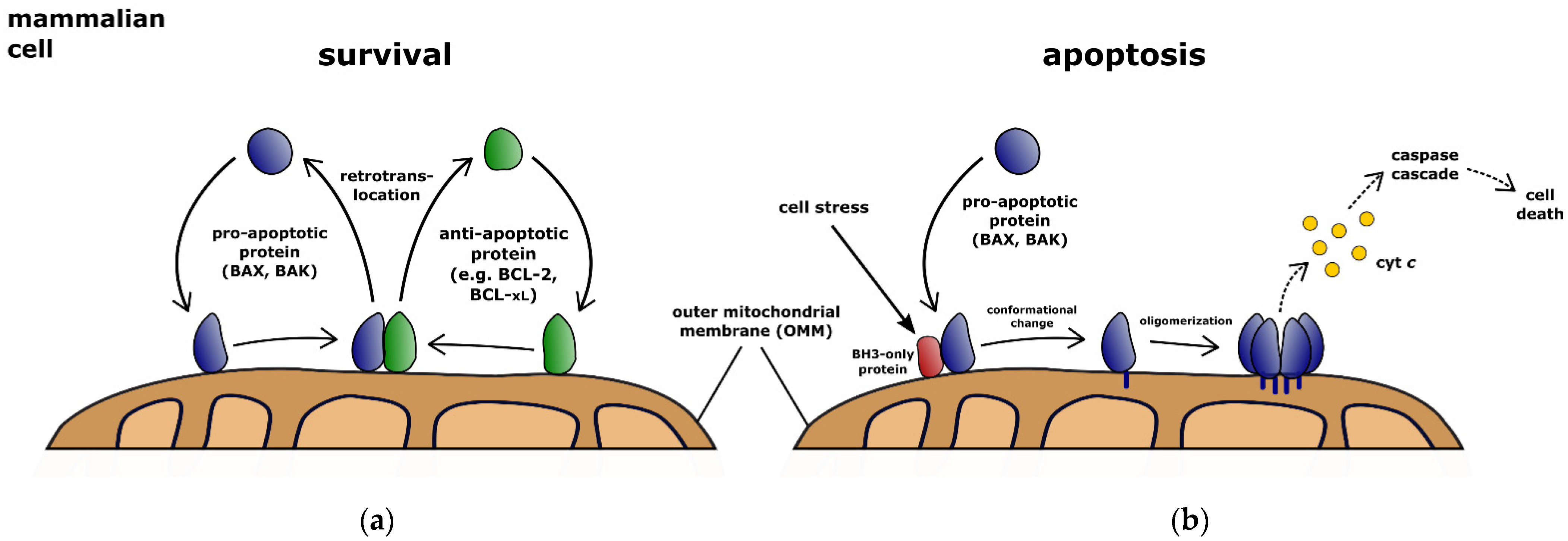
1. Pro-Apoptotic BCL-2 Activities Control the Molecular Decision to Apoptosis

Superfluous, infected, or damaged cells remove themselves from the organism through apoptosis [1,2,3]. This cell death program is essential for the survival of multicellular animals and the most important protective mechanism against tumor development. Apoptotic cells fragment into vesicles termed “apoptotic bodies” which are completely eliminated by phagocytosis [4]. Thereby, the dying cell protects neighboring cells from their harmful contents by being removed from the body.
Intrinsic apoptosis is the most common form of programmed death, involving the permeabilization of the outer mitochondrial membrane (OMM) [5]. This pathway is governed by proteins of the B-cell lymphoma-2 (BCL-2) family (Table 1). The pro-apoptotic activities of BCL-2-associated X protein (BAX) and BCL-2 antagonist killer 1 (BAK) can permeabilize the OMM. Then, proteins from the mitochondrial intermembrane space (IMS) such as cytochrome c (cyt c) are released, which initiates the caspase (cysteinyl aspartate protease) cascade that disassembles the cell [6,7,8]. Therefore, BAX/BAK activation is the first irreversible step in intrinsic apoptosis. However, reduced or inhibited caspase activation can lead to cell survival despite OMM permeabilization [9,10]. Limited OMM permeabilization of a subset of mitochondria has also been demonstrated to be insufficient to induce apoptosis [11,12]. Thus, cells can bypass commitment to apoptosis after BAX/BAK activation. The efficient activation of the caspase cascade is the principal function of the BCL-2 proteins. If the underlying mechanisms are impaired, a pathway for tumor formation will be cleared. Incidentally, this is also true when tumor therapy fails to induce apoptosis in targeted cells.
In addition to cell intrinsic signaling, extracellular death receptor ligands can trigger apoptosis. Cytotoxic T cells eliminate tumor cells by this mechanism, which is started or enhanced by immunotherapy. Extracellular domains of transmembrane receptors such as Fas (CD95) bind to their trimeric ligands. The apoptotic signal is transmitted by the clustering of activated receptors and the formation of an intracellular adaptor protein scaffold. Within this scaffold, the initiator caspase-8 self-activates and initiates the caspase cascade by substrate cleavage. Processing of one of the caspase-8 substrates, the BCL-2 homology domain 3 (BH3)-only protein BID, links the death receptor apoptosis pathway to OMM permeabilization. Truncated BID (tBID) can inhibit pro-survival BCL-2 proteins, thereby activating the pro-apoptotic BCL-2 proteins BAX and BAK [13,14,15]. BAX/BAK-dependent permeabilization of the OMM is often necessary to trigger apoptosis by extrinsic signals [5]. Even cells competent of undergoing apoptosis by receptor-mediated caspase activation alone show a greatly enhanced apoptotic response by BAX or BAK [16,17].

2. Dynamic Retrotranslocation Determines the Effective BCL-2 Protein Pool

BAX and BAK are antagonized by the large group of anti-apoptotic BCL-2 proteins with high functional redundancy, including BCL-2, B-cell lymphoma-extra large (BCL-xL), and myeloid cell leukemia 1 (MCL-1). The BCL-2 protein fold, shared by both pro-apoptotic and anti-apoptotic family members, creates a hydrophobic surface groove that is occupied by the C-terminal transmembrane domain (TMD) in the cytosolic forms of BAX and BCL-xL [18,19]. Intermolecular interactions between BCL-2 proteins are primarily mediated by binding of a BH3 domain to another protein’s hydrophobic groove [20]. One precondition for interaction between BCL-2 proteins, however, is the presence of a lipid membrane [15]. In cells, BCL-2 proteins localize predominantly to the OMM depending on their TMDs and interact with this membrane [21].
To this end, BAX and BAK translocate permanently to the OMM [22,23]. At the OMM, anti-apoptotic BCL-2 proteins inhibit BAX and BAK by constant retrotranslocation into the cytosol (Figure 1) [22,24,25,26]. Different shuttling rates of BAX and BAK lead to their distinct localization in the cell [25,27,28]. The shuttling of BAX and BAK establishes an equilibrium between protein pools in the cytosol and on the mitochondria. At the same time, only the mitochondrial population of proteins is available for activation [28]. Retrotranslocation, therefore, determines the cellular response to apoptosis stimulation [29,30,31]. BCL-2 proteins are also found in other compartments of the cell, yet only retrotranslocation between mitochondria and cytosol has been studied. For this purpose, the transition out of the membrane seems to be necessary [32]. Theoretically, the transition between other compartments and the mitochondria is also conceivable. To the ER, BCL-2 proteins could pass through the cytosol via retrotranslocation on the one hand and via lateral sorting from the mitochondria on the other hand [33,34]. In principle, it remains open whether BCL-2 proteins can be transported exclusively via the cytosol or also directly between different compartments. BAX/BAK retrotranslocation itself depends on recognition of exposed BH3 motifs by the hydrophobic groove of the pro-survival BCL-2 proteins [28]. BH3-only proteins, such as tBID, inhibit BCL-2 protein-dependent retrotranslocation of BAX and BAK [22]. Consequently, the presence of BH3-only proteins slows down the inhibitory retrotranslocation of BAX and BAK. The resulting increase in effective mitochondrial protein pools and prolongation of the dwell time of BAX and BAK molecules at the OMM increases the probability for apoptosis induction [28]. In other words, more stress means more BAX at the OMM and thus a greater chance for full activation of caspases.

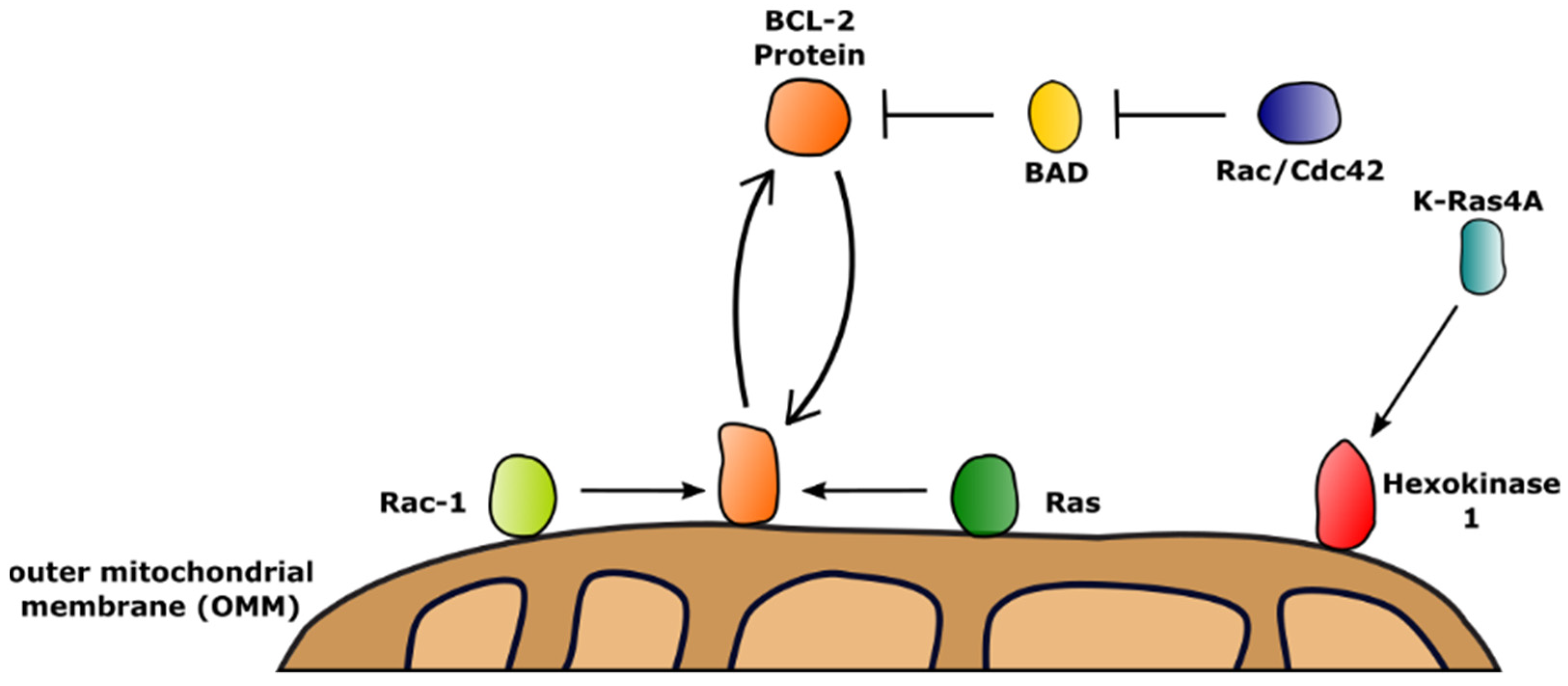
3. Membrane Receptors Guide the Function of BCL-2 Proteins by Activation of Downstream GTPases

Cellular commitment to apoptosis is directed by the activity and localization of BCL-2 proteins. In addition to regulatory interactions between members of the BCL-2 family, proteins outside this family participate in apoptosis regulation. There is increasing evidence that the superfamily of GTPases interacts with various members of the BCL-2 family on different molecular levels. GTPases are characterized by their activation via GTP binding and their subsequent deactivation after GTP hydrolysis by an intrinsic enzyme activity [35]. Based on their structure, different subfamilies were identified within the GTPase superfamily such as the heterotrimeric G proteins (large GTPases) and small Ras-like GTPases [36].
G protein signaling is heavily tied to the activity of a prominent class of plasma membrane-embedded proteins, the G protein-coupled receptor (GPCR) family, which engage intracellular G proteins upon ligand binding [37,38]. Despite the huge number of different GPCRs, only a limited set of G proteins is available to guide (multi-) cellular survival [39,40]. Activation of G proteins by different GPCRs such as the angiotensin II receptor type 1, the vasopressin receptor 2, and the N-formyl peptide receptor induces the activation of executioner caspase-3, leading to apoptosis [41]. This pro-apoptotic effect was negatable by homologous receptor desensitization, shutting off G protein signaling [41]. Interestingly, cleavage of arrestin-2, an adaptor protein mediating homologous desensitization, by different caspases, reduced cellular resistance to apoptosis and enhanced tBID-mediated release of cyt c from mitochondria [42]. This indicates a feedback loop, balancing pro- and anti-apoptotic effects, as well as a temporal scale of G protein signaling, determining its pro- and anti-survival actions. Yet, this seems to strongly depend on the cellular system, which is reflected by controversial reports of pro- and anti-apoptotic G protein signaling as demonstrated for the muscarinic receptor M1 [43,44].
In contrast to the small family of heterotrimeric G proteins, which are characterized by their enzymatic active Gα subunits, the Ras-like GTPases contain 167 monomeric members, which are homologous to Gα [45,46]. A summary of known interactions between small GTPases and BCL-2 family members is given in Table 2 and Figure 2. One way of regulating cellular survival by small GTPases is by activation of downstream kinases as shown for the Ras effector kinase Raf-1. Activated Raf-1 subsequently regulates BCL-2 protein function by interaction with and phosphorylation of the BH3-only proteins BAD and BIM, interfering with their binding to anti-apoptotic BCL-2 and BCL-xL [47,48,49]. Similarly to the Ras subfamily, members of the Rho subfamily of GTPases can protect cells from committing to apoptosis by regulation of BAD/BCL-xL interactions via downstream kinase signaling as shown for Rac and Cdc42 [50].
In addition to employing downstream effectors to modulate cell survival, small GTPases also regulate BCL-2 proteins by direct interactions, potentially linking metabolic pathways to cellular survival [51]. Rac-1, a crucial participant in insulin-dependent glucose uptake and metabolism, was shown to interact with BCL-2 on mitochondria, increasing the pro-survival effects of the BCL-2 protein [52,53]. Besides Rac-1, the GTPase Ras engages the N-terminal BH4 domain of BCL-2 at the OMM using its C-terminal CAAX motif, which enables BCL-2 to suppress the apoptotic influence of Ras signaling [54].
Table 2. Overview of BCL-2 family interactions with the superfamily of small GTPases, regulating cellular commitment to apoptosis.
GTPase
Superfamily
Mode of Action
RasRaf-1-dependent phosphorylation of pro-apoptotic BAD/BIM [47,48,49]
Ras binding to BCL-2, increasing its anti-apoptotic effect [54]
Activation of hexokinase I by K-Ras4A binding [55]
RhoRac-1 binding to BCL-2, increasing its anti-apoptotic effect [50]
PAK-dependent phosphorylation of pro-apoptotic BAD by Rac/Cdc42 [50]
Generally, large GTPases such as the heterotrimeric G proteins and small GTPases such as Ras balance a vast field of signaling pathways, impacting cellular homeostasis. Their participation in various metabolic pathways indicate an important role in guiding cellular survival. For example, the K-Ras isoform K-Ras4A was recently found to directly interact with hexokinase I and hexokinase II, key enzymes of glucose metabolism, on the OMM. Whereas K-Ras4A was able to bind to hexokinase II, it failed to increase its enzymatic activity [55]. Contrary, binding of K-Ras4A to hexokinase I increased the enzymatic activity of hexokinase I [55].

4. Hexokinases: At the Crossroads between Glucose Metabolism and Apoptosis

Hexokinases phosphorylate hexose sugars, primarily glucose, trapping glucose within the cytoplasm and keeping the intracellular concentration of plain glucose low. Hexokinases, therefore, play a critical role in cellular uptake and disposition of glucose by committing glucose to glycolytic and pentose phosphate pathways or storage. In mammalian tissues, four isoforms of hexokinases (I–IV) are found. They are constitutively expressed in most tissues but differ significantly in their tissue-specific distribution, subcellular localization, and functional properties (Table 3).
There is growing evidence that there is a direct link between glucose metabolism and apoptosis. First discoveries showed physical interactions between hexokinases and mitochondria [67]. It was later found that mainly hexokinase isoforms I and II bind to the mitochondria [68]. The subcellular distribution of hexokinase II has been reported to be dependent on glucose availability, whereas the distribution of hexokinase I is unaffected by varying glucose levels [69]. The interaction of hexokinases I and II with the mitochondria is facilitated by an N-terminal binding motif containing a short hydrophobic α-helix that is likely to be inserted into the OMM [60,70,71]. Truncated hexokinase lacking this hydrophobic region is unable to bind to the mitochondria [72]. In fact, it has been shown that a single mutation at the N-terminal domain of hexokinase II is enough to prevent binding to the OMM [73]. Hexokinases III and IV lack this hydrophobic N-terminal domain, and thus do not interact with the mitochondria.
Due to their binding to the OMM, hexokinases I and II have been linked to apoptosis. Hexokinase II has been shown to inhibit indomethacin-induced cyt c release and caspase-3 activation by preventing BAX from binding to the mitochondria [74]. Overexpression of hexokinase I has been shown to inhibit staurosporine-induced apoptosis [75]. Furthermore, overexpression of hexokinase I or II seems to decrease stress-induced accumulation of mitochondrial BAX, leading to the suggestion that hexokinases and BAX may compete for common binding sites on the mitochondria [76]. However, activation of BAX, as indicated by exposure of the carboxy-terminal 6A7 epitope, was not affected [76]. It has thus been hypothesized that hexokinase II sequesters active BAX in the cytosol, although no such interaction could be detected by immunoprecipitation [76]. The anti-apoptotic effect of hexokinase II is in line with a hexokinase II upregulation observed in many types of cancer [77]. Consequently, it has been suggested that overexpression of hexokinases in tumor cells contributes to resistance against chemotherapeutic drugs [78,79,80]. However, a survival advantage of tumor cells could not only be attributed to the inhibition of apoptosis by hexokinases but also to an increased rate of glycolysis and perhaps ATP production. Further studies showed that the anti-apoptotic effect of hexokinases is significantly reduced when truncated hexokinase isoforms that lack the N-terminal mitochondrial binding domain are overexpressed [72]. Interestingly, similar results have been obtained with full-length but catalytically inactive forms of both hexokinase I and II. These effects could either result from impaired hexokinase localization or the lack of glucose conversion to glucose-6-phosphate. In contrast, knockdown of hexokinase I has been reported to increase mitochondrial BAX and to promote TNF-induced BAX oligomerization [81]. In addition, hexokinase II knockdown has been suggested to enhance the expression of BAX and caspase-3, while BCL-2 could be downregulated [82]. Further studies revealed that depletion of hexokinase II decreases cancer cell proliferation and increases sensitivity to cell death inducers [58,83]. Effects similar to the hexokinase II knockdown have been observed when this enzyme gets displaced from mitochondria or from interaction sites between mitochondria and ER (mitochondria-associated membranes; MAMs) by selective peptides [59,84]. Pro-apoptotic effects could also be achieved by detaching mitochondrial-bound hexokinase I with clotrimazole [74,81].
Specific binding of hexokinases to the mitochondria is mediated by the voltage-dependent anion channel (VDAC), which is the major transport channel mediating the passage of ions and metabolites across the OMM [85,86,87,88]. The interaction with VDACs is thought to provide hexokinases preferred access to mitochondrially generated ATP [62]. Three isoforms of VDAC have been identified, VDAC1, VDAC2, and VDAC3 [89]. Different degrees of colocalization between these isoforms and hexokinase I have been revealed by STED microscopy [90]. Moreover, evidence suggests that distinct hexokinase I pools exist on the mitochondria that are not colocalized with any of the isoforms [90]. Although hexokinases seem to bind to the mitochondria without VDAC, knockout of VDAC results in a significant decrease in mitochondrial hexokinase [91].
The structural basis of complex formation between VDAC and hexokinases is yet to be elucidated. It has been suggested that hexokinase first inserts into the OMM and then interacts with VDAC on the outer leaflet of OMM [92]. Complex formation between both proteins seems to be mediated by their N-terminal domains [93,94]. Removal of the N-terminal domain of hexokinases or VDACs abolishes their interaction [93,95]. Studies show that their association protects cells against apoptosis [74,75]. A single mutation is sufficient to largely abolish hexokinase I binding to VDACs and prevent hexokinase I-mediated protection from cell death [57]. Interaction of hexokinases and VDACs has also been implicated in aerobic glycolysis (“Warburg effect”) and proliferation of tumor cells [96]. Strikingly, hexokinases have been reported to bind to VDACs more tightly in cancer cells compared to control cells [96]. However, the molecular basis of hexokinase-mediated apoptosis inhibition remained unresolved. VDAC is believed to adopt a closed state upon activation of apoptosis [97]. It has been suggested that binding of hexokinase to VDACs leads to channel opening [98,99]. There is also evidence, however, indicating that hexokinase I induces VDAC1 closure (“low-conducting” state), leading to diminished metabolic exchange [95,100,101]. Thus, hexokinase-dependent VDAC closure has been speculated to be an anti-apoptotic event by preventing cyt c release [95]. These discrepancies may be explained by the assumption that VDACs, besides an open and closed state, can adopt a partially closed conformation through their interaction with hexokinases [92]. This conformation would still allow some flux of low molecular weight substances while preventing the release of apoptotic factors. Interestingly, it has been shown that hexokinase-mediated VDAC closure can be reversed by glucose-6-phosphate [95,102], indicating a direct link between glucose metabolism and apoptosis perhaps through the influence of hexokinase localization on apoptosis. It has been further suggested that closing of VDACs by hexokinases could be inhibited by AKT signaling and overexpressed BCL-2 or BCL-xL [75,97]. However, opposing effects were also reported [64]. Nonetheless, a competition between hexokinases and BCL-2 proteins for VDAC binding sites has been proposed [63]. This assumption is consistent with the recent observation that under high glucose condition the interaction between VDAC1 and BAX is enhanced [103]. These effects could be partly reversed by overexpression of hexokinase II [103].
In addition, other binding partners of hexokinase II have been identified. The Tp53-induced Glycolysis and Apoptosis Regulator (TIGAR) is a p53 target gene. TIGAR has been shown to translocate to the mitochondria under hypoxic conditions, where it forms a complex with hexokinase II and increases hexokinase activity [104]. Similarly, the phosphoprotein enriched in astrocytes (PEA15) binds to hexokinase II following hypoxia and seems to increase anti-apoptotic effects [105]. These findings suggest that hexokinase II interacts with both ubiquitously and tissue-specific expressed binding partners.
Another regulator of hexokinases is glycogen synthase kinase 3β (GSK3β), which is a downstream effector of the phosphatidylinositol 3-kinase (PI3K)/AKT signaling pathway. GSK3β has been suggested to promote apoptosis [56,61]. Activation of GSK3β seems to induce the dissociation of hexokinase II from the OMM via phosphorylation of VDAC [74,106]. Inhibition of GSK3β has been shown to increase hexokinase II binding to the mitochondria and to protect against rotenone-induced apoptosis [106]. In contrast to the GSK3β-mediated disruption of hexokinase/VDAC interaction, phosphorylation of VDAC by PKC-ε has been reported to promote hexokinase binding [72,107]. These findings indicate an intricate regulatory web for the complex formation of hexokinases and VDACs.

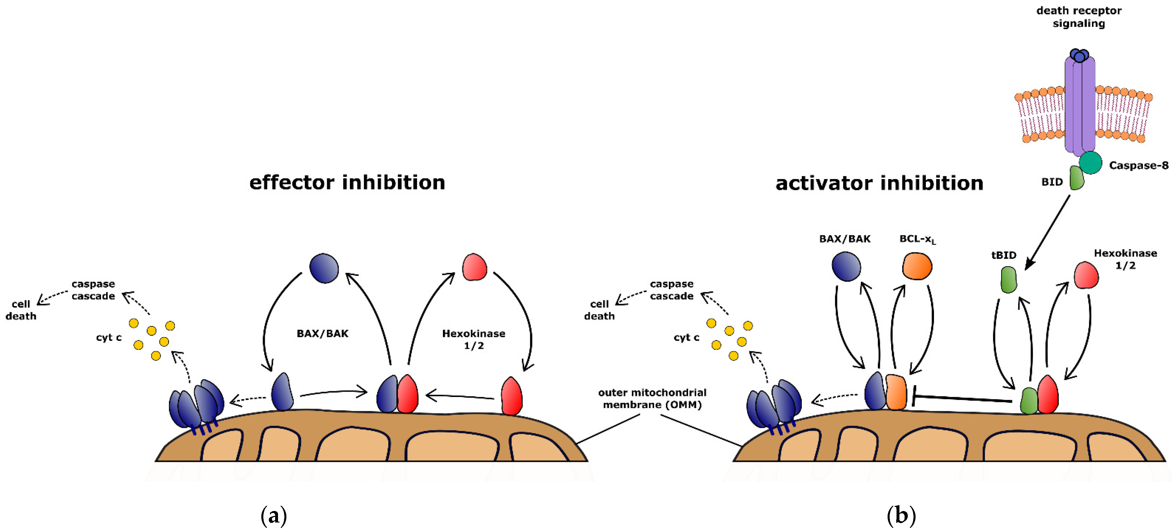
5. Hexokinase-Dependent Retrotranslocation Protects Cells against Extrinsic Apoptosis

Although a role for hexokinases has been suspected in the regulation of mitochondrial apoptosis for some time, only recently the molecular link has been discovered [108]. Studying transient protein interactions with BAX, hexokinases I and II were identified. Owing to the pronounced regulation observed in tumors, hexokinase II is likely involved in cell death regulation. However, it shares mitochondrial association and probably the mechanism of binding to mitochondria and dissociation with hexokinase I. Hence, it is not too surprising that both hexokinases are involved in apoptosis control.
The analysis of transient hexokinase interactions with BAX revealed involvement in inhibition of BAX and also BAK by retrotranslocation from mitochondria to the cytosol. Hexokinases also accelerate BCL-xL retrotranslocation comparable to the anti-apoptotic BCL-2 protein MCL-1. In turn, overexpression of BCL-xL shifts the localization of hexokinases to the cytosol. In other words, there is evidence for a strong interdependence of the proteins, which retrotranslocate BAX and BAK. A function of hexokinases in retrotranslocation seems to imply that hexokinases can protect cells from apoptosis in general. However, this is not the case. In fact, inhibition of only receptor-mediated apoptosis by hexokinases can be observed. In a reduced cell system lacking the prominent members of the BCL-2 family, hexokinases alone inhibited BAX only to a modest extent [108]. A substantial effect of hexokinase was observed only when cell survival was dependent on the inhibition of tBID. The discrepancy between minimal direct effect of hexokinases on BAX activity and paramount role in specific inhibition of tBID-dependent receptor-mediated apoptosis led to the discovery of hexokinase-dependent retrotranslocation of tBID [108].
This function of the hexokinases is independent of the phosphorylation of glucose to glucose-6-phosphate. However, mitochondrial association of the hexokinases is essential for retrotranslocation. On the mitochondria hexokinases form complexes with VDAC2 similar to pro-survival BCL-2 proteins [32,109]. However, VDAC2 seems to either interact with pro-survival BCL-2 proteins or hexokinases. Neither BAX nor BAK are present in stable complexes of VDAC2 with pro-survival BCL-2 proteins or those VDAC2 complexes containing hexokinases. Thus, stable complexes between BCL-2 proteins in association with the OMM seem lacking. Only the interaction between tBID and BCL-xL seems to be an exception to this rule: BCL-xL decreases the rate of tBID retrotranslocation [108]. Therefore, the mitochondrial pool of both proteins increases due to OMM-embedded tBID/BCL-xL complexes [110,111]. By stabilizing common complexes, tBID appears to have the exceptional ability to reduce the effective BCL-xL protein pool for BAX/BAK retrotranslocation. It follows that death receptor signaling could be particularly effective in cells addicted to BCL-xL activity. The tBID-specific hexokinase-dependent retrotranslocation protects cells particularly from this apoptosis signaling axis.
The dependence on hexokinase localization and the specificity toward receptor-mediated apoptosis can be explanations as to why this mechanism remained hidden. Mitochondrial hexokinases inhibit apoptosis by effector inhibition through BAX/BAK retrotranslocation and activator inhibition by retrotranslocating mitochondrial tBID (Figure 3). Hexokinase-mediated BAX/BAK retrotranslocation potentially counteracts any mitochondrial apoptosis signaling. This process occurs in all mammalian cells and depends on the localization and expression of hexokinases I and II. Elevated levels of specifically hexokinase II in some tumor entities suggest a more important role. Increased glucose metabolism was observed to stabilize HKII glucose dependently. The resulting anti-apoptotic effect could thus contribute to the development of vascular complications of diabetes, diabetic embryopathy, and insulin resistance. This is also supported by increased HKII expression levels in cancer-associated adipose tissue [112]. Indeed, apoptosis is linked to decreased HKII levels and mitochondrial binding in obesity and type II diabetes [113].
Nevertheless, in experiments, hexokinases protected tumor cells significantly less than anti-apoptotic BCL-2 proteins [108]. Crucial to cell survival after death receptor signaling, however, is the retrotranslocation of tBID. Hexokinase-dependent retrotranslocation of tBID can prevent cell death in cells that require mitochondria for receptor-mediated apoptosis (type II). Even if mitochondrial signaling is not essential for receptor-mediated apoptosis (type I), the proportion of apoptotic cells is likely reduced. Thus, inhibition of the activator tBID reduces apoptosis triggered by cytotoxic T cells. This mechanism is also likely to be more pronounced in tumors with high hexokinase levels. Future studies should investigate the role of hexokinase-dependent retrotranslocation in immune evasion.
In addition to association with mitochondria, the cytosolic domains of hexokinases are also required for interaction and retrotranslocation of BCL-2 proteins. tBID retrotranslocation can be inhibited by BH3 mimetics. The role of the BH3 motif is supported by reduced interactions between hexokinases and BAX variants of the BH3 motif [108]. The retrotranslocation of BAX, BAK, and tBID, the inhibitory effect of BH3 mimetics, the binding to tBID, BIM, and BAX, and the disruptive effect of BAX-BH3 variants on hexokinases suggest that hexokinases interact with BCL-2 proteins via the BH3 motif. Binding of hexokinases to the BH3 motif may explain why hexokinases can retrotranslocate BAX, BAK, and tBID. Differences in the BH3 motif or secondary binding sites could impede BIM shuttling. Despite structural and functional differences, hexokinases and pro-survival BCL-2 proteins retrotranslocate BCL-2 proteins.
Hexokinase-dependent retrotranslocation of tBID is a direct countermeasure to death receptor-dependent initiation of OMM permeabilization. This additional layer of apoptosis regulation contributes to cell-to-cell differences in apoptosis induction [114]. Recently, it was suggested that tBID permeabilizes the OMM in the absence of BAX and BAK [115]. Indeed, such a special role of tBID among BH3-only proteins would explain why a tBID-specific inhibitory mechanism is necessary to protect the healthy cell. Hexokinase-dependent resistance to death receptor ligands, such as TRAIL and FasL, provides further complexity to the design of successful anti-tumor strategies. Apoptosis induction by cytotoxic T cells requires inhibition of hexokinase-dependent BCL-2 protein retrotranslocation when progression relies on caspase-mediated BID cleavage. Observations of cell type-specific differential apoptosis induction by tBID and BIM could be caused by differential hexokinase-dependent tBID retrotranslocation [116,117]. Varying hexokinase activities could create apparent differences in tBID and BIM activities in some cell types while lacking from others [118].
Hexokinases also directly inhibit commitment to apoptosis by BAX/BAK retrotranslocation. Strikingly, hexokinases enhance BAX/BAK retrotranslocation to the same rates as pro-survival BCL-2 proteins, while protection from BAX in cells lacking BCL-2 proteins is considerably lower [108]. Therefore, the dominant effect of apoptosis inhibition by hexokinases results from tBID shuttling. Hexokinase-dependent tBID retrotranslocation frees pro-survival BCL-2 proteins to retrotranslocate BAX and BAK into the cytosol in a ‘primed to death’ scenario [119,120]. BAX/BAK retrotranslocation especially following intrinsic stress-induced signaling is dominated by pro-survival BCL-2 proteins. Interestingly, BID has also been reported as a substrate of caspase-2 in response to ER stress or DNA damage signaling [65,66]. Nonetheless, hexokinase-dependent apoptosis inhibition seems specific to death receptor signaling [108].

6. Conclusions

The BCL-2 protein family interacts with a variety of different protein species to regulate the cellular fate. Hexokinases provide a link from glucose metabolism to cell survival. Mitochondrial hexokinases inhibit apoptosis by two different mechanisms: effector inhibition through BAX/BAK retrotranslocation and activator inhibition by tBID retrotranslocation specifically preventing mitochondrial apoptosis engagement following death receptor signaling. Hexokinase-dependent retrotranslocation safeguards, therefore, cells from apoptosis induced by cytotoxic T cells and could reduce apoptosis in obesity and type II diabetes.

Author Contributions

Conceptualization, F.E.; writing—original draft preparation, A.S. and P.W.; writing—review and editing, F.E.; visualization, A.S. and P.W.; supervision, F.E.; project administration, F.E.; funding acquisition, F.E. All authors have read and agreed to the published version of the manuscript.

Funding

F.E. is supported by DFG ED 190/15-1.

Conflicts of Interest

The authors declare no conflict of interest.

References

  1. Sulston, J.E.; Horvitz, H.R. Post-Embryonic Cell Lineages of the Nematode, Caenorhabditis Elegans. Dev. Biol. 1977, 56, 110–156. [Google Scholar] [CrossRef]
  2. Zychlinsky, A.; Prevost, M.C.; Sansonetti, P.J. Shigella Flexneri Induces Apoptosis in Infected Macrophages. Nature 1992, 358, 167–169. [Google Scholar] [CrossRef] [PubMed]
  3. Clavería, C.; Giovinazzo, G.; Sierra, R.; Torres, M. Myc-Driven Endogenous Cell Competition in the Early Mammalian Embryo. Nature 2013, 500, 39–44. [Google Scholar] [CrossRef] [PubMed]
  4. Kerr, J.F.; Wyllie, A.H.; Currie, A.R. Apoptosis: A Basic Biological Phenomenon with Wide-Ranging Implications in Tissue Kinetics. Br. J. Cancer 1972, 26, 239–257. [Google Scholar] [CrossRef] [Green Version]
  5. Hotchkiss, R.S.; Strasser, A.; McDunn, J.E.; Swanson, P.E. Cell Death. N. Engl. J. Med. 2009, 361, 1570–1583. [Google Scholar] [CrossRef] [Green Version]
  6. Oltvai, Z.N.; Milliman, C.L.; Korsmeyer, S.J. Bcl-2 Heterodimerizes in Vivo with a Conserved Homolog, Bax, That Accelerates Programmed Cell Death. Cell 1993, 74, 609–619. [Google Scholar] [CrossRef]
  7. Shimizu, S.; Narita, M.; Tsujimoto, Y. Bcl-2 Family Proteins Regulate the Release of Apoptogenic Cytochrome c by the Mitochondrial Channel VDAC. Nature 1999, 399, 483–487. [Google Scholar] [CrossRef]
  8. Eskes, R.; Antonsson, B.; Osen-Sand, A.; Montessuit, S.; Richter, C.; Sadoul, R.; Mazzei, G.; Nichols, A.; Martinou, J.C. Bax-Induced Cytochrome c Release from Mitochondria Is Independent of the Permeability Transition Pore but Highly Dependent on Mg2+ Ions. J. Cell Biol. 1998, 143, 217–224. [Google Scholar] [CrossRef] [Green Version]
  9. Martinou, I.; Desagher, S.; Eskes, R.; Antonsson, B.; André, E.; Fakan, S.; Martinou, J.C. The Release of Cytochrome c from Mitochondria during Apoptosis of NGF-Deprived Sympathetic Neurons Is a Reversible Event. J. Cell Biol. 1999, 144, 883–889. [Google Scholar] [CrossRef]
  10. Potts, M.B.; Vaughn, A.E.; McDonough, H.; Patterson, C.; Deshmukh, M. Reduced Apaf-1 Levels in Cardiomyocytes Engage Strict Regulation of Apoptosis by Endogenous XIAP. J. Cell Biol. 2005, 171, 925–930. [Google Scholar] [CrossRef]
  11. Ichim, G.; Lopez, J.; Ahmed, S.U.; Muthalagu, N.; Giampazolias, E.; Delgado, M.E.; Haller, M.; Riley, J.S.; Mason, S.M.; Athineos, D.; et al. Limited Mitochondrial Permeabilization Causes DNA Damage and Genomic Instability in the Absence of Cell Death. Mol. Cell 2015, 57, 860–872. [Google Scholar] [CrossRef] [PubMed] [Green Version]
  12. Tait, S.W.G.; Parsons, M.J.; Llambi, F.; Bouchier-Hayes, L.; Connell, S.; Muñoz-Pinedo, C.; Green, D.R. Resistance to Caspase-Independent Cell Death Requires Persistence of Intact Mitochondria. Dev. Cell 2010, 18, 802–813. [Google Scholar] [CrossRef] [PubMed] [Green Version]
  13. Huang, K.; O’Neill, K.L.; Li, J.; Zhou, W.; Han, N.; Pang, X.; Wu, W.; Struble, L.; Borgstahl, G.; Liu, Z.; et al. BH3-Only Proteins Target BCL-XL/MCL-1, Not BAX/BAK, to Initiate Apoptosis. Cell Res. 2019, 29, 942–952. [Google Scholar] [CrossRef] [PubMed]
  14. Llambi, F.; Moldoveanu, T.; Tait, S.W.G.; Bouchier-Hayes, L.; Temirov, J.; McCormick, L.L.; Dillon, C.P.; Green, D.R. A Unified Model of Mammalian BCL-2 Protein Family Interactions at the Mitochondria. Mol. Cell 2011, 44, 517–531. [Google Scholar] [CrossRef] [PubMed] [Green Version]
  15. Roucou, X.; Montessuit, S.; Antonsson, B.; Martinou, J.-C. Bax Oligomerization in Mitochondrial Membranes Requires TBid (Caspase-8-Cleaved Bid) and a Mitochondrial Protein. Biochem. J. 2002, 368, 915–921. [Google Scholar] [CrossRef]
  16. Wei, M.C.; Lindsten, T.; Mootha, V.K.; Weiler, S.; Gross, A.; Ashiya, M.; Thompson, C.B.; Korsmeyer, S.J. TBID, a Membrane-Targeted Death Ligand, Oligomerizes BAK to Release Cytochrome c. Genes Dev. 2000, 14, 2060–2071. [Google Scholar] [CrossRef]
  17. Schleicher, R.I.; Reichenbach, F.; Kraft, P.; Kumar, A.; Lescan, M.; Todt, F.; Göbel, K.; Hilgendorf, I.; Geisler, T.; Bauer, A.; et al. Platelets Induce Apoptosis via Membrane-Bound FasL. Blood 2015, 126, 1483–1493. [Google Scholar] [CrossRef] [Green Version]
  18. Hinds, M.G.; Lackmann, M.; Skea, G.L.; Harrison, P.J.; Huang, D.C.S.; Day, C.L. The Structure of Bcl-w Reveals a Role for the C-Terminal Residues in Modulating Biological Activity. EMBO J. 2003, 22, 1497–1507. [Google Scholar] [CrossRef]
  19. Suzuki, M.; Youle, R.J.; Tjandra, N. Structure of Bax: Coregulation of Dimer Formation and Intracellular Localization. Cell 2000, 103, 645–654. [Google Scholar] [CrossRef] [Green Version]
  20. Sattler, M.; Liang, H.; Nettesheim, D.; Meadows, R.P.; Harlan, J.E.; Eberstadt, M.; Yoon, H.S.; Shuker, S.B.; Chang, B.S.; Minn, A.J.; et al. Structure of Bcl-XL-Bak Peptide Complex: Recognition between Regulators of Apoptosis. Science 1997, 275, 983–986. [Google Scholar] [CrossRef]
  21. Jeong, S.-Y.; Gaume, B.; Lee, Y.-J.; Hsu, Y.-T.; Ryu, S.-W.; Yoon, S.-H.; Youle, R.J. Bcl-x(L) Sequesters Its C-Terminal Membrane Anchor in Soluble, Cytosolic Homodimers. EMBO J. 2004, 23, 2146–2155. [Google Scholar] [CrossRef] [PubMed] [Green Version]
  22. Edlich, F.; Banerjee, S.; Suzuki, M.; Cleland, M.M.; Arnoult, D.; Wang, C.; Neutzner, A.; Tjandra, N.; Youle, R.J. Bcl-x(L) Retrotranslocates Bax from the Mitochondria into the Cytosol. Cell 2011, 145, 104–116. [Google Scholar] [CrossRef] [PubMed] [Green Version]
  23. Edlich, F. BCL-2 Proteins and Apoptosis: Recent Insights and Unknowns. Biochem. Biophys. Res. Commun. 2018, 500, 26–34. [Google Scholar] [CrossRef] [PubMed]
  24. Edlich, F. The Great Migration of Bax and Bak. Mol. Cell. Oncol. 2015, 2, e995029. [Google Scholar] [CrossRef] [Green Version]
  25. Todt, F.; Cakir, Z.; Reichenbach, F.; Emschermann, F.; Lauterwasser, J.; Kaiser, A.; Ichim, G.; Tait, S.W.G.; Frank, S.; Langer, H.F.; et al. Differential Retrotranslocation of Mitochondrial Bax and Bak. EMBO J. 2015, 34, 67–80. [Google Scholar] [CrossRef] [Green Version]
  26. Schellenberg, B.; Wang, P.; Keeble, J.A.; Rodriguez-Enriquez, R.; Walker, S.; Owens, T.W.; Foster, F.; Tanianis-Hughes, J.; Brennan, K.; Streuli, C.H.; et al. Bax Exists in a Dynamic Equilibrium between the Cytosol and Mitochondria to Control Apoptotic Priming. Mol. Cell 2013, 49, 959–971. [Google Scholar] [CrossRef] [Green Version]
  27. Edlich, F.; Martinou, J.-C. Bcl-2 Protein Interplay on the Outer Mitochondrial Membrane. In Mitochondria and Cell Death; Springer: New York, NY, USA, 2016; pp. 69–83. ISBN 978-1-4939-3612-0. [Google Scholar]
  28. Todt, F.; Cakir, Z.; Reichenbach, F.; Youle, R.J.; Edlich, F. The C-Terminal Helix of Bcl-x(L) Mediates Bax Retrotranslocation from the Mitochondria. Cell Death Differ. 2013, 20, 333–342. [Google Scholar] [CrossRef]
  29. Reichenbach, F.; Wiedenmann, C.; Schalk, E.; Becker, D.; Funk, K.; Scholz-Kreisel, P.; Todt, F.; Wolleschak, D.; Döhner, K.; Marquardt, J.U.; et al. Mitochondrial BAX Determines the Predisposition to Apoptosis in Human AML. Clin. Cancer Res. Off. J. Am. Assoc. Cancer Res. 2017, 23, 4805–4816. [Google Scholar] [CrossRef] [Green Version]
  30. Funk, K.; Czauderna, C.; Klesse, R.; Becker, D.; Hajduk, J.; Oelgeklaus, A.; Reichenbach, F.; Fimm-Todt, F.; Lauterwasser, J.; Galle, P.R.; et al. BAX Redistribution Induces Apoptosis Resistance and Selective Stress Sensitivity in Human HCC. Cancers 2020, 12, 1437. [Google Scholar] [CrossRef]
  31. Marquardt, J.U.; Edlich, F. Predisposition to Apoptosis in Hepatocellular Carcinoma: From Mechanistic Insights to Therapeutic Strategies. Front. Oncol. 2019, 9, 1421. [Google Scholar] [CrossRef] [Green Version]
  32. Lauterwasser, J.; Todt, F.; Zerbes, R.M.; Nguyen, T.N.; Craigen, W.; Lazarou, M.; van der Laan, M.; Edlich, F. The Porin VDAC2 Is the Mitochondrial Platform for Bax Retrotranslocation. Sci. Rep. 2016, 6, 32994. [Google Scholar] [CrossRef] [PubMed]
  33. Andreu-Fernández, V.; García-Murria, M.J.; Bañó-Polo, M.; Martin, J.; Monticelli, L.; Orzáez, M.; Mingarro, I. The C-Terminal Domains of Apoptotic BH3-Only Proteins Mediate Their Insertion into Distinct Biological Membranes. J. Biol. Chem. 2016, 291, 25207–25216. [Google Scholar] [CrossRef] [PubMed] [Green Version]
  34. García-Murria, M.J.; Duart, G.; Grau, B.; Diaz-Beneitez, E.; Rodríguez, D.; Mingarro, I.; Martínez-Gil, L. Viral Bcl2s’ Transmembrane Domain Interact with Host Bcl2 Proteins to Control Cellular Apoptosis. Nat. Commun. 2020, 11, 6056. [Google Scholar] [CrossRef] [PubMed]
  35. Grunicke, H.H.; Maly, K. Role of GTPases and GTPase Regulatory Proteins in Oncogenesis. Crit. Rev. Oncog. 1993, 4, 389–402. [Google Scholar]
  36. Li, H.; Yao, X.-Q.; Grant, B.J. Comparative Structural Dynamic Analysis of GTPases. PLoS Comput. Biol. 2018, 14, e1006364. [Google Scholar] [CrossRef] [Green Version]
  37. Oldham, W.M.; Hamm, H.E. Heterotrimeric G Protein Activation by G-Protein-Coupled Receptors. Nat. Rev. Mol. Cell Biol. 2008, 9, 60–71. [Google Scholar] [CrossRef]
  38. Wingler, L.M.; Lefkowitz, R.J. Conformational Basis of G Protein-Coupled Receptor Signaling Versatility. Trends Cell Biol. 2020, 30, 736–747. [Google Scholar] [CrossRef]
  39. Fredriksson, R.; Lagerström, M.C.; Lundin, L.-G.; Schiöth, H.B. The G-Protein-Coupled Receptors in the Human Genome Form Five Main Families. Phylogenetic Analysis, Paralogon Groups, and Fingerprints. Mol. Pharmacol. 2003, 63, 1256–1272. [Google Scholar] [CrossRef] [Green Version]
  40. Yanamadala, V.; Negoro, H.; Denker, B.M. Heterotrimeric G Proteins and Apoptosis: Intersecting Signaling Pathways Leading to Context Dependent Phenotypes. Curr. Mol. Med. 2009, 9, 527–545. [Google Scholar] [CrossRef] [Green Version]
  41. Revankar, C.M.; Vines, C.M.; Cimino, D.F.; Prossnitz, E.R. Arrestins Block G Protein-Coupled Receptor-Mediated Apoptosis. J. Biol. Chem. 2004, 279, 24578–24584. [Google Scholar] [CrossRef] [Green Version]
  42. Kook, S.; Zhan, X.; Cleghorn, W.M.; Benovic, J.L.; Gurevich, V.V.; Gurevich, E.V. Caspase-Cleaved Arrestin-2 and BID Cooperatively Facilitate Cytochrome c Release and Cell Death. Cell Death Differ. 2014, 21, 172–184. [Google Scholar] [CrossRef] [PubMed] [Green Version]
  43. Leloup, C.; Michaelson, D.M.; Fisher, A.; Hartmann, T.; Beyreuther, K.; Stein, R. M1 Muscarinic Receptors Block Caspase Activation by Phosphoinositide 3-Kinase- and MAPK/ERK-Independent Pathways. Cell Death Differ. 2000, 7, 825–833. [Google Scholar] [CrossRef] [PubMed] [Green Version]
  44. Graham, E.S.; Woo, K.K.; Aalderink, M.; Fry, S.; Greenwood, J.M.; Glass, M.; Dragunow, M. M1 Muscarinic Receptor Activation Mediates Cell Death in M1-HEK293 Cells. PLoS ONE 2013, 8, e72011. [Google Scholar] [CrossRef] [PubMed] [Green Version]
  45. Rojas, A.M.; Fuentes, G.; Rausell, A.; Valencia, A. The Ras Protein Superfamily: Evolutionary Tree and Role of Conserved Amino Acids. J. Cell Biol. 2012, 196, 189–201. [Google Scholar] [CrossRef] [PubMed] [Green Version]
  46. Syrovatkina, V.; Alegre, K.O.; Dey, R.; Huang, X.-Y. Regulation, Signaling and Physiological Functions of G-Proteins. J. Mol. Biol. 2016, 428, 3850–3868. [Google Scholar] [CrossRef] [PubMed] [Green Version]
  47. Zha, J.; Harada, H.; Yang, E.; Jockel, J.; Korsmeyer, S.J. Serine Phosphorylation of Death Agonist BAD in Response to Survival Factor Results in Binding to 14-3-3 Not BCL-XL. Cell 1996, 87, 619–628. [Google Scholar] [CrossRef] [Green Version]
  48. Ley, R.; Balmanno, K.; Hadfield, K.; Weston, C.; Cook, S.J. Activation of the ERK1/2 Signaling Pathway Promotes Phosphorylation and Proteasome-Dependent Degradation of the BH3-Only Protein, Bim. J. Biol. Chem. 2003, 278, 18811–18816. [Google Scholar] [CrossRef] [Green Version]
  49. Jin, S.; Zhuo, Y.; Guo, W.; Field, J. P21-Activated Kinase 1 (Pak1)-Dependent Phosphorylation of Raf-1 Regulates Its Mitochondrial Localization, Phosphorylation of BAD, and Bcl-2 Association. J. Biol. Chem. 2005, 280, 24698–24705. [Google Scholar] [CrossRef] [Green Version]
  50. He, H.; Yim, M.; Liu, K.H.; Cody, S.C.; Shulkes, A.; Baldwin, G.S. Involvement of G Proteins of the Rho Family in the Regulation of Bcl-2-like Protein Expression and Caspase 3 Activation by Gastrins. Cell Signal. 2008, 20, 83–93. [Google Scholar] [CrossRef]
  51. Rebollo, A.; Pérez-Sala, D.; Martínez-A, C. Bcl-2 Differentially Targets K-, N-, and H-Ras to Mitochondria in IL-2 Supplemented or Deprived Cells: Implications in Prevention of Apoptosis. Oncogene 1999, 18, 4930–4939. [Google Scholar] [CrossRef] [Green Version]
  52. Ueda, S.; Kitazawa, S.; Ishida, K.; Nishikawa, Y.; Matsui, M.; Matsumoto, H.; Aoki, T.; Nozaki, S.; Takeda, T.; Tamori, Y.; et al. Crucial Role of the Small GTPase Rac1 in Insulin-Stimulated Translocation of Glucose Transporter 4 to the Mouse Skeletal Muscle Sarcolemma. FASEB J. Off. Publ. Fed. Am. Soc. Exp. Biol. 2010, 24, 2254–2261. [Google Scholar] [CrossRef] [PubMed] [Green Version]
  53. Velaithan, R.; Kang, J.; Hirpara, J.L.; Loh, T.; Goh, B.C.; Le Bras, M.; Brenner, C.; Clement, M.-V.; Pervaiz, S. The Small GTPase Rac1 Is a Novel Binding Partner of Bcl-2 and Stabilizes Its Antiapoptotic Activity. Blood 2011, 117, 6214–6226. [Google Scholar] [CrossRef] [PubMed] [Green Version]
  54. Denis, G.V.; Yu, Q.; Ma, P.; Deeds, L.; Faller, D.V.; Chen, C.-Y. Bcl-2, via Its BH4 Domain, Blocks Apoptotic Signaling Mediated by Mitochondrial Ras. J. Biol. Chem. 2003, 278, 5775–5785. [Google Scholar] [CrossRef] [Green Version]
  55. Amendola, C.R.; Mahaffey, J.P.; Parker, S.J.; Ahearn, I.M.; Chen, W.-C.; Zhou, M.; Court, H.; Shi, J.; Mendoza, S.L.; Morten, M.J.; et al. KRAS4A Directly Regulates Hexokinase 1. Nature 2019, 576, 482–486. [Google Scholar] [CrossRef] [PubMed]
  56. Pastorino, J.G.; Hoek, J.B.; Shulga, N. Activation of Glycogen Synthase Kinase 3β Disrupts the Binding of Hexokinase II to Mitochondria by Phosphorylating Voltage-Dependent Anion Channel and Potentiates Chemotherapy-Induced Cytotoxicity. Cancer Res. 2005, 65, 10545–10554. [Google Scholar] [CrossRef] [PubMed] [Green Version]
  57. Zaid, H.; Abu-Hamad, S.; Israelson, A.; Nathan, I.; Shoshan-Barmatz, V. The Voltage-Dependent Anion Channel-1 Modulates Apoptotic Cell Death. Cell Death Differ. 2005, 12, 751–760. [Google Scholar] [CrossRef] [PubMed]
  58. Wolf, A.; Agnihotri, S.; Micallef, J.; Mukherjee, J.; Sabha, N.; Cairns, R.; Hawkins, C.; Guha, A. Hexokinase 2 Is a Key Mediator of Aerobic Glycolysis and Promotes Tumor Growth in Human Glioblastoma Multiforme. J. Exp. Med. 2011, 208, 313–326. [Google Scholar] [CrossRef] [PubMed] [Green Version]
  59. Arzoine, L.; Zilberberg, N.; Ben-Romano, R.; Shoshan-Barmatz, V. Voltage-Dependent Anion Channel 1-Based Peptides Interact with Hexokinase to Prevent Its Anti-Apoptotic Activity. J. Biol. Chem. 2009, 284, 3946–3955. [Google Scholar] [CrossRef] [Green Version]
  60. Xie, G.; Wilson, J.E. Rat Brain Hexokinase: The Hydrophobie N-Terminus of the Mitochondrially Bound Enzyme Is Inserted in the Lipid Bilayer. Arch. Biochem. Biophys. 1988, 267, 803–810. [Google Scholar] [CrossRef]
  61. Beurel, E.; Jope, R.S. The Paradoxical Pro- and Anti-Apoptotic Actions of GSK3 in the Intrinsic and Extrinsic Apoptosis Signaling Pathways. Prog. Neurobiol. 2006, 79, 173–189. [Google Scholar] [CrossRef] [Green Version]
  62. Arora, K.K.; Pedersen, P.L. Functional Significance of Mitochondrial Bound Hexokinase in Tumor Cell Metabolism. Evidence for Preferential Phosphorylation of Glucose by Intramitochondrially Generated ATP. J. Biol. Chem. 1988, 263, 17422–17428. [Google Scholar] [CrossRef]
  63. Pastorino, J.G.; Hoek, J.B. Regulation of Hexokinase Binding to VDAC. J. Bioenerg. Biomembr. 2008, 40, 171–182. [Google Scholar] [CrossRef] [PubMed] [Green Version]
  64. Duan, S.; Hájek, P.; Lin, C.; Shin, S.K.; Attardi, G.; Chomyn, A. Mitochondrial Outer Membrane Permeability Change and Hypersensitivity to Digitonin Early in Staurosporine-Induced Apoptosis. J. Biol. Chem. 2003, 278, 1346–1353. [Google Scholar] [CrossRef] [PubMed] [Green Version]
  65. Guo, Y.; Srinivasula, S.M.; Druilhe, A.; Fernandes-Alnemri, T.; Alnemri, E.S. Caspase-2 Induces Apoptosis by Releasing Proapoptotic Proteins from Mitochondria. J. Biol. Chem. 2002, 277, 13430–13437. [Google Scholar] [CrossRef] [PubMed] [Green Version]
  66. Upton, J.-P.; Austgen, K.; Nishino, M.; Coakley, K.M.; Hagen, A.; Han, D.; Papa, F.R.; Oakes, S.A. Caspase-2 Cleavage of BID Is a Critical Apoptotic Signal Downstream of Endoplasmic Reticulum Stress. Mol. Cell. Biol. 2008, 28, 3943–3951. [Google Scholar] [CrossRef] [Green Version]
  67. Rose, I.A.; Warms, J.V.B. Mitochondrial Hexokinase. J. Biol. Chem. 1967, 242, 1635–1645. [Google Scholar] [CrossRef]
  68. Sui, D.; Wilson, J.E. Structural Determinants for the Intracellular Localization of the Isozymes of Mammalian Hexokinase: Intracellular Localization of Fusion Constructs Incorporating Structural Elements from the Hexokinase Isozymes and the Green Fluorescent Protein. Arch. Biochem. Biophys. 1997, 345, 111–125. [Google Scholar] [CrossRef]
  69. John, S.; Weiss, J.N.; Ribalet, B. Subcellular Localization of Hexokinases I and II Directs the Metabolic Fate of Glucose. PLoS ONE 2011, 6, e17674. [Google Scholar] [CrossRef] [Green Version]
  70. Nawaz, M.H.; Ferreira, J.C.; Nedyalkova, L.; Zhu, H.; Carrasco-López, C.; Kirmizialtin, S.; Rabeh, W.M. The Catalytic Inactivation of the N-Half of Human Hexokinase 2 and Structural and Biochemical Characterization of Its Mitochondrial Conformation. Biosci. Rep. 2018, 38, BSR20171666. [Google Scholar] [CrossRef] [Green Version]
  71. Polakis, P.G.; Wilson, J.E. An Intact Hydrophobic N-Terminal Sequence Is Critical for Binding of Rat Brain Hexokinase to Mitochondria. Arch. Biochem. Biophys. 1985, 236, 328–337. [Google Scholar] [CrossRef]
  72. Sun, L.; Shukair, S.; Naik, T.J.; Moazed, F.; Ardehali, H. Glucose Phosphorylation and Mitochondrial Binding Are Required for the Protective Effects of Hexokinases I and II. Mol. Cell. Biol. 2008, 28, 1007–1017. [Google Scholar] [CrossRef] [PubMed] [Green Version]
  73. Bryan, N.; Raisch, K.P. Identification of a Mitochondrial-Binding Site on the N-Terminal End of Hexokinase II. Biosci. Rep. 2015, 35, e00205. [Google Scholar] [CrossRef] [PubMed]
  74. Pastorino, J.G.; Shulga, N.; Hoek, J.B. Mitochondrial Binding of Hexokinase II Inhibits Bax-Induced Cytochrome c Release and Apoptosis. J. Biol. Chem. 2002, 277, 7610–7618. [Google Scholar] [CrossRef] [PubMed] [Green Version]
  75. Abu-Hamad, S.; Zaid, H.; Israelson, A.; Nahon, E.; Shoshan-Barmatz, V. Hexokinase-I Protection against Apoptotic Cell Death Is Mediated via Interaction with the Voltage-Dependent Anion Channel-1. J. Biol. Chem. 2008, 283, 13482–13490. [Google Scholar] [CrossRef] [PubMed] [Green Version]
  76. Gall, J.M.; Wong, V.; Pimental, D.R.; Havasi, A.; Wang, Z.; Pastorino, J.G.; Bonegio, R.G.B.; Schwartz, J.H.; Borkan, S.C. Hexokinase Regulates Bax-Mediated Mitochondrial Membrane Injury Following Ischemic Stress. Kidney Int. 2011, 79, 1207–1216. [Google Scholar] [CrossRef] [PubMed] [Green Version]
  77. Mathupala, S.P.; Ko, Y.H.; Pedersen, P.L. Hexokinase II: Cancer’s Double-Edged Sword Acting as Both Facilitator and Gatekeeper of Malignancy When Bound to Mitochondria. Oncogene 2006, 25, 4777–4786. [Google Scholar] [CrossRef] [Green Version]
  78. Ahn, K.J.; Hwang, H.S.; Park, J.H.; Bang, S.H.; Kang, W.J.; Yun, M.; Lee, J.D. Evaluation of the Role of Hexokinase Type II in Cellular Proliferation and Apoptosis Using Human Hepatocellular Carcinoma Cell Lines. J. Nucl. Med. 2009, 50, 1525–1532. [Google Scholar] [CrossRef] [Green Version]
  79. Lemasters, J.J.; Holmuhamedov, E. Voltage-Dependent Anion Channel (VDAC) as Mitochondrial Governator—Thinking Outside the Box. Biochim. Biophys. Acta BBA-Mol. Basis Dis. 2006, 1762, 181–190. [Google Scholar] [CrossRef] [Green Version]
  80. Gu, J.J.; Singh, A.; Xue, K.; Mavis, C.; Barth, M.; Yanamadala, V.; Lenz, P.; Grau, M.; Lenz, G.; Czuczman, M.S.; et al. Up-Regulation of Hexokinase II Contributes to Rituximab-Chemotherapy Resistance and Is a Clinically Relevant Target for Therapeutic Development. Oncotarget 2018, 9, 4020–4033. [Google Scholar] [CrossRef] [Green Version]
  81. Schindler, A.; Foley, E. Hexokinase 1 Blocks Apoptotic Signals at the Mitochondria. Cell. Signal. 2013, 25, 2685–2692. [Google Scholar] [CrossRef]
  82. Liu, C.; Wang, X.; Zhang, Y. The Roles of HK2 on Tumorigenesis of Cervical Cancer. Technol. Cancer Res. Treat. 2019, 18, 153303381987130. [Google Scholar] [CrossRef] [PubMed]
  83. Fan, K.; Fan, Z.; Cheng, H.; Huang, Q.; Yang, C.; Jin, K.; Luo, G.; Yu, X.; Liu, C. Hexokinase 2 Dimerization and Interaction with Voltage-dependent Anion Channel Promoted Resistance to Cell Apoptosis Induced by Gemcitabine in Pancreatic Cancer. Cancer Med. 2019, 8, 5903–5915. [Google Scholar] [CrossRef] [PubMed]
  84. Ciscato, F.; Filadi, R.; Masgras, I.; Pizzi, M.; Marin, O.; Damiano, N.; Pizzo, P.; Gori, A.; Frezzato, F.; Chiara, F.; et al. Hexokinase 2 Displacement from Mitochondria-associated Membranes Prompts Ca2+ -dependent Death of Cancer Cells. EMBO Rep. 2020, 21, e49117. [Google Scholar] [CrossRef] [PubMed]
  85. Felgner, P.L.; Messer, J.L.; Wilson, J.E. Purification of a Hexokinase-Binding Protein from the Outer Mitochondrial Membrane. J. Biol. Chem. 1979, 254, 4946–4949. [Google Scholar] [CrossRef]
  86. Lindén, M.; Gellerfors, P.; Dean Nelson, B. Pore Protein and the Hexokinase-Binding Protein from the Outer Membrane of Rat Liver Mitochondria Are Identical. FEBS Lett. 1982, 141, 189–192. [Google Scholar] [CrossRef] [Green Version]
  87. Liu, M.Y.; Colombini, M. Regulation of Mitochondrial Respiration by Controlling the Permeability of the Outer Membrane through the Mitochondrial Channel, VDAC. Biochim. Biophys. Acta BBA-Bioenerg. 1992, 1098, 255–260. [Google Scholar] [CrossRef]
  88. Ujwal, R.; Cascio, D.; Colletier, J.-P.; Faham, S.; Zhang, J.; Toro, L.; Ping, P.; Abramson, J. The Crystal Structure of Mouse VDAC1 at 2.3 Å Resolution Reveals Mechanistic Insights into Metabolite Gating. Proc. Natl. Acad. Sci. USA 2008, 105, 17742–17747. [Google Scholar] [CrossRef] [Green Version]
  89. de Cerqueira Cesar, M.; Wilson, J.E. All Three Isoforms of the Voltage-Dependent Anion Channel (VDAC1, VDAC2, and VDAC3) Are Present in Mitochondria from Bovine, Rabbit, and Rat Brain. Arch. Biochem. Biophys. 2004, 422, 191–196. [Google Scholar] [CrossRef]
  90. Neumann, D.; Bückers, J.; Kastrup, L.; Hell, S.W.; Jakobs, S. Two-Color STED Microscopy Reveals Different Degrees of Colocalization between Hexokinase-I and the Three Human VDAC Isoforms. PMC Biophys. 2010, 3, 4. [Google Scholar] [CrossRef] [Green Version]
  91. Miyamoto, S.; Murphy, A.; Brown, J. Akt Mediates Mitochondrial Protection in Cardiomyocytes through Phosphorylation of Mitochondrial Hexokinase-II. Cell Death Differ. 2008, 15, 521–529. [Google Scholar] [CrossRef] [Green Version]
  92. Haloi, N.; Wen, P.-C.; Cheng, Q.; Yang, M.; Natarajan, G.; Camara, A.K.S.; Kwok, W.-M.; Tajkhorshid, E. Structural Basis of Complex Formation between Mitochondrial Anion Channel VDAC1 and Hexokinase-II. Commun. Biol. 2021, 4, 667. [Google Scholar] [CrossRef] [PubMed]
  93. Abu-Hamad, S.; Arbel, N.; Calo, D.; Arzoine, L.; Israelson, A.; Keinan, N.; Ben-Romano, R.; Friedman, O.; Shoshan-Barmatz, V. The VDAC1 N-Terminus Is Essential Both for Apoptosis and the Protective Effect of Anti-Apoptotic Proteins. J. Cell Sci. 2009, 122, 1906–1916. [Google Scholar] [CrossRef] [PubMed] [Green Version]
  94. Shoshan-Barmatz, V.; Zakar, M.; Rosenthal, K.; Abu-Hamad, S. Key Regions of VDAC1 Functioning in Apoptosis Induction and Regulation by Hexokinase. Biochim. Biophys. Acta BBA-Bioenerg. 2009, 1787, 421–430. [Google Scholar] [CrossRef] [Green Version]
  95. Azoulay-Zohar, H.; Israelson, A.; Abu-Hamad, S.; Shoshan-Barmatz, V. In Self-Defence: Hexokinase Promotes Voltage-Dependent Anion Channel Closure and Prevents Mitochondria-Mediated Apoptotic Cell Death. Biochem. J. 2004, 377, 347–355. [Google Scholar] [CrossRef] [PubMed]
  96. Pedersen, P.L. Warburg, Me and Hexokinase 2: Multiple Discoveries of Key Molecular Events Underlying One of Cancers’ Most Common Phenotypes, the “Warburg Effect”, i.e., Elevated Glycolysis in the Presence of Oxygen. J. Bioenerg. Biomembr. 2007, 39, 211–222. [Google Scholar] [CrossRef] [PubMed]
  97. Gottlob, K.; Majewski, N.; Kennedy, S.; Kandel, E.; Robey, R.B.; Hay, N. Inhibition of Early Apoptotic Events by Akt/PKB Is Dependent on the First Committed Step of Glycolysis and Mitochondrial Hexokinase. Genes Dev. 2001, 15, 1406–1418. [Google Scholar] [CrossRef] [Green Version]
  98. Majewski, N.; Nogueira, V.; Robey, R.B.; Hay, N. Akt Inhibits Apoptosis Downstream of BID Cleavage via a Glucose-Dependent Mechanism Involving Mitochondrial Hexokinases. Mol. Cell. Biol. 2004, 24, 730–740. [Google Scholar] [CrossRef] [Green Version]
  99. Robey, R.B.; Hay, N. Mitochondrial Hexokinases, Novel Mediators of the Antiapoptotic Effects of Growth Factors and Akt. Oncogene 2006, 25, 4683–4696. [Google Scholar] [CrossRef] [Green Version]
  100. Perevoshchikova, I.V.; Zorov, S.D.; Kotova, E.A.; Zorov, D.B.; Antonenko, Y.N. Hexokinase Inhibits Flux of Fluorescently Labeled ATP through Mitochondrial Outer Membrane Porin. FEBS Lett. 2010, 584, 2397–2402. [Google Scholar] [CrossRef] [Green Version]
  101. Vander Heiden, M.G.; Chandel, N.S.; Li, X.X.; Schumacker, P.T.; Colombini, M.; Thompson, C.B. Outer Mitochondrial Membrane Permeability Can Regulate Coupled Respiration and Cell Survival. Proc. Natl. Acad. Sci. USA 2000, 97, 4666–4671. [Google Scholar] [CrossRef] [Green Version]
  102. Bobba, A.; Amadoro, G.; La Piana, G.; Petragallo, V.A.; Calissano, P.; Atlante, A. Glucose-6-Phosphate Tips the Balance in Modulating Apoptosis in Cerebellar Granule Cells. FEBS Lett. 2015, 589, 651–658. [Google Scholar] [CrossRef] [PubMed] [Green Version]
  103. Zhang, J.; Guo, Y.; Ge, W.; Zhou, X.; Pan, M. High Glucose Induces Apoptosis of HUVECs in a Mitochondria-dependent Manner by Suppressing Hexokinase 2 Expression. Exp. Ther. Med. 2019, 18, 621–629. [Google Scholar] [CrossRef] [PubMed] [Green Version]
  104. Cheung, E.C.; Ludwig, R.L.; Vousden, K.H. Mitochondrial Localization of TIGAR under Hypoxia Stimulates HK2 and Lowers ROS and Cell Death. Proc. Natl. Acad. Sci. USA 2012, 109, 20491–20496. [Google Scholar] [CrossRef] [PubMed] [Green Version]
  105. Mergenthaler, P.; Kahl, A.; Kamitz, A.; van Laak, V.; Stohlmann, K.; Thomsen, S.; Klawitter, H.; Przesdzing, I.; Neeb, L.; Freyer, D.; et al. Mitochondrial Hexokinase II (HKII) and Phosphoprotein Enriched in Astrocytes (PEA15) Form a Molecular Switch Governing Cellular Fate Depending on the Metabolic State. Proc. Natl. Acad. Sci. USA 2012, 109, 1518–1523. [Google Scholar] [CrossRef] [PubMed] [Green Version]
  106. Gimenez-Cassina, A.; Lim, F.; Cerrato, T.; Palomo, G.M.; Diaz-Nido, J. Mitochondrial Hexokinase II Promotes Neuronal Survival and Acts Downstream of Glycogen Synthase Kinase-3. J. Biol. Chem. 2009, 284, 3001–3011. [Google Scholar] [CrossRef] [Green Version]
  107. Baines, C.P.; Song, C.-X.; Zheng, Y.-T.; Wang, G.-W.; Zhang, J.; Wang, O.-L.; Guo, Y.; Bolli, R.; Cardwell, E.M.; Ping, P. Protein Kinase Cε Interacts With and Inhibits the Permeability Transition Pore in Cardiac Mitochondria. Circ. Res. 2003, 92, 873–880. [Google Scholar] [CrossRef] [Green Version]
  108. Lauterwasser, J.; Fimm-Todt, F.; Oelgeklaus, A.; Schreiner, A.; Funk, K.; Falquez-Medina, H.; Klesse, R.; Jahreis, G.; Zerbes, R.M.; O’Neill, K.; et al. Hexokinases Inhibit Death Receptor-Dependent Apoptosis on the Mitochondria. Proc. Natl. Acad. Sci. USA 2021, 118, e2021175118. [Google Scholar] [CrossRef]
  109. Lauterwasser, J.; Fimm-Todt, F.; Edlich, F. Assessment of Dynamic BCL-2 Protein Shuttling Between Outer Mitochondrial Membrane and Cytosol. Methods Mol. Biol. 2019, 1877, 151–161. [Google Scholar] [CrossRef]
  110. Bogner, C.; Leber, B.; Andrews, D.W. Apoptosis: Embedded in Membranes. Curr. Opin. Cell Biol. 2010, 22, 845–851. [Google Scholar] [CrossRef]
  111. Lovell, J.F.; Billen, L.P.; Bindner, S.; Shamas-Din, A.; Fradin, C.; Leber, B.; Andrews, D.W. Membrane Binding by TBid Initiates an Ordered Series of Events Culminating in Membrane Permeabilization by Bax. Cell 2008, 135, 1074–1084. [Google Scholar] [CrossRef] [Green Version]
  112. Kalezic, A.; Udicki, M.; Srdic Galic, B.; Aleksic, M.; Korac, A.; Jankovic, A.; Korac, B. Tissue-Specific Warburg Effect in Breast Cancer and Cancer-Associated Adipose Tissue-Relationship between AMPK and Glycolysis. Cancers 2021, 13, 2731. [Google Scholar] [CrossRef] [PubMed]
  113. Liu, Y.; Cheng, H.; Zhou, Y.; Zhu, Y.; Bian, R.; Chen, Y.; Li, C.; Ma, Q.; Zheng, Q.; Zhang, Y.; et al. Myostatin Induces Mitochondrial Metabolic Alteration and Typical Apoptosis in Cancer Cells. Cell Death Dis. 2013, 4, e494. [Google Scholar] [CrossRef] [PubMed] [Green Version]
  114. Spencer, S.L.; Gaudet, S.; Albeck, J.G.; Burke, J.M.; Sorger, P.K. Non-Genetic Origins of Cell-to-Cell Variability in TRAIL-Induced Apoptosis. Nature 2009, 459, 428–432. [Google Scholar] [CrossRef] [PubMed]
  115. Flores-Romero, H.; Hohorst, L.; John, M.; Albert, M.-C.; King, L.E.; Beckmann, L.; Szabo, T.; Hertlein, V.; Luo, X.; Villunger, A.; et al. BCL-2-Family Protein TBID Can Act as a BAX-like Effector of Apoptosis. EMBO J. 2022, 41, e108690. [Google Scholar] [CrossRef]
  116. Lopez, J.; Bessou, M.; Riley, J.S.; Giampazolias, E.; Todt, F.; Rochegüe, T.; Oberst, A.; Green, D.R.; Edlich, F.; Ichim, G.; et al. Mito-Priming as a Method to Engineer Bcl-2 Addiction. Nat. Commun. 2016, 7, 10538. [Google Scholar] [CrossRef] [Green Version]
  117. Sarosiek, K.A.; Chi, X.; Bachman, J.A.; Sims, J.J.; Montero, J.; Patel, L.; Flanagan, A.; Andrews, D.W.; Sorger, P.; Letai, A. BID Preferentially Activates BAK While BIM Preferentially Activates BAX, Affecting Chemotherapy Response. Mol. Cell 2013, 51, 751–765. [Google Scholar] [CrossRef] [Green Version]
  118. Mason, K.D.; Carpinelli, M.R.; Fletcher, J.I.; Collinge, J.E.; Hilton, A.A.; Ellis, S.; Kelly, P.N.; Ekert, P.G.; Metcalf, D.; Roberts, A.W.; et al. Programmed Anuclear Cell Death Delimits Platelet Life Span. Cell 2007, 128, 1173–1186. [Google Scholar] [CrossRef] [Green Version]
  119. Certo, M.; Del Gaizo Moore, V.; Nishino, M.; Wei, G.; Korsmeyer, S.; Armstrong, S.A.; Letai, A. Mitochondria Primed by Death Signals Determine Cellular Addiction to Antiapoptotic BCL-2 Family Members. Cancer Cell 2006, 9, 351–365. [Google Scholar] [CrossRef] [Green Version]
  120. Ni Chonghaile, T.; Sarosiek, K.A.; Vo, T.-T.; Ryan, J.A.; Tammareddi, A.; Moore, V.D.G.; Deng, J.; Anderson, K.C.; Richardson, P.; Tai, Y.-T.; et al. Pretreatment Mitochondrial Priming Correlates with Clinical Response to Cytotoxic Chemotherapy. Science 2011, 334, 1129–1133. [Google Scholar] [CrossRef] [Green Version]
Figure 1. BCL-2 proteins inhibit apoptosis or commit the cell to mitochondrial apoptosis. (a) Pro-apoptotic BCL-2 proteins BAX and BAK (blue) constantly translocate to the outer mitochondrial membrane (OMM). After a change in the protein conformation of BAX or BAK the pro-apoptotic BCL-2 proteins are recognized by anti-apoptotic BCL-2 proteins, e.g., BCL-xL (green), and retrotranslocate back into the cytosol due to transient interactions between the two types of BCL-2 proteins. Retrotranslocation stabilizes the inactive forms of BAX and BAK and prevents, therefore, the activation of BAX or BAK in cells. (b) Lack of retrotranslocation of BAX or BAK commits the cell to apoptosis. BAX or BAK undergo further conformational changes at increased mitochondrial dwell times, oligomerize and permeabilize the OMM. The subsequent release of intermembrane space proteins, such as cytochrome c (cyt c), initiates the caspase cascade that dismantles the cell.
Biology 11 00412 g001
Figure 2. Interactions of small GTPases with the BCL-2 family, influencing BCL-2 protein function. Anti-apoptotic BCL-2 proteins such as BCL-2 (orange) shuttle between the cytoplasm and the outer mitochondrial membrane (OMM). BCL-2 protein function is regulated by members of the GTPase family. GTPases from the Rac and Ras subfamilies (light green and green, respectively) can bind to OMM-integrated BCL-2, enhancing its anti-apoptotic function. The anti-apoptotic effect of BCL-2 function is enhanced by inhibition of BAD through phosphorylation in a Rac/Cdc42 (dark blue)-dependent manner. Further, GTPases such as K-Ras4A (light blue) activate hexokinase I (red) on the OMM, participating in apoptosis regulation.
Biology 11 00412 g002
Figure 3. BCL-2 proteins inhibit apoptosis or commit the cell to mitochondrial apoptosis as a result of receptor-mediated apoptosis or intrinsic stress. (a) Hexokinase-dependent effector inhibition functions in analogy to BCL-2 protein-mediated retrotranslocation are involved in cell protection from any signal that can potentially trigger apoptosis. This mechanism is universally expected in mammalian cells but seems to be of lesser importance compared to the role of anti-apoptotic BCL-2 proteins. Constant OMM association of pro-apoptotic BCL-2 proteins (blue) is counteracted when BAX and BAK are recognized after a major conformational change by hexokinases (red) and retrotranslocated into the cytosol. In the absence of retrotranslocation of BAX or BAK, apoptosis is initiated through oligomerization of BAX and/or BAK and permeabilizes the OMM leading to cytochrome c (cyt c) release and caspase activity. Therefore, hexokinases prevent OMM permeabilization and commitment to apoptosis. (b) Additional inhibition of the activator tBID by hexokinases specifically protects cells from receptor-mediated apoptosis by cytotoxic T cells. In response to ligands death receptors, e.g., Fas or TRAIL-R, trimerize and initiate the formation of a caspase-8 (dark green) activating scaffold. Caspase-8 activity results in BID (green) cleavage. Mitochondrial tBID inhibits BAX/BAK (blue) retrotranslocation by competing with BAX and BAK for pro-survival BCL-2 protein, e.g., BCL-xL (orange), interactions. Therefore, tBID shifts BAX and BAK towards the active forms by forming OMM-embedded complexes with BCL-xL. Mitochondrial hexokinase I/II (red) selectively retrotranslocate tBID into the cytosol. Therefore, hexokinases prevent BAX/BAK activation in response to death receptor-mediated apoptosis.
Biology 11 00412 g003
Table 1. Classification, role, and interactions of BCL-2 family members.
BCL-2 Family
Member
Gene
Name
ActivityAssociated DiseasesInteracting BCL-2 Family
Proteins in Cancer
BCL-2BCL2Anti-apoptoticFollicular lymphoma 1,BAX, BAD, BIM, tBID, PUMA
high-grade B-cell lymphoma
BCL-xLBCL2L1Anti-apoptoticAbsolute glaucoma,BAX, BAK, BAD, BIM, tBID, PUMA
tongue carcinoma
MCL-1MCL1Anti-apoptoticMyeloid leukemia,BAX, BAK, BIM, tBID, NOXA, PUMA
chlamydia
BAXBAXPro-apoptoticT-cell acute lymphoblastic leukemia,MCL-1, BFL-1, BCL-xL, BCL-2, BCL-w,
colorectal cancerBCL-B, PUMA, BIM, tBID
BAKBAK1Pro-apoptoticAbsolute glaucoma,MCL-1, BFL-1, BCL-xL, PUMA, BIM, tBID
keratoacanthoma
BIDBIDPro-apoptoticBladder transitional cell papilloma,MCL-1, BFL-1, BCL-xL, BCL-2, BCL-w,
colon adenocarcinomaBCL-B, BAX, BAK
BIMBCL2L11Pro-apoptoticInterleukin-7 receptor alpha deficiency,MCL-1, BFL-1, BCL-xL, BCL-2, BCL-w,
lymphoproliferative syndromeBCL-B, BAX, BAK
BADBADPro-apoptoticB-cell lymphoma,BCL-2, BCL-xL, BCL-w
transient cerebral ischemia
Table 3. Classification, functions, and interactions of hexokinases.
HKTissue DistributionSubcellular
Localization
FunctionsSuggested
Interactions in Cell Death Signaling
References
IAll mammalian tissues,OMM, cytosolGlucose catabolism,BCL-xL[56]
main isoform in the brain apoptosis regulatorBID[56,57] *
BIM[56]
BAX[56]
BAK[56]
VDAC[58,59]
IIHeart, skeletal muscle,OMM, cytosolGlucose catabolism,BAX[56,60] *
adipose tissue glycogen synthesis,BAK[56]
apoptosis regulatorVDAC[58,59]
PKCε[61]
AKT[62]
PEA15[63]
TIGAR[64]
IIIUbiquitously expressed at low levels,PerinuclearGlucose catabolism
highest expression in lung, kidneycompartment
and liver
IVLiver, pancreatic islets, certainCytosolGlucose catabolism,BAD[65]
parts of the brain and gut intracellular glucose sensorVDAC[66]
* Competitive interaction reported.; HK: hexokinase.
Publisher’s Note: MDPI stays neutral with regard to jurisdictional claims in published maps and institutional affiliations.

