Article

Entomopathogenic Nematodes and Their Symbiotic Bacteria from the National Parks of Thailand and Larvicidal Property of Symbiotic Bacteria against Aedes aegypti and Culex quinquefasciatus

by
Aunchalee Thanwisai
1,2,3,
Paramaporn Muangpat
1,
Wipanee Meesil
1,
Pichamon Janthu
1,
Abdulhakam Dumidae
1,
Chanakan Subkrasae
1,
Jiranun Ardpairin
1,
Sarunporn Tandhavanant
4,
Timothy P. Yoshino
5 and
Apichat Vitta
1,2,3,*
1
Department of Microbiology and Parasitology, Faculty of Medical Science, Naresuan University, Phitsanulok 65000, Thailand
2
Centre of Excellence in Medical Biotechnology (CEMB), Faculty of Medical Science, Naresuan University, Phitsanulok 65000, Thailand
3
Center of Excellence for Biodiversity, Faculty of Sciences, Naresuan University, Phitsanulok 65000, Thailand
4
Department of Microbiology and Immunology, Faculty of Tropical Medicine, Mahidol University, Bangkok 10400, Thailand
5
Department of Pathobiological Sciences, School of Veterinary Medicine, University of Wisconsin, Madison, WI 53706, USA
*
Author to whom correspondence should be addressed.
Biology 2022, 11(11), 1658; https://doi.org/10.3390/biology11111658
Submission received: 6 October 2022 / Revised: 10 November 2022 / Accepted: 11 November 2022 / Published: 13 November 2022
(This article belongs to the Section Zoology)

Abstract

:

Simple Summary

Entomopathogenic nematodes (EPNs) are insect-parasitizing nematodes of the genera Heterorhabditis and Steinernema that are symbiotically associated with the symbiotic bacteria Photorhabdus and Xenorhabdus, respectively. Heterorhabditis indica, H. baujardi, Heterorhabditis SGmg3, Steinernema guangdongense, S. surkhetense, S. minutum, and S. longicaudum were isolated from soil samples in the national parks of Thailand. For symbiotic bacterial isolates, P. luminescens subsp. akhurstii, P. luminescens subsp. hainanensis, P. luminescens subsp. australis, Xenorhabdus stockiae, X. indica, X. griffiniae, X. japonica, and X. hominickii were isolated from those EPNs. In mosquito larvicidal bioassays, Photorhabdus isolates were effective against both Aedes aegypti and Culex quinquefasciatus. In conclusion, a wide diversity of entomopathogenic nematodes and symbiotic bacteria was found in the national parks of Thailand. Moreover, isolated Photorhabdus bacteria were shown to have potential as biocontrol agents to control culicine mosquitoes.

Abstract

Entomopathogenic nematodes (EPNs) are insect parasitic nematodes of the genera Het-erorhabditis and Steinernema. These nematodes are symbiotically associated with the bacteria, Photorhabdus and Xenorhabdus, respectively. National parks in Thailand are a potentially rich resource for recovering native EPNs and their symbiotic bacteria. The objectives of this study are to isolate and identify EPNs and their bacterial flora from soil samples in four national parks in Thailand and to evaluate their efficacy for controlling mosquito larvae. Using a baiting method with a Galleria mellonella moth larvae and a White trap technique, 80 out of 840 soil samples (9.5%) from 168 field sites were positive for EPNs. Sequencing of an internal transcribed spacer resulted in the molecular identification of Heterorhabditis nematode isolates as H. indica, H. baujardi and Heterorhabditis SGmg3, while using 28S rDNA sequencing, Steinernema nematode species were identified as S. guang-dongense, S. surkhetense, S. minutum, S. longicaudum and one closely related to S. yirgalemense. For the symbiotic bacterial isolates, based on recA sequencing, the Photorhabdus spp. were identified as P. luminescens subsp. akhurstii, P. luminescens subsp. hainanensis and P. luminescens subsp. australis. Xenorhabdus isolates were identified as X. stockiae, X. indica, X. griffiniae, X. japonica and X. hominickii. Results of bioassays demonstrate that Photorhabdus isolates were effective on both Aedes aegypti and Culex quinquefasciatus. Therefore, we conclude that soil from Thailand’s national parks contain a high diversity of entomopathogenic nematodes and their symbiotic bacteria. Photorhabdus bacteria are larvicidal against culicine mosquitoes and may serve as effective biocontrol agents.

Graphical Abstract

1. Introduction

Entomopathogenic nematodes (EPNs), in the genus Steinernema Travassos, 1927, Heterorhabditis Poinar, 1976 and Neosteinernema Nguyen and Smart, 1994, are soil-dwelling and insect-parasitizing nematodes [1,2,3]. Steinernema, in the family Steinernematidae, and Heterorhabditis, in the family Heterorhabditidae, are symbiotically associated with bacteria in genera Xenorhabdus and Photothabdus, respectively, while the symbiotic bacterial species associated with Neosteinernema in the family Steinernematidae is still unclear. The EPNs have been successfully used in the biological control of soil-dwelling insect larvae [4,5]. The infective juvenile (IJ) is the invasive, non-feeding stage of EPNs that parasitized and kills the insect host, usually within 48 hours aided by their bacterial partners [6]. Several groups of economically important insect pests can be effectively controlled by using EPNs including the larval stages of termites, mustard sawfly, cabbage leaf webber, fig moth, rice stem borers, carob moth and white grub [5,7]. Thus, promoting the use of EPNs in agriculture has clear implications for environmental sustainability and food safety [4]. Surveying and accurately identifying native EPNs and their symbiotic bacteria provide important baseline data for applying the EPNs as biocontrol agents in a local area. Native EPNs more easily adapt to local environmental and ecological conditions during the application process.
Molecular techniques based on sequencing of specific genes have been used to identify species of EPNs. For example, DNA sequence analysis of the 18S rDNA gene was used to distinguish between the genera Steinernema and Heterorhabditis. However, it was unable to distinguish between species due to low 18S rDNA sequence variation [8]. By contrast, the sequencing of the 28S rDNA gene and the highly variable internal transcribe spacer (ITS) region of Steinernema [9] and Heterorhabditis [10], respectively, were effective in differentiating members of these genera at the species level. For symbiotic bacteria, molecular identification of these bacterial species has been based on the sequencing of the several genetic markers including the 16S rDNA, 50S ribosomal protein L2 (rplB) gene, recombinase A (recA), DNA gyrase beta subunit (gyrB), DNA polymerase III subunit beta (dnaN), and glutamyl-tRNA synthetase (gltX) [11,12,13]. Analysis of the DNA sequences of the housekeeping gene, recA, was sufficient to discriminate between Photorhabdus and Xenorhabdus species [13].
The previously studied locations for surveying EPNs and symbiotic bacteria in Thai-land included roadside verges, areas of fruit crops, field crops, rice fields, and the banks of rivers and ponds [14,15,16,17], and typically have yielded low prevalence (<10%) and species diversity (<10 species) of EPNs and symbiotic bacteria. Surveys of EPNs and symbiotic bacteria in the national parks have resulted in several new records in Thailand [18,19,20]. In addition, at least two new species of EPNs were isolated from national parks in Vietnam [21,22]. The national parks in Thailand are defined as areas that contain vast natural resources of ecological importance or unique beauty, or that possess flora and fauna of special importance. We hypothesize that these largely uninhabited areas may be exceptionally good natural resources containing a diversity of previously undiscovered EPNs and bacterial symbionts. In the present study, four national parks, including Kaeng Krachan National Park in Phetchaburi Province, Namtok Samlan National Park in Saraburi Province, Phu Phan National Park in Sakhon Nakhon Province, and Huai Nam Dang National Park in Chiang Mai Province, were selected as primary sampling locations to search for EPNs and their symbiotic bacteria. In addition, randomly selected symbiotic bacterial species were laboratory tested to evaluate their larvicidal activity against the mosquitos, Aedes aegypti and Culex quinquefasciatus. Aedes aegypti is an important vector for the dengue virus, while Cx. quinquefasciatus serves as the main vector for filarial worms. Chemical insecticides are the primary methods used to control these mosquitoes. However, their use is associated with known adverse effects on humans, animals and the environment [23]. Moreover, repeated applications of insecticides can result in the creation and establishment of chemical resistant mosquito strains [24]. Therefore, the aims of the present study are to identify the entomopathogenic nematodes and their bacterial symbionts from national parks in Thailand and to evaluate the larvicidal activity of the selected symbiotic bacteria against Ae. aegypti and Cx. quinquefasciatus.

2. Materials and Methods
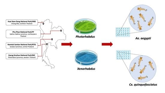
2.1. Study Sites

Four national parks in Thailand were selected for study sites to collect EPNs and symbiotic bacteria: Kaeng Krachan National Park (Phetchaburi Province, western Thai-land), Namtok Samlan National Park (Saraburi Province, central Thailand), Phu Phan National Park (Sakhon Nakhon Province, northeastern Thailand) and Huai Nam Dang National Park (Chiang Mai Province, northern Thailand) (Figure 1). The protocol for soil sample collection in the national parks was approved and permitted by the Department of National Park, Wildlife and Plant Conservation, Thailand (Permit number 0907.4/5514).

2.2. Collection of Soil Samples

Soil samples were randomly collected from 4 national parks of Thailand. We collected 840 soil samples from 168 soil sites by hand shovel. At each site, 5 soil samples were taken from an area of approximately 10 m2 at a depth of 5–10 cm. Approximately 300–600 g of each soil sample was taken by hand shovel and transferred to a plastic bag. Soil parameters for each sample, including temperature, pH, and moisture, were recorded using a soil survey instrument. Site location and soil texture were also recorded. GPS Navigation was used to determine the longitude, latitude and altitude of each site.

2.3. Isolation of EPNs from Soil Samples

The infective juvenile (IJ) stage of the EPNs was isolated from soil samples using larval Galleria mellonella baiting, as described by Bedding and Akhurst [25]. The G. mellonella cadavers were collected and placed into a White trap that was maintained at room temperature (25–30 °C) to allow emergence of infective EPN juveniles [26]. All soil samples were rebaited using fresh insect larvae to maximize EPN recovery. Emergent nematodes were collected and re-exposed to insect larvae to confirm entomopathogenicity and increase EPN yields. The nematodes were kept in a culture flask containing distilled water at 13–15 °C prior to molecular identification.

2.4. Species Identification of EPNs

For preliminary identification of nematodes, the skin color of G. mellonella cadavers was observed to predict the cause of death by Steinernema (beige/ochre or black) or Heterorhabditis (red, burgundy) [27]. The genomic DNA was extracted from approximately 200–500 IJs by using the NucleoSpin Tissue Kit (Macherey-Nagel, Duren, Germany) according to the manufacturer’s protocol. To check the quality of genomic DNA, five µl of purified DNA were examined on 0.8% agarose gel electrophoresis running in 0.5X TBE buffer at 80 V. After completion, the gel was stained with 10 µg/ml ethidium bromide for 1 min and destained with dH2O for 30 min. The DNA band was visualized, compared to a 100 bp molecular size marker and photographed under UV illumination. The genomic DNA was stored at −20 °C until use.
Polymerase chain reaction (PCR) was performed for molecular identification and sequence analysis based on different gene targets for Steinernema and Heterorhabditis. Partial sequence of 28S rDNA for Steinernema nematodes was amplified using primers; 539_F (5′-GGATTTCCTTAGTAACTGCGAGTG-3′) and 535_R (5′-TAGTCTTTCGCCCCTATAC-3′) to obtain an 870 bp amplicon [9]. The reaction was carried out in a 30 µl volume containing 15 µl of EconoTaq® PLUS 2× Master mix (1×; Luci-gen Corporation, Middleton, WI, USA), 1.5 µl of 5 µM of each primer (0.25 µM), 9 µl of dH20, and 3 µl of the DNA template (20–200 ηg). Partial internal transcribed spacer (ITS) sequence for Heterorhabditis nematode was amplified using the primers: 18S_F (5′-TTGATTACGTCCCTGCC CTTT-3′), TW81_F (5′-GTTTCCGTAGGTGAACCTGC-3′), 16S_R (5′-TTTCAC TCGCCGTTACTAAGG-3′) and AB28_R (5′-ATATGCTTAAGTTCAGCGGGT-3′) to obtain amplicons that varied in size between strains at a range of 800-850 bp for TW81_F and AB28_R primers and 1000–1100 bp for 18S_F and 16S_R or AB28_R primers [10]. The PCR components (30 µl total volume) were also used for amplifying Steinernema nematodes except for primers. All PCR reactions were performed in a Biometra TOne Thermal Cycler (Analytik Jena AG, Jena, Germany) using a temperature profile as previously described in Thanwisai et al. [15]. The amplified products were separated by 1.2% agarose-gel electrophoresis and visually examined. The PCR products were purified using a NucleoSpin® Gel and PCR Clean-Up Kit (Ma-cherey-Nagel, Düren, Germany) according to the manufacturer’s instructions before sequencing at Macrogen, Inc. South Korea (http://www.macrogen.com, accessed on 6 August 2022).

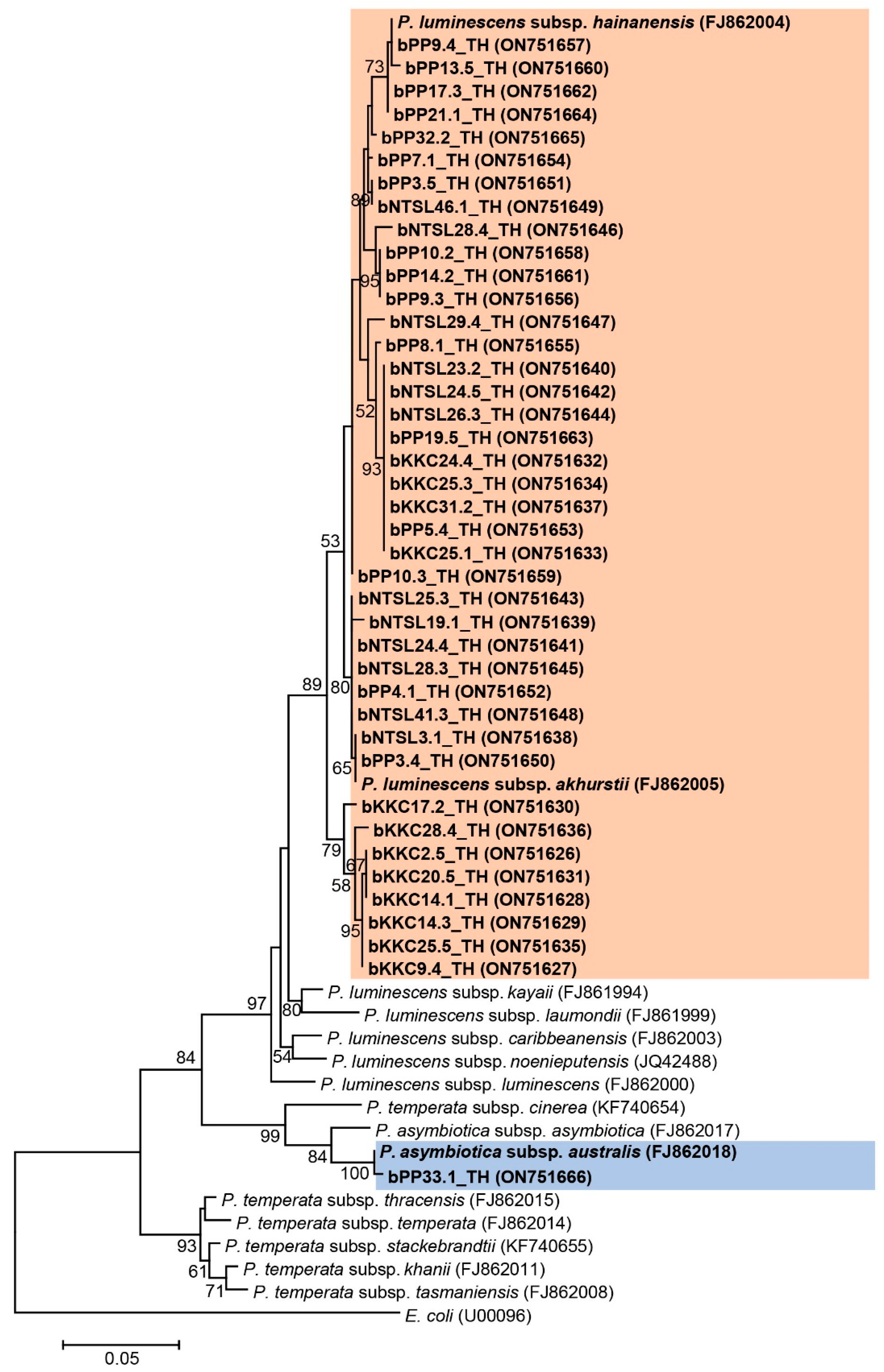
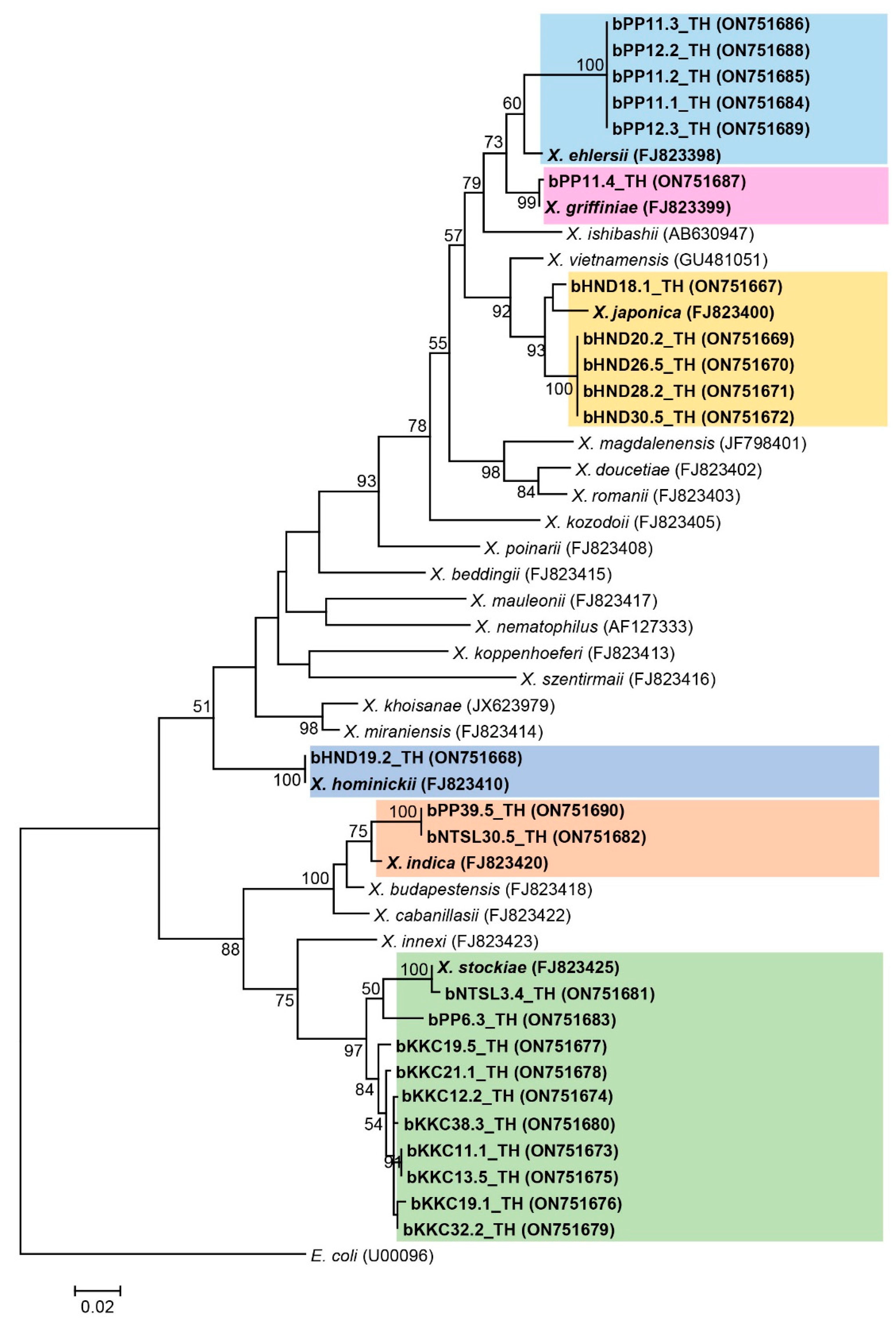
2.5. Species Identification of Symbiotic Bacteria

Xenorhabdus and Photorhabdus were isolated from the haemolymph of the G. mellonella infected with the IJ of EPNs according to Fukruksa et al. [28]. Colony morphology of Xenorhabdus and Photorhabdus was observed on NBTA agar plates and examined for size, color, edge morphology and surface texture [15].
Preparation of bacterial cell genomic DNA extraction was performed according to the methods described by Yooyangket et al. [19] using a Genomic DNA Mini Kit (blood/Cultured Cell) (Geneaid Biotech Ltd., New Taipei City, Taiwan). One microliter of genomic DNA was examined using 0.8% agarose gel electrophoresis.
The PCR mixture (30 μl total volume) targeting the recA gene contained 3 μl of 10× buffer (1×), 4.2 μl of 25 mM MgCl2 (3.5 mM), 0.6 μl of 10 mM dNTPs (200 μM), 1.2 μl of 5 μM from each primer (0.8 μM), 0.3 μl of 2.5-unit Taq DNA polymerase (0.1 U/ml), 3 μl of genomic DNA solution (20–200 ηg) and 16.5 μl of sterile distilled water. The recA primer sequences were recA1_F (5′-GCTATTGATGAAAATAAACA-3′) and recA2_R were (5′-RATTTTRTCWCCRTTRTAGCT-3′) [13]. The PCR reaction was performed in a Biometra TOne Thermal Cycler (Analytik Jena AG, Jena, Germany). PCR parameters for recA gene of Xenorhabdus were an initial denature step of 94 °C for 5 min, followed by 30 cycles of denaturation of 94 °C for 1 min, annealing temperature of 50 °C for 1 min and extension of 72 °C for 2 min with a final extension of 72 °C for 7 min. Parameters for Photorhabdus were an initial denature step of 94 °C for 5 min, followed by 30 cycles of denaturation of 94 °C for 1 min, annealing temperature of 50 °C for 45 s and extension of 72 °C for 1.5 min with a final extension of 72 °C for 7 min. The PCR products of recA of both genera (890 bp) were examined on 1.2% agarose gel electrophoresis. Twenty-nine microliters of PCR products were purified using Gel/PCR DNA Fragments Extraction Kit (Geneaid Biotech Ltd., Taiwan), as previously described by Yooyangket et al. [19]. The sequencing of recA gene was performed by Macrogen Inc. Service, South Korea (http://www.macrogen.com, accessed on 6 August 2022).

2.6. Analysis of the ITS, 28S rDNA, and recA Sequences

Chromatogram sequence ambiguity resolution was visually checked using SeqManII software (DNASTAR Inc., Madison, WI, USA). Species identification was performed using a BLASTN search against all nucleotide sequences (excluding human and mouse genomes) currently available in GenBank (http://blast.ncbi.nlm.nih.gov/Blast.cgi, accessed on 16 August 2022), and the match with the highest similarity score was selected. All nucleotide sequences of the ITS and 28S rRNA genes of EPNs and recA sequences of symbiotic bacteria were downloaded and aligned with our sequences using Clustal-W [29], which was included in the MEGA software version 7.0. [30]. Maximum likelihood trees were reconstructed using Nearest-Neighbor-Interchange (NNI) and Tamura-Nei model using MEGA software version 7.0 [30]. Bootstrap analysis was carried out with 1000 datasets.

2.7. Bioassay for Larvicidal Property against Aedes aegypti and Culex quinquefasciatus

Mortality rates of Aedes aegypti and Culex quinquefasciatus larvae (3rd–4th instar) were observed in the laboratory to measure bacterial larvicidal activity. The batched eggs of Ae. aegypti and the larvae of Cx. quinquefasciatus were obtained from the Medical Entomology Division, National Institute of Health, Department of Medical Sciences, Ministry of Public Health of Thailand. They were transported to the Department of Microbiology and Parasitology, Faculty of Medical Science, Naresuan University. The larvae of Cx. quinquefasciatus were maintained in dechlorinated water for one day prior to testing. The eggs of Ae. aegypti were allowed to hatch, and the first instar larvae were similarly maintained in dechlorinated water. The mosquito larvae were fed with minced pet food. The late 3rd and 4th instar larvae of both mosquitoes were used in the bioassays.
Six isolates of symbiotic bacteria (2 isolates of Xenorhabdus: Xenorhabdus bPP39.5_TH, and Xenorhabdus bHND30.5_TH and 4 isolates of Photorhabdus: Photorhabdus bKKC20.5_TH, Photorhabdus bKKC25.3_TH, Photorhabdus bPP3.5_TH, and Photorhabdus bPP7.1_TH) were selected to test for their mosquito larvicidal potential. A single colony of Photorhabdus or Xenorhabdus isolate was selected from an NBTA plate and sterilely transferred to a 15 ml tube containing 5 ml of 5YS broth medium composed of 5% yeast extract (w/v), 0.5% NaCl (w/v), 0.05% K2HPO4 (w/v), 0.05% NH2H2PO4 (w/v), 0.02% MgSO4.7H2O (w/v) [31]. In the control group, a single colony of Escherichia coli ATCC 25922 on TSA agar was subcultured in 5YS broth and then processed under the same condition used for Photorhabdus and Xenorhabdus.
Bioassays of both mosquito species were performed according to a previous study by Yooyangket et al. [19]. Thirty larvae in 3 wells of a 24-well plate (10 larvae/well) for each mosquito was tested against each symbiotic bacterial (108 cfu/ml) isolate. The assay for each mosquito species was carried out 3 times. After incubation of test plates for 96 h at room temperature, the number of dead larvae was assessed based on observing no movement after teasing with a fine sterile toothpick.

2.8. Statistical Analysis

The survival of mosquito larvae, when exposed to the symbiotic bacteria isolated from EPNs, was compared with the controls (E. coli and distilled water). Analysis of the Log-rank test for equality of survivor functions was performed using the STATA version 13.0. A p-value less than 0.05 was considered as statistically significant differences between the 2 groups.

3. Results

3.1. Recovery of EPNs

A total of 80 out of 840 soil samples (9.5%) from 168 soil sites were positive with EPNs. More Heterorhabditis isolates (n = 47) than Steinernema (n = 33) were found in loam-like textures. Most EPNs were recovered from Phu Phan National Parks in Sakhon Nakhon province, Northeast Thailand. Twelve isolates for Steinernema and 18 isolates for Heterorhabditis were identified to the species levels (Table 1). However, other EPNs were not identified at the species level due to contamination with fungi during collection or poor sequencing data. The soil parameters of pH, temperature and moisture showed similar ranges between samples with EPNs and without EPNs (Table 2).

3.2. Identification and Phylogeny of EPNs

Eighteen isolates of EPNs were molecularly identified based on 680 bp of the ITS region for Heterorhabditis (GenBank accession numbers ON710863-ON710880). Eleven Heterorhabditis isolates were identified as H. indica with a high similarity score (98–100%), and two others, identified as H. baujardi, also exhibiting high identity scores (99–100%). In addition, five isolates of Heterorhabditis were similar to Heterorhabditis SGmg3 (97–99%) (Table S1). A maximum likelihood tree of the Heterorhabditis isolates showed three main groups; the first group contained eleven Heterorhabditis isolates in the present study and one isolate of H. indica (accession number KP970842), the second group contained two Heterorhabditis isolates together with a H. baujardi (accession number MF618321), and the remaining group contained five Heterorhabditis isolates together with a Heterorhabditis SGmg3 (accession number FJ751864) (Figure 2).
For Steinernema, 15 isolates (GenBank accession numbers ON715452-ON715466) were molecularly identified based on a 633 bp of the 28S rDNA. Twelve isolates of Steinernema were identified as S. guangdongense (five isolates) with 97% identity, S. surkhetense (three isolates) with 100% identity, and S. minutum (four isolates) with 98–99% identity. One isolate of Steinernema was closely related to S. minutum (95% identity), and one isolate of Steinernema was closely related to S. longicaudum (96% identity). The remaining isolate of Steinernema was closely related to S. yirgalemense (93% identity) (Table S2). The phylogenetic tree of the Steinernema isolates was divided into five main groups: group one contained one Steinernema isolate in the present study and one isolate of S. longicaudum (accession number GU395644); group two contained three Steinernema isolates in the present study together with S. surkhetense (accession number MF621004); group three consisted of five Steinernema isolates in this study and an isolate of S. minutum (accession number GU64715); group four contained only one Steinernema isolate in the present study together with S. yirgalemense (accession number AY748450); and last group contained five Steinernema isolates in the present study together with S. guangdongense (accession number AY170341) (Figure 3). C. elegans (accession number JN636101) was used as an outgroup.

3.3. Identification and Phylogeny of Symbiotic Bacteria

Photorhabdus and Xenorhabdus were preliminarily discriminated by colony morphology after culture on NBTA for 3–4 days in the dark at room temperature. Photorhabdus (41 isolates) were light or dark green colonies with smooth edges and convex or umbonated surfaces on NBTA, whereas 24 isolates of Xenorhabdus were dark blue colonies with rough edges and convex or umbonated surfaces on NBTA. All sixty-five isolates of symbiotic bacteria were molecularly identified based on 588 bp of a partial sequence of the recA gene. Photorhabdus isolates (n = 41; GenBank accession numbers ON751626-ON751666) were identified as P. luminescens subsp. akhurstii (31 isolates), P. luminescens subsp. hainanensis (nine isolates) and P. luminescens subsp. australis (one isolate) with high identity ranging from 97% to 100% (Table S3). Phylogeny showed most Photorhabdus isolates (n = 40) in the present study fell into group one, which contained P. luminescens subsp. akhurstii (accession number FJ862005) and P. luminescens subsp. hainanensis (accession number FJ862004), and only one isolate closely related to P. asymbiotica subsp. australis (accession number FJ862018) (Figure 4).
For twenty-four Xenorhabdus isolates (GenBank accession numbers ON751667-ON751690), nineteen were identified as X. stockiae (ten isolates) and X. indica (two isolates) with 97–99% identity, X. griffiniae (one isolate) with 99% identity, X. japonica (five isolates) with 97–98% identity, and X. hominickii (one isolate) with 100% identity. The remaining five Xenorhabdus isolates in the present study were closely related to X. ehlersii (96% identity) (Table S4). Phylogenic analysis of the Xenorhabdus isolates in the present study revealed a wider distribution as indicated by six groups. Group one contained five Xenorhabdus isolates that were closely related to X. ehlersii (accession number FJ823398); group two contained only one Xenorhabdus isolate related to X. griffiniae (accession number FJ823399); group three had five Xenorhabdus isolates related to X. japonica (accession number FJ823400); group four contained two Xenorhabdus isolates related to one Xenorhabdus isolate in the present study and X. hominickii (accession number FJ823410); and finally, group five contained two isolates of Xenorhabdus in this study together with X. indica (accession number FJ823420). Group six, the largest number of 10 Xenorhabdus isolates in this study, grouped together with one X. stockiae sequence (accession number FJ823425) downloaded from GenBank (Figure 5). E. coli (accession number U00096) was used as an outgroup.

3.4. Whole Cell Suspension of Symbiotic Bacteria against the Larvae of Aedes aegypti and Culex quinquefasciatus

Whole cell suspensions of symbiotic bacteria were tested against larvae of two mosquitoes, Aedes aegypti (Figure 6A) and Culex quinquefasciatus (Figure 6B), to evaluate their larvicidal activities. The larvae of Ae. aegypti began to die at 24 h postexposure to the symbiotic bacteria. The cumulative mortality of Ae. aegypti larvae was as high as 48.89% after exposure to Photorhabdus bPP7.1_TH (Table S5). In contrast, the mortality in the control of Ae. aegypti larvae was at its lowest at 0% and 2.22% after exposure to Escherichia coli ATCC 25922 and distilled water for 96 h, respectively. All isolates of symbiotic bacteria tested against Ae. aegypti larvae showed significant differences compared to controls at p-value ≤ 0.05 (Table S6). The larvae of Cx. quinquefasciatus exhibited a high mortality after exposure to Photorhabdus bPP3.5_TH for 24 h. The mortality of Cx. quinquefasciatus was similarly high (83.33–96.67%) after exposure to Photorhabdus isolates (bKKC25.3_TH, bPP3.5_TH and bPP7.1_TH) for 96 h. However, in the control, the mortality of Cx. quinquefasciatus was also high after exposure to Escherichia coli ATCC 25922 and distilled water for 96 h. Other isolates of symbiotic bacteria tested against Cx. quinquefasciatus larvae showed significant differences compared to control groups at p-value ≤ 0.05 (Table S7).

4. Discussion

Several surveys of the local EPNs over a wide diversity of ecological locations have been reported worldwide, including Thailand [32]. However, there have been few studies specifically surveying EPNs in national parks. In 2005, Steinernema robustispiculum, a new species of EPN, was isolated from the woodlands of Chumomray National Park, Sason, Sathay, Kontum, Vietnam [21]. In 2014, Steinernema huense, a novel EPN species, was isolated from the soil in the forest in Bach Ma National Park, Thua Thien Hue Province, Vietnam [22]. In Thailand, several species of EPNs and symbiotic bacteria were isolated from 6 national parks: Mae Wong, Nam Nao, Thung Salaeng Luang, Phu Hin Rong Kla, Namtok Chat Trakan and Kaeng Chet Khwae National Parks. In the first survey of the EPNs in a Thai national park (Mae Wong National Park, Kamphaeng Phet Province), Heterorhabditis indica, H. baujardi, H. zealandica, S. websteri, and S. kushidai were identified. This survey recorded H. zealandica and S. kushidai for the first time in the country [18]. Yooyangket et al. reported the finding of H. baujardi and S. websteri in Nam Nao National Park, Phetchabun Province [19], while, most recently, S. longicuadum was isolated from soil samples in Kaeng Chet Khwae National Park in Phitsanulok Province, Thailand [20]. In the present study, H. indica, H. baujardi, Heterorhabditis SGmg3, S. guangdongense, S. surkhetense, and S. minutum were isolated from soil samples in Kaeng Krachan National Park, Phetchaburi Province, Namtok Samlan National Park, Saraburi Province, Phu Phan National Park, Sakhon Nakhon Province and Huai Nam Dang National Park, Chiang Mai Province, Thailand. This suggests that the forest soil environments of the national parks contain a wide diversity of EPN species. This diversity is due, at least in part, to a large number of different insect host species that coexist in Thailand’s national parks. The prevalence of EPNs isolated from soil samples in national parks of Thailand are variable, ranging from 2.87% in Nam Nao National Park [19], 4.36% in Mae Wong National Park [18], 9.1% in four national parks of Phitsanulok Province [20] to 9.5% in the present study. The presence of EPNs in the surveyed areas is influenced by several factors. Although soil pH, moisture and temperature between soil samples do not appear to be correlated with EPN presence or absence in this study, abiotic factors such as soil chemistry and pH, temperature, texture and structure and moisture are important for occurrence/distribution/survival of the EPNs [33]. In addition, biotic factors, e.g., natural predators/pathogens, interspecific or intraspecific competition among EPNs, and cooperation among EPNs can influence the EPN abundance and species diversity [33].
At present, Steinernema (100 species) and Heterorhabditis (16 species) are found globally in diverse geographical regions [32,34]. In an earlier study of EPNs in Thailand, one survey reported nematode distribution by genus only [14], while other studies identified Steinernema species, including the novel S. siamkayai [35] and S. minutum in southern Thailand [36]. Subsequently, H. indica hosting Photorhabdus luminescens was reported from northeastern and southern Thailand [37]. This was followed by the identification of numerous species found in one study included S. websteri, S. khoisanae, H. indica, H. bacteri-ophora and H. baujardi [15], and subsequently, S. websteri reported from lower northern Thailand [16,17]. At present, 15 EPN species representing a variety of ecological soil types in different geographic regions of Thailand have been reported including, S. siamkayai, S. surkhetense, S. websteri (synonym S. carpocapsae), S. scarabiae, S. kushidai, S. minutum, S. khoisanae, S. longicaudum, S. guangdongense, S. huense, S. sangi, H. indica (synonym H. gerrardi), H. baujardi (synonym H. somsookae), H. bacteiophora and H. zealandica [15,17,18,19,20,28,35,36,38,39,40,41]. In addition, three Steinernema isolates (Steinernema sp. YNd80, Steinernema sp. YNc215, and Steinernema eKK26.2_TH) and one Heterorhabditis isolate (Heterorhabditis sp. SGmg3) were unclassified as to species. In addition, isolates of a Steinernema sp. closely related to S. minutum, and others closely related to S. longicaudum and S. yirgalemense were reported in the present study. Confirmation of the identities of these Steinernema isolates requires further follow up morphological and molecular characterizations. In the present study, only a partial region of the 28S rDNA was used to identify EPNs. Additional nucleotide regions are suggested to clarify the taxonomic status of these EPN isolates (Steinernema closely related to S. minutum or S. longicaudum or S. yirgalemense). Taken together, the number of species thus far identified indicates a rich and diverse EPN fauna in Thailand.
At present, 24 species of bacteria Xenorhabdus [13,42] and five species of Photorhabdus [12,13,42] have been documented worldwide. In Thailand, X. stockiae, X. miraniensis, P. luminescens subsp. hainanensis, P. luminescens subsp. akhurstii, P. luminescens subsp. laumondii and P. luminescens subsp. australis were reported from 13 provinces [15]. Additional symbiotic bacterial species were reported as X. vietnamensis, X. indica, X. ehlersii, X. japonica, X. griffiniae, X. eapokensis, P. luminescens subsp. namnaonensis and P. temperata subsp. temperata [18,19,20,28,38,40,43]. Recently, Photorhabdus australis subsp. thailandensis was reported as novel subspecies from Thailand [44]. In the present study, X. hominickii associated with an unidentified EPN represents the first record in Thailand. This bacterium, previously found associated with Steinernema karii in Kenya [45] and with S. monticolum in Korea [46], exhibits an expanded geographic range and adds to the diversity of symbiotic bacteria in Thailand.
Symbiotic bacteria are capable of producing bioactive compounds including antimicrobial, antiparasitic, insecticidal and other cytotoxic compounds. These bacteria have been widely used for biological control of many insect pests, including lepidopterans [47], coleopterans [48] and mosquitoes [49,50]. Several symbiotic bacteria strains have been used to test mosquitocidal activity against the larvae of Aedes species [20,38,49,51]. In addition, some EPN strains were reported to be pathogens for the larvae of mosquitoes [41,52,53,54]. This indicated that both symbiotic bacteria and EPNs may be employed to control insects living in water and might be useful in the development of biocontrol agents for the control of mosquitoes and other disease vectors. Our findings confirm that Ae. aegypti and Cx. quinquefasciatus larvae were susceptible to selected symbiotic bacteria. Aedes aegypti larvae were most susceptible to Photorhabdus bPP7.1_TH, whilst Cx. quinquefasciatus larvae were most susceptible to several Photorhabdus isolates. This indicates that Photorhabdus isolates are superior in killing Ae. aegypti and Cx. quinquefasciatus than Xenorhabdus isolates. Previous reports, however, have shown that X. ehlersii and X. griffiniae also serve as effective larvicide microbes due to the high mortality against larval Ae. aegytpi [20,28]. Orally ingested cell suspensions from Photorhabdus spp. [55], Photorhabdus luminescens and X. nematophila [50] exhibited high toxicity against culicine mosquitoes, while several bioactive compounds/proteins from Photorhabdus, such as anthraquinones [56] and PirAB protein [57], have been shown to be toxic against mosquitoes. Similarly, several secreted proteins and secondary metabolites from Xenorhabdus spp., including phenethylamides and indol derivatives [58,59], xenorhabdins and xenooxides [59], xenocoumacins [60], benzylideneacetone [61] and iodine [62] were effective in controlling culicine mosquitoes [63,64]. Novel formulations of symbiotic bacterial toxins also have been studied. For example, synthesized gold and silver nanoparticles (AuNPs and AgNPs) coated with supernatant of Photorhabdus luminescens strain KPR-8B showed high mortality on Ae. aegypti, An. stephensi, and Cx. quinquefasciatus [65], while combining Xenorhabdus or Photorhabdus with Cry4Ba toxin from Bacillus thuringiensis enhanced larvicidal activity against Ae. aegypti [66]. Therefore, it continues to be important to identify and test strains or isolates of symbiotic bacteria to develop alternative or new strategies to control mosquito vectors of human disease.

5. Conclusions

In summary, we identified several species of entomopathogenic nematodes and their symbiotic bacteria from the national parks of Thailand, including several yet unidentified species. The finding of Xenorhabdus hominickii represents a new record of the symbiotic bacteria in Thailand. The EPNs and their symbiotic bacteria identified in this study from national parks of Thailand represent a diverse population worthy of further research. Several of the Photorhabdus bacteria discovered in park soil samples show potential to control Ae. aegypti and Cx. quinquefasciatus. These bacterial symbionts may be used for controlling the larvae of culicine mosquitoes through the development of bacteria-derived larvicides.

Supplementary Materials

The following supporting information can be downloaded at: https://www.mdpi.com/article/10.3390/biology11111658/s1, Table S1: BLASTN search of ITS (680 bp) for Heterorhabditis isolates from National Parks in Thailand; Table S2: BLASTN search of 28S rDNA (633 bp) for Steinernema isolates from National Parks in Thailand; Table S3: BLASTN search of recA (588 bp) for Photorhabdus isolates from National Parks in Thailand; Table S4: BLASTN search of recA (588 bp) for Xenorhabdus isolates from National Parks in Thailand; Table S5: Mortality of Aedes aegypti and Culex quinquefasciatus after exposure to whole cell suspension of symbiotic bacteria; Table S6: The p-values for bacteria tested against Aedes aegypti larvae showing; significant differences compared to control groups at p-value ≤ 0.05; Table S7: The p-values for bacteria tested against Culex quinquefasciatus larvae showing significant differences compared to control groups at p-value ≤ 0.05.

Author Contributions

Conceptualization, A.T. and A.V.; methodology, A.T., P.M., W.M., A.D., C.S., J.A., P.J. and A.V.; formal analysis, A.T., P.M., S.T. and A.V.; writing—original draft preparation, A.T. and A.V.; writing—review and editing, A.T., T.P.Y. and A.V.; supervision, A.V. and T.P.Y.; project administration, A.V.; visualization, A.T.; funding acquisition, A.V. All authors have read and agreed to the published version of the manuscript.

Funding

This research was funded by the National Research Council of Thailand (NRCT) and Thailand Science Research and Innovation (TSRI) (former Thailand Research Fund) and Naresuan University (grant number RSA6080037).

Institutional Review Board Statement

Not applicable.

Informed Consent Statement

Not applicable.

Data Availability Statement

All relevant data in this study are included in this published article and its supplementary information files. The nucleotide sequences that support the findings of this study have been deposited in GenBank under accession numbers ON710863-ON710880, ON715452-ON715466, ON751626-ON751666, ON751667-ON751690.

Acknowledgments

We would like to thank Bancha Ramsiri, Head of Huai Nam Dang National Park, Kumjohndech Sriwilai, Head of Phu Soi Dao National Park, Wimol Ungprombundith, Head of Phu Phan National Park, Yannawut Seangwong, Head of Namtok Samlan National Park and Mana Phermpool, Head of Kaeng Krachan National Park for facilitating the soil collection in the national parks.

Conflicts of Interest

The authors declare no conflict of interest. The funders had no role in the design of the study, in the collection, analyses, or interpretation of data to publish the results.

References

  1. Travassos, L. Uma nova Cappillaria parasite de peixes be agua doce: Capillaria sentinosa n. sp. Boletim Biologico. 1927, 10, 215–217. [Google Scholar]
  2. Poinar, G.O., Jr. Description and biology of a new insect parasitic rhabditoid, Heterorhabditis bacteriophora n. gen. n. sp. (Rhabditida; Heterorhabditidae n. fam.). Nematologica 1976, 21, 463–470. [Google Scholar] [CrossRef]
  3. Nguyen, K.B.; Smart, G.C., Jr. Neosteinernema longicurvicauda n. gen., n. sp. (Rhabditida: Steinernematidae), a parasite of the termite Reticulitermes flavipes (Koller). J. Nematol. 1994, 26, 162–174. [Google Scholar] [PubMed]
  4. Smart, G.C. Entomopathogenic nematodes for the biological control of insects. J. Nematol. 1995, 27, 529–534. [Google Scholar] [PubMed]
  5. Divya, K.; Sankar, M. Entomopathogenic nematodes in pest management. Indian J. Sci. Technol. 2009, 2, 53–60. [Google Scholar] [CrossRef]
  6. Dowds, B.C.A.; Peters, A. Virulence mechanisms. In Entomopathogenic Nematology; Gaugler, R., Ed.; CABI Publishing: Wallingford, UK, 2002; pp. 79–98. [Google Scholar]
  7. Alotaibi, S.S.; Darwish, H.; Zaynab, M.; Alharthi, S.; Alghamdi, A.; Al-Barty, A.; Asif, M.; Wahdan, R.H.; Baazeem, A.; Noureldeen, A. Isolation, identification, and biocontrol potential of entomopathogenic nematodes and associated bacteria against Virachola livia (Lepidoptera: Lycaenidae) and Ectomyelois ceratoniae (Lepidoptera: Pyralidae). Biology 2022, 11, 295. [Google Scholar] [CrossRef] [PubMed]
  8. Blaxter, M.L.; De Ley, P.; Garey, J.R.; Lui, L.X.; Scheldeman, P.; Vierstraete, J.R.; Thomas, W.K. A molecular evolutionary framework for the phylum Nematoda. Nature 1998, 392, 71–75. [Google Scholar] [CrossRef]
  9. Stock, S.P.; Campbell, J.F.; Nadler, S.A. Phylogeny of Steinernema Travassos 1927 (Cephalobina: Steinernematidae) inferred from ribosomal DNA sequences and morphological characters. J. Parasitol. 2001, 87, 877–889. [Google Scholar] [CrossRef]
  10. Hominick, W.M.; Briscoe, B.R.; del Pino, F.G.; Heng, J.; Hunt, D.J.; Kozodoy, E.; Mracek, Z.; Nguyen, K.B.; Reid, A.P.; Spiridonov, S.; et al. Biosystematics of entomopathogenic nematodes: Current status, protocols and definitions. J. Helminthol. 1997, 71, 271–298. [Google Scholar] [CrossRef]
  11. Fischer-Le Saux, M.; Viallard, V.; Brunel, B.; Normand, P.; Boemare, N.E. Polyphasic classification of the genus Photorhabdus and proposal of new taxa: P. luminescens subsp. luminescens subsp. nov., P. luminescens subsp. akhurstii subsp. nov., P. luminescens subsp. laumondii subsp. nov., P. temperata sp. nov., P. temperata subsp. temperata subsp. nov. and P. asymbiotica sp. nov. Int. J. Syst. Bacteriol. 1999, 49, 1645–1656. [Google Scholar]
  12. Akhurst, R.J.; Boemare, N.E.; Janssen, P.H.; Peel, M.M.; Alfredson, D.A.; Beard, C.E. Taxonomy of Australian clinical isolates of the genus Photorhabdus and proposal of Photorhabdus asymbiotica subsp. asymbiotica subsp. nov. and P. asymbiotica subsp. australis subsp. nov. Int. J. Syst. Evol. Microbiol. 2004, 54, 1301–1310. [Google Scholar] [CrossRef] [PubMed] [Green Version]
  13. Tailliez, P.; Laroui, C.; Ginibre, N.; Paule, A.; Pages, S.; Boemare, N. Phylogeny of Photorhabdus and Xenorhabdus based on universally conserved protein-coding sequences and implications for the taxonomy of these two genera. Proposal of new taxa: X. vietnamensis sp. nov., P. luminescens subsp. caribbeanensis subsp. nov., P. luminescens subsp. hainanensis subsp. nov., P. temperata subsp. khanii subsp. nov., P. temperate subsp. tasmaniensis subsp. nov., and the reclassification of P. luminescens subsp. thracensis as P. temperate subsp. thracensis. Int. J. Syst. Evol. Microbiol. 2010, 60, 1921–1937. [Google Scholar] [PubMed]
  14. Tangchitsomkid, N.; Sontirat, S. Occurrence of entomopathogenic nematode in Thailand. Kasetsart J. (Nat. Sci.). 1998, 32, 347–354. [Google Scholar]
  15. Thanwisai, A.; Tandhavanant, S.; Saiprom, N.; Waterfield, N.R.; Phan, K.L.; Bode, H.B.; Peacock, S.J.; Chantratita, N. Diversity of Xenorhabdus and Photorhabdus spp. and their symbiotic entomopathogenic nematodes from Thailand. PLoS ONE 2012, 7, e43835. [Google Scholar] [CrossRef] [PubMed] [Green Version]
  16. Vitta, A.; Yimthin, T.; Fukruksa, C.; Wongpeera, W.; Yotpanya, W.; Polseela, R.; Thanwisai, A. Distribution of entomopathogenic nematodes in lower northern Thailand. Southeast Asian J. Trop. Med. Public Health 2015, 46, 564–573. [Google Scholar] [PubMed]
  17. Vitta, A.; Fukruksa, C.; Yimthin, T.; Deelue, K.; Sarai, C.; Polseela, R.; Thanwisai, A. Preliminary survey of entomopathogenic nematodes in upper northern Thailand. Southeast Asian J. Trop. Med. Public Health 2017, 48, 18–26. [Google Scholar]
  18. Muangpat, P.; Yooyangket, T.; Fukruksa, C.; Suwannaroj, M.; Yimthin, T.; Sitthisak, S.; Chantratita, N.; Vitta, A.; Tobias, N.J.; Bode, H.B.; et al. Identification and characterization of the antimicrobial activity against drug resistant bacteria of Photorhabdus and Xenorhabdus associated with entomopathogenic nematodes from Mae Wong National Park, Thailand. Front. Microbiol. 2017, 8, 1142. [Google Scholar] [CrossRef]
  19. Yooyangket, T.; Muangpat, P.; Polseela, R.; Tandhavanant, S.; Thanwisai, A.; Vitta, A. Identification of entomopathogenic nematodes and symbiotic bacteria from Nam Nao National Park in Thailand and larvicidal activity of symbiotic bacteria against Aedes aegypti and Aedes albopictus. PLoS ONE 2018, 13, e0195681. [Google Scholar] [CrossRef]
  20. Thanwisai, A.; Muangpat, P.; Dumidae, A.; Subkrasae, C.; Ardpairin, J.; Tandhavanant, S.; Vitta, A. Identification of entomopathogenic nematodes and their symbiotic bacteria in national parks of Thailand, and mosquitocidal activity of Xenorhabdus griffiniae against Aedes aegypti larvae. Nematology 2021, 24, 193–203. [Google Scholar] [CrossRef]
  21. Phan, L.K.; Subbotin, S.A.; Waeyenberge, L.; Moens, M. A new entomopathogenic nematode, Steinernema robustispiculum n. sp. (Rhabditida: Steinernematidae), from Chumomray National Park in Vietnam. Syst. Parasitol. 2005, 60, 23–32. [Google Scholar] [CrossRef]
  22. Phan, K.L.; Mráček, Z.; Půža, V.; Nermut, J.; Jarošová, A. Steinernema huense sp. n., a new entomopathogenic nematode (Nematoda: Steinernematidae) from Vietnam. Nematology 2014, 16, 761–775. [Google Scholar] [CrossRef]
  23. Casida, J.E.; Quistad, G.B. Golden age of insecticide research: Past, present, or future? Annu. Rev. Entomol. 1998, 43, 1–16. [Google Scholar] [CrossRef] [PubMed] [Green Version]
  24. Hemingway, J.; Ranson, H. Insecticide resistance in insect vectors of human disease. Annu. Rev. Entomol. 2000, 45, 371–391. [Google Scholar] [CrossRef] [PubMed]
  25. Beding, R.A.; Akhurst, R.J. A simple technique for the determination of insect parasitic Rhabditid nematodes in soil. Nematologica 1975, 21, 109–110. [Google Scholar] [CrossRef]
  26. White, G.F. A method for obtaining infective nematode larvae from cultures. Science 1927, 66, 302–303. [Google Scholar] [CrossRef]
  27. Gulcu, B.; Cimen, H.; Raja, R.K.; Hazir, S. Entomopathogenic nematodes and their mutualistic bacteria: Their ecology and application as microbial control agents. Biopestic. Int. 2017, 13, 79–112. [Google Scholar]
  28. Fukruksa, C.; Yimthin, T.; Suwannaroj, M.; Muangpat, P.; Tandhavanant, S.; Thanwisai, A.; Vitta, A. Isolation and identification of Xenorhabdus and Photorhabdus bacteria associated with entomopathogenic nematodes and their larvicidal activity against Aedes aegypti. Parasite Vectors 2017, 10, 440. [Google Scholar] [CrossRef] [Green Version]
  29. Thompson, J.D.; Higgins, D.G.; Gibson, T.J. CLUSTAL W: Improving the sensitivity of progressive multiple sequence alignment through sequence weighting, position specific gap penalties and weight matrix choice. Nucleic Acids Res. 1994, 22, 4673–4680. [Google Scholar] [CrossRef] [Green Version]
  30. Kumar, S.; Steche, G.; Tamura, K. MEGA7: Molecular Evolutionary Genetics Analysis Version 7.0 for Bigger Datasets. Mol. Biol. Evol. 2016, 33, 1870–1874. [Google Scholar] [CrossRef] [Green Version]
  31. Shrestha, Y.K.; Lee, K.Y. Oral toxicity of Photorhabdus culture media on gene expression of the adult sweet potato whitefly, Bemisia tabaci. J. Invertebr. Pathol. 2012, 109, 91–96. [Google Scholar] [CrossRef]
  32. Bhat, A.H.; Chaubey, A.K.; Askary, T.H. Global distribution of entomopathogenic nematodes, Steinernema and Heterorhabditis. Egypt. J. Biol. Pest Control 2020, 30, 31. [Google Scholar] [CrossRef] [Green Version]
  33. Stuart, R.J.; Barbercheck, M.E.; Grewal, P.S. Entomopathogenic nematodes in the soil environment: Distributions, interactions and the influence of biotic and abiotic factors. In Nematode Pathogenesis of Insects and Other Pests. Sustainability in Plant and Crop Protection; Campos-Herrera, R., Ed.; Springer: Cham, Switzerland, 2015. [Google Scholar] [CrossRef]
  34. Hunt, D.J. Nematology monographs and perspectives. In Advances in Entomopathogenic Nematodes Taxonomy and Phylogeny; Hunt, D.J., Nguyen, K.B., Eds.; Bill: Lieden, The Netherlands, 2016; pp. 1–11. [Google Scholar]
  35. Stock, S.P.; Somsook, V.; Reid, A.P. Steinernema siamkayai n. sp. (Rhabditida: Steinernematidae), an entomopathogenic nematode from Thailand. Syst. Parasitol. 1998, 41, 105–113. [Google Scholar] [CrossRef]
  36. Maneesakorn, P.; Grewal, P.S.; Chandrapatya, A. Steinernema minutum sp. nov. (Rhabditida: Steinernema): A new entomopathogenic from Thailand. Int. J. Nematol. 2010, 20, 27–42. [Google Scholar]
  37. Maneesakorn, P.; An, R.; Daneshvar, H.; Taylor, K.; Bai, X.; Adams, B.J.; Grewal, P.S.; Chandrapatya, A. Phylogenetic and cophylogenetic relationships of entomopathogenic nematodes (Heterorhabditis: Rhabditida) and their symbiotic bacteria (Photorhabdus: Enterobacteriaceae). Mol. Phylogenet. Evol. 2011, 59, 271–280. [Google Scholar] [CrossRef]
  38. Suwannaroj, M.; Yimthin, T.; Fukruksa, C.; Muangpat, P.; Yooyangket, T.; Tandhavanant, S.; Thanwisai, A.; Vitta, A. Survey of entomopathogenic nematodes and associate bacteria in Thailand and their potential to control Aedes aegypti. J. Appl. Entomol. 2020, 144, 212–223. [Google Scholar] [CrossRef]
  39. Ardpairin, J.; Muangpat, P.; Sonpom, S.; Dumidae, A.; Subkrasae, C.; Tandhavanant, S.; Thanwisai, A.; Vitta, A. A survey of entomopathogenic nematodes and their symbiotic bacteria in agricultural areas of northern Thailand. J. Helminthol. 2020, 94, e192. [Google Scholar] [CrossRef]
  40. Yimthin, T.; Fukruksa, C.; Muangpat, P.; Dumidae, A.; Wattanachaiyingcharoen, W.; Vitta, A.; Thanwisai, A. A study on Xenorhabdus and Photorhabdus isolates from Northeastern Thailand: Identification, antibacterial activity, and association with entomopathogenic nematode hosts. PLoS ONE 2021, 16, e0255943. [Google Scholar] [CrossRef]
  41. Subkrasae, C.; Ardpairin, J.; Dumidae, A.; Janthu, P.; Meesil, W.; Muangpat, P.; Tandhavanant, S.; Thanwisai, A.; Vitta, A. Molecular identification and phylogeny of Steinernema and Heterorhabditis nematodes and their efficacy in controlling the larvae of Aedes aegypti, a major vector of the dengue virus. Acta Trop. 2022, 228, 106318. [Google Scholar] [CrossRef]
  42. An, R.; Grewal, P.S. Photorhabdus luminescens subsp. kleinii subsp. nov. (Enterobacteriales: Enterobacteriaceae). Curr. Microbiol. 2011, 62, 539–543. [Google Scholar] [CrossRef]
  43. Glaeser, S.P.; Tobias, N.J.; Thanwisai, A.; Chantratita, N.; Bode, H.B.; Kämpfer, P. Photorhabdus luminescens subsp. namnaonensis subsp. nov., isolated from Heterorhabditis baujardi nematodes. Int. J. Syst. Evol. Microbiol. 2017, 67, 1046–1051. [Google Scholar] [CrossRef]
  44. Machado, R.; Muller, A.; Ghazal, S.M.; Thanwisai, A.; Pagès, S.; Bode, H.B.; Hussein, M.A.; Khalil, K.M.; Tisa, L.S. Photorhabdus heterorhabditis subsp. aluminescens subsp. nov., Photorhabdus heterorhabditis subsp. heterorhabditis subsp. nov., Photorhabdus australis subsp. thailandensis subsp. nov., Photorhabdus australis subsp. australis subsp. nov., and Photorhabdus aegyptia sp. nov. isolated from Heterorhabditis entomopathogenic nematodes. Int. J. Syst. Evol. Microbiol. 2021, 71, 004610. [Google Scholar] [CrossRef]
  45. Tailliez, P.; Pagès, S.; Ginibre, N.; Boemare, N. New insight into diversity in the genus Xenorhabdus, including the description of ten novel species. Int. J. Syst. Evol. Microbiol. 2006, 56, 2805–2818. [Google Scholar] [CrossRef] [PubMed] [Green Version]
  46. Fischer-Le Saux, M.; Maule´on, H.; Constant, P.; Brunel, B.; Boemare, N. PCR-ribotyping of Xenorhabdus and Photorhabdus isolates from the Caribbean region in relation to the taxonomy and geographic distribution of their nematode hosts. Appl. Environ. Microbiol. 1998, 64, 4246–4254. [Google Scholar] [CrossRef] [Green Version]
  47. Kalia, V.; Sharma, G.; Shapiro-Ilan, D.I.; Ganguly, S. Biocontrol potential of Steinernema thermophilum and its symbiont Xenorhabdus indica against lepidopteran pests: Virulence to egg and larval stages. J. Nematol. 2014, 46, 18–26. [Google Scholar] [PubMed]
  48. Manachini, B.; Schillaci, D.; Arizza, V. Biological responses of Rhynchophorus ferrugineus (Coleoptera: Curculionidae) to Steinernema carpocapsae (Nematoda: Steinernematidae). J. Econ. Entomol. 2013, 106, 1582–1589. [Google Scholar] [CrossRef]
  49. Da Silva, O.S.; Prado, G.R.; Da Silva, J.L.R.; Silva, C.E.; Da Costa, M.; Heermann, R. Oral toxicity of Photorhabdus luminescens and Xenorhabdus nematophila (Enterobacteriaceae) against Aedes aegypti (Diptera: Culicidae). Parasitol Res. 2013, 112, 2891–2896. [Google Scholar] [CrossRef]
  50. Da Silva, W.J.; Pilz-Júnior, H.L.; Heermann, R.; Da Silva, O.S. The great potential of entomopathogenic bacteria Xenorhabdus and Photorhabdus for mosquito control: A review. Parasit Vectors 2020, 13, 376. [Google Scholar] [CrossRef]
  51. Vitta, A.; Thimpoo, P.; Meesil, W.; Yimthin, T.; Fukruksa, C.; Polseela, R.; Mangkit, B.; Tandhavanant, S.; Thanwisai, A. Larvicidal activity of Xenorhabdus and Photorhabdus bacteria against Aedes aegypti and Aedes albopictus. Asian Pac. J. Trop. Biomed. 2018, 8, 31–36. [Google Scholar] [CrossRef]
  52. Dilipkumar, A.; Raja Ramalingam, K.; Chinnaperumal, K.; Govindasamy, B.; Paramasivam, D.; Dhayalan, A.; Pachiappan, P. Isolation and growth inhibition potential of entomopathogenic nematodes against three public health important mosquito vectors. Exp. Parasitol. 2019, 197, 76–84. [Google Scholar] [CrossRef]
  53. Silva, B.; Almeida, A.M.; Dolinski, C.; Souza, R.M. Efficacy of Heterorhabdits indica LPP35 against Aedes aegypti in domiciliary oviposition sites. J. Nematol. 2019, 51, 1–7. [Google Scholar] [CrossRef] [Green Version]
  54. Liu, W.T.; Chen, T.L.; Hou, R.F.; Chen, C.C.; Tu, W.C. The invasion and encapsulation of the entomopathogenic nematode, Steinernema abbasi, in Aedes albopictus (Diptera: Culicidae) larvae. Insects 2020, 11, 832. [Google Scholar] [CrossRef] [PubMed]
  55. Shrestha, Y.K.; Jang, E.K.; Yu, Y.S.; Kwon, M.; Shin, J.H.; Lee, K.Y. Oral toxicity of symbiotic bacteria Photorhabdus spp. against immature stages of insects. J. Asia Pac. Entomol. 2011, 14, 127–130. [Google Scholar] [CrossRef]
  56. Ahn, J.Y.; Lee, J.Y.; Yang, E.J.; Lee, Y.J.; Koo, K.B.; Song, K.S.; Lee, K.Y. Mosquitocidal activity of anthraquinones isolated from symbiotic bacteria Photorhabdus of entomopathogenic nematode. J. Asia Pac. Entomol. 2013, 16, 317–320. [Google Scholar] [CrossRef]
  57. Ahantarig, A.; Chantawat, N.; Waterfield, N.R.; Ffrench-Constant, R.; Kittayapong, P. PirAB toxin from Photorhabdus asymbiotica as a larvicide against dengue vectors. Appl. Environ. Microbiol. 2009, 75, 4627–4629. [Google Scholar] [CrossRef] [PubMed] [Green Version]
  58. McInerney, B.V.; Gregson, R.P.; Lacey, M.J.; Akhurst, R.J.; Lyons, G.R.; Rhodes, S.H.; Smith, D.R.; Engelhardt, L.M.; White, A.H. Biologically active metabolites from Xenorhabdus spp. part 1 dithiolopyrrolone derivatives with antibiotic activity. J. Nat. Prod. 1991, 54, 774–784. [Google Scholar] [CrossRef] [PubMed]
  59. Li, J.; Hu, K.; Webster, J.M. Antibiotics from Xenorhabdus spp. and Photorhabdus spp. (enterobacteriaceae): (Review). Chem. Heterocycl. Compd. 1998, 34, 1331–1339. [Google Scholar] [CrossRef]
  60. McInerney, B.V.; Gregson, R.P.; Lacey, M.J.; Akhurst, R.J.; Taylor, W.C. Biologically active metabolites from Xenorhabdus spp. part 2. Benzopyran-1-one derivatives with gastroprotective activity. J. Nat. Prod. 1991, 54, 785–795. [Google Scholar] [CrossRef]
  61. Ji, D.; Yi, Y.; Kang, G.H.; Choi, Y.H.; Kim, P.; Baek, N.I.; Kim, Y. Identification of an antibacterial compound, benzylideneacetone, from Xenorhabdus nematophila against major plant-pathogenic bacteria. FEMS Microbiol. Lett. 2004, 239, 241–248. [Google Scholar] [CrossRef] [Green Version]
  62. Furgani, G.; Böszörményi, E.; Fodor, A.; Máthé-Fodor, A.; Forst, S.; Hogan, J.S.; Katona, Z.; Klein, M.G.; Stackebrandt, E.; Szentirmai, A.; et al. Xenorhabdus antibiotics: A comparative analysis and potential utility for controlling mastitis caused by bacteria. J. Appl. Microbiol. 2008, 104, 745–758. [Google Scholar] [CrossRef]
  63. Bode, H.B. Entomopathogenic bacteria as a source of secondary metabolites. Curr. Opin. Chem. Biol. 2009, 13, 224–230. [Google Scholar] [CrossRef]
  64. Chaston, J.M.; Suen, G.; Tucker, S.L.; Andersen, A.W.; Bhasin, A.; Bode, E.; Bode, H.B.; Brachmann, A.O.; Cowles, C.E.; Cowles, K.N.; et al. The entomopathogenic bacterial endosymbionts Xenorhabdus and Photorhabdus: Convergent lifestyles from divergent genomes. PLoS ONE 2011, 6, e27909. [Google Scholar] [CrossRef] [PubMed]
  65. Aiswarya, D.; Raja, R.K.; Kamaraj, C.; Balasubramani, G.; Deepak, P.; Arul, D.; Amutha, V.; Sankaranarayanan, C.; Hazir, S.; Perumal, P. Biosynthesis of gold and silver nanoparticles from the symbiotic bacterium, Photorhabdus luminescens of entomopathogenic nematode: Larvicidal properties against three mosquitoes and Galleria mellonella larvae. J. Clust. Sci. 2019, 30, 1051–1063. [Google Scholar] [CrossRef]
  66. Park, Y. Entomopathogenic bacterium, Xenorhabdus nematophila and Photorhabdus luminescens, enhances Bacillus thuringiensis Cry4Ba toxicity against yellow fever mosquito, Aedes aegypti (Diptera: Culicidae). J. Asia Pac. Entomol. 2015, 18, 459–463. [Google Scholar] [CrossRef]
Figure 1. Map of Thailand shows the location of national parks where soil samples were collected. The number of identified positive EPN soil samples for each locality is listed.
Biology 11 01658 g001
Figure 2. Maximum likelihood tree constructed based on a partial sequence of internal transcribe spacer (608 bp) of Heterorhabditis nematodes.
Biology 11 01658 g002
Figure 3. Maximum likelihood tree constructed based on a partial sequence of 28S rDNA (633 bp) of Steinernema nematodes.
Biology 11 01658 g003
Figure 4. Maximum likelihood tree constructed based on a partial sequence of recA gene (588 bp) of Photorhabdus bacteria.
Biology 11 01658 g004
Figure 5. Maximum likelihood tree constructed based on a partial sequence of recA gene (588 bp) of Xenorhabdus bacteria.
Biology 11 01658 g005
Figure 6. Survival analysis of the larvae of Aedes aegypti (A) and Culex quinquefasciatus (B) after exposure to the symbiotic bacteria isolated from entomopathogenic nematodes in national parks of Thailand. (bHND30.5_TH = Xenorhabdus japonica; bPP39.5_TH = Xenorhabdus indica; bPP3.5_TH = Photorhabdus luminescens subsp. hainanensis; bPP7.1_TH = Photorhabdus luminescens subsp. akhurstii, bKKC20.5_TH = Photorhabdus luminescens subsp. akhurstii; bKKC25.3_TH = Photorhabdus luminescens subsp. akhurstii; E. coli = Escherichia coli ATCC 25922; DW = Distilled water). Asterisks (*) indicate significant difference with control.
Biology 11 01658 g006
Table 1. Number and identification of EPN/bacterial species recovery form soil samples in the national parks of Thailand.
National Park/CodeProvince/Region of ThailandNo. of Soil Site No. of Soil Samples
(No. of Soil Sample with EPN)
No. of Positive with Molecular Sequences (Isolate)
Symbiotic BacteriaEPNs
XenorhabdusPhotorhabdusSteinernemaHetorhabditis
Huai Nam Dang National Park/HNDChiang Mai/Northern40200
(6)
6---
Kaeng Krachan National Park/KKCPhetchaburi/
Western
40200
(23)
81244
Namtok Samlan National Park/NTSLSaraburi/
Central
48240
(24)
21248
Phu Phan National Park/PPSakhon Nakhon/
Northeastern
40200
(27)
81776
Total 168840 (80)24411218
Table 2. Soil pH, temperature and moisture of the samples collected from 4 of Thailand’s national parks.
National Parks/CodeMean ± SD of Soil pH
(Minimum–Maximum)
Mean ± SD of Soil Temperature
(Minimum–Maximum)
Mean ± SD of Soil Moisture
(Minimum–Maximum)
With
EPN
Without
EPN
All With
EPN
Without
EPN
All With
EPN
Without
EPN
All
Huai Nam Dang National Park/HND
(n = 6 for with EPN, n = 194 for without EPN, n = 200 for all)
6.9 ± 0.11
(6.8–7.0)
6.87 ± 0.20
(5.8–7.0)
6.87 ± 0.19
(5.8–7.0)
21.17 ± 0.98
(20–23)
20.67 ± 1.08
(19–23)
20.68 ± 1.07
(19–23)
1.0 ± 0.00
(1.00)
1.12 ± 0.49
(1.0–5.0)
1.11 ± 0.48
(1.0–5.0)
Kaeng Krachan National Park/KKC
(n = 23 for with EPN, n = 177 for without EPN, n = 200 for all)
6.75 ± 0.40
(5.6–7.0)
6.61 ± 0.58
(4.4–9.0)
6.62 ± 0.56
(4.4–9.0)
23.0 ± 1.00
(22–25)
22.79 ± 0.82
(22–26)
22.81 ± 0.83
(22–26)
1.87 ± 1.56
(1.0–7.0)
2.38 ± 2.04
(1.0–8.0)
2.31 ± 1.99
(1.0–8.0)
Namtok Samlan National Park/NTSL
(n = 24 for with EPN, n = 216 for without EPN, n = 240 for all)
6.68 ± 0.21
(6.2–7.0)
6.62 ± 0.41
(4.2–7.0)
6.63 ± 0.39
(4.2–7.0)
26.21 ± 1.18
(24–28)
25.99 ± 1.03
(24–28)
26.0 ± 1.04
(24–28)
1.38 ± 0.58
(1.0–3.0)
1.40 ± 1.00
(1.0–8.0)
1.39 ± 0.96
(1.0–8.0)
Phu Phan National Park/PP
(n = 27 for with EPN, n = 173 for without EPN, n = 200 for all)
6.61± 0.47
(5.0–7.0)
6.78 ± 0.38
(4.0–8.0)
6.75 ± 0.39
(4.0–7.0)
26.07 ± 0.87
(25–29)
26.43 ± 1.14
(20–30)
26.38 ± 1.11
(20–30)
1.98 ± 1.92
(1.0–7.0)
1.50 ± 1.33
(1.0–8.0)
1.56 ± 1.42
(1.0–8.0)
Publisher’s Note: MDPI stays neutral with regard to jurisdictional claims in published maps and institutional affiliations.

