Article

New Insights on Leptospira Infections in a Canine Population from North Sardinia, Italy: A Sero-Epidemiological Study

1
Istituto Zooprofilattico Sperimentale della Sardegna, Department of Animal Health, Laboratory of Seroimmunology, 07100 Sassari, Italy
2
Azienda Socio Sanitaria Locale, Azienda Tutela Salute, 09047 Cagliari, Italy
*
Author to whom correspondence should be addressed.
Biology 2021, 10(6), 507; https://doi.org/10.3390/biology10060507
Submission received: 19 May 2021 / Revised: 4 June 2021 / Accepted: 5 June 2021 / Published: 7 June 2021
(This article belongs to the Special Issue Advances in the Biology of Leptospira, Borrelia and Other Spirochetes)

Abstract

:

Simple Summary

Leptospirosis is a widespread zoonosis caused by pathogenic spirochaetes of the genus Leptospira. Pathogenic leptospires live in the kidneys of different mammalian species and are excreted in the environment with the urine. The infection of humans is mainly caused by direct contact with infected animals or indirectly by contact with a contaminated environment. The aim of this study was to perform a sero-epidemiological survey to assess the presence of antibodies against Leptospira serovars in a representative sample of kennel and owned dogs from a selected area of Sardinia Island. In addition, kidney homogenates from rodents collected from the same study area were also analyzed by culture-based and real-time PCR-based testing methods. Higher antibody titers were observed against Leptospira Icterohaemmorrhagiae, which is believed to be responsible for the majority of severe cases of leptospirosis in humans. The detection of pathogenic Leptospira serotypes in dogs from northern Sardinia may represent a potential risk of infection for humans and contribute to the spread of the bacteria in the environment. Public health strategies to control possible Leptospira outbreaks should be implemented to prevent the disease from becoming a major medical and veterinary problem in this region.

Abstract

Leptospirosis is a widespread zoonosis recognized as a re-emerging infectious disease in a wide variety of animal species, including humans and dogs. No data exist regarding the presence of Leptospira species in the canine population of Sardinia Island. This study reports the first sero-survey for leptospirosis in kennel and owned dogs from six areas of the north of Sardinia. Sera from 1296 dogs were tested by microscopic agglutination test (MAT) specific for nine different serovars that are known to be well widespread in the Mediterranean environment. Moreover, kidney homogenates from rodents collected from the study area were also analyzed by LipL32 real-time PCR and multi-locus sequence type (MLST) on the basis of the analysis of seven concatenated loci. A total of 13% of the examined sera (95%CI: 11–15) tested positive for one or more serovars of Leptospira MAT detected; antibodies for serogroup Icterohaemorrhagiae (57%; 95%CI: 49–65) were the most common, followed by serovars Bratislava (22%; 95%CI: 16–28), Canicola (14%; 95%CI: 9–19), and Grippotyphosa (7%; 95%CI: 3–11). MLST analyses on isolates from rodents identified L. interrogans and L. borgpetersenii genomospecies. Different serovars belonging to pathogenic Leptospira serogroups are circulating in dogs from the island. Moreover, data obtained from rodents, indicated that rodents likely act as reservoir of spirochetes. Further sero-epidemiological studies are needed in order to obtain data from other collection sites in Sardinia and to increase the information on Leptospira species circulating in this area.

1. Introduction

Leptospirosis is a cosmopolitan zoonosis caused by bacteria belonging to the genus Leptospira, which is well known for infecting humans and a wide variety of domesticated and wild vertebrates [1]. Leptospira species have been described also in several ‘unconventional’ hosts [2] such as reptiles [3], amphibians [4], and cetaceans [5,6], as well as from many other different orders, suggesting that most microbial diversity in hosts is largely unknown. Humans can become infected by direct exposure to infected animals and their products (urines or body fluids) [7]. Canine leptospirosis due to pathogenic Leptospira species has been described on almost every continent [8,9,10]. Dogs are known to be reservoir hosts for L. interrogans serovar Canicola; therefore, shedding a huge amount of leptospires in urine can be expected without initiation of an appropriate antibiotic therapy [11,12,13]. It is increasingly recognized that dogs can also shed other Leptospira serovars or species in the absence of clinical signs, raising concerns for zoonotic transmission [14,15]. In dogs, Leptospira infection is usually associated with activities that include drinking from an infected water sources, swimming in contaminated water, or eating food that has been exposed to contaminated water or potentially infected by rodents or others carrier wildlife [16]. Since dogs act as bridge between wildlife and humans, they could be used as a useful indicator of the presence and distribution of the bacteria in specific areas [17,18]. However, since Leptospira does not grow easily with the use of standard culture techniques, specific indirect tools have been mainly used for diagnosis [19]. Microscopic agglutination test (MAT) is the gold standard method for testing Leptospira infection, even if cross-reaction between different Leptospira serovars could complicate the diagnosis [7,20]. Currently, direct detection of Leptospira DNA by real-time PCR and genotyping by multi-locus sequence typing (MLST) from various clinical samples allows for the establishment of the presence of the pathogen [19,21]. The aim of this study was to carry out a sero-epidemiological investigation in order to evaluate the presence of antibodies against Leptospira serovars in a representative sample of stray and non-stray dogs from a specific area of the island of Sardinia. The molecular detection and characterization of Leptospira strains from wild rodents collected from areas near those of study was also performed.

2. Materials and Methods

2.1. Ethical Statement

Animal experiments carried out in this study were approved by the ethical committee of the Istituto Zooprofilattico Sperimentale della Sardegna (IZS) and further authorized by the Italian Ministry of Health (Ministero della Salute) in accordance with Council Directive 2010/63/EEC of the European Union and the Italian D.Igs 26/2014 (protocol 1248/2015-PR).

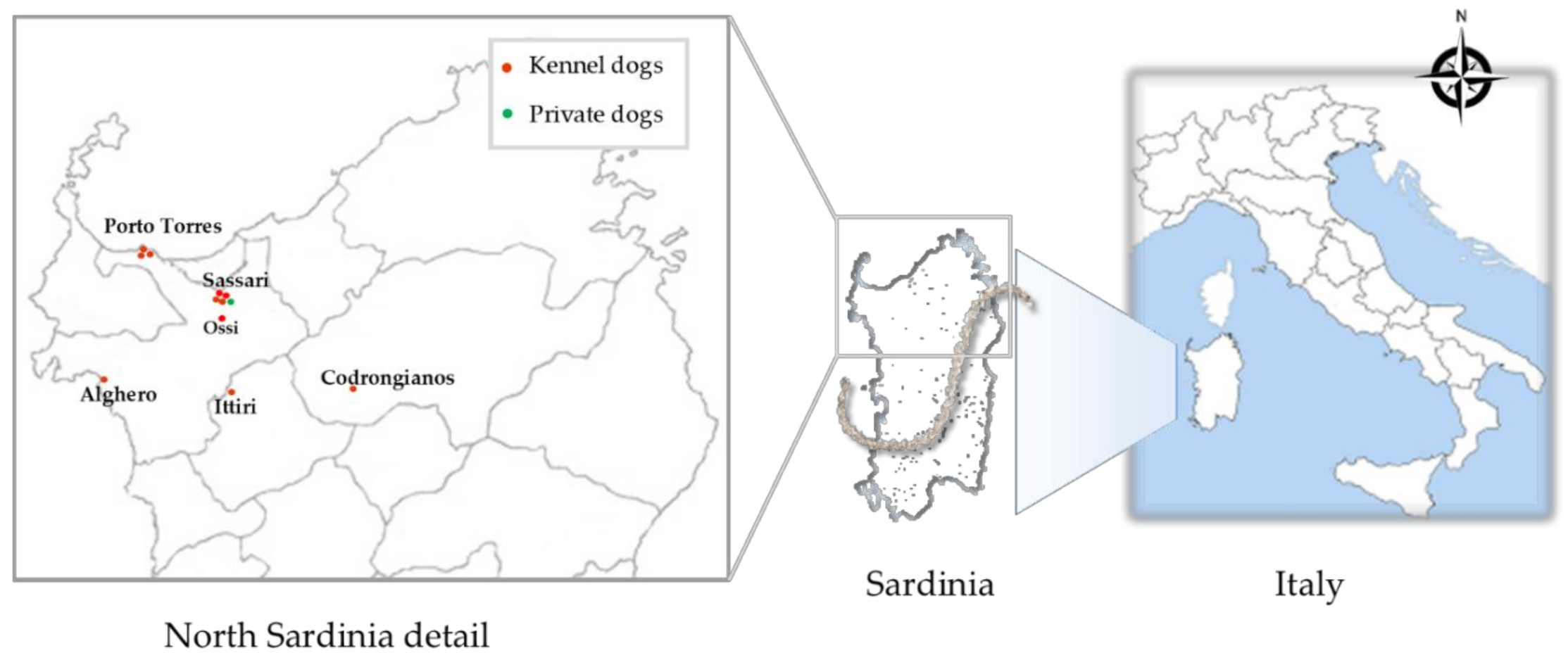
2.2. Study Design and Sample Collection

Between November 2016 and March 2018, a cross sectional study was conducted in order to investigate the presence of Leptospira in dogs from 11 kennels located in different sites of North Sardinia (Figure 1). A database including information about dog species, gender, age, size, and collection sites of kennels was created for each animal. The microchip number, the last vaccination date and presence or absence of any physical signs were also evaluated.
Moreover, blood samples from 88 client-owned dogs presented for routine vaccination or health assessment at veterinary clinics located mainly in the area of Sassari city were included in this study, as shown in Table 1. Dog owners signed a consent of availability and acceptance to include their dogs in this study. For each participating dog, place of origin, gender, age, household variables (indoor/outdoor or strictly indoor), breed, and history of vaccination were recorded. All dogs underwent to a clinical examination in which body condition, color of mucous membranes, temperature, and clinical signs of leptospirosis were evaluated. Blood samples were collected from each dog and stored at 4 °C until serum separation (obtained within 24 hours after centrifugation). Serum was also frozen at 20 °C until further serological and molecular analyses. From a total of 18 dogs casually selected, urine samples were obtained by cystocentesis and sent to IZS laboratories for Leptospira detection. In order to obtain a representative sample of the rodent population, we placed snap and/or mechanical traps in each kennel identified for this study and investigated them daily (Table 1). When the rodents were captured, they were transported to the IZS and euthanized using the pentobarbital IP euthanasia technique [22] and morphologically classified by an experienced veterinarian [23]. Blood samples were obtained by intracardiac puncture (n = 17). Kidney and liver tissues were also collected from 237 rats and included in this study. Where possible, urine samples were also obtained (n = 4).

2.3. Microscopic Agglutination Test

An aliquot of serum was examined by MAT following the recommendation performed by National Reference Centre of Leptospirosis (IZSLER, Brescia, Italy). Live strains of eight 7–10 days of culture of Leptospira species belonging to nine different serogroups (most frequently found from Mediterranean area) were used (Table 2). The density of leptospires was assessed using a counting chamber (Petroff-Hausser, USA) and adjusted to 2 × 10⁸ leptospires/mL.
All sera were diluted using phosphate buffered saline (PBS) (pH 7.6) at 10−2 dilution, mixed individually with each serovar suspension in the proportion 1:1, and incubated at 29 °C for 2 h. Pure PBS solution was used as negative control. The analysis was performed in dark field microscopy (Olympus BX50; Olympus Corp., Tokyo, Japan) with magnification of 100×. Samples were considered positive when 50% or more leptospires were agglutinated at 10−2 dilution, considering a cut-off titer of 100. The reactive serovar was considered the one that presented the highest titer. Positive samples were further diluted in the proportions 1:200, 1:400, 1:800, and 1:1600, and tested only to the reacted serovar to determine their titer.

2.4. Culturing of Leptospires

Ellinghausen–McCullough–Johnson–Harris (EMJH) semi-solid and liquid medium was used for the isolation of Leptospira from urine and organs. 5-Fluorouracil (5-FU) was added due to minimize bacterial contamination. Samples of kidney and liver were homogenized as previously described [24]. A total of 500 µL of urine (500 µL diluted 1:100 in PBS) and 25 mg of tissue were suspended in EMJH–fluorouracil medium at 28 °C and cultured for a period of three months. The media were examined under dark-field microscopy for the presence of leptospires approximately every seven days. Pure isolates, free of contaminants, were used for further molecular identification.

2.5. DNA Extraction and PCR Assays

Blood, homogenized organs, urine samples, and isolates were extracted using the DNeasy Blood and Tissue Kit® (Qiagen, Hilden, Germany), according to the manufacturer’s instructions. The final elution volume reduced to 50 µL was the unique modification performed on the original protocol. All DNA samples were then screened for detection of pathogenic Leptospira by real-time PCR (qPCR) assay with primers LipL32-45F (5′-AAGCATTACCGCTTGTGGTG-3′) and LipL32-286R (5′-GAA CTCCCA TTT CAG CGA TT-3′), and the probeLipL32-189P (FAM-5′-AA AGCCAG GAC AAG CGCCG-3′-BHQ1), targeting a fragment of the lipL32 gene [25]. PCR reaction mix composition and PCR temperature cycling conditions were the same as those used in a previous study [24]. A negative control (DNA extracted from water) and a positive control (DNA extracted from the reference strain of L. interrogans ATCC® BAA1198D5TM) were included in each run.

2.6. Multi Locus Sequence Types (MLST)

In order to reveal sequence types (STs) of Leptospira isolates, we performed MLST assay using the 7 housekeeping genes pntA, sucA, tpiA, pfkB, mreA, glmU, and caiB proposed by Boonsilp in 2013 [26]. Each allele and the allelic profiles (glmU-pntA-sucA-tpiA-pfkB-mreA-caiB) were submitted to the Leptospira database (http://pubmlst.org/leptospira accessed on 10 May 2021) to define the STs.

3. Results

A total of 1296 dogs were tested for leptospirosis in this study. Among them, kennel dogs were 1208 (93%; 95%CI: 92–94) and owned dogs were 88 (7%; 95%CI: 6–8) in number (Table 3). The examined dogs did not show any apparent clinical signs related to Leptospira infection. The dogs were classified on the basis of sex as female (51%; 95%CI: 48–54) and male dogs (49%; 95%CI: 46–52). With regards to age, 15% of dogs were under 2 years old (95%CI: 13–17), 52% were between 2 and 8 years (52%; 95%CI: 49–55), and 33% were over 8 years (95%CI: 30–36). Regarding the size of the dogs, the percentages of 10% (95%CI: 8–12), 86% (95%CI: 84–88), and 4% (95%CI: 3–5) were attributed to dogs of small, medium, and large size, respectively. Of the 1296 tested dogs, 88% (95%CI: 86–90) were vaccinated using a bivalent vaccine including serovars Icterohaemorrhagiae and Canicola. A total of 12% (95%CI: 10–14) were not immunized.
From a total of 1296 dog sera examined by MAT, 164 (13%; 95%CI: 11–15) tested positive for one or more serovars of Leptospira (Figure 2A). Specifically, the prevalence rate was of 89% (n = 154; 94%CI: 90–98) in kennel dogs and 11% (n = 10; 6%; 95%CI: 2–10) in owned dogs (Table 4). In Table 4, dogs with confirmed Leptospira diagnosis are classified on the basis of the different variables (sex, age, size, and vaccination status). Female and male positive dogs were 80 (49%) and 84 (51%) in number, respectively. MAT-positive results were more frequently diagnosed in adult dogs (93%; 95%CI: 89–97) as compared with young animals (7%; 95%CI: 3–11), and 67% of medium-sized dogs confirmed the presence of Leptospira infection.
Positive samples included 77 (47%; 95%CI: 39–55) non-vaccinated dogs (including those with no history of recent vaccination) and 87 (53%; 95%CI: 45–61) dogs that received the vaccine from less than six months since the blood collection. Among the non-vaccinated dogs, 38 (49%; 95%CI: 38–60), 27 (35%; 95%CI: 24–46), 11 (4%; 95%CI: 6–22), and 1 (2%; 95%CI: 0–5) showed MAT positivity for one, two, three, and four serovars, respectively (Figure 2A). Copenhageni and Icterohaemorrhagiae (61%; 95%CI: 50–72) were the most common serovars, followed by Bratislava (17%; 95%CI: 9–25), Grippothyphosa, and Canicola (both 8%; 95%CI: 2–14). Titers ranging from 1:100 to 1:400 were the most commonly detected (Table 5).
Positive results associated with dogs recently vaccinated are listed in Table 5. A total of 56 (64%; 95%CI: 54–74), 24 (28%; 95%CI: 19–37), and 7 (8%; 95%CI: 2–14) dogs showed positivity for one, two, and three serovars, respectively (Figure 2B). Antibody titers for Copenhageni were 1:100 and 1:1400 in 52% (95%CI: 42–62) and 2% (95%CI: 0–5) of the vaccinated dogs tested. One dog had titers of 1:3200 for Icterohaemorrhagiae serovar, which resulted in the second most common serovar detected.
All of the 21 urine samples collected from dogs (n = 18) and rats (n = 4) were negative when the lipL32 gene qPCR specific for pathogenic leptospires was used. Of 237 rodent organs tested, Leptospira DNA was obtained from 28 (12%; 95%CI: 8–16) kidneys and from 7 (3%; 95%CI: 1–5) cultured isolates. MLST analysis (Table 6) by using the database generated for the 7-locus scheme (http://pubmlst.org/leptospira 10 May 2021) highlighted that three different STs were obtained, namely, ST17 from one Rattus rattus, ST149 from two Rattus rattus and three Apodemus sylvaticus, and ST198 from one Rattus norvegicus (Table 6).

4. Discussion

This work represents the first cross-sectional study in which the presence of Leptospira serovars was evaluated in Sardinian dogs by microscopic agglutination test (MAT). The 13% of examined dogs (154/1208) showed sero-reactivity to five of the nine Leptospira serovars of the MAT panel used herein. Despite the fact that MAT is considered to be the reference method for the serodiagnosis of leptospirosis both in humans and animals, including dogs [20], this test suffers from bias related to non-specific cross reactions, such as those linked to a recent dog vaccination [27].
Among the 26 dogs that did not receive any vaccine at all and the 51 with no history of recent vaccination, the serovar Bratislava, Icterohaemorrhagiae, and Copenhageni were prevalent and MAT titers ranged from 1:100 to 1:400. Canicola and Grippotyphosa were also obtained at low titer, indicating a possible transmission through contaminated shared environment. One dog showed higher antibody titer of 1:1600 for Bratislava serovar, which is commonly associated with disease in domestic animals, in particular cats [28], pigs [29,30], horses [31,32], and dogs [33,34]. One 8-year-old dog with no history of recent vaccination had a double Leptospira co-infection showing high titers both for Copenhageni (1:1600) and Icterohaemorrhagiae (1:3200) serovars, indicating that vaccines do not ensure an immunity protection one year after vaccination [35,36]. Although dogs of all ages can be infected with Leptospira, in this study, only 12 dogs younger than 2 years (7%) were positive after MAT analyses. These results were in accordance with other studies [37,38,39], where the rate of positives in younger exemplars was related to better immunologic protection as a result of puppy vaccinations and maternally acquired antibodies. Icterohaemorrhagiae and Copenhageni serovars are considered to be the most representative and virulent strains of the Icterohaemorrhagiae serogroup and are typically responsible for the majority of severe cases of leptospirosis in humans [40,41]. In Italy, Icterohaemorrhagiae and Copenhageni serovars (serogroup Icterohaemorrhagiae) have been reported from several domestic mammals, including dogs [27,42,43]. Rattus species are considered the main reservoir for both serovars [41], suggesting a participation of these hosts in the environmental persistence of the bacterium. The large number of infected animals resulted positives for the different serovars could be also correlated with the location of kennels in rural setting populated by wild rodents (as also demonstrated by the high number of rodents collected in these areas). Thus, according to other studies, the widespread circulation of the bacterium is related to the close proximity to rural areas, making transmission easier than it is elsewhere [44,45,46]. The 12% of non-vaccinated dogs also tested seropositive for Bratislava and Canicola serovars, probably indicating the widespread circulation of these strains in the territory, associated with the low level of immune protection in this population. In humans, severe leptospirosis is most frequently associated with serogroup Icterohaemorrhagiae, in particular serovar Copenhageni [7,47]. Antibodies to Leptospira interrogans serovar Bratislava have been detected in dogs [33,48]; humans [49]; horses from Italy [32]; and in hedgehogs, which are indicated as potential reservoirs leading to canine infection in France and Ireland [30,50]. Similar data have been reported in dogs from Spain, where seropositive sera (MAT ≥1:100) to serovars Icterohaemorrhagiae (19.4%), Bratislava (8.5%), Grippotyphosa (7.2%), and Canicola (3.4%) have also been described. However, a significant bias of these studies is that the majority of dogs (n = 85) were vaccinated, and thus positive sera could also have been the result of a possible cross-reaction commonly linked to post-vaccination.
Eighty-seven sera of dogs (53%) that had been recently vaccinated (six months before the sample collection) showed low titers of antibodies against Icterohaemorrhagiae (n = 41), Copenhageni (n = 45), and Canicola (n = 16; 19% (≥1:100/1:200) serovars, which are included in the bivalent vaccine formulation. However, one vaccinated dog showed a seropositivity (MAT ≥ 1:3200) against Icterohaemorrhagiae serovar, and we hypothesized an absence of immune response after vaccine administration. Another important aspect linked to this result is that vaccination should be administered in non-infected dogs. When dogs are already infected, vaccination cannot prevent the disease. Although vaccination should act as available option to decrease the spread of the infection, standard hygiene measures before and after the contact with potentially infected animals or contaminated water should be taken to prevent dissemination of Leptospira in the environment and between animals.
Seropositive cases recorded against serovar that are not included in vaccine composition such as Bratislava (titers from 1:100 to 1:800) and Grippotyphosa (1:100 titer) could indicate a recent exposure to Leptospira infection. This also could be interpretated as cross-reactivity to nonvaccine serovars, demonstrating that MAT titers are not serovar specific. According to other studies [51], these results indicate that vaccination used until now is not always effective to dog immunization since the number of serovars involved in Leptospira infections and circulating among animals is very high, and the epidemiology of human infections always reflects the circulation of the bacterium in animal reservoirs. Dogs that can shed leptospires without being symptomatic represent a possible risk for humans and other animals [17]. The real-time PCR (qPCR) targeting the lipL32 gene conducted on urine samples collected from 18 dogs showed low diagnostic sensitivity, and no positive results were obtained. PCR results are usually less sensitive when compared to the serological reactions because molecular results depend on the phase of the diseases, as well as potentially being related to the low concentration of leptospires in urine. Moreover, PCR-based diagnosis does not allow for identification of the circulating serovars in a particular geographical area. However, further studies are needed to better isolate Leptospira strains by collecting more dog urine samples and assessing whether dogs are shedding the pathogen in the environment. Wild rodents represent the main reservoir of leptospires, and the epidemiology of Leptospira infections reflects the circulation of the bacterium in animal reservoirs [52]. Out of 237 kidney samples from rodents, 7 were successfully cultured, illustrating the difficulty of the diagnosis of Leptospira by the standard routine laboratory culture techniques and the importance of development of new easy techniques that could allow better diagnosis of leptospirosis. Results obtained from the seven isolates from rat kidneys with MLST allowed for the characterization of the Leptospira strains by sequencing and analyzing specific fragments of some bacterial housekeeping genes, thus identifying specific sequence types (STs). These results were in agreement with the recent detection of pathogenic Leptospira from wild mammals [24] in the same study area and indicate that serovar diversity of Leptospira species has yet to be fully investigated in Sardinia Island.

5. Conclusions

Despite the role of dogs as possible source of human infection and their contribution to the circulation of Leptospira in the environment having been widely reported worldwide [34,35,46,53], cases of dog–human transmission have not yet been reported in the study area. However, detection of pathogenic Leptospira serovars in dogs from North Sardinia highlights that dogs represent a potential infection risk for people and contribute to bacteria spread in the environment. Public health strategies for controlling possible outbreak of Leptospira should be implemented in order to avoid the disease becoming a major medical and veterinary problem in this region.

Author Contributions

Conceptualization, I.P. and M.N.P.; methodology, I.P.; validation, I.P. and V.C.; formal analysis, I.P.; investigation, A.P. (Angela Piras), B.P., P.P., and A.P. (Aureliana Pedditzi); resources, all authors; data curation, I.P. and M.N.P.; writing—original draft preparation, I.P. and V.C.; writing—review and editing, all authors; visualization, I.P.; supervision, M.N.P. All authors have read and agreed to the published version of the manuscript.

Funding

This research received no external funding.

Institutional Review Board Statement

The study was conducted according to the guidelines of the Declaration of Helsinki and approved by the Ethics Committee of Istituto Zooprofilattico Sperimentale della Sardegna in accordance with Council Directive 2010/63/EEC of the European Union and the Italian D.Igs 26/2014 (protocol 1248/2015-PR).

Informed Consent Statement

Informed consent was obtained from all subjects involved in the study.

Data Availability Statement

The data presented in this study are available in the article.

Acknowledgments

The authors would like to thank dog owners and kennels directors for their contributions.

Conflicts of Interest

The authors declare no conflict of interest.

References

  1. Cilia, G.; Bertelloni, F.; Fratini, F. Leptospira Infections in Domestic and Wild Animals. Pathogens 2020, 9, 573. [Google Scholar] [CrossRef] [PubMed]
  2. Cilia, G.; Bertelloni, F.; Albini, S.; Fratini, F. Insight into the Epidemiology of Leptospirosis: A Review of Leptospira Isolations from “Unconventional” Hosts. Animals 2021, 11, 191. [Google Scholar] [CrossRef] [PubMed]
  3. Ebani, V.V. Domestic reptiles as source of zoonotic bacteria: A mini review. Asian Pac. J. Trop. Med. 2017. [Google Scholar] [CrossRef] [PubMed]
  4. Vieira, A.S.; Pinto, P.S.; Lilenbaum, W. A systematic review of leptospirosis on wild animals in Latin America. Trop. Anim. Health Prod. 2018, 50, 229–238. [Google Scholar] [CrossRef]
  5. Piredda, I.; Palmas, B.; Noworol, M.; Tola, S.; Longheu, C.; Bertasio, C.; Scaltriti, E.; Denurra, D.; Cherchi, M.; Picardeau, M.; et al. Isolation of Leptospira interrogans from a Bottlenose Dolphin (Tursiops truncatus) in the Mediterranean Sea. J. Wildl. Dis. 2020. [Google Scholar] [CrossRef]
  6. Loffler, S.G.; Rago, V.; Martinez, M.; Uhart, M.; Florin-Christensen, M.; Romero, G.; Brihuega, B. Isolation of a seawater tolerant Leptospira spp. from a southern right whale (Eubalaena australis). PLoS ONE 2015, 10. [Google Scholar] [CrossRef] [Green Version]
  7. Haake, D.A.; Levett, P.N. Leptospirosis in humans. Curr. Top. Microbiol. Immunol. 2015, 387, 65–97. [Google Scholar] [CrossRef] [Green Version]
  8. Miotto, B.A.; Guilloux, A.G.A.; Tozzi, B.F.; Moreno, L.Z.; Da Hora, A.S.; Dias, R.A.; Heinemann, M.B.; Moreno, A.M.; de Souza Filho, A.F.; Lilenbaum, W.; et al. Prospective study of canine leptospirosis in shelter and stray dog populations: Identification of chronic carriers and different Leptospira species infecting dogs. PLoS ONE 2018, 13. [Google Scholar] [CrossRef] [Green Version]
  9. Altheimer, K.; Jongwattanapisan, P.; Luengyosluechakul, S.; Pusoonthornthum, R.; Prapasarakul, N.; Kurilung, A.; Broens, E.M.; Wagenaar, J.A.; Goris, M.G.A.; Ahmed, A.A.; et al. Leptospira infection and shedding in dogs in Thailand. BMC Vet. Res. 2020, 16, s12917-s020. [Google Scholar] [CrossRef] [Green Version]
  10. Marami, L.M.; Gebremedhin, E.Z.; Sarba, E.J.; Tola, G.K.; Endalew, S.S.; Melkamsew Tesfaye, A.; Di Marco Lo Presti, V.; Vitale, M. Seroprevalence and Associated Risk Factors of Canine Leptospira and Brucella Species Infection in West Shewa Zone, Central Ethiopia. Vet. Med. Res. Rep. 2021, Volume 12, 33–42. [Google Scholar] [CrossRef]
  11. Rojas, P.; Monahan, A.M.; Schuller, S.; Miller, I.S.; Markey, B.K.; Nally, J.E. Detection and quantification of leptospires in urine of dogs: A maintenance host for the zoonotic disease leptospirosis. Eur. J. Clin. Microbiol. Infect. Dis. 2010, 29, 1305–1309. [Google Scholar] [CrossRef]
  12. Zakeri, S.; Khorami, N.; Ganji, Z.F.; Sepahian, N.; Malmasi, A.A.; Gouya, M.M.; Djadid, N.D. Leptospira wolffii, a potential new pathogenic Leptospira species detected in human, sheep and dog. Infect. Genet. Evol. 2010, 10, 273–277. [Google Scholar] [CrossRef]
  13. Gay, N.; Soupé, M.E.G.; Goarant, C. Though not reservoirs, dogs might transmit Leptospira in New Caledonia. Int. J. Environ. Res. Public Health 2014, 11, 4316–4325. [Google Scholar] [CrossRef] [Green Version]
  14. Llewellyn, J.R.; Krupka-Dyachenko, I.; Rettinger, A.L.; Dyachenko, V.; Stamm, I.; Kopp, P.A.; Straubinger, R.K.; Hartmann, K. Urinary shedding of leptospires and presence of Leptospira antibodies in healthy dogs from Upper Bavaria. Berl. Munch. Tierarztl. Wochenschr. 2016, 129, 251–257. [Google Scholar] [CrossRef]
  15. Samir, A.; Soliman, R.; El-Hariri, M.; Abdel-Moein, K.; Hatem, M.E. Leptospirosis in animals and human contacts in Egypt: Broad range surveillance. Rev. Soc. Bras. Med. Trop. 2015, 48, 272–277. [Google Scholar] [CrossRef] [Green Version]
  16. Narkkul, U.; Thaipadungpanit, J.; Srisawat, N.; Rudge, J.W.; Thongdee, M.; Pawarana, R.; Pan-ngum, W. Human, animal, water source interactions and leptospirosis in Thailand. Sci. Rep. 2021, 11, s41598-s021. [Google Scholar] [CrossRef]
  17. Schuller, S.; Francey, T.; Hartmann, K.; Hugonnard, M.; Kohn, B.; Nally, J.E.; Sykes, J. European consensus statement on leptospirosis in dogs and cats. J. Small Anim. Pract. 2015, 56, 159–179. [Google Scholar] [CrossRef]
  18. Millán, J.; Velarde, R.; Chirife, A.D.; León-Vizcaíno, L. Carriage of pathogenic Leptospira in carnivores at the wild/domestic interface. Pol. J. Vet. Sci. 2019, 22, 781–784. [Google Scholar] [CrossRef]
  19. Samrot, A.V.; Sean, T.C.; Bhavya, K.S.; Sahithya, C.S.; Chandrasekaran, S.; Palanisamy, R.; Robinson, E.R.; Subbiah, S.K.; Mok, P.L. Leptospiral infection, pathogenesis and its diagnosis—A review. Pathogens 2021, 10, 145. [Google Scholar] [CrossRef]
  20. Reagan, K.L.; Sykes, J.E. Diagnosis of Canine Leptospirosis. Vet. Clin. North Am. Small Anim. Pract. 2019, 49, 719–731. [Google Scholar] [CrossRef]
  21. Bertasio, C.; Papetti, A.; Scaltriti, E.; Tagliabue, S.; D’incau, M.; Boniotti, M.B. Serological survey and molecular typing reveal new Leptospira serogroup pomona strains among pigs of northern Italy. Pathogens 2020, 9, 332. [Google Scholar] [CrossRef]
  22. Zatroch, K.K.; Knight, C.G.; Reimer, J.N.; Pang, D.S.J. Refinement of intraperitoneal injection of sodium pentobarbital for euthanasia in laboratory rats (Rattus norvegicus). BMC Vet. Res. 2017, 13, s12917-s017. [Google Scholar] [CrossRef] [Green Version]
  23. Nagorsen, D.W. An Identification Manual to The Small Mammals of British Columbia; Ministry of Sustainable Resource Management Ministry of Water, Land and Air Protection Royal British Columbia Museum: Victoria, BC, Canada, 2002. [Google Scholar]
  24. Piredda, I.; Ponti, M.N.; Palmas, B.; Noworol, M.; Pedditzi, A.; Rebechesu, L.; Chisu, V. Molecular Typing of Pathogenic Leptospira Species Isolated from Wild Mammal Reservoirs in Sardinia. Animals 2021, 11, 1109. [Google Scholar] [CrossRef]
  25. Stoddard, R.A.; Gee, J.E.; Wilkins, P.P.; McCaustland, K.; Hoffmaster, A.R. Detection of pathogenic Leptospira spp. through TaqMan polymerase chain reaction targeting the LipL32 gene. Diagn. Microbiol. Infect. Dis. 2009, 64, 247–255. [Google Scholar] [CrossRef]
  26. Boonsilp, S.; Thaipadungpanit, J.; Amornchai, P.; Wuthiekanun, V.; Bailey, M.S.; Holden, M.T.G.; Zhang, C.; Jiang, X.; Koizumi, N.; Taylor, K.; et al. A Single Multilocus Sequence Typing (MLST) Scheme for Seven Pathogenic Leptospira Species. PLoS Negl. Trop. Dis. 2013, 7. [Google Scholar] [CrossRef] [Green Version]
  27. Balboni, A.; Zamagni, S.; Bertasio, C.; Boniotti, M.B.; Troìa, R.; Battilani, M.; Dondi, F. Identification of Serogroups Australis and Icterohaemorrhagiae in Two Dogs with a Severe Form of Acute Leptospirosis in Italy. Pathogens 2020, 9, 351. [Google Scholar] [CrossRef]
  28. Sprißler, F.; Jongwattanapisan, P.; Luengyosluechakul, S.; Pusoonthornthum, R.; Prapasarakul, N.; Kurilung, A.; Goris, M.; Ahmed, A.; Reese, S.; Bergmann, M.; et al. Leptospira infection and shedding in cats in Thailand. Transbound. Emerg. Dis. 2019, 66, 948–956. [Google Scholar] [CrossRef]
  29. Shiokawa, K.; Welcome, S.; Kenig, M.; Lim, B.; Rajeev, S. Epidemiology of Leptospira infection in livestock species in Saint Kitts. Trop. Anim. Health Prod. 2019, 51, s11250-s019. [Google Scholar] [CrossRef] [PubMed]
  30. Arent, Z.; Frizzell, C.; Gilmore, C.; Allen, A.; Ellis, W.A. Leptospira interrogans serovars Bratislava and Muenchen animal infections: Implications for epidemiology and control. Vet. Microbiol. 2016, 190, 19–26. [Google Scholar] [CrossRef] [Green Version]
  31. Divers, T.J.; Chang, Y.F.; Irby, N.L.; Smith, J.L.; Carter, C.N. Leptospirosis: An important infectious disease in North American horses. Equine Vet. J. 2019, 51, 287–292. [Google Scholar] [CrossRef]
  32. Vera, E.; Taddei, S.; Cavirani, S.; Schiavi, J.; Angelone, M.; Cabassi, C.S.; Schiano, E.; Quintavalla, F. Leptospira seroprevalence in bardigiano horses in northern Italy. Animals 2020, 10, 23. [Google Scholar] [CrossRef] [PubMed] [Green Version]
  33. López, M.C.; Vila, A.; Rodón, J.; Roura, X. Leptospira seroprevalence in owned dogs from Spain. Heliyon 2019, 5. [Google Scholar] [CrossRef] [PubMed] [Green Version]
  34. Weis, S.; Rettinger, A.; Bergmann, M.; Llewellyn, J.R.; Pantchev, N.; Straubinger, R.K.; Hartmann, K. Detection of Leptospira DNA in urine and presence of specific antibodies in outdoor cats in Germany. J. Feline Med. Surg. 2017, 19, 470–476. [Google Scholar] [CrossRef] [PubMed]
  35. Lau, S.F.; Wong, J.Y.; Khor, K.H.; Roslan, M.A.; Abdul Rahman, M.S.; Bejo, S.K.; Radzi, R.; Bahaman, A.R. Seroprevalence of Leptospirosis in Working Dogs. Top. Companion Anim. Med. 2017, 32, 121–125. [Google Scholar] [CrossRef] [Green Version]
  36. Martin, L.E.R.; Wiggans, K.T.; Wennogle, S.A.; Curtis, K.; Chandrashekar, R.; Lappin, M.R. Vaccine-associated Leptospira antibodies in client-owned dogs. J. Vet. Intern. Med. 2014, 28, 789–792. [Google Scholar] [CrossRef] [Green Version]
  37. Lee, H.S.; Levine, M.; Guptill-Yoran, C.; Johnson, A.J.; von Kamecke, P.; Moore, G.E. Regional and temporal variations of Leptospira seropositivity in dogs in the United States, 2000-2010. J. Vet. Intern. Med. 2014, 28, 779–788. [Google Scholar] [CrossRef] [Green Version]
  38. Yao, P.J.; Stephenson, N.; Foley, J.E.; Toussieng, C.R.; Farver, T.B.; Sykes, J.E.; Fleer, K.A. Incidence rates and risk factors for owner-reported adverse events following vaccination of dogs that did or did not receive a Leptospira vaccine. J. Am. Vet. Med. Assoc. 2015, 247, 1139–1145. [Google Scholar] [CrossRef]
  39. Azócar-Aedo, L.; Monti, G.; Jara, R. Leptospira spp. in domestic cats from different environments: Prevalence of antibodies and risk factors associated with the seropositivity. Animals 2014, 4, 612–626. [Google Scholar] [CrossRef] [PubMed] [Green Version]
  40. Goris, M.G.A.; Kikken, V.; Straetemans, M.; Alba, S.; Goeijenbier, M.; van Gorp, E.C.M.; Boer, K.R.; Wagenaar, J.F.P.; Hartskeerl, R.A. Towards the Burden of Human Leptospirosis: Duration of Acute Illness and Occurrence of Post-Leptospirosis Symptoms of Patients in The Netherlands. PLoS ONE 2013, 8. [Google Scholar] [CrossRef] [Green Version]
  41. De Brito, T.; da Silva, A.M.G.; Abreu, P.A.E. Pathology and pathogenesis of human leptospirosis: A commented review. Rev. Inst. Med. Trop. Sao Paulo 2018, 60. [Google Scholar] [CrossRef]
  42. Tagliabue, S.; Figarolli, B.M.; D’Incau, M.; Foschi, G.; Gennero, M.S.; Giordani, R.; Natale, A.; Papa, P.; Ponti, N.; Scaltrito, D.; et al. Indagine sierologica sulla presenza di Leptospira spp. in Italia: Dati nazionali 2010–2011. Vet. Ital. 2016, 52, 129–138. [Google Scholar] [CrossRef] [PubMed]
  43. Bertasio, C.; Boniotti, M.B.; Lucchese, L.; Ceglie, L.; Bellinati, L.; Mazzucato, M.; Furlanello, T.; D’incau, M.; Natale, A. Detection of new Leptospira genotypes infecting symptomatic dogs: Is a new vaccine formulation needed? Pathogens 2020, 9, 484. [Google Scholar] [CrossRef] [PubMed]
  44. Gkentzi, D.; Lagadinou, M.; Bountouris, P.; Dimitrakopoulos, O.; Triantos, C.; Marangos, M.; Paliogianni, F.; Assimakopoulos, S.F. Epidemiology, clinical and laboratory findings of leptospirosis in Southwestern Greece. Infect. Dis. (Auckl). 2020, 52, 413–418. [Google Scholar] [CrossRef]
  45. Briskin, E.A.; Casanovas-Massana, A.; Ryff, K.R.; Morales-Estrada, S.; Hamond, C.; Perez-Rodriguez, N.M.; Benavidez, K.M.; Weinberger, D.M.; Castro-Arellano, I.; Wunder, E.A.; et al. Seroprevalence, Risk Factors, and Rodent Reservoirs of Leptospirosis in an Urban Community of Puerto Rico, 2015. J. Infect. Dis. 2019, 220, 1489–1497. [Google Scholar] [CrossRef]
  46. Lelu, M.; Muñoz-Zanzi, C.; Higgins, B.; Galloway, R. Seroepidemiology of leptospirosis in dogs from rural and slum communities of Los Rios Region, Chile. BMC Vet. Res. 2015, 11, s12917-s015. [Google Scholar] [CrossRef] [Green Version]
  47. Bharti, A.R.; Nally, J.E.; Ricaldi, J.N.; Matthias, M.A.; Diaz, M.M.; Lovett, M.A.; Levett, P.N.; Gilman, R.H.; Willig, M.R.; Gotuzzo, E.; et al. Leptospirosis: A zoonotic disease of global importance. Lancet Infect. Dis. 2003, 3, 757–771. [Google Scholar] [CrossRef]
  48. Renaud, C.; Andrews, S.; Djelouadji, Z.; Lecheval, S.; Corrao-Revol, N.; Buff, S.; Demont, P.; Kodjo, A. Prevalence of the Leptospira serovars bratislava, grippotyphosa, mozdok and pomona in French dogs. Vet. J. 2013, 196, 126–127. [Google Scholar] [CrossRef]
  49. Cacciapuoti, B.; Cacciapuoti, B.; Pinto, A.; Nuti, M.; Sabrie, A.M. Human leptospirosis in somalia: A serological survey. Trans. R. Soc. Trop. Med. Hyg. 1982, 76, 178–182. [Google Scholar] [CrossRef]
  50. Ayral, F.C.; Bicout, D.J.; Pereira, H.; Artois, M.; Kodjo, A. Short report: Distribution of Leptospira serogroups in cattle herds and dogs in France. Am. J. Trop. Med. Hyg. 2014, 91, 756–759. [Google Scholar] [CrossRef] [Green Version]
  51. Wunder, E.A.; Adhikarla, H.; Hamond, C.; Bonner, K.A.O.; Liang, L.; Rodrigues, C.B.; Bisht, V.; Nally, J.E.; Alt, D.P.; Reis, M.G.; et al. A live attenuated-vaccine model confers cross-protective immunity against different species of the Leptospira genus. Elife 2021, 10, 1–20. [Google Scholar] [CrossRef]
  52. Karpagam, K.B.; Ganesh, B. Leptospirosis: A neglected tropical zoonotic infection of public health importance—An updated review. Eur. J. Clin. Microbiol. Infect. Dis. 2020, 39, 835–846. [Google Scholar] [CrossRef] [PubMed]
  53. Ngasaman, R.; Saechan, V.; Prachantasena, S.; Yingkajorn, M.; Sretrirutchai, S. Investigation of Leptospira infection in stray animals in Songkhla, Thailand: Leptospirosis Risk Reduction in Human. Vector-Borne Zoonotic Dis. 2020, 20, 432–435. [Google Scholar] [CrossRef]
Figure 1. Geographical distribution of the 11 kennels analyzed in this study (red dot) and of the owned dogs (green dot).
Biology 10 00507 g001
Figure 2. Graphic representations of the single and multiple seropositivity for one or more Leptospira serovars detected in vaccinated (A) and unvaccinated dogs (B).
Biology 10 00507 g002
Table 1. Summary of dogs and rodents analyzed in this study, sites where kennels were located, and relative number of captured rodents.
Collection of DogsKennel Location (Abbreviation Letter)No. of Hosted DogsNo. of Tested DogsNo. of Captured Rodents
Ossi (A)1306330
Alghero (B)45029848
Porto Torres (C)804112
Codrongianos (D)13010022
Porto Torres (E)1005027
KennelSassari (F)28014936
Porto Torres (G)1004911
Ittiri (H)32019415
Sassari (I)55317
Sassari (L)28016015
Sassari (M)1507314
PrivateSassari-88-
Total1120751296237
Table 2. Genomospecies, serovar, strain, and serogroup of Leptospira used as antigens for the microscopic agglutination test.
GenomospeciesSerogroupSerovarStrain
L. interrogansSejroeHardjoHardjoprajitno n°224
AustralisBratislavaHedgehog n°47
PomonaPomonaPomona n°222
Icterohaem.Icterohaem.RGA 20
Icterohaem.CopenhageniWijnberg n°1
CanicolaCanicolaAlarik n°2
L. kirschneriGrippotyphosaGrippotyphosaMoska V n°54
L. borgpeterseniiTarassoviTarassoviMitis Johnson n°6
BallumBallumMus 127 n°217
Table 3. Summary of MAT-positive dogs analyzed in this study.
KennelsMAT AnalysisMAT Positives ( x - ; 95% CI)
A6320 (32; 20–44)
B2984 (1; 0–2)
C419 (22; 9–35)
D1008 (8; 3–13)
E507 (14; 4–24)
F14934 (23; 16–30)
G491 (2; 0–6)
H19432 (17; 12–22)
I310
L16029 (18; 12–24)
M7310 (14; 6–22)
Owned8810 (11; 4–18)
Total1296164 (13%; 11–15)
Table 4. Prevalence of positive MAT results in the dogs’ population divided by kennel location sex, age, size, and vaccination status.
Dog InformationKennel DogsOwned DogsTot.
ABCDEFGHILM
Sex
Male93622170200126380
Female111365171120174784
Total2049873413202910164
Age
<200010001090112
2–883560221240126996
>81214171207084 56
Total204987341320291010164
Size
Large110140405021129
Medium128332412702696110
Small82014600010325
Total204987341320291010164
Vaccination status
No200400002401110877
<6 months0458734180180287
Total204987341320291010164
Table 5. Number of positive microscopic agglutination test (MAT) results among dogs never vaccinated and vaccinated from less than six months.
GenospecieSerogroupSerovarNumber of Dogs with Respective MAT Titers
1:1001:2001:4001:8001:16001:3200
Vaccination in MonthsTotal (%)
No<6No<6No<6No<6No<6No<6No<6
L. interrogansAustralisBratislava5135-24-21---13 (17)19 (22)
L. interrogansCanicolaCanicola3163--1------6(8)17 (20)
L. interrogansIcterohaem.Copenhageni304514-22--1---47 (61)47 (54)
L. interrogansIcterohaem.Icterohaem.204116-862---1147 (61)48 (55)
L. kirschneriGrippotyphosaGrippotyphosa561---------6 (8)6 (7)
Table 6. Results of serology by MAT, qPCR, culture assay, and MLST performed on samples collected from rodents.
Rodent GroupScientific Name (n.)SourceMATqPCRCultural IsolationMLST (n.)
TestedPositives
(%; 95%CI)
TestedPositives
(%; 95%CI)
TestedPositives
(%;95%CI)
Rat (160)Rattus rattus (158)Kidney--15813 (8; 4–12)1583 (2; 0–4)ST17 (1); ST 149 (2)
Liver--31 (33; 0–83)30
Urine--2020
Serum150----
Rattus norvegicus (2)Kidney--21 (50; 0–120)21 (50;ST 198 (1)
Liver--1010
Urine--1010
Serum20----
Mouse (77)Apodemus sylvaticus (70)Kidney--709 (13; 5–21)703 (4; 0–9)ST 149 (3)
Liver--21 (50; 0–120)20
Urine--1010
Mus musculus (7)Kidney--72 (29; 18–40)70
Liver--11 (100)10
Total237 17023728 (12; 8–16)2377 (3; 1–5)
Publisher’s Note: MDPI stays neutral with regard to jurisdictional claims in published maps and institutional affiliations.

