Article

Assessment of Composite with Fibers as a Support for Antibacterial Nanomaterials: A Case Study of Bacterial Cellulose, Polylactide and Usual Textile

1
Faculty of Chemistry, Lomonosov Moscow State University, 119991 Moscow, Russia
2
N.M. Emanuel Institute of Biochemical Physics, Russian Academy of Sciences, 119334 Moscow, Russia
3
Cryoelectronics Lab, Faculty of Physics, Lomonosov Moscow State University, 119991 Moscow, Russia
*
Author to whom correspondence should be addressed.
Fibers 2022, 10(9), 70; https://doi.org/10.3390/fib10090070
Submission received: 18 July 2022 / Revised: 17 August 2022 / Accepted: 22 August 2022 / Published: 24 August 2022
(This article belongs to the Collection Feature Papers in Fibers)

Abstract

:
To obtain composite fiber materials with antibacterial properties, the samples of bacterial cellulose (BC), polylactide (PLA) and usual fibers (FM) were modified by poly-ε-caprolactone or polyhydroxybutyrate and then functionalized by the enzyme-polyelectrolyte complex of quorum-quenching enzymes, such as hexahistidine-tagged organophosphorus hydrolase with poly(glutamic acid) or by suspension of tantalum nanoparticles (Ta NPs) in ethanol. The structures of the composite fibers were analyzed using scanning electron microscopy. It was shown that the introduction of additional natural polymers into the matrix of BC, PLA and FM resulted in decreasing of the structural porosity. Comparative studies of the antibacterial activity of the composite materials were carried out using Escherichia coli and Bacillus subtilis cells. The decrease of adenosine triphosphate concentration in cell samples loaded onto fiber materials was applied as a measurable characteristic of antibacterial effect typical for the new fiber materials. The profound improvement of antibacterial activity was determined in composite materials with polyhydroxybutyrate and Ta NPs.

Graphical Abstract

1. Introduction

Modern trends in the production of special fiber materials suggest their multiple functionalization [1], which can be provided by introducing various nanoobjects into them (carbon or metal nanomaterials, electrically conducting or semi-conducting nanoelements, enzymes and biologically active substances, including antibiotics). Research often focuses on the combination of several nanoobjects loaded into fibers, according to the materials modified by them [2], and as a result of such combinations, fiber materials with special properties appear [3].
The idea of functionalization of fibers and fiber materials by nanoobjects, for example, by enzymes is not new, and research in this area has been conducted for a long time (in particular, in the development of self-cleaning and defense materials [4]). Recently, much attention has been attracted by the different variants of possible functionalization of nanofiber materials by enzymes [5,6]. In particular, we have recently found that it is possible to successfully combine antimicrobials and the enzyme hexahistidine-tagged organophosphorus hydrolase (His6-OPH) stabilized in enzyme-polyelectrolyte complexes (EPCs) with poly(amino acids), e.g., with polyglutamic acid (PLE50) [7,8]. This enzyme has a lactonase activity and can hydrolyze a wide range of quorum sensing signaling molecules that are used by Gram-negative bacterial cells during development of antibiotic resistance. Such a combination of stabilized enzyme and antimicrobial polypeptides in EPCs makes it possible to increase the effectiveness of widely used antibiotics, reduce their doses and limit the development of resistance in pathogens [9,10], as well as to lower the level of possible negative consequences from the use of antibiotics (dysbacteriosis, allergic reactions, etc.).
At the same time, the use of enzymes in the form of EPCs allows artificially enlarging the size of the enzyme due to its environment with a partner-polypeptide, which can contribute to more effective retention of the biologically active component in the fiber material without the need for its covalent binding to such a carrier [8,11,12]. Nevertheless, many researchers strive to covalently fix the enzymes in fiber materials [13].
However, the use of additional crosslinking agents always complicates the process of obtaining functional fiber materials, creates the possible presence of residual amounts of usually aggressive reagents (from the chemical point of view) in the created fiber content, contact with which becomes undesirable and not safe, and the activity of the enzyme can significantly decrease despite its high stability. The possible application of an enzymatic EPC with an antibiotic to the surface of synthetic and natural fibers was shown, thus obtaining a functionalized material with antimicrobial properties [14]. At the same time, bacterial cellulose (BC), which is a biodegradable natural polymer with unique characteristics arising due to its nanocrystalline structure and nanoporosity, has become popular as a basis for fiber composite materials with antibacterial properties [15,16].
When varying materials that were functionalized by the same EPCs with an antimicrobial substance (using the example of antimicrobial polypeptides), it was found that the modified material itself has a significant effect on the properties manifested by the enzyme and antimicrobial substance when they are introduced into the material [17]. It was shown that a change in the composition of the material, particularly due to the creation of its composite version, can also significantly affect the change in the characteristics of the material itself, in particular, sorption properties and strength, as well as the level and stability of the functions manifested by the enzyme and antibiotic [18].
Varying the composition of composite materials with the same “functionalizing content” in the form of an enzyme that catalyzes bacterial quorum quenching (QQ) and antimicrobial substance allows you to expand the spectrum of applications of such a combination and to create new protective [14,19] and dressing materials [17]. At the same time, in addition to the functional effectiveness of the tissue variants, the possibility of using biodegradable materials in their creation becomes extremely important. Today, poly-ε-caprolactone (PCL) [20] and poly-4-hydroxybutyrate (PHB) [21] are most often considered among biodegradable polymers that can be used for the development of composite materials and for modification of both natural and synthetic fibrous materials. The reason for this lies in the development of biotechnological approaches to scaling the processes of obtaining these polymers, which form the basis for their wide potential commercialization.
It should be noted that bacterial cellulose (BC) has recently been used as the most popular natural fibrous bases for polymer modifications [22] and polylactide (PLA) [23]. At the same time, interest continues to grow in (semi)synthetic and natural fibrous materials, e.g., cellulose-containing materials [24], for which production has already been well developed, but their modification by the above-mentioned natural biodegradable polymers and subsequent functionalization by nanoobjects may open up new directions for the use of such fibers.
One of the interesting techniques that can significantly increase the antimicrobial activity of fibers is the introduction of various metal nanoparticles (NPs) through ab(d)sorption into fibrous materials [25,26]. The increased effectiveness of such composite materials is ensured due to the absence of resistance in the cells of microorganisms to the effects of metallic NPs. At the same time, the combination of these nanomaterials with a QQ-enzyme and/or antibiotics can improve the overall efficiency of modified fibers [14]. Noteworthily, tantalum NPs (Ta NPs) rather than widely used zinc NPs appeared to be more preferable [27].
The aim of this work was to obtain composite materials based on BC, PLA and usual fibers, further modified by PCL or PHB. Then, these composites were functionalized using Ta NPs or nanocomplexes of His6-OPH/PLE50, investigated by antibacterial and enzyme activity and compared with unmodified materials. Such composite materials were interesting due to the possibility of their application in various areas, including but not limited to biomedicine, food packaging, personal protective equipment and filtration systems for water purification. The applied functionalization of fibrous materials by non-covalent enzyme-polyelectrolyte nanocomplex His6-OPH/PLE50 generally refers this research to the number of advanced developments in the field of fibrous materials. This approach takes into account the achievements in the field of bioinformatics and biocatalysis, since the “partners” (His6-OPH and PLE50) for such complexes are selected as a result of computer modeling [8].

2. Materials and Methods

2.1. Materials

Nanofibrillar BC was produced using immobilized Komagataeibacter xylinum B-12429 cells as it was previously described [18]. Briefly, a bacterial biomass was grown in a Hestrin-Schramm nutrient broth (pH 5.6) containing a 20 g/L of fructose, 5 g/L of yeast extract, 5 g/L of tryptone, 2.7 g/L of K2HPO4, 0.5 g/L of MgSO4, and 1.15 g/L of citric acid at 28 °C for 19 h and 180 rpm. Then, biomass was centrifuged (10,000× g, 10 min) in Avanti J25 (Beckman, Brea, CA, USA), mixed with 12 wt.% poly(vinyl alcohol) in a dosage of 30 wt.%, pelleted and frozen at −20 °C. Next, 50 g/L of immobilized biocatalyst was placed in the same media (pH 7.0) and maintained under static conditions at 28 °C for 6 days. BC was separated from the medium, washed with 1 M of NaOH and then with water to remove all cell and protein residues. BC samples were dried under sterile conditions at room temperature overnight.
A nonwoven fiber material (FM) consisting of 70% viscose and 30% polyester was purchased locally and used as is. The 24-h water vapor transmission rate through it (367 g/m2) was measured according to ISO 2528:2017. The mass per area, measured according to ISO 3801:1977, was 32.4 ± 4.5 g/m2 and reported recently [14].
Polylactide fiber materials were obtained from Ahlstrom-Munksjö (Helsinki, Finland), had different mass per area being equal to 23.5 ± 0.2 and 17.6 ± 1.4 g/m2 and were marked as PLA(S) and PLA(B), respectively. Poly(4-hydroxybutyrate) (PHB) with molar weight of ca. 500 kg/mol was a kindly gift from Metabolix GmbH (Cologne, Germany). Poly-ε-caprolactone (PCL) for modeling was obtained locally (Polymorphous, Moscow, Russia).
A patented method [28] was used to produce His6-OPH with recombinant Escherichia coli SG13009[pREP4] (Qiagen, Hilden, Germany). Briefly, cells were transformed by plasmid pTES-His-OPH, and the overnight cell culture was grown at 30 °C in a Luria–Bertani (LB) medium broth containing a 5 g/L of yeast extract, 10 g/L of tryptone and 10 g/L of NaCl and supplemented with a 100 mg/L of ampicillin and 25 mg/L of kanamycin. Then, cells were seeded to the rich medium containing 24 g/L of yeast extract, 12 g/L of tryptone, 4 g/L of glycerin, 6.95 g/L of KH2PO4, 12.54 g/L of K2HPO4 × 3H2O and 237 mg/L of CoCl2 × 6H2O, and biosynthesis of His6-OPH was induced by addition of 0.25 mM of isopropyl-β-D-thiogalactoside (Fermentas, Vilnius, Lithuania). The cells were cultivated using a thermostatically controlled Adolf Kuhner AG shaker (Basel, Switzerland) with stirring of 180 rpm at 30 °C for 20 h and harvested at 8000× g for 20 min. Cell biomass was suspended in 50 mL of 50 mM K-phosphate buffer containing 0.3 M NaCl (pH 7.5) and homogenized by sonication on ice. Cell debris was removed by centrifugation (15,000× g, 30 min).
His6-OPH was purified using Ni-NTA agarose (Sigma-Aldrich, Darmstadt, Germany) by the published procedure [29]. Briefly, cell supernatant was loaded onto a column equilibrated with a 50 mM K-phosphate buffer (pH 7.5). The column was washed with the same buffer followed by buffer containing 10 mM imidazole until OD280 became less than 0.01. The protein fractions obtained after elution in linear imidazole gradient (0–500 mM) were dialyzed overnight against 50 mM K-phosphate buffer (pH 7.5) followed by the same buffer containing 50 µM CoCl2.
According to the published methods [30], to determine His6-OPH concentration, a Bradford assay was performed with Coomassie Brilliant Blue G-250 dye and bovine serum albumin (Sigma-Aldrich) for calibration curve. To analyze protein purity, denaturing electrophoresis with sodium dodecyl sulfate was carried out in a 12% polyacrylamide gel using a Mini-PROTEAN II cell (Bio-Rad, Hercules, CA, USA) and Coomassie Brilliant Blue R-250 (Sigma-Aldrich) staining, and it was ca. 98.5 ± 0.5%.
Non-covalent EPCs of His6-OPH with poly(L-glutamic acid) (PLE50) (Alamanda Polymers, Huntsville, AL, USA) were obtained as it was described previously [8,9] by ordinary mixing of enzyme with polymer solution at a molar ratio of 1:1. Further, they were labeled His6-OPH/PLE50 in the text.
Ta NPs in ethanol were obtained in a plasma electric arc discharge as published previously [14,27]. The size of individual NPs determined using a transmission electron microscope LEO 912 AB OMEGA (Carl Zeiss, Oberkochen, Germany) with energy filter and Keller system was 1–3 nm. Concentration of Ta NPs (by metal) preliminarily dissolved in HNO3 and analyzed using an atomic-emission spectrometer iCAP 6300 Radial View (Thermo Fisher Scientific Inc., Waltham, MA, USA) with inductively coupled plasma was 18.3 mg/mL.

2.2. Functionalization of Composite Materials

To obtain composite materials, initially, granules of PCL and PHB were grinded at room temperature. Then, 0.22–0.24 g of their powder was evenly placed onto BC or FM (3 × 3 cm) and wrapped in aluminum foil. After that, samples were loaded under press during 3–5 min at 140–150 and 180–200 °C for PCL and PHB, respectively. Samples with PHB were further annealed for 5 min at 180 °C in air oven.
To combine BC and polylactide fiber materials, their samples (4 × 4 cm) were layered and as previously put under pressure during 3–5 min at 150–160 °C. Before further use, all materials were cut into square samples 1 × 1 cm.
A 50-µL volume of 18.3 mg/L Ta NPs in ethanol were loaded onto material (1 × 1 cm) dropwise as described previously [27] and dried at a room temperature.
To load His6-OPH/PLE50 nanocomplexes onto materials, the previously published general procedure [17] was used with minor modifications. To wit, a 10-µL volume of 50 µg/mL His6-OPH/PLE50 in a PBS buffer (pH 7.4) was dropped onto samples (1 × 1 cm) and dried for 20–24 h at +8 °C under sterile conditions.
To perform the scanning electron microscopy (SEM) analysis of the fiber materials, the samples were freeze-dried with a Freeze Dry System (Labconco, Kansas City, MO, USA), then sectioned, sputtered with gold and studied at various magnifications with a Supra 40-30-87 microscope (Carl Zeiss, Oberkochen, Germany).

2.3. Estimation of Antibacterial Activity of Modified Fibers

To analyze the antibacterial activity of composite materials, the previously published procedure [17,27] was applied. The Gram-negative bacterial cells of Escherichia coli DH5α (Thermo Fisher Scientific, Waltham, MA, USA) and Gram-positive bacterial cells of Bacillus subtilis B-522 (All-Russian Collection of Microorganisms, Moscow, Russia) were used.
Briefly, the cells were aerobically cultivated in an LB broth at 28 °C and 180 rpm overnight. Then, cells were separated (8000× g, 10 min) and suspended in a sterile 0.9% NaCl solution. A 50-µL volume of cell suspension of (1 ± 0.5) × 106 cells/mL, monitored using an Agilent UV-8453 spectroscopy system (Agilent Technology, Waldbronn, Germany) at 540 nm, was loaded onto the fiber samples. After 24 h of exposure, samples were placed in 1 mL of DMSO and gently stirred for 1 h.
The residual concentration of ATP in the extract was analyzed using a standard luciferin–luciferase method. ATP reagent (Lyumtek OOO, Moscow, Russia) was applied for the estimation of antibacterial activity of composite materials by known protocol [31,32].
Briefly, the intensity of bioluminescence produced in the ATP-dependent reaction was measured with a Microluminometer 3560 (New Horizons Diagnostic, Arbutus, MD, USA). A sample was diluted 10 times with distilled water, and a 50-µL volume of diluted sample was added into cuvette with a 50-µL volume of luciferin–luciferase reagent. Measured luminescence intensity was converted into ATP concentration using a calibration curve for standard ATP solutions (0.0001–100 nM). The residual concentrations of living cells in the fiber materials were calculated using both data of measured ATP concentrations and the calibration curves on colony-forming units (CFU). The experiments were undertaken no less than in triplicate.

2.4. Enzymatic Activity of Composit Fibrous Materials

Samples of fibrous materials modified by His6-OPH/PLE50 were put in a stirred vessel containing a 1.8 mL of Na-carbonate buffer (pH 10.5). Then, a 0.2-mL volume of 10 mM paraoxon (Sigma-Aldrich) in water solution was added as a substrate, and the kinetics of 4-nitrophenol accumulation in the reaction medium was determined for 5 min by sampling a 0.1-mL volume of the medium, diluting it by a 0.9-mL volume of the same buffer and measuring OD with an Agilent UV-8453 at 405 nm. After that, a reaction medium was decanted, and samples were gently washed once with 2 mL of the same buffer before further use. Similar samples of fiber materials without enzyme were concurrently investigated and used as negative controls.
Enzymatic activity of modified fiber materials was calculated using the initial linear parts of kinetic curves (Vo = tg α). One unit of enzyme activity (OPH activity) was defined as the quantity of the enzyme necessary to hydrolyze 1 µmol of paraoxon per min at 25 °C.

3. Results

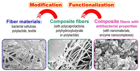
3.1. Structure of Composite Fiber Materials with PCL and PHB

Obtained samples were examined by SEM method (Figure 1). Both PCL and PHB formed almost smooth films on the surface of all fibrous materials under applied conditions. An even smoother surface can be obtained by introducing a polymer in a solvent with subsequent removal of the latter [33]. However, many fibrous materials can be also dissolved in this solvent and lose their structure, and therefore, this method is only partially applicable, e.g., to inert fibers.
Being more refractory, PHB almost did not penetrate through the fibrous support. At the same time, PCL freely passed through (micro)porous matrices, and was also sequestered by the nanoporous matrix of BC.
The aggregates of Ta NPs can be visualized on the surface of different composite samples (Figure 1). However, the distribution of NPs was not as good in the case of PHB modification as it was in the case of PCL. The accumulation of some clusters with enriched amounts of NPs can be observed in some localizations. That can negatively affect the antimicrobial activity of modified composite material.

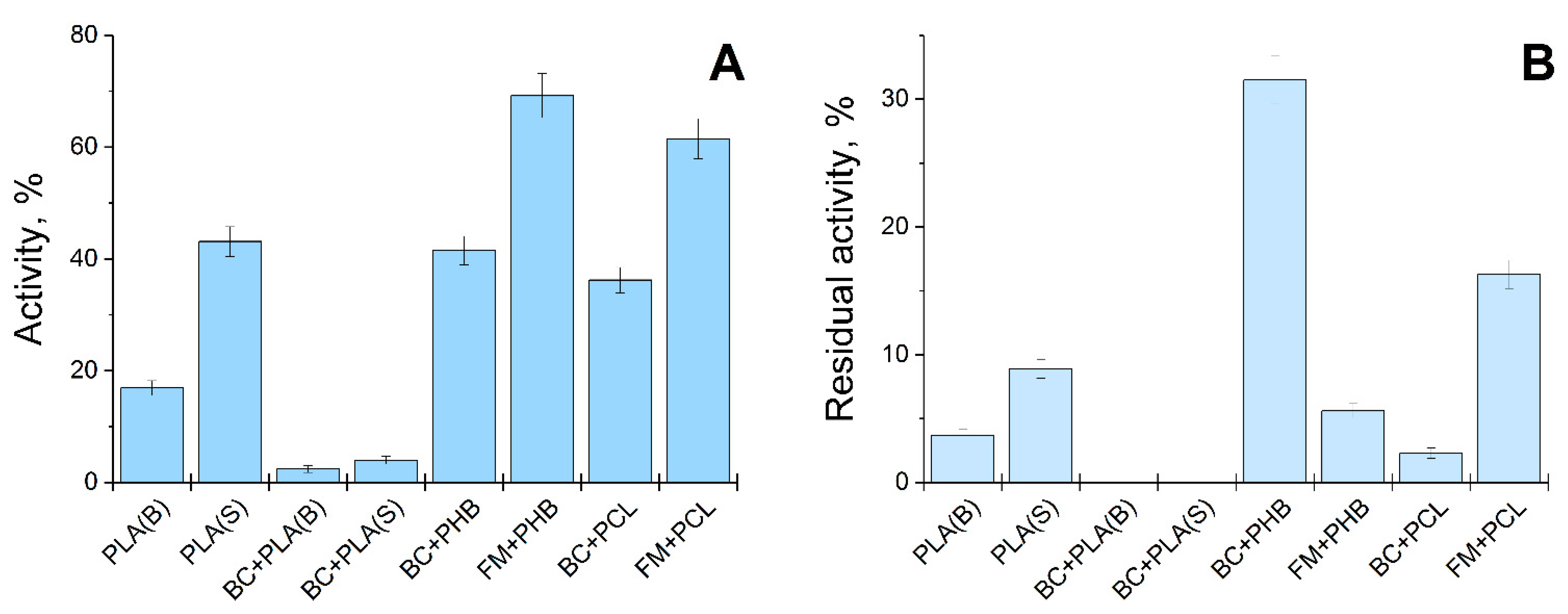
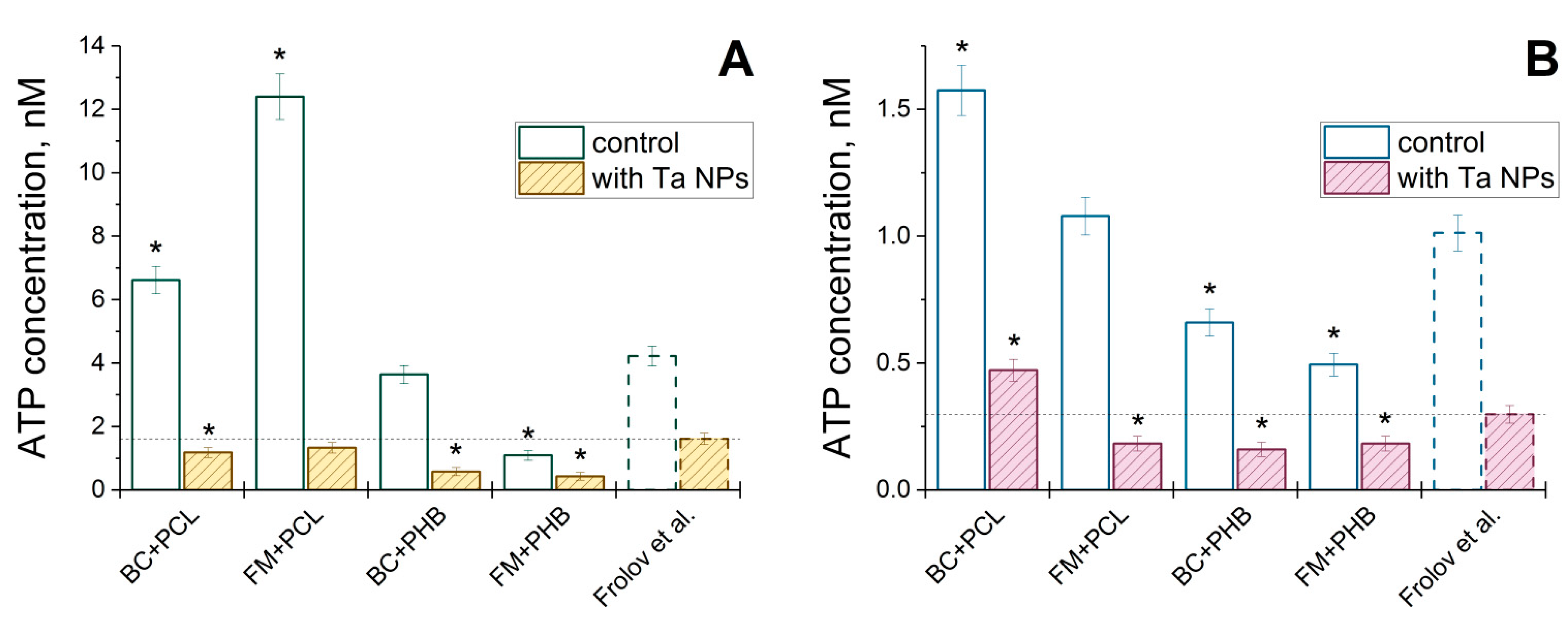
3.2. Funcional Characteristics of Composite Fiber Materials

Initially, EPC of His6-OPH/PLE50 was applied to the composites, and enzymatic activity of modified materials was determined (Figure 2). The greatest activity (up to 70% of the initial one) was demonstrated by composite materials based on FM coated with PHB or PCL. Slightly lower activity (less than 45% of the initial level) was possessed by samples of denser PLA material and BC coated with PHB or PCL. However, with repeated use of the same material samples, the maximum preservation of OPH activity (more than 30%) was revealed in BC modified by PHB. Thus, this variant of composite material was of the greatest interest for combination with nanocomplexes of His6-OPH.
Antibacterial activity of composite materials modified by Ta NPs was issued using Gram-negative (E. coli) and Gram-positive (Bacillus subtilis) bacterial cells (Figure 3). Both strains had higher survivability on the PCL-based composites without Ta NPs as compared to previously investigated material [27].
Modification by Ta NPs resulted in notable bacterial death, especially in the case of E. coli cells (up to 9-fold), with the levels being lower or comparable to material [27]. Composites with PHB alone had even better antibacterial activity as compared to material [27]. Application of Ta NPs led to synergetic effects with PHB and up to 4-fold harder bacterial elimination as compared to material [27].
Interestingly, composite of FM with PHB possessed the highest initial enzymatic activity among other fiber samples (Figure 2A), but it appeared to be non-reusable since only 5% of residual activity was revealed for it after single use.

4. Discussion

Introduction of novel antimicrobial nanomaterials differing from silver, zinc, copper, titanium, iron, etc. widely implemented in various studies is highly relevant since it allows flexibly counteracting urgent challenges rising in the face of humankind. Two main groups of antimicrobial mechanisms of nanomaterial action are generally accepted to be oxidative and non-oxidative [34]. The first one is associated with reactive oxygen species (ROS) generated onto nanomaterials and damaging the biochemical machinery of living cells. Non-oxidative mechanisms include multiple routes and predominantly damage to the cell envelope. Ta NPs used in the work are unlikely to generate ROS and thus may be interesting for application. From this standpoint, the current work is a logical extension of previous studies implementing Ta NPs as major antibacterial agents [14,27].
Multiple composite fiber materials are known to date, among which the most recent ones applying composite nanofibers of PLA, PCL and cotton [35] should be noted. However, the antibacterial activity of such composites en masse is still determined indirectly, and thus it is impossible to exactly compare them with the results of the current work. As compared to the previous work of the same methodology [27], composite structure of fiber materials resulted in improved antibacterial activity, especially when combined with PHB. Interestingly, composites with PHB better retained nanocomplexes His6-OPH/PLE50 in single- or multiple-use regimes. This means that such PHB composites have a great potential for combined deposition of enzymes and nanomaterials via the methodology described previously [14].
Noteworthily, composites with PLA obtained via thermo-impregnation had a poor sorption capacity and undetectable retention of nanocomplexes His6-OPH/PLE50. Nanoparticles having comparable sizes with His6-OPH/PLE50 will also be likely to be eluted. This is good for single-use materials releasing antibacterials into a medium. However, this is inapplicable for obtaining stable fiber materials in the long term suggested in the current work. Activation of polymer fibers and/or other additional modifications to the method will be required in the following work(s).
Thus, the combination of fiber materials with biodegradable polymers was shown in this work to allow multiplying both the number of possible final composite options and the range of their characteristics. Functionalization of these composites by metal-containing NPs or nanoscale EPC of His6-OPH that is capable of destroying not only bacterial QS-signaling molecules in a highly efficient manner [10,17,36], but can catalyze the hydrolysis of different toxic organophosphorus compounds [7,37,38] and mycotoxins [39], results in obtaining not just antibacterial, but also multifunctional, materials ready to show detoxifying abilities in relation to various toxic substances.

5. Conclusions

Thus, novel samples of composite fiber materials with antibacterial activity were demonstrated. Modern methods of nanotechnology and biotechnology were combined to obtain highly effective composite fiber materials. The prospectively suggested approach to the creation of fibrous composite versions may make it possible to widely expand the spectrum of the enzymes and nanomaterials that can be introduced to the materials, giving them useful features. Such new protective composite antibacterial materials may be attractive for possible various industrial applications as replaceable elements of membrane (bio) reactors, tourist equipment, fiber products for pets, construction finishing materials and protective clothing for agricultural workers.

Author Contributions

Conceptualization, E.E.; methodology, I.L., N.S. and D.P.; validation, I.L. and N.S.; formal analysis, E.E.; investigation, I.L., O.M., N.S. and D.P.; data curation, E.E.; writing—original draft preparation, I.L., O.M., E.E. and N.S.; writing—review and editing, I.L. and E.E.; supervision, E.E. All authors have read and agreed to the published version of the manuscript.

Funding

The research was funded by Russian Foundation of Basic Research (Project 18-29-25065). The stabilization of enzyme by poly(amino acid) was supported by State Task of Lomonosov Moscow State University (121041500039-8), and the samples of BC as biodegradable polymer was produced and studied with support from State Task of N.M. Emanuel Institute of Biochemical Physics RAS (01201253305).

Institutional Review Board Statement

Not applicable.

Informed Consent Statement

Not applicable.

Data Availability Statement

The data presented in this study are available by request from corresponding author.

Acknowledgments

This research was performed according to the Development program of the Interdisciplinary Scientific and Educational School of Lomonosov Moscow State University “The future of the planet and global environmental change”. The research was carried out using the equipment of the shared research infrastructure of the “Educational and Methodical Center of Lithography and Microscopy”, Lomonosov Moscow State University. This study was made possible with the support of the Applied Genetics Resource Facility of MIPT (Support Grant 075-15-2021-684). Authors are thankful to George Frolov for providing Ta NPs.

Conflicts of Interest

The authors declare no conflict of interest. I.L., O.M., N.S. and E.E. are named co-inventors of several Russian patents that involve organophosphorus hydrolase and its modified forms as a base active compound.

References

  1. Smith, M.K.; Mirica, K.A. Self-Organized Frameworks on Textiles (SOFT): Conductive Fabrics for Simultaneous Sensing, Capture, and Filtration of Gases. J. Am. Chem. Soc. 2017, 139, 16759–16767. [Google Scholar] [CrossRef] [PubMed]
  2. Ali, F.; Khan, S.B.; Kamal, T.; Anwar, Y.; Alamry, K.A.; Asiri, A.M. Anti-bacterial chitosan/zinc phthalocyanine fibers supported metallic and bimetallic nanoparticles for the removal of organic pollutants. Carbohydr. Polym. 2017, 173, 676–689. [Google Scholar] [CrossRef] [PubMed]
  3. Selvasudha, N.; Sweety, J.P.; Dhanalekshmi, U.M.; Devi, N.S.D. Smart antimicrobial textiles for healthcare professionals and individuals. In Antimicrobial Textiles from Natural Resources, 1st ed.; Mondal, I.H., Ed.; Woodhead Publishing: Sawston, UK, 2021; pp. 455–484. [Google Scholar] [CrossRef]
  4. Singh, A.; Lee, Y.; Dressick, W.J. Self-Cleaning Fabrics for Decontamination of Organophosphorous Pesticides and Related Chemical Agents. Adv. Mater. 2004, 16, 2112–2115. [Google Scholar] [CrossRef]
  5. Conte, M.P.; Lau, K.H.; Ulijn, R.V. Biocatalytic Self-Assembly Using Reversible and Irreversible Enzyme Immobilization. ACS Appl. Mater. Interfaces 2017, 9, 3266–3271. [Google Scholar] [CrossRef] [Green Version]
  6. Wong, D.E.; Senecal, K.J.; Goddard, J.M. Immobilization of chymotrypsin on hierarchical nylon 6,6 nanofiber improves enzyme performance. Colloids Surf. B Biointerfaces 2017, 154, 270–278. [Google Scholar] [CrossRef] [Green Version]
  7. Efremenko, E.N.; Lyagin, I.V.; Klyachko, N.L.; Bronich, T.; Zavyalova, N.V.; Jiang, Y.; Kabanov, A.V. A simple and highly effective catalytic nanozyme scavenger for organophosphorus neurotoxins. J. Control. Release 2017, 247, 175–181. [Google Scholar] [CrossRef] [PubMed]
  8. Lyagin, I.V.; Efremenko, E.N. Biomolecular engineering of biocatalysts hydrolyzing neurotoxic organophosphates. Biochimie 2018, 144, 115–121. [Google Scholar] [CrossRef] [PubMed]
  9. Aslanli, A.; Lyagin, I.; Efremenko, E. Novel approach to Quorum Quenching: Rational design of antibacterials in combination with hexahistidine-tagged organophosphorus hydrolase. Biol. Chem. 2018, 399, 869–879. [Google Scholar] [CrossRef]
  10. Aslanli, A.; Lyagin, I.; Efremenko, E. Charges’ interaction in polyelectrolyte (nano)complexing of His6-OPH with peptides: Unpredictable results due to imperfect or useless concept? Int. J. Biol. Macromol. 2019, 140, 368–376. [Google Scholar] [CrossRef]
  11. Kim, M.; Gkikas, M.; Huang, A.; Kang, J.W.; Suthiwangcharoen, N.; Nagarajan, R.; Olsen, B.D. Enhanced activity and stability of organophosphorus hydrolase via interaction with an amphiphilic polymer. Chem. Commun. (Camb) 2014, 50, 5345–5348. [Google Scholar] [CrossRef] [Green Version]
  12. vander Straeten, A.; Bratek-Skicki, A.; Germain, L.; D’Haese, C.; Eloy, P.; Fustin, C.A.; Dupont-Gillain, C. Protein-polyelectrolyte complexes to improve the biological activity of proteins in layer-by-layer assemblies. Nanoscale 2017, 9, 17186–17192. [Google Scholar] [CrossRef] [PubMed]
  13. Sharifi, M.; Robatjazi, S.M.; Sadri, M.; Mosaabadi, J.M. Immobilization of organophosphorus hydrolase enzyme by covalent attachment on modified cellulose microfibers using different chemical activation strategies: Characterization and stability studies. Chin. J. Chem. Eng. 2019, 27, 191–199. [Google Scholar] [CrossRef]
  14. Lyagin, I.; Stepanov, N.; Frolov, G.; Efremenko, E. Combined Modification of Fiber Materials by Enzymes and Metal Nanoparticles for Chemical and Biological Protection. Int. J. Mol. Sci. 2022, 23, 1359. [Google Scholar] [CrossRef] [PubMed]
  15. Volova, T.G.; Prudnikova, S.V.; Kiselev, E.G.; Nemtsev, I.V.; Vasiliev, A.D.; Kuzmin, A.P.; Shishatskaya, E.I. Bacterial cellulose (BC) and BC composites: Production and properties. Nanomaterials 2022, 12, 192. [Google Scholar] [CrossRef]
  16. Xu, Y.; Li, S.; Yue, X.; Lu, W. Review of silver nanoparticles (AgNPs)-cellulose antibacterial composites. BioRes 2018, 13, 2150–2170. [Google Scholar] [CrossRef]
  17. Aslanli, A.; Lyagin, I.; Stepanov, N.; Presnov, D.; Efremenko, E. Bacterial Cellulose Containing Combinations of Antimicrobial Peptides with Various QQ Enzymes as a Prototype of an “Enhanced Antibacterial” Dressing: In Silico and In Vitro Data. Pharmaceutics 2020, 12, 1155. [Google Scholar] [CrossRef]
  18. Aslanli, A.; Stepanov, N.; Razheva, T.; Podorozhko, E.A.; Lyagin, I.; Lozinsky, V.I.; Efremenko, E.N. Enzymatically functionalized composite materials based on nanocellulose and poly(vinylalcohol) cryogel and possessing antimicrobial activity. Materials 2019, 12, 3619. [Google Scholar] [CrossRef] [Green Version]
  19. Bhuiyan, M.A.R.; Wang, L.; Shaid, A.; Shanks, R.A.; Ding, J. Advances and applications of chemical protective clothing system. J. Ind. Text. 2019, 49, 97–138. [Google Scholar] [CrossRef]
  20. Li, T.T.; Sun, L.; Zhong, Y.; Peng, H.K.; Ren, H.T.; Zhang, Y.; Lin, J.-H.; Lou, C.-W. Silk fibroin/polycaprolactone-polyvinyl alcohol directional moisture transport composite film loaded with antibacterial drug-loading microspheres for wound dressing materials. Int. J. Biol. Macromol. 2022, 207, 580–591. [Google Scholar] [CrossRef]
  21. Sabapathy, P.C.; Devaraj, S.; Anburajan, P.; Parvez, A.; Kathirvel, P.; Qi, X. Active polyhydroxybutyrate (PHB)/sugarcane bagasse fiber-based anti-microbial green composite: Material characterization and degradation studies. Appl. Nanosci. 2021; in press. [Google Scholar] [CrossRef]
  22. Makarov, I.; Vinogradov, M.; Gromovykh, T.; Lutsenko, S.; Feldman, N.; Shambilova, G.; Sadykova, V. Antifungal Composite Fibers Based on Cellulose and Betulin. Fibers 2018, 6, 23. [Google Scholar] [CrossRef] [Green Version]
  23. Pušnik Črešnar, K.; Aulova, A.; Bikiaris, D.N.; Lambropoulou, D.; Kuzmič, K.; Fras Zemljič, L. Incorporation of metal-based nanoadditives into the PLA matrix: Effect of surface properties on antibacterial activity and mechanical performance of PLA nanoadditive films. Molecules 2021, 26, 4161. [Google Scholar] [CrossRef] [PubMed]
  24. Antunes, J.C.; Moreira, I.P.; Gomes, F.; Cunha, F.; Henriques, M.; Fangueiro, R. Recent Trends in Protective Textiles against Biological Threats: A Focus on Biological Warfare Agents. Polymers 2022, 14, 1599. [Google Scholar] [CrossRef] [PubMed]
  25. Wang, C.; Yin, J.; Han, S.; Jiao, T.; Bai, Z.; Zhou, J.; Zhang, L.; Peng, Q. Preparation of palladium nanoparticles decorated polyethyleneimine/polycaprolactone composite fibers constructed by electrospinning with highly efficient and recyclable catalytic performances. Catalysts 2019, 9, 559. [Google Scholar] [CrossRef] [Green Version]
  26. Cho, Y.S.; Kim, H.K.; Ghim, M.S.; Hong, M.W.; Kim, Y.Y.; Cho, Y.S. Evaluation of the antibacterial activity and cell response for 3D-printed polycaprolactone/nanohydroxyapatite scaffold with zinc oxide coating. Polymers 2020, 12, 2193. [Google Scholar] [CrossRef]
  27. Frolov, G.; Lyagin, I.; Senko, O.; Stepanov, N.; Pogorelsky, I.; Efremenko, E. Metal nanoparticles for improving bactericide functionality of usual fibers. Nanomaterials 2020, 10, 1724. [Google Scholar] [CrossRef]
  28. Efremenko, E.; Votchitceva, Y.; Aliev, T.; Varfolomeev, S. Recombinant Plasmid DNA pTes-His-oph and Producer of Oligohistidine-Containing Organophosphate Hydrolase. RU Patent 2255975C1, 10 July 2005. Available online: https://patents.google.com/patent/RU2255975C1 (accessed on 23 August 2022).
  29. Efremenko, E.; Votchitseva, Y.; Plieva, F.; Galaev, I.; Mattiasson, B. Purification of His6-organophosphate hydrolase using monolithic supermacroporous polyacrylamide cryogels developed for immobilized metal affinity chromatography. Appl. Microbiol. Biotechnol. 2006, 70, 558–563. [Google Scholar] [CrossRef]
  30. Votchitseva, Y.A.; Efremenko, E.N.; Aliev, T.K.; Varfolomeyev, S.D. Properties of hexahistidine-tagged organophosphate hydrolase. Biochemistry 2006, 71, 167–172. [Google Scholar] [CrossRef]
  31. Ismayilov, I.T.; Stepanov, N.A.; Efremenko, E.N.; Abbasov, V.M. Evaluation of biocidal properties of vegetable oil-based corrosion inhibitors using bioluminescent enzymatic method. Mosc. Univ. Chem. Bull. 2015, 70, 197–201. [Google Scholar] [CrossRef]
  32. Stepanov, N.; Senko, O.; Perminova, I.; Efremenko, E. A new approach to assess the effect of various humic compounds on the metabolic activity of cells participating in methanogenesis. Sustainability 2019, 11, 3158. [Google Scholar] [CrossRef] [Green Version]
  33. Bychuk, M.A. Preparation and Properties of Polymer Films Based on Poly-3-Hydroxybutyrate and Poly-ɛ-Caprolactone. Ph.D Dissertation, Moscow State University of Design and Technology, Moscow, Russia, 2016. [Google Scholar]
  34. Wang, L.; Hu, C.; Shao, L. The antimicrobial activity of nanoparticles: Present situation and prospects for the future. Int. J. Nanomed. 2017, 12, 1227–1249. [Google Scholar] [CrossRef] [PubMed] [Green Version]
  35. Cheng, Y.; Li, J.; Chen, M.; Zhang, S.; He, R.; Wang, N. Environmentally friendly and antimicrobial bilayer structured fabrics with integrated interception and sterilization for personal protective mask. Separat. Purificat. Technol. 2022, 294, 121165. [Google Scholar] [CrossRef]
  36. Sirotkina, M.; Efremenko, E.N. Rhodococcus lactonase with organophosphate hydrolase (OPH) activity and His6-tagged OPH with lactonase activity: Evolutionary proximity of the enzymes and new possibilities in their application. Appl. Microbiol. Biotechnol. 2014, 98, 2647–2656. [Google Scholar] [CrossRef] [PubMed]
  37. Varfolomeev, S.D.; Efremenko, E.N. (Eds.) Organophosphorus Neurotoxins, 1st ed.; RIOR: Moscow, Russia, 2020; 379p. [Google Scholar] [CrossRef]
  38. Lyagin, I.; Efremenko, E. Enzymes, Reacting with Organophosphorus Compounds as Detoxifiers: Diversity and Functions. Int. J. Mol. Sci. 2021, 22, 1761. [Google Scholar] [CrossRef]
  39. Lyagin, I.; Efremenko, E. Enzymes for detoxification of various mycotoxins: Origins and mechanisms of catalytic action. Molecules 2019, 24, 2362. [Google Scholar] [CrossRef] [PubMed] [Green Version]
Figure 1. (A) SEM images of PLA(S). (B) SEM of FM modified by PCL. (C) SEM of BC modified by PCL. (D) SEM of BC modified by PCL and functionalized by Ta NPs in ethanol. (E) SEM of BC modified by PHB. (F) SEM of BC modified by PHB and functionalized by Ta NPs in ethanol.
Fibers 10 00070 g001
Figure 2. (A) OPH activity of composite materials based on polylactide fiber (PLA(S), PLA(B)), BC and FM modified with PLA(S), PLA(B), PCL or PHB and then functionalized by His6-OPH/PLE50. Enzyme activity deposited onto samples was accepted as 100%. (B) Residual OPH activity of the same samples after their single use and washing. Enzyme activity in the first working cycle was accepted as 100%.
Fibers 10 00070 g002
Figure 3. Antibacterial activity of composite fiber materials modified with PCL or PHB and functionalized with or without Ta NPs toward E. coli (A) and Bacillus subtilis (B) bacterial cells. It was estimated via analysis of residual intracellular ATP concentration in bacteria exposed onto composites for 24 h. Previously investigated fiber material [27] (labeled as “Frolov et al.”) was issued under the same conditions, and samples with statistically significant differences compared to reference material (one-way ANOVA with Holm-Sidak multiple comparison test, p < 0.05) were marked with an asterisk.
Fibers 10 00070 g003
Publisher’s Note: MDPI stays neutral with regard to jurisdictional claims in published maps and institutional affiliations.

