Article

Eu2+-Activated Ba0.5Sr0.5Al2O4 Phosphors for Screen Printing and Anti-Counterfeiting Flexible Film

1
Beijing Key Laboratory of Printing and Packaging Materials and Technology, Beijing Institute of Graphic Communication, Beijing 102600, China
2
School of Physics, Beihang University, Beijing 100191, China
3
Department of Physics, The University of Texas at Arlington, Arlington, TX 76019-0059, USA
4
National Center for Nanoscience and Technology, CAS Key Laboratory of Nanophotonic Materials and Devices (Preparatory), Beijing 100190, China
5
The GBA Research Innovation Institute for Nanotechnology, Guangzhou 510700, China
*
Authors to whom correspondence should be addressed.
These authors contributed equally to this work.
Coatings 2023, 13(7), 1247; https://doi.org/10.3390/coatings13071247
Submission received: 13 June 2023 / Revised: 6 July 2023 / Accepted: 11 July 2023 / Published: 14 July 2023
(This article belongs to the Special Issue Optoelectronic Thin Films)

Abstract

:
Herein, a series of Ba0.5Sr0.5Al2O4: xEu2+ (x = 0.01, 0.02, 0.03, 0.04, 0.06) nanophosphors were synthesized by a combustion method. The investigation encompassed the characterization of the phase purity, morphology, elemental composition, and photoluminescence behavior of Ba0.5Sr0.5Al2O4: xEu2+ nanoparticles. Under excitation by 303 nm and 365 nm ultraviolet light, the nanoparticles exhibited blue-green emission arising from the 4f65d→4f7 transition of Eu2+ ions. The optimal doping concentration was determined to be 2%. Notably, the nanoparticles demonstrated fluorescence lifetimes and quantum yields of 1010 ns ( λ ex = 303 nm), 112 ns ( λ ex = 365 nm), 10.5%, and 10.3%, respectively. Additionally, a comprehensive analysis of the band structure and electronic density of states was conducted, revealing a theoretical direct band gap of 4.05 eV for the Ba0.5Sr0.5Al2O4 host. In addition, the prepared fluorescent powder can be used to prepare fluorescent flexible films. This film does not change the characteristic emission of Eu2+ ions and has more stable physicochemical properties, which may be more suitable for use in harsh environments. Also, the fluorescent powder can be blended with polyacrylic acid to form colorless anti-counterfeiting ink that can be applied to banknotes as an anti-counterfeiting mark. A clover pattern was successfully printed using screen-printing technology, proving its potential application in the field of anti-counterfeiting.

1. Introduction

Nowadays, all kinds of counterfeit products, such as fake banknotes, fake drugs, and counterfeit food, have penetrated every aspect of our life, posing a great threat to our personal property and life and health as well as the reputation and profit of brand owners [1,2,3]. And with the development of economic globalization, the proliferation of these problematic products will also affect the image of the country’s products, thus losing its advantage in international competition [4]. To maintain the healthy development of the market and the safety of people’s life and property, in addition to using legal means to crack down on the production and sale of counterfeits, countries around the world have been continuously exploring various advanced anti-counterfeiting technologies [5], for example, multiple laser anti-counterfeiting, 1D barcode anti-counterfeiting, 2D barcode anti-counterfeiting, temperature change anti-counterfeiting, watermark anti-counterfeiting, fluorescent anti-counterfeiting, etc. [6,7,8,9,10,11]. Among them, fluorescent materials with rare-earth doped inorganic substances are favored because of their photochemical stability, rich energy levels, and ability to achieve multi-color emission from UV to NIR [12,13,14,15]. For example, Xue Bai et al. prepared CaWO4: Yb3+, Er3+, Bi3+ photochromic phosphors showing reversible photochromic and dual-mode upconversion (UC) and downshifting (DS) luminescence properties, which have potential applications as composite anti-counterfeits and optical storage media [16]. Mengxiao Li et al. synthesized dual-mode luminescent NaYF4: Er, Yb(Tm) carbon quantum dots by a facile solvothermal method and successfully printed a variety of dual-mode fluorescent patterns using a mixture of a fluorescent material and an aqueous poly(acrylic acid) solution as a colorless anti-counterfeit ink, showing good dual-mode fluorescent properties [17].
Although these materials have achieved many remarkable results, these materials are simply phosphors or fluorescent inks, which have many problems, such as the single excitation mode, not being easy to preserve, not being recyclable, etc. [18,19]. Moreover, as the counterfeiting technology continues to iterate and update, the possibility of its being cracked or counterfeited remains high. In recent years, the application of a rare earth anti-counterfeiting film in anti-counterfeiting has become increasingly widespread. These films usually appear colorless and transparent in daylight but show specific anti-counterfeiting patterns when exposed to UV light, etc. This design combines the properties of both thin film and rare-earth light-emitting materials, enabling more advanced anti-counterfeiting. Polydimethylsiloxane (PDMS) is known for its high chemical and thermal stability, corrosion resistance, flexibility, and repeatability, and it is widely used in electronic components, sensors, fiber-optic waveguide coatings, and circuit board packaging [20,21,22,23]. The composite of rare-earth fluorescent material and PDMS elastic substrate can prepare an excellent flexible anti-counterfeiting material by combining the luminescent properties of rare-earth fluorescent material with the compressible and stretchable physical properties of PDMS.
Recently, Eu2+-activated Ba/Sr-based aluminate phosphors have been extensively studied, and the phosphors have unique luminescence properties through Ba/Sr mutual substitution. Daisuke Nakauchi et al. synthesized Eu-doped BaxSr1−xAl2O4 (x = 0, 0.1, 0.3, and 0.5) single crystals by the floating zone method [24]. They investigated their scintillation properties, observing the high optical yield of Eu: Ba0.5Sr0.5Al2O4. Max Volhard synthesized Sr0.97−xBaxEu0.03Al2O4 (x = 0–0.97) phosphor by the solid-phase reaction and studied the phase change and luminescence characteristics with the changing Ba content [25]. Marcos et al. investigated the X-ray excited optical luminescence (XEOL) properties of Eu-doped Ba1−xSrxAl2O4 phosphors. They found that the XEOL yield was higher in the Ba-rich samples and the decay rate of long-lasting phosphorescence (LLP) was slower in the Sr-rich samples [26].
In this study, Ba0.5Sr0.5Al2O4: xEu2+ (x = 0.01, 0.02, 0.03, 0.04, 0.06) phosphor was synthesized by a simple combustion method. To study the optical properties of the phosphor, the synthesized samples were measured and characterized by scanning electron microscope (SEM), photoluminescence (PL), X-ray diffraction (XRD), etc. Additionally, the optimal doping concentration of Eu ion x = 0.02, and the optimal reaction temperature was 700 °C. Finally, fluorescent flexible films and anti-counterfeiting inks were prepared using the optimized samples, showing their potential applications in the field of anti-counterfeiting.

2. Experimental Section

2.1. Materials and Method

A series of Ba0.5Sr0.5Al2O4: xEu2+ (x = 0.01, 0.02, 0.03, 0.04, 0.06) samples were prepared by the combustion method. The raw materials used were BaCO3 (purity: 99.99%), SrCO3 (99.99%), Al2O3 (99.99%), Eu2O3 (99.99%), HNO3 (80%), and urea (99.99%), which were all purchased from Tianjin Chemical Reagent Factory, Tianjin, China. The deionized water was homemade. All reagents were directly used as received without further purification.

2.2. Synthesis of Nanomaterials

First, appropriate amounts of HNO3 and deionized water were added to BaCO3, SrCO3, Al2O3, and Eu2O3 to prepare 0.2 mmol/mL of Ba(NO3)2, 0.5 mmol/mL of Sr(NO3)3, 1 mmol/mL of Al(NO3)3, and 0.1 mmol/mL of Eu(NO3)2 solutions, respectively. Then, Ba(NO3)2, Sr(NO3)3, Al(NO3)3 and Eu(NO3)2 solutions were measured in stoichiometric ratios (0.5 − 0.5x:0.5 − 0.5x:2:x) and placed in a crucible, then 2.2 g of urea for the combustion agent was added and mixed homogeneously, and placed in a preheated muffle furnace. After waiting 3 to 5 min, a loose white and porous solid powder is obtained. A series of samples can be obtained by adjusting the Eu ion doping concentration (x = 0.01, 0.02, 0.03, 0.04, 0.06) and reaction temperature (500, 600, 700, 800, 900 °C). Ultimately, the obtained samples were naturally cooled to room temperature and ground into powder for a test.

2.3. Preparation of Anti-Counterfeit Flexible Film and Ink

Polydimethylsiloxane (PDMS, Sylgard184, Dow Corning, Midland, MI, USA) was used to assemble the elastomer for Ba0.5Sr0.5Al2O4: Eu2+. The milled phosphor was mixed with the uncured PDMS material and put into a mold, and then the molds were dried at 100 °C for 1 h to obtain the “ML” pattern composite phosphor. The final flexible, stretchable anti-counterfeit film was obtained by placing the resulting “ML” composite in an uncured PDMS matrix and curing it at 120 °C for 40 min.
The fluorescent powder was introduced into a mixed solution of ethanol and polyacrylic acid, followed by vigorous stirring. The amounts of polyacrylic acid and ethanol were adjusted to attain a viscosity suitable for screen printing, resulting in the final fluorescent ink.

2.4. Characterizations

The crystalline phases were collected on an X-ray diffraction (Rigaku D/Max-2400, Rigaku, Tokyo, Japan) with Cu Kα radiation (λ = 1.54 Å), operating at 40 kV and 150 mA. The PLE and PL spectra were measured by a fluorescence spectrometer (FluoroMax-4, Horiba, Ltd., Kyoto, Japan). The morphology of phosphors was observed through scanning electron microscopy (Quanta 250 FEG, Thermo Fisher Scientific, Waltham, MA, USA). The fluorescence lifetime and quantum yield of phosphors were measured by a transient steady-state fluorescence spectrometer (FLS1000, Edinburgh Instruments, Edinburgh, UK). The UV-Vis absorption spectra were performed using a UV-3600 UV-Vis Near Infrared Spectrophotometer (Shimadzu Corporation, Kyoto, Japan). The energy-dispersive X-ray spectroscopy (EDS) spectrum was obtained with a desktop scanning electron microscope energy dispersive spectrometer (SU8020, Hitachi, Ltd., Hitachi, Japan).

2.5. Theoretical Calculations

The structural optimization and electronic structure calculations of Ba0.5Sr0.5Al2O4 were performed using density functional theory (DFT) based on first-principles calculations. The structure consists of a unit cell. All DFT calculations were performed using the Vienna ab initio Simulation Package (VASP) [27]. The Perdew–Burke–Ernzerhof (PBE) exchange–correlation functional and projector-augmented wave (PAW) pseudopotentials were employed for spin-polarized calculations [28,29,30]. During the structural optimization process, a convergence criterion of 10−6 eV for the total energy was set, and atomic positions were relaxed until the forces acting on each atom were less than 0.01 eV/Å. Gaussian smearing with a width of 0.05 eV was used for the occupation of the electronic orbitals. A plane-wave cutoff energy of 500 eV was applied in all calculations. The Brillouin zone integration was performed using the Monkhorst–Pack (MP) special k-point mesh with a spacing of 0.03 Å−1.

3. Results and Discussion

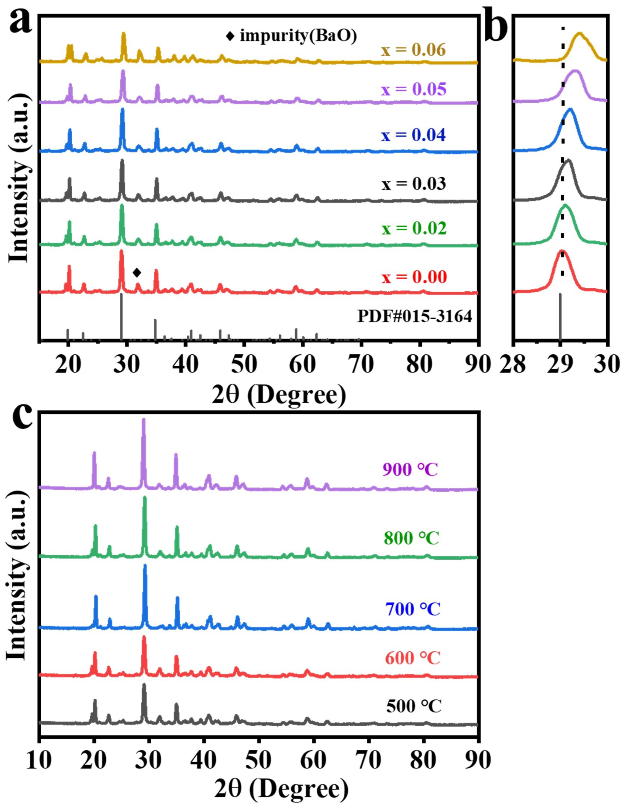
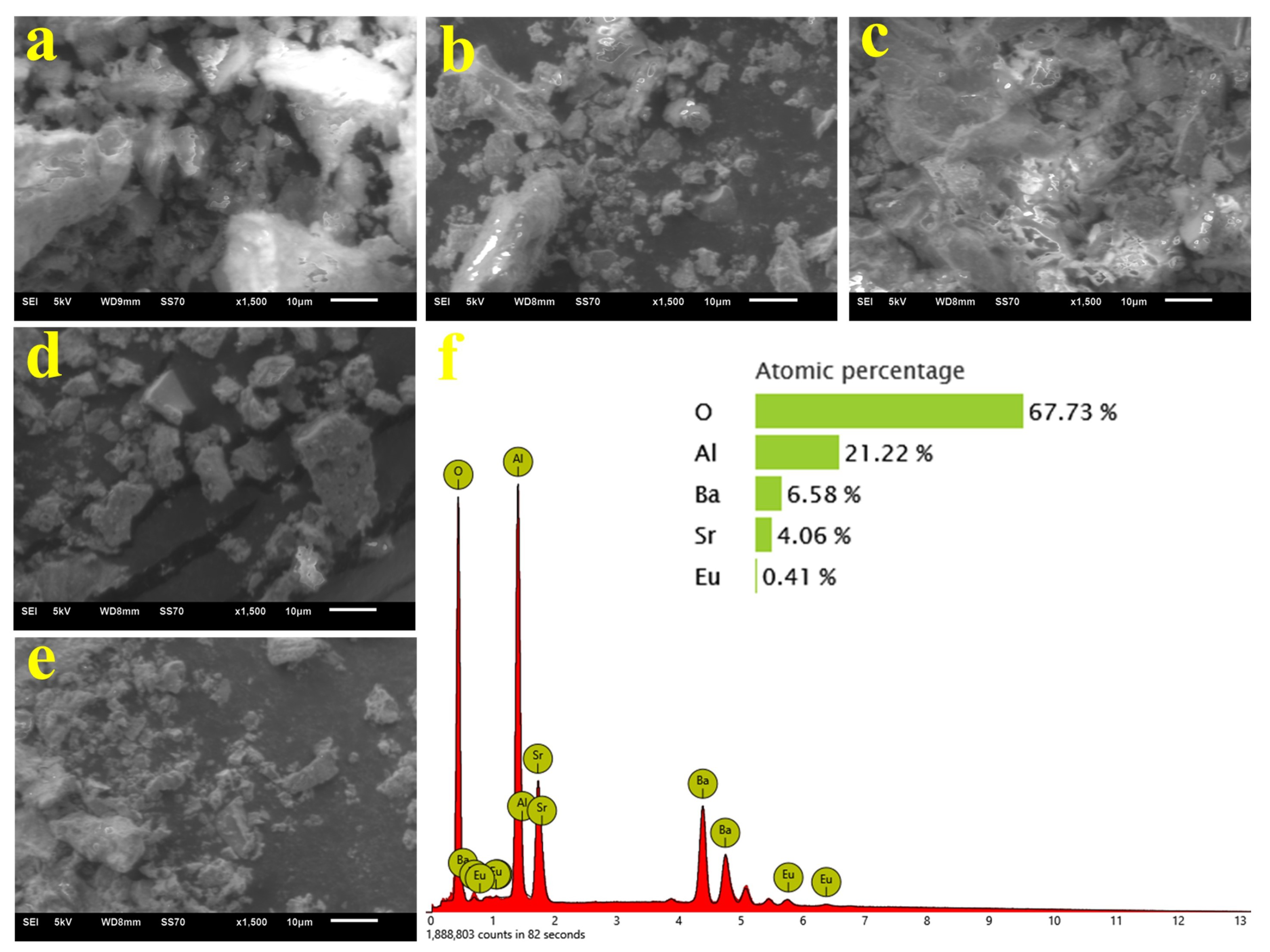
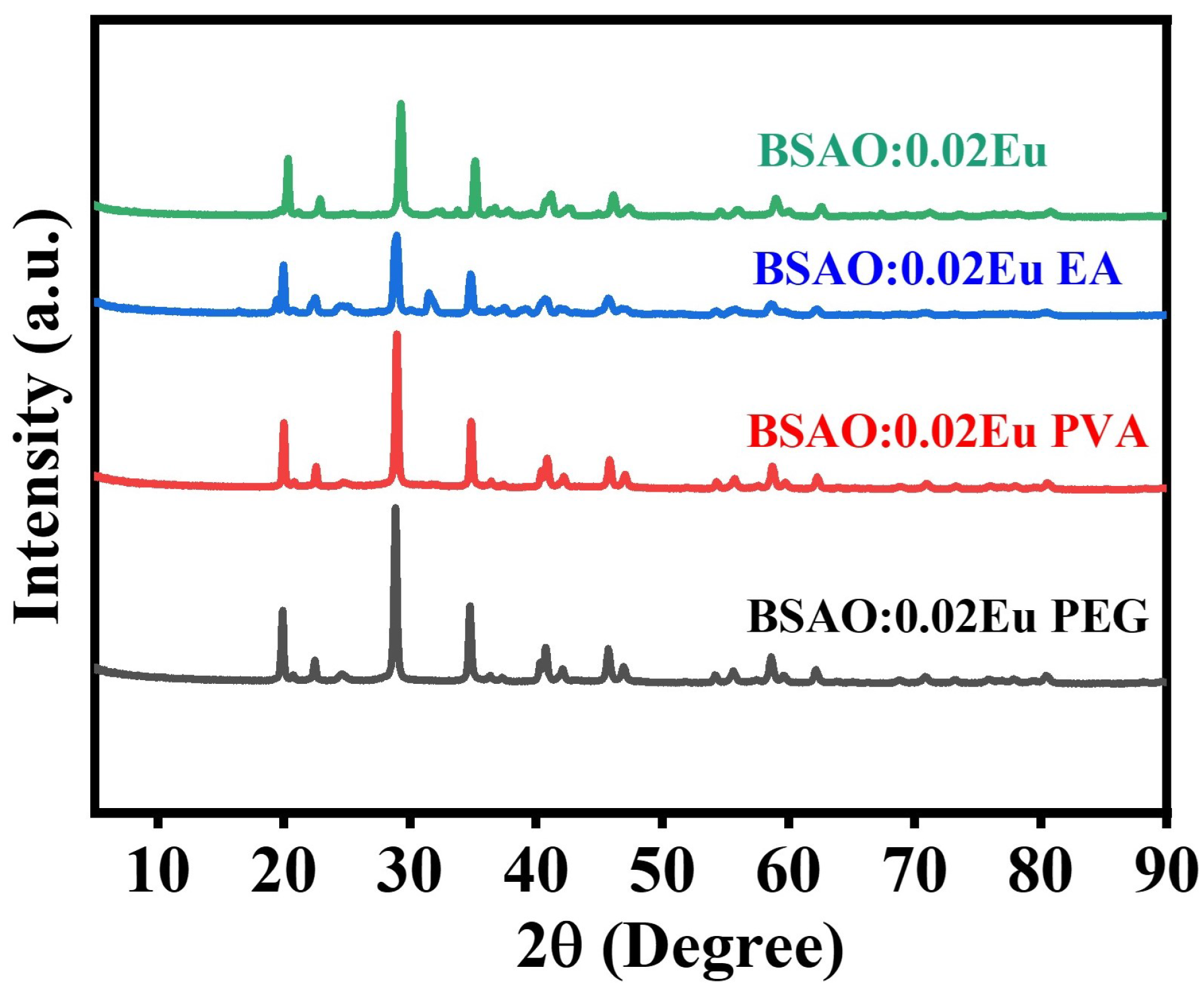
3.1. Structure and Morphology Analysis of BSAO: Eu2+

The phase composition and purity of Ba0.5Sr0.5Al2O4 (abbreviated as BSAO) phosphor were investigated by X-ray powder diffraction. Figure 1a shows the XRD patterns of the BSAO host and BSAO: xEu2+ (x = 0, 0.01, 0.02, 0.03, 0.04, 0.06) phosphors at a reaction temperatures of 600 °C. Most of the diffraction peaks of the samples can be indexed to the standard data of PDF#97-015-3164. However, for the BSAO sample, some byproducts can be observed, as noted by the rhombus. The impurity phase is inferred to be BaO, and the weight ratio to be approximately 0.4%, by a simple quantitative analysis, which can be almost ignored on the effect of the luminescence properties. According to the XRD standard cards, it is identified that the BSAO belongs to the P63(173) space group of the hexagonal phase. The cell parameters a = b = 8.972 Å, c = 8.606 Å calculated for BSAO are between BaAl2O4 (a = b = 10.449 Å, c = 8.793 Å) and SrAl2O4 (PDF#97-015-3164) [31,32]. Generally, it is reasonable to assume that the Eu2+ dopants tend to occupy the Sr2+ or Ba2+ sites based on similar effective ionic radii (IR) of the cation with varying coordination numbers (CNs) and valences [33,34,35]. Moreover, the main diffraction peak of (112) shifts toward a higher angle with the introduction of Eu ions as shown in Figure 1b. It means that the lattice parameters and the cell volume of the substituted samples will be reduced to some extent, which is in line with the shift of the diffraction peaks, which could be well accepted based on Bragg’s law (2d sin  θ  = n λ ) [36,37]. It confirms again that the Eu ions with smaller ionic radii are constantly replacing the potentially substitutable ions. Moreover, the presence of additional peaks at about 20° of the 2 θ  angle may be due to the unstable crystalline phase of the sample caused by the insufficient reaction temperature.
The effect of the reaction temperature on the phase and morphology of the final sample will be further investigated. Figure 1c shows the XRD patterns of BSAO: 0.02Eu2+ synthesized at different reaction temperatures. With the increase in temperature, the strength of the major diffraction peak progressively grows, and the position of its diffraction peak is basically unchanged compared with Figure 1a. It means that the crystals of the BSAO: 0.02Eu2+ samples are formed successfully at different reaction temperatures and crystallized better with increasing temperature. In addition, the XRD pattern of the sample shows a decrease in the impurity peak when the reaction temperature is 700 °C. Furthermore, the intensity of the extra diffraction peak at the 2 θ  angle of about 20° gradually decreases with the increase in the reaction temperature. When the reaction temperature reaches 900 °C, the extra peaks disappear, and the crystalline phase starts to stabilize. The influence of the reaction temperature on the microscopic morphology of the phosphor is investigated using SEM pictures. Figure 2 depicts the crystal morphology of BSAO: 0.02Eu2+ produced at different reaction temperatures. In all five of these samples, the phosphor particles are aggregated into tiny clusters of varied shapes and sizes. In addition, there are lamellar structures with fractures and air pores. At 500–600 °C, the gaps in the sample structure progressively become smaller and gradually tend to be united, and at 700 °C, the gaps are negligible, showing a laminar structure, and the structure shows no obvious fracture symptoms. At 800–900 °C, the lamellar structure disappears gradually, and the gaps in the sample structure keep rising again, which may be caused by the violent outward burst of the gas created during the burning of the sample [38]. The irregularities in the shape, size, and pores of these samples may be related to the irregular mass flow and unequal temperature distribution of the samples during combustion [39]. Moreover, the existence of the elements Ba, Sr, Al, O, and Eu is demonstrated by the EDS spectrum of BSAO: 0.02Eu2+ as shown in Figure 2f. The ratio of Ba to Sr can be observed to be close to 1:1, and the successful doping of Eu ions into the host material is achieved.

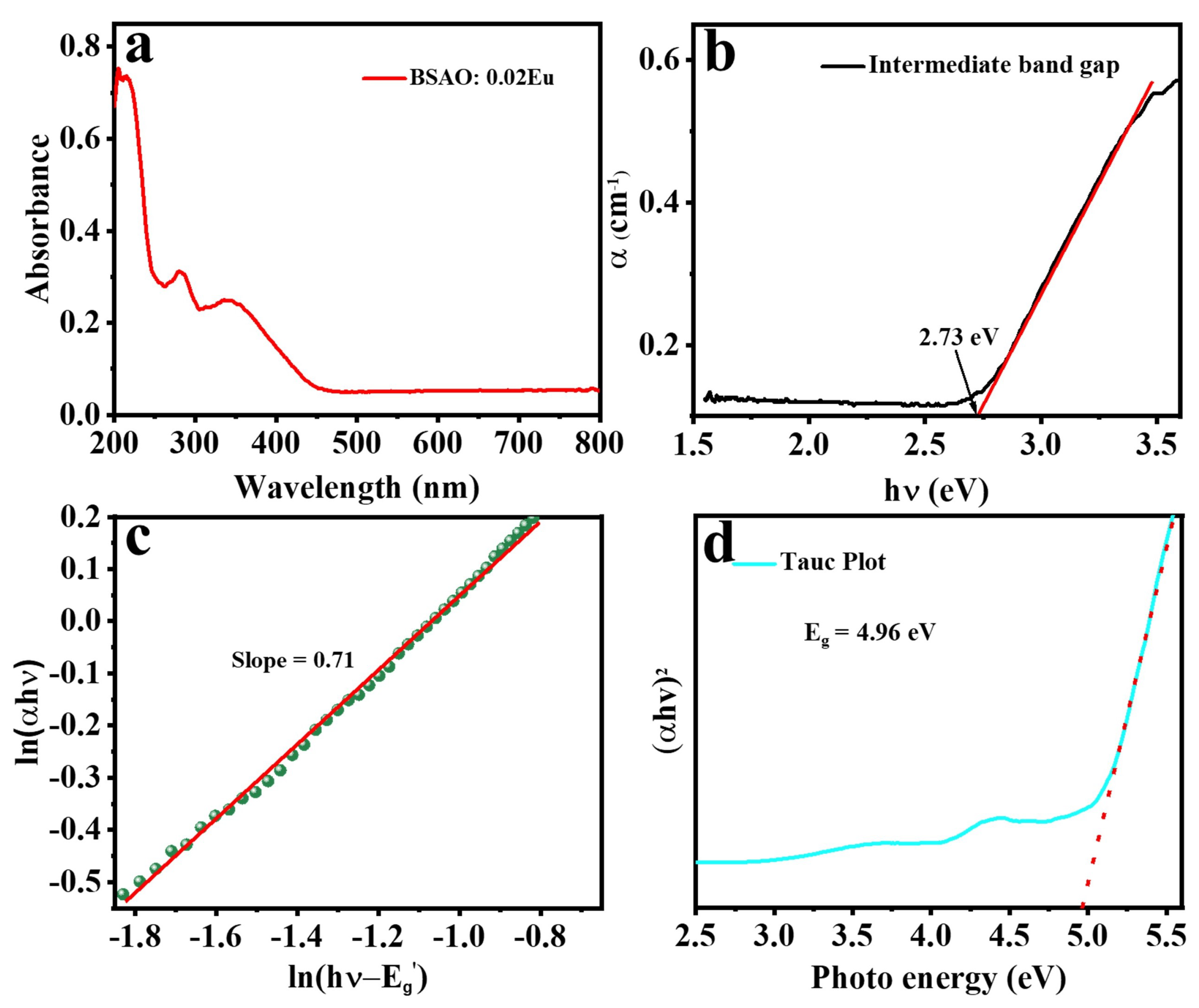
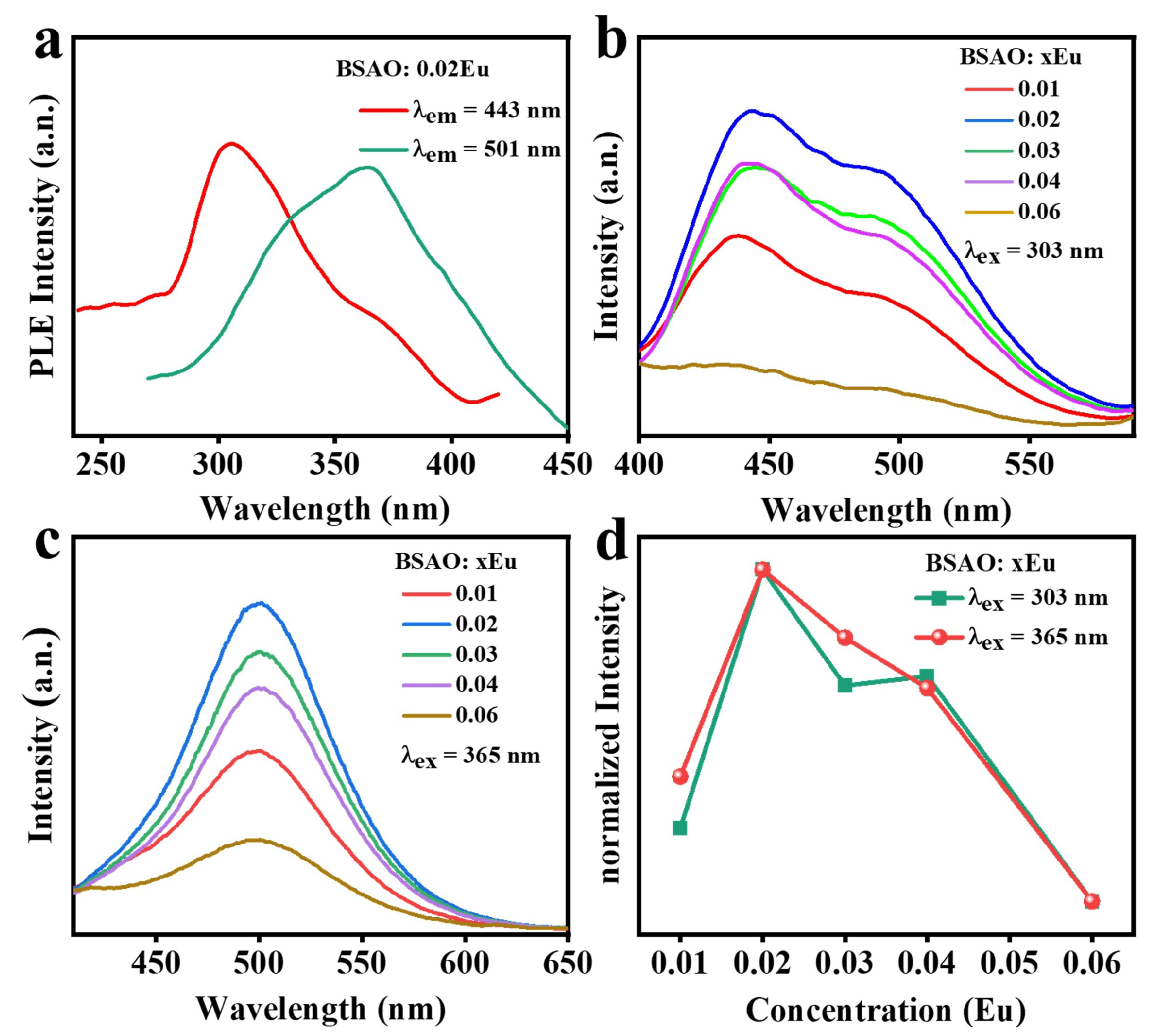
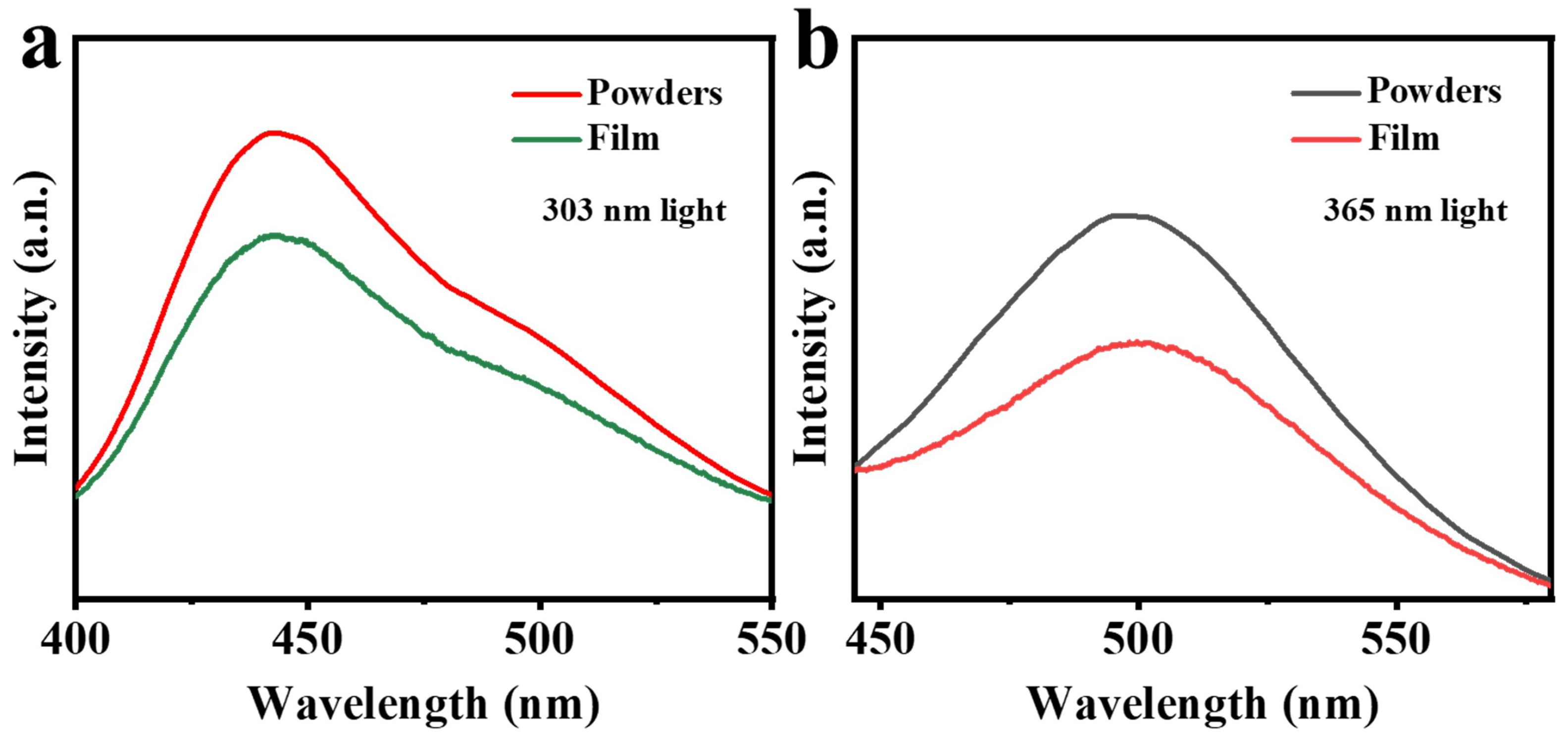
3.2. UV-Vis Absorption and Photoluminescence Properties of BSAO: Eu2+

UV-vis absorption spectroscopy was used to study the absorption performance of BSAO: 0.02Eu2+ (Figure 3a). The absorption spectrum shows two absorption peaks from 230 to 400 nm. Absorption near 230 nm is attributed to the host band absorption. The broad absorption band covering the spectral range of 300–400 nm is assigned to the 4f→5d electronic transitions of Eu2+. To experimentally estimate the bandgap scale, the Eg of the BSAO: 0.02Eu2+ can be estimated according to the following equation [40]:
( α h ν ) 1 / n = A ( h ν E g )
where is the photon energy,  α  is the absorption coefficient, Eg is the band gap energy (eV), and A is a constant. The value of n is determined according to the type of interband conversion. It is equal to 2 for indirect transitions and 1/2 for direct transitions [41]. In addition, the intermediate optical band gap ( E g ) can be obtained by the linear fit in the flat part in Figure 3b. Then, the value of n is calculated using the following equation [42]:
n = ln ( α h ν ) / ln ( h ν E g )
As shown in Figure 3c, the value of n is approximated by 1/2. Thus, the plot of (αhν)2 versus (Eg) is shown in Figure 3d. The absorption Tauc plot shows that the band gap is 4.96 eV.
The luminescence properties of the BSAO: Eu2+ phosphors are investigated in detail by photoluminescence (PL) and photoluminescence excitation (PLE) spectroscopy. It is found that the peak positions and spectral shapes of emission spectra do not vary with Eu2+ concentration for the BSAO: Eu2+ nanophosphor, but the intensity of emission peaks for BSAO: xEu2+ (x = 0.01, 0.02, 0.03, 0.04, and 0.06) nanophosphor strongly depends on the doping concentration of Eu2+ ions. As shown in Figure 4a, the PLE spectrum of BSAO: 0.02Eu2+ encompasses the UV to the blue area and exhibits two broad absorption bands centered at 365 and 303 nm, which is attributable to the 4f7→4f65d transition of Eu2+ [43]. Figure 4b,c show the emission spectra of the samples under UV excitation at 303 and 365 nm, and their emission peaks are 443 and 501 nm, respectively. The PL spectrum shows a broad asymmetric band with a maximum at 443 nm under 303 nm excitation, corresponding to the allowed 4f65d→4f7 transition of the Eu2+. As shown in Figure 4c, the narrow-band green emission, correlating to the 5d-4f transition of Eu2+, is observed under 365 nm excitation. The full width at half-maximum (FWHM) of the emission spectrum is around 62 nm. Furthermore, concentration quenching typically occurs in the emission center when certain structural defects and nonradiative transition rates reach a saturation state. In general, the critical value of the concentration (having intense emission) is determined as the optimal doping concentration. Therefore, the strongest PL emission intensities of the BSAO: Eu2+ phosphors are recorded when x = 0.02, revealing that the optimal doping concentrations of Eu2+ ions in the BSAO host are 2% as illustrated in Figure 4d. Moreover, it should be clarified whether the mechanism of concentration quenching is the critical reason for their different optimal concentrations. Indeed, the energy transfer concentration quenching mechanism can be investigated by calculating the interaction coefficient (Q) as shown below [44,45,46]:
log I x ) = A Q 3 ) log x
where x denotes the Eu2+ ion concentration, and I is the luminescence intensity. In this respect, the fitting slope (−(Q/3)) is determined to be −1.72 and −2.25, respectively. The interaction coefficient (Q) values are analyzed to be about 5.16 ( λ ex = 303 nm) and 6.75 ( λ ex = 365 nm) as shown in Figure 5. All of them are close to 6, suggesting that the dipole–dipole interaction (Q = 6 ) is the main mechanism to control concentration quenching rather than the dipole–quadrupole (Q = 8) or quadrupole–quadrupole (Q = 10) interaction.
The reaction temperature is a highly essential aspect that directly impacts the structure and luminescent properties of the sample. The produced solutions are sintered at 500, 600, 700, 800, and 900 °C, and the emission spectra of the synthesized products are given in Figure 6. When the reaction temperatures are different, the characteristics of the spectra do not change significantly under the same wavelength of excitation, except for the emission intensity. Figure 6a,b show the emission spectra of the samples under the excitation of the UV light at 303 and 365 nm. It shows a broad emission band centered at 442 and 500 nm, respectively. The luminescence intensities of the samples all reach the highest when the reaction temperature is 700 °C. These blue-green emission bands are attributed to the 4f65d→4f7 electron transition of the Eu2+ ion. In addition, the emission bandwidth of the samples becomes narrower when the reaction temperature is 800 °C. It may be due to the improved crystallinity of the material at higher reaction temperatures, which reduces the defects and impurities that may lead to broader emission bands. Figure 6c provides a thorough illustration of this procedure.
Figure 6d depicts the coherent infrared energy (CIE) chromaticity coordinate positions of the BSAO: 0.02Eu2+ phosphor under the excitation wavelength of 303 nm and 365 nm. The corresponding CIE chromaticity coordinates are (0.166, 0.208) and (0.176, 0.365). It is observed from Figure 6d that the CIE chromaticity coordinate varies with the excitation wavelength and the corresponding colors are changed from blue to green.
Moreover, the luminescence decay curves of the BSAO: 0.02Eu2+ phosphor ( λ ex = 303 nm,  λ em = 442 nm;  λ ex = 365 nm,  λ em = 501 nm) are shown in Figure 7, and the decay curves are well fitted by a second-order and triple-order exponential function expressed as follows [47]:
I ( t ) = I 0 + A 1 e x p ( t / τ 1 ) + A 2 e x p ( t / τ 2 ) +
where I0 represents the background constant, A1 and A2 are constants, t is the decay time,  τ 1 and  τ 2 refer to the decay times of the exponential components and the average lifetimes of all BSAO with different Eu2+ doping concentrations are calculated by the following equation:
τ * = ( A 1 × τ 1 2 + A 2 × τ 2 2 + ) / ( A 1 × τ 1 + A 2 × τ 2 + )
According to the formula, the average lifetime of BSAO: 0.02Eu2+ is 1010 ns ( λ ex = 303 nm) and 112 ns ( λ ex = 365 nm), respectively. Furthermore, the photoluminescence quantum yield (PLQY) is an important parameter for luminescent materials. The QY of the phosphors can usually be measured according to the percentage of the photon number emitted by the sample ( ε ) versus the photon number absorbed by the sample ( α ). The PLQY spectrum of the BSAO: 0.02Eu2+ phosphor is presented in Figure 7c,d. The QY can be determined by the following equation [48]:
P L Q Y = ε / α = S e m / ( S 0 S )
where Sem is the integrated intensity of the emission light of the phosphor, S0 and S is integrated with the intensity of the scattered light of the reference background plate and the phosphor, respectively. Thus, the absorbed photon number of the BSAO: 0.02Eu2+ phosphor is obtained by subtracting the scattered light intensity (S) of the BSAO: 0.02Eu2+ phosphor from the scattered light intensity (S0 ) of the reference background plate. Therefore, it is clear that the QY could reach up to 10.5% ( λ ex = 303 nm) and 10.3% ( λ ex = 365 nm). The detailed parameters of the lifetimes and QY are shown in Table 1.

3.3. Electronic Structure

The band structure and density of states (DOSs) of the BSAO host are calculated using density functional theory (DFT). Considering the mutual substitution between Ba and Sr and their atomic ratios close to 1:1 (Figure 2f), we design a structural model of Ba0.5Sr0.5Al2O4. The structural model is based on the substitution of 50% of Sr atoms with Ba in the SrAl2O4 system. As shown in Figure 8a,b, the valence band maximum (VBM) and the conduction band minimum (CBM) are located at the same point, indicating that BSAO exhibits a direct band gap (Eg). The predicted value of Eg is approximately 4.05 eV. In addition, as shown in Figure 8d, the experimental band gap of the BSAO host can be obtained by Equation (1). The experimental Eg (5.18 eV) is slightly larger than the calculated Eg (4.05 eV), which can be attributed to the general limitations of the generalized gradient approximation scheme in the DFT calculations [49,50]. Figure 8c displays the total density of states (TDOS) and partial density of states (PDOS) of the BSAO host. It is observed that the d orbitals of the Ba and Sr atoms predominantly contribute to the DOS at the CBM, while the VBM is primarily derived from the p orbitals of the O atoms.

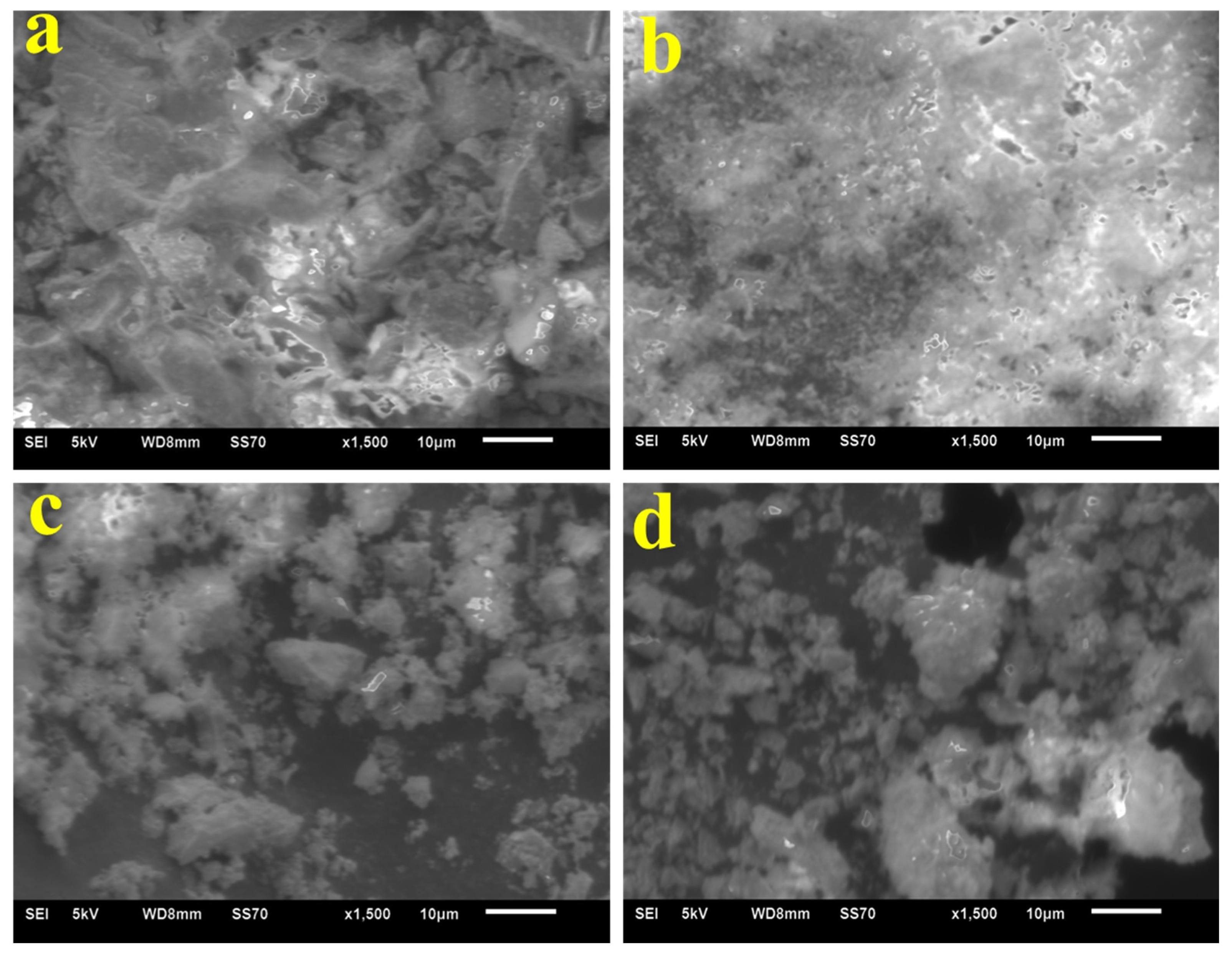
3.4. Morphology Control of BSAO: Eu2+ via Surfactants

Numerous factors, such as grain size, shape, and dimensionality can alter the luminescence of rare earth ions. Surfactants are extensively employed to modulate inorganic nanocrystal size, shape, and crystallinity [51]. To examine the effect of surfactants on the morphology of fluorescent materials, BSAO: 0.02Eu2+ phosphors were synthesized with the assistance of surfactants. Figure 9 shows the XRD patterns of BSAO: 0.02Eu2+ at a reaction temperature of 700 °C with the addition of different surfactants (polyvinyl alcohol (PVA), ethyl acetate (EA), and polyethylene glycol (PEG)). The results show that the impurity peaks decrease with the entry of the surfactant, and the position of the main diffraction peak is basically the same as that of the diffraction peak without the addition of the surfactant. In particular, the addition of PVA and PEG shows a significant reduction in the additional peaks at about 20° of the 2 θ  angle. Moreover, the intensity of the main diffraction peak of BSAO: 0.02Eu2+ with the addition of PVA is higher, which indicates that the samples with the addition of PVA are better crystallized. Compared with the samples without the surfactant addition, the development of the samples is significantly improved. Crystals with higher crystallinity generally have fewer trapping sites and exhibit stronger luminescence. Figure 10 shows the SEM images of BSAO: 0.02Eu2+ phosphor without and with different surfactants added. Figure 10b,c,d are the SEM images of the samples doped with surfactants PVA, EA, and PEG, respectively. By comparison, it is found that the samples with the addition of PVA are more compact and show an obvious lamellar structure. Therefore, adding surfactant PVA can improve the morphology of BSAO: Eu2+ phosphor.

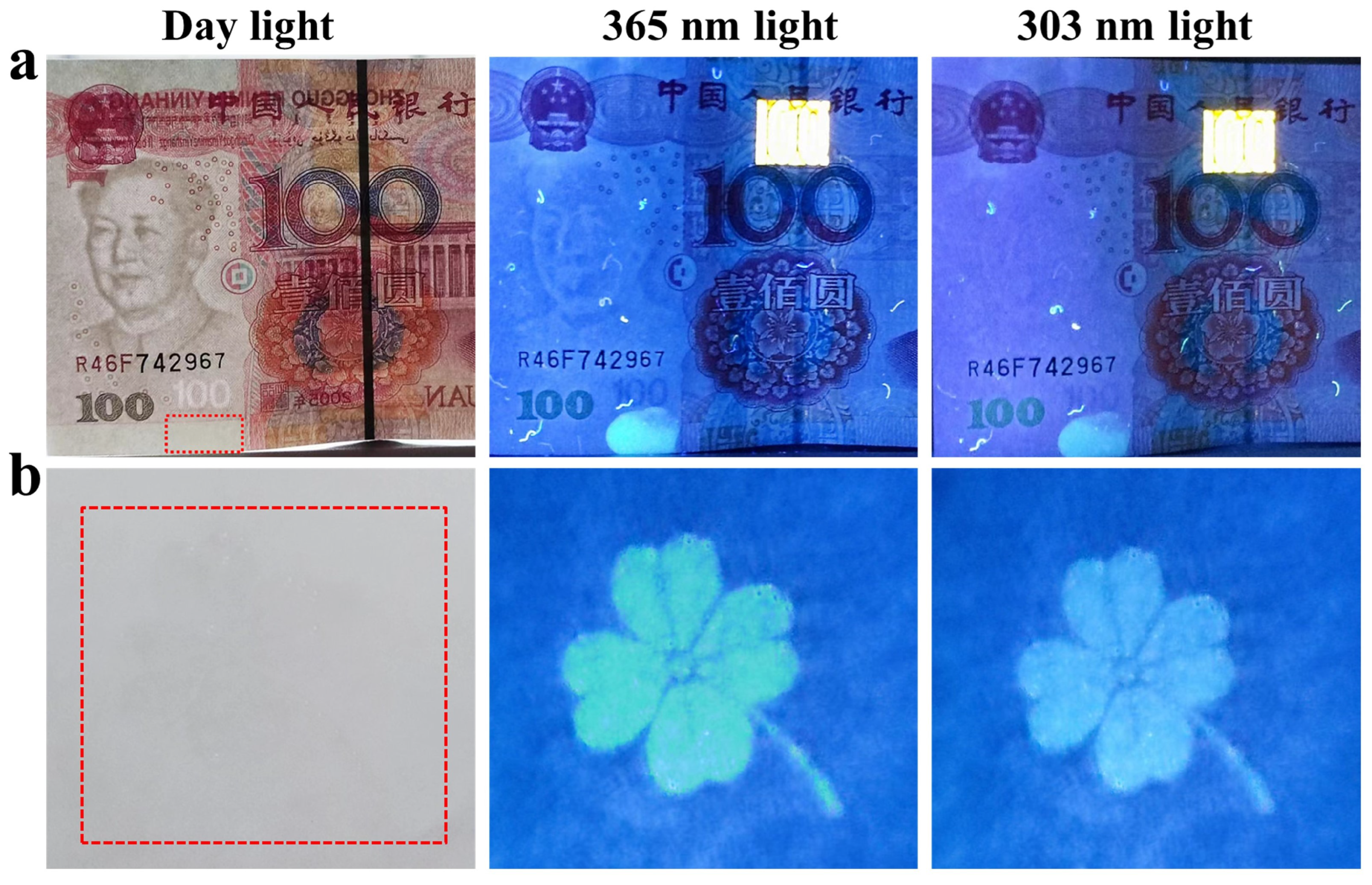
3.5. Anticounterfeiting Application

To further investigate the anti-counterfeiting properties of phosphors, we fabricate PDMS composite films by embedding optimized Eu2+ doped BSAO phosphors in the polydimethylsiloxane (PDMS) polymer matrix. The fabricated luminescent film is well proportioned with flexible performance as presented in Figure 11a. As shown in Figure 11b,c, the BSAO: 0.02Eu2+/PDMS films emit blue and green emissions under 303 nm and 365 nm UV light excitation, respectively. Figure 11d,e show SEM images of the surface and a cross section of the composite film with the letter portion, respectively. It can be seen that the particles are well dispersed in the PDMS substrate. Also, the emission spectra of PDMS film are recorded to confirm that the featured transitions of Eu2+ ions are barely affected by the PDMS film as shown in Figure 12. In addition, the good physical and chemical stability and thermal stability of the phosphor-PDMS film are confirmed [52,53,54]. It means that the resulting luminescent film can meet the requirements of our daily lives and that the performance will not be compromised, even in more extreme environments. Furthermore, fluorescent inks are also prepared for anti-counterfeiting marks and screen-printing applications. As shown in Figure 13a, the ink is coated on the banknote, and the coated area is not significantly different from other parts of the substrate under daylight. However, fluorescence emission can be observed under UV light. Finally, as depicted in Figure 13b, the prepared ink is printed on white paper using screen-printing technology, forming a four-leaf clover pattern. The pattern is almost invisible under visible light but becomes clearly visible under UV light. From the above findings, we suggest that the Eu2+ ion-activated BSAO/PDMS composite films and ink are anticipated for high-level anti-counterfeiting applications.

4. Conclusions

In summary, BSAO: Eu2+ phosphors with the hexagonal phase are successfully synthesized via a simple combustion reaction from XRD patterns and EDS spectra. The optical band gap, concentration quenching mechanism, and optimal doping concentration are confirmed. Meanwhile, the optimal BSAO: 0.02Eu2+ has a blue-green emission band under the excitation of UV lamps due to the 4f-5d transition of Eu2+. In addition, the fluorescence lifetime and the quantum yield are determined as 1010, 111.98 ns, 10.49%, and 10.25%, respectively. Furthermore, an anti-counterfeiting strategy is presented, utilizing phosphors/PDMS films that offer flexibility and exhibit distinct colors when exposed to different UV wavelengths. Finally, by blending the fluorescent powder with ethanol and polyacrylic acid, a transparent ink is prepared, demonstrating the excellent anti-counterfeiting application potential of the fluorescent powder.

Author Contributions

J.W. (Jiao Wu), Q.L. and P.G. designed and carried out the experiment. J.W. (Jiao Wu), J.W. (Jigang Wang), J.H. and W.C. processed the test data. J.Z. provides DFT computational support. With the help of Y.Q., Z.L. and M.C. wrote and completed the paper. All authors have read and agreed to the published version of the manuscript.

Funding

This research is supported by Beijing Natural Science Foundation (No. 2202018), General Project of Beijing Municipal Education Commission Science and Technology Program (No. KM202010015004), Research and development of intelligent packaging for cultural relics (Ed202001), Construction and application transformation of cross media cloud platform for printing and packaging anticounterfeiting and traceability (27170121005), National Natural Science Foundation of China (No. 21604005, Grant No. 52072084), the general project of fundamental research of BIGC (Nos. Ed202208, Eb202001, 20190122041, 22150122042, 22150122034, 27170122012), Initial funding for the Doctoral Program of BIGC (No. 27170121001/002), and the general project of science and technology of Beijing Municipal Education Commission (No. KM202110015008), the Key Area Research and Development Program of Guangdong Province (Grant No. 2020B0101020002), the GBA National Institute for Nanotechnology Innovation (Grant No. 2020GN0106), the National Key R&D Program of China (Grant No. 2021YFC2802000), and National key research and development program (2019YFB1707202).

Institutional Review Board Statement

Not applicable.

Informed Consent Statement

Not applicable.

Data Availability Statement

Research data are not shared.

Conflicts of Interest

The authors declare no conflict of interest.

References

  1. Ren, W.; Lin, G.; Clarke, C.; Zhou, J.; Jin, D. Optical nanomaterials and enabling technologies for high-security-level anticounterfeiting. Adv. Mater. 2020, 32, 1901430. [Google Scholar] [CrossRef]
  2. Liu, F.; Liu, K.; Xie, X.L. Monitoring and limiting deceptive counterfeiting: A two-stage model. J. Oper. Res. Soc. China 2016, 4, 265–308. [Google Scholar] [CrossRef]
  3. Ofori-Parku, S.S.; Park, S.E. I (Don’t) want to consume counterfeit medicines: Exploratory study on the antecedents of consumer attitudes toward counterfeit medicines. BMC Public Health 2022, 22, 1–13. [Google Scholar] [CrossRef]
  4. Conley, T. Defining and understanding economic globalisation. Policy Organ. Soc. 2000, 19, 87–115. [Google Scholar] [CrossRef]
  5. Abdollahi, A.; Roghani-Mamaqani, H.; Razavi, B.; Salami-Kalajahi, M. Photoluminescent and chromic nanomaterials for anticounterfeiting technologies: Recent advances and future challenges. ACS Nano 2020, 14, 14417–14492. [Google Scholar] [CrossRef]
  6. Han, S.; Bae, H.J.; Kim, J.; Shin, S.; Choi, S.E.; Lee, S.H.; Kwon, S.; Park, W. Lithographically encoded polymer microtaggant using high-capacity and error-correctable QR code for anti-counterfeiting of drugs. Adv. Mater. 2012, 24, 5924–5929. [Google Scholar] [CrossRef]
  7. Li, D.; Tang, L.; Wang, J.; Liu, X.; Ying, Y. Multidimensional SERS barcodes on flexible patterned plasmonic metafilm for anticounterfeiting applications. Adv. Opt. Mater. 2016, 4, 1475–1480. [Google Scholar] [CrossRef]
  8. Fan, Y.; Zhang, C.; Gao, Z.; Zhou, W.; Hou, Y.; Zhou, Z.; Yao, J.; Zhao, Y.S. Randomly Induced Phase Transformation in Silk Protein-Based Microlaser Arrays for Anticounterfeiting. Adv. Mater. 2021, 33, 2102586. [Google Scholar] [CrossRef]
  9. Jeon, H.J.; Leem, J.W.; Ji, Y.; Park, S.M.; Park, J.; Kim, K.Y.; Kim, S.W.; Kim, Y.L. Cyber-Physical Watermarking with Inkjet Edible Bioprinting. Adv. Funct. Mater. 2022, 32, 2112479. [Google Scholar] [CrossRef]
  10. Lv, S.; Shanmugavelu, B.; Wang, Y.; Mao, Q.; Zhao, Y.; Yu, Y.; Hao, J.; Zhang, Q.; Qiu, J.; Zhou, S. Transition metal doped smart glass with pressure and temperature sensitive luminescence. Adv. Opt. Mater. 2018, 6, 1800881. [Google Scholar] [CrossRef]
  11. Ding, M.; Dong, B.; Lu, Y.; Yang, X.; Yuan, Y.; Bai, W.; Wu, S.; Ji, Z.; Lu, C.; Zhang, K.; et al. Energy manipulation in lanthanide-doped core–shell nanoparticles for tunable dual-mode luminescence toward advanced anti-counterfeiting. Adv. Mater. 2020, 32, 2002121. [Google Scholar] [CrossRef]
  12. Dorenbos, P. f→d transition energies of divalent lanthanides in inorganic compounds. J. Phys. Condens. Matter 2003, 15, 575. [Google Scholar] [CrossRef]
  13. Liu, Y.; Tu, D.; Zhu, H.; Li, R.; Luo, W.; Chen, X. A strategy to achieve efficient dual-mode luminescence of Eu3+ in lanthanides doped multifunctional NaGdF4 nanocrystals. Adv. Mater. 2010, 22, 3266–3271. [Google Scholar] [CrossRef]
  14. Dorenbos, P. The 4fn ↔ 4fn−15d transitions of the trivalent lanthanides in halogenides and chalcogenides. J. Lumin. 2000, 91, 91–106. [Google Scholar] [CrossRef]
  15. Li, Q.; Yan, B. Multi-component assembly of luminescent rare earth hybrid materials. J. Rare Earths 2019, 37, 113–123. [Google Scholar] [CrossRef]
  16. Bai, X.; Cun, Y.; Xu, Z.; Zi, Y.; Haider, A.A.; Ullah, A.; Khan, I.; Qiu, J.; Song, Z.; Yang, Z. Multiple Anti-Counterfeiting and optical storage of reversible dual-mode luminescence modification in photochromic CaWO4: Yb3+, Er3+, Bi3+ phosphor. Chem. Eng. J. 2022, 429, 132333. [Google Scholar] [CrossRef]
  17. Li, M.; Yao, W.; Liu, J.; Tian, Q.; Liu, L.; Ding, J.; Xue, Q.; Lu, Q.; Wu, W. Facile synthesis and screen printing of dual-mode luminescent NaYF4: Er, Yb (Tm)/carbon dots for anti-counterfeiting applications. J. Mater. Chem. C 2017, 5, 6512–6520. [Google Scholar] [CrossRef]
  18. Zhang, X.; Qin, X.; Zhang, W. NaYF4: Yb, Er with N-GQDs mixture: One-pot hydrothermal synthesis and its luminescent film. Opt. Mater. 2021, 114, 110910. [Google Scholar] [CrossRef]
  19. Pei, P.; Bai, Y.; Su, J.; Yang, Y.; Liu, W. Achieving mechano-upconversion-downshifting-afterglow multimodal luminescence in a lanthanide-doped LaCaAl3O7 phosphor for multidimensional anticounterfeiting. Sci. China Mater. 2022, 65, 2809–2817. [Google Scholar] [CrossRef]
  20. Sohn, K.S.; Timilsina, S.; Singh, S.P.; Lee, J.W.; Kim, J.S. A mechanoluminescent ZnS: Cu/rhodamine/SiO2/PDMS and piezoresistive CNT/PDMS hybrid sensor: Red-light emission and a standardized strain quantification. ACS Appl. Mater. Interfaces 2016, 8, 34777–34783. [Google Scholar] [CrossRef]
  21. Hou, Y.F.; Wang, J.; Wang, X.; Liao, Y.P.; Yang, L.; Cai, E.L.; Wang, S.S. Simultaneous measurement of pressure and temperature in seawater with PDMS sealed microfiber Mach-Zehnder interferometer. J. Light. Technol. 2020, 38, 6412–6421. [Google Scholar] [CrossRef]
  22. Zhao, X.; Ounaies, Z. A facile method to enhance the flexibility and triboelectric output of PDMS using ionic liquid-coated single-wall carbon nanotubes. Nano Energy 2022, 94, 106908. [Google Scholar] [CrossRef]
  23. Zhao, X.; Li, L.; Li, B.; Zhang, J.; Wang, A. Durable superhydrophobic/superoleophilic PDMS sponges and their applications in selective oil absorption and in plugging oil leakages. J. Mater. Chem. A 2014, 2, 18281–18287. [Google Scholar] [CrossRef]
  24. Nakauchi, D.; Okada, G.; Kawaguchi, N.; Yanagida, T. Luminescent and scintillation properties of Eu-doped (Ba,Sr)Al2O4 crystals. Opt. Mater. 2019, 87, 58–62. [Google Scholar] [CrossRef]
  25. Volhard, M.; Yu, L.; den Engelsen, D.; Fern, G.R.; Ireland, T.G.; Silver, J. Crystal structure, photoluminescence and cathodoluminescence of Ba1−xSrxAl2O4 doped with Eu2+. Opt. Mater. Express 2020, 10, 1951–1961. [Google Scholar] [CrossRef]
  26. Rezende, M.V.d.S.; Andrade, A.B.; Valerio, M.E.G.; Montes, P.J.R. The effect of the host composition on the lifetime decay properties of barium/strontium aluminates compounds. J. Appl. Phys. 2014, 115, 103510. [Google Scholar] [CrossRef]
  27. Kresse, G.; Furthmüller, J. Efficient iterative schemes for ab initio total-energy calculations using a plane-wave basis set. Phys. Rev. B 1996, 54, 11169. [Google Scholar] [CrossRef]
  28. Kresse, G.; Joubert, D. From ultrasoft pseudopotentials to the projector augmented-wave method. Phys. Rev. B 1999, 59, 1758. [Google Scholar] [CrossRef]
  29. Perdew, J.P.; Burke, K.; Ernzerhof, M. Generalized gradient approximation made simple. Phys. Rev. Lett. 1996, 77, 3865. [Google Scholar] [CrossRef]
  30. Blöchl, P.E. Projector augmented-wave method. Phys. Rev. B 1994, 50, 17953. [Google Scholar] [CrossRef]
  31. Malkamäki, M.; Bos, A.J.; Dorenbos, P.; Lastusaari, M.; Rodrigues, L.C.; Swart, H.C.; Hölsä, J. Persistent luminescence excitation spectroscopy of BaAl2O4:Eu2+,Dy3+. Phys. B Condens. Matter 2020, 593, 411947. [Google Scholar] [CrossRef]
  32. Fukuda, K.; Fukushima, K. Crystal structure of hexagonal SrAl2O4 at 1073K. J. Solid State Chem. 2005, 178, 2709–2714. [Google Scholar] [CrossRef]
  33. Zheng, B.; Fan, J.; Chen, B.; Qin, X.; Wang, J.; Wang, F.; Deng, R.; Liu, X. Rare-earth doping in nanostructured inorganic materials. Chem. Rev. 2022, 122, 5519–5603. [Google Scholar] [CrossRef] [PubMed]
  34. Wang, Y.; Seto, T.; Ishigaki, K.; Uwatoko, Y.; Xiao, G.; Zou, B.; Li, G.; Tang, Z.; Li, Z.; Wang, Y. Pressure-Driven Eu2+-Doped BaLi2Al2Si2N6: A New Color Tunable Narrow-Band Emission Phosphor for Spectroscopy and Pressure Sensor Applications. Adv. Funct. Mater. 2020, 30, 2001384. [Google Scholar] [CrossRef]
  35. Shannon, R.D. Revised effective ionic radii and systematic studies of interatomic distances in halides and chalcogenides. Acta Crystallogr. Sect. A Cryst. Phys. Diffr. Theor. Gen. Crystallogr. 1976, 32, 751–767. [Google Scholar] [CrossRef]
  36. Wang, H.; Li, Z.; Kang, R.; Ji, R.; Wang, Y. Sr2BN2Cl: Eu2+-Based Narrow-Band Blue-Emitting Phosphor: A Potential Color Converter for Illumination and Displays. Inorg. Chem. 2022, 61, 18245–18252. [Google Scholar] [CrossRef]
  37. Li, J.; Liu, J.; Ni, Q.; Zhu, Q.; Zeng, Z.; Huo, J.; Long, C.; Wang, Q. Key Role Effect of Samarium in Realizing Zero Thermal Quenching and Achieving a Moisture-Resistant Reddish-Orange Emission in Ba3LaNb3O12: Sm3+. Inorg. Chem. 2022, 61, 17883–17892. [Google Scholar] [CrossRef] [PubMed]
  38. Peng, T.; Yang, H.; Pu, X.; Hu, B.; Jiang, Z.; Yan, C. Combustion synthesis and photoluminescence of SrAl2O4:Eu,Dy phosphor nanoparticles. Mater. Lett. 2004, 58, 352–356. [Google Scholar] [CrossRef]
  39. Gedekar, K.; Wankhede, S.; Moharil, S.; Belekar, R. d–f luminescence of Ce3+ and Eu2+ ions in BaAl2O4, SrAl2O4 and CaAl2O4 phosphors. J. Adv. Ceram. 2017, 6, 341–350. [Google Scholar] [CrossRef]
  40. Tauc, J. Optical properties and electronic structure of amorphous Ge and Si. Mater. Res. Bull. 1968, 3, 37–46. [Google Scholar] [CrossRef]
  41. Inaba, K.; Suzuki, S.; Noguchi, Y.; Miyayama, M.; Toda, K.; Sato, M. Metastable Sr0.5TaO3 perovskite oxides prepared by nanosheet processing. Eur. J. Inorg. Chem. 2008, 2008, 5471–5475. [Google Scholar] [CrossRef]
  42. Sangiorgi, N.; Aversa, L.; Tatti, R.; Verucchi, R.; Sanson, A. Spectrophotometric method for optical band gap and electronic transitions determination of semiconductor materials. Opt. Mater. 2017, 64, 18–25. [Google Scholar] [CrossRef]
  43. Dorenbos, P. Energy of the first 4f7→4f65d transition of Eu2+ in inorganic compounds. J. Lumin. 2003, 104, 239–260. [Google Scholar] [CrossRef]
  44. Van Uitert, L. Characterization of energy transfer interactions between rare earth ions. J. Electrochem. Soc. 1967, 114, 1048. [Google Scholar] [CrossRef]
  45. Zhao, K.; Yin, L.; Ma, Z.; Yang, T.; Tang, H.; Cao, P.; Huang, S. Investigation of the solid-solution limit, crystal structure, and thermal quenching mitigation of Sr-substituted Rb2CaP2O7: Eu2+ phosphors for white LED applications. Inorg. Chem. 2022, 61, 1627–1635. [Google Scholar] [CrossRef]
  46. Zhu, H.; Huang, X.; Li, Y.n.; She, Y.l.; Wang, J.; Wong, W.Y.; Liu, M.; Li, W.; Zhou, Z.; Xia, M. Novel ultra-high-temperature zero-thermal quenching plant-protecting type blue-green dual-emission KAl11O17: Eu2+, Mn2+ phosphors for urban ecological lighting. J. Mater. Chem. C 2022, 10, 3461–3471. [Google Scholar] [CrossRef]
  47. Pei, P.; Liu, K.; Ju, Z.; Wei, R.; Liu, W. Achieving mechano-upconversion-downshifting-afterglow multimodal luminescence in Pr3+/Er3+ coactivated Ba2Ga2GeO7 for multidimensional anticounterfeiting. J. Mater. Chem. C 2022, 10, 5240–5248. [Google Scholar] [CrossRef]
  48. Zhang, Y.; Qiao, X.; Wan, J.; Wu, L.A.; Chen, B.; Fan, X. Facile synthesis of monodisperse YAG: Ce3+ microspheres with high quantum yield via an epoxide-driven sol–gel route. J. Mater. Chem. C 2017, 5, 8952–8957. [Google Scholar] [CrossRef]
  49. Cheng, X.; Xie, Z.; Zheng, W.; Li, R.; Deng, Z.; Tu, D.; Shang, X.; Xu, J.; Gong, Z.; Li, X.; et al. Boosting the Self-Trapped Exciton Emission in Alloyed Cs2(Ag/Na)InCl6 Double Perovskite via Cu+ Doping. Adv. Sci. 2022, 9, 2103724. [Google Scholar] [CrossRef]
  50. Locardi, F.; Sartori, E.; Buha, J.; Zito, J.; Prato, M.; Pinchetti, V.; Zaffalon, M.L.; Ferretti, M.; Brovelli, S.; Infante, I.; et al. Emissive Bi-Doped Double Perovskite Cs2Ag1−xNaxInCl6 Nanocrystals. ACS Energy Lett. 2019, 4, 1976–1982. [Google Scholar] [CrossRef]
  51. Wang, X.; Xu, J.; Yu, J.; Bu, Y.; Marques-Hueso, J.; Yan, X. Morphology control, spectrum modification and extended optical applications of rare earth ion doped phosphors. Phys. Chem. Chem. Phys. 2020, 22, 15120–15162. [Google Scholar] [CrossRef] [PubMed]
  52. Hua, Y.; Ran, W.; Yu, J.S. Advantageous occupation of europium (III) in the B site of double-perovskite Ca2BB′O6 (B = Y, Gd, La; B′ = Sb, Nb) frameworks for white-light-emitting diodes. ACS Sustain. Chem. Eng. 2021, 9, 7960–7972. [Google Scholar] [CrossRef]
  53. Patnam, H.; Hussain, S.K.; Yu, J.S. Rare-earth-free Mn4+ ions activated Ba2YSbO6 phosphors for solid-state lighting, flexible display, and anti-counterfeiting applications. Ceram. Int. 2023, 49, 2967–2977. [Google Scholar] [CrossRef]
  54. Hua, Y.; Seo, Y.U.; Yun Kim, S.; Kim, H.J.; Su Yu, J. Rare-earth-free Sr2YSb1−xO6:xMn4+: Synthesis, structure, luminescence behavior, thermal stability, and applications. Chem. Eng. J. 2021, 412, 128633. [Google Scholar] [CrossRef]
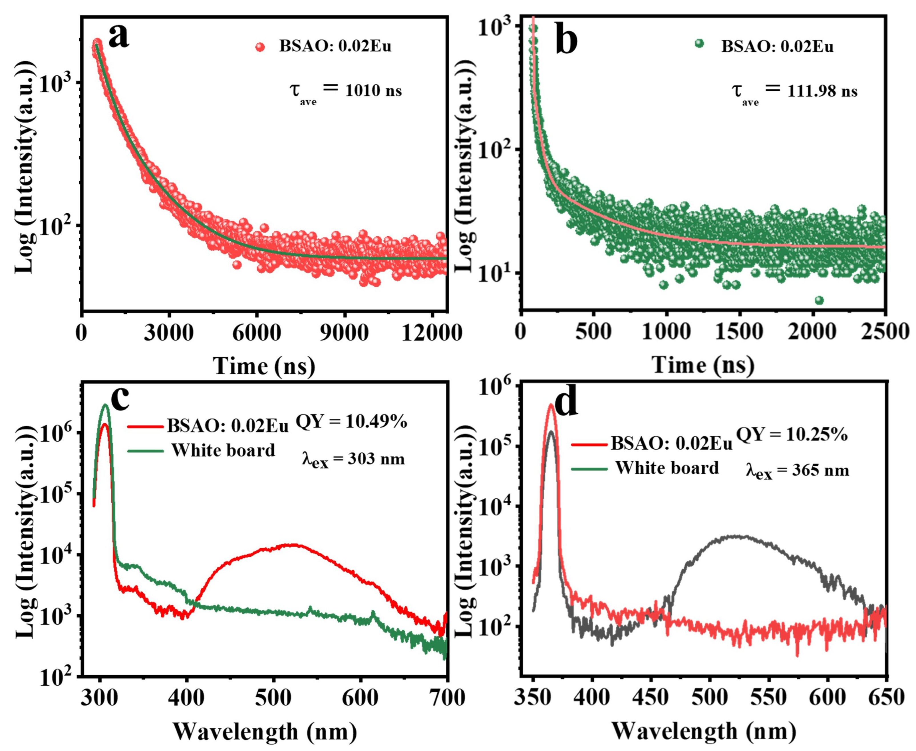
Figure 1. (a) XRD patterns of BSAO doped with different concentrations of Eu and SrAl2O4 standard cards; (b) the diffraction peak at (211) of BSAO: Eu2+; (c) XRD patterns of BSAO: 0.02Eu2+ at different reaction temperatures (500, 600, 700, 800, 900 °C).
Coatings 13 01247 g001
Figure 2. SEM images of BSAO: 0.02Eu2+ synthesized at (ae) 500 °C–900 °C; (f) the EDS of BSAO: 0.02Eu2+ particles synthesized at 700 °C.
Coatings 13 01247 g002
Figure 3. (a) UV-vis absorption spectra; (b) intermediate optical band gap ( E g ) of BSAO: 0.02Eu2+; (c) graphical elaboration of Equation (2); (d) Tauc plot of absorption measurements of BSAO: 0.02Eu2+.
Coatings 13 01247 g003
Figure 4. (a) PLE spectra of BSAO: 0.02Eu2+; PL spectra of BSAO doped with different Eu ion concentrations (x = 0.01, 0.02, 0.03, 0.04, 0.06) under (b) 303 nm, and (c) 365 nm; (d) Concentration dependence of the PL intensity.
Coatings 13 01247 g004
Figure 5. Linear fitting of log(x) versus log (I/x) for spectra of BSAO: 0.02Eu2+ phosphors. (a λ ex = 303 nm and (b λ ex = 365 nm.
Coatings 13 01247 g005
Figure 6. PL spectra of BSAO: 0.02Eu2+ synthesized at different reaction temperatures (500–900 °C) under the excitation of (a) 303 nm, (b) 365 nm; (c) PL schematic diagram of Eu2+ ions in the BSAO host; (d) CIE (1931) chromaticity coordinates diagram for BSAO: 0.02Eu2+ under the excitation of 303 and 365 nm ultraviolet light.
Coatings 13 01247 g006
Figure 7. The luminescence decay curves of the BSAO: 0.02Eu2+ ((a λ ex = 303 nm, (b λ ex = 365 nm) phosphor; (c,d) are the quantum yields of the BSAO: 0.02Eu2+.
Coatings 13 01247 g007
Figure 8. (a,b) Band structure of BSAO; (c) total DOS and partial DOS of BSAO. (d) UV-vis absorption spectra of BSAO host. Inset: The experimental Eg of BSAO host.
Coatings 13 01247 g008
Figure 9. XRD patterns of BSAO: 0.02Eu2+ with different surfactants (PEG, PVA, EA).
Coatings 13 01247 g009
Figure 10. (a) SEM image of BSAO: 0.02Eu2+ without surfactant; SEM images of (b) PVA, (c) EA, and (d) PEG were added, respectively.
Coatings 13 01247 g010
Figure 11. BSAO: 0.2Eu2+/PDMS film under (a) daylight, (b) 303 nm light, and (c) 365 nm light; surface (d) and cross section (e) SEM images of the BSAO: 0.02Eu2+/PDMS film.
Coatings 13 01247 g011
Figure 12. PL emission spectra of the BSAO: 0.02Eu2+/PDMS film under (a) 303 nm and (b) 365 nm light irradiations.
Coatings 13 01247 g012
Figure 13. (a) The anti-counterfeiting markings applied on banknotes; (b) the four-leaf clover pattern printed through screen printing.
Coatings 13 01247 g013
Table 1. Parameters of fluorescence lifetime decay curves of BSAO: 0.02Eu2+ and PLQY.
ExcitationPLQYDecay Lifetimes (ns)
τ 1 τ 2 τ 3 τ avg
303 nm10.5% ± 0.01%407.711330.3null1010 ± 0.03
365 nm10.3% ± 0.05%36.94368.61.4112 ± 0.02
Disclaimer/Publisher’s Note: The statements, opinions and data contained in all publications are solely those of the individual author(s) and contributor(s) and not of MDPI and/or the editor(s). MDPI and/or the editor(s) disclaim responsibility for any injury to people or property resulting from any ideas, methods, instructions or products referred to in the content.

