Article

The 2′,4′-Dichloro-chalcone Inhibits the In Vitro Growth and Pathogenicity of Fusarium tricinctum and Trichothecium roseum by Activating Cyanide-Resistant Respiration

1
College of Food Science and Engineering, Gansu Agricultural University, Lanzhou 730070, China
2
Lanzhou Industrial Research Institute, Lanzhou 730000, China
3
Institute of Potato, Gansu Academy of Agricultural Sciences, Lanzhou 730070, China
*
Author to whom correspondence should be addressed.
Coatings 2023, 13(10), 1789; https://doi.org/10.3390/coatings13101789
Submission received: 19 September 2023 / Revised: 13 October 2023 / Accepted: 17 October 2023 / Published: 18 October 2023
(This article belongs to the Special Issue Bio-Based and Bio-Inspired Polymers and Composites)

Abstract

:
Chalcones are a class of flavonoids possessing antimicrobial properties and have potential for use as coatings of plant products for the control of postharvest diseases. The effects of 2′,4′-dichloro-chalcone on the in vitro growth and in vivo pathogenicity of Fusarium tricinctum and Trichothecium roseum were investigated. First, 1 µM of 2′,4′-dichloro-chalcone strongly inhibited the mycelial growth and conidial production of F. tricinctum (32.3%) and T. roseum (65.2%) in vitro. Meanwhile, the cell membrane permeability was increased by 25% and 22.5% and the accumulation of reactive oxygen species was increased by 41.7 and 65.4%, respectively, of F. tricinctum and T. roseum. This treatment also significantly inhibited the total respiration rate and activated the cyanide-resistant respiratory pathway in both pathogens. The expression level of AOX was enhanced in F. tricinctum and T. roseum by 52.76 and 39.13%, respectively. This treatment also significantly inhibited the expansion of potato dry rot from F. tricinctum (48.6%) and apple rot spot from T. roseum (36.2%). Therefore, 2′,4′-dichloro-chalcone has potential use as an alternative safety method in the control of postharvest diseases by F. tricinctum and T. roseum in agricultural practices.

1. Introduction

Fusarium tricinctum and Trichothecium roseum are important fungal pathogens of various plant diseases worldwide. F. tricinctum infests many cereal crops, vegetables, and traditional Chinese medicinal plants including rice, wheat, barley, maize, potato, lily and licorice [1,2,3,4,5,6]. T. roseum infests a wide range of fruits and vegetables such as apples, peaches, melons and tomatoes [7,8,9,10], causing severe preharvest and postharvest rots. Infection by F. tricinctum and T. roseum not only leads to a reduction in product quality, but also to increased food safety risks through their production of toxic and carcinogenic mycotoxins [11]. Currently, chemical fungicides are commonly used in the postharvest control of these two pathogens, which can have negative impacts on the development of fungal resistance and environmental pollution [12]. Therefore, there is a need for the development of alternative and environmentally safe means for postharvest disease prevention during the storage of important crop products.
Chalcones are a natural class of open-chain flavonoids that are widely found in edible or medicinal plants [13]. The presence of active α, β-unsaturated carbonyl functional groups (-CO-CH=CH-) in their molecular structure and the delocalized active π-electrons in the aryl ring provide for a variety of biological activities such as antioxidant, antitumor and anti-inflammatory effects [14]. Several studies have found that chalcones have a significant inhibitory activity against fungi and bacteria [15]. The phenolic groups in chalcones show high affinities for some microbial proteins and can therefore selectively inhibit microbial growth and development [16,17]. López et al. (2001) found that chalcone derivatives inhibited the activity of enzymes involved in yeast cell wall synthesis and significantly inhibited yeast growth [18]. López et al. (2020) found that chalcones had a strong inhibitory effect on root rot and fruit blight caused by Oomycetes [19]. Tsukiyama et al. (2002) isolated licorice chalcone A from a licorice root and showed its significant inhibitory effect on the growth of Bacillus subtilis [6]. Natural chalcones and chalcone derivatives have emerged as preferred biomolecules that are safe and efficient alternatives to chemical fungicides. The antimicrobial effects of natural chalcones have motivated the study of chalcone structures and the synthesis of new chalcone derivatives for the enhancement of their antibacterial effects [20]. Wang et al. (2023) found that chalcones containing the triazole structure were all inhibitory to bacterial growth, particularly against Staphylococcus aureus [21]. Génesis et al. (2020) found that three allyl-structured chalcones had a strong inhibitory effect on the growth of the fungal pathogen, Mycobacterium avium [19]. Liu et al. (2017) synthesized halogenated chalcone aminothiourea Schiff bases with good inhibitory activities against tyrosinase and fungal growth [22]. Meaningfully, Chalcone compounds such as 3-Hydroxy-4-methoxychalcone have been used to coat plastics, resulting in a significant decrease in bacterial adhesion and biofilm formation [23]. Aminoethyl phloretin, a soluble chalcone, was shown to reduce growth of the food borne pathogens, Listeria monocytogenes and Staphylococcus aureus when incorporated into packaging [24]. Some studies have reported that chalcone derivatives with halogen substituents were highly fungicidal [25,26]. However, the inhibitory effects of chlorinated chalcones on pathogenic fungi have been less characterized. The objective of this study was to assess the effects of 2′,4′-dichloro-chalcone on the pathogenicities of F. tricinctum and T. roseum during the postharvest storage of potatoes and apples, respectively, and to provide mechanistic insight into its antifungal action. The results provide a meaningful theoretical basis for chalcone bioactive molecules to be used as film preservation materials.

2. Materials and Methods

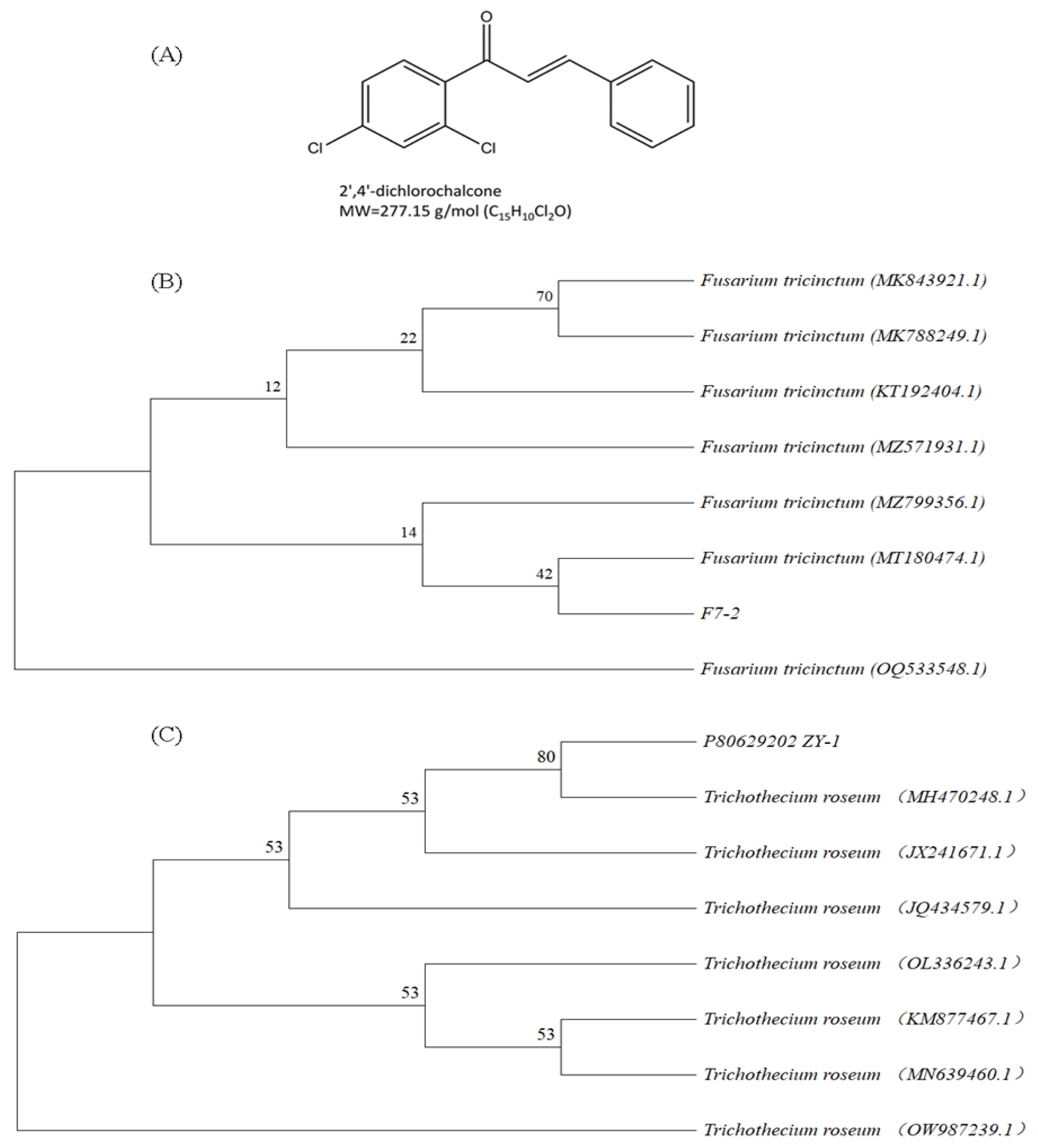
2.1. Biological Materials and Test Reagents

The F. tricinctum and T. roseum were isolated from potato tubers and apple fruits with typical symptoms of dry rot and mold heart disease, respectively, by the post-harvest laboratory of the College of Food Science and Engineering, Gansu Agricultural University. The confirmation of the fungal species utilized their ITS DNA sequences. Isolated fungal DNA was extracted with a fungal genomic DNA extraction Kit (Solarbio, Beijing, China) and PCR amplification of their ITS regions was carried out using the ITS gene-rot symptoms and from apples with typical specific primers ITS1 5′-TCCGTAGGTGAACCTGCGG-3′, and ITS4 5′-TCCTCCGCTTATTGATATATGC-3′ with reference to the method of Schoch et al. (2012) [27]. The sequencing results were queried against the National Center for Biotechnology Information (NCBI) database using BlastP [28], and sequence similarities of >99% were used to choose the reference sequences. The phylogenetic analysis was conducted using MEGA 11.0 software (https://www.megasoftware.net, accessed on 22 July 2023) using ClustalW for sequence alignment and the neighbor joining (NJ) method for sequence clustering. The phylogenetic tree of F. tricinctum and T. roseum isolates are shown in Figure 1B,C. The identified pathogens were maintained in potato dextrose agar (PDA) medium for future use.
Minitubers of potatoes (Solanum tuberosum L. cv. Atlantic) were purchased from Gansu Yihang Potato Industry Co., Ltd. (Dingxi, China) in September 2022 and used to obtain mature tubers in a virus-free, mini potato planting greenhouse. Apple fruits tested (Malus Mill cv. Fuji) were purchased from the local market. The potatoes and apples were dried for 4 h, then refrigerated at 4 ± 1 °C with a relative humidity of (80 ± 5)% to test for the development of F. tricinctum or T. roseum, respectively.
The 2′,4′-dichloro-chalcone (CAS No. 19672-60-7; molecular structure is shown in Figure 1A) and was purchased from Shanghai Yuanye Biological Company (Shanghai, China). Potassium cyanide (analytically pure) was obtained from Professor Shijun Bao (College of Animal Medicine, Gansu Agricultural University).

2.2. Methods

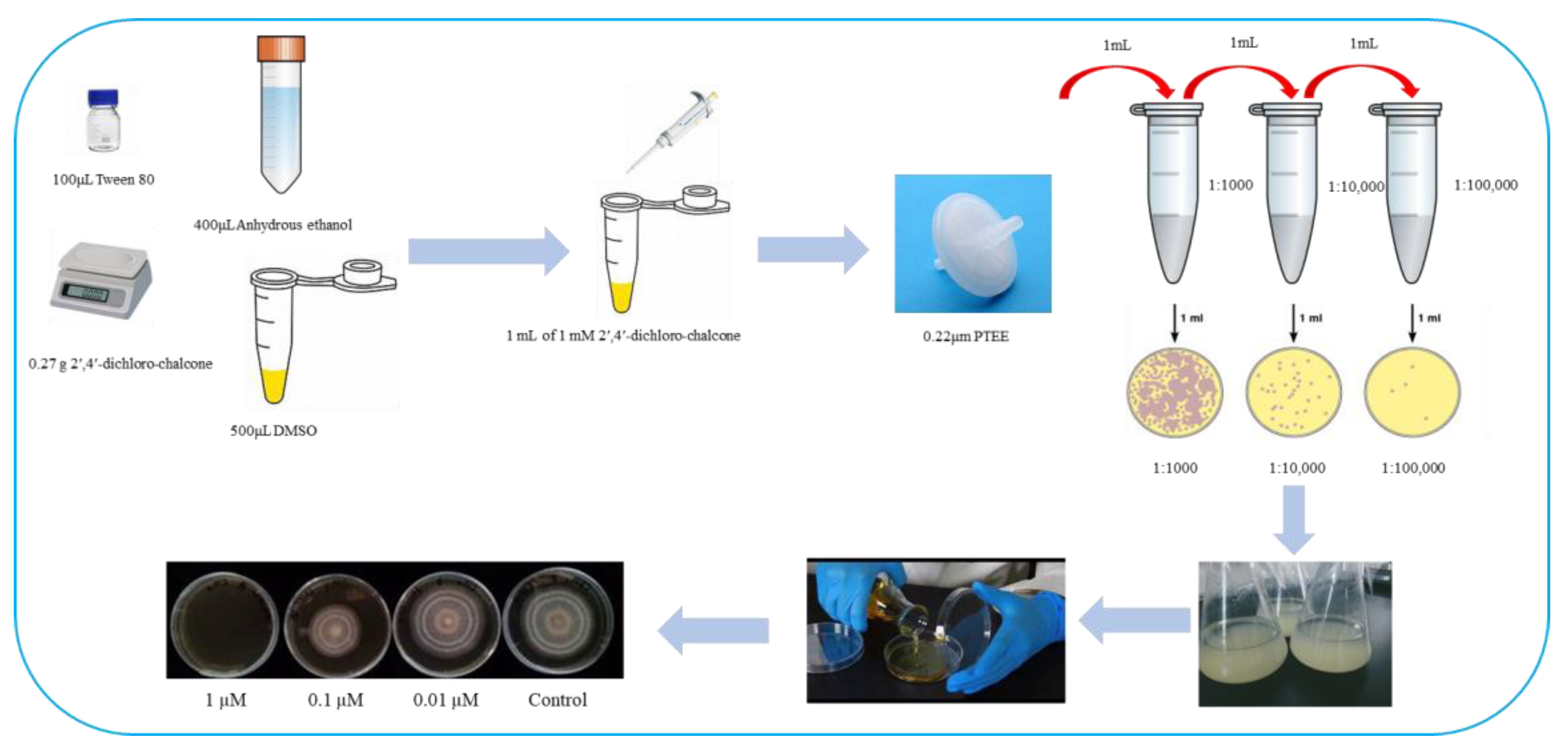
2.2.1. Preparation of 2′,4′-Dichloro-chalcone Stock Solution

A stock solution of 1 mM 2′,4′-dichloro-chalcone, was prepared by dissolving 0.27 g in 500 μL of DMSO, 100 μL Tween 80 and 400 μL of anhydrous ethanol (Figure 2). After filtration with 0.22µm Polytetrafluoroethylene, chalcone solution of 0.01 µM, 0.1 µM and 1 µM were prepared by multiple dilution method (1:100,000; 1:10,000; 1:1000). The same solution without 2′,4′-dichloro-chalcone was prepared for use as a negative control. Both solutions were stored at 4 °C and used within 7 d.

2.2.2. Preparation of Spore Suspension of F. tricinctum and T. roseum

F. tricinctum and T. roseum were cultured on a PDA medium for 7 d at 25 °C and their conidia collected by agitation in 10 mL sterile water for 15 s. The conidial suspension was filtered through two layers of sterile gauze, then counted using a hemocytometer and adjusted to 1 × 10⁶ CFU/mL in sterile water for subsequent experiments.

2.2.3. Determination of the Effects of 2′,4′-Dichloro-chalcone on F. tricinctum and T. roseum Mycelial Growth and Sporulation

Overall 5 μL of 1 × 10⁶ CFU/mL spore suspension was inoculated onto solid PDA plates containing 0, 0.01, 0.1 or 1 µM 2′,4′-dichloro-chalcone and incubated at 28 °C. The average diameter (n = 3) of each colony was measured every 24 h and the growth inhibition (%) of mycelial growth relative to the control was calculated according to the following formula:
Mycelial   growth   inhibition   % = Colony   diameter control Colony   diameter Treated Colony   diameter   Control
The spore count from plates was determined with a hemocytometer as in Section 2.2.2 after 7 d of culture (n = 5). The relative inhibition of sporulation was calculated from the following formula:
Inhibition   of   spore   production   % = Number   of   spores control Number   of   spores Treated Number   of   spores control

2.2.4. The Effects of 2′,4′-Dichloro-chalcone on Hyphal Cell Membrane Permeability

A 1 cm diameter sterile punch was used to collect three mycelial samples (about 1 g of mycelium per sample) that were rinsed in water, incubated in 10 mL deionized water for 0, 30, 60, 90 or 120 min, then centrifuged to collect the supernatant for conductivity measurements with a DS-307A conductivity meter (Shanghai Youke Instrument Co., Ltd., Shanghai, China). The cell membrane permeability was calculated according to the following formula:
Cell   membrane   permeability   % = Conductivity   before   water   bath Final   conductivity × 100

2.2.5. Kinetics of Oxygen Consumption Rates in Total and Cyanide Resistant Respiration in F. tricinctum and T. roseum

The oxygen consumption rates of total and cyanide-resistant respiration were measured essentially according to Xu et al. (2021) [29]. Overall, 20 μL of 106 CFU/mL spore suspensions was added into 30 mL potato dextrose broth (PDB) and cultured in an orbital shaker at 28 °C and 160 rpm (QYC-2102C, Shanghai Shengke Instrument Equipment Co., Ltd., Shanghai, China) at 28 °C. After 24 h, 2′,4′-dichloro-chalcone was added to either 0.01, 0.1 or 1 µM and incubated further. Additionally, 2 mL of the culture solution was taken after a further 4 and 8 h for the measurement of oxygen consumption rates with a Clark oxygen electrode (Oxygraph+, Hansha Scientific Instrument Co., Ltd., Norfolk, UK) at a constant temperature of 25 °C and expressed as nmol(O2)/mL/min. The total oxygen consumption rates without inhibitors (RIt) were determined. Then, 1 mM KCN was added to determine the contribution from the cytochrome respiration pathway (RIcyt). Next, 1 mM KCN and 1 mM salicylhydroxamic acid were added to measure the residual oxygen consumption rate (RIres). The rate of cyanide-resistant respiration (RIalt) was subsequently determined from RIcyt–Rires [30].

2.2.6. Protein Immunoblotting Analysis of AOX Expression in F. tricinctum and T. roseum

To facilitate the collection of mycelia, 2 μL of 1 × 10⁶ CFU/mL spore suspension was placed on cellophane in contact with PDA medium and incubated at 25 °C for 3–4 days. Protein extracts of the mycelium were prepared by incubating the collected mycelia in 50 mM Tris.HCl (pH 6.8) containing 0.1% β-mercaptoethanol and 2% (w/v) SDS at 40 °C for 30 min and clarified by centrifugation at 10,000× g for 5 min. Proteins were separated by 12% SDS-PAGE and transferred to nitrocellulose membranes (0.22 mm pore size) using the procedure described by Affourtit [31]. Filters were washed in TBST and incubated for 1 h at room temperature with 1:1000 anti-AOX and anti-actin monoclonal antibodies (Agrisera, Vännäs, Sweden). Following a wash in TBST, filters were incubated with HRP-labeled secondary antibody (Proteintech, Wuhan Sanying, Wuhan, China) and the immunoblots developed with ECLTM Western Blotting Detection Reagents GE Healthcare (Sigma-Aldrich, St. Louis, MO, USA). The intensity of the detected bands was determined from their grey scale values AOX using Image J software (https://imagej.nih.gov/ij/, accessed on 1 August 2023) followed by normalization of AOX intensity with respect to that of the actin reference.

2.2.7. Measurement of ROS Production in F. tricinctum and T. roseum

The production of ROS was detected based on the method of Xin et al. (2021) [32]. Briefly, 1 × 10⁶ CFUs were introduced into 1 mL PDB containing either 0, 0.01, 0.1, or 1 µM 2′,4′-dichloro-chalcone. After 16 h of incubation, the fungal cultures were washed three times in PBS (pH 7) by centrifugation at 5000× g for 10 min, then resuspended in 30 µL of 30 µg/mL of dichloro-dihydrofluorescein diacetate (DCFH-DA) and incubated for 15 min at 28 °C. The hypha were then washed twice by centrifugation, resuspended in 1 mL PBS (pH 7) for 1 h for signal development and the ROS levels determined by fluorescence spectrophotometry (U-LH100-3, Shanghai Yongke Optical Instrument Co., Ltd., Shanghai, China) with 488 nm excitation and 500–600 nm emission filters.

2.2.8. Determination of F. tricinctum and T. roseum Pathogenicities In Vivo

The pathogenicity of F. tricinctum was determined in potato, while that of T. roseum was determined in apple using the methods established by Zhu et al. (2023) [33] and Ge et al. (2015) [34], respectively. Briefly, tubers and fruits of uniform size and no mechanical injuries or symptoms of disease were selected for testing. The selected samples of each were surface sterilized with 2% sodium hypochlorite solution for 2 min, rinsed in running water then dried naturally. The halved potato and intact apple samples were immersed in sterile water containing 0, 0.01, 0.1 or 1 µM 2′,4′-dichloro-chalcone solution for 10 min, then air-dried for 4 h before inoculation with 20 μL of F. tricinctum or T. roseum spore suspension (1 × 10⁶ CFU/mL), respectively, and incubation in sterile boxes (RH = 70%) at room temperature in the dark. The expansion of the mycelial diameter (disease progression) was monitored and the symptoms of disease were photographed from 2 to 20 d on a daily basis. The inhibition rate of the disease diameter was calculated according to the following formula:
Inhibition   of   disease   spot   growth   % = Diameter   of   spots   control Diameter   of   spots   treated Diameter   of   spots   control

2.3. Statistical Analysis

All experiments were repeated three times, and all data were calculated as mean and standard error (±SE). ANOVA tests were used to test statistical significance, where p < 0.05 was considered as significant.

3. Results

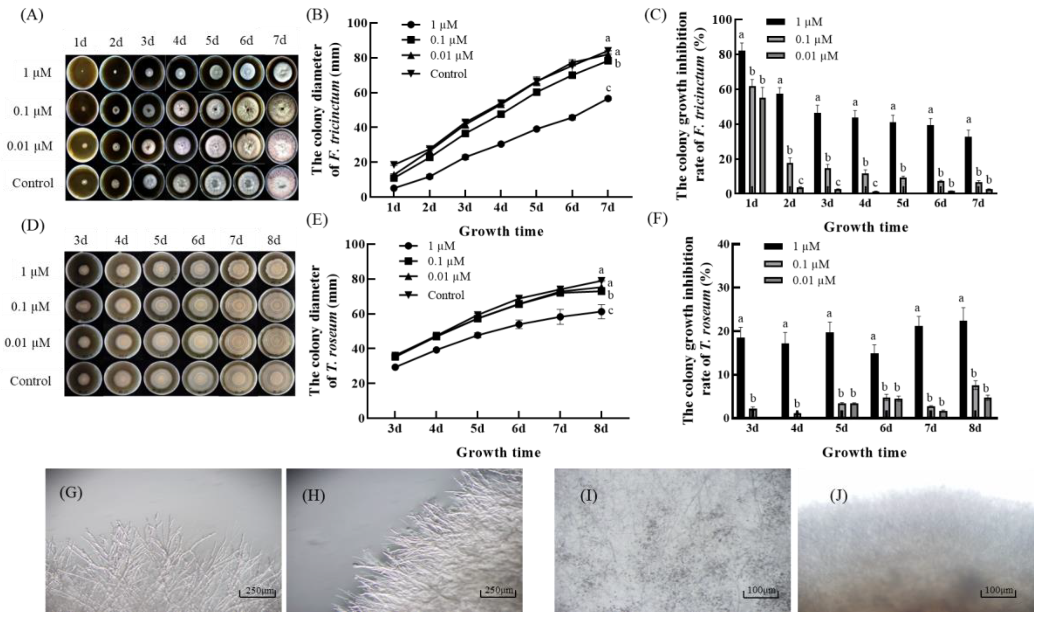
3.1. Effect of 2′,4′-Dichloro-chalcone on Mycelial Growth of F. tricinctum and T. roseum

As shown in Figure 3, 2′,4′-dichloro-chalcone significantly inhibited the mycelial growth of both pathogens on the PDA medium (A, D). The effects on mycelial growth were low (2.5–7.2%) and insignificant at 0.01 and 0.1 µM, but clearly significant at 1 µM, inhibiting F. tricinctum by 32.3% after 7 d and T. roseum by 22.8% after 8d (p < 0.05) (B, C and E, F, respectively).
The hyphae of F. tricinctum cultivated on control PDA plates were slender and relatively highly branched, indicating a strong growth ability (G), while the mycelium grown under 1 µM 2′,4′-dichloro-chalcone (H) consisted of thinner and twisted hyphae with relatively fewer branches, which were restricted to the hyphal apices, indicating a poorer growth ability. Under control conditions, T. roseum presented uniformly thin hyphae with a smooth, slender surface and a pointed apical tip (I). However, in the presence of 1 µM 2′,4′-dichloro-chalcone, the hyphae produced showed increased branching, but were of uneven thickness and showed blunt or enlarged apices (J). The results indicated that 2′,4′-dichloro-chalcone inhibited mycelium growth and colony formation in F. tricinctum and T. roseum with substantial effects on the hypha growth and morphology.

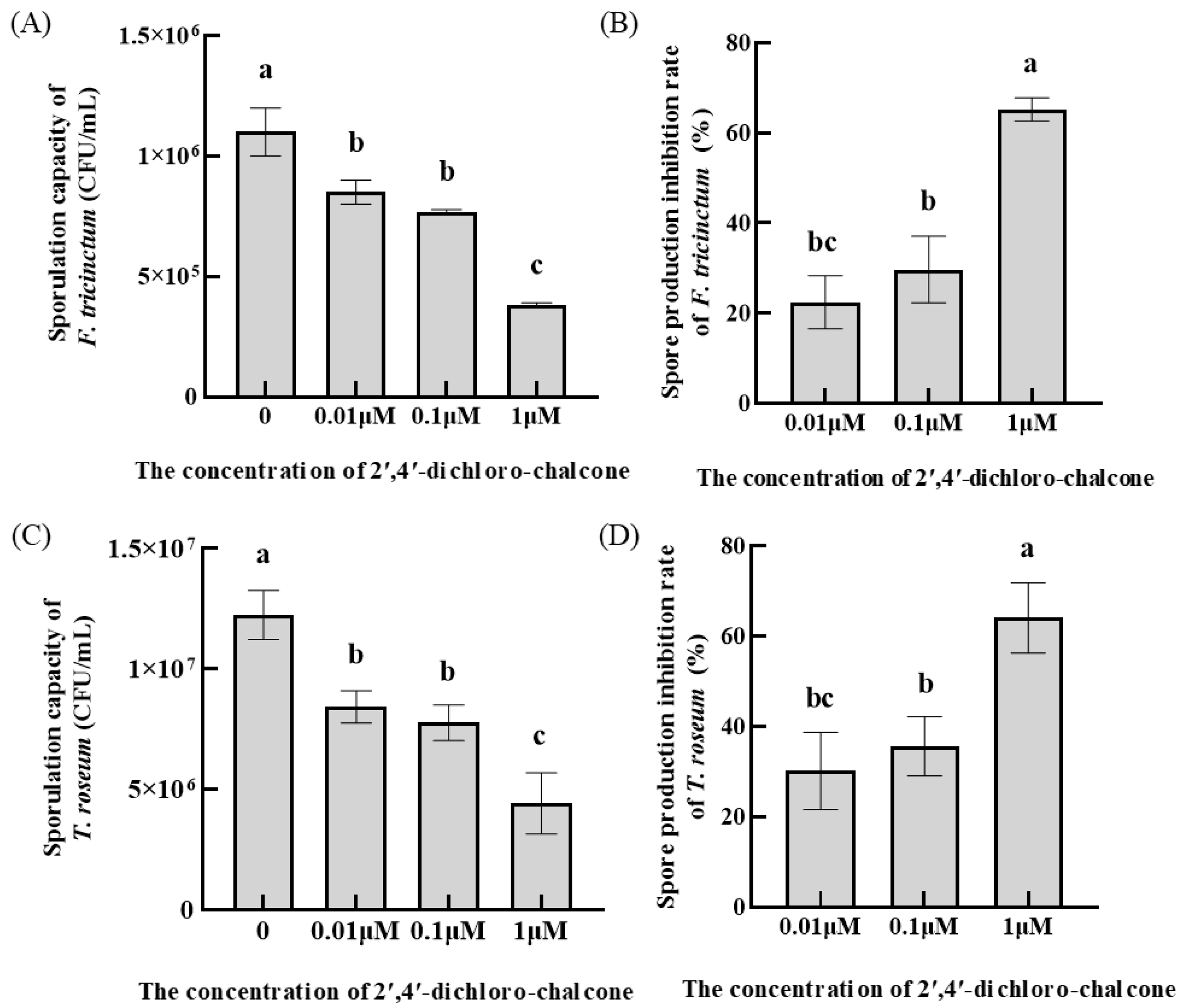
3.2. Effect of 2′,4′-Dichloro-chalcone on Spore Production in F. tricinctum and T. roseum

Significant effects (p < 0.05) of 2′,4′-dichloro-chalcone on sporulation were observed at 1 µM for both pathogens, resulting in a 64–65% decrease in conidial production from both pathogens (Figure 4). The lower concentrations of 2′,4′-dichloro-chalcone tested (0.01 or 0.1 µM) showed smaller (22–36%) and less significant effects.

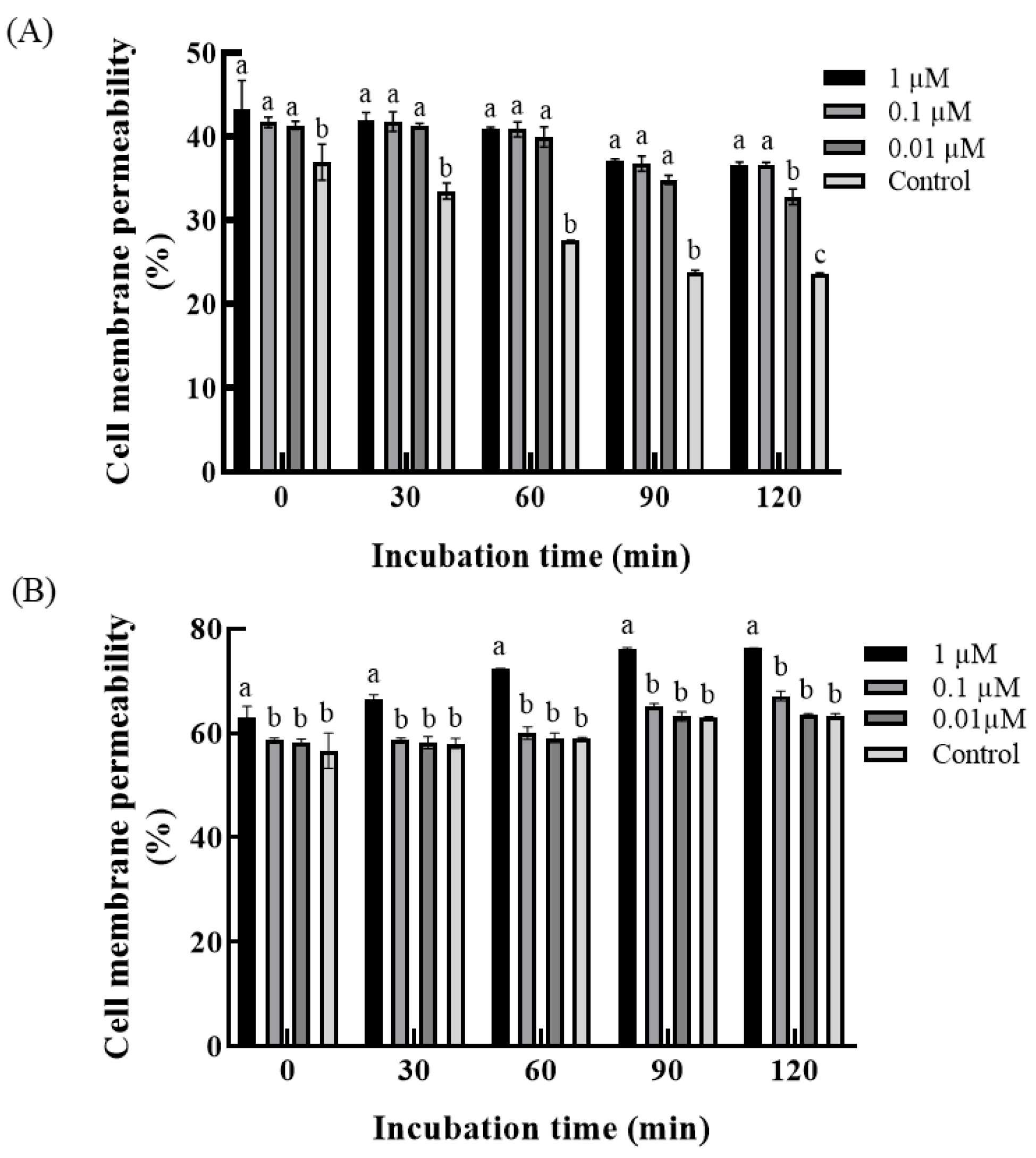
3.3. Effect of 2′,4′-Dichloro-chalcone on Membrane Permeability of F. tricinctum and T. roseum

The membrane permeability of intact hyphae can be measured through their effect on the conductivity of incubating solutions [35]. Figure 5 shows that 2′,4′-dichloro-chalcone at 1 µM significantly enhanced the membrane permeability (p < 0.05) in the two pathogens, while lower concentrations produced smaller and insignificant effects.

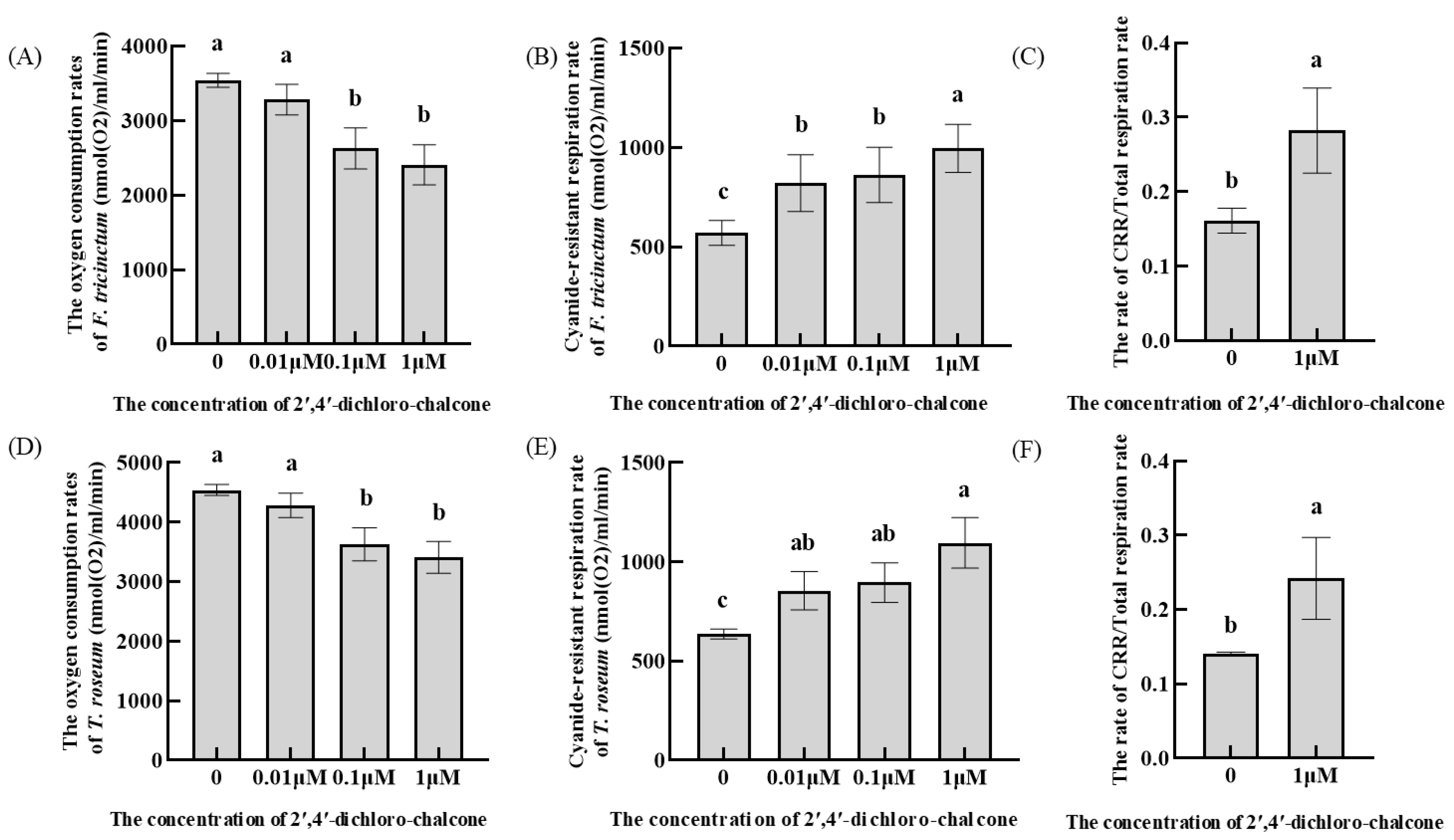
3.4. Effect of 2′,4′-Dichloro-chalcone on the Oxygen Consumption Rates in Respiratory Pathways of F. tricinctum and T. roseum

The total oxygen consumption rate largely reflects the activity of the respiratory pathway in aerobic fungi and the relative contributions from total and cyanide-resistant respiration is reflected in the electron distribution in the mitochondrial respiratory chain. Relative to control conditions, the total oxygen consumption rate in F. tricinctum and T. roseum was slightly reduced by 0.01 and 0.1 µM 2′,4′-dichloro-chalcone, but reduced by 32% and 25% at 100 µM, respectively (Figure 6A,D). In contrast, the oxygen consumption rate in the cyanide-resistant respiration pathway was increased by 42–43% in response to 1 µM 2′,4′-dichloro-chalcone treatment in the two pathogens (B, E). The cyanide-resistant respiration rate was significantly induced (p < 0.05). For clarity, the ratio of cyanide-resistant respiration to total respiration is shown in panels C and F. The results indicated that 2′,4′-dichloro-chalcone significantly inhibited the total respiration rate but activated an increased contribution from cyanide-resistant respiration in both pathogens.

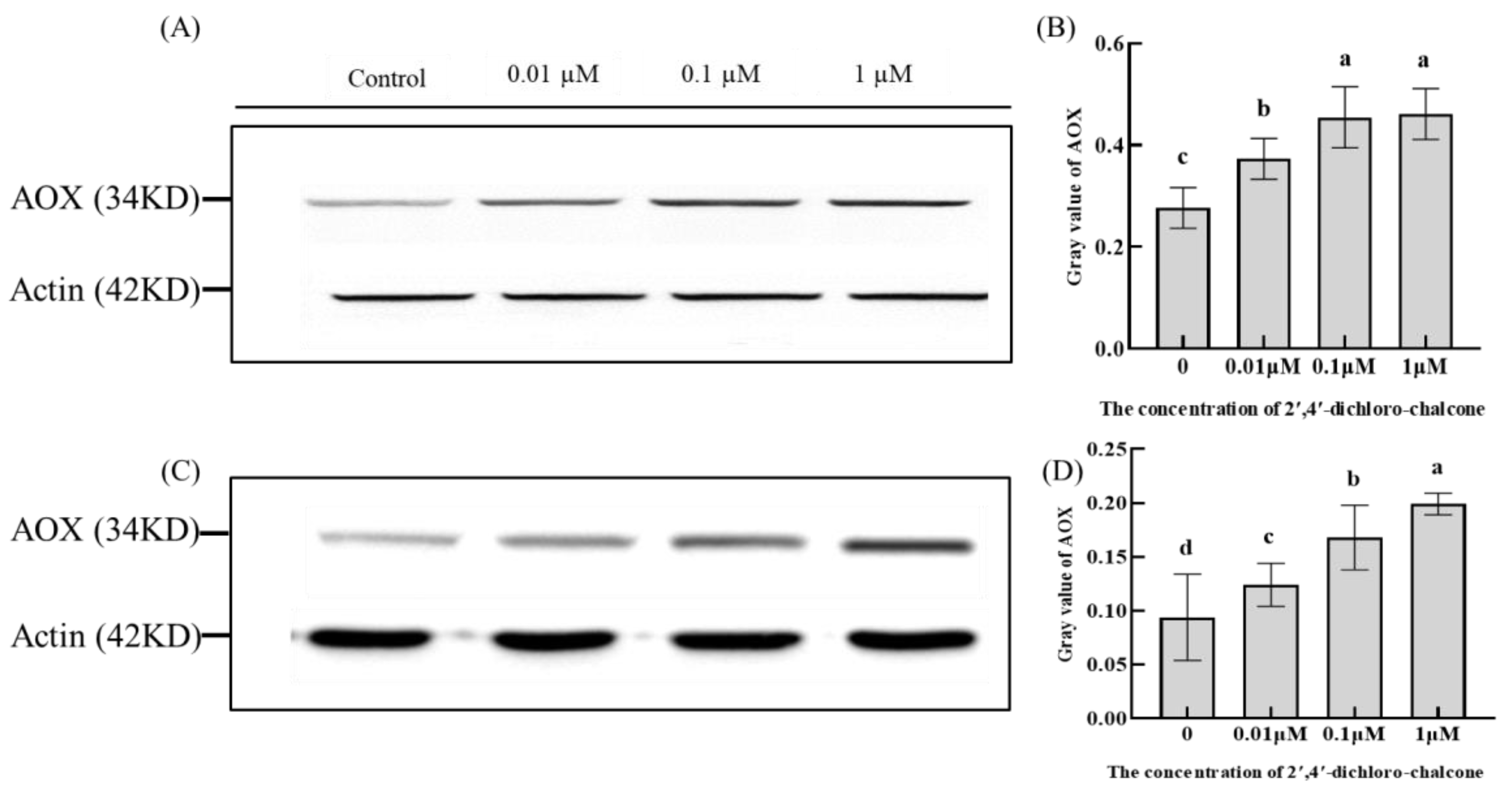
3.5. Effect of 2′,4′-Dichloro-chalcone on AOX Leveles in F. tricinctum and T. roseum

AOX is the terminal oxidase of the cyanide resistant respiratory pathway and its expression at the protein level reflects the activity of cyanide resistant respiratory pathways [36]. As shown in Figure 7, AOX was highly increased in response to 0.1 and 1 µM 2′,4′-dichloro-chalcone in both F. tricinctum and T. roseum. These results are consistent with the increased contribution of the cyanide-resistant respiration pathway to total respiration shown in Figure 6B,E.

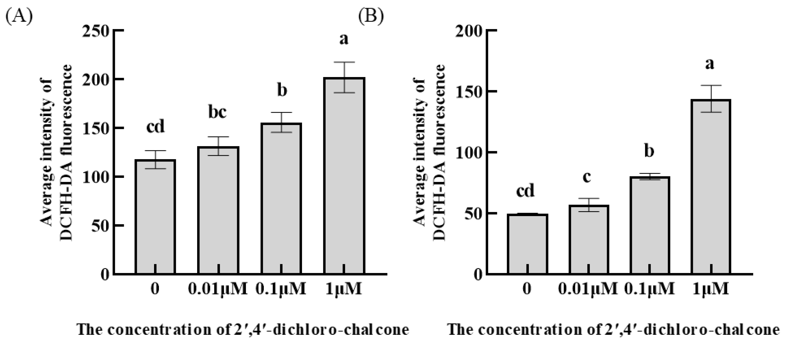
3.6. Effect of 2′,4′-Dichloro-chalcone on ROS Accumulation in F. tricinctum and T. roseum

The levels of intracellular ROS were assayed by fluorescence spectroscopy using the fluorescent probe, DCFH-DA. As shown in Figure 8, relative to the control group, 1 µM 2′,4′-dichloro-chalcone significantly increased the intracellular ROS levels in F. tricinctum and T. roseum conidia by 41.7 and 65.4%, respectively, whereas 0.1 µM 2′,4′-dichloro-chalcone induced lesser, but significant, increases of 25 and 38.2%, respectively. No significant effects were observed with 0.01µM 2′,4′-dichloro-chalcone.

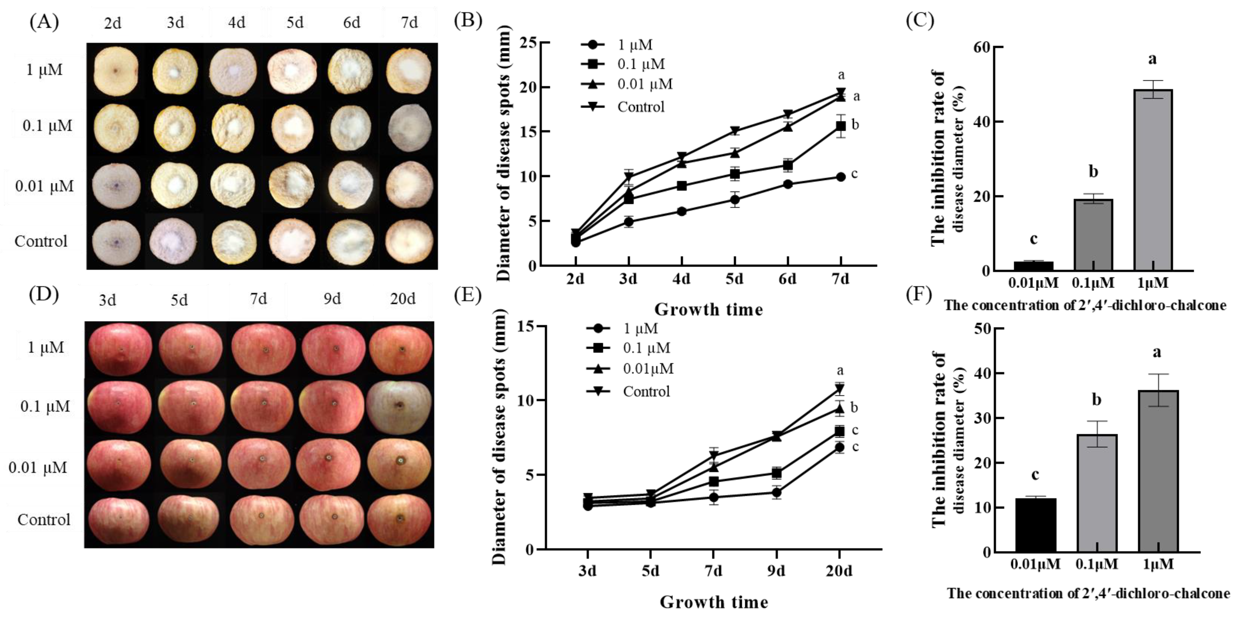
3.7. Effect of 2′,4′-Dichloro-chalcone on the Pathogenicities of F. tricinctum and T. roseum

The effect of 2′,4′-dichloro-chalcone on the pathogenicities of F. tricinctum and T. roseum were assessed by colony growth (disease spot diameter) after their inoculation onto the cut surface of potatoes and apples, respectively. Under the control conditions, F. tricinctum displayed faster growth kinetics, achieving an average disease spot size of 19.38 mm after 7 d (Figure 9A). In contrast, the development of T. roseum was relatively slower, achieving 10.77 mm after 20 d (D). The pre-inoculation treatment of potatoes and apples with 0.1 or 1 µM 2′,4′-dichloro-chalcone significantly inhibited disease spot development from both pathogens (A, D). After 7 d, 0.1 and 1 µM 2′,4′-dichloro-chalcone were observed to inhibit F. tricinctum spot development by 19.3 and 48.6%, respectively (B, C), while after 20 d, the T. roseum spot diameter was reduced by 26.4 and 36.3%, respectively (E, F). The results indicate that 1 µM 2′,4′-dichloro-chalcone treatment could effectively reduce product damage from potato dry rot and apple rot spot by inhibiting their pathogenicity.

4. Discussion

The present study demonstrated the does-dependent growth inhibitory activity of 2′,4′-dichloro-chalcone to F. tricinctum and T. roseum. The treatment of 1 µM 2′,4′-dichloro-chalcone caused significant growth inhibition of F. tricinctum and T. roseum in vitro and in vivo.

4.1. Antifungial Activity of 2′,4′-Dichloro-chalcone In Vitro

Chalcones are members of the flavonoid family, and are precursors for the synthesis of flavonoids and isoflavones in plants. Due to their content of active α and β unsaturated carbonyl functional groups, they are chemically very reactive, can effectively inhibit the growth of bacteria, mold and yeast, and prolong the shelf life of foods. In postharvest preservation, chalcone compounds can be used as film-forming materials that can be applied in vegetables and fruits to protect them from microorganisms [37]. However, the fungicidal activity of chalcone derivatives with different substituents varies greatly. Previous studies have reported that chalcone derivatives with halogen substituents were highly fungicidal [25,26]. Therefore, the aim of this study was to investigate the inhibitory effect of 2′,4′-dichloro-chalcone on postharvest fungal pathogens, to explore its inhibitory mechanism and to provide a theoretical basis for the use of chlorinated chalcones in the postharvest preservation of fruits and vegetables.
Tests of 2′,4′-dichloro-chalcone against the pathogens F. tricinctum and T. roseum showed it had inhibitory effects on mycelium growth and sporulation (Figure 3 and Figure 4). This is consistent with previous research, which demonstrated that the growth of Candida glabrata and Trichophyton interdigitatum were inhibited by 12.5 µg/mL non-alkylated chalcone derivatives containing 2-bromine or 2-chloride subunits [38]. Kumar et al. (2013) also found that 0.1 mg/mL chalcone compounds containing p-fluorinated substituents in the benzene ring usually showed high antimicrobial activity [39]. However, our results indicated that 2′,4′-dichloro-chalcone showed significant inhibitory effects against F. tricinctum and T. roseum at 1 µM, which is substantially lower than the reported inhibitory concentrations required for these other halogenated chalcone derivatives.

4.2. The Possible Antifungial Mechanism of 2′,4′-Dichloro-chalcone against F. tricinctum and T. roseum

Changes in the permeability of cell membranes in response to treatments can provide an important measure of membrane integrity [30]. In this study, 1 µM 2′,4′-dichloro-chalcone was observed to rapidly increase the cell membrane permeability of F. tricinctum and T. roseum conidia, indicating the cell membrane integrity was substantially compromised (Figure 5). It has been reported that phenolic chalcones can destroy the integrity of the cell membrane of Gram-negative and Gram-positive bacteria [40]. Chalcone compounds have also been reported to show high affinity to the cell membrane of Staphylococcus aureus and that their binding results in the loss of cell membrane integrity [41].
In addition to normal respiration via the cytochrome respiratory pathway, the cyanide-resistant respiratory pathway can operate when normal respiration is compromised, in which AOX functions as the terminal mitochondrial oxidase in fungi. Studies have indicated that AOX was induced in unfavorable conditions to activate cyanide-resistant respiration as a survival mechanism [42]. AOX can also regulate the response of fungi to oxidative stress to reduce the oxidative stress damage to cells [43]. In addition, AOX also plays a role in determining the fungal susceptibility to some fungicides [44]. It has been shown that Quinol oxidation-inhibiting fungicides (such as Azoxystrobin, Kresoxim-methyl, Metominostrobin) acted on the fungal mitochondrial respiratory complex III, with inhibition of fungal growth, but that AOX expression affected this response [45]. Xu et al. (2013) reported that the expression of AOX in Sclerotinia sclerotiorum reduced its sensitivity to Azoxystrobin but could increase the sensitivity to Procymidone [46]. Consistent with these earlier reports, our results showed that the total respiratory rate of F. tricinctum and T. roseum was gradually inhibited after treatment with 1 µM 2′,4′-dichloro-chalcone, while the cyanide-resistant respiratory rate and AOX levels were increased, normal respiration pathway were disrupted (Figure 6 and Figure 7).
ROS are mainly generated by the mitochondrial respiratory chain in fungi, and unregulated ROS accumulation can enhanced the oxidative stress and damage to cell components, resulting in reduced cell viability [47,48]. ROS accumulation can also disrupt mitochondrial functions [49]. Some researchers have found that quinoline chalcone derivatives significantly induced ROS accumulation in Candida albicans, with damage to the mitochondrial membrane and inhibition of growth [50]. The 2-hydroxychalcone treatment can also promote the production of ROS in dermatophytes, leading to fungal cell apoptosis and necrosis [51]. Synthetic coumarin-chalcone was reported to inhibit thioredoxin reductase, induce significant ROS accumulation and activate the mitochondrial apoptosis pathway [52]. In this study, 2′,4′-dichloro-chalcone treatment was also found to promote ROS accumulation in F. tricinctum and T. roseum (Figure 8).

4.3. 2′,4′-Dichloro-chalcone Treatment Significantly Inhibited the Pathogenicities of F. tricinctum and T. roseum In Vivo

Zhan et al. (2016) found that the host contents of pyrimidinyl chalcone compounds reduced infection by Rhizoctorzia solani, Physolospora piricola, Fusarium graminearum, and Bipolaris maydis in vivo [53]. Our results showed that 2′,4′-dichloro-chalcone treatment significantly reduced the pathogenicities of F. tricinctum and T. roseum in potato and apple, respectively (Figure 9).
AOX is the terminal oxidase of the cyanide resistant respiratory pathway in the fungal mitochondrial respiratory chain and can reduce the mitochondrial dysfunction caused by the excessive production of ROS [54]. During excessive ROS is accumulation, oxidative and functional damage to the mitochondrial membrane can occur, ultimately resulting in depressed respiration and overall physiological status of the cell [55], with a substantial decrease in the fungal growth rate and pathogenicity [56]. Singh et al. (2021) reported that an excessive intracellular ROS accumulation induces the expression of AOX and an increase in the cyanide-resistant respiration rate of Ascochyta rabiei, resulting in inhibition of the mycelial growth, spore production and cell vitality [57], which is consistent with our results. Therefore, 2′,4′-dichloro-chalcone affects the integrity of cell membranes, thereby affecting the mitochondrial respiratory electron transport chain, leading to a large production of ROS, directly or indirectly inducing the expression of AOX, activating the cyanide resistant respiratory pathway, and affecting growth and pathogenicity of fungi. In the future, 2′,4′-dichloro-chalcone compounds can be used as film forming materials be applied in postharvest control of these pathogens and sustainable agricultural practices. Although chalcones have very good antifungial activity, it can easily decompose at high temperatures, and the antimicrobial effect will also decline.
The inhibition mechanism of chlorinated chalcones against pathogen fungi still needs to be further analyzed at the molecular level.

5. Conclusions

Our results showed that 1 µM of 2′,4′-dichloro-chalcone strongly inhibited mycelial growth and conidial production of F. tricinctum (32.3%) and T. roseum (65.2%) in vitro. In addition, this treatment also significantly inhibited the expansion of potato dry rot from F. tricinctum (48.6%) and apple rot spot from T. roseum (36.2%). The present investigation might be helpful to explore the application of 2′,4′-dichloro-chalcone as an effective antifungal agent in treating postharvest infections and in agricultural practices caused by F. tricinctum and T. roseum. However, the characteristics of film formation of 2′,4′-dichloro-chalcone and other chlorinated chalcones molecular should be further investigated as alternative film-forming materials for the postharvest preservation of fruits and vegetables, sustainable agriculture and food security.

Author Contributions

Conceptualization, F.Z. and Y.Z. (Yan Zhu); methodology, W.Z. (Wei Zhang), F.Z. and J.D.; software, W.S. and Y.S.; validation, W.Z. (Weibing Zhang) and P.W.; formal analysis, F.C. and Y.Z. (Yuanshou Zhao); investigation, F.Z. and Y.Z. (Yan Zhu); resources, F.C.; data curation, W.S. and Y.L.; writing—original draft preparation, F.Z. and Y.Z. (Yan Zhu); writing—review and editing, Y.Z. (Yan Zhu) and W.S.; visualization, W.Z. (Weibing Zhang) and J.D.; project administration, Y.Z. (Yan Zhu); funding acquisition, Y.Z. (Yan Zhu); Yan Zhu is co-first author. All authors have read and agreed to the published version of the manuscript.

Funding

This research was funded by the National Natural Science Foundation (31760473), the Gansu Natural Science Foundation (23JRRA1464), the Lanzhou Science and Technology Plan Project (2023-3-107), the Natural Science Foundation for Young Scientists of Gansu Province (22JR5RA879),and the Excellent Postgraduate Innovation Star project of Gansu Province (2023CXZX-687).

Institutional Review Board Statement

Not applicable.

Informed Consent Statement

Not applicable.

Data Availability Statement

No new data were created or analyzed in this study. Data sharing is not applicable to this article.

Acknowledgments

We acknowledge the assistance of Xiaojing Wang in College of Food Science and Engineering, Gansu Agricultural University for providing language revising of manuscript and partial funding support.

Conflicts of Interest

The authors declare that they have no known competing financial interest or personal relationships that could have appeared to influence the work reported in this paper.

References

  1. Dong, F.; Xu, J.H.; Zhang, X.; Wang, S.F.; Xing, Y.J.; Mokoena, M.P.; Olaniran, A.O.; Shi, J.R. Gramineous weeds near paddy fields are alternative hosts for the Fusarium graminearum species complex that causes fusarium head blight in rice. Plant Pathol. 2020, 69, 433–441. [Google Scholar] [CrossRef]
  2. Amato, B.; Pfohl, K.; Tonti, S.; Nipoti, P.; Prodi, A. Fusarium proliferatum and fumonisin B1 co-occur with Fusarium species causing Fusarium Head Blight in durum wheat in Italy. J. Appl. Bot. Food Qual. 2015, 88, 228–233. [Google Scholar]
  3. Arino, A.A.; Bullerman, L.B. Fungal colonization of corn grown in nebraska in relation to year, genotype and growing conditions1. J. Food Prot. 1994, 57, 1084–1087. [Google Scholar] [CrossRef]
  4. Gachango, E.; Hanson, L.E.; Rojas, A.; Hao, J.J.; Kirk, W.W. Fusarium spp. causing dry rot of seed potato tubers in michigan and their sensitivity to fungicides. Plant Dis. 2012, 96, 1767–1774. [Google Scholar] [CrossRef] [PubMed]
  5. Lakshman, D.; Vieira, P.; Pandey, R.; Slovin, J.; Kamo, K. Symptom development in response to combined infection of in vitro-grown lilium longiflorum with pratylenchus penetrans and soilborne fungi collected from diseased roots of field-grown lilies. Plant Dis. 2017, 101, 882–889. [Google Scholar] [CrossRef]
  6. Tsukiyama, R.I.; Katsura, H.; Tokuriki, N.; Kobayashi, M. Antibacterial activity of licochalcone A against spore-forming bacteria. Antimicrob. Agents Chemother. 2002, 46, 1226–1230. [Google Scholar] [CrossRef] [PubMed]
  7. Dai, P.; Jiang, Y.; Liang, X.; Gleason, M.L.; Sun, G. Trichothecium roseum enters ‘fuji’ apple cores through stylar fissures. Plant Dis. 2019, 104, 1060–1068. [Google Scholar] [CrossRef] [PubMed]
  8. May-De Mio, L.L.; Negri, G.; Michailides, T.J. Effect of Trichothecium roseum, lime sulphur and phosphites to control blossom blight and brown rot on peach. Can. J. Plant Pathol. 2014, 36, 428–437. [Google Scholar] [CrossRef]
  9. Gong, D.; Bi, Y.; Li, S.; Li, Y.; Wang, Y. Trichothecium roseum infection promotes ripening of harvested muskmelon fruits and induces the release of specific volatile compound. J. Plant Pathol. 2019, 101, 529–538. [Google Scholar] [CrossRef]
  10. Ghazanfar, M.U.; Hussain, M.; Hamid, M.I.; Ansari, S.U. Utilization of biological control agents for the management of postharvest pathogens of tomato. Pak. J. Bot. 2016, 48, 2093–2100. [Google Scholar]
  11. Zhu, Y.H.; Shao, Y.Y.; Li, L.; Zhao, L.; Zhang, M.J.; Dong, C.M. The plant growth-promoting endophytic Fusarium oxysporum GG22 enhances Rehmannia glutinosa secondary metabolites accumulation. Ind. Crops Prod. 2022, 182, 114881. [Google Scholar] [CrossRef]
  12. Rizk, S.A.; Elsayed, G.A.; El-Hashash, M.A. One-pot synthesis, spectroscopic characterization and DFT study of novel 8-azacoumarin derivatives as eco-friendly insecticidal agents. J. Iran. Chem. Soc. 2018, 15, 2093–2105. [Google Scholar] [CrossRef]
  13. Ouyang, Y.; Li, J.J.; Chen, X.Y.; Fu, X.Y.; Sun, S.; Wu, Q. Chalcone Derivatives: Role in Anticancer Therapy. Biomolecules 2021, 11, 894. [Google Scholar] [CrossRef]
  14. Cavusoglu, B.K.; Atli, O.; Gormus, G.; Ozkay, Y.; Kaplancikli, Z.A. Synthesis and Evaluation of Heterocycles Based Chalcone Derivatives as Anti-proliferative Agents. Anti-Cancer Agents Med. Chem. 2018, 18, 1044–1053. [Google Scholar] [CrossRef] [PubMed]
  15. Parikh, K.; Joshi, D. Antibacterial and antifungal screening of newly synthesized benzimidazole-clubbed chalcone derivatives. Med. Chem. 2013, 22, 3688–3697. [Google Scholar] [CrossRef]
  16. Zhuang, C.; Zhang, W.; Sheng, C.; Zhang, W.; Xing, C.; Miao, Z. Chalcone: A Privileged Structure in Medicinal Chemistry. Chem. Rev. 2017, 117, 7762–7810. [Google Scholar] [CrossRef]
  17. Shukla, S.; Sood, A.K.; Goyal, K.; Singh, A.; Sharma, V.; Guliya, N.; Gulati, S.; Kumar, S. Chalcone Scaffolds as Anticancer Drugs: A Review on Molecular Insight in Action of Mechanisms and Anticancer Properties. Anti-Cancer Agents Med. Chem. 2021, 21, 1650–1670. [Google Scholar] [CrossRef] [PubMed]
  18. Lopez, S.N.; Cortes, J.C.; Ribas, J.C.; Castelli, M.V.; Zacchino, S.A.; Dominguez, J.N.; Lobo, G.; Charris-Charris, J.; Devia, C.; Rodriguez, A.M.; et al. In Vitro antifungal evaluation and structure-activity relationships of a new series of chalcone derivatives and synthetic analogues, with inhibitory properties against polymers of the fungal cell wall. Bioorgan. Med. Chem. 2001, 9, 1999–2013. [Google Scholar] [CrossRef]
  19. Lopez, G.; Mellado, M.; Werner, E.; Said, B.; Godoy, P.; Caro, N.; Besoain, X.; Montenegro, I.; Madrid, A. Sonochemical Synthesis of 2′-Hydroxy-Chalcone Derivatives with Potential Anti-Oomycete Activity. Antibiotics 2020, 9, 576. [Google Scholar] [CrossRef]
  20. Kakati, D.; Sarma, R.K.; Saikia, R.; Barua, N.C.; Sarma, J.C. Rapid microwave assisted synthesis and antimicrobial bioevaluation of novel steroidal chalcones. Steroids 2013, 78, 321–326. [Google Scholar] [CrossRef]
  21. Wang, Z.W.; Sun, Y.J.; Zhang, L.; Zhang, P. Synthesis and Antifungal Activity of Chalcones Containing Triazole Moiety. Chin. J. Synth. Chem. 2023, 31, 405–414. [Google Scholar]
  22. Liu, J.B. Bioactivities of chalcones with halogen atom and their thiosemicarbazide derivatives. Chemistry 2017, 8, 77–83. [Google Scholar]
  23. Sivakumar, P.M.; Iyer, G.; Natesan, L.; Doble, M. 3′-Hydroxy-4-methoxychalcone as a potential antibacterial coating on polymeric biomaterials. Appl. Surf. Sci. 2010, 256, 6018–6024. [Google Scholar] [CrossRef]
  24. Hu, Z. Preparation and Properties of pH-Responsive Chalcone-Loaded PVA/PAA Functional Films; Hefei University of Technology: Hefei, China, 2021. [Google Scholar]
  25. Shakil, N.A.; Singh, M.; Kumar, J.; Sathiyendiran, M.; Kumar, G.; Singh, M.; Pandey, R.P.; Pandey, A.; Parmar, V.S. Microwave synthesis and antifungal evaluations of some chalcones and their derived diaryl-cyclohexenones. J. Environ. Sci. Health B 2010, 45, 524–530. [Google Scholar] [CrossRef] [PubMed]
  26. Shakhatreh, M.A.; Al-Smadi, M.L.; Khabour, O.F.; Shuaibu, F.A.; Hussein, E.I.; Alzoubi, K.H. Study of the antibacterial and antifungal activities of synthetic benzyl bromides, ketones, and corresponding chalcone derivatives. Drug Des. Dev. Ther. 2016, 10, 3653–3660. [Google Scholar] [CrossRef]
  27. Schoch, C.L.; Seifert, K.A.; Huhndorf, S.; Robert, V.; Spouge, J.L.; Levesque, C.A.; Chen, W.; Bolchacova, E.; Voigt, K.; Crous, P.W. Nuclear ribosomal internal transcribed spacer (ITS) region as a universal DNA barcode marker for Fungi. Proc. Natl. Acad. Sci. USA 2012, 109, 6241–6246. [Google Scholar] [CrossRef]
  28. Wang, Z.; Lu, K.H.; Liu, X.; Zhu, Y.P.; Liu, C.H. Comparative Functional Genome Analysis Reveals the Habitat Adaptation and Biocontrol Characteristics of Plant Growth-Promoting Bacteria in NCBI Databases. Microbiol. Spectr. 2023, 11, e05007-22. [Google Scholar] [CrossRef]
  29. Xu, F.; Copsey, A.C.; Young, L.; Barsottini, M.R.O.; Albury, M.S.; Moore, A.L. Comparison of the Kinetic Parameters of Alternative Oxidases from Trypanosoma brucei and Arabidopsis thaliana-A Tale of Two Cavities. Front. Plant Sci. 2021, 12, 744218. [Google Scholar] [CrossRef] [PubMed]
  30. Feng, H.Q.; Sun, K.; Wei, Y.; Wang, R.F.; Jia, L.Y.; Zhang, J.P.; Li, Y. Role of cyanide-resistant respiration during light-induced attraction of predators to herbivore-infested leaves. Photosynthetica 2013, 51, 583–592. [Google Scholar] [CrossRef]
  31. Affourtit, C.; Moore, A.L. Purification of the plant alternative oxidase from Arum maculatum: Measurement, stability and metal requirement. Biochim. Biophys. Acta (BBA)-Bioenerg. 2004, 1608, 181–189. [Google Scholar] [CrossRef]
  32. Xin, Y.H.; Yang, D.; Yang, J.; Zhang, T.D.; Wang, Y.; Zhang, J.H.; Shi, Y.F. Effects of chalcone derivatives on mitochondrial structure and function of Aspergillus niger. Mycosystema 2021, 40, 2114–2122. [Google Scholar]
  33. Zhu, Y.; Zhu, F.P.; Chen, F.; Shi, G.Y.; Zhao, Y.T.; Zhang, F.; Gong, W.J.; Zhang, W.; Wang, X.J.; Li, Y.C. Isolation, identification and pathogenicity of pathogens causing dry rot of stored potato tubers in Gansu Province. J. Food Saf. Qual. 2023, 14, 240–248. [Google Scholar]
  34. Ge, Y.H.; Deng, H.W.; Bi, Y.; Li, C.Y.; Liu, Y.Y.; Dong, B.Y. Postharvest ASM dipping and DPI pre-treatment regulated reactive oxygen species metabolism in muskmelon (Cucumis melo L.) fruit. Postharvest. Biol. Technol. 2015, 99, 160–167. [Google Scholar] [CrossRef]
  35. Liu, J.J.; Hagberg, I.; Novitsky, L.; Hadj-Moussa, H.; Avis, T.J. Interaction of antimicrobial cyclic lipopeptides from Bacillus subtilis influences their effect on spore germination and membrane permeability in fungal plant pathogens. Fungal Biol. 2014, 118, 855–861. [Google Scholar] [CrossRef] [PubMed]
  36. El-Khoury, R.; Rak, M.; Benit, P.; Jacobs, H.T.; Rustin, P. Cyanide resistant respiration and the alternative oxidase pathway: A from to mammals. BBA-Bioenergetics 2022, 1863, 148567. [Google Scholar] [CrossRef]
  37. Yadav, N.; Dixit, S.K.; Bhattacharya, A.; Mishra, L.C.; Sharma, M.; Awasthi, S.K.; Bhasin, V.K. Antimalarial activity of newly synthesized chalcone derivatives in vitro. Chem. Biol. Drug Des. 2012, 80, 340–347. [Google Scholar] [CrossRef] [PubMed]
  38. Kucerova-Chlupacova, M.; Vyskovska-Tyllova, V.; Richterova-Finkova, L.; Kunes, J.; Buchta, V.; Vejsova, M.; Paterova, P.; Semelkova, L.; Jandourek, O.; Opletalova, V. Novel halogenated pyrazine-based chalcones as potential antimicrobial drugs. Molecules 2016, 21, 1421. [Google Scholar] [CrossRef] [PubMed]
  39. Kumar, C.; Loh, W.S.; Ooi, C.; Quah, C.; Fun, H.K. Heteroaryl chalcones: Design, synthesis, X-ray crystal structures and biological evaluation. Molecules 2013, 18, 12707–12724. [Google Scholar] [CrossRef] [PubMed]
  40. Erguden, B.; Unver, Y. Phenolic chalcones lead to ion leakage from gram-positive bacteria prior to cell death. Arch. Microbiol. 2021, 204, 3. [Google Scholar] [CrossRef]
  41. Tagousop, C.N.; Tamokou, J.D.; Ekom, S.E.; Ngnokam, D.; Voutquenne-Nazabadioko, L. Antimicrobial activities of flavonoid glycosides from Graptophyllum grandulosum and their mechanism of antibacterial action. BMC Complem. Altern. Med. 2018, 18, 252. [Google Scholar] [CrossRef] [PubMed]
  42. Jian, W.; Zhang, D.W.; Zhu, F.; Wang, S.X.; Zhu, T.; Pu, X.J.; Zheng, T.; Feng, H.; Lin, H.H. Nitrate reductase-dependent nitric oxide production is required for regulation alternative oxidase pathway involved in the resistance to Cucumber mosaic virus infection in Arabidopsis. Plant Growth Regul. 2015, 77, 99–107. [Google Scholar] [CrossRef]
  43. Xu, T.; Yao, F.; Liang, W.S.; Li, Y.H.; Li, D.R.; Wang, H.; Wang, Z.Y. Involvement of alternative oxidase in the regulation of growth, development, and resistance to oxidative stress of Sclerotinia sclerotiorum. J. Microbiol. 2012, 50, 594–602. [Google Scholar] [CrossRef] [PubMed]
  44. Bartlett, D.W.; Clough, J.M.; Godwin, J.R.; Hall, A.A.; Hamer, M.; Parr-Dobrzanski, B. The strobilurin fungicides. Pest. Manag. Sci. 2002, 58, 649–662. [Google Scholar] [CrossRef] [PubMed]
  45. Banno, S.; Yamashita, K.; Fukumon, F.; Okada, K.; Uekusa, H.; Takagaki, M.; Kimura, M.; Fujimura, M. Characterization of QoI resistance in Botrytis cinerea and identification of two types of mitochondrial cytochrome b gene. Plant Pathol. 2009, 58, 120–129. [Google Scholar] [CrossRef]
  46. Xu, T.; Wang, Y.T.; Liang, W.S.; Yao, F.; Li, Y.H.; Li, D.R.; Wang, H.; Wang, Z.Y. Involvement of alternative oxidase in the regulation of Sclerotinia sclerotiorum sensitivity to fungicides of azoxystrobin and procymidone. J. Microbiol. 2013, 51, 352–358. [Google Scholar] [CrossRef] [PubMed]
  47. Tian, J.; Ban, X.; Zeng, H.; He, J.; Chen, Y.; Wang, Y. The mechanism of antifungal action of essential oil from dill (Anethum graveolens L.) on Aspergillus flavus. PLoS ONE 2012, 7, 30147. [Google Scholar] [CrossRef] [PubMed]
  48. Kobayashi, D.; Kondo, K.; Uehara, N.; Otokozawa, S.; Tsuji, N.; Yagihashi, A. Watanabe, N Endogenous reactive oxygen species is an important mediator of miconazole antifungal effect. Antimicrob. Agents Chemother. 2002, 46, 3113–3117. [Google Scholar] [CrossRef] [PubMed]
  49. Pozniakovsky, A.I.; Knorre, D.A.; Markova, O.V.; Hyman, A.A.; Skulachev, V.P.; Severin, F.F. Role of mitochondria in the pheromone- and amiodaroneinduced programmed death of yeast. J. Cell Biol. 2005, 168, 257–269. [Google Scholar] [CrossRef]
  50. Chai, N.N.; Sun, A.M.; Zhu, X.H.; Li, Y.P.; Wang, R.R.; Zhang, Y.; Mao, Z.W. Antifungal evaluation of quinoline-chalcone derivatives combined with FLC against drug-resistant Candida albicans. Bioorg. Med. Chem. Lett. 2023, 86, 129242. [Google Scholar] [CrossRef] [PubMed]
  51. Bila, N.M.; Costa-Orlandi, C.B.; Vaso, C.O.; Bonatti, J.L.C.; Regasini, L.O.; Fontana, C.R.; Fusco-Almeida, A.M. 2-Hydroxychalcone as a Potent Compound and Photosensitizer against Dermatophyte Biofilms. Front. Cell. Infect. Microbiol. 2021, 11, 679470. [Google Scholar] [CrossRef]
  52. Wang, Y.; Zhang, W.; Dong, J.; Gao, J. Design, synthesis and bioactivity evaluation of coumarin-chalcone hybrids as potential anticancer agents. Bioorg. Chem. 2019, 95, 103530. [Google Scholar] [CrossRef] [PubMed]
  53. Zhan, J.S.; Jin, H.; Tao, T.; Hou, T.P. Synthesis and Anti-fungal Activity of Chalcones with Pyridyls. J. Shangdong Agric. Univ. 2016, 47, 166–171. [Google Scholar]
  54. Helmerhorst, E.J.; Stan, M.; Murphy, M.P.; Sherman, F.; Oppenheim, F.G. The concomitant expression and availability of conventional and alternative, cyanide-insensitive, respiratory pathways in Candida albicans. Mitochondrion 2005, 5, 200–211. [Google Scholar] [CrossRef] [PubMed]
  55. Lehmann, S.; Serrano, M.; L’Haridon, F.; Tjamos, S.E.; Metraux, J.P. Reactive oxygen species and plant resistance to fungal pathogens. Phytochemistry 2015, 112, 54–62. [Google Scholar] [CrossRef]
  56. Marschall, R.; Tudzynski, P. A new and reliable method for live imaging and quantification of reactive oxygen species in Botrytis cinerea Technological advancement. Fungal Genet. Biol. 2014, 71, 68–75. [Google Scholar] [CrossRef] [PubMed]
  57. Singh, Y.; Nair, A.M.; Verma, P.K. Surviving the odds: From perception to survival of fungal phytopathogens under host-generated oxidative burst. Plant Commun. 2021, 2, 100142. [Google Scholar] [CrossRef]
Figure 1. Chemical structure of 2′,4′-dichloro-chalcone (A); phylogenetic trees of F. tricinctum (B) and T. roseum isolates (C).
Coatings 13 01789 g001
Figure 2. The preparation of the chalcone stock solution and preparation of PDA medium containing the agent.
Coatings 13 01789 g002
Figure 3. The effects of 2′,4′-dichloro-chalcone on colony growth and hypha morphology of F. tricinctum (AC,G,H) and T. roseum (DF,I,J). (A,D) Representative examples of colony sizes after 7 d growth. (B,E) Mycelial growth curves (diameter). (C,F) The calculated growth inhibition by 2′,4′-dichloro-chalcone. Representative examples of mycelial morphology grown under control conditions (G,I) and with 100 µM 2′,4′-dichloro-chalcone (H,J). The error bars in panels (BF) represent the standard error of three replicate assays. Different letters above the bars represent significant differences between treatments (p < 0.05).
Coatings 13 01789 g003
Figure 4. The effect of 2′,4′-dichloro-chalcone on sporulation in F. tricinctum (A,B) and T. roseum (C,D). (A,C) Spore production capacity under control conditions. (B,D) Inhibition rate of sporulation (%). Vertical bars indicate the standard error of three replicate assays. The error bars in panels (AD) represent the standard error of three replicate assays. Different letters above the bars represent significant differences between treatments (p < 0.05).
Coatings 13 01789 g004
Figure 5. The effect of 2′,4′-dichloro-chalcone on membrane permeability in F. tricinctum and T. roseum. (A) F. tricinctum. (B) T. roseum. Vertical bars indicate the standard error of three replicate assays. Different letters above the bars represent significant differences between treatments (p < 0.05).
Coatings 13 01789 g005
Figure 6. The effect of 2′,4′-dichloro-chalcone on oxygen consumption rates in total and cyanide-resistant respiration pathways in F. tricinctum (A,D) and T. roseum (B,E). For clarity, the ratios of cyanide-resistant respiration to total respiration in F. tricinctum and T. roseum are shown in panels (C,F), respectively. Vertical bars indicate the standard error of three replicate assays. Different letters above the bars represent significant differences between treatments (p < 0.05).
Coatings 13 01789 g006
Figure 7. The effect of 2′,4′-dichloro-chalcone on AOX levels in F. tricinctum (A,B) and T. roseum (C,D). The immunoblots are shown in panels (A,C). The grey-scale values of AOX are shown in panels (B,D). The values presented were normalized with respect to actin. Vertical bars indicate the standard error of three replicate assays. Different letters above bars indicate significant differences between the treatments (p < 0.05).
Coatings 13 01789 g007
Figure 8. The effect of 2′,4′-dichloro-chalcone on intracellular ROS levels in conidia of F. tricinctum (A) and T. roseum (B). ROS were detected using DCFH-DA as a fluorescent probe. Vertical bars indicate the standard error of three replicate assays. Differing letters above bars indicate significantly differences between the treatments (p < 0.05).
Coatings 13 01789 g008
Figure 9. The effect of 2′,4′-dichloro-chalcone treatment on disease spot development from F. tricinctum (AC) and T. roseum (DF). (A,D) The diameter of disease spots. (B,E) The inhibition of disease spot development. (C,F) The effect of 2′,4′-dichloro-chalcone treatment on the disease spots. Vertical bars indicate the standard error of three replicate assays. Differing letters above each time point represent significant differences (p < 0.05).
Coatings 13 01789 g009
Disclaimer/Publisher’s Note: The statements, opinions and data contained in all publications are solely those of the individual author(s) and contributor(s) and not of MDPI and/or the editor(s). MDPI and/or the editor(s) disclaim responsibility for any injury to people or property resulting from any ideas, methods, instructions or products referred to in the content.

