Communication

Partitional Behavior of Janus Dumbbell Microparticles in a Polyethylene Glycol (PEG)-Dextran (DEX) Aqueous Two-Phase System (ATPS)

Department of Bio-Applied Chemistry, Daejeon University, 62, Daehak-ro, Dong-gu, Daejeon 34520, Korea
Coatings 2022, 12(3), 415; https://doi.org/10.3390/coatings12030415
Submission received: 28 February 2022 / Revised: 12 March 2022 / Accepted: 16 March 2022 / Published: 21 March 2022
(This article belongs to the Special Issue Surface Chemical Modification II)

Abstract

:
Janus particles are known to be useful to various fields such as biomolecule-probing sensors, reaction catalysts, surfactants, and so on. They have two chemically different surfaces which possess contradictory characteristics such as polarity, hydrophobicity, etc. Here, a simple fabrication of dumbbell-shaped Janus microparticles was tested by the chemical reaction of carboxyl groups and amino groups to form amide bonds. They were distributed to the interface between polyethylene glycol (PEG)-rich phase and dextran (DEX)-rich phase, while the unreacted particles having carboxyl groups located at the top PEG-rich phase and particles having amine ligands went to the bottom DEX-rich phase of an aqueous two-phase system (ATPS). The fabrication procedures, observations, and possible applications of results are discussed.

1. Introduction

Janus particles are defined as micrometer- to nanometer-sized bead-shaped materials having more than one composition or surface property [1]. The word “Janus” originates from the Roman god with two opposite faces. Therefore, Janus particles possess at least two chemically different surfaces, which have different chemical properties such as polarity, hydrophobicity, optical property, and so on. Additionally, there have been a large number of studies to manufacture and probe these unprecedented materials in order to apply them to a variety of areas such as optical traps, E-paper display, self-propulsion, biosensing, and imaging [1,2,3,4].
The partitioning of micro/nanoparticles in ATPS is intriguing for its potency and need for biocompatible systems in many applications [5,6], such as the recovery of viruses, viral particles, cells, extracellular vesicles, and so forth. ATPS has been studied not only for fundamental research but also for biotechnological applications due to its low surface tension and hydrophilic nature [5,6,7,8]. Partitioning of molecules and particles in ATPS is of great importance for the study of biologically valuable materials such as proteins, enzymes, nucleic acids, viruses, cells, drugs, and metallic components [6]. Examples of these such as vanillin and lipase have been reported. Nouri et al. [7] used modified carbon nanotube particles in ATPS by adsorbing vanillin. The partition coefficient increased more than a hundred percent and claimed the nanotube material as a potential drug delivery carrier. Amid et al. [8] introduced a novel ATPS comprising a surfactant and xylitol to successfully purify lipase, an important enzyme in industries, from pumpkin seeds. A few cases dealt with the partitioning of metal particles in ATPS [9,10,11]. Those cases are valuable for the idea of Janus particles in ATPS and their applications because gold and silver are well-known materials for biological purposes [12]. The concept of combining Janus particles and ATPS, however, has very rarely been reported [13] in contrast to the importance and accumulation of knowledge of each technique. Additionally, few issues on non-metal Janus particles and their partitional behaviors have been reported, especially in ATPS.
Janus dumbbell particles, a kind of Janus particle, look like two particles that are connected to become a non-spherical one. Park and Lee [14] studied equilibrium orientations of Janus ellipsoid and Janus dumbbell particles at the oil–water interface theoretically and concluded that the orientation strongly depends on the characteristics of the particle. Janus dumbbell nanoparticles were synthesized and assembled at the oil–water interface to stabilize emulsions by Yang et al. [15]. This catalyst provided greater efficiency than a platinum–carbon catalyst in aqueous hydrogenation reactions. Kim et al. [16] reported that Janus dumbbell gold nanoparticles could be synthesized by dimerization reaction and proved their existence by transmission electron microscopy (TEM).
Here, ATPS is applied to the fabrication and assembly of Janus dumbbell-shaped microparticles. The partition effect of surface-modified polystyrene (PS)-coated magnetic microparticles and silica microparticles in a PEG-DEX ATPS were shown. Carboxylated PS particles were partitioned mainly to PEG-rich upper phase, while amino-silica particles were located mostly in DEX-rich bottom phase. The chemically combined dumbbell-shaped Janus particles were mainly found at the interface of PEG-DEX ATPS. The procedures and results, limitations, and possible extension of the study are described, along with future applications.

2. Materials and Methods

2.1. Materials

2-(N-morpholino) ethanesulfonic acid (MES) hydrate (Cat# M8250) and N-(3-Dimethylaminopropyl)-N’-ethyl carbodiimide hydrochloride (EDC, Cat# E7750) were obtained from Merck (Kenilworth, NJ, USA). SpheroTM Carboxyl Magnetic Particles (2.5% w/v, Cat# CM-20-10) and SpheroTM Amino Silica Particles (5% w/v, Cat# ASIP-30-10) were purchased from Spherotech, Inc. (Lake Forest, IL, USA). The polystyrene (PS) magnetic particles are composed of a layer of iron oxide and polystyrene coated onto polystyrene core particles. The amount of iron oxide is about 15%, and it contains around 20 μeq/g of carboxyl groups on their surface, according to the vendor’s information. Other important parameters of PS and silica microparticles were summarized in Table 1. Phosphate buffer saline (PBS) tablet and polyethylene glycol (PEG, average MW 8000) were purchased from Fisher Scientific (Hampton, NH, USA). Dextran T20 (DEX, average MW 20,000) was purchased from Pharmacosmos (Holbaek, Denmark). Deionized (DI) water was made by a water purification system (Human Power I+ Scholar-UV, Human Corporation, Seoul, South Korea).

2.2. Preparation of the ATPS Solutions

The procedures for how to make PEG-DEX ATPS have already been described elsewhere [17,18]. First, 10% PEG and 5% DEX ATPS solutions were prepared by mixing 20% polyethylene glycol (PEG) and 10% dextran (DEX) solutions at a 1:1 ratio (10 mL of each PEG and DEX solutions were mixed for 20 mL). Both polymer solutions were prepared with DI water.

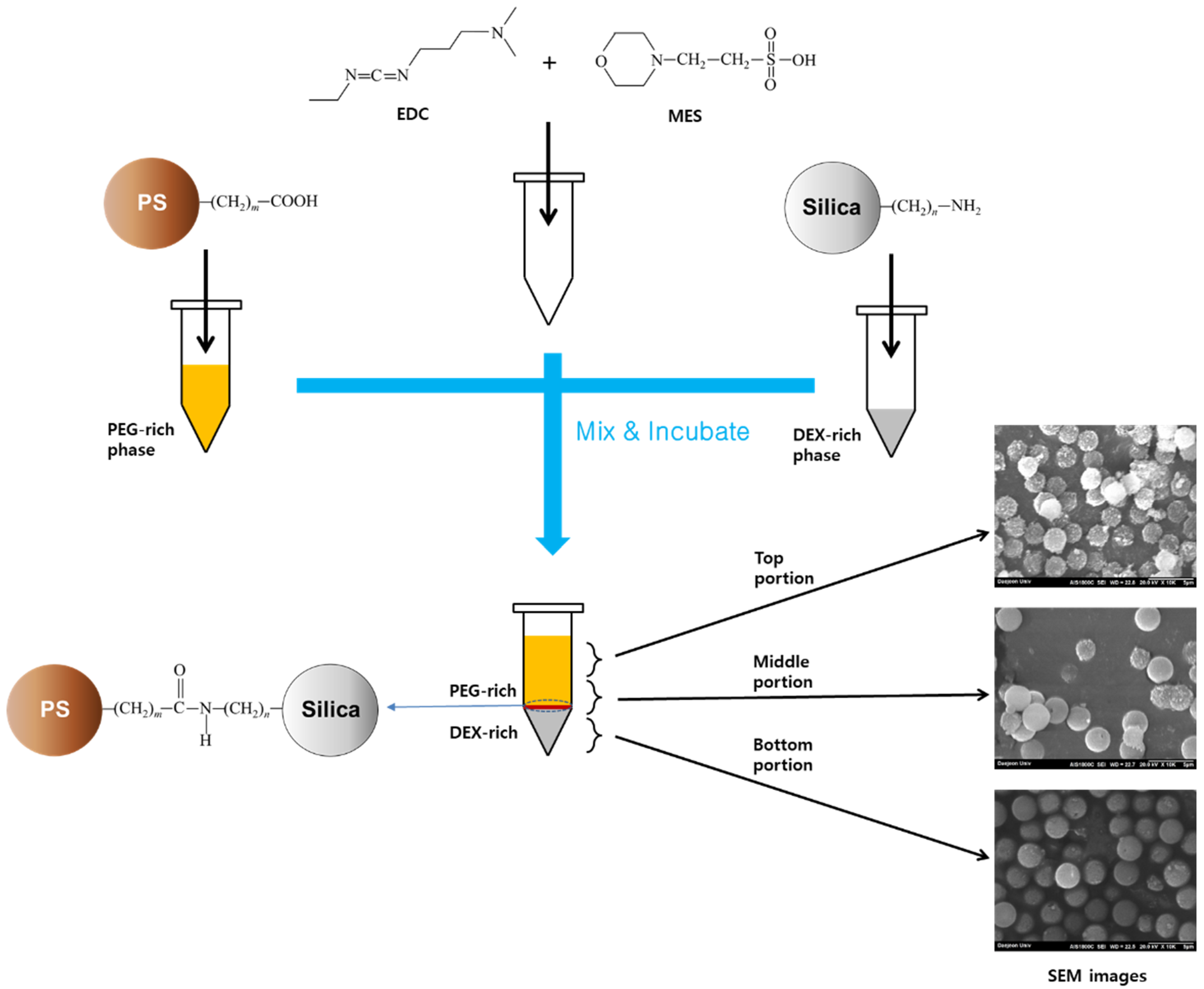
2.3. Preparation of Janus Particles in ATPS

Carboxyl-modified polystyrene (PS) magnetic particles and amino-silica non-magnetic particles were introduced from Spherotech®. Figure 1 describes the reaction of two surface-modified microparticles to form a dumbbell-shaped Janus particle.
The coupling reaction of a carboxyl group and an amino group which is well-known for protein and peptide synthesis provides an amide bond, which forms a dumbbell-shaped or number 8-shaped Janus particle comprising one PS sphere and the other silica sphere which are connected. The detailed procedures are referred from a part of SpheroTM Technical Note: particle coating procedures (STN-1 rev C, 041106) for covalent coupling (one step EDC coupling from “coating of amino particles with ligand or proteins using EDC”) and described as follows. The original procedures were modified for the ATPS application. First, 0.1 mL of 2.5% carboxyl magnetic PS particle suspension was added to 1 mL of PEG-rich phase in a 1.5 mL-volume plastic tube. Second, 0.05 mL of 5% amino silica particle suspension was added to a 0.5 mL DEX-rich phase in another 1.5 mL plastic tube. Third, the covalent coupling reaction solution was made in a different 1.5 mL plastic tube by weighing and adding 16 mg of MES hydrate and 5 mg EDC, then transferring 1 mL of PEG-rich phase and 0.5 mL DEX-rich phase with microparticles which prepared above, shook shortly to dissolve EDC and MES and placed in a vertical rotary mixer (Intelli-Mixer RM-2M, Elmi Ltd., Riga, Latvia), vortexed at 30~60 rpm at ambient temperature for a few hours to promote the reaction.

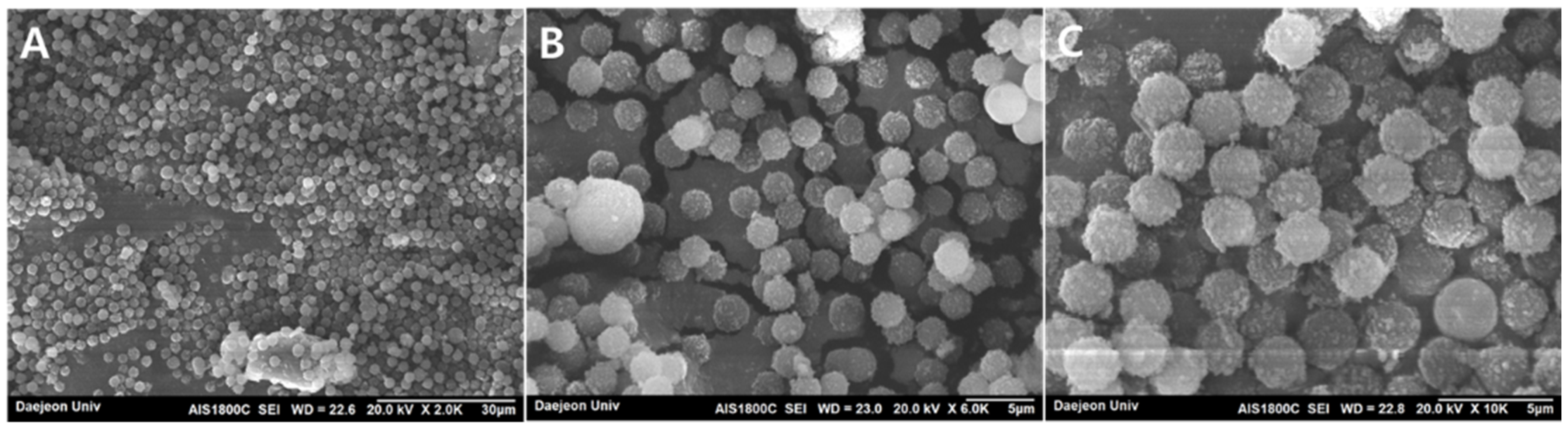
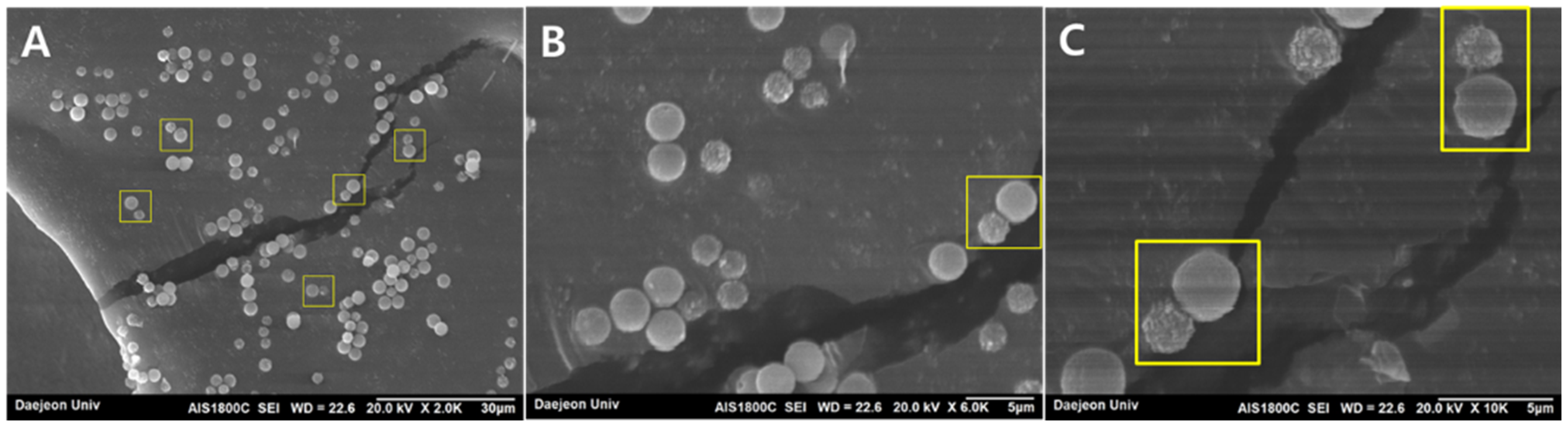
2.4. Partition Observation of Janus Particles with SEM Characterization

After the PEG-DEX ATPS layers were formed, the three parts of the whole solutions were taken separately by pipetting 0.4–0.5 mL of the top, middle, and bottom suspension (Figure 2), and each part was transferred to a new plastic tube, then centrifuged for 5 min. The supernatant was removed then the remaining particles were washed with 1 mL PBS buffer. The reduced particle concentrations of one-tenth or one-hundredth of the original procedures, by adding 10 μL PS and 5 μL of silica particle suspensions or 1 μL PS and 0.5 μL of silica suspensions, were also tested to help the observation of fewer particles and the reduction in overlapped particles in the Scanning Electron Microscopy (SEM) images.
Electron microscopic images were taken on an SEM model AIS1800C (Seron Technologies, Uiwang-si, Korea) in the separation analysis lab of Daejeon University, with a secondary electron (SE) detector at an accelerating voltage of 20 kV. The particle samples were dried in a centrivap concentrator (Labconco, Kansas City, MO, USA) for a couple of hours. A piece of carbon tape was first placed on the SEM mount then particles were spread on the carbon tape using cotton swaps and sputtered with gold (Au) for charge dissipation. Four to five minutes of Au ion sputtering at about 15 kV, is known to coat gold to the surface of particle samples around 2 nm thickness according to vendor’s information (Seron Technologies, Uiwang-si, Korea).

3. Results and Discussion

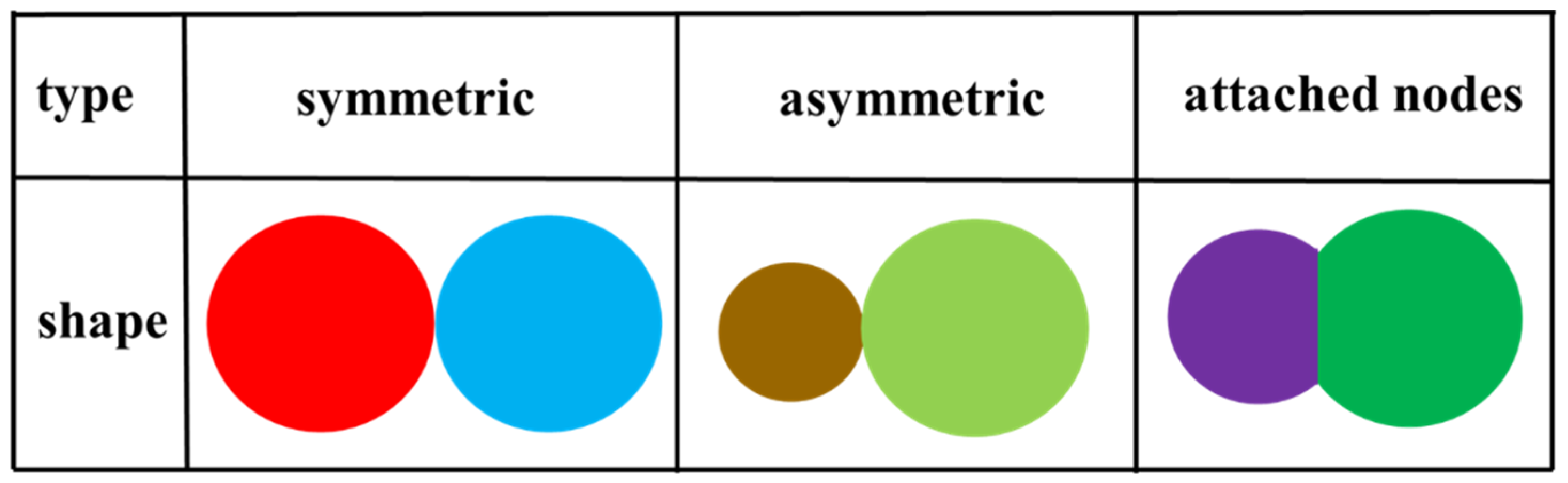
3.1. Janus Dumbbell-like Microparticles and Partition Factors of ATPS

Various types of Janus particles including Janus dumbbell particles have been reported. Among them, typical shapes of Janus dumbbell-like particles are described in Scheme 1.
The partitioning mechanism of ATPS is known to be complex [5,6]. Diverse parameters can be expressed by the following equation. K is the concentration ratio of target material in two immiscible phases, called partition coefficient.
ln K = ln Kelectrochemical + ln Khydrophobicity + ln Kbiospecific affinity + ln Ksize + ln Kconformation + ln Kenvironment
Among them, the hydrophobicity factor is reported as the major ruling one for protein partitioning.

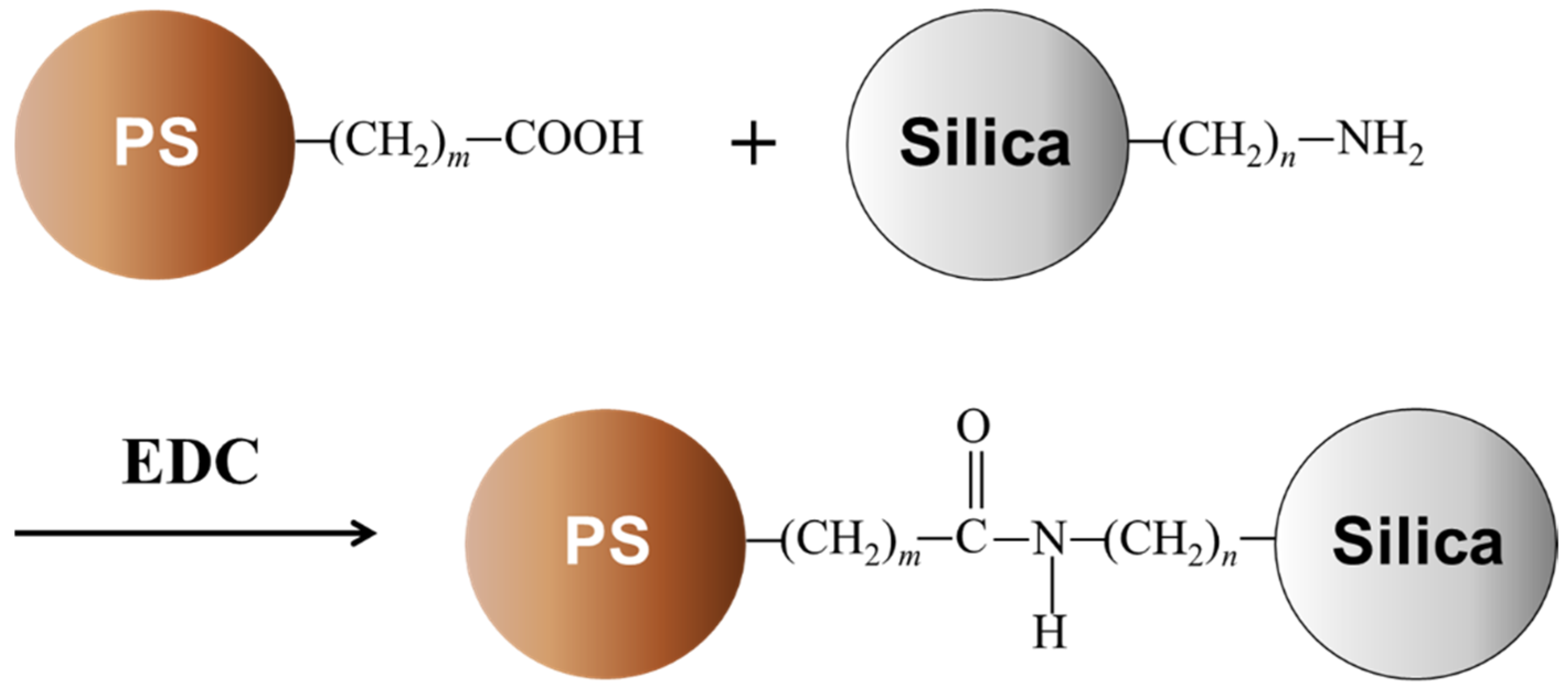
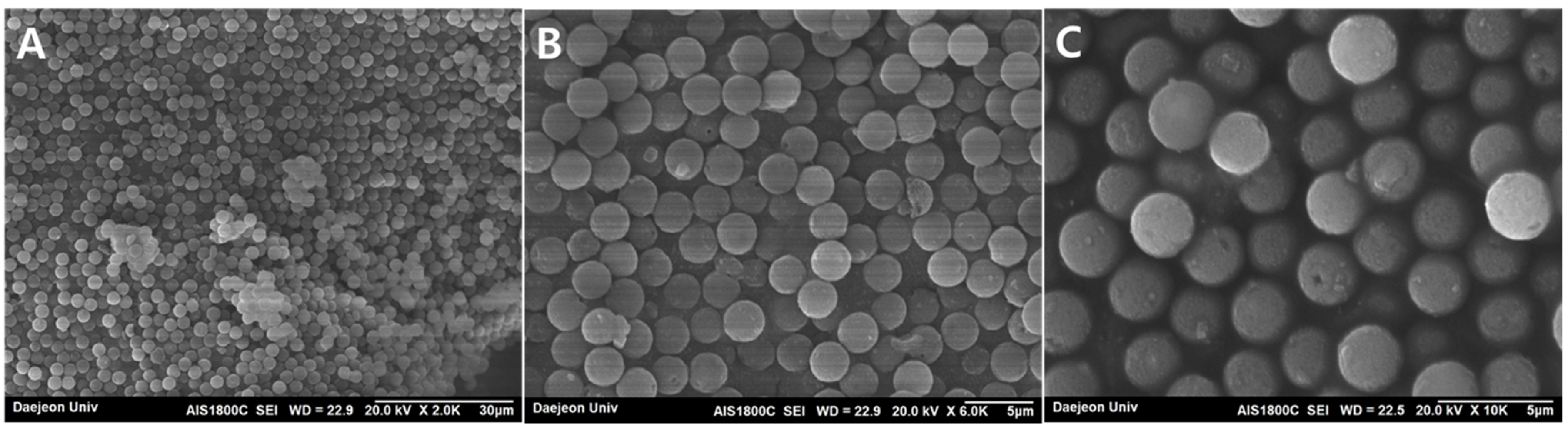
3.2. EDC Coupling Reaction to Form Janus Dumbbell-like Microparticles

EDC coupling reaction is one of the well-known reactions to form amide bonds. It is often introduced to alter the surface of particles with biological molecules. The importance of using EDC is that it is water-soluble. MES does not participate in the coupling reaction but it is a commonly used buffer with EDC, which does not have carboxyl or amine functional groups therefore it does not interfere with EDC coupling reaction. More information about amide bond formation reagents including EDC can be found elsewhere [19].

3.3. Partitioning of Carboxyl PS Microbeads in PEG-DEX ATPS

Polystyrene (PS) is known to be hydrophobic because of its aromatic functional group or benzene ring structure. Tosylated PS magnetic microparticles of 2.8 μm average diameter were distributed to the upper phase of PEG-DEX in the prior studies [17,18], without surface modification. The modified functional groups located at the surface of the microparticle may affect the partitional behavior. However, the basic substrate material, PS, in this case, seems to have a greater affect and determine the partition in ATPS. Figure 3 shows the SEM images of microparticles that were withdrawn and dried from the top portion of PEG-DEX ATPS after EDC coupling reaction shown in Figure 2, with different image magnification values. The particles seem to be homogeneous and mainly composed of carboxyl PS microparticles from the SEM image observation.
The reaction and partition procedures (Figure 2) were performed with three different concentration combinations of PS and silica microparticles, which are described in Section 2.3, to promote dispersity of particles with scanning electron microscopy (SEM), but other reaction conditions are the same, and the same PS particles were also mainly found with lower (ten times and hundred times lower) reaction particles concentrations, at the top portions.

3.4. Partitioning of PS-Silica Dumbbell Microbeads in PEG-DEX ATPS

Figure 4 demonstrates the SEM images of microparticles from the middle portion of PEG-DEX ATPS, after EDC coupling reaction, with different magnifications. Combinations of PS-silica dumbbell-like particles (inside the yellow boxes in Figure 4A) were found, which are different from images of the top portion (Figure 3) or bottom portion (Figure 5). It means the EDC coupling reaction can be possible not only between particles and molecules but also between microparticles, although the yield of the intended Janus dumbbell-shaped microparticle seemed to be low. The carboxyl PS particles prefer to be located in the upper PEG-rich phase, while the amino-silica particles prefer to be at the lower DEX-rich phase in ATPS. To make two different particles as one, they have to collide (or be very close to each other) and the covalent bond has to be formed, probably at the interface of ATPS. The vortexing of the plastic tube container may promote the collisions but it seems not enough to make all particles react to obtain a high yield of Janus dumbbell-shaped particles at the interface of ATPS. Because the diameters of PS and silica microparticles are different (2.17 and 3.0 μm), the resulting Janus microparticles look like the number “8”, snowman, or tumbling doll shape in Figure 4C.
The reason why dumbbell-shaped Janus particles were only found at the middle portion of PEG-DEX ATPS can be explained as follows. Because PS particles prefer the PEG-rich upper phase and silica particles prefer the DEX-rich lower phase, the possibility of locating Janus PS-silica dumbbell particles after EDC coupling reaction is expected to be greater at the interfacial region of PEG-DEX ATPS, or the middle portion. Chiu et al. [9] reported the extraction and concentration of radiolabeled antibody proteins using gold nanoparticles (GNP) at the interface of PEG-salt ATPS. Compared to bulk phase extraction, interfacial extraction provides a faster result and higher concentration factor because the volume of the interface is smaller compared to that of the upper or lower phase of ATPS. This could be a reason why PS-silica dumbbell particles were mainly observed at the middle portion of PEG-DEX ATPS.
For the lower concentration combinations of PS and silica microparticles, the Janus dumbbell particles were not easily found from the SEM image observations, because the ratio of dumbbell particles to bare PS or silica particles seems to be low and the number of Janus particles formed is expected to be smaller compared to the original concentratrational procedure case.

3.5. Partitioning of Amino-Silica Microbeads in PEG-DEX ATPS

Silica particles are known to be hydrophilic because of their silanol functional groups. They are known to be introduced in diverse biomedical applications [20,21]. The basic substrate material, silica, in this case, would have more affinity with dextran than with PEG because silanol groups can form hydrogen bonds with dextran molecules, rather than PEG. The density of silica microparticles is also greater than PS. Figure 5 describes the SEM images of microparticles that were taken and dried from the bottom portion of PEG-DEX ATPS after the EDC coupling reaction shown in Figure 2, with different magnifications. The particles also seem to be homogeneous and mainly amino-silica microparticles from the SEM image observation.
When lower concentration combinations of PS and silica microparticles were tried, the same silica particles were found, mainly at the bottom portions.

4. Conclusions

The synthesis of Janus dumbbell microparticles in ATPS has not been reported and was attempted for the first time. A simple approach using carboxyl-modified and amino-modified particles with water-soluble crosslinking reagent was proven to be successful for the manufacture of Janus dumbbell-shaped particles in aqueous conditions.
Although it seems that the trial has not achieved a high yield of Janus particles, it can be improved by considering a few aspects. The coupling reaction was performed in a tube of non-flowing or static way. The introduction of dynamic flow for effective mixing, reaction, and collection of target particles in ATPS is possible using a microfluidic device [22] because the Janus dumbbell particles are mainly found at the interface of PEG-rich and DEX-rich phase. The more sophisticated design of flow, such as circulating channel flows, can increase the yield of Janus dumbbell particles by promoting continuous contact and chance of collision for the reaction of unreacted particles at the interface of ATPS without vortexing.
The Janus dumbbell microparticle in ATPS is potential for a variety of purposes such as selective extraction, concentration, and detection of biologically important targets because ATPS provides a non-harmful environment to biomaterials. It may be utilized to capture target molecules by the sandwiched way (particle–target–particle) then located at the interface of ATPS for further use.
Janus particles with metal surfaces also have been reported because of their wide applications, besides non-metal Janus particles. The fabrication of Janus particles with two different material surfaces in a particle, such as gold–PS or gold–silica, along with the study of their partition behaviors in ATPS, is expected to be the next aim in the future. The study of Janus dumbbell-shaped nanometer-sized beads with ATPS may also help to purify viral particles or even to develop novel molecular machines, such as artificial ribosomes [23], in an aqueous environment.

Funding

This research received no external funding.

Institutional Review Board Statement

Not applicable.

Informed Consent Statement

Not applicable.

Data Availability Statement

Not applicable.

Acknowledgments

This research was supported by the Daejeon University Research Grants (20200152).

Conflicts of Interest

The authors declare no conflict of interest. The founding sponsors had no role in the design of the study; in the collection, analyses, or interpretation of data; in the writing of the manuscript; or in the decision to publish the results.

Abbreviations

ATPS: aqueous two-phase system; DEX: dextran; DI: deionized; EDC: N-(3-Dimethylaminopropyl)-N’-ethyl carbodiimide hydrochloride; GNP: gold nanoparticles; MES: 2-(N-morpholino) ethanesulfonic acid; PBS: phosphate-buffered saline; PEG: polyethylene glycol; PS: polystyrene, SE: secondary electron; SEM: scanning electron microscopy; TEM: transmission electron microscopy.

References

  1. Walther, A.; Müller, A.H.E. Janus Particles: Synthesis, Self-Assembly, Physical Properties, and Applications. Chem. Rev. 2013, 113, 5194–5261. [Google Scholar] [CrossRef] [PubMed]
  2. Zhang, J.; Grzybowski, B.A.; Granick, S. Janus Particle Synthesis, Assembly, and Application. Langmuir 2017, 33, 6964–6977. [Google Scholar] [CrossRef] [PubMed]
  3. Le, T.C.; Zhai, J.; Chiu, W.-H.; Tran, P.A.; Tran, N. Janus Particles: Recent Advances in the Biomedical Applications. Int. J. Nanomed. 2019, 14, 6749–6777. [Google Scholar] [CrossRef] [PubMed] [Green Version]
  4. Hu, J.; Zhou, S.; Sun, Y.; Fang, X.; Wu, L. Fabrication, properties and applications of Janus particles. Chem. Soc. Rev. 2012, 41, 4356–4378. [Google Scholar] [CrossRef] [PubMed]
  5. Pereira, J.F.B.; Coutinho, J.A.P. Chapter 5 Aqueous Two-Phase Systems in Liquid-Phase Extraction; Poole, C.F., Ed.; Elsevier: Amsterdam, The Nederlands, 2020; pp. 157–182. [Google Scholar]
  6. Iqbal, M.; Tao, Y.; Xie, S.; Zhu, Y.; Chen, D.; Wang, X.; Huang, L.; Peng, D.; Satter, A.; Shabbir, M.A.B.; et al. Aqueous two-phase system (ATPS): An overview and advances in its applications. Biol. Proced. Online 2016, 18, 18. [Google Scholar] [CrossRef] [PubMed] [Green Version]
  7. Nouri, M.; Shahriari, S.; Pazuki, G. Increase of vanillin partitioning using aqueous two phase system with promising nanoparticles. Sci. Rep. 2009, 9, 19665. [Google Scholar] [CrossRef] [Green Version]
  8. Amid, M.; Manap, M.Y.; Hussin, M.; Mustafa, S. A Novel Aqueous Two Phase System Composed of Surfactant and Xylitol for the Purification of Lipase from Pumpkin (Cucurbita moschata) Seeds and Recycling of Phase Components. Molecules 2015, 20, 11184–11201. [Google Scholar] [CrossRef] [Green Version]
  9. Chiu, R.Y.T.; Thach, A.V.; Wu, C.M.; Wu, B.M.; Kamei, D.T. An Aqueous Two-Phase System for the Concentration and Extraction of Proteins from the Interface for Detection Using the Lateral-Flow Immunoassay. PLoS ONE 2015, 10, e0142654. [Google Scholar] [CrossRef]
  10. Long, M.S.; Keating, C.D. Nanoparticle Conjugation Increases Protein Partitioning in Aqueous Two-Phase Systems. Anal. Chem. 2006, 78, 379–386. [Google Scholar] [CrossRef]
  11. Helfrich, M.R.; El-Kouedi, M.; Etherton, M.R.; Keatings, C.D. Partitioning and Assembly of Metal Particles and Their Bioconjugates in Aqueous Twos-Phase Systems. Langmuir 2005, 21, 8478–8486. [Google Scholar] [CrossRef]
  12. Shankar, P.D.; Shobana, S.; Karuppusamy, I.; Pugazhendhi, A.; Ramkumar, V.S.; Arvindnarayan, S.; Kumar, G. A review on the biosynthesis of metallic nanoparticles (gold and silver) using bio-components of microalgae: Formation mechanism and applications. Enz. Microbial. Technol. 2016, 95, 28–44. [Google Scholar] [CrossRef] [PubMed]
  13. Innes-Gold, S.N.; Luby, C.J.; Mace, C.R. Experimental and Theoretical Validation of System Variables That Control the Position of Particles at the Interface of Immiscible Liquids. Langmuir 2018, 34, 7673–7680. [Google Scholar] [CrossRef] [PubMed]
  14. Park, B.J.; Lee, D. Equilibrium Orientation of Nonspherical Janus Particles at Fluid-Fluid Interfaces. ACS Nano 2012, 6, 782–790. [Google Scholar] [CrossRef]
  15. Yang, T.; Wei, L.; Jing, L.; Liang, J.; Zhang, X.; Tang, M.; Monteiro, M.J.; Chen, Y.; Wang, Y.; Gu, S.; et al. Dumbbell-Shaped Bi-component Mesoporous Janus Solid Nanoparticles for Biphasic Interface Catalysis. Angew. Chem. Int. Ed. 2017, 56, 8459–8463. [Google Scholar] [CrossRef] [PubMed] [Green Version]
  16. Kim, H.; Carney, R.P.; Reguera, J.; Ong, Q.K.; Liu, X.; Stellaci, F. Synthesis and Characterization of Janus Gold Nanoparticles. Adv. Mater. 2012, 24, 3857–3863. [Google Scholar] [CrossRef] [PubMed] [Green Version]
  17. Byun, C.K.; Hwang, H.; Choi, W.S.; Yaguchi, T.; Park, J.; Kim, D.; Mitchell, R.J.; Kim, T.; Cho, Y.-K.; Takayama, S. Productive Chemical Interaction between a Bacterial Microcolony Couple Is Enhanced by Periodic Relocation. J. Am. Chem. Soc. 2013, 135, 2242–2247. [Google Scholar] [CrossRef]
  18. Byun, C.K.; Kim, M.; Kim, D. Modulating the Partitioning of Microparticles in a Polyethylene Glycol (PEG)-Dextran (DEX) Aqueous Biphasic System by Surface Modification. Coatings 2018, 8, 85. [Google Scholar] [CrossRef] [Green Version]
  19. Valeur, E.; Bradley, M. Amide bond formation: Beyond the myth of coupling reagents. Chem. Soc. Rev. 2009, 38, 606–631. [Google Scholar] [CrossRef]
  20. Wang, L.; Zhao, W.; Tan, W. Bioconjugated Silica Nanoparticles: Development and Applications. Nano. Res. 2008, 1, 99–115. [Google Scholar] [CrossRef] [Green Version]
  21. Tan, W.; Wang, K.; He, X.; Zhao, X.J.; Drake, T.; Wang, L.; Bagwe, R.P. Bionanotechnology Based on Silica Nanoparticles. Med. Res. Rev. 2004, 24, 621–638. [Google Scholar] [CrossRef]
  22. Yang, S.; Guo, F.; Kiraly, B.; Mao, X.; Lu, M.; Leong, K.W.; Huang, T.J. Microfluidic synthesis of multifunctional Janus particles for biomedical applications. Lab Chip 2012, 12, 2097–2102. [Google Scholar] [CrossRef] [PubMed]
  23. De Bo, G.; Gall, M.A.Y.; Kuschel, S.; De Winter, J.; Gerbaux, P.; Leigh, D.A. An artificial molecular machine that builds an asymmetric catalyst. Nat. Nanotechnol. 2018, 13, 381–385. [Google Scholar] [CrossRef] [PubMed]
Figure 1. Reaction scheme between a carboxyl-activated polystyrene (PS) magnetic particle and an amino-coated silica particle (both from Spherotech®). m and n are natural numbers between 2 to 8, according to vendor’s information.
Coatings 12 00415 g001
Figure 2. Preparation of Janus dumbbell particles in PEG-DEX ATPS. Details were shown in Materials and Methods.
Coatings 12 00415 g002
Scheme 1. Different types of Janus dumbbell particles. Different colors indicate the different surface properties of each particle.
Coatings 12 00415 sch001
Figure 3. SEM images of microparticles of the top portion of PEG-DEX ATPS after the EDC coupling reaction with different magnifications. (A) 2000 times, (B) 6000 times, (C) 10,000 times magnified. Scale bars are (A) 30, (B) 5, (C) 5 μm. See Experimental for other conditions.
Coatings 12 00415 g003
Figure 4. SEM images of microparticles of the middle portion of PEG-DEX ATPS after the EDC coupling reaction with different magnifications. (A) 2000 times, (B) 6000 times, (C) 10,000 times magnified. Janus dumbbell particles are shown in yellow boxes. Scale bars are (A) 30, (B) 5, (C) 5 μm. See Experimental for other conditions.
Coatings 12 00415 g004
Figure 5. SEM images of microparticles of the bottom portion of PEG-DEX ATPS after the EDC coupling reaction with different magnifications. (A) 2000 times, (B) 6000 times, (C) 10,000 times magnified. Scale bars are (A) 30, (B) 5, (C) 5 μm. See Experimental for other conditions.
Coatings 12 00415 g005
Table 1. Characteristics of polystyrene (PS) and silica microparticles used.
Particle TypeDensity (g/cm3)ShapeSize (μm)Functionalized Group
Polystyrene (PS)1.05Spherical,
non-porous
2.17Carboxyl
Silica1.96Spherical,
non-porous
3.0Amino
Publisher’s Note: MDPI stays neutral with regard to jurisdictional claims in published maps and institutional affiliations.

