
The Cytotoxicity of OptiBond Solo Plus and Its Effect on Sulfur Enzymes Expression in Human Fibroblast Cell Line Hs27

 , , and
Abstract
:1. Introduction
2. Materials and Methods
2.1. OptiBond Solo Plus Solution Preparation
2.2. Cell Cultures
2.3. LDH Cell Membrane Integrity Assay
2.4. The Number of Cells by Crystal Violet Staining
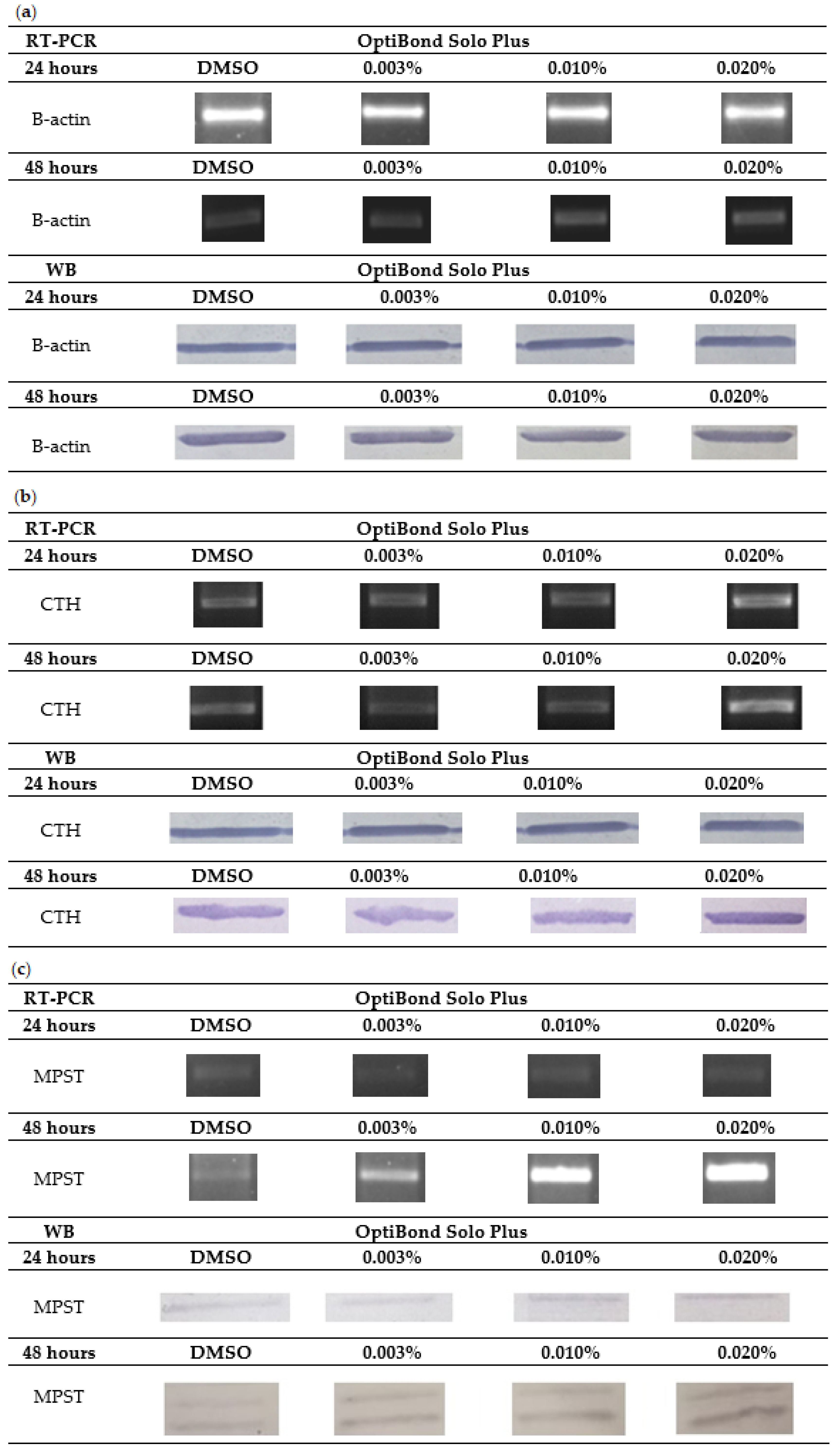
2.5. Gene Expression at the mRNA Level by RT-PCR
2.6. Gene Expression at the Protein Level by Western Blot
- (a)
- SDS-PAGE electrophoresis
- (b)
- Western Blot with immunohistochemical identification of proteins
2.7. RP-HPLC (Reverse Phase High Performance Liquid Chromatography)
2.8. Data Analysis
3. Results
4. Discussion
5. Limitations
6. Conclusions
Author Contributions
Funding
Institutional Review Board Statement
Informed Consent Statement
Data Availability Statement
Acknowledgments
Conflicts of Interest
References
- Zieniewska, I.; Maciejczyk, M.; Zalewska, A. The effect of selected dental materials used in conservative dentistry, endodontics, surgery, and orthodontics as well as during the periodontal treatment on the redox balance in the oral cavity. Int. J. Mol. Sci. 2020, 21, 9684. [Google Scholar] [CrossRef] [PubMed]
- Taubmann, A.; Willershausen, I.; Walter, C.; Al-Maawi, S.; Kaina, B.; Gölz, L. Genotoxic and cytotoxic potential of methacrylate-based orthodontic adhesives. Clin. Oral Investig. 2021, 25, 2569–2581. [Google Scholar] [CrossRef] [PubMed]
- Montagner, A.F.; Sarkis-Onofre, R.; Pereira-Cenci, T.; Cenci, M.S. MMP inhibitors on dentin stability: A systematic review and meta-analysis. J. Dent. Res. 2014, 93, 733–743. [Google Scholar] [CrossRef] [PubMed] [Green Version]
- Sofan, E.; Sofan, A.; Palaia, G.; Tenore, G.; Romeo, U.; Migliau, G. Classification review of dental adhesive systems: From the IV generation to the universal type. Ann. Stomatol. 2017, 8, 1–17. [Google Scholar]
- Cadenaro, M.; Antoniolli, F.; Sauro, S.; Tay, F.R.; Di Lenarda, R.; Prati, C.; Biasotto, M.; Contardo, L.; Breschi, L. Degree of conversion and permeability of dental adhesives. Eur. J. Oral Sci. 2005, 113, 525–530. [Google Scholar] [CrossRef]
- Kharouf, N.; Eid, A.; Hardan, L.; Bourgi, R.; Arntz, Y.; Jmal, H.; Foschi, F.; Sauro, S.; Ball, V.; Haikel, Y.; et al. Antibacterial and bonding properties of universal adhesive dental polymers doped with pyrogallol. Polymers 2021, 13, 1538. [Google Scholar] [CrossRef] [PubMed]
- Hardan, L.; Bourgi, R.; Kharouf, N.; Mancino, D.; Zarow, M.; Jakubowicz, N.; Haikel, Y.; Cuevas-Suárez, C.E. Bond strength of universal adhesives to dentin: A systematic review and meta-analysis. Polymers 2021, 13, 814. [Google Scholar] [CrossRef]
- Tanasiewicz, M.; Skucha-Nowak, M.; Gibas, M.; Pawlak, J.; Wiȩckiewicz, W.; Mertas, A.; Król, W. The analysis of cytotoxicity of an experimental preparation used for the reduction of dentin hypersensitivity. Adv. Clin. Exp. Med. 2017, 26, 15–22. [Google Scholar] [CrossRef] [Green Version]
- Fischer, M.; Mertas, A.; Czuba, Z.P.; Skucha-Nowak, M. Study of cytotoxic properties of an experimental preparation with features of a dental infiltrant. Materials 2021, 14, 2442. [Google Scholar] [CrossRef]
- Małkiewicz, K.; Turło, J.; Marciniuk-Kluska, A.; Grzech-Leśniak, K.; Gąsior, M.; Kluska, M. Release of bisphenol A and its derivatives from orthodontic adhesive systems available on the European market as a potential health risk factor. Ann. Agric. Environ. Med. 2015, 22, 172–177. [Google Scholar] [CrossRef] [Green Version]
- Fröb, L.; Rüttermann, S.; Romanos, G.E.; Herrmann, E.; Gerhardt-Szép, S. Cytotoxicity of self-etch versus etch-and-rinse dentin adhesives: A screening study. Materials 2020, 13, 452. [Google Scholar] [CrossRef] [PubMed] [Green Version]
- Goël Brackett, M.; Bouillaguet, S.; Lockwood, P.E.; Rotenberg, S.; Lewis, J.B.; Messer, R.L.W.; Wataha, J.C. In vitro cytotoxicity of dental composites based on new and traditional polymerization chemistries. J. Biomed. Mater. Res.—Part B Appl. Biomater. 2007, 81, 397–402. [Google Scholar] [CrossRef] [PubMed]
- Atabek, D.; Aktaş, N.; Uyar, D.S. Two Years Clinical Evaluation of Sonic-Resin Placement System with Self-Etch and Total-Etch Adhesive Modes. J. Res. Med. Dent. Sci. 2021, 48, 743–751. [Google Scholar]
- Brackett, W.W.; Tay, F.R.; Brackett, M.G.; Dib, A.; Sword, R.J.; Pashley, D.H. The effect of chlorhexidine on dentin hybrid layers in vivo. Oper. Dent. 2007, 32, 107–111. [Google Scholar] [CrossRef]
- Szep, S.; Kunkel, A.; Ronge, K.; Heidemann, D. Cytotoxicity of modern dentin adhesives—In vitro testing on gingival fibroblasts. J. Biomed. Mater. Res. 2002, 63, 53–60. [Google Scholar] [CrossRef] [PubMed]
- Wang, R. Physiological implications of hydrogen sulfide: A whiff exploration that blossomed. Physiol. Rev. 2012, 92, 791–896. [Google Scholar] [CrossRef] [PubMed] [Green Version]
- Nagahara, N. Multiple role of 3-mercaptopyruvate sulfurtransferase: Antioxidative function, H2S and polysulfide production and possible SOx production. Br. J. Pharmacol. 2018, 175, 577–589. [Google Scholar] [CrossRef] [Green Version]
- Santos, S.; Mascarenhas, P.; Bandarra, S.; Ribeiro, A.; Maurício, P.; Barahona, I. Evaluation of the Cytotoxic Potential of Adhesives, with Two on the Market: Scotchbond Universal and Optibond Solo Plus, and an Adhesive in the Experimental Phase: T1. Med. Sci. Forum 2021, 5, 7. [Google Scholar]
- Chomczynski, P.; Sacchi, N. Single-step method of RNA isolation by acid guanidinium thiocyanate-phenol-chloroform extraction. Anal. Biochem. 1987, 162, 156–159. [Google Scholar] [CrossRef]
- Bronowicka-Adamska, P.; Bentke, A.; Wróbel, M. Hydrogen sulfide generation from l-cysteine in the human glioblastoma-astrocytoma U-87 MG and neuroblastoma SHSY5Y cell lines. Acta Biochim. Pol. 2017, 64, 171–176. [Google Scholar] [PubMed]
- Laemmli, U.K. Cleavage of structural proteins during the Assembly of the Head of Bacteriophage T4. Nature 1970, 227, 680–685. [Google Scholar] [CrossRef] [PubMed]
- Dominick, P.K.; Cassidy, P.B.; Roberts, J.C. A new and versatile method for determination of thiolamines of biological importance. J. Chromatogr. B. Biomed. Sci. Appl. 2001, 761, 1–12. [Google Scholar] [CrossRef]
- Bronowicka-Adamska, P.; Zagajewski, J.; Czubak, J.; Wróbel, M. RP-HPLC method for quantitative determination of cystathionine, cysteine and glutathione: An application for the study of the metabolism of cysteine in human brain. J. Chromatogr. B. Analyt. Technol. Biomed. Life Sci. 2011, 879, 2005–2009. [Google Scholar] [CrossRef] [PubMed]
- Spyrou, G.E.; Watt, D.A.L.; Naylor, I.L. The origin and mode of fibroblast migration and proliferation in granulation tissue. Br. J. Plast. Surg. 1998, 51, 455–461. [Google Scholar] [CrossRef]
- Wawrzynkiewicz, A.; Rozpedek-Kaminska, W.; Galita, G.; Lukomska-Szymanska, M.; Lapinska, B.; Sokolowski, J.; Majsterek, I. The cytotoxicity and genotoxicity of three dental universal adhesives—an in vitro study. Int. J. Mol. Sci. 2020, 21, 3950. [Google Scholar] [CrossRef]
- Prabhakar, A.R.; Dhanraj, K.; Sugandhan, S. Comparative evaluation in vitro of caries inhibition potential and microtensile bond strength of two fluoride releasing adhesive systems. Eur. Arch. Paediatr. Dent. 2014, 15, 385–391. [Google Scholar] [CrossRef] [PubMed]
- Pace, M.; Pierote, J.J.; Câmara, J.V.; Barbosa, I.; Araújo, C.T.; Prieto, L.; Carvalho, G.A.; Pereira, G.; Vianna, R.; Fried, H.; et al. Influence of the volume of restorative material on the concentration of stresses in the restorative interface. J. Clin. Exp. Dent. 2021, 13, 555–563. [Google Scholar] [CrossRef] [PubMed]
- Engn, A.; Yaln, M.; Lker, H.E.; Ztrk, B.; Hakk, S.S. Cytotoxicity evaluation of dentin bonding agents by dentin barrier test on 3-dimensional pulp cells. Oral Surg. Oral Med. Oral Pathol. Oral Radiol. Endodontol. 2011, 112, 83–88. [Google Scholar]
- Ayar, M.K. Bond durability of contemporary adhesive systems to pulp chamber dentin. Acta Biomater. Odontol. Scand. 2015, 1, 76–80. [Google Scholar] [CrossRef]
- Vajrabhaya, L.; Korsuwannawong, S.; Bosl, C.; Schmalz, G. The cytotoxicity of self-etching primer bonding agents in vitro. Oral Surg. Oral Med. Oral Pathol. Oral Radiol. Endodontol. 2009, 107, e86–e90. [Google Scholar] [CrossRef]
- Pagano, S.; Lombardo, G.; Costanzi, E.; Balloni, S.; Bruscoli, S.; Flamini, S.; Coniglio, M.; Valenti, C.; Cianetti, S.; Marinucci, L. Morpho-functional effects of different universal dental adhesives on human gingival fibroblasts: An in vitro study. Odontology 2021, 109, 524–539. [Google Scholar] [CrossRef] [PubMed]
- Kaszuba, K.; Bentke, A.; Krawczyk, A.; Szlęzak, D.; Wróbel, M. A study on the cytotoxic effect of fluoride on the human fibroblast cell line HS27. Fluoride 2020, 53, 124–135. [Google Scholar]
- Lee, D.H.; Lim, B.S.; Lee, Y.K.; Ahn, S.J.; Yang, H.C. Involvement of oxidative stress in mutagenicity and apoptosis caused by dental resin monomers in cell cultures. Dent. Mater. 2006, 22, 1086–1092. [Google Scholar] [CrossRef]
- Żukowski, P.; Maciejczyk, M.; Waszkiel, D. Sources of free radicals and oxidative stress in the oral cavity. Arch. Oral Biol. 2018, 92, 8–17. [Google Scholar] [CrossRef] [PubMed]
- Krifka, S.; Hiller, K.A.; Spagnuolo, G.; Jewett, A.; Schmalz, G.; Schweikl, H. The influence of glutathione on redox regulation by antioxidant proteins and apoptosis in macrophages exposed to 2-hydroxyethyl methacrylate (HEMA). Biomaterials 2012, 33, 5177–5186. [Google Scholar] [CrossRef] [PubMed]
- Demirci, M.; Hiller, K.A.; Bosl, C.; Galler, K.; Schmalz, G.; Schweikl, H. The induction of oxidative stress, cytotoxicity, and genotoxicity by dental adhesives. Dent. Mater. 2008, 24, 362–371. [Google Scholar] [CrossRef]
- Krifka, S.; Spagnuolo, G.; Schmalz, G.; Schweikl, H. A review of adaptive mechanisms in cell responses towards oxidative stress caused by dental resin monomers. Biomaterials 2013, 34, 4555–4563. [Google Scholar] [CrossRef]
- Spagnuolo, G.; Annunziata, M.; Rengo, S. Cytotoxicity and oxidative stress caused by dental adhesive systems cured with halogen and LED lights. Clin. Oral Investig. 2004, 8, 81–85. [Google Scholar] [CrossRef]
- Schafer, F.Q.; Buettner, G.R. Redox environment of the cell as viewed through the redox state of the glutathione disulfide/glutathione couple. Free Radic. Biol. Med. 2001, 30, 1191–1212. [Google Scholar] [CrossRef]
- Schweikl, H.; Spagnuolo, G.; Schmalz, G. Genetic and cellular toxicology of dental resin monomers. J. Dent. Res. 2006, 85, 870–877. [Google Scholar] [CrossRef]
- Engelmann, J.; Jankea, V.; Volka, J.; Leyhausen, G.; Von Neuhoff, N.; Schlegelberger, B.; Geurtsen, W. Effects of BisGMA on glutathione metabolism and apoptosis in human gingival fibroblasts in vitro. Biomaterials 2004, 25, 4573–4580. [Google Scholar] [CrossRef] [PubMed]


| Gene | Initiation | Denaturation | Amplification | Elongation | Termination |
|---|---|---|---|---|---|
| MPST | 5 min at 95 °C | 30 s at 95 °C | 30 s at 55 °C | 2 min at 72 °C for 29 cycles | 72 °C for 8 min |
| CTH | 5 min at 95 °C | 30 s at 95 °C | 1 min at 51 °C | 8 min at 72 °C for 30 cycles | 72 °C for 8 min |
| TST | 5 min at 95 °C | 30 s at 95 °C | 30 s at 54.5 °C | 2 min at 72 °C for 34 cycles | 72 °C for 8 min |
| β-actin | 5 min at 95 °C | 30 s at 95 °C | 30 s at 55 °C | 2 min at 72 °C for 30 cycles | 72 °C for 8 min |
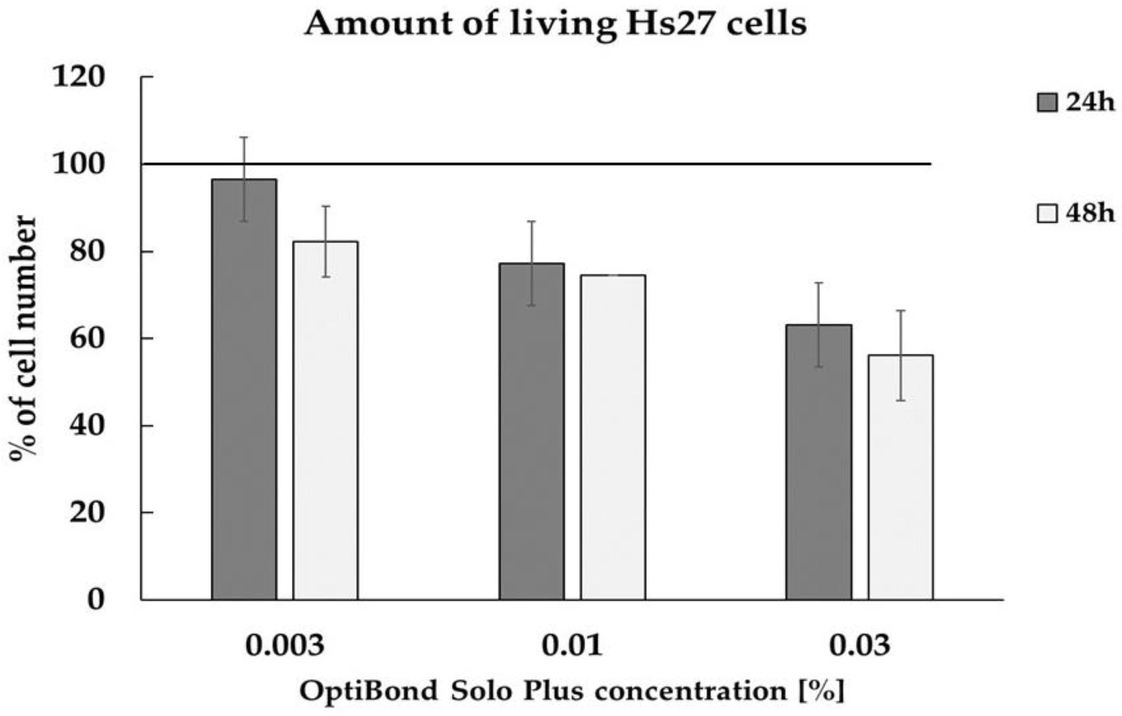
| OptiBond Solo Plus | % Cytotoxicity | Time |
|---|---|---|
| 0.003% | <5% | |
| 0.010% | <5% | 24 h |
| 0.03% | 43.7 | |
| 0.003% | <5% | |
| 0.010% | <5% | 48 h |
| 0.03% | 41.0 |
| GSSG | GSH | GSH/GSSG | |
|---|---|---|---|
| 0% | 7.24 ± 1.18 | 83.62 ± 3.46 | 11.54 |
| 0.03% | 29.06 ± 6.9 | 166.97 ± 6.6 | 5.74 |
Publisher’s Note: MDPI stays neutral with regard to jurisdictional claims in published maps and institutional affiliations. |
© 2022 by the authors. Licensee MDPI, Basel, Switzerland. This article is an open access article distributed under the terms and conditions of the Creative Commons Attribution (CC BY) license (https://creativecommons.org/licenses/by/4.0/).
Share and Cite
Bentke-Imiolek, A.; Kaszuba, K.; Bronowicka-Adamska, P.; Czopik, B.; Zarzecka, J.; Wróbel, M. The Cytotoxicity of OptiBond Solo Plus and Its Effect on Sulfur Enzymes Expression in Human Fibroblast Cell Line Hs27. Coatings 2022, 12, 382. https://doi.org/10.3390/coatings12030382
Bentke-Imiolek A, Kaszuba K, Bronowicka-Adamska P, Czopik B, Zarzecka J, Wróbel M. The Cytotoxicity of OptiBond Solo Plus and Its Effect on Sulfur Enzymes Expression in Human Fibroblast Cell Line Hs27. Coatings. 2022; 12(3):382. https://doi.org/10.3390/coatings12030382
Chicago/Turabian StyleBentke-Imiolek, Anna, Kinga Kaszuba, Patrycja Bronowicka-Adamska, Barbara Czopik, Joanna Zarzecka, and Maria Wróbel. 2022. "The Cytotoxicity of OptiBond Solo Plus and Its Effect on Sulfur Enzymes Expression in Human Fibroblast Cell Line Hs27" Coatings 12, no. 3: 382. https://doi.org/10.3390/coatings12030382