
The Influence of Diamond Nanoparticles on Fibroblast Cell Line L929, Cytotoxicity and Bacteriostaticity of Selected Pathogens

 , , ,
, , ,
Abstract
:1. Introduction
2. Materials and Methods
2.1. Cytotoxicity
2.2. Bacteriostaticity
2.3. SEM
2.4. XRD
2.5. Raman Spectroscopy
2.6. FT–IR Spectroscopy
3. Results
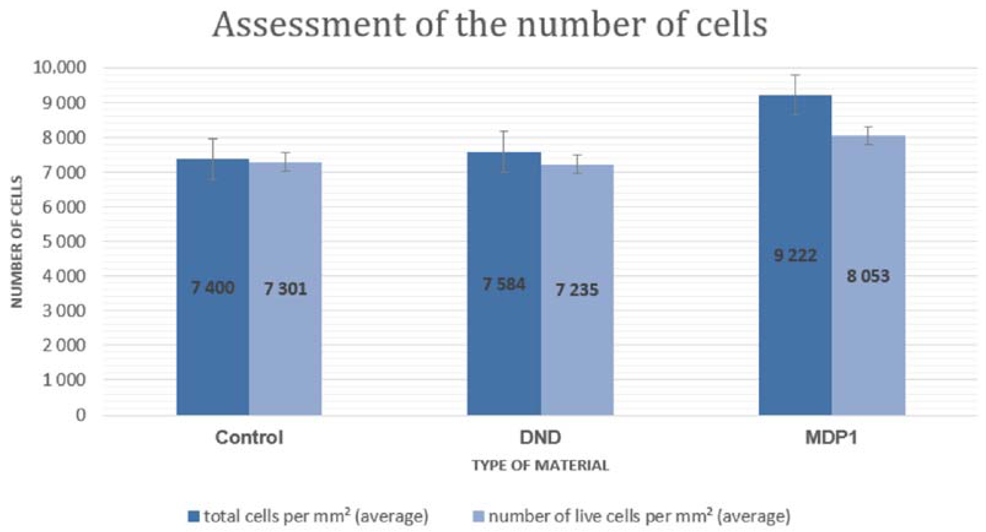
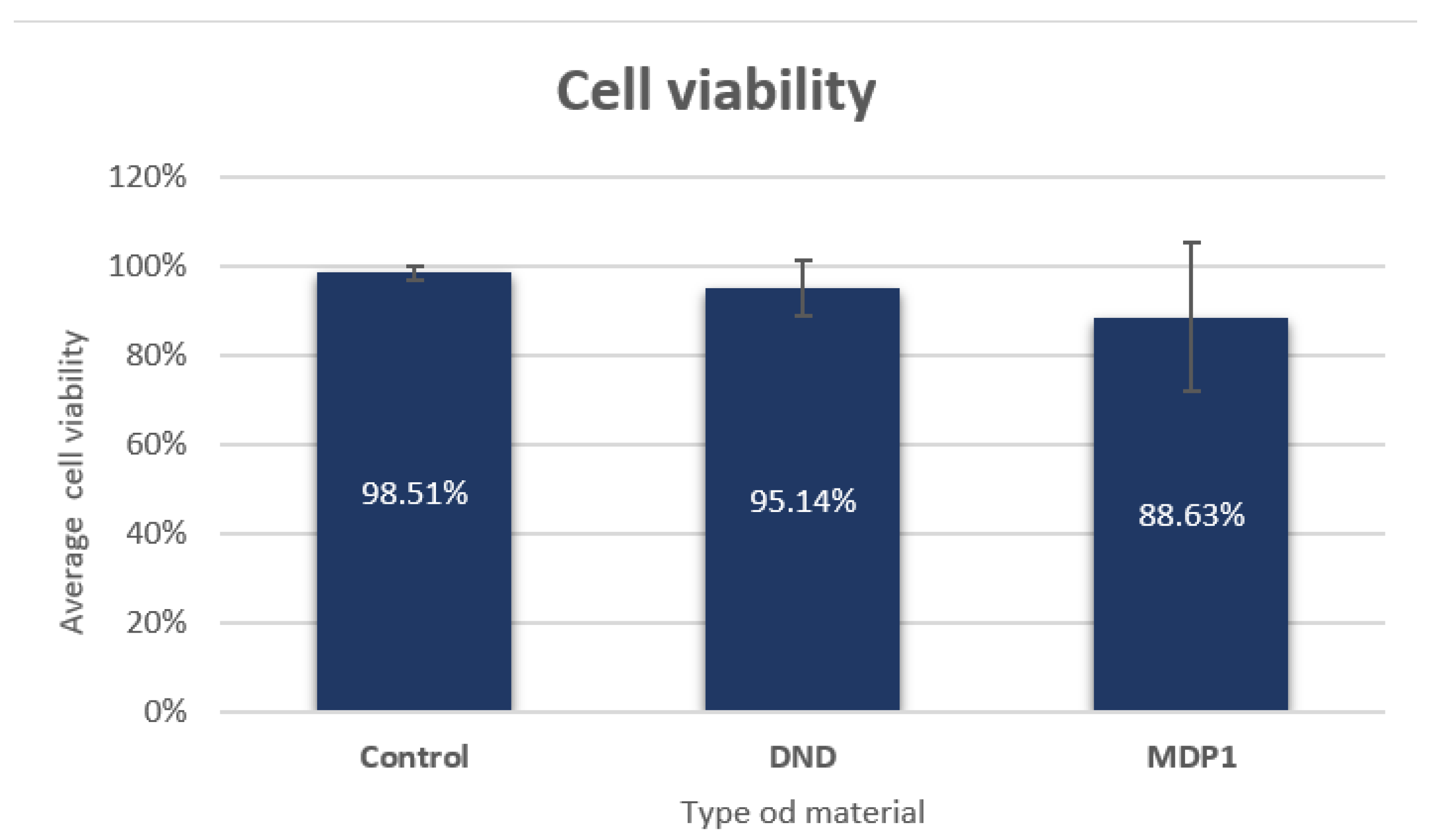
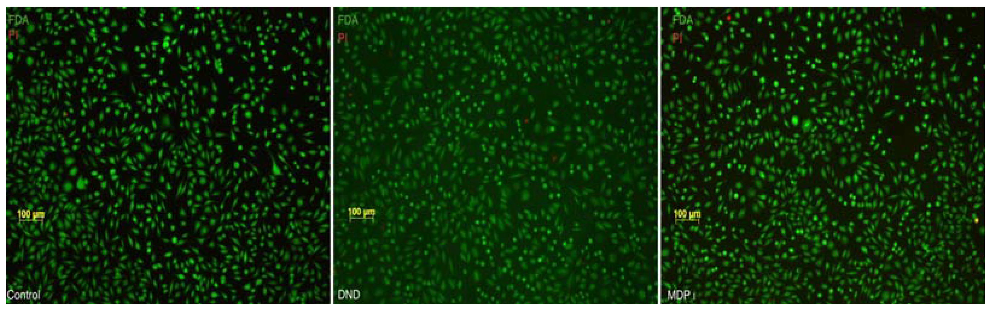
3.1. Cytotoxicity Tests
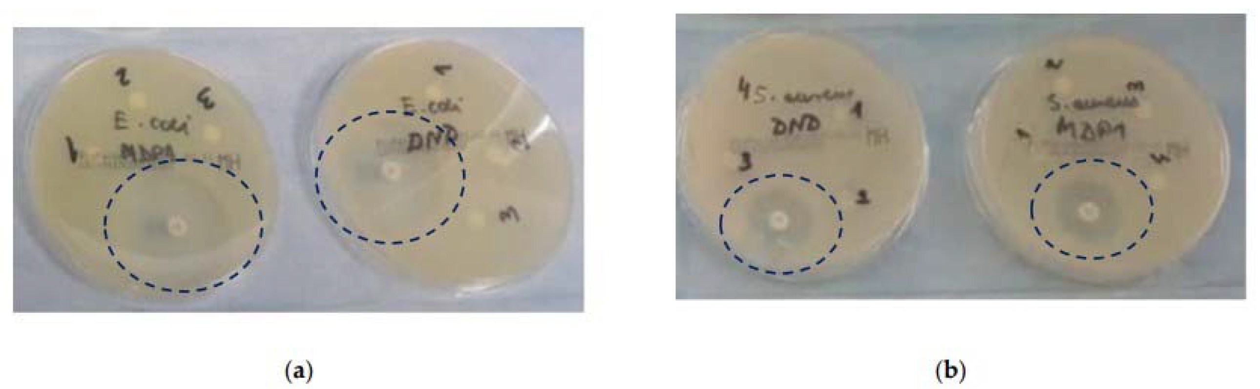
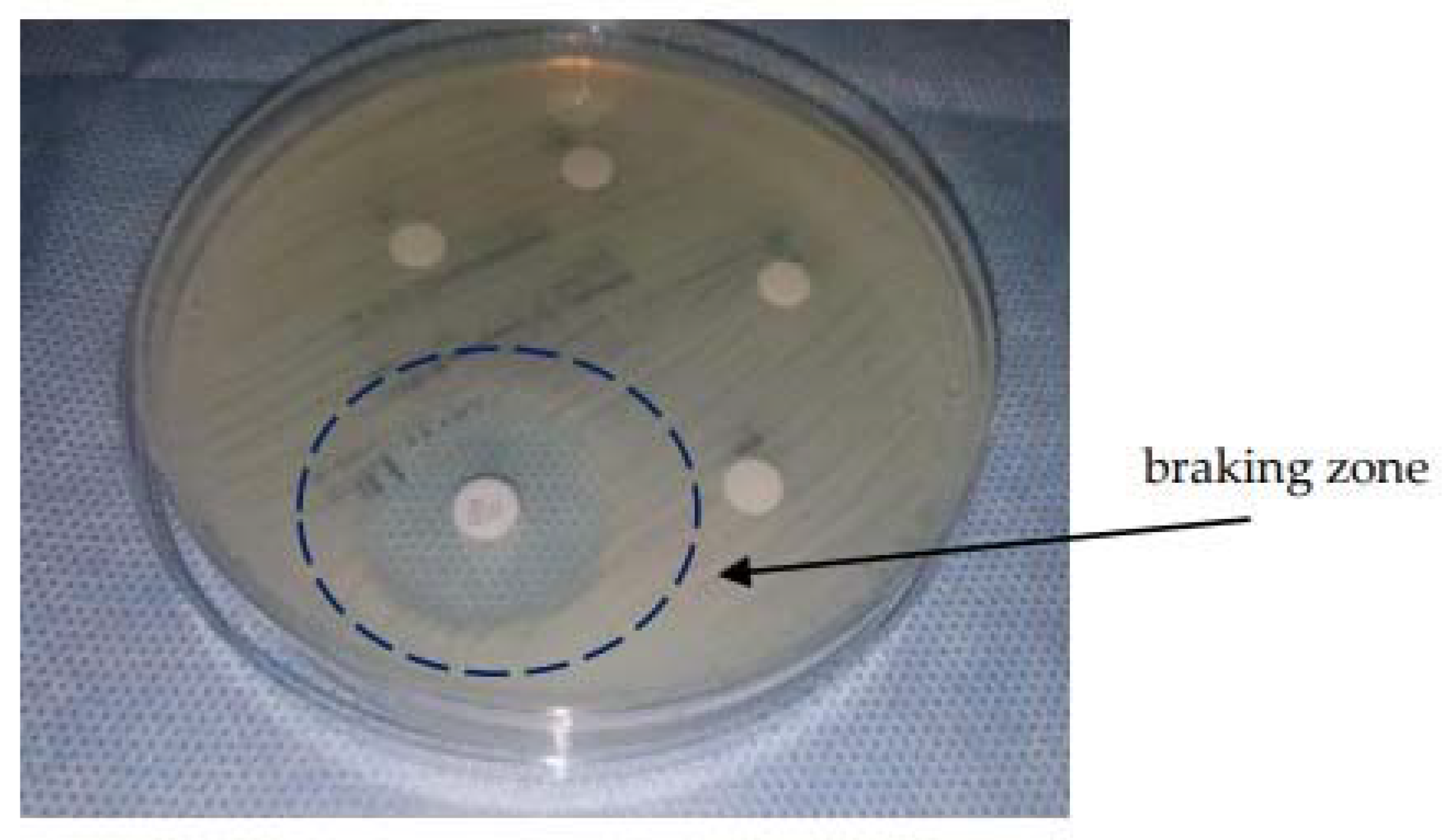
3.2. Bacteriostaticity Test
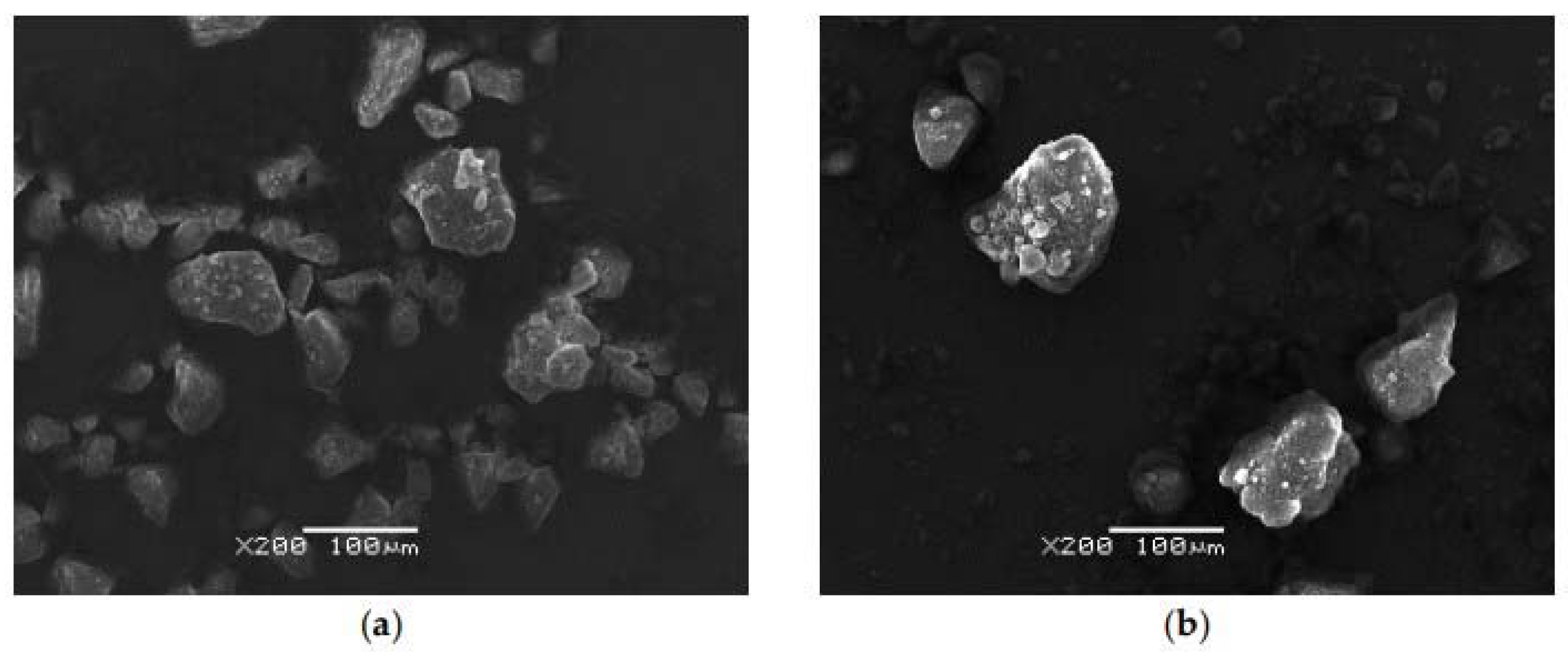
3.3. SEM Analysis
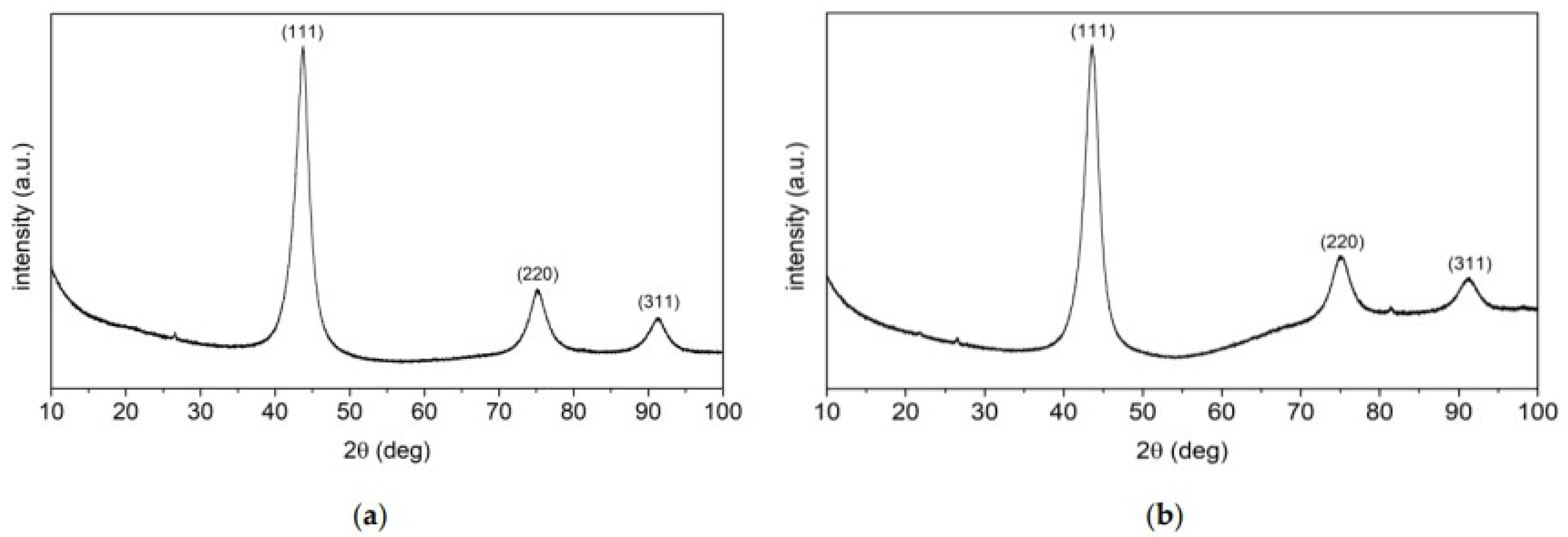
3.4. X-ray Diffraction (XRD) Analysis
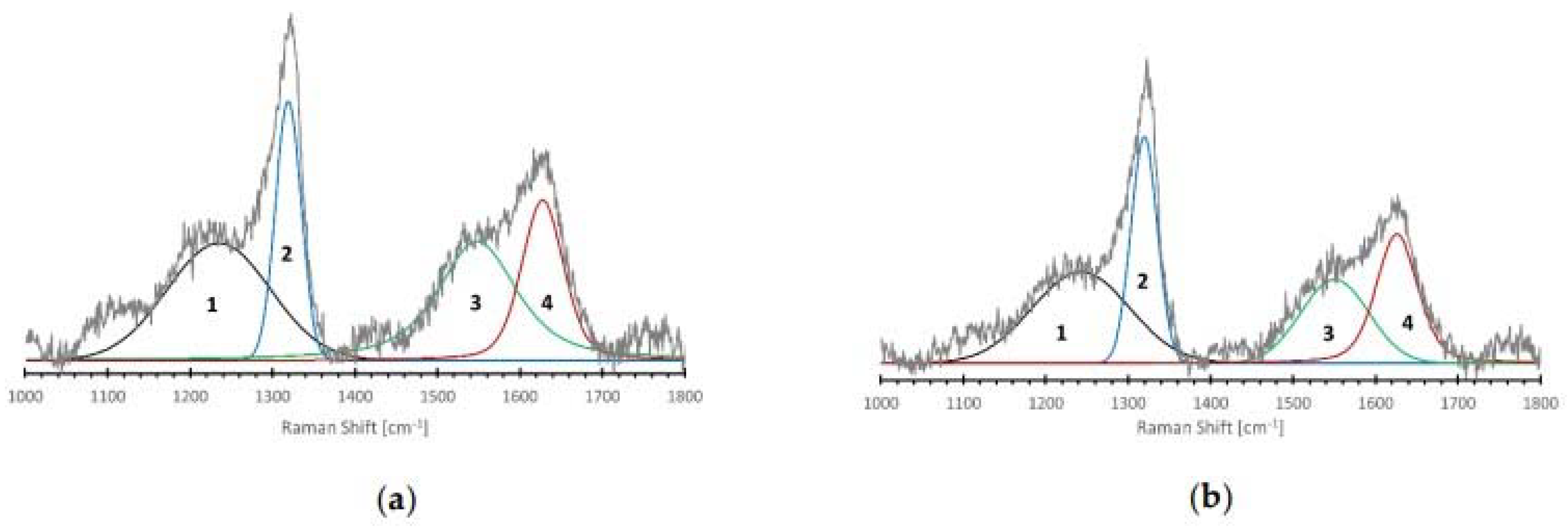
3.5. Raman Spectroscopy Analysis
3.6. FT–IR Spectroscopy Analysis
4. Discussion and Conclusions
Author Contributions
Funding
Institutional Review Board Statement
Informed Consent Statement
Data Availability Statement
Conflicts of Interest
References
- Danilenko, V.V. Nanodiamonds: Problems and prospects. J. Superhard Mater. 2010, 32, 301–310. [Google Scholar] [CrossRef]
- Ceynowa, P.; Mitura, K.; Zinka, W.; Mitura, S. Diamond nanopowder modification system (DPP) in the rotating chamber of a plasma-chemical reactor (MW PACVD). Elektronika 2014, 10, 47–50. [Google Scholar]
- Danilenko, V.V. On the history of the discovery of nanodiamond synthesis. Phys. Solid State 2004, 46, 595. [Google Scholar] [CrossRef]
- Schrand, A.M.; Huang, H.; Carlson, C.; Schlager, J.J.; Osawa, E. Are diamond nanoparticles cytotoxic? J.Phys. Chem B. 2007, 111, 2–7. [Google Scholar] [CrossRef]
- Mitura, K.; Kornacka, J.; Kopczyńska, E.; Kalisz, J.; Czerwińska, E.; Affeltowicz, M.; Kaczorowski, W.; Kolesińska, B.; Frączyk, J.; Bakalova, T. Active Carbon-Based Nanomaterials in Food Packaging. Coatings 2021, 11, 161. [Google Scholar] [CrossRef]
- Niemiec, T.; Szmidt, M.; Sawosz, E.; Grodzik, M.; Mitura, K. The effect of diamond nanoparticles on redox and immune parameters in rats. J. Nanosci. Nanotechnol. 2011, 11, 9072–9077. [Google Scholar] [CrossRef]
- Eivazzadeh-Keihan, R.; Maleki, A.; de la Guardia, M.; Bani, M.S.; Chenab, K.K.; Pashazadeh-Panahi, P.; Baradaran, B.; Mokhtarzadeh, A.; Hamblin, M.R. Carbon based nanomaterials for tissue engineering of bone: Building new bone on small black scaffolds: A review. J. Adv. Res. 2019, 18, 185–201. [Google Scholar] [CrossRef]
- Chauhan, S.; Jain, N.; Nagaich, U. Nanodiamonds with powerful ability for drug delivery and biomedical applications: Recent updates on in vivo study and patents. J. Pharm. Anal. 2020, 10, 1–12. [Google Scholar] [CrossRef]
- Zakrzewska, K.; Samluk, A.; Wierzbicki, M.; Jaworski, S.; Kutwin, M.; Sawosz, E.; Chwalibog, A.; Pijanowska, D.G.; Pluta, K.D. Analysis of the Cytotoxicity of Carbon-Based Nanoparticles, Diamond and Graphite, in Human Glioblastoma and Hepatoma Cell Lines. PLoS ONE 2015. [Google Scholar] [CrossRef] [Green Version]
- Zupančič, D.; Kreft, M.E.; Grdadolnik, M.; Mitev, D.; Iglič, A.; Veranič, P. Detonation nanodiamonds are promising nontoxic delivery system for urothelial cells. Protoplasma 2017, 255, 419–423. [Google Scholar] [CrossRef]
- Norouzi, N.; Ong, Y.; Damle, V.G.; Najafi, M.B.H.; Schirhagl, R. Effect of medium and aggregation on antibacterial activity of nanodiamonds. Mater. Sci. Eng. C 2020, 112, 110930. [Google Scholar] [CrossRef] [PubMed]
- Beranová, J.; Seydlová, G.; Kozak, H.; Benada, O.; Fišer, R.; Artemenko, A.; Konopásek, I.; Kromka, A. Sensitivity of bacteria to diamond nanoparticles of various size differs in gram-positive and gram-negative cells. FEMS Microbiol. Lett. 2014, 351, 179–186. [Google Scholar] [CrossRef] [PubMed] [Green Version]
- Lišková, P.; Beranová, J.; Ukraintsev, E.; Fišer, R.; Kofroňová, O. Diamond nanoparticles suppress lateral growth of bacterial colonies. Colloids Surf. B 2018, 170, 544–552. [Google Scholar] [CrossRef] [PubMed]
- Wehling, J.; Dringen, R.; Zare, R.N.; Maas, M.; Rezwan, K. Bactericidal Activity of Partially Oxidized Nanodiamonds. ACS Nano 2014, 8, 6475–6483. [Google Scholar] [CrossRef]
- Jariwala, D.H.; Patel, D.; Wairkar, S. Surface functionalization of nanodiamonds for biomedical applications. Mater. Sci. Eng. C 2020, 113, 110996. [Google Scholar] [CrossRef]
- Mitura, K.; Jędrzejewska-Szczerska, M.; Ceynowa, P.; Dudek, M.; Cicha, M. Hemokompatibility of non-functionalized and plasma-chemical functionalized detonation nanodiamond particles. Arch. Metall. Mater. 2015, 60, 73–79. [Google Scholar] [CrossRef] [Green Version]
- Mitura, K.; Włodarczyk, E. Fluorescent Nanodiamonds in Biomedical Applications. J. AOAC Int. 2018, 101, 1297–1307. [Google Scholar] [CrossRef]
- Bajpai, V.K.; Kamle, M.; Shukla, S.; Mahato, D.K.; Chandra, P.; Hwang, S.K.; Kumar, P.; Huh, Y.S.; Han, Y.-K. Prospects of using nanotechnology for food preservation, safety, and security. J. Food Drug Anal. 2018, 26, 1201–1214. [Google Scholar] [CrossRef]
- Khaneghaha, A.M.; Hashemi, S.M.B.; Limbo, S. Antimicrobial agents and packaging systems in antimicrobial active food packaging: An overview of approaches and interactions. Food Bioprod. Process. 2018, 111, 1–19. [Google Scholar] [CrossRef]
- Aliofkhazraei, M.; Ali, N.; Milne, W.I.; Ozkan, C.S.; Mitura, S. Graphene Science Handbook; CRC Press: Boca Raton, FL, USA, 2016. [Google Scholar]
- Kurantowicz, N.; Sawosz, E.; Jaworski, S.; Kutwin, M.; Strojny, B.; Wierzbicki, M.; Szeliga, J.; Hotowy, A.; Lipińska, L.; Koziński, R. Interaction of graphene family materials with Listeria monocytogenes and Salmonella enterica. Nanoscale Res. Lett. 2015, 10, 23. [Google Scholar] [CrossRef] [Green Version]
- Jaworski, S.; Sawosz, E.; Kutwin, M.; Wierzbicki, M.; Hinzmann, M.; Grodzik, M.; Winnicka, A.; Lipinska, L.; Wlodyga, K.; Chwalibog, A. In vitro and in vivo effects of graphene oxide and reduced graphene oxide on glioblastoma. Int. J. Nanomed. 2015, 10, 1585–1596. [Google Scholar] [CrossRef] [Green Version]
- Maas, M. Carbon Nanomaterials as Antibacterial Colloids. Materials 2016, 9, 617. [Google Scholar] [CrossRef] [PubMed] [Green Version]
- Szunerits, S.; Barras, A.; Boukherroub, R. Antibacterial Applications of Nanodiamonds. Int. J. Environ. Res. Public Health 2016, 13, 413. [Google Scholar] [CrossRef] [PubMed]
- Plotnikov, V.A.; Makarov, S.V.; Bogdanov, D.G.; Bogdanov, A.S. The structure of detonation nanodiamond particles. In Proceedings of the 10th International Conference on Mechanics, Resource and Diagnostics of Materials and Structures, Ekaterinburg, Russia, 16–20 May 2016; AIP: New York, NY, USA, 2016. [Google Scholar] [CrossRef] [Green Version]
- Mitura, K. HRTEM examinations of nanodiamond particles for biomedical application. J. Achiev. Mater. Manuf. Eng. 2009, 37, 317–322. [Google Scholar]
- Bogdanov, D.; Plotnikov, V.; Bogdanov, A.; Makarov, S.; Vins, V.; Yelisseyev, A.; Lin, V.; Chepurov, A. Consolidation of nanocrystals of detonation diamonds at high-pressure high-temperature sintering. Int. J. Refract. Met. Hard Mater. 2018, 71, 101–105. [Google Scholar] [CrossRef]
- Kryshtal, A.P.; Mchedlov-Petrossyan, N.O.; Laguta, A.N.; Kriklya, N.N.; Kruk, A.; Osawa, E. Primary detonation nanodiamond particles: Their core-shell structure and the behavior in organo-hydrosols. Colloids Surf.A Physicochem. Eng. Asp. 2020, 614, 126079. [Google Scholar] [CrossRef]
- Korepanov, V.I.; Hamaguchi, H.; Osawa, E.; Ermolenkov, V.; Lednev, I.K.; Etzold, B.J.M.; Levinson, O.; Zousman, B.; Epperla, C.P.; Chang, H.-C. Carbon structure in nanodiamonds elucidated from Raman spectroscopy. Carbon 2017, 121, 322–329. [Google Scholar] [CrossRef]
- Lambert, J.B. Introduction to Organic Spectroscopy; Macmillan: New York, NY, USA, 1987. [Google Scholar]
- Kartick, B.S.; Srivastava, S.K.; Srivastava, I. Green Synthesis of Graphene. J. Nanosci. Nanotechnol. 2013, 13, 4320–4324. [Google Scholar] [CrossRef]
- Ou, J.; Yan Bing, H.; Lan Zhen, H.; Zhang, R.; Xu, T.; Zhao, J. pH-sensitive nanocarriers for Ganoderma applanatum polysaccharide release via host–guest interactions. J Mater Sci. 2018, 53, 7963–7975. [Google Scholar] [CrossRef]
- Kumaravel, V. Antimicrobial activity of menthol modified nanodiamond particles. Chem. Eng. Sci. 2021, 416, 129071. [Google Scholar] [CrossRef]
- Solarska, K.; Gajewska, A.; Kaczorowski, W.; Bartosz, G.; Mitura, K. Effect of nanodiamond powders on the viability and prodution of reactive oxygen and nitrogen species in endothelial cells by unmodified and Fenton modified ultradisperse detonation diamond. Diam. Relat. Mater. 2012, 21, 9037–9046. [Google Scholar] [CrossRef]


| Degree of Cytotoxicity | Reactivity | Cell Condition |
|---|---|---|
| 0 | not available | Discrete intra-plasmatic granules, no lysis, no reduction in cell growth. |
| 1 | slight | No more than 20% round cells, loosely suffused without intracytoplasmic granules, showing morphological changes, few cell lysis, little inhibition of cell growth. |
| 2 | mild | Not more than 50% round cells, devoid of intra-plasmacytic granules, strong cell lysis, not more than 50% inhibition of cell growth. |
| 3 | moderate | Not more than 70% of surface containing round cells and lysed, not completely damaged, cell growth inhibition greater than 50%. |
| 4 | strong | Almost complete and total cell destruction |
| Type of Powder | Amount of Sample Weighed |
|---|---|
| DND | 0.051 g |
| MDP1 | 0.026 g |
| Sample | DND | MDP1 | |
|---|---|---|---|
| Amorphous sp3 peak FWHM/Position [cm−1] | (1) | 147/1235 | 139/1242 |
| Diamond peak FWHM/Position [cm−1] | (2) | 37/1319 | 37/1319 |
| GLS peaks FWHM/Position [cm−1] | (3) | 112/1548 | 97/1550 |
| (4) | 59/1627 | 59/1625 | |
| Diamond to amorphous ratio | 0.55 | 0.67 | |
| Diamond to GLC ratio | 0.33 | 0.49 | |
| ID/IG | 0.93 | 1.24 | |
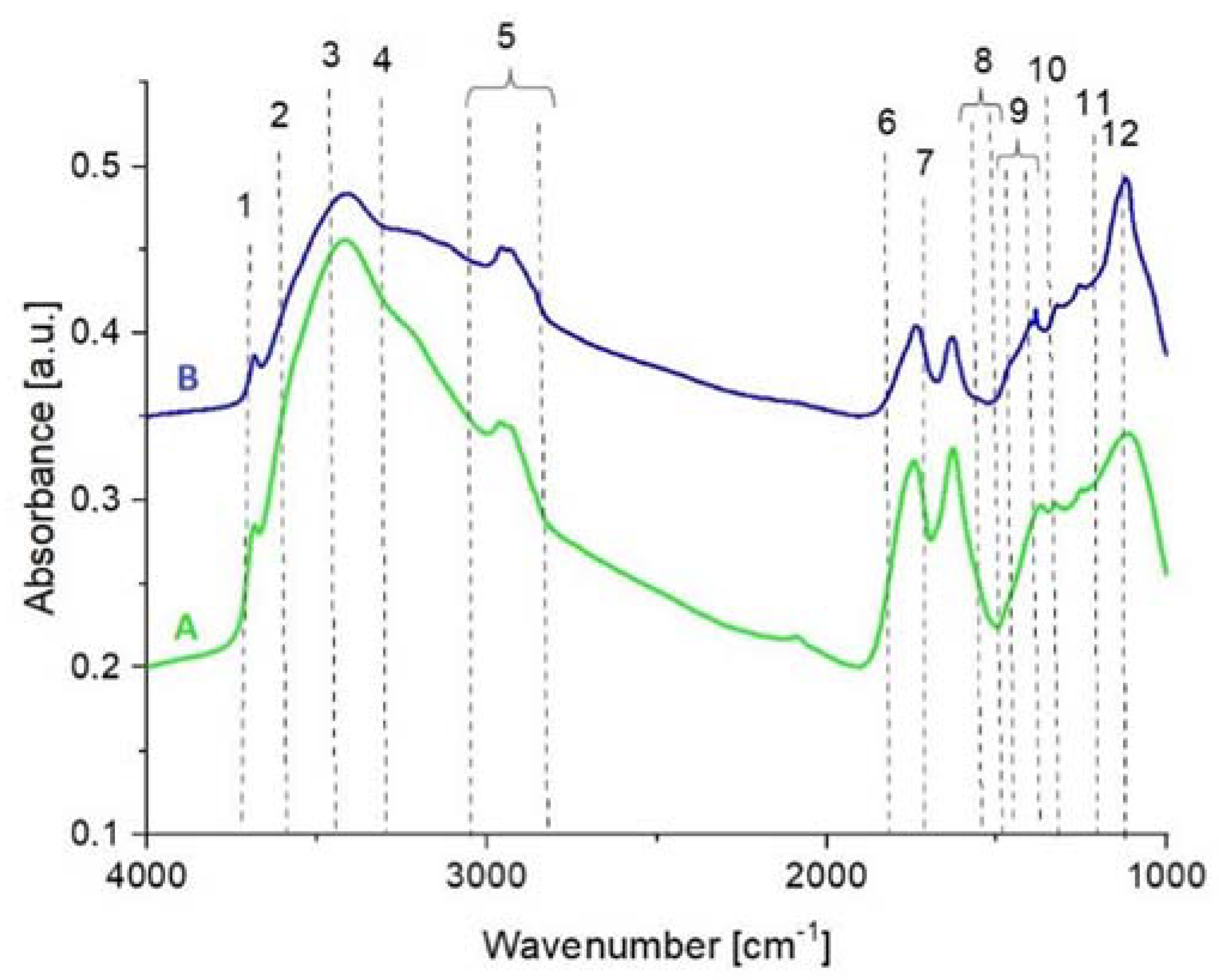
| No. in Fig. X | Vibrational Mode | Wavenumber (cm−1) | A | B |
|---|---|---|---|---|
| 1 | Stretching O–H (from isolated water) | 3680 | + | + |
| 2 | Stretching O–H (water on Lewis acid sites) | 3640 | − | − |
| 3 | Stretching O–H (like in alkohols or fenols) | 3400 | + | + |
| 4 | Stretching O–H (with strong intermolecular bonding | 3290 | + | + |
| 5 | Stretching CH3 and CH2 (asym and sym) | 3000–2800 | + | + |
| 6 | Stretching C=O (in esters) | 1740 | + | + |
| 7 | Stretching C=C | 1630 | + | + |
| 8 | Deformation CH2 and CH3 | 1480–1440 | + | + |
| 9 | Stretching C–O (in carboxylic anhydride) | 1385–1370 | + | + |
| 10 | bending C–O (in ester) | 1270–1250 | + | + |
| 11 | Stretching C–O–C | 1124 | + | + |
| 12 | Stretching C–O–C | 1080 | − | − |
Publisher’s Note: MDPI stays neutral with regard to jurisdictional claims in published maps and institutional affiliations. |
© 2022 by the authors. Licensee MDPI, Basel, Switzerland. This article is an open access article distributed under the terms and conditions of the Creative Commons Attribution (CC BY) license (https://creativecommons.org/licenses/by/4.0/).
Share and Cite
Mitura, K.; Kornacka, J.; Niemiec-Cyganek, A.; Pawlus-Łachecka, L.; Mydłowska, K.; Sobczyk-Guzenda, A.; Kaczorowski, W.; Ossowska, P.; Bałasz, B.; Wilczek, P. The Influence of Diamond Nanoparticles on Fibroblast Cell Line L929, Cytotoxicity and Bacteriostaticity of Selected Pathogens. Coatings 2022, 12, 280. https://doi.org/10.3390/coatings12020280
Mitura K, Kornacka J, Niemiec-Cyganek A, Pawlus-Łachecka L, Mydłowska K, Sobczyk-Guzenda A, Kaczorowski W, Ossowska P, Bałasz B, Wilczek P. The Influence of Diamond Nanoparticles on Fibroblast Cell Line L929, Cytotoxicity and Bacteriostaticity of Selected Pathogens. Coatings. 2022; 12(2):280. https://doi.org/10.3390/coatings12020280
Chicago/Turabian StyleMitura, Katarzyna, Joanna Kornacka, Aleksandra Niemiec-Cyganek, Lucyna Pawlus-Łachecka, Katarzyna Mydłowska, Anna Sobczyk-Guzenda, Witold Kaczorowski, Paulina Ossowska, Błażej Bałasz, and Piotr Wilczek. 2022. "The Influence of Diamond Nanoparticles on Fibroblast Cell Line L929, Cytotoxicity and Bacteriostaticity of Selected Pathogens" Coatings 12, no. 2: 280. https://doi.org/10.3390/coatings12020280