Article

Antimicrobial Activity and Molecular Docking Studies of the Biotransformation of Diterpene Acanthoic Acid Using the Fungus Xylaria sp.

by
Andrey Moacir do Rosario Marinho
1,*,
Claudia Maria S. C. de Oliveira
2,
João Victor Silva-Silva
3,*,
Samara C. Anchieta de Jesus
1,
José Edson S. Siqueira
1,
Luana C. de Oliveira
1,
Jéssica Fernandes Auzier
4,
Liviane N. Soares
4,
Maria Lúcia Belém Pinheiro
4,
Sebastião C. Silva
2,
Lívia S. Medeiros
5,
Emmanoel V. Costa
4 and
Patrícia S. Barbosa Marinho
1
1
Post-Graduation in Chemistry, Federal University of Pará, Belém 66075-110, PA, Brazil
2
Post-Graduation in Chemistry, Federal University of South and Southeast of Pará, Marabá 68507-590, PA, Brazil
3
Laboratory of Medicinal and Computational Chemistry, Institute of Physics of São Carlos, University of São Paulo, São Carlos 13418-900, SP, Brazil
4
Post-Graduation in Chemistry, Federal University of Amazonas, Manaus 69077-000, AM, Brazil
5
Post-Graduation in Chemistry, Federal University of São Paulo, Diadema 09920-000, SP, Brazil
*
Authors to whom correspondence should be addressed.
Antibiotics 2023, 12(8), 1331; https://doi.org/10.3390/antibiotics12081331
Submission received: 30 June 2023 / Revised: 4 August 2023 / Accepted: 11 August 2023 / Published: 18 August 2023
(This article belongs to the Special Issue New Insights into Antimicrobial Discovery)

Abstract

:
Biotransformations are reactions mediated by microorganisms, such as fungi. These bioreactions have high chemo- and stereoselectivity on organic substrates and can be applied in the search for new bioactive compounds. In this study, acanthoic acid (AA) was biotransformed using the fungus Xylaria sp., giving the novel compound 3β,7β-dihydroxyacanthoic acid (S1). Both the AA and the product S1 were tested against Gram-positive and Gram-negative bacteria. To identify and validate possible biological targets as enzymes or proteins involved in the activity observed in vitro, we used the molecular docking method. Hydroxylation at the C-3 and C-7 positions of the biotransformation product enhanced its activity against Escherichia coli as well as its binding affinity and interactions with superoxide dismutase 1 (SOD1; PDB ID 4A7G). Based on our results, the SOD1 enzyme was suggested to be a possible target for the antioxidant activity of product S1.

1. Introduction

Biotransformation methods are increasingly being used in chemical procedures for the formation of new products. Biotransformation, or biocatalysts, is a chemical process in which an organic compound is subjected to structural modification via whole-cell or isolated enzymes [1,2]. Whole-cell biocatalysts can be used for different types of processes, such as biotransformation, biodegradation, and fermentation. These processes can involve one or more steps to produce the desired chemicals [3,4].
Xylaria is one of 40 genera belonging to the Xylariaceae family and is globally distributed. Classification at the species level is difficult because of variations in the coloration, shape, size, and developmental stage [5,6]. Previous studies have demonstrated the enzymatic capacity of fungi for the genus Xylaria in biocatalyst processes, such as the biotransformation and biodegradation of the synthetic compounds assisting environmental processes [7] and the biotransformation of saponins [8]. In this context, Xylaria is a promising genus in biotransformation methods [5,6].
Bacterial resistance to antibiotics is a serious public health issue and has resulted in the search for new antibiotic compounds, such as diterpenes, to improve the therapeutic arsenal used in the treatment of bacterial infections [9]. Casbane diterpene showed biocidal and biostatic activity against several clinically relevant species (bacteria and yeasts) [10]. Conidiogenone D diterpene, isolated from solid fermentation cultures of the endophytic fungus Leptosphaeria sp., showed antibacterial activity against Bacillus cereus and Pseudomonas aeruginosa [11]. This bioactive potential has been attributed to the structural diversity of diterpenes [12,13,14].
Some diterpenes have been submitted to biotransformation to improve their bioactivity. For example, the fungal biotransformation of ent-pimaran-dienoic acid resulted in three products, one of which inhibited the growth of the main microbes responsible for tooth decay [15]. Acanthoic acid (AA) is a pimaradiene diterpene with a wide range of pharmacological activities, such as anti-cancer, anti-inflammatory, anti-diabetes, liver protection, gastrointestinal protection, and cardiovascular protection activity. Structurally modified AA has cytotoxic and anti-inflammatory activity [16,17,18].
Thus, natural compounds modified via biotransformation are promising in the search for new bioactive compounds. In this work, we reported, for the first time, the biotransformation of AA diterpene using the endophytic fungus Xylaria sp., leading to the dihydroxylated product 3β,7β-dihydroxyacanoic acid (S1).

2. Results

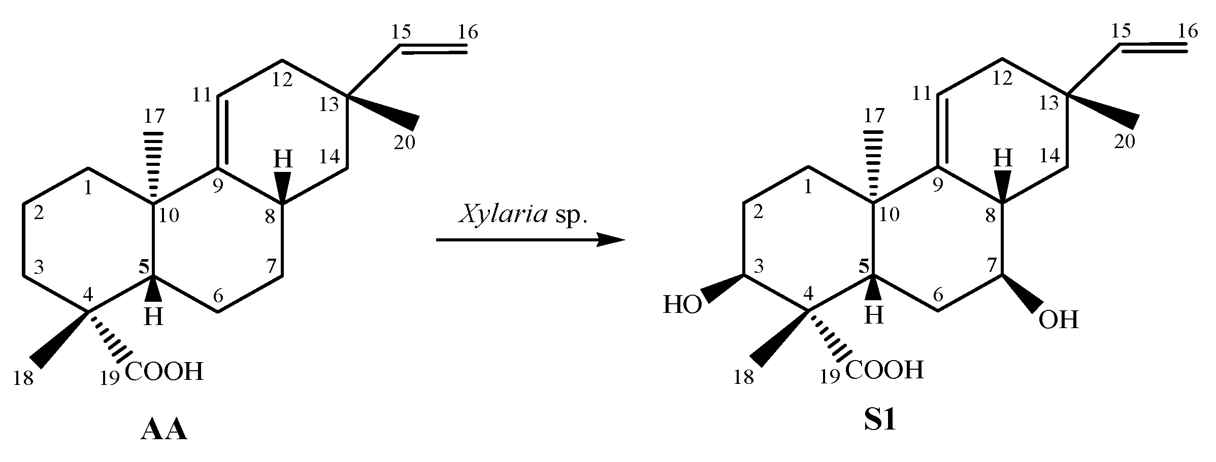
2.1. Identification of Chemical Constituents

The structure of the biotransformation product S1 (Figure 1) was elucidated using spectroscopic data. Detailed analyses of 1D and 2D NMR (one-dimensional and two-dimensional resonance magnetic nuclear) and MS (mass spectrometry) data were performed. Signal attribution was described according to the results of the spectral analysis and using data from the literature (Table 1).
Subsequently, the 1H NMR values of AA and S1 were compared, and the absence of the signals for hydrogens H-2, H-3, H-6, and H-7 was observed. An analysis of 1H and 13C NMR data suggested AA oxidation. Following heteronuclear multiple bond correlation (HMBC) and correlation spectroscopy (COSY), a correlation was observed between all NMR data and compound S1 (Figure 2).

2.2. Antimicrobial Activity

The antimicrobial activity results for both AA and S1 are presented in Table 2. AA demonstrated activity against Bacillus subtilis, with an MIC of 31.25 µg.mL−1, while S1 showed better activity against Escherichia coli, with an MIC of 31.25 µg.mL−1. Both AA and S1 exhibited an MIC of 62.5 µg.mL−1 against Salmonella typhimurium.

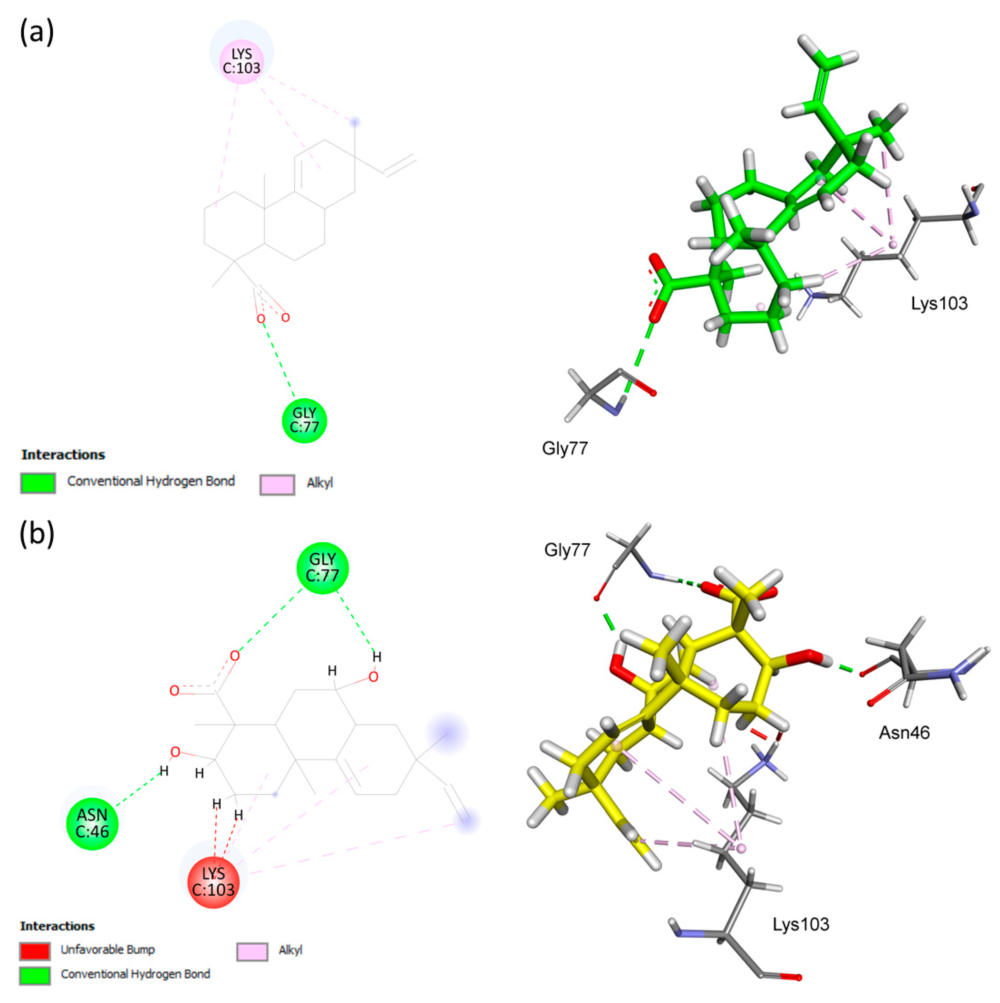
2.3. Molecular Docking

To understand the difference observed in the antimicrobial activities between substrate AA and product S1, without disregarding other possibilities due to the complexity of this system, studies of enzyme regulation were developed through molecular docking using GOLD v. 2020.2.0.
Bactericidal antimicrobials can act by inhibiting DNA synthesis, RNA synthesis, cell-wall synthesis or protein synthesis, and generate reactive oxygen species (ROS) [19,20]. With this in mind, molecular docking simulations were used to show the interaction patterns of AA and S1 with the penicillin-binding protein 2 protein (PBP2; PDB ID 6G9S) [21], DNA gyrase subunit b (GyrB; PDB ID 4DUH) [22], topoisomerase IV (Topo IV; PDB ID 4HZ0) [23] and superoxide dismutase 1 (SOD1; PDB ID 4A7G) [24] in the hope of finding the mode of action for compounds AA and S1 based on antimicrobial activity.
To validate the docking configuration, the co-crystallized inhibitors of each protein were redocked into the active sites of FimH, PBP2, GyrB, Topo IV, and SOD1. This validation confirmed the suitability of the docking protocol used for this study. This was demonstrated by the Root Mean Square Deviation (RMSD) values between the experimental pose of the co-crystallized inhibitor and the docked pose of 1.6146 Å in PBP2 (PDB ID 6G9S), 0.5448 Å in GyrB (PDB ID 4DUH), 0.3511 Å in Topo IV (PDB ID 4HZ0), and 0.7531 Å in SOD1 (PDB ID 4A7G), and by the ability of the docking conformation to reproduce all the key interactions achieved by the co-crystallized ligands in the active site.
The analysis of the results from molecular docking simulations revealed that compound AA demonstrated an affinity for the catalytic site region of PBP2 with a docking score value of 50.47. On the other hand, compound S1 showed a better fit in the catalytic region of SOD1, with a docking score value of 42.45. Moreover, the detailed information and interactions of these ligands with PBP2, SOD1, GyrB, and Topo IV proteins are provided in Table 3.
In Figure 3, possible chemical interactions that explain the binding of AA and S1 to the active site of the penicillin-binding protein 2 can be observed. At the Trp370 residue, hydrophobic interactions of the Pi-Alkyl and Pi-Sigma type with aromatic systems occurred. Additionally, there were hydrogen–bonding interactions with the carboxyl functional group of AA. In the case of S1, fewer interactions with the active site residues were identified. In this case, the aromatic systems interacted with the methyl group at position C-10, and hydrogen–bonding interactions occurred between Ser387 and the carbonyl group present in the structure of S1 (Figure 4).
In the binding site of the DNA gyrase subunit B (gyrB) protein, the AA ligand was involved in three alkyl-type hydrophobic interactions with Lys103, while hydrogen–bonding interactions with Gly77 were recognized, interacting with the ligand’s carbonyl group. Furthermore, hydrogen–bonding interactions between Asn46 and Gly77, as well as hydrophobic binding to Lys103 in different regions of S1, as shown in Figure 4, were also observed.
The interactions between AA and S1 with the residues present in the catalytic site of topoisomerase IV can be observed in Figure 5. Hydrophobic pi–alkyl interactions between His95 and the vinyl group C15=C16 of AA differed from the hydrogen bonding between the residue Asn42 and the carbonyl group of S1.
Figure 6 illustrates that AA binds tightly through a hydrophobic interaction with the His110 residue, which exhibits the pi–alkyl interaction. Furthermore, AA forms hydrogen–bonding interactions with the Ser25 residue. Both AA and S1 exhibited a similar binding site with the Ser25 residue, but in the S1 compound, most of the interactions had shorter distances compared to AA (Table 3). Additionally, S1 strongly interacted through hydrophobic binding with Val103 (alkyl interaction) and His110 (pi-alkyl and pi–pi stacked interactions) and through hydrogen bonding with Val103, Ile104, Ser105, Asp109, and His110, which was not observed for AA (Figure 7).
To complete the analysis, we reiterated from the hydrophobic surface and hydrogen bond diagrams (Figure 7) that compound S1 showed effective interactions in the active site of the SOD1 of 4A7G. These results indicate that the structural contrast between AA and S1, with hydroxylation at C-3 and C-7 positions of the biotransformation product S1, increased the binding.

3. Discussion

The high-resolution mass spectrometry–electrospray negative mode, HRMS-ESI (-), spectrum of S1 showed m/z 333.2067 [M-H]- and IR spectrum was observed with an OH bond stretching band at 3.423 cm−1; these data are compatible to the molecular formula C20H30O4. This translates into two additional hydroxyl groups in the molecule of S1 compared to AA, which was confirmed by NMR data analysis. A comparison between the 1H NMR spectra of AA and S1 showed chemical shifts at δH 0.95 (s) and δH 0.97 (s), which could be attributed to the methyl groups Me-17 and Me-20, respectively; their signals did not suffer significant changes. For methyl Me-18, we observed an increase in the shift from δH 1.24 to δH 1.36 (Δ = +0.12). The signals for vinylic hydrogens of the olefin group were observed as a double doublet (dd) at δH 5.85 (J = 17.4 and 10.7 Hz, 1H) to H-15, δH 4.91 (cis, J = 10.7 and 1.2 Hz, 1H) and δH 4.96 (trans, J = 17.5 and 1.2 Hz, 1H) to H-16a and H-16b. The signals at δH 1.85 and 2.08 (J = 17.4, 4.0 and 2.3 Hz, 1H) were attributed to hydrogens H-12a and H-12b.
Based on the analysis of 13C and 2D NMR data, the signals characteristic to methyl groups at δC 21.6, δC 23.6, and δC 22.5 could be attributed to Me-17, Me-18, and Me-20, respectively. The signal at δ 181.0 was typical to the carboxyl group of pimarane diterpenes and was attributed to C-19. For the olefin carbons, we observed signals at δC 145.7 (C-9), δC 119.9 (C-11), δC 149.7 (C-15), and δC 109.6 (C-16). The chemical shifts in the carbon C-10, C-12, and C-13 to S1 were similar to those observed for substrate AA. The signal for oximetinic carbon at δC 70.5 was attributed to C-3, according to the HMBC to Me-18 to 3J HMBC, as well as the HMBC with C-4 (δC 47.6). In the heteronuclear single quantum coherence (HSQC) spectrum, the signal at δH 4.12 correlated with δC 70.5 and was attributed to H-3. The signal at δC 39.5 was attributed to C-5 based on the HMBC between Me-17 and C-5. The location of the second hydroxylation was defined through the HMBC of H-5 and H-14 with the signal at δC 72.6 (C-7); this showed the HSQC correlation with signal δH 3.65, which was to H-7. Moreover, a correlation spin–spin 1H–1H was observed for H-5, H-6, H-7, H-8, and H-14, confirming that the second hydroxylation was located at the C-7 position. Chemical shifts in the carbon C-1 (δC 34.8) and C-2 (δC 27.5) were compared with data from the literature, showing a similarity to pimarane diterpenes with the C-3 position being hydroxylated [25,26,27,28,29]. The NMR, MS and IV spectra for S1 are available in Supplementary Materials.
The configuration of carbon C-3 and C-7 was determined based on the coupling constant observed. For H-3 (t, J = 2.8 Hz), equatorial–equatorial coupling with H-2a and H-2b was suggested. The coupling constant for H-7 (ddd, J = 10.1, 9.0, and 4.9 Hz) showed axial–axial coupling with H-6a and H-8 and axial–equatorial coupling with H-6b. Thus, both OH-3 and OH-7 were β-oriented [30,31,32,33,34]. Therefore, the product S1, 3β,7β-dihydroxy-acanthoic acid is novel and reported here for the first time. The main HMBC and COSY correlations for S1 are shown in Figure 2.
Among the natural secondary metabolites, diterpenoids hold significant importance due to their broad spectrum of antimicrobial activity. In recent years, several in vitro studies have shown that diterpenoid compounds have the capability to inhibit the growth of different strains of antibiotic-resistant bacteria, which have emerged due to the indiscriminate use of antibiotics [35]. Biotransformation enables the generation of a variety of structural analogs that can be evaluated for their antimicrobial activity, thereby expanding the diversity of compounds available for the development of new therapeutic agents against microbial infections [36,37]. In this context, the fungus Xylaria sp. was employed to biotransform diterpene acanthoic acid, leading to the introduction of hydroxylation at the C-3 and C-7 positions of the resulting compound S1. This modification resulted in a more active molecule against Gram-negative bacteria. Specifically, hydroxylation at these positions improved the antibacterial activity of S1 against E. coli when compared to AA.
Studies have reported that bactericidal antimicrobials have an inhibitory action on DNA synthesis, RNA synthesis, cell wall synthesis, and protein synthesis, as well as generating ROS [19,20]. These antimicrobial activities may involve the inhibition of DNA gyrase (Gyr) and topoisomerase IV (Topo IV), two essential enzymes for bacterial survival, as they perform complementary functions in bacterial DNA processing [38]. Penicillin-binding protein 2 (PBP2) is involved in the inhibition of bacterial cell wall synthesis and belongs to the group of enzymes known as transpeptidases. Penicillin-class antibiotics inhibit transpeptidases, resulting in the disruption of proper bacterial cell wall formation and consequential bacterial death [39]. This mechanism of action demonstrates its overall effectiveness against various bacterial species, including E. coli [21]. Additionally, bacteria employ defense mechanisms, such as superoxide dismutase (SOD), to prevent oxidative stress caused by ROS and maintain a cellular redox balance [40].
To obtain a better understanding of the mechanism by which S1 exhibited superior inhibition against E. coli, in silico enzymatic evaluation assays were developed using molecular docking studies. This molecular docking technique was highly useful in determining the binding affinity of drug molecules to the biological target [41]. In this case, the docking simulations evaluated the binding patterns of diterpenoids at the active sites of proteins 6G9S (PBP2), 4DUH (GyrB), 4HZ0 (Topo IV), and 4A7G (SOD1).
In this study, a difference in affinity between AA and S1 for the active site region of the analyzed proteins was observed. An analysis of the molecular docking simulations revealed that AA showed an affinity for the catalytic region of PBP2 (PDB ID 6G9S), while S1 exhibited a better fit in the catalytic region of SOD1 (PDB ID 4A7G). It can be observed that AA interacts with residues Trp370, Ser387, and Ser545, which are part of the active site structure of PBP2 from E. coli (Ser330, Thr331, Val332, Lys333, Arg368, Asp369, Trp370, Lys371, Ser387, Ala388, Asp389, Ile453, Gly454, Gln455, Gly456, Lys544, Ser545, Gly546, and Thr547) [21]. This result suggests that the inhibition pathway of AA likely has an affinity for the same active site region as penicillin-class antibiotics [39], indicating its role in disrupting the proper formation of the bacterial cell wall. The docked pose clearly showed that both AA and S1 bound within the active site of the SOD1; however, the binding mode was improved in S1 because of significant hydrogen bonding within the catalytic active site with Ser25, Val103, Ile104, Ser105, Asp109, and His110. This result suggests that S1 inhibits E. coli via the superoxide dismutase pathway. Hydrogen bonds play a central role in protein–ligand binding affinity, including enzymatic catalysis, to stabilize a ligand in a binding pocket [42,43]. Published results regarding the antimicrobial activity of pimarane diterpenes against different bacteria indicate that the presence of a hydrogen-bond donor group (HBD) is important for the antimicrobial activity of diterpenes. Moreover, the distance between HDB groups can interfere with antimicrobial activity [44]. Based on the observations above, we could assume that S1 showed improved activity against E. coli because of additional HDB groups at C-3 and C-7, favoring the chemical interactions between S1 and the protein target by the formation of a stable enzyme–substrate complex.
Therefore, molecular docking simulations provided a clearer understanding of the mechanisms of action for diterpenes AA and S1 on target proteins, revealing valuable insights into their interactions at the active site. These results contribute to a better comprehension of the results obtained in the in vitro tests. However, further experimental studies are needed to confirm the proposed association between SOD1 and the observed antioxidant activity of S1.

4. Materials and Methods

4.1. Microorganisms

The fungus Xylaria sp. (code EJCP07) was obtained from the collection of the Laboratory of Bioassays and Chemistry of the Microorganisms (LaBQuiM), Federal University of Pará, Brazil. It was reactivated for 7 days in a Petri dish containing BDA medium to be used in the biotransformation reactions in this work.

4.2. Acanthoic Acid

The diterpene used as a substrate in this work was an authentic sample of acanthoic acid (AA) isolated from Annona amazonica (Annonaceae) in a previous study [45].

4.3. Biotransformation Reactions Procedure

Biotransformation was performed in five 500 mL Erlenmeyer flasks, each containing 200 mL of the Czapek medium. The flasks were autoclaved for 15 min at 121 °C and 1 atm pressure. Subsequently, three small disks of 2 mm2, containing Xylaria sp. (EJCP07) mycelium, were added to two Medium + Fungus + Substrate (MFS) flasks and used for biotransformation with one Medium + Substrate (MS) flask (control). The Medium (M), Medium + Fungus (MF) flasks were also used as control. The flaks were placed on an orbital shaker (Quimis Q315IA) at 32 °C and 150 rpm. After 3 days, 30 mg (per flask) of the AA was solubilized in DMSO and added to flasks MFS and MS. The flaks remained on the shaker for another 5 days, and after this, the mixtures were filtered to obtain the mycelium and the aqueous phase. The mycelium was discarded, and the aqueous phase was used for liquid–liquid extraction with ethyl acetate (EA) (3 × 100 mL). After this, the EA phase was dried with anhydrous sodium sulfate (Na2SO4), filtered, and concentrated in a rotary evaporator to obtain the EA phase of the biotransformation extract (PEABE).

4.4. Fractionation of PEABE Extract and Biotransformation Product Purification

We obtained 40.5 mg of the PEABE extract, which was submitted to an exclusion column chromatograph Sephadex LH-20 eluted with methanol, resulting in 11 fractions (Fr1–Fr11). The fractions were analyzed via TLC and 1H NMR, and the biotransformation product was detected in fraction Fr5 (16.7 mg). This fraction was then fractionated using a silica gel column chromatography, increasing the polarity gradient elution of Hex/EtAcO 40% (100 mL), Hex/EtAcO 60% (60 mL), EtAcO 100% (50 mL), EtAcO/MeOH (50%) and MeOH 100% (50 mL), which resulted in 19 fractions (Frs1–Frs19). After TLC, similar fractions were pooled, and the biotransformation product S1 was obtained (1.7 mg).

4.5. Characterisation of the Compounds

The 1D and 2D NMR spectra were obtained using a Bruker Ascend 400 (400 MHz) nuclear magnetic resonance (NMR) spectrometer (Bruker, Fällanden, Switzerland) with chloroform-d as the solvent. The HRMS values were obtained on a MicroTOF-QII (Bruker Daltonics, EUA) equipped with an electrospray source (ESI) operating in the negative ion mode.

4.6. Antimicrobial Assay

The antimicrobial susceptibility test was carried out using the microbroth dilution assay [46]. Tests were performed on 96-well plates with 100 μL of Mueller–Hinton broth (MHB) (HiMedia, Mumbai, India), 100 μL of the test compound, and 5 μL of test bacteria at 1.0 × 10⁴ CUF/mL, followed by incubation at 37 °C (24 h). The compounds were dissolved (initially 1 mg) in 100 μL of dimethyl sulfoxide (DMSO) and 900 μL of the brain heart infusion (BHI) broth, resulting in a 1 mg/mL stock solution. The stock solution was diluted from 500 to 7.81 µg/mL for testing. E. coli (ATCC 25922), S. typhimurium (ATCC 14028), and B. subtilis (ATCC 6633) were provided by Evandro Chagas Institute, Belém, Pará State, Brazil. Bioactivity was registered as the absence of red coloration in the wells after the addition of 10 μL of 2,3,5-triphenyltetrazolium chloride. The microorganisms were then sub-cultured on MHB plates. The activities of the test compounds were classified as bacteriostatic or bactericidal according to the behavior of the microorganisms in these sub-cultures. Amoxicillin and terramycin were used as positive controls, and the MHB culture medium was used as a negative control.

4.7. Molecular Docking

The molecules (AA and the biotransformation product S1) were drawn using ChemDraw Professional 16.0 and were optimised via Avogadro (version 1.2.0) with a UFF (Universal Force Field) up to dE = 1 × 10⁻¹² kJ/mol and exported as mol2 [47]. Molecular docking simulations were performed using the GOLD v. 2022.1 program, which is available free of charge from CSDS (bdec.dotlib.com.br/inicio_csds/application/Hermes (accessed on 25 February 2022)). Four different scoring functions (ASP, ChemScore, GoldScore and ChemPLP) were applied in enzyme–substrate interaction calculations, as described by Silva-Silva et al. [48]. Protein structures (PDB ID 6G9S, 2.00 Å; PDB ID 4DUH, 1.50 Å; PDB ID 4HZ0, 2.20 Å; PDB ID 4A7G, 1.24 Å) were treated as rigid, and the compounds were treated as fully flexible. Only chain A of the receptor was used, and no crystallographic water molecules were considered [49,50]. The binding site was defined as all receptor atoms up to 10 Å of the reference crystallographic inhibitor. At least 10 poses were generated for the ligand, using the default parameters of the genetic algorithm. The validation process of the applied calculation was carried out through redocking studies, evaluating the RMSD value of the co-crystallised ligand pose, which presented the best score value and its alignment between the generated poses. The results were visually analysed with the help of Discovery Studio Visualizer v. 19.1.0.18287 (BIOVIA, San Diego, CA, USA). The 2D and 3D enzyme–substrate interaction diagrams were produced in Discovery Studio Visualizer v. 19.1.0.18287.

5. Conclusions

The biotransformation of AA using the fungus Xylaria sp. led to the formation of a new compound, 3β,7β-dihydroxy-acanthoic acid (S1), with improved activity against E. coli compared to AA. In docking studies, S1 showed promising characteristics and can, therefore, be used to design new antibacterial agents that are effective against protein superoxide dismutase 1.

Supplementary Materials

The following supporting information can be downloaded at: https://www.mdpi.com/article/10.3390/antibiotics12081331/s1, Figures S1–S7: NMR, IR, and MS spectra of the active compound are available online.

Author Contributions

Conceptualization, A.M.d.R.M., C.M.S.C.d.O. and J.V.S.-S.; data curation, A.M.d.R.M., C.M.S.C.d.O., J.V.S.-S. and P.S.B.M.; formal analysis, A.M.d.R.M., J.V.S.-S., S.C.S., P.S.B.M. and E.V.C.; funding acquisition, A.M.d.R.M., P.S.B.M. and E.V.C.; investigation, C.M.S.C.d.O., J.V.S.-S., S.C.A.d.J., J.E.S.S., L.C.d.O., L.N.S., J.F.A., M.L.B.P. and L.S.M.; methodology, C.M.S.C.d.O., J.V.S.-S., A.M.d.R.M., P.S.B.M., S.C.S., J.F.A. and E.V.C.; project administration, A.M.d.R.M., J.V.S.-S. and P.S.B.M.; resources, A.M.d.R.M., S.C.S., P.S.B.M. and E.V.C.; supervision, A.M.d.R.M. and J.V.S.-S.; validation, A.M.d.R.M. and J.V.S.-S. visualization, A.M.d.R.M., S.C.S., P.S.B.M. and E.V.C.; writing—original draft, A.M.d.R.M. and J.V.S.-S.; writing—review and editing, A.M.d.R.M., C.M.S.C.d.O., J.V.S.-S. and P.S.B.M. All authors discussed the data obtained and collaborated in drafting the manuscript. All authors have read and agreed to the published version of the manuscript.

Funding

This research was funded by the Fundação Amazônia de Amparo a Estudos e Pesquisa do Pará (FAPESPA), Conselho Nacional de Desenvolvimento Científico e Tecnológico (CNPq, grant 308631/2021-8), Coordenação de Aperfeiçoamento de Pessoal de Ensino Superior (CAPES). Emmanoel V. Costa is grateful to Conselho Nacional de Desenvolvimento Científico e Tecnológico (CNPq) (Grant: 304577/2020-0), and CNPq/Institutos Nacionais de Ciência e Tecnologia (INCT) (Grant; 465357/2014-8). Andrey M R Marinho is grateful to CNPq (Grant: 310540/2022-4).

Institutional Review Board Statement

Not applicable.

Informed Consent Statement

Not applicable.

Data Availability Statement

The data presented in this study are available in the Supplementary Material.

Acknowledgments

The authors thank all the collaborators of the UFPA, PROPESP/UFPA, UFAM and USP São Carlos for technical support.

Conflicts of Interest

The authors declare no conflict of interest.

Sample Availability

Samples of the compounds are available from the authors.

References

  1. Shakour, Z.T.A.; Fayek, N.M.; Farag, M.A. How Do Biocatalysis and Biotransformation Affect Citrus Dietary Flavonoids Chemistry and Bioactivity? A Review. Crit. Rev. Biotechnol. 2020, 40, 689–714. [Google Scholar] [CrossRef]
  2. Adams, J.P.; Brown, M.J.B.; Diaz-Rodriguez, A.; Lloyd, R.C.; Roiban, G. Biocatalysis: A Pharma Perspective. Adv. Synth. Catal. 2019, 361, 2421–2432. [Google Scholar] [CrossRef]
  3. Pan, T.; Wang, Z. Microbial Biocatalysis. Catalysts 2023, 13, 629. [Google Scholar] [CrossRef]
  4. Liu, X.; Zhou, Z.-Y.; Cui, J.-L.; Wang, M.-L.; Wang, J.-H. Biotransformation Ability of Endophytic Fungi: From Species Evolution to Industrial Applications. Appl. Microbiol. Biotechnol. 2021, 105, 7095–7113. [Google Scholar] [CrossRef] [PubMed]
  5. Macías-Rubalcava, M.L.; Sánchez-Fernández, R.E. Secondary Metabolites of Endophytic Xylaria Species with Potential Applications in Medicine and Agriculture. World J. Microbiol. Biotechnol. 2017, 33, 15. [Google Scholar] [CrossRef]
  6. Arunrattiyakorn, P.; Kuno, M.; Aree, T.; Laphookhieo, S.; Sriyatep, T.; Kanzaki, H.; Garcia Chavez, M.A.; Wang, Y.A.; Andersen, R.J. Biotransformation of β-Mangostin by an Endophytic Fungus of Garcinia Mangostana to Furnish Xanthenes with an Unprecedented Heterocyclic Skeleton. J. Nat. Prod. 2018, 81, 2244–2250. [Google Scholar] [CrossRef]
  7. Rusch, M.; Spielmeyer, A.; Zorn, H.; Hamscher, G. Biotransformation of Ciprofloxacin by Xylaria Longipes: Structure Elucidation and Residual Antibacterial Activity of Metabolites. Appl. Microbiol. Biotechnol. 2018, 102, 8573–8584. [Google Scholar] [CrossRef] [PubMed]
  8. Murgu, M.; Santos, L.F.A.; de Souza, G.D.; Daolio, C.; Schneider, B.; Ferreira, A.G.; Rodrigues-Filho, E. Hydroxylation of a Hederagenin Derived Saponin by a Xylareaceous Fungus Found in Fruits of Sapindus Saponaria. J. Braz. Chem. Soc. 2008, 19, 831–835. [Google Scholar] [CrossRef]
  9. Pina, J.R.S.; Silva-Silva, J.V.; Carvalho, J.M.; Bitencourt, H.R.; Watanabe, L.A.; Fernandes, J.M.P.; de Souza, G.E.; Aguiar, A.C.C.; Guido, R.V.C.; Almeida-Souza, F.; et al. Antiprotozoal and Antibacterial Activity of Ravenelin, a Xanthone Isolated from the Endophytic Fungus Exserohilum Rostratum. Molecules 2021, 26, 3339. [Google Scholar] [CrossRef]
  10. Carneiro, V.A.; dos Santos, H.S.; Arruda, F.V.S.; Bandeira, P.N.; Albuquerque, M.R.J.R.; Pereira, M.O.; Henriques, M.; Cavada, B.S.; Teixeira, E.H. Casbane Diterpene as a Promising Natural Antimicrobial Agent against Biofilm-Associated Infections. Molecules 2011, 16, 190–201. [Google Scholar] [CrossRef]
  11. Chen, H.-Y.; Liu, T.-K.; Shi, Q.; Yang, X.-L. Sesquiterpenoids and Diterpenes with Antimicrobial Activity from Leptosphaeria sp. XL026, an Endophytic Fungus in Panax Notoginseng. Fitoterapia 2019, 137, 104243. [Google Scholar] [CrossRef] [PubMed]
  12. Li, Q.-J.; Zhao, C.-L.; Ku, C.F.; Zhu, Y.; Zhu, X.-J.; Zhang, J.-J.; Deyrup, S.T.; Pan, L.-T.; Zhang, H.-J. Two New Bioactive Diterpenes Identified from Isodon Interruptus. Bioorg. Chem. 2020, 95, 103512. [Google Scholar] [CrossRef] [PubMed]
  13. Ullah, A.; Munir, S.; Mabkhot, Y.; Badshah, S. Bioactivity Profile of the Diterpene Isosteviol and Its Derivatives. Molecules 2019, 24, 678. [Google Scholar] [CrossRef] [PubMed]
  14. Schmidt, T.; Khalid, S.; Romanha, A.; Alves, T.; Biavatti, M.; Brun, R.; Da Costa, F.; de Castro, S.; Ferreira, V.; de Lacerda, M.; et al. The Potential of Secondary Metabolites from Plants as Drugs or Leads Against Protozoan Neglected Diseases—Part I. Curr. Med. Chem. 2012, 19, 2128–2175. [Google Scholar] [CrossRef]
  15. Ribeiro, V.; Oliveira, L.; Santos, M.; Ambrósio, S. Biotransformation of Diterpenes from Brazilian Brown Propolis by Cunninghamella Echinulata. Planta Med. 2022, 88, 1538–1539. [Google Scholar] [CrossRef]
  16. Dou, J.-Y.; Jiang, Y.-C.; Cui, Z.-Y.; Lian, L.-H.; Nan, J.-X.; Wu, Y.-L. Acanthoic Acid, Unique Potential Pimaradiene Diterpene Isolated from Acanthopanax Koreanum Nakai (Araliaceae): A Review on Its Pharmacology, Molecular Mechanism, and Structural Modification. Phytochemistry 2022, 200, 113247. [Google Scholar] [CrossRef]
  17. Han, X.; Cui, Z.-Y.; Song, J.; Piao, H.-Q.; Lian, L.-H.; Hou, L.-S.; Wang, G.; Zheng, S.; Dong, X.-X.; Nan, J.-X.; et al. Acanthoic Acid Modulates Lipogenesis in Nonalcoholic Fatty Liver Disease via FXR/LXRs-Dependent Manner. Chem. Biol. Interact 2019, 311, 108794. [Google Scholar] [CrossRef]
  18. Kasemsuk, T.; Saehlim, N.; Arsakhant, P.; Sittithumcharee, G.; Okada, S.; Saeeng, R. A Novel Synthetic Acanthoic Acid Analogues and Their Cytotoxic Activity in Cholangiocarcinoma Cells. Bioorg. Med. Chem. 2021, 29, 115886. [Google Scholar] [CrossRef]
  19. Kohanski, M.A.; Dwyer, D.J.; Collins, J.J. How Antibiotics Kill Bacteria: From Targets to Networks. Nat. Rev. Microbiol. 2010, 8, 423–435. [Google Scholar] [CrossRef]
  20. Vaishampayan, A.; Grohmann, E. Antimicrobials Functioning through ROS-Mediated Mechanisms: Current Insights. Microorganisms 2021, 10, 61. [Google Scholar] [CrossRef]
  21. Levy, N.; Bruneau, J.-M.; Le Rouzic, E.; Bonnard, D.; Le Strat, F.; Caravano, A.; Chevreuil, F.; Barbion, J.; Chasset, S.; Ledoussal, B.; et al. Structural Basis for E. coli Penicillin Binding Protein (PBP) 2 Inhibition, a Platform for Drug Design. J. Med. Chem. 2019, 62, 4742–4754. [Google Scholar] [CrossRef] [PubMed]
  22. Brvar, M.; Perdih, A.; Renko, M.; Anderluh, G.; Turk, D.; Solmajer, T. Structure-Based Discovery of Substituted 4,5′-Bithiazoles as Novel DNA Gyrase Inhibitors. J. Med. Chem. 2012, 55, 6413–6426. [Google Scholar] [CrossRef] [PubMed]
  23. Tari, L.W.; Trzoss, M.; Bensen, D.C.; Li, X.; Chen, Z.; Lam, T.; Zhang, J.; Creighton, C.J.; Cunningham, M.L.; Kwan, B.; et al. Pyrrolopyrimidine Inhibitors of DNA Gyrase B (GyrB) and Topoisomerase IV (ParE). Part I: Structure Guided Discovery and Optimization of Dual Targeting Agents with Potent, Broad-Spectrum Enzymatic Activity. Bioorg. Med. Chem. Lett. 2013, 23, 1529–1536. [Google Scholar] [CrossRef]
  24. Kershaw, N.M.; Wright, G.S.A.; Sharma, R.; Antonyuk, S.V.; Strange, R.W.; Berry, N.G.; O’Neill, P.M.; Hasnain, S.S. X-ray Crystallography and Computational Docking for the Detection and Development of Protein–Ligand Interactions. Curr. Med. Chem. 2013, 20, 569–575. [Google Scholar] [CrossRef]
  25. Asili, J.; Lambert, M.; Ziegler, H.L.; Stærk, D.; Sairafianpour, M.; Witt, M.; Asghari, G.; Ibrahimi, I.S.; Jaroszewski, J.W. Labdanes and Isopimaranes from Platycladus orientalis and Their Effects on Erythrocyte Membrane and on Plasmodium falciparum Growth in the Erythrocyte Host Cells. J. Nat. Prod. 2004, 67, 631–637. [Google Scholar] [CrossRef]
  26. Fan, S.-Y.; Pei, Y.-H.; Zeng, H.-W.; Zhang, S.-D.; Li, Y.-L.; Li, L.; Ye, J.; Pan, Y.-X.; Li, H.-L.; Zhang, W.-D. Compounds from Platycladus orientalis and Their Inhibitory Effects on Nitric Oxide and TNF-α Production. Planta Med. 2011, 77, 1623–1630. [Google Scholar] [CrossRef] [PubMed]
  27. Marquina, S.; Parra, J.L.; González, M.; Zamilpa, A.; Escalante, J.; Trejo-Hernández, M.R.; Álvarez, L. Hydroxylation of the Diterpenes Ent-Kaur-16-En-19-Oic and Ent-Beyer-15-En-19-Oic Acids by the Fungus Aspergillus Niger. Phytochemistry 2009, 70, 2017–2022. [Google Scholar] [CrossRef]
  28. Nogueira, M.; Da Costa, F.; Brun, R.; Kaiser, M.; Schmidt, T. Ent-Pimarane and Ent-Kaurane Diterpenes from Aldama Discolor (Asteraceae) and Their Antiprotozoal Activity. Molecules 2016, 21, 1237. [Google Scholar] [CrossRef]
  29. Porto, T.S.; Simão, M.R.; Carlos, L.Z.; Martins, C.H.G.; Furtado, N.A.J.C.; Said, S.; Arakawa, N.S.; dos Santos, R.A.; Veneziani, R.C.S.; Ambrósio, S.R. Pimarane-Type Diterpenes Obtained by Biotransformation: Antimicrobial Properties Against Clinically Isolated Gram-Positive Multidrug-Resistant Bacteria. Phytother. Res. 2013, 27, 1502–1507. [Google Scholar] [CrossRef]
  30. Matsumori, N.; Nonomura, T.; Sasaki, M.; Murata, M.; Tachibana, K.; Satake, M.; Yasumoto, T. Long-Range Carbon-Proton Coupling Constants for Stereochemical Assignment of Acyclic Structures in Natural Products: Configuration of the C5-C9 Portion of Maitotoxin. Tetrahedron. Lett. 1996, 37, 1269–1272. [Google Scholar] [CrossRef]
  31. López-Vallejo, F.; Fragoso-Serrano, M.; Suárez-Ortiz, G.A.; Hernández-Rojas, A.C.; Cerda-García-Rojas, C.M.; Pereda-Miranda, R. Vicinal 1 H–1 H NMR Coupling Constants from Density Functional Theory as Reliable Tools for Stereochemical Analysis of Highly Flexible Multichiral Center Molecules. J. Org. Chem. 2011, 76, 6057–6066. [Google Scholar] [CrossRef] [PubMed]
  32. Silva, D.M.; Costa, E.V.; de Nogueira, P.C.L.; de Moraes, V.R.S.; de Cavalcanti, S.C.H.; Salvador, M.J.; Ribeiro, L.H.G.; Gadelha, F.R.; Barison, A.; Ferreira, A.G. Ent-Kaurane Diterpenoids and Other Constituents from the Stem of Xylopia Laevigata (Annonaceae). Quim. Nova 2012, 35, 1570–1576. [Google Scholar] [CrossRef]
  33. Li, J.-C.; Dai, W.-F.; Liu, D.; Jiang, M.-Y.; Zhang, Z.-J.; Chen, X.-Q.; Chen, C.-H.; Li, R.-T.; Li, H.-M. Bioactive Ent-Isopimarane Diterpenoids from Euphorbia Neriifolia. Phytochemistry 2020, 175, 112373. [Google Scholar] [CrossRef]
  34. Thongnest, S.; Mahidol, C.; Sutthivaiyakit, S.; Ruchirawat, S. Oxygenated Pimarane Diterpenes from Kaempferia marginata. J. Nat. Prod. 2005, 68, 1632–1636. [Google Scholar] [CrossRef]
  35. Saha, P.; Rahman, F.I.; Hussain, F.; Rahman, S.M.A.; Rahman, M.M. Antimicrobial Diterpenes: Recent Development from Natural Sources. Front. Pharmacol. 2022, 12, 820312. [Google Scholar] [CrossRef]
  36. Hao, Y.; Wei, Z.; Wang, Z.; Li, G.; Yao, Y.; Dun, B. Biotransformation of Flavonoids Improves Antimicrobial and Anti-Breast Cancer Activities In Vitro. Foods 2021, 10, 2367. [Google Scholar] [CrossRef]
  37. Aminudin, N.I.; Amran, N.A.; Zainal Abidin, Z.A.; Susanti, D. Biotransformation of Curcumin and Structure–Activity Relationship (SAR) of Its Analogues: A Systematic Review. Biocatal. Biotransformation 2023, 41, 1–14. [Google Scholar] [CrossRef]
  38. Fàbrega, A.; Madurga, S.; Giralt, E.; Vila, J. Mechanism of Action of and Resistance to Quinolones. Microb. Biotechnol. 2009, 2, 40–61. [Google Scholar] [CrossRef] [PubMed]
  39. Cochrane, S.A.; Lohans, C.T. Breaking down the Cell Wall: Strategies for Antibiotic Discovery Targeting Bacterial Transpeptidases. Eur. J. Med. Chem. 2020, 194, 112262. [Google Scholar] [CrossRef]
  40. Li, Y.; Zhan, F.; Li, F.; Lu, Z.; Shi, F.; Xu, Z.; Yang, Y.; Zhao, L.; Qin, Z.; Lin, L. Immune Function of Cytosolic Manganese Superoxide Dismutase from Macrobrachium Rosenbergii in Response to Bacterial Infection. Aquaculture 2021, 541, 736771. [Google Scholar] [CrossRef]
  41. Ahmmed, F.; Islam, A.U.; Mukhrish, Y.E.; El Bakri, Y.; Ahmad, S.; Ozeki, Y.; Kawsar, S.M.A. Efficient Antibacterial/Antifungal Activities: Synthesis, Molecular Docking, Molecular Dynamics, Pharmacokinetic, and Binding Free Energy of Galactopyranoside Derivatives. Molecules 2023, 28, 219. [Google Scholar] [CrossRef]
  42. Kostal, J. Chapter Four—Computational Chemistry in Predictive Toxicology: Status Quo et Quo Vadis? In Advances in Molecular Toxicology; Elsevier: Amsterdam, The Netherlands, 2016; Volume 10, pp. 139–186. [Google Scholar]
  43. Schiebel, J.; Gaspari, R.; Wulsdorf, T.; Ngo, K.; Sohn, C.; Schrader, T.E.; Cavalli, A.; Ostermann, A.; Heine, A.; Klebe, G. Intriguing Role of Water in Protein-Ligand Binding Studied by Neutron Crystallography on Trypsin Complexes. Nat. Commun. 2018, 9, 3559. [Google Scholar] [CrossRef]
  44. Porto, T.S.; Furtado, N.A.J.C.; Heleno, V.C.G.; Martins, C.H.G.; Da Costa, F.B.; Severiano, M.E.; Silva, A.N.; Veneziani, R.C.S.; Ambrósio, S.R. Antimicrobial Ent-Pimarane Diterpenes from Viguiera Arenaria against Gram-Positive Bacteria. Fitoterapia 2009, 80, 432–436. [Google Scholar] [CrossRef] [PubMed]
  45. Pinheiro, M.L.B.; Xavier, C.M.; deSouza, A.D.L.; de Rabelo, D.M.; Batista, C.L.; Batista, R.L.; Costa, E.V.; Campos, F.R.; Barison, A.; Valdez, R.H.; et al. Acanthoic Acid and Other Constituents from the Stem of Annona Amazonica (Annonaceae). J. Braz. Chem. Soc. 2009, 20, 1095–1102. [Google Scholar] [CrossRef]
  46. CLSI. M07: Methods for Dilution Antimicrobial Susceptibility Tests for Bacteria That Grow Aerobically, 11th ed.; Clinical and Laboratory Standards Institute: Wayne, PA, USA, 2018; ISBN 1562388363. [Google Scholar]
  47. Hanwell, M.D.; Curtis, D.E.; Lonie, D.C.; Vandermeersch, T.; Zurek, E.; Hutchison, G.R. Avogadro: An Advanced Semantic Chemical Editor, Visualization, and Analysis Platform. J. Cheminform. 2012, 4, 17. [Google Scholar] [CrossRef] [PubMed]
  48. Silva-Silva, J.V.; Moreira, R.F.; Watanabe, L.A.; de Souza, C.d.S.F.; de Hardoim, D.J.; Taniwaki, N.N.; Bertho, A.L.; Teixeira, K.F.; Cenci, A.R.; Doring, T.H.; et al. Monomethylsulochrin Isolated from Biomass Extract of Aspergillus sp. against Leishmania amazonensis: In Vitro Biological Evaluation and Molecular Docking. Front. Cell Infect. Microbiol. 2022, 12, 974910. [Google Scholar] [CrossRef] [PubMed]
  49. Matin, M.M.; Hasan, M.d.S.; Uzzaman, M.; Bhuiyan, M.d.M.H.; Kibria, S.M.; Hossain, M.d.E.; Roshid, M.H.O. Synthesis, Spectroscopic Characterization, Molecular Docking, and ADMET Studies of Mannopyranoside Esters as Antimicrobial Agents. J. Mol. Struct. 2020, 1222, 128821. [Google Scholar] [CrossRef]
  50. Alam, A.; Hosen, M.A.; Hosen, A.; Fujii, Y.; Ozeki, Y.; Kawsar, S.M.A. Synthesis, Characterization, and Molecular Docking Against a Receptor Protein FimH of Escherichia coli (4XO8) of Thymidine Derivatives. J. Mex. Chem. Soc. 2021, 65, 256–276. [Google Scholar] [CrossRef]
Figure 1. Biotransformation of acanthoic acid (AA) using the fungus Xylaria sp. resulting in product S1.
Antibiotics 12 01331 g001
Figure 2. HMBC and COSY correlations to compound S1.
Antibiotics 12 01331 g002
Figure 3. Analysis of docking in the active site of the 6G9S target protein of Escherichia coli. (a) AA. (b) Biotransformation product S1. The receptor–ligand interaction is represented on a 2D diagram (Left) and a 3D diagram (Right). The figure was generated using Biovia Discovery Studio Visualizer software (v. 19.1.0.18287, BIOVIA, San Diego, CA, USA).
Antibiotics 12 01331 g003
Figure 4. Analysis of the docking in the active site of the 4DUH target protein of Escherichia coli. (a) Substrate AA. (b) Biotransformation product S1. The receptor–ligand interaction is represented on a 2D diagram (Left) and a 3D diagram (Right). This figure was generated using the Biovia Discovery Studio Visualizer software (v. 19.1.0.18287, BIOVIA, San Diego, CA, USA).
Antibiotics 12 01331 g004
Figure 5. Analysis of the docking in the active site of the 4HZ0 target protein of Escherichia coli. (a) Substrate AA. (b) Biotransformation product S1. The receptor–ligand interaction is represented on a 2D diagram (Left) and a 3D diagram (Right). The figure was generated using Biovia Discovery Studio Visualizer software (v. 19.1.0.18287, BIOVIA, San Diego, CA, USA).
Antibiotics 12 01331 g005
Figure 6. Analysis of the docking in the active site of the 4A7G target protein of Homo sapiens. (a) Substrate AA. (b) Biotransformation product S1. The receptor–ligand interaction is represented on a 2D diagram (Left) and a 3D diagram (Right). The figure was generated using Biovia Discovery Studio Visualizer software (v. 19.1.0.18287, BIOVIA, San Diego, CA, USA).
Antibiotics 12 01331 g006
Figure 7. Diagram of hydrophobic surfaces and hydrogen bonds between S1 and the superoxide dismutase of Homo sapiens (PD ID 4A7G). This figure was generated using Biovia Discovery Studio Visualizer software (v. 19.1.0.18287, BIOVIA, San Diego, CA, USA).
Antibiotics 12 01331 g007
Table 1. 1H and 13C NMR (400 MHz, CDCl3) data for compounds AA and S1.
AAS1
n1H
δ (Mult, J in Hz)
13C1H
δ (Mult, J in Hz)
13C
1ax 1.28 (m)
eq 1.79 (m)
41.91.55
1.70
34.8
2ax 2.19 (m)
eq 1.92 (m)
18.91.68 (m)
2.22 (m)
27.5
3ax 1.05 (m)
eq 2.15 (m)
38.14.12 (t, 2.8)70.5
4-44.2-47.6
51.66 (dd, 12.9 and 6.0)48.02.30 (dd, 12.8 and 5.0)39.5
6ax 1.48 (m)
eq 1.90 (m)
20.31.70 (m)
2.75 (m)
30.8
7ax 1.21 (m)
eq 1.73 (m)
27.83.65 (ddd, 10.1, 9.0 and 4.9)72.6
82.32 (m)28.72.40 (m)37.2
9-149.9-145.7
10-38.4-38.2
115.39 (dt, 5.0 and 2.1)116.65.53 (dt, 5.3 and 2.2)119.9
12ax 1.75 (m)
eq 2.03 (ddd, 17.4; 4.0 and 2.3)
37.51.85 (m)
2.08 (ddd, 17.4; 4.0 and 2.3)
37.3
13-34.8-34.4
14ax 1.02 (m)
eq 1.45 (m)
41.81.15 (m)
1.26 (m)
38.7
155.81 (dd, 17.5 and 10.7)150.25.86 (dd, 17.4 and 10.7)149.7
164.86 cis (dd, 10.7 and 1.4)
4.93 (dd, 17.5 and 1.4)
109.14.92 (dd, 10.7 and 1.2)
4.95 (dd 17.5 and 1.2)
109.6
170.96 (s)22.20.95 (s)21.6
181.24 (s)28.51.37 (s)23.6
19-184.3-181.0
200.99 (s)22.40.98 (s)22.5
Table 2. Antimicrobial activity of substrate and biotransformation product.
CompoundMIC (µg.mL−1)
Gram (+) BacteriaGram (−) Bacteria
Bacillus subtilis
(ATCC 6633)
Escherichia coli
(ATCC 25922)
Salmonella typhimurium
(ATCC 14028)
AA31.2525062.5
S150031.2562.5
Amoxicillin7.817.817.81
Tetracycline7.817.817.81
MIC: minimum inhibitory concentration; AA: acanthoic acid; S1: biotransformation product.
Table 3. Docking score and interactions of AA and biotransformation product S1 after docking at the penicillin-binding protein 2 (PDB ID 6G9S), DNA gyrase (PDB ID 6G9S), topoisomerase IV (PDB ID 4HZ0) and superoxide dismutase (PDB ID 4A7G).
CompoundsDocking ScoreBond CategoryResidues in ContactInteraction TypesDistance (Å)
Docking inside the penicillin-binding protein 2 (PDB: 6G9S)
AAGoldscore fitness: 50.47HydrophobicTRP370PA4.02, 4.05, 5.11, 5.48
HydrophobicTRP370PS2.55
HydrogenSER387H2.43
HydrogenSER545H2.54
S1Goldscore fitness: 44.29HydrophobicTRP370PA3.20, 3,70, 5.15
HydrogenSER387H2.87
ET5Goldscore fitness: 49.58HydrogenSER330H2.23, 2.88
HydrogenLYS333H3.02
HydrophobicTRP370PA4.98
HydrophobicTRP370PPS5.01
UnfavorableTYR393UDD2.38
HydrogenILE453H2.35
HydrogenGLN455H2.50
HydrogenLYS544H5.18
HydrogenTHR547H1.86
Docking inside the DNA gyrase subunit B (PDB: 4DUH)
AAChemPLP Score: 50.62HydrophobicGLY77A2.88
HydrogenLYS103H3.81, 4.08, 5.43
S1ChemPLP Score: 40.70HydrogenASN46H1.71
HydrogenGLY77H1.76, 2.70
HydrophobicLYS103UB1.26, 1.33
HydrophobicLYS103A4.36, 5.13, 5.15
RLIChemPLP Score: 78.99HydrophobicVAL43A4.70
HydrogenASN46H3.03
HydrophobicVAL71A4.17
HydrogenASP73H1.70
HydrogenARG76H2.56
HydrophobicILE78PA4.65, 4.90, 4.99
HydrophobicPRO79PA4.14
HydrophobicILE94PA4.84
HydrophobicLYS103PA4.64
HydrogenARG136H1.80, 2.10
HydrophobicVAL167A4.21
Docking inside the topoisomerase IV (PDB: 4HZ0)
AAChemPLP Score: 41.28HydrophobicHIS95PA3.82, 4.18
S1ChemPLP Score: 43.29HydrogenASN42H1.98
1AVChemPLP Score: 57.00HydrophobicASN42APS4.67
HydrogenASP69H1.85
HydrophobicARG72PA5.48
HydrophobicMET74PS2.76
HydrophobicMET74PA4.15, 4.74
HydrophobicPRO75PA4.06
HydrophobicILE90PA4.51
Docking inside the superoxide dismutase (PDB: 4A7G)
AAChemPLP Score: 31.34HydrogenSER25H2.61
HydrophobicHIS110PA4.26, 5.14
S1ChemPLP Score: 42.45HydrogenSER25H1.68
HydrophobicVAL103A3.76
HydrogenVAL103H2.02
HydrogenILE104H2.99
HydrogenSER105H1.89
HydrogenASP109H2.39
HydrophobicHIS110PA4.58
HydrogenHIS110H2.75
12IChemPLP Score: 41.74HydrogenASP109H2.55
HydrogenHIS110H2.13
HydrophobicHIS110PPS3.68, 4.31
AA: acanthoic acid; S1: biotransformation product; KGM: (2S,3S,4S,5S,6R)-2-heptoxy-6-(hydroxymethyl)oxane-3,4,5-triol; ET5: (3R,6S)-6-(aminomethyl)-4-(1,3-oxazol-5-yl)-3-(sulfooxyamino)-3,6-dihydro-2H-pyridine-1-carboxylic acid; RLI: 4-[[4-[4-methyl-2-(propanoylamino)-1,3-thiazol-5-yl]-1,3-thiazol-2-yl]amino]benzoic acid; 1AV: 7-imidazol-1-yl-2-pyridin-3-yl-[1,3]thiazolo[5,4-d]pyrimidin-5-amine; 12I: 4-(4-methylpiperazin-1-yl)quinazoline; A: Alkyl; APS: Amide-Pi Stacked; H: conventional hydrogen bond; PA: Pi-Alkyl; PPS: Pi-Pi Stacked; PS: Pi-Sigma; UDD: Unfavourable donor-donor; UB: Unfavourable Bump.
Disclaimer/Publisher’s Note: The statements, opinions and data contained in all publications are solely those of the individual author(s) and contributor(s) and not of MDPI and/or the editor(s). MDPI and/or the editor(s) disclaim responsibility for any injury to people or property resulting from any ideas, methods, instructions or products referred to in the content.

