Article

Screening for Antimicrobial Resistance and Genes of Exotoxins in Pseudomonas aeruginosa Isolates from Infected Dogs and Cats in Poland

Division of Infectious Diseases of Animals and Veterinary Administration, Department of Epizootiology and Clinic of Birds and Exotic Animals, Faculty of Veterinary Medicine, Wroclaw University of Environmental and Life Sciences, pl. Grunwaldzki 45, 50-366 Wroclaw, Poland
*
Author to whom correspondence should be addressed.
Antibiotics 2023, 12(7), 1226; https://doi.org/10.3390/antibiotics12071226
Submission received: 16 May 2023 / Revised: 14 July 2023 / Accepted: 17 July 2023 / Published: 24 July 2023
(This article belongs to the Special Issue Antimicrobial Resistance and Zoonoses)

Abstract

:
Pseudomonas aeruginosa has assumed an increasingly prominent role as the aetiological agent in serious hard-to-treat infections in animals and humans. In this study, 271 P. aeruginosa strains collected from dogs and cats were investigated. The aim of the research was to screen these P. aeruginosa strains for antibiotic resistance and the presence of selected virulence factor genes. Antibiotic resistance was determined using the Kirby–Bauer method, while virulence genes were detected by polymerase chain reaction (PCR). The most frequently detected resistance was to fluoroquinolones, ranging in prevalence from 17.3% for ciprofloxacin up to 83% for enrofloxacin. The resistance to carbapenems was 14% and 4.8% for imipenem and meropenem, respectively. Almost all P. aeruginosa strains harboured the exoT (97.8%) and lasB (93.4%) genes, while the lowest prevalence was found for exoU (17.3%) and plcH (17.3%). P. aeruginosa strains isolated from dogs that harboured the toxA gene were more frequently resistant to ceftazidime (p = 0.012), while the presence of the exoU gene was found to be connected with resistance to marbofloxacin (p = 0.025) and amikacin (p = 0.056). In strains originating from cats, only the connection between the presence of the exoU gene and resistance to enrofloxacin (p = 0.054) was observed. The confirmation of associations between virulence-factor-encoding genes and antibiotic resistance indicates that problems of antibiotic resistance may not only cause complications at the level of antibiotic dosage but also lead to changes in the virulence of the bacteria; thus, further studies in this area are required.

1. Introduction

Pseudomonas aeruginosa (P. aeruginosa) is an oxidase-positive Gram-negative bacillus with minimal nutrient requirements, a feature that, combined with the possibility of its growth across a broad range of temperature (4–44 °C), is recognized to contribute to its ubiquity [1]. In nature, P. aeruginosa can exist in biofilm, attached to a surface or substrate, or in planktonic form, in which it can actively move via its flagellum [2]. P. aeruginosa is an opportunistic bacterium and very rarely causes infection in healthy individuals; however, in certain circumstances, it can directly threaten the health or the life of an individual [1,3,4]. Nevertheless, due to its high resistance level to antimicrobials and disinfectants, P. aeruginosa is a major cause of nosocomial infections in humans (7.1% in European countries) [5,6]. The risk factors to humans associated with P. aeruginosa infections are chronic obstructive pulmonary disease, diabetes, cystic fibrosis, immunosuppression (after organ or bone marrow transplantation), severe kidney and liver failure, and multiorgan failure [7,8,9]. In dogs the most frequent P. aeruginosa is isolated from patients with ear infections, nevertheless, the bacteria can cause a range of other infections including skin infections, urinary tract infections, and respiratory infections [10,11,12]. While in cats P. aeruginosa is mostly described in clinical material from wounds, respiratory tract infections, and conjunctivitis [13,14].
The diversity of the antimicrobial mechanism of P. aeruginosa causes difficulties in eradicating the bacteria from healthcare environments. The outer membrane of P. aeruginosa contains a variety of porins that affect movement in and out of the cell and efflux pumps that can actively pump antimicrobials out. Moreover, compared with other Gram-negative bacteria, it is less permeable to antibiotics [15]. Pseudomonas are naturally resistant to penicillin G, aminopenicillin, cephalosporin of the I and II generation, macrolides, tetracyclines, chloramphenicol, quinolones, sulphonamides, and trimethoprim [1], which narrows down the available clinical options, especially for veterinary patients [16]. In the case of P. aeruginosa, there is always a risk to public health [17]. According to the European Food Safety Authority, Panel on Animal Health and Welfare, P. aeruginosa is classified, with >90% certainty, as one of the most relevant antimicrobial-resistant bacteria in the EU [18].
The possibility of antimicrobial-resistant strain transfer from dogs and cats to humans should be an important point in international infectious disease control [19]. Particular attention within the context of public health is given to the carbapenem resistance of P. aeruginosa due to its increased prevalence outside of hospital environments. Carbapenems, such as imipenem and meropenem, are effective antibiotics for the treatment of P. aeruginosa infections [20]. In recent years, the rate of carbapenem resistance in P. aeruginosa has increased worldwide and has become of great concern, since it significantly restricts the therapeutic options for patients. Increased carbapenem resistance among these organisms has been found worldwide, with around 10% to 50% of P. aeruginosa isolates recorded as being carbapenem resistant in most countries [21]. Carbapenem resistance is omitted and underestimated in veterinary screening studies. This may be the result of its omission from the World Organisation for Animal Health (WOAH) List of Antimicrobial Agents of Veterinary Importance [22]. The European Medicine Agency included carbapenems in category A (“Avoid”); so, their use in veterinary medicine in the European Union (EU) is not authorized. There are exceptions for emergency clinical cases in companion animals, under the cascade according to Article 112 of the Veterinary Medicinal Products Regulation 2019 of the European Union Legislation [18].
Because of their close contact, the transmission of resistant strains of P. aeruginosa can occur between these species of animals and humans. Even in cases when chemotherapeutics are not in use in some species through the interspecies transmission of bacteria, the resistance determinants can also be transferred. In the current study, resistance to carbapenems was included in screening following the One Health Concept [19]. Moreover, carbapenem-resistant or multidrug-resistant P. aeruginosa strains are also equipped with other virulence factors, which directly influence the severity of infection [23]. Some of these are characteristic of acute or chronic infection. Pili, exotoxin S, exotoxin A, and phospholipase C are important for the acute phase of the disease, while siderophores (pyoverdin and pyochelin) and pseudo-capsules of alginate are essential for the chronic phase of P. aeruginosa infections [24]. Additionally, the characteristics of the secretion system of P. aeruginosa, being a type III secretion system, play an important role in the virulence of this bacteria. This system delivers virulence factors such as exotoxins S (exoS), T (exoT), U (exoU), and Y (exoY) [25]. Pseudomonas exotoxins have properties that enable invasion, colonization, and spreading in the host organism: exoS changes the function of the host cytoskeleton; exoU causes cell lysis and necroptosis in epithelial cells, macrophages, and neutrophils; exoT impairs the production of reactive oxygen species burst in neutrophils and promotes the apoptosis of host cells; exoY disrupts the actin cytoskeleton and increases endothelial permeability; and toxA inhibits protein synthesis [26]. Moreover, P. aeruginosa possesses other exoenzymes: lasB (Elastase B) has zinc metalloprotease activity and causes elastin degradation; plcN (non-haemolytic phospholipase C) releases phosphate esters from phosphatidylserine and phosphatidylcholine; and plcH (haemolytic phospholipase C) releases phosphate esters from sphingomyelin and phosphatidylcholine [26].
The current study aims to screen and compare the distribution of selected virulence factor genes and antibiotic resistance in P. aeruginosa strains of pet origin. The results of this study will help guide empirical antimicrobial selection for the treatment of dogs and cats infected with P. aeruginosa in veterinary medicine. In addition, the data generated from this study will contribute to antimicrobial resistance surveillance programmes in animal health.

2. Materials and Methods
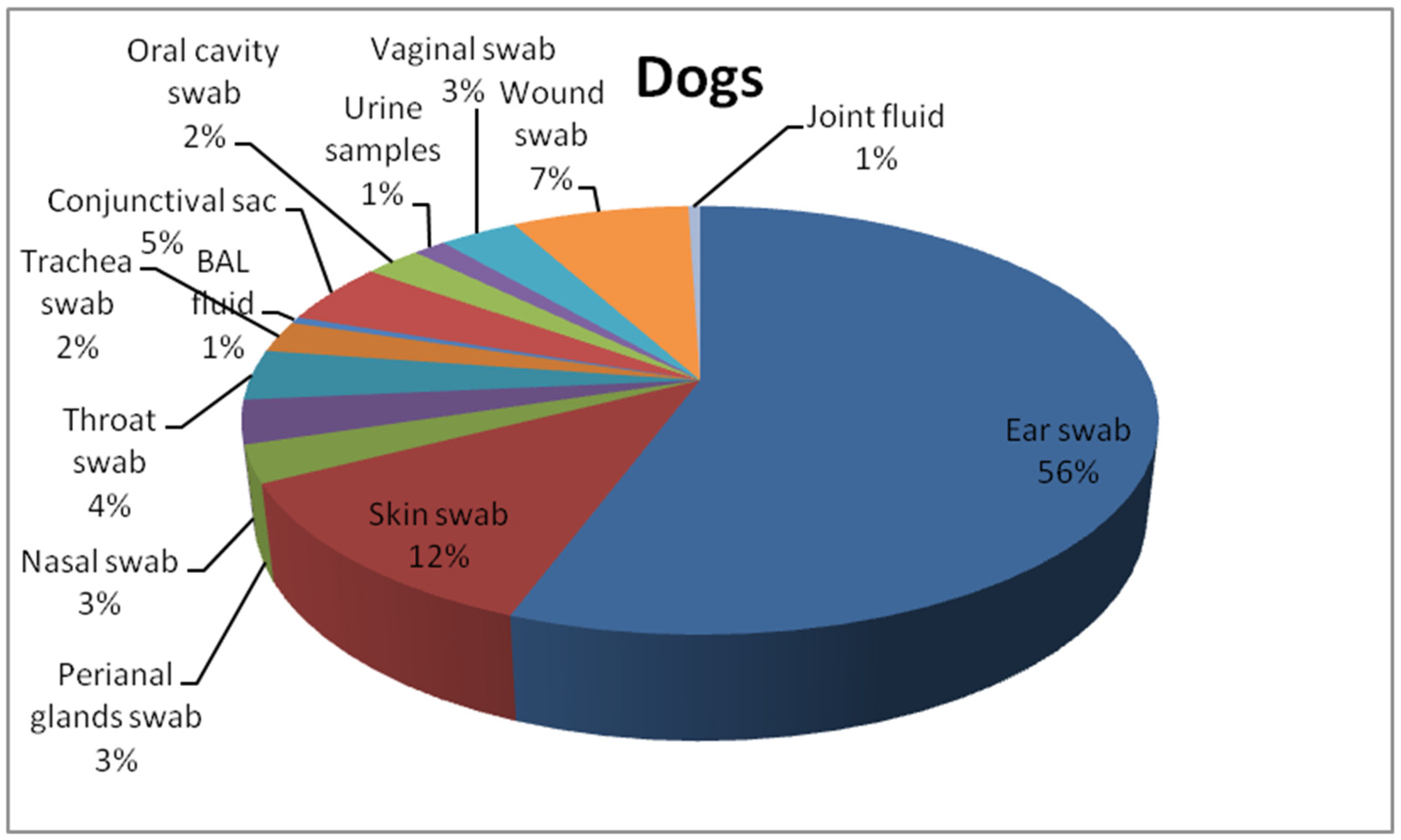
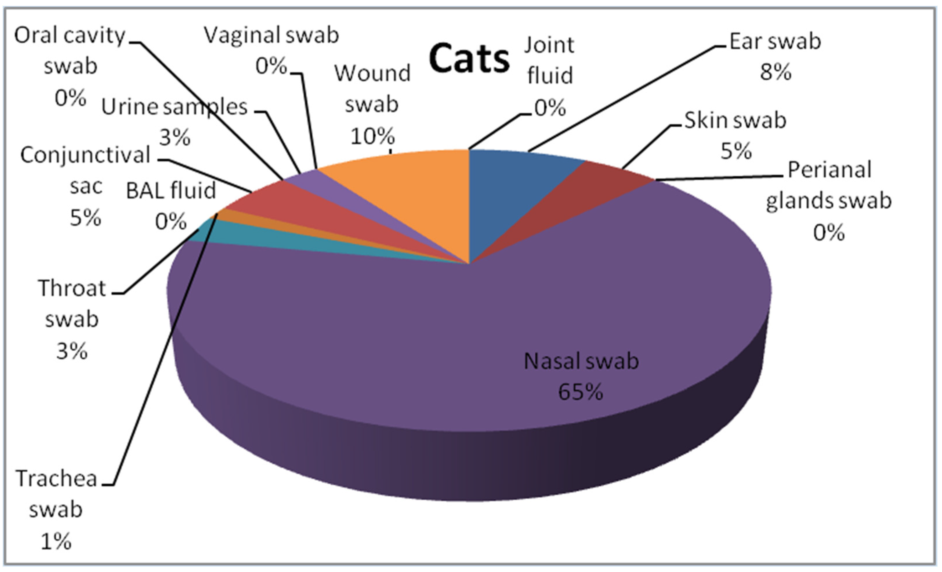
2.1. Isolates

This study includes 271 P. aeruginosa isolates collected from cats (n = 59) and dogs (n = 212) from the Lower Silesia area between 2017 and 2020 in the commercial veterinary laboratory, VETLAB Ltd., in Wrocław, Poland. The bacterial isolates were transferred to the Department of Epizootiology and Clinic of Birds and Exotic Animals, Wrocław University of Environmental and Life Sciences. The isolates were cultured on the Columbia Blood Agar Base (OXOID, Basingstoke, UK) with 5% sheep blood and MacConkey Agar (OXOID, Basingstoke, UK). Then isolates were checked for oxidase production (Oxidase Detection Strips MICROGEN MID-61g GRASO BIOTECH, Starogard Gdański, Poland). Pure strains of nonfermenting bacilli, suspected to be P. areuginosa strains were frozen in 1 mL of Brain Heart Infusion Broth (BHI) (OXOID, Basingstoke, UK) supplemented with 30% glycerol and storage in −80 °C for up to six months for further analysis. Subsequently they were used for molecular investigation. Belonging to the P. aeruginosa species was provided by polymerase chain reaction (PCR). The details of procedure used for species identification of the P. aeruginosa strains used in the current study is described in Płókarz et al. (2022) [27]. The bacterial strains originated from pets with clinical manifestations of respiratory tract infections (swabs from the nasal cavity, conjunctival sacs, oral cavity, throat, trachea, and bronchi), skin, wounds, joint fluid, and genitourinary tract. Details are included in Figure 1 and Figure 2.

2.2. Antibiotic Resistance

All P. aeruginosa isolates were screened for antibiotic resistance using the Kirby–Bauer disc diffusion method. The concentration of bacterial suspensions was adjusted according to the 0.5 McFarland turbidity standard. The antibiotics and their corresponding amounts (μg/disc) used in antimicrobial disc diffusion tests are as follows: enrofloxacin (5), ciprofloxacin (5), ceftazidime (30), amikacin (30), gentamicin (10), tobramycin (10), ticarcillin (75), imipenem (10), meropenem (10) (OXOID, Basingstoke, UK), marbofloxacin (5), and pradofloxacin (5) (MAST Group, Liverpool, UK). Antimicrobial-resistant phenotyping of the isolates was performed and interpreted according to the Clinical and Laboratory Standards Institute document M100-S32 and VETO1S [28]. P. aeruginosa ATCC 27853 was used as a quality control strain.

2.3. Detection of Virulence Genes

The presence of toxin genes and occurrence of other virulence factors genes was assayed in DNA isolated from the P. aeruginosa strains. Isolation of DNA was carried out as previously described by Płókarz et al. (2022) [27]. The DNA was amplified in a thermocycler (Bio-Rad, Marnes-la-Coquette, France) in 2.5 µL buffer (MgCl2 at 20 mM concentration), 0.2 µL Taq DNA polymerase (5 U/L) (Thermo Fisher Scientific, Vilnius, Lithuania), 0.2 µL dNTP mix 10 mM concentration (Thermo Fisher Scientific, Vilnius, Lithuania), 0.2 µL of each specific primer, and 2 µL template DNA. The reaction volume was adjusted to 25 µL with sterile water. The used primers and cycle conditions are presented in Table 1.

2.4. Statistical Analysis

Statistical analysis was performed using TIBCO Statistica 13.3 (TIBCO Software Inc., Palo Alto, CA, USA). Spearman’s rank correlation coefficient was used to analyse the correlation between antibiotic resistance and the origin P. aeruginosa isolates Wilson’s test was used to establish the confidence interval (95% PU). The chi-square test and Fisher’s exact test were used to determine the significant difference in the virulence factor’s gene occurrence between P. aeruginosa strains susceptible and resistant to selected antibiotics. The statistical significance level was configured as p < 0.05.

3. Results

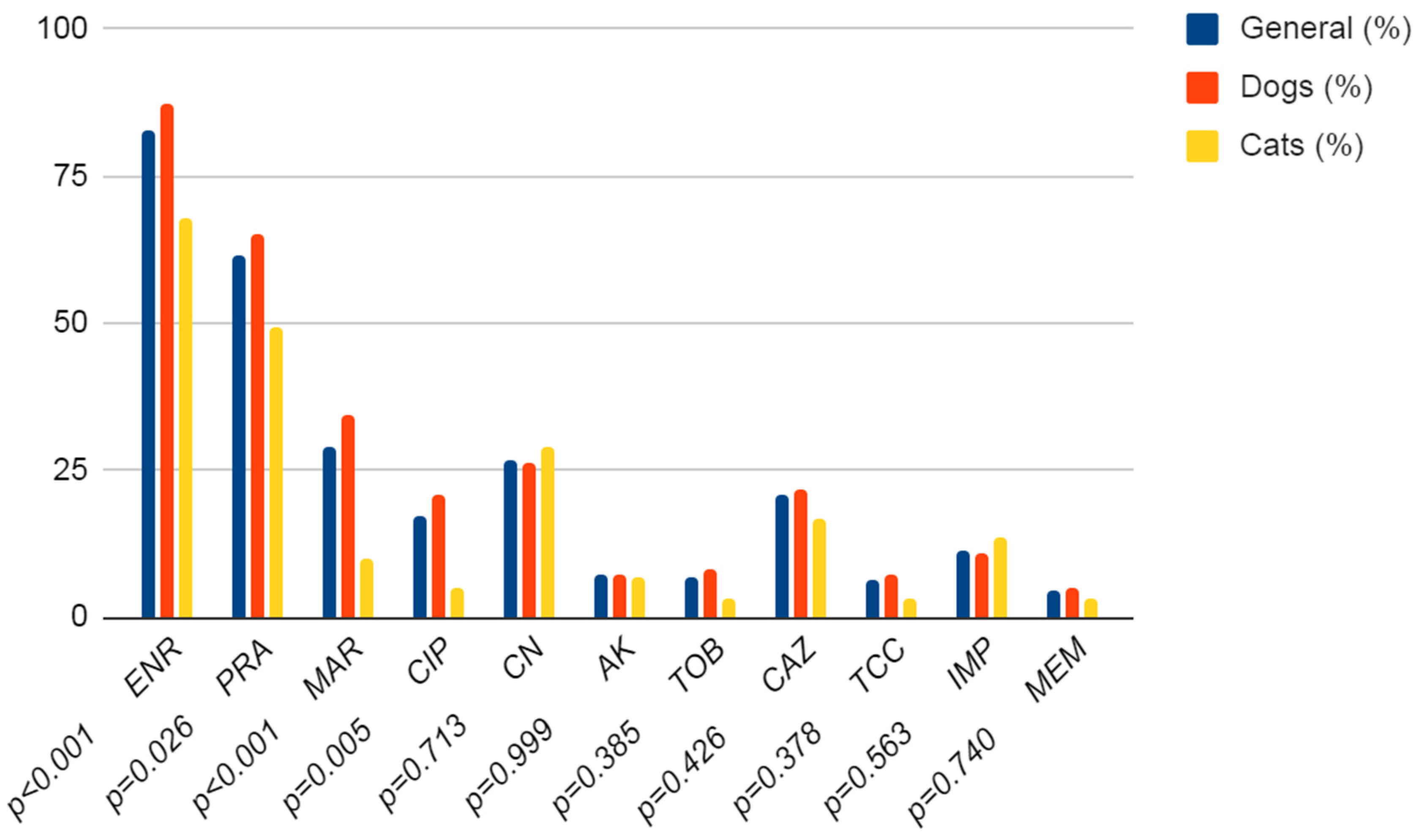
3.1. Antibiotic Resistance

In the current study, the resistance of 271 P. aeruginosa strains to specific antibiotics was investigated. In the beginning, the differences between the antibiotic resistance of P. aeruginosa originating from dogs or cats were tested. Only in the fluoroquinolone class significant differences between dogs and cats occur. In dogs, P. aeruginosa isolates were statistically significantly more often resistant to fluoroquinolone than isolates from cats (p ≤ 0.001). In other groups, there were no statistically significant differences. In P. aeruginosa strains from both dogs and cats, the most frequently detected resistance was to fluoroquinolones (from 17.3% (CI 95%: 13.3–22.3%) for ciprofloxacin to 83% (CI 95%: 78.1–87%) for enrofloxacin). Among the aminoglycosides, the highest resistance was observed to gentamicin at 26.9% (CI 95%: 22–32.5%), whereas in the carbapenem class, resistance to imipenem (14% (CI 95%: 8.2–15.8%)) was around two times higher than to meropenem (4.8% (CI 95%: 2.8–8%)). The detailed data are presented in Figure 3.
In the P. aeruginosa strains from dogs, there were some statistically significant correlations between the resistance to a particular antibiotic and the material from which bacteria was isolated; for example, the strains from wounds were frequently resistant to ticarcillin (p = 0.014), the strains from the urogenital system to meropenem (p = 0.042), the strains from the external auricular canals to marbofloxacin (p = 0.016), and from the skin and appendages to ciprofloxacin (p = 0.023) and gentamicin (p = 0.022). The detailed data are presented in Table 2. In cats, this correlation occurred only in the case of tobramycin, where strains from the respiratory system were significantly less resistant to this antibiotic (p = 0.026) (Table 3).

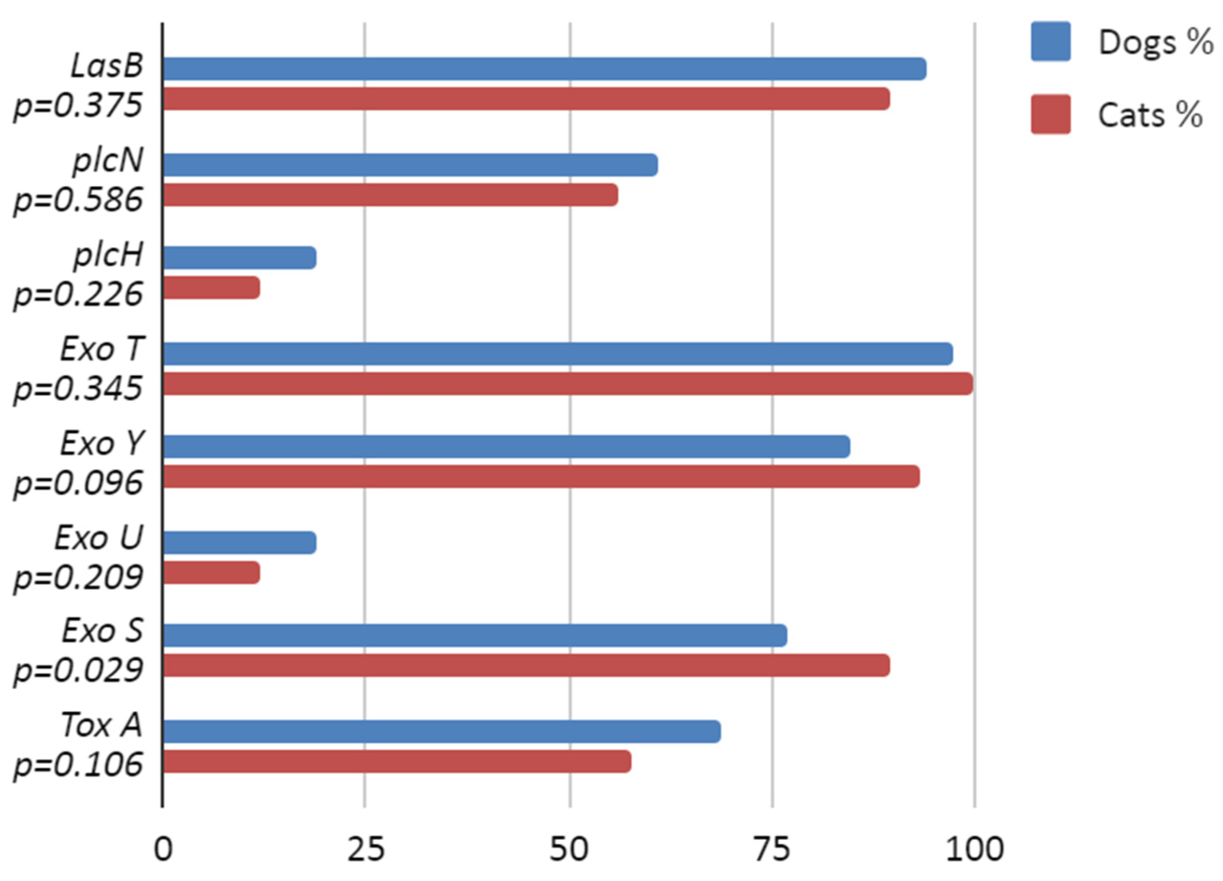
3.2. Virulence Factor Genes

Almost all P. aeruginosa strains harboured the exoT (97.8%) and lasB (93.4%) genes, while the lowest prevalence was found for the exoU (17.3%) and plcH (17.3%) genes. There was no observation of differences in the presence of exoenzyme genes in strains of dog or cat origin except for the occurrence of exoS, which was significantly more frequently detected in the P. aeruginosa from cats (p = 0.029). The detailed data are presented in Figure 4. There were only a few statistically significant associations between harbouring the virulence gene and exhibiting resistance to a particular antibiotic. In the P. aeruginosa strains from dogs, there were connections such as toxA and ceftazidime (p = 0.012) and exoU and marbofloxacin (p = 0.025) and amikacin (p = 0.056), while in the strains from cats, exoU and enrofloxacin (p = 0.054) were associated. The detailed data are presented in the Supplementary Materials Tables S1 and S2.

4. Discussion

The disc diffusion method is still a widespread way to diagnose antibiotic resistance in commercial laboratories in Poland, because it is a relatively cheap and reliable method. Previous reports have indicated good effectiveness in the confirmation of resistance to antibiotics compared with determination of the minimal inhibitory concentration method (MIC) [31]. The disc diffusion method can be useful for screening antibiotic resistance. As our research considered a large number of P. aeruginosa strains under investigation, we decided to assess antibiotic resistance using this method.
In the current study, P. aeruginosa strains were most frequently resistant to fluoroquinolones. The majority of strains, 225 from among 271, presented resistance to enrofloxacin (83%), while the P. aeruginosa strains were the most susceptible to ciprofloxacin (only 47 from among 271 (17.3%) of P. aeruginosa were resistant). In bacteria originating from cats, the resistance to ciprofloxacin was low, at 3 from among 59 P. aeruginosa strains (5.1%), compared with the P. aeruginosa strains from dogs, where 44 from among 212 were resistant (20.8%). The obtained results are similar to those previously reported by Penna et al. (2011) [32], where 13.8% of strains of P. aeruginosa were found to be resistant to ciprofloxacin, while 63.6% were resistant to enrofloxacin. In the study by Eliasy et al. (2020), where the antimicrobial resistance patterns of P. aeruginosa from clinical samples from dogs were investigated, a high proportion of strains resistant to fluoroquinolones (enrofloxacin 73% and orbifloxacin 90%) were also detected [33]. Similar tendencies were also observed in strains of human origin where the resistance to ciprofloxacin was 31.36% and the resistance to levofloxacin was 79.57% [34]. However, we observed significantly lower resistance of P. aeruginosa strains to aminoglycosides compared with the previous study by Penna et al. (2011), where the resistance of strains was 71.4% for gentamicin and 64.4% for tobramycin [32]. Accordance with our results resistance to aminoglycosides was as follows: amikacin (16%), gentamicin (18%), and tobramycin (12%). The differences in the obtained results could be caused by the origin of the bacteria. In the earlier study, only P. aeruginosa strains from the causes of canine otitis externa were investigated [32], whereas, in the current study, we analysed the material from different pathological changes in pets. Considering also that aminoglycosides are frequently used in topical therapy for external ear canal infections, the increased resistance to these antibiotics is very probable. In contrast, very low levels of P. aeruginosa strains resistant to aminoglycosides were detected in Japan, where only 4.5% of strains exhibited resistance to gentamicin and 2.5% to amikacin [35].
There were also differences in resistance levels to ceftazidime, where 58% was reported by Penna et al. (2011) [32] and 77% by Eliasi et al. (2020) [33], while in the current study 21.7% of the P. aeruginosa strains from dogs and 16.9% from cats were detected as resistant. This is in contrast to other veterinary reports, where the prevalence of resistance to ceftazidime was 0–0.5% [35,36,37]. The resistance to ticarcillin (6.6% of P. aeruginosa) in the current study is very similar to the results obtained by Pottier et al. (2022) [36] in strains from veterinary samples (7.1%). In similar studies, the reported resistance of P. aeruginosa to carbapenems was consistent with our results 6–10% [33,38,39] to imipenem and 2–6% [36,38] to meropenem. While the results provided by Dégi et al. (2021) are significantly different to the results obtained in the current study [40]. In P. aeruginosa strains isolated from dogs in Western Romania, the resistance rate was 74.14% to meropenem and 77.59% to imipenem and was significantly higher than in the current study. The reason could be that samples in the mentioned research were collected the most frequently from superficial infections while our study shows mainly samples collected from external auricular canal [40]. The resistance to carbapenems in animal P. aeruginosa strains is still at the control level and is much lower than in the bacteria that cause nosocomial infections. Occurrence of resistance against this group of antibiotics in dogs and cats are probably results of close contact between owners and pets. It suggests exchanging genetic elements such as self-transmissible conjugative plasmids through horizontal gene transfer (HGT) [41]. The recent study of the antibiotic resistance of P. aeruginosa from Polish hospitals found that 67.8% of strains are resistant to imipenem and 29.2% to meropenem [42]. According to a 2017 ECDC survey, the average resistance rate to carbapenems (imipenem + meropenem) was 24.2% across Poland [43].
Our report also describes the prevalence of the chosen virulence factor genes in the P. aeruginosa strains under investigation. Overall, the frequency determined for the exoU (17.3%) and exoS (79,7%) genes is comparable to the results obtained by Hayashi et al. (2021) of 15.8% and 82.5%, respectively [38]. Similar to our results, lasB was detected in almost all P. aeruginosa (98.7%) [44]. The high prevalence of exoT and exoY genes in the current study (over 80% of P. aeruginosa strains) was consistent with previous reports, where all or almost all strains harboured such genes (100% exoT and 89–100% exoY) [45,46].
Our results show the presence of the toxA gene in 66.4% of strains, which is significantly lower than the 91.7% obtained by Hattab et al. (2021) [47]. Significant differences were also found for the prevalence of the plcH gene, with only 17.3% found to harbour the plcH gene in our study versus 91.7% in the earlier study. This difference could be caused by the very small number of samples (n = 24) compared with our study (n = 271), which included only canine samples. In our study, 59.8% of isolates were found to possess the plcN gene, which was lower than found by Aslataş et al. (2022) in clinical cases of bovine mastitis (100%), but they examined isolates from other anatomical locations and species, and the number of samples was significantly lower [48]. It is very difficult to compare the prevalence of P. aeruginosa isolates across different studies because of the differences in examined animal species, and geographical regions, all of which can exert varying influences on the results.
The association between the presence of toxA and the antibiotic resistance of P. aeruginosa strains found in our study is supported by the earlier results of Amormozafari et al. (2016), who used the same statistical analysis [49]. The presence of exoU, exoS, exoT, and exoY was analysed in humans by Horna et al. (2019). The results of their research confirm the association between the resistance to fluoroquinolone and aminoglycoside and the occurrence of the exoU gene [45]. In our research, we found a significant association indicating that the presence of the exoU gene in P. aeruginosa strains is linked with resistance to marbofloxacin (p = 0.025) and to amikacin (p = 0.056). For the other toxin genes, however, there were no statistically significant associations. The study performed by Hayashi et al. in 2021 in canine and feline patients from Japanese animal hospitals showed a significant association of isolates possessing exoU and resistance to aminoglycoside [38].

5. Conclusions

Pseudomonas aeruginosa isolates tested in the current study showed the highest resistance to fluoroquinolone. This can be useful in clinical veterinary practice in experimental antibiotic therapy and suggests that veterinarians should use greater caution in the selection of antibiotics. Of note is the low resistance to aminoglycosides in cases of otitis externa, despite commercial usage of this antibiotics class as a component of ear drops. Of considerable epidemiological value, our study included an antibiogram assessment of resistance to meropenem, imipenem, and ticarcillin in isolates from dogs and cats. In our knowledge carbapenems are not used in veterinary medical practice in Poland which suggests transferring P.aeruginosa resistant strains from human to animals. Furthermore, results of research showed a higher rate of resistance in humans which supports the thesis: humans are a source of P.aeruginosa resistance strains for animals. Although the rate of the resistant strains was still low, the increasing tendency indicates control of this potential reservoir is important. The analysis of the associations between the strains possessing virulence-factor-encoding genes and antibiotic resistance allows us to broaden the horizons considering antibiotic resistance. Further research on the relationships between virulence factors and antibiotic resistance in animals is required.

Supplementary Materials

The following supporting information can be downloaded at: https://www.mdpi.com/article/10.3390/antibiotics12071226/s1, Table S1: Statistical analysis of virulence-factor-encoding genes and selected antibiotics relationship in Pseudomonas aeruginosa isolates from cats; Table S2: Statistical analysis of virulence-factor-encoding genes and selected antibiotics relationship in Pseudomonas aeruginosa isolates from dogs.

Author Contributions

All authors of this research paper have directly participated in the planning and/or execution and/or analysis of this study. D.P. wrote the manuscript, provided statistical analysis, designed the research, table and figures, and analyzed data; K.B. corrected the manuscript, tables and figures; K.R. contributed to the design of the research, analyzed data and supervised the findings of this work. All authors have read and agreed to the published version of the manuscript.

Funding

The APC/BPC is financed/co-financed by Wroclaw University of Environmental and Life Sciences.

Institutional Review Board Statement

The samples used in this study originally came from the samples used as material for routine diagnostics.

Informed Consent Statement

This study was provided based on material from VETLAB Ltd. in Wroclaw. All animals had their own veterinarian who was responsible for providing information about potential zoonotic risk. The study did not involve humans.

Data Availability Statement

The datasets used and/or analyzed during the current study are available via e-mail from the corresponding author on reasonable request.

Acknowledgments

The authors thank Kamila Bobrek for help in the laboratory work and the technicians from the Diagnostic Laboratory EPI-VET of the Faculty of Veterinary Medicine in Wroclaw, and Józef Galli from VETLAB Ltd. in Wroclaw, who performed part of the laboratory work.

Conflicts of Interest

The authors declare no conflict of interest. The funders had no role in the design of the study, the collection, analyses, or interpretation of data, writing of the manuscript, or the decision to publish the results.

References

  1. Wolska, K.; Kot, B.; Piechota, M.; Frankowska, A. Resistance of Pseudomonas aeruginosa to antibiotics. PHMD 2013, 67, 1300–1311. [Google Scholar] [CrossRef]
  2. Thi, M.T.T.; Wibowo, D.; Rehm, B.H.A. Pseudomonas aeruginosa Biofilms. Int. J. Mol. Sci. 2020, 21, 8671. [Google Scholar] [CrossRef]
  3. González-Alsina, A.; Mateu-Borrás, M.; Doménech-Sánchez, A.; Albertí, S. Pseudomonas aeruginosa and the Complement System: A Review of the Evasion Strategies. Microorganisms 2023, 11, 664. [Google Scholar] [CrossRef]
  4. Wood, S.J.; Kuzel, T.M.; Shafikhani, S.H. Pseudomonas aeruginosa: Infections, Animal Modeling, and Therapeutics. Cells 2023, 12, 199. [Google Scholar] [CrossRef]
  5. Litwin, A.; Rojek, S.; Gozdzik, W.; Duszynska, W. Pseudomonas aeruginosa device associated—Healthcare associated infections and its multidrug resistance at intensive care unit of University Hospital: Polish, 8.5-year, prospective, single-centre study. BMC Infect. Dis. 2021, 21, 180. [Google Scholar] [CrossRef] [PubMed]
  6. European Centre for Disease Prevention and Control. Point Prevalence Survey of Healthcare-Associated Infections and Antimicrobial Use in European Long-Term. Care Facilities 2016–2017; ECDC: Stockholm, Sweden, 2023. [Google Scholar]
  7. Rello, J.; Ausina, V.; Ricart, M.; Puzo, C.; Quintana, E.; Net, A.; Prats, G. Risk factors for infection by Pseudomonas aeruginosa in patients with ventilator-associated pneumonia. Intensive Care Med. 1994, 20, 193–198. [Google Scholar] [CrossRef] [PubMed]
  8. Bassetti, M.; Vena, A.; Croxatto, A.; Righi, E.; Guery, B. How to manage Pseudomonas aeruginosa infections. Drugs Context. 2018, 7, 212527. [Google Scholar] [CrossRef] [PubMed]
  9. González-Díaz, A.; Medina-Polo, J.; Abad-López, P.; González-Padilla, D.A.; García-Rojo, E.; Santos-Pérez de la Blanca, R.; Hernández-Arroyo, M.; Tejido-Sánchez, Á. The challenge of Pseudomonas aeruginosa related infections in a Urology Ward: Epidemiology, risk factors and patterns of antibiotic resistance. Arch. Esp. Urol. 2020, 73, 299–306. [Google Scholar]
  10. Robinson, V.H.; Paterson, S.; Bennett, C.; Steen, S.I. Biofilm production of Pseudomonas spp. isolates from canine otitis in three different enrichment broths. Vet. Dermatol. 2019, 30, 218-e67. [Google Scholar] [CrossRef]
  11. Bajwa, J. Canine otitis externa—Treatment and complications. Can. Vet. J. 2019, 60, 97–99. [Google Scholar]
  12. De Sousa, T.; Garcês, A.; Silva, A.; Lopes, R.; Alegria, N.; Hébraud, M.; Igrejas, G.; Poeta, P. The Impact of the Virulence of Pseudomonas aeruginosa Isolated from Dogs. Vet. Sci. 2023, 10, 343. [Google Scholar] [CrossRef]
  13. Gentilini, F.; Turba, M.E.; Pasquali, F.; Mion, D.; Romagnoli, N.; Zambon, E.; Terni, D.; Peirano, G.; Pitout, J.D.D.; Parisi, A.; et al. Hospitalized Pets as a Source of Carbapenem-Resistance. Front. Microbiol. 2018, 6, 2872. [Google Scholar] [CrossRef]
  14. Sharma, D.; Pakravanm, N.; Pritchard, J.C.; Hartmann, F.A.; Young, K.M. Mucoid Pseudomonas aeruginosa infection in a cat with severe chronic rhinosinusitis. Vet. Clin. Pathol. 2019, 48, 300–304. [Google Scholar] [CrossRef] [PubMed] [Green Version]
  15. Lister, P.D.; Wolter, D.J.; Hanson, N.D. Antibacterial-resistant Pseudomonas aeruginosa: Clinical impact and complex regulation of chromosomally encoded resistance mechanisms. Clin. Microbiol. Rev. 2009, 22, 582–610. [Google Scholar] [CrossRef] [PubMed] [Green Version]
  16. De Briyne, N.; Atkinson, J.; Pokludová, L.; Borriello, S.P. Antibiotics used most commonly to treat animals in Europe. Vet. Rec. 2014, 175, 325. [Google Scholar] [CrossRef] [PubMed] [Green Version]
  17. Langendonk, R.F.; Neill, D.R.; Fothergill, J.L. The building blocks of antimicrobial resistance in Pseudomonas aeruginosa: Implications for current resistance-breaking therapies. Front. Cell Infect. 2021, 11, 665759. [Google Scholar] [CrossRef] [PubMed]
  18. European Medicines Agency (EMA). Categorisation of Antibiotics in the European Union; European Medicines Agency: London, UK, 2019. [Google Scholar]
  19. Velazquez-Meza, M.E.; Galarde-López, M.; Carrillo-Quiróz, B.; Alpuche-Aranda, C.M. Antimicrobial resistance: One Health approach. Vet. World. 2022, 15, 743–749. [Google Scholar] [CrossRef]
  20. Wi, Y.M.; Choi, J.Y.; Lee, J.Y.; Kang, C.I.; Chung, D.R.; Peck, K.R.; Song, J.H.; Ko, K.S. Antimicrobial effects of β-lactams on imipenem-resistant ceftazidime-susceptible Pseudomonas aeruginosa. Antimicrob. Agents Chemother. 2017, 61, e00054-17. [Google Scholar] [CrossRef] [Green Version]
  21. Çopur Çiçek, A.; Ertürk, A.; Ejder, N.; Rakici, E.; Kostakoğlu, U.; Esen Yıldız, İ.; Özyurt, S.; Sönmez, E. Screening of Antimicrobial Resistance Genes and Epidemiological Features in Hospital and Community-Associated Carbapenem-Resistant Pseudomonas aeruginosa Infections. Infect. Drug Resist. 2021, 14, 1517–1526. [Google Scholar] [CrossRef]
  22. World Organisation for Animal Health (OIE). OIE List of Antimicrobial Agents of Veterinary Importance. In Proceedings of the FAO2/OIE/WHO3 Expert Workshop on Non-Human Antimicrobial Usage and Antimicrobial Resistance, Geneva, Switzerland, 1–5 December 2003; Volume Xxviii, pp. 1–10. [Google Scholar]
  23. Ellappan, K.; Belgode Narasimha, H.; Kumar, S. Coexistence of multidrug resistance mechanisms and virulence genes in carbapenem-resistant Pseudomonas aeruginosa strains from a tertiary care hospital in South India. J. Glob. Antimicrob. Resist. 2018, 12, 37–43. [Google Scholar] [CrossRef]
  24. Fadhil, L.; Al-Marzoqi, A.H.; Zahraa, M.A.; Shalan, A.A. Molecular and phenotypic study of virulence genes in a pathogenic strain of Pseudomonas aeruginosa isolated from various clinical origins by PCR: Profiles of genes and toxins. Res. J. Pharm. Biol. Chem. Sci. 2016, 7, 590–598. [Google Scholar]
  25. Yang, J.J.; Tsuei, K.C.; Shen, E.P. The role of Type III secretion system in the pathogenesis of Pseudomonas aeruginosa microbial keratitis. Tzu. Chi. Med. J. 2022, 34, 8–14. [Google Scholar] [PubMed]
  26. Bradbury, R.S.; Roddam, L.F.; Merritt, A.; Reid, D.W.; Champion, A.C. Virulence gene distribution in clinical, nosocomial and environmental isolates of Pseudomonas aeruginosa. J. Med. Microbiol. 2010, 59, 881–890. [Google Scholar] [CrossRef] [PubMed]
  27. Płókarz, D.; Czopowicz, M.; Bierowiec, K.; Rypuła, K. Virulence Genes as Markers for Pseudomonas aeruginosa Biofilm Formation in Dogs and Cats. Animals 2022, 12, 422. [Google Scholar] [CrossRef] [PubMed]
  28. James, S. Performance Standards of Antimicrobial Susceptibility Testing; Twenty-Fourth International Supplement; Clinical and Laboratory Standards Institute: Wayne, PA, USA, 2022. [Google Scholar]
  29. Wolska, K.; Szweda, P. Genetic features of clinical Pseudomonas aeruginosa strains. Pol. J. Microbiol. 2009, 58, 255–260. [Google Scholar]
  30. Mokhtari, A.; Amini, K. Genotyping of Pseudomonas Aeruginosa Strains As A Multidrug Resistant (MDR) Bacterium And Evaluating The Prevalence of Esbls and Some Virulence Factors Encoding Genes By PFGE and ERIC-PCR Methods. Iran. J. Pharm. Res. 2019, 18, 1580–1594. [Google Scholar]
  31. Rémy, A.; Bonnin, C.E.; Agnès, B.; Jousset, T.N.; Laurent, D. Comparison of disk diffusion, MIC test strip and broth microdilution methods for cefiderocol susceptibility testing on carbapenem-resistant enterobacterales. Clin. Microbiol. Inf. 2022, 28, e1–e1156. [Google Scholar]
  32. Penna, B.; Thomé, S.; Martins, R.; Martins, G.; Lilenbaum, W. In Vitro antimicrobial resistance of Pseudomonas aeruginosa isolated from canine otitis externa in Rio de Janeiro. Braz. J. Microbiol. 2011, 42, 1434–1436. [Google Scholar] [CrossRef] [Green Version]
  33. Eliasi, U.L.; Sebola, D.; Oguttu, J.W.; Qekwana, D.N. Antimicrobial resistance patterns of Pseudomonas aeruginosa isolated from canine clinical cases at a veterinary academic hospital in South Africa. J. S. Afr. Vet. Assoc. 2020, 91, e1–e6. [Google Scholar] [CrossRef]
  34. Djuikoue, C.I.; Djouela Djoulako, P.D.; Same Njanjo, H.V.; Kiyang, C.P.; Djantou Biankeu, F.L.; Guegang, C.; Tchouotou, A.S.D.; Wouambo, R.K.; Thumamo Pokam, B.D.; Apalata, T.; et al. Phenotypic Characterization and Prevalence of Carbapenemase-Producing Pseudomonas aeruginosa Isolates in Six Health Facilities in Cameroon. BioMed 2023, 3, 77–88. [Google Scholar] [CrossRef]
  35. Yukawa, S.; Tsuyuki, Y.; Sato, T.; Fukuda, A.; Usui, M.; Tamura, Y. Antimicrobial Resistance of Pseudomonas aeruginosa Isolated from Dogs and Cats in Primary Veterinary Hospitals in Japan. Jpn. J. Infect. Dis. 2017, 70, 461–463. [Google Scholar] [CrossRef] [Green Version]
  36. Pottier, M.; Castagnet, S.; Gravey, F.; Leduc, G.; Sévin, C.; Petry, S.; Giard, J.-C.; Le Hello, S.; Léon, A. Antimicrobial Resistance and Genetic Diversity of Pseudomonas aeruginosa Strains Isolated from Equine and Other Veterinary Samples. Pathogens 2023, 12, 64. [Google Scholar] [CrossRef] [PubMed]
  37. Mekić, S.; Matanović, K.; Šeol, B. Antimicrobial susceptibility of Pseudomonas aeruginosa isolates from dogs with otitis externa. Vet. Rec. 2011, 169, 125. [Google Scholar] [CrossRef] [PubMed]
  38. Hayashi, W.; Izumi, K.; Yoshida, S.; Takizawa, S.; Sakaguchi, K.; Iyori, K.; Minoshima, K.I.; Takano, S.; Kitagawa, M.; Nagano, Y.; et al. Antimicrobial Resistance and Type III Secretion System Virulotypes of Pseudomonas aeruginosa Isolates from Dogs and Cats in Primary Veterinary Hospitals in Japan: Identification of the International High-Risk Clone Sequence Type 235. Microbiol. Spectr. 2021, 9, e0040821. [Google Scholar] [CrossRef] [PubMed]
  39. Oliveira, L.C.; Medeiros, C.M.O.; Silva, I.N.G.; Monteiro, A.J.; Leite, C.A.L.; Carvalho, C.B.M. Susceptibilidade a antimicrobianos de bactérias isoladas de otite externa em cães. Arq. Bras. Med. Veterinária Zootec. 2005, 57, 405–408. [Google Scholar] [CrossRef]
  40. Dégi, J.; Moțco, O.A.; Dégi, D.M.; Suici, T.; Mareș, M.; Imre, K.; Cristina, R.T. Antibiotic Susceptibility Profile of Pseudomonas aeruginosa Canine Isolates from a Multicentric Study in Romania. Antibiotics. 2021, 10, 846. [Google Scholar] [CrossRef]
  41. Callens, M.; Scornavacca, C.; Bedhomme, S. Evolutionary responses to codon usage of horizontally transferred genes in Pseudomonas aeruginosa: Gene retention, amelioration and compensatory evolution. Microb. Genom. 2021, 7, 000587. [Google Scholar] [CrossRef]
  42. Brzozowski, M.; Krukowska, Ż.; Galant, K.; Jursa-Kulesza, J.; Kosik-Bogacka, D. Genotypic characterisation and antimicrobial resistance of Pseudomonas aeruginosa strains isolated from patients of different hospitals and medical centres in Poland. BMC Infect. Dis. 2020, 20, 693. [Google Scholar] [CrossRef]
  43. European Centre for Disease Prevention and Control. Surveillance of Antimicrobial Resistance in Europe; European Centre for Disease Prevention and Control: Stockholm, Sweden, 2017. [Google Scholar]
  44. Elmouaden, C.; Laglaoui, A.; Ennanei, L.; Bakkali, M.; Abid, M. Virulence genes and antibiotic resistance of Pseudomonas aeruginosa isolated from patients in the Northwestern of Morocco. J. Infect. Dev. Ctries. 2019, 13, 892–898. [Google Scholar] [CrossRef]
  45. Feltman, H.; Schulert, G.; Khan, S.; Jain, M.; Peterson, L.; Hauser, A.R. Prevalence of type III secretion genes in clinical and environmental isolates of Pseudomonas aeruginosa. Microbiology 2001, 147, 2659–2669. [Google Scholar] [CrossRef] [Green Version]
  46. Horna, G.; Amaro, C.; Palacios, A.; Guerra, H.; Ruiz, J. High frequency of the exoU+/exoS+ genotype associated with multidrug-resistant “high-risk clones” of Pseudomonas aeruginosa clinical isolates from Peruvian hospitals. Sci. Rep. 2019, 9, 10874. [Google Scholar] [CrossRef] [PubMed] [Green Version]
  47. Hattab, J.; Mosca, F.; Di Francesco, C.E.; Aste, G.; Marruchella, G.; Guardiani, P.; Tiscar, P.G. Occurrence, antimicrobial susceptibility, and pathogenic factors of Pseudomonas aeruginosa in canine clinical samples. Vet. World 2021, 14, 978–985. [Google Scholar] [CrossRef] [PubMed]
  48. Aslataş, Ö.; Tűrkyilmaz, S.; KESKİN, O.; Gűllű Yűcetepe, A.; Bűykatal, K. Molecular Characterization of Pseudomonas aeruginosa Isolated From Clinical Bovine Mastitis Cases. Kafkas. Univ. Vet. Fak. Derg. 2022, 28, 747–759. [Google Scholar]
  49. Amirmozafari, N.; Fallah, J.; Habibi, A. Association of the Exotoxin A and Exoenzyme S with Antimicrobial Resistance in Pseudomonas Aeruginosa Strains. Arch. Iran. Med. 2016, 19, 353–358. [Google Scholar]
Figure 1. Collection sites of swabs or another sort of material where P. aeruginosa was isolated in dogs.
Antibiotics 12 01226 g001
Figure 2. Collection sites of swabs or another sort of material where P. aeruginosa was isolated in cats.
Antibiotics 12 01226 g002
Figure 3. Antibiotic resistance of the P. aeruginosa strains originating from dogs and cats. ENR—enrofloxacin; PRA—pradofloxacin; MAR—marbofloxacin; CIP—ciprofloxacin; CN—gentamicin; AK—amikacin; TOB—tobramycin; CAZ—ceftazidime; TCC—ticarcillin; IMP—imipenem; MEM—meropenem, p—value.
Antibiotics 12 01226 g003
Figure 4. Virulence factor genes’ prevalence: comparison of P. aeruginosa isolates from dogs and cats. Genes encoding: LasB—elastase B, plcN—non-haemolytic phospholipase C, plcH—haemolytic phospholipase, exoT—exotoxin T, exoY—exotoxin Y, Exo U—exotoxin U, exoS—exotoxin T, tox A—exotoxin A.
Antibiotics 12 01226 g004
Table 1. The primers and PCR assay conditions used in the current study.
GenPrimersAnnealing TemperatureProduct SizeReferences
Las BF:5′-GGAATGAACGAAGCGTTCTCCGAC-3′
R:5′-TTGGCGTCGACGAACACCTCG-3′
55 °C a284Wolska et al., 2009 [29]
plcNF:5′-TCCGTTATCGCAACCAGCCCTACG-3′
R:5′-TCGCTGTCGAGCAGGTCGAAC-3′
55 °C a481Wolska et al., 2009 [29]
plcHF:5′-GCACGTGGTCATCCTGATGC-3′
R:5′-TCCGTAGGCGTCGACGTAC-3′
55 °C a608Wolska et al., 2009 [29]
ToxAF:5′-CTGCGCGGGTCTATGTGCC-3′
R:5′-GATGCTGGACGGGTCGAG-3′
55 °C a270Wolska et al., 2009 [29]
ExoSF:5′-CGTCGTGTTCAAGCAGATGGTGCTG-3′
R:5′-CCGAACCGCTTCACCAGGC-3′
55 °C a444Wolska et al., 2009 [29]
ExoUF:5′-CCGTTGTGGTGCCGTTGAAG-3′
R:5′-CCAGATGTTCACCGACTCGC-3′
58 °C b134Mokhtaria and Amini 2019 [29]
ExoYF:5′-CGGATTCTATGGCAGGGAGG-3′
R:5′-GCCCTTGATGCACTCGACCA-3′
58 °C b289Mokhtaria and Amini 2019 [30]
ExoTF:5′-AATCGCCGTCCAACTGCATGCG-3′
R: 5′-TGTTCGCCGAGGTACTGCTC-3′
58 °C b152Mokhtaria and Amini 2019 [30]
a 30 cycles of 30 s at 94 °C, 1 min at annealing temperature, and 1.5 min at 72 °C. b 36 cycles of 30 s at 94 °C, 30 s at annealing temperature, and 1 min at 72 °C.
Table 2. Correlation between the antibiotic resistance and the location of origin of the P. aeruginosa strains in dogs.
AntibioticExternal Auricular Canal (n = 118)Skin and Appendages
(n = 31)
Respiratory System and Oral Cavity (n = 36)Uro-Genital System
(n = 10)
Wounds
(n = 16)
p
n% (95% PU)n% (95% PU)n% (95% PU)n% (95% PU)n% (95% PU)
ENR10387.3 (80.1–92.1)2477.4 (60.2–88.6)3186.1 (713–93.9)10100 (72.2–100)16100 (8.6–100)0.052
MAR5042.4 (33.8–51.4)619.4 (9.2–36.3)719.4 (9.8–35.0)220.0 (5.7–51.0)743.8 (23.1–66.8)0.016 *
CIP3126.3 (19.2–34.9)13.2 (0.6–16.2)719.4 (9.8–35.0)110.0 (1.8–40.4)425.0 (10.2–49.5)0.023 *
PRA8067.8 (58.9–75.6)1651.6 (34.8–68.0)2569.4 (53.1–82.0)440.0 (16.8–68.7)1275.0 (50.5–89.8)0.172
CAZ3227.1 (19.9–35.8)26.5 (1.8–20.7)616.7 (7.9–31.9)220.0 (5.7–51.0)425.0 (10.2–49.5)0.089
AK119.3 (5.3–15.9)13.2 (0.6–16.2)25.6 (1.5–18.1)110.0 (1.8–40.4)16.3 (1.1–28.3)0.757
CN4033.9 (26.0–42.8)39.7 (3.3–24.9)719.4 (9.8–350)110.0 (1.8–40.4)531.3 (14.2–55.6)0.022 *
TOB119.3 (5.3–15.9)26.5 (1.8–20.7)12.8 (0.5–14.2)00 (0–27.8)318.8 (6.6–43.0)0.225
TTC86.8 (3.5–12.8)00 (0–11.0)12.8 (0.5–14.2)220.0 (5.7–51.0)425.0 (10.2–49.5)0.014 *
IMP119.3 (5.3–15.9)26.5 (1.8–20.7)411.1 (4.4–25.3)440.0 (16.8–68.7)212.5 (3.5–36.0)0.150
MEM75.9 (2.9–11.7)00 (0–11.0)00 (0–9.6)220.0 (5.7–51.0)16.3 (1.1–28.3)0.042 *
* statistically significant differences. ENR—enrofloxacin; PRA—pradofloxacin; MAR—marbofloxacin; CIP—ciprofloxacin; CN—gentamicin; AK—amikacin; TOB—tobramycin; CAZ—ceftazidime; TCC—ticarcillin; IMP—imipenem; MEM—meropenem.
Table 3. Correlation between antibiotic resistance and origin place of P. aeruginosa strains in cats.
AntibioticRespiratory System (n = 49)Others (n = 10)
n% (95% PU)n% (95% PU)p
ENR3265.3 (51.3–77.1)880.0 (49.0–94.3)0.476
MAR510.2 (4.4–21.8)110.0 (1.8–40.4)0.999
CIP24.1 (1.1–13.7)110.0 (1.8–40.4)0.433
PRA2346.9 (33.7–60.6)660.0 (31.3–83.2)0.452
CAZ816.3 (8.5–29.0)220.0 (5.7–51.0)0.673
AK36.1 (2.1–16.5)110.0 (1.8–40.4)0.535
CN1326.5 (16.2–40.3)440.0 (16.8–68.7)0.453
TOB00 (0–7.3)220.0 (5.7–51.0)0.026 *
TTC24.1 (1.1–13.7)00 (0–27.8)0.999
IMP612.2 (5.7–24.2)220.0 (5.7–51.0)0.613
MEM12.0 (0.4–10.7)110.0 (1.8–40.4)0.313
* statistically significant differences. ENR—enrofloxacin; PRA—pradofloxacin; MAR—marbofloxacin; CIP—ciprofloxacin; CN—gentamicin; AK—amikacin; TOB—tobramycin; CAZ—ceftazidime; TCC—ticarcillin; IMP—imipenem; MEM—meropenem.
Disclaimer/Publisher’s Note: The statements, opinions and data contained in all publications are solely those of the individual author(s) and contributor(s) and not of MDPI and/or the editor(s). MDPI and/or the editor(s) disclaim responsibility for any injury to people or property resulting from any ideas, methods, instructions or products referred to in the content.

