Article

Antibacterial and Antibiofilm Effects of Different Samples of Five Commercially Available Essential Oils

by
Răzvan Neagu
1,2,
Violeta Popovici
3,*,
Lucia Elena Ionescu
4,*,
Viorel Ordeanu
4,
Diana Mihaela Popescu
2,
Emma Adriana Ozon
5 and
Cerasela Elena Gîrd
1
1
Department of Pharmacognosy, Phytochemistry, and Phytotherapy, Faculty of Pharmacy, Carol Davila University of Medicine and Pharmacy, 6 Traian Vuia Street, 020956 Bucharest, Romania
2
Regenerative Medicine Laboratory, “Cantacuzino” National Military Medical Institute for Research and Development, 103 Spl. Independentei, 050096 Bucharest, Romania
3
Department of Microbiology and Immunology, Faculty of Dental Medicine, Ovidius University of Constanta, 7 Ilarie Voronca Street, 900684 Constanta, Romania
4
Experimental Microbiology Laboratory, “Cantacuzino” National Military Medical Institute for Research and Development, 103 Spl. Independentei, 050096 Bucharest, Romania
5
Department of Pharmaceutical Technology and Biopharmacy, Faculty of Pharmacy, Carol Davila University of Medicine and Pharmacy, 6 Traian Vuia Street, 020956 Bucharest, Romania
*
Authors to whom correspondence should be addressed.
Antibiotics 2023, 12(7), 1191; https://doi.org/10.3390/antibiotics12071191
Submission received: 16 June 2023 / Revised: 12 July 2023 / Accepted: 12 July 2023 / Published: 14 July 2023

Abstract

:
Essential oils (EOs) have gained economic importance due to their biological activities, and increasing amounts are demanded everywhere. However, substantial differences between the same essential oil samples from different suppliers are reported—concerning their chemical composition and bioactivities—due to numerous companies involved in EOs production and the continuous development of online sales. The present study investigates the antibacterial and antibiofilm activities of two to four samples of five commercially available essential oils (Oregano, Eucalyptus, Rosemary, Clove, and Peppermint oils) produced by autochthonous companies. The manufacturers provided all EOs’ chemical compositions determined through GC-MS. The EOs’ bioactivities were investigated in vitro against Gram-positive (Staphylococcus aureus) and Gram-negative bacteria (Escherichia coli and Pseudomonas aeruginosa). The antibacterial and antibiofilm effects (ABE% and, respectively, ABfE%) were evaluated spectrophotometrically at 562 and 570 nm using microplate cultivation techniques. The essential oils’ calculated parameters were compared with those of three standard broad-spectrum antibiotics: Amoxicillin/Clavulanic acid, Gentamycin, and Streptomycin. The results showed that at the first dilution (D1 = 25 mg/mL), all EOs exhibited antibacterial and antibiofilm activity against all Gram-positive and Gram-negative bacteria tested, and MIC value > 25 mg/mL. Generally, both effects progressively decreased from D1 to D3. Only EOs with a considerable content of highly active metabolites revealed insignificant differences. E. coli showed the lowest susceptibility to all commercially available essential oils—15 EO samples had undetected antibacterial and antibiofilm effects at D2 and D3. Peppermint and Clove oils recorded the most significant differences regarding chemical composition and antibacterial/antibiofilm activities. All registered differences could be due to different places for harvesting the raw plant material, various technological processes through which these essential oils were obtained, the preservation conditions, and complex interactions between constituents.

1. Introduction

Essential oils (EOs) are highly concentrated plant derivatives defined based on their physicochemical properties [1]. The Eos’ chemical composition includes phenolic compounds, terpenes, terpenoids, phenylpropanoids, and other aliphatic and aromatic constituents. European Pharmacopoeia (Ph. Eur.) defines EOs as odorous products, usually of complex composition, obtained from a botanically defined plant raw material by steam distillation, dry distillation, or a suitable mechanical process without heating [2]. Generally, EOs have high contents (20–70%) of two or three major phytoconstituents; other compounds are quantified in trace concentrations [3]. The pharmacological potential of EOs has been extensively explored: ranging from antioxidant [4,5], anti-inflammatory [6,7], immunomodulatory [8,9], antinociceptive [10,11], antiulcer [12,13], anticancer [14,15], insecticidal [16,17], larvicidal [18,19], anthelmintic [20,21], antiviral [22,23], antibacterial and antibiofilm [24,25,26,27,28] properties.
Currently, substantial growth in the EOs’ general use worldwide has been reported in various domains: the food industry, fragrances, aromatherapy, and cosmetics, personal care, spa and relaxation, home care, and healthcare (pharmaceuticals and nutraceuticals) [29]. Especially, aromatherapy (implying—professionals—aromatherapists—individual customers) has gained extensive applications in numerous countries.
Therefore, the global market for essential oils is anticipated to have a continuous expansion, with regional differences. For example, in the USA, its size is expected to approximately double in 2030 compared to 2022 [30]. The USA Food and Drug Administration (FDA) divides EOs into three categories:
  • Cosmetics—Products intended to clean the body (except for soap);
  • Household items/Other—Fragrance products, like scented candles, household cleaners, and air fresheners;
  • Drugs—Products intended for therapeutic use that can treat or prevent various diseases or affect the body structure or function [31,32,33].
The EOs from doTERRA (Pleasant Grove, UT, USA) [34] are commercialized with the label CPTG (Certified Pure Therapeutic Grade). However, the FDA did not regulate essential oils as foods or dietary supplements. Any EO product cannot be marketed with the following mention: “It is intended to treat, prevent, cure or mitigate any disease or other health condition”—even when scientific research supports the claims’ validity.
In Europe, the EOs industry growth is promoted by the European Federation of Essential Oils (EFEO) [35]. Currently, EFEO is discussing with the European Commission and the EU Parliament to amend or introduce legislation concerning essential oils. In contrast, due to numerous bioactivities, the European Medicinal Agency (EMA) considers EOs as herbal preparations and as active pharmaceutical ingredients (API) in two groups of herbal products [36]:
  • Herbal medicinal products (HMPs), both for human and veterinary use;
  • Traditional herbal medicinal products (THMPs) for human use.
Thus, EMA established rigorous quality documentation for all manufacturers, and competent national authorities can refer to one unique set of information concerning registered EOs as HMPs/THMPs when evaluating marketing applications [37,38]. In 2022, EMA revised the “Guideline on specifications: test procedures and acceptance criteria for herbal substances, herbal preparations, and herbal medicinal products/traditional herbal medicinal products” [39]. When essential oils are used as APIs of HMPs, the quality guidelines require an analytical characterization of the raw material. Moreover, according to the monograph “Herbal Drugs” from Ph. Eur., other tests must be performed on the essential oils [36]. According to GMP standards, the manufacturing process is another point, implying the quality of water used for the EOs distillation from fresh plants. The composition of essential oils should be within the Ph. Eur. Monograph’s limits. Compliance with the ISO standards and the Ph. Eur. limits is critical for revealing adulterated EOs, evaluating correct plant material, chemotypes, and provenance, and recognizing changes during fabrication and storage [40].
All manufacturers must have suitable quality documentation for EOs, following EMA regulations. However, it is difficult to achieve all documents [36] when farmers or very small companies are implied in the manufacturing processes.e Therefore, substantial differences could be recorded between the same essential oil samples from different suppliers due to a lack of regulation, numerous companies involved in EOs production, and continuous development of online sales. Thus, measuring the metal content of 34 EOs from various manufacturers, Iordache et al. [41] identified Peppermint oil with Hg levels over six times higher than Ph. Eur. permissible limits. Vargas Jentzsch et al. [42] investigated commercial Clove essential oil samples. They found three adulterated samples containing benzyl alcohol and vegetable oil [42]. Recently, Pierson et al. [43] tested 31 EO samples purchased online by evaluating their compliance with ISO standards; they found that more than 45% did not pass the test, and more than 19% were diluted with various solvents (propylene and dipropylene glycol, triethyl citrate, or vegetable oil) [43]. In a previous study, Brun et al. [44] investigated 10 commercially available Tea Tree essential oils, finding that only 5 samples had significant antimicrobial activity [44].
The present study aims to explore the antibacterial and antibiofilm effects of five commercially available EOs—well-known for their phytotherapeutic applications—against Gram-positive and Gram-negative bacteria. Four essential oils (Eucalyptus oil, Rosemary oil, Clove oil, and Peppermint oil) are registered by EMA as HMPs for human and veterinary use [45], having a periodically updated monography (Table 1) [46]. Moreover, they have individual monographs in Ph. Eur., indicating the bioactive constituents’ concentration limits, which are the basis of EMA regulation.
Only Oregano oil is authorized as a feed additive for animal species [47], and these data follow ISO 13171:2016 from International Organization for Standardization, Geneva, Switzerland [48]. Phytogenic feed additives (phytobiotics) are currently used in traditional European animal healthcare [49]. The effect of Oregano oil dietary supplementation in poultry on production parameters, intestinal villi height, and broiler breast’s antioxidant capacity is well studied [50,51].
The antibacterial effects of essential oils investigated in our study are implied in all their therapeutical benefits, as mentioned in Table 1.
Table 1. The main applications of essential oils, according to the European Medicinal Agency.
Essential Oil
Name
Botanical
Name
Therapeutic Area/
Applications
1.Origani aetheroleum
(Oregano oil)
Origanum vulgare ssp. hirtum (Link) Ietsw.Feed additive for specific
animal species [47]
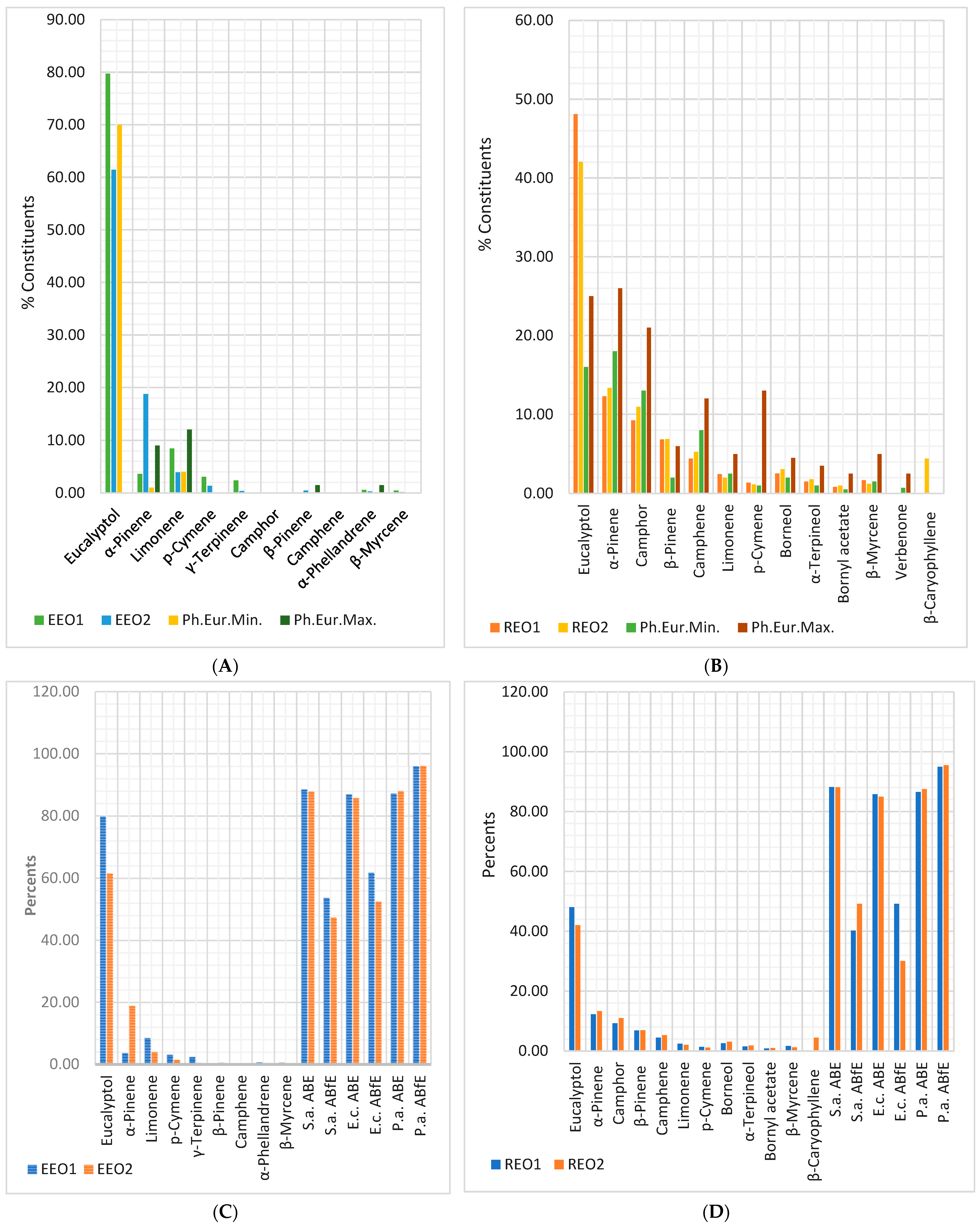
2.Eucalypti
aetheroleum
(Eucalyptus oil)
Eucalyptus globulus Labill.
Eucalyptus polybractea R.T. Baker.
Eucalyptus smithii R.T. Baker.
Pain and inflammation
Cough and cold [52]
3.Rosmarini
aetheroleum
(Rosemary oil)
Rosmarinus officinalis L.Circulatory disorders
Gastrointestinal disorders [53]
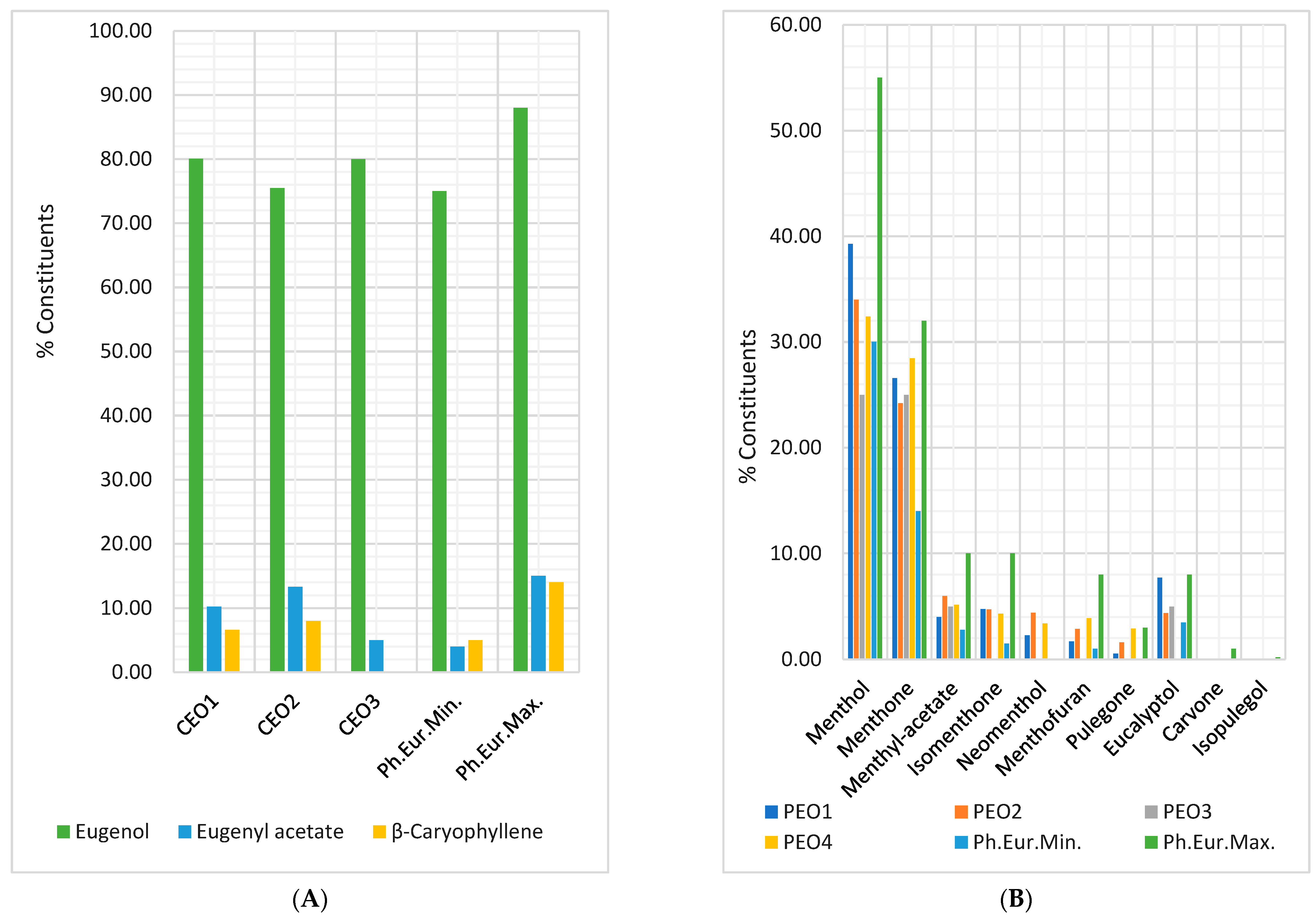
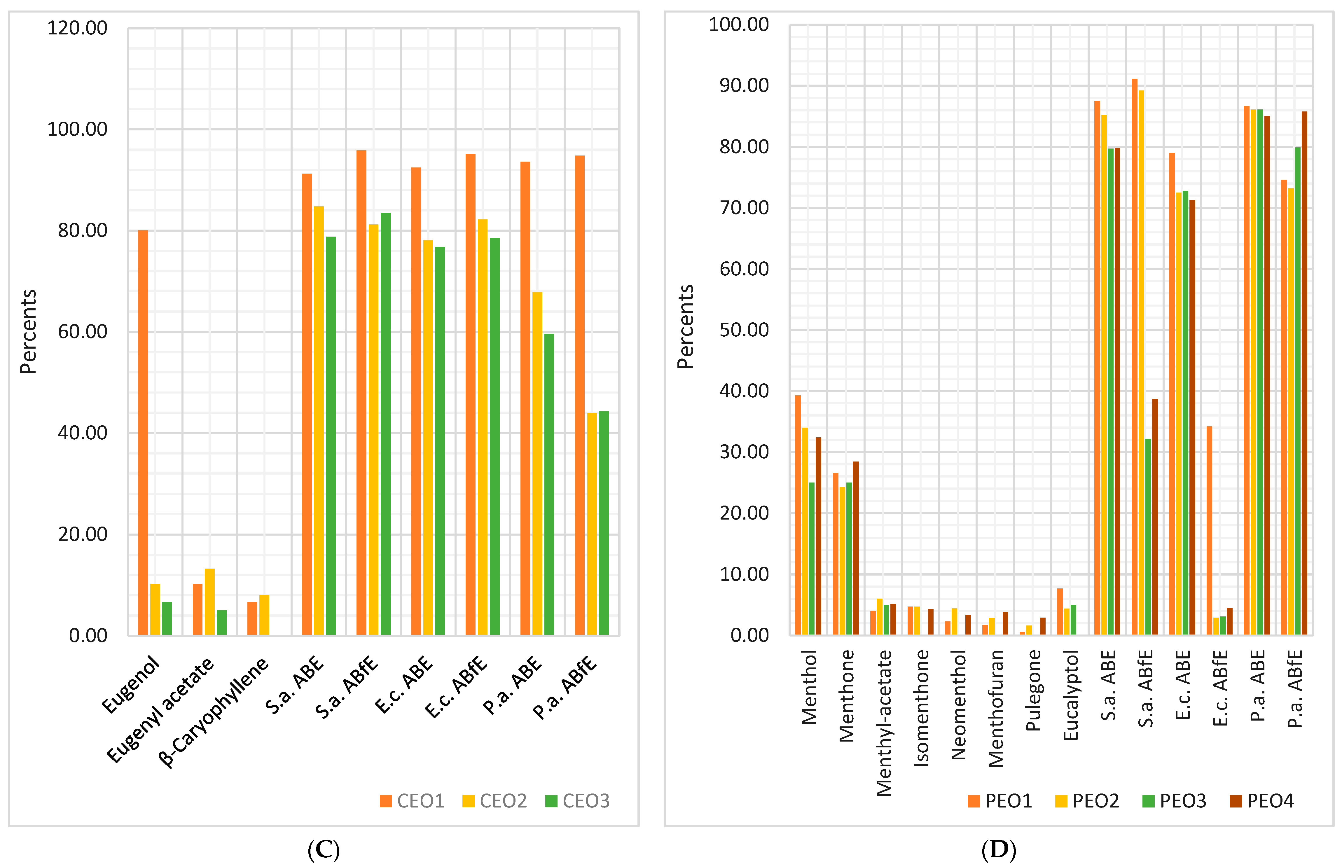
4.Caryophylli floris
aetheroleum
(Clove oil)
Syzygium aromaticum (L.) Merr. et L.M. Perry,
syn. Eugenia caryophyllus (Spreng.) Bullock et S.G. Harrison
Mouth and throat disorders [54]
5.Menthae piperitae aetheroleum
(Peppermint oil)
Mentha piperita L.Pain and inflammation
Skin disorders and minor wounds
Cough and cold
Gastrointestinal disorders [55]
Our study novelty consists of a different design. We selected five essential oils with well-known antibacterial effects [40,41,42,43,44,45,46,47] to facilitate the detection of the potential differences between the results obtained from the tested samples and those mentioned in the literature data. As ordinary consumers, we checked various Romanian markets (including online suppliers) and selected only four that concomitantly provided the essential oils’ chemical composition. We compared the bioactive metabolite contents with those mentioned in each monograph in Ph. Eur. 10 and ISO 13171:2016 according to EMA’s current regulation. Then, the antibacterial and antibiofilm effects of two to four samples of each EO were evaluated, correlating the data obtained with the bioactive compounds. The results were compared with the most known data from the scientific literature. In addition, a complex statistical analysis was performed to support our results.

2. Results

2.1. Antibacterial and Antibiofilm Activity on S. aureus

The percentage values of antibacterial and antibiofilm efficacy of essential oils against Gram-positive bacteria (S. aureus) tested, compared to standard antibiotics, are displayed in Table 2.
Table 2 shows that, at 50 µg/mL, AMC had remarkable antibacterial efficacy (ABE = 96.83%), while both aminoglycosides exhibited a good one (ABE > 85%). All EOs recorded good antibacterial activity (ABE > 75%) against S. aureus at D1 = 25 mg/mL. OEOs and CEO1 proved to have considerable antibacterial potentials (ABE = 92.76, 90.40, 91.26%) like the AMC one and higher than GEN and STR. The CEO3, PEO3, and PEO4 had the lowest ABE (78.80, 79.73, 79.83%). S. aureus sensitivity commonly decreases directly proportional to EOs concentration. Only a few EOs recorded good antibacterial effects at all D1, D2, and D3 dilutions: EEOs and REO1 (ABE% = 75–89%). The anti-staphylococcal effect slowly diminished at progressive dilutions (as in the case of REO2) or intensely decreased (OEOs, CEOs, PEOs 1 and 2). PEOs 3 and 4 exhibited antibacterial effects only at 25 mg/mL. As an overview, we could appreciate that the MIC value for all EOs tested is higher than 25 mg/mL. In addition, no significant differences (p > 0.05) between the antibacterial efficacy values of the tested samples at D1 were recorded in the case of OEOs, EEOs, and REOs (Table 2). At D1, the ABE values of all three CEOs were statistically significant (91.26, 84.80, 78.80%, p < 0.05), and of PEOs (PEO1 and PEO2 vs. PEO3 and PEO4: 87.50 and 85.23% vs. 79.73 and 79.83%, p < 0.05).
Data from Table 2 also show that all standard antibiotics and EOs have antibiofilm activity at D1 and D2.
At D1, AMC shows a moderate ABfE, and both aminoglycosides report a satisfactory one. For EOs, it decreases from “very good” (OEOs, CEO1, and PEO1) to good (CEOs 2 and 3 and PEO2), moderate (EEO1), and satisfactory (EEO2, REOs, PEOs 3 and 4). Excepting OEOs, the samples of each EO recorded significant differences (p < 0.05, Table 2). The highest differences were recorded in the case of PEOs 1 and 2 vs. PEOs 3 and 4 (91.13 and 89.23% vs. 32.17 and 38.73%, Table 2). Moderate differences in ABfE values at D1 were also registered in CEOs (95.40, 77.13, 81.77%, Table 2).
At D2, most EOs recorded significant differences in antibiofilm effect compared to D1 (p < 0.05), except CEO1 and CEO3 (95.87 vs. 95.40%, respectively, 83.50 vs. 81.77%, p > 0.05, Table 2).
At D3 (0.25 mg/mL), the antibiofilm activity registered the lowest values for all standard antibiotics and a significant part of EOs samples (ABfE < 50%). It was not detected for EEOs and CEOs 2 and 3. The CEO1 and PEOs 1 and 2 showed AbfE values > 50% (56.87, 77.20, and 81.87%, respectively, Table 2).

2.2. Antibacterial and Antibiofilm Activity on E. coli

The percentage values of antibacterial and antibiofilm efficacy of essential oils against Gram-negative bacteria E. coli, compared to standard antibiotics, are displayed in Table 3. Table 3 shows that at D1 = 50 µg/mL, AMC exhibits a very good ABE, while both aminoglycosides display a good one (ABE < 90%).
All EOs inhibited E. coli strains growing at D1 = 25 mg/mL; the antibacterial effect decreased from "very good" (OEO1 and CEO1) to moderate (PEO2–4). Most EOs recorded good inhibitory effects against E. coli (Table 3). Statistically significant differences were recorded in CEOs—between CEO1 and CEOs 2 and 3 (92.47% vs. 78.10 and 77.80%, p < 0.05)—and PEOs—PEO1 vs. PEOs 2, 3, and 4 (79.00% vs. 72.50, 72.77, and 71.30%, p < 0.05, Table 3).
At D2, most EOs samples registered significant statistical differences compared to D1; only EEOs and REOs showed similar ABE values (Table 3). The PEOs 2, 3, and 4 (72.50 vs. 43.87%, 72.77 vs. ND, and 71.30 vs. 4.60%) and CEOs 1, 2, and 3 (92.47 vs. 46.27%, 78.10 vs. 34.70%, and 76.80 vs. 18.50%) displayed the highest differences (Table 3).
At D3, the antibacterial activity of CEOs 2 and 3 and PEOs 3 and 4 was undetected. At the same time, in the case of other EOs, it was substantially decreased compared to D2: OEO1 (13.40 vs. 79.40%), PEO1 (45.57 vs. 70.00%), PEO2 (29.80 vs. 43.87%), and CEO1 (22.30 vs. 46.27%). Only ABE of EEO1 showed insignificant differences (83.73 vs. 84.23%, p > 0.05, Table 3). From standard antibiotics, AMC reported an intense diminution of ABE value (19.60 vs. 84.47%, Table 3).
The antibiofilm activity of standard antibiotics and EOs differs significantly at D1 (Table 3). AMC and GEN show a high ABfE, while STR has a moderate one. However, OEOs and CEO1 significantly inhibited E. coli biofilm formation (95.13, 95.63, 95.10%, p > 0.05), while CEOs 2 and 3 had a good antibiofilm effect (82.20, 78.50%, p > 0.05). Table 2 also shows that EEOs have a moderate antibiofilm action (61.73 and 52.33%, p < 0.05), followed by REOs and PEO1 with satisfactory ABfE values (49.23 and 30.07%, p < 0.05, and 34.23%). The PEOs 2, 3, and 4 recorded the lowest inhibitory activity on E. coli biofilm formation (2.90, 3.10, and 4.50%, respectively, Table 3).
Insignificant differences between D1 and D2 were observed at CEO1, STR, and AMC (Table 3). In the case of other EOs and GEN, the ABfE values significantly decreased (p < 0.05, Table 3). On CEOs 2 and 3 and all PEOs, the antibiofilm activity was undetected at D2 and D3 (Table 3). At D3, the other five EOs (OEO2, EEOs, and REOs) reported undetected antibiofilm activity on E. coli; in addition, the ABfE values of OEO1 and CEO1 substantially decreased (2.93 and 11.20%, respectively, Table 3).

2.3. Antibacterial and Antibiofilm Activity on P. aeruginosa
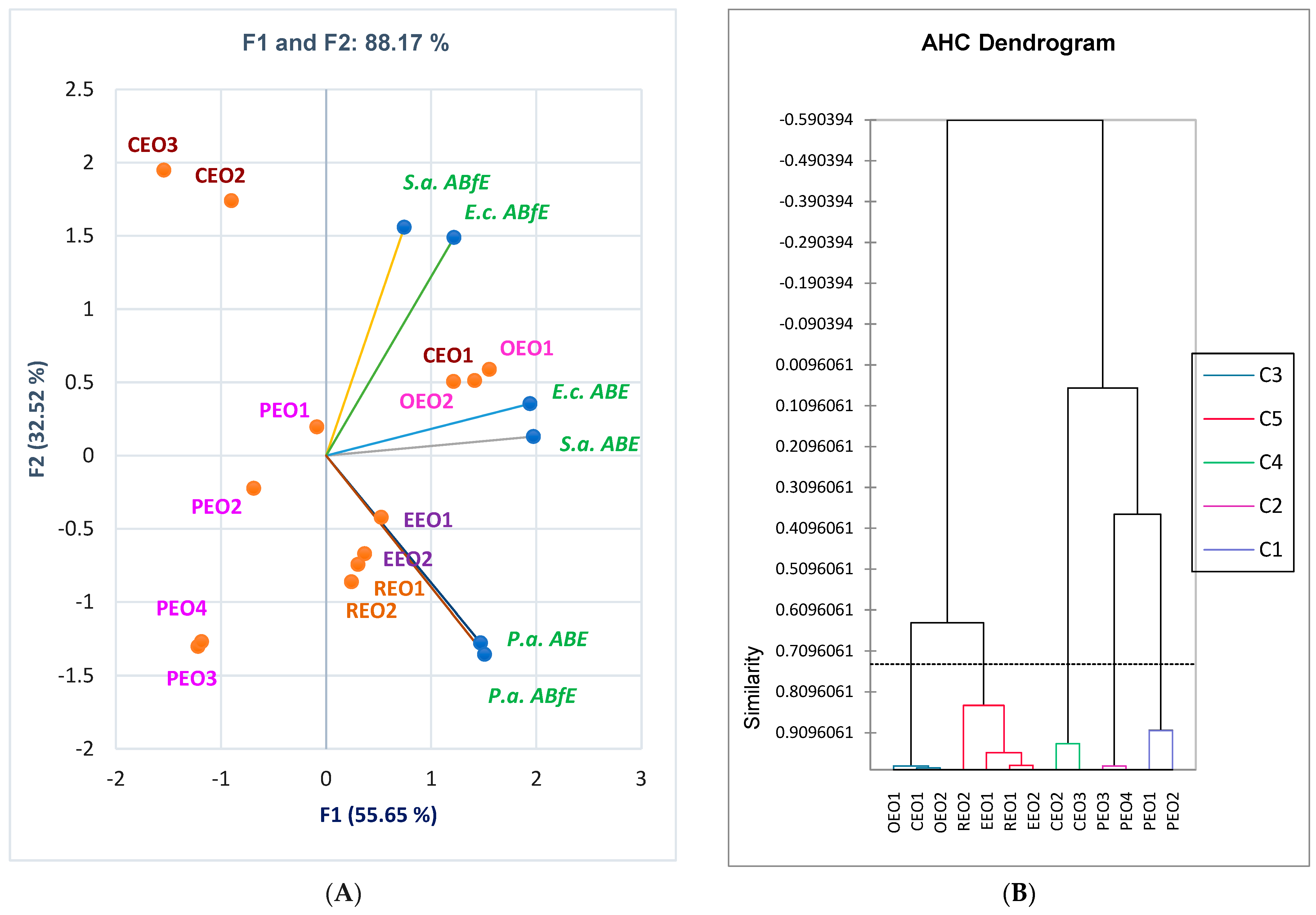
The percentage values of antibacterial and antibiofilm efficacy of essential oils against P. aeruginosa, compared to standard antibiotics, are displayed in Table 4.
Table 4 shows that all standard antibiotics (GEN, STR, and AMC) exhibited very good inhibitory activity on P. aeruginosa strains growing at 50 µg/mL (91.80, 92.80, and 91.40%, p > 0.05), AMC having a lower one than both aminoglycosides. At 25 mg/mL, both OEOs and CEO1 (92.00, 91.40, and 93.60%, p > 0.05) revealed an antibacterial efficacy similar to standard antibiotics, while CEOs 2 and 3 reported a moderate one (67.80 and 59.60%, p < 0.05, Table 4). All the other EOs displayed good antibacterial efficacy against P. aeruginosa. Only between CEO samples (CEO1, 2, and 3) were registered significant differences at D1, p < 0.05 (Table 4).
At D2, EEOs and REOs did not show significant differences compared to D1: ABE = 84.70–87.90%, p > 0.05 (Table 4). The same observation is available for PEO2 (79.20 vs. 86.10%, p > 0.05, Table 4). Most EOs (OEOs, CEOs, and PEOs 1 and 4) reported a significant diminution in ABE values (p < 0.05, Table 4). The PEO 2 antibacterial activity at D2 was undetected (Table 4).
At D3, EEOs maintained the same antibacterial activity without significant differences reported to D1 and D2 (ABE = 84.93–87.90%, p > 0.05, Table 4). The REOs 1 and 2 (75.73 and 66.10% vs. 84.70 and 85.77%, p < 0.05) and PEOs 1 and 2 (69.70 and 61.40% vs. 77.44 and 79.20%, p < 0.05) displayed more significant differences than D2 but not as high (Table 4). The OEO2, CEO1, and standard antibiotics exhibited low antibacterial activity at D3, undetected at CEOs 2 and 3, and PEOs 3 and 4 (Table 4).
Table 4 reveals that OEOs, EEOs, REOs, and CEO1 displayed substantial antibiofilm activity at D1 and D2 (ABE > 90%, p > 0.05). Their biofilm formation inhibition is higher than standard antibiotics (Table 4). At D1, ABfE values for CEO2 and CEO3 are <50% of CEO1 (43.90 and 44.30% vs. 94.80%, p < 0.05). It strongly diminished at D2 (7.10 and 16.40% vs. 92.40%, p < 0.05, Table 4). Moreover, PEO4 has ABfE value higher than PEOs 1, 2, and 3: 85.80% vs. 74.60 and 73.20% (p < 0.05), and 79.90% (p > 0.05); at D2, the antibiofilm activity of PEOs 3 and 4 was undetected, and that of PEOs 1 and 2 was similar (68.80 vs. 65.90%, p > 0.05, Table 4).
At D3, REOs and EEOs maintained a good antibiofilm effect (ABfE > 85%), while the other EOs reported a high diminution: OEOs (27.77 and 8.50%, p < 0.05), CEOs 1 and 2 (16.00 vs. 4.10%, p < 0.05), and PEOs 1 and 2 (57.20 vs. 48.40%, p < 0.05). Moreover, CEO3 and PEOs 3 and 4, like standard aminoglycosides, did not show antibiofilm activity at D3 against P. aeruginosa (Table 4).

2.4. Data Analysis

Pearson correlation applied on OEOs showed a substantial positive correlation between carvacrol and linalool and antibacterial effect against all three bacteria and antibiofilm activity on S. aureus and P. aeruginosa (r = 0.999, p < 0.05). Thymol, p-cymene, γ-terpinene, and α-terpinene strongly correlate with antibiofilm activity in E. coli (r = 0.998, p < 0.05).
Principal Component Analysis (PCA) was used to evaluate the correlation between the bioactive compounds and antibacterial and antibiofilm efficacy of EOs on Gram-positive and Gram-negative bacteria.
The PCA-Biplot from Figure 1 shows the correlation between the previously mentioned variable parameters for two EOs (EEO and REO) with common constituents (eucalyptol, α-pinene, β-pinene, camphene, limonene, p-cymene, and β-myrcene). The Correlation Matrix from Supplementary Material shows evidence of substantial correlations between several secondary metabolites and antibacterial and antibiofilm effects of EEOs and REOs. Eucalyptol, limonene, p-cymene, and γ-terpinene substantially correlate with antibacterial effects against E. coli (r = 0.931, r = 0.937, r = 0.944, r = 0.913, p > 0.05). At the same time, they show a good and moderate correlation with antibiofilm activity on E. coli (r = 0.882, r = 0.803, r = 0.759, r = 0.731, p > 0.05). They also moderately correlate with antibiofilm effects on S. aureus (r = 0.633, r = 0.711, r = 0.658, r = 0.746, p > 0.05) and P. aeruginosa (r = 0.728, r = 0.662, r = 0.503, r = 0.612, p > 0.05). Moreover, limonene, p-cymene, and γ-terpinene moderately correlate with antibacterial activity against S. aureus (r = 0.610, r = 0.759, r = 0.701, p > 0.05). α-Phellandrene displays a considerable correlation with antibacterial activity against E. coli (r = 0.873, p > 0.05) and a good and moderate one with antibiofilm effects on P. aeruginosa, E. coli, and S. aureus (r = 0.811, r = 0.806, r = 0.734, p > 0.05). Figure 1 also shows the place of each EEO and REO sample, correlated to chemical composition and antibacterial and antibiofilm activities.
Figure 2 displays the correlations between bioactive constituents and CEOs’ antibacterial and antibiofilm effects.
Thus, eugenol substantially correlates with all antibacterial and antibiofilm activities: E.c. ABE and P.a. ABfE (r = 0.999, p < 0.05), E.c. ABfE (r = 0.986, p > 0.05), P.a. ABE and S.a. ABfE (r = 0.982, p > 0.05), S.a. ABE (r = 0.897, p > 0.05). It has the highest content compared to other volatile compounds quantified in CEOs and shows the most substantial correlations with antibacterial and antibiofilm activities.
Eugenyl acetate and β-caryophyllene correlate moderately with S.a. ABE (r = 0.611, r = 0.760, p > 0.05). β-caryophyllene displays a low correlation (r = 0.543, r = 0.559, p > 0.05) with E.c. ABfE and P.a. ABE. Finally, the PCA-Biplot shows each CEO sample’s place considering these variable parameters; it provides evidence that CEO1 is substantially active regarding antibacterial and antibiofilm effects. All detailed data are found in Supplementary Material.
Figure 3 shows the correlations between bioactive constituents and PEOs’ antibacterial and antibiofilm effects. From bioactive constituents, eucalyptol shows a remarkable and significant statistical correlation with antibacterial activity against P. aeruginosa (r = 0.995, p < 0.05). It is also considerably correlated with ABE against E. coli (r = 0.830, p > 0.05) and moderately associated with ABE against S. aureus (r = 0.716, p > 0.05) and ABfE in S. aureus and E. coli (r = 0.593, r = 0.685, p > 0.05). However, eucalyptol negatively correlates with ABfE in P. aeruginosa (r = −0.803, p > 0.05). Concomitantly, menthol is considerably correlated with both ABE and ABfE in S. aureus (r = 0.847, r = 0.826, p > 0.05) and E. coli (r = 0.680, r = 0.754, p > 0.05). Moderate correlations were evidenced between menthone and pulegone and antibiofilm activity against P. aeruginosa (r = 0.736, r = 0.503, p > 0.05); both previously mentioned constituents and menthofuran are negatively correlated with P.a. ABE (r = −0.579, r = −0.818, r = −0.617, p > 0.05). Isomenthone and neomenthol with ABfE on S. aureus (r = 0.708, r = 0.550, p > 0.05). Moreover, isomenthone is moderately correlated with antibacterial activity against S. aureus (r = 0.636, p > 0.05).
Finally, considering all discussed variable parameters—extensively described in Supplementary MaterialFigure 3 shows the place of each PEO sample, thus explaining all differences between them and supporting the results.
The correlation matrix from Supplementary Material and Figure 4A highlights a strong correlation between antibacterial and antibiofilm effects against both Gram-negative bacteria, P. aeruginosa (r = 0.912, p > 0.05) and E. coli (r = 0.760, p > 0.05). On S. aureus, both effects are poorly correlated (r = 0.425, p > 0.05) when we evaluate this effect on the entire EOs group. However, for all EOs and standard antibiotics, the antibacterial activity against S. aureus is considerably associated with that against E. coli (r = 0.895, p < 0.05) and moderately with that against P. aeruginosa (r = 0.628, p < 0.05). Antibacterial effects against Gram-negative bacteria also show a moderate correlation (r = 0.878, p < 0.05). All data are statistically significant (p < 0.05). Generally, antibiofilm activities on all bacteria tested are poorly correlated. As an overview, on the first dilution (D1 = 25 mg/mL), only OEOs show similar percentage values of antibacterial and antibiofilm effects.
The registered data from Results are summarized in Figure 4A, evidencing the place of essential oils reported to both ABE and ABfE against all bacteria tested.
In a simplified manner, the dendrogram obtained by Agglomerative Hierarchical Clustering (AHC) from Figure 4B and Supplementary Material shows how EO samples act similarly. Figure 4B shows that PEO1 acts similarly to PEO2, PEO3 to PEO4, and CEO2 to CEO3. On the other hand, both OEOs have similar effects at D1. At the same time, OEO2 acts closely to CEO1. The same observation is available on EEOs 1 and 2 and REOs 1 and 2.

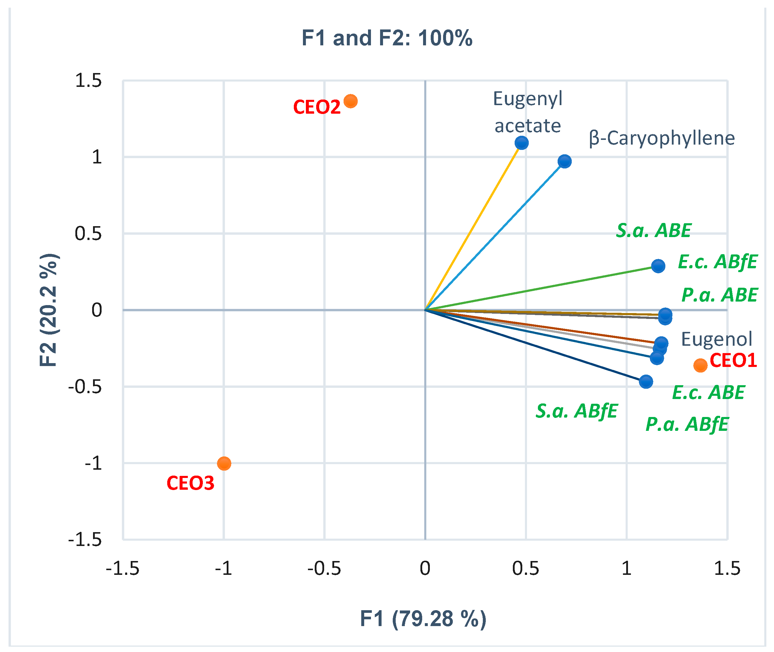
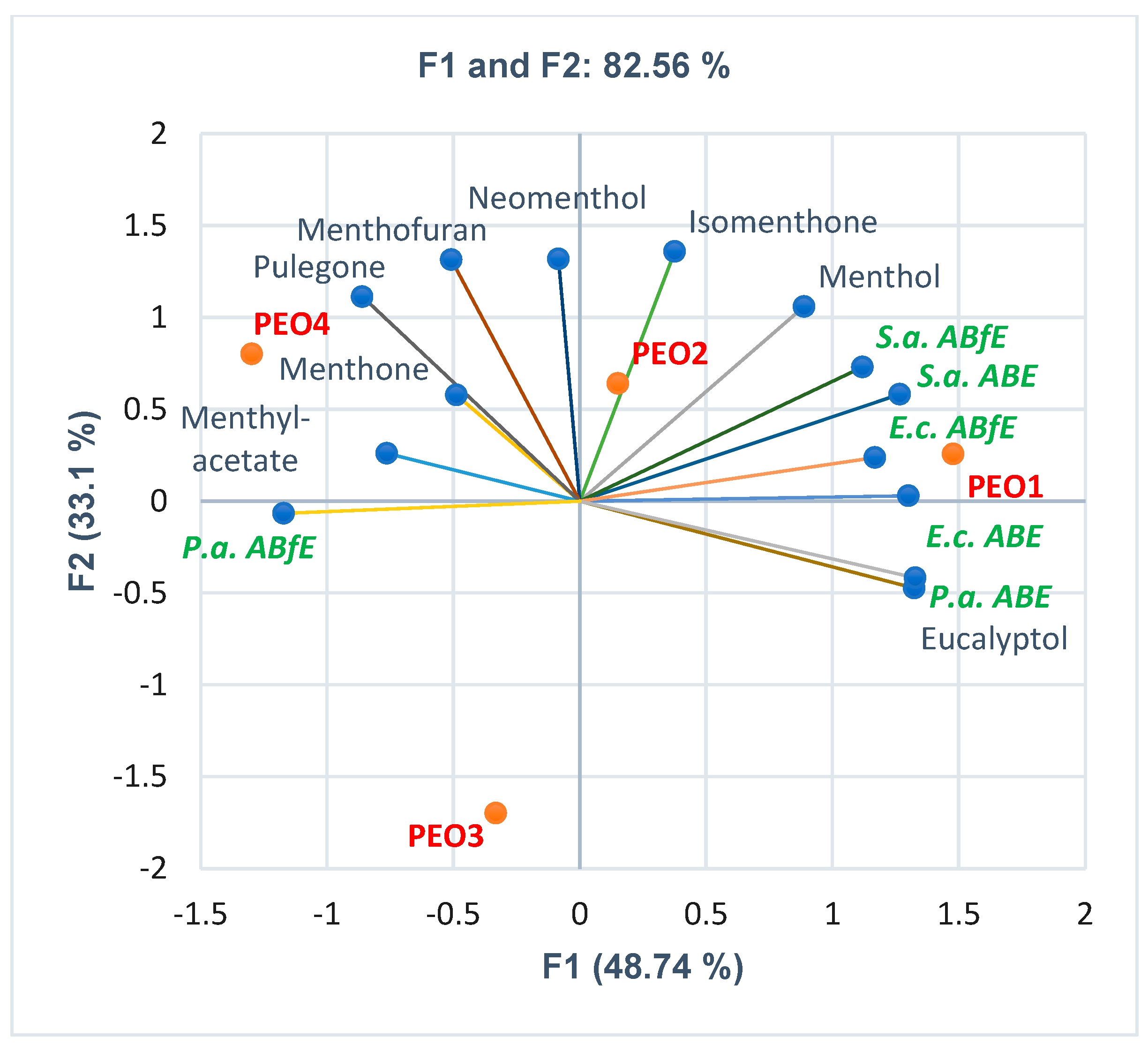
2.5. Correlations between EOs Chemical Constituents and Their Antibacterial and Antibiofilm Effects

Knowing each EO’s chemical composition, the correlations between bioactive constituents and antibacterial and antibiofilm effects were analyzed to explain the differences between the corresponding samples.
Figure 5A indicates that only p-cymene’s contents in both OEO samples are included in Standard limits (4–10%). OEO1 displays 69.60% carvacrol, 5.60% γ-terpinene, and 2.60% thymol; all values are in the ISO 13171:2016 limits. In contrast, OEO2 has significantly lower carvacrol content (44.40% < ISO St.Min.) and considerably higher thymol (3×) and γ-terpinene (2×) concentrations than ISO St.Max. Furthermore, linalool was detected in OEO1 and α-terpinene in OEO2, unmentioned in ISO 13171:2016. Figure 5B shows that OEOs’ antibacterial and antibiofilm activities against Gram-positive and Gram-negative bacteria are similar. All constituents contribute to these effects. OEO1 has the highest carvacrol content, but OEO2 contains thymol and γ-terpinene more than ISO 13171:2016 maximal limits. Their effects are augmented by p-cymene, linalool, and α-terpinene; the last two volatile compounds are missing in the standard.
Figure 6A shows that the eucalyptol content in EEO2 is lower than EEO1 and Ph. Eur.Min., while the α-pinene content is higher. Both EEOs have three compounds unmentioned in Ph. Eur. 10 (p-cymene, γ-terpinene, and α-phellandrene). Figure 6B reveals that both REOs have eucalyptol content substantially higher than Ph. Eur.Max. (48.10 and 42.05 > 25%); β-pinene ones are slightly increased than Ph. Eur. 10 maximal value (6.84 and 6.89 > 6.00%). α-Pinene (12.29 and 13.35%), camphor (9.23 and 10.26%), and camphene (4.42 and 5.28%) are significantly lower than Ph. Eur.Min. (18, 13, and 8%). EEOs and REOs display considerable antibacterial effects on all bacteria tested (Figure 6C,D). The antibiofilm activities are substantial on P. aeruginosa and moderate on S. aureus and E. coli. Eucalyptol is the primary metabolite responsible for it in both EOs; its content in REOs was higher than maximal limits from Ph. Eur. 10. Its activity is potentiated by other compounds (limonene, p-cymene, and γ-terpinene) in both EOs and α-phellandrene in EEOs.
Figure 7A shows that all three CEO samples contain eugenol and eugenyl acetate in the Ph. Eur. 10 range (75–88%, respectively, 4–13%). CEO1 and CEO3 have the highest eugenol content (80.00–80.06%). The eugenyl acetate content decreases in order: CEO2, CEO1, and CEO3 (13.27 > 10.25 > 5%). CEO3 did not have β-aryophyllene. Some aspects concerning PEOs’ chemical composition were observed, analyzing Figure 7B. Thus, all PEOs contain menthone and menthyl acetate in the Ph. Eur 10 limits (14–32% and, respectively, 2.80–10%). PEO1 has the highest contents of menthol, isomenthone, and eucalyptol (39.27, 4.75, and 7.70%), while PEO4 shows the greatest ones for menthone (28.45%), menthofuran (3.89%), and pulegone (2.90%), and PEO2 for menthyl acetate (6.01%) and neomenthol (4.42%). PEO3 contains only four bioactive compounds: menthol (<30%), menthone, menthyl acetate, and eucalyptol; PEO4 has no eucalyptol. Moreover, two constituents mentioned in Ph. Eur. 10 (carvone and isopulegol) were not found in PEOs samples. At the same time, neomenthol, contained by PEO1, PEO2, and PEO4, did not appear in Menthae piperitae aetheroleum monograph from Ph. Eur. 10.; the same observation is available for menthofuran (3.89%) and pulegone (2.90%) and PEO2 for menthyl acetate (6.01%) and neomenthol (4.42%). PEO3 contains only four bioactive compounds: menthol (< 30%), menthone, menthyl acetate, and eucalyptol; PEO4 has no eucalyptol. Moreover, two constituents mentioned in Ph. Eur. 10 (carvone and isopulegol) were not found in PEOs samples. At the same time, neomenthol, contained by PEO1, PEO2, and PEO4, did not appear in Menthae piperitae aetheroleum monograph from Ph. Eur. 10.
The antibacterial activity of CEO and PEO samples is different. CEO1, due to substantial eugenol content, acts like OEOs (Figure 4). The antibacterial and antibiofilm effects decrease in order of CEO1, CEO2, and CEO3. In addition, eugenyl acetate and β-caryophyllene synergistically act with eugenol. CEO2 and CEO3 display similar antibacterial and antibiofilm potential, significantly lower than CEO1.
Figure 4 and Figure 7D show that PEO1 and PEO2 and, respectively, PEO3 and PEO4, act similarly. The primary metabolites implied in antibacterial and antibiofilm effects are menthol, menthone, menthyl acetate, and eucalyptol. They synergistically act with the others in lower content.
All previously mentioned observations are available for D1 = 25 mg/mL. At the following two dilutions (D2 = 2.5 mg/mL and D3 = 0.25 mg/mL), the antibacterial and antibiofilm effects could remain similar, slowly decrease, substantially diminish, or be undetected.
On S. aureus and P. aeruginosa, the antibacterial activity of both EEOs did not report significant differences (Table 2 and Table 4), while on E. coli, only EEO1 reported similar ABE values (Table 3). Regarding antibiofilm activity, EEO1 and both REOs recorded similar effects (p > 0.05) at all three dilutions against P. aeruginosa.
On S. aureus, the following EOs exhibited no significant differences in antibiofilm activity between D1 and D2: PEOs 1 and 2, CEOs 1 and 3, and OEO1; on E. coli, EEO2, and REOs, regarding antibacterial activity; finally, on P. aeruginosa, REOs and PEOs (ABE) and PEO1, CEO1, EEO2, OEOs (ABfE). In contrast, at D2 and D3, the antibacterial activity of PEOs 3 and 4 against S. aureus was undetected. Similar observations are available on E. coli: PEO3 has no antibacterial effects, and CEO2, CEO3, and all PEOs did not record antibiofilm activities (Table 3). On P. aeruginosa, PEOs 3 and 4 did not show antibiofilm activity, and only PEO3 had no antibacterial efficacy (Table 4).

3. Discussion

According to European Pharmacopoeia, an EO is an odorous product with complex composition obtained by steam distillation, dry distillation, or other mechanical processes without heating from a botanically defined plant raw material. The Eos’ separation from the aqueous phase uses a physical process without significantly affecting their composition. This definition reveals that other extraction procedures involving different solvents lead to extracts, not EOs. Due to increased demands and insufficient regulation, the adulteration of essential oils became a common practice along supply chains, generating safety concerns in the EOs industry [56]. The EOs adulteration could be performed using various methods: a cheaper oil addition [57] to the original one (e.g., corn mint to Peppermint oil), EOs’ dilution with vegetable oils, and synthetic phytochemicals’ inclusion [51] in the original EO [58,59,60]. Thus, supplementary quality control measures should be taken to ensure safety for human use [61,62]. Because aromatherapy is the most crucial application of essential oils, the best way to test the properties of various samples of the same EO is by using biological systems [63]. Pierson et al. [38] recently signaled the potential consumer vulnerability to neroli, mandarin, and bergamot essential oils purchased online.
Many patients request pharmacists’ counseling, aiming to know how to select from numerous types of commercially available essential oils the most suitable ones for therapeutic purposes. In Romania, the most known manufacturers mention the following data on each EO packaging and leaflet [64,65].
  • The scientific name of the raw plant.
  • 100% Pure—It is not mixed with other essential oils, synthetic components, plant oils, or mineral oils and has no chemical solvents.
  • 100% Natural—It is obtained by steam distillation.
  • 100% Verified—It is biochemically and botanically defined.
  • Notification number at the National Service for Medicinal, Aromatic Plants and Hive Products [66], National Research and Development Institute for Food Bioresources—IBA Bucharest [67] according to the National Regulation on Food Supplements.
On some manufacturers’ websites, the GC-MS analysis reports are available, indicating the chemical composition of the EOs [68,69]. The others provide GC-MS reports upon request. Therefore, putting ourselves in the patient’s place, we have chosen well-known, most studied, and widely used EOs in therapy due to their antibacterial properties precisely so that we can relate our results to the data from the literature. We purchased and analyzed several samples of the same EO from different suppliers. All investigated EOs samples were manufactured by four autochthonous companies and regularly commercialized in pharmacies, pharma markets [70], and online markets. We requested the GS-MS reports with chemical composition to compare them with the one from the Ph. Eur. 10 monograph, or ISO standard, as stipulated by the EMA regulation. The manufacturers provided each EO’s chemical composition, and all statistical analyses were performed with available data [71]. Next, we analyzed their influence on antibacterial and antibiofilm effects against Gram-positive and Gram-negative bacteria tested using the microdilution method. Three decimal dilutions, 25, 2.5, and 0.25 mg/mL, were used for EOs; the values were established based on the data used in previously published studies from the scientific literature. We selected the other three (D1 = 50 µg/mL, D2 = 5 µg/mL, and D3 = 0.5 µg/mL) for standard antibiotics, considering their MIC values on bacterial strains according to CLSI and EUCAST [71].
Regarding the OEOs’ chemical composition, it was observed that OEO1 contains all four constituents (carvacrol, p-cymene, γ-terpinene, and thymol) in the suitable Ph. Eur. limits (Figure 5A). OEO2 has a lower carvacrol content, but other metabolites in augmented concentrations than OEO1: thymol content—seven times higher, γ-terpinene—three-point-five times higher, and p-cymene—two times higher. A similar thymol concentration was quantified by Salehi et al. in OEO from Greece [72]. Moreover, another two compounds, unmentioned in Ph. Eur., were found in the GC-MS report: linalool and β-caryophyllene, in up to 2% concentration. They could contribute to the antibacterial effects due to complex interaction with the other bioactive metabolites [44]. Our results confirm these aspects (Figure 5B).
The main constituents of OEO are carvacrol and thymol, which have solid pharmacological potential, including antibacterial, anti-inflammatory, and antioxidant activities. At the same time, carvacrol and thymol, obtained by chemical synthesis, could be adulterants of Oregano oil [48]. Gavaric et al. [73] reported the additive antibacterial effect of thymol and carvacrol against tested bacterial strains (S. aureus, E. coli, Salmonella infantis, and Bacillus cereus). p-Cymene is the biological precursor of carvacrol; when used alone, it exhibits a lower antibacterial effect than carvacrol; however, it synergistically acts with carvacrol against E. coli [74] and B. cereus [75]. Furthermore, carvacrol can form a chimera with DNA [76], while thymol reduces enterotoxins A, B, and α-hemolysin secreted by S. aureus isolates [77].
Thus, OEO could be considered a broad-spectrum natural antibiotic [78,79]. Investigating the antibacterial mechanism against MRSA, Cui et al. proved that OEO affects bacterial wall permeability, leading to an irreversible depletion [76]. It can inhibit bacterial respiratory metabolism (perturbing the tricarboxylic acids cycle) and the expression of MRSA’s crucial pathogenic factor PVL.
In the present study, both OEOs display similar antibacterial and antibiofilm activities, reporting the highest effects compared to all EOs investigated. Their inhibitory activity against all bacteria tested is similar to Amoxicillin-Clavulanic acid. Our results were similar to those obtained in other studies, which confirm both activities (antibacterial and antibiofilm) on S. aureus [80], E. coli [81], and P. aeruginosa [82,83].
Our PCA analysis, separately performed on OEOs, reported a strong statistically significant correlation between the bioactive constituents and ABE, respectively, ABfE, thus justifying OEOs’ records. Carvacrol and linalool were substantially correlated (r = 0.999, p < 0.05) with ABE and ABfE against S. aureus and P. aeruginosa and only with ABE against E. coli. In contrast, thymol, linalool, α-, and γ-terpinene evidenced the same correlation with antibiofilm activity in E. coli. Numerous studies from scientific literature reported the antibacterial and antibiofilm properties of all previously mentioned phenolic compounds [84,85,86,87,88,89,90,91,92,93,94,95].
For all bacteria tested, MIC > 25 mg/mL. Other studies indicated various MIC values: 0.16–0.32 mg/mL against MRSA [96], >3.2 mg/mL against E. coli [97,98], and 0.16–0.64 mg/mL against P. aeruginosa [96].
Of six metabolites mentioned in the EEO monograph from Ph. Eur. 10, only four (eucalyptol, α-pinene, α-phellandrene, and limonene) appear in the available GC-MS reports (Figure 6A). Their concentration in EEO1 is included in regulatory limits. EEO2 has a lower content of eucalyptol (61.45% vs. 79.73%) and a six times higher concentration of α-pinene (18.77% vs. 3.60%). The samples have small contents of other compounds: myrcene (in EEO1), p-cymene, and γ-terpinene (in EEO1 and EEO2).
Bachir et al. [99,100], reported the antibacterial efficacy of Eucalypti aetheroleum against S. aureus due to its phytoconstituents (eucalyptol, linalool, β-pinene). Moreover, its bactericidal effect against E. coli and P. aeruginosa was reported [101]. The EEO’s bioactive constituent responsible for the antibiofilm activity is eucalyptol [102,103,104]. Therefore, Eucalyptus oil penetrates the biofilm matrix, interfering with the essential constituents’ synthesis and the metabolic processes of the biofilm. EEO has synergistic antibacterial activity against Gram-positive bacteria, while against Gram-negative ones, it is additive [105]. Furthermore, eucalyptol obtained by chemical synthesis could be mixed with Eucalyptus oil for adulteration [106].
The present study proved Eucalypti aetheroleum’s appreciable antibacterial effectiveness against S. aureus, E. coli, and P. aeruginosa (MIC > 25 mg/mL). The EEO’s MIC against MRSA varies between 0.032–307 mg/mL [28]. Mulyaningsih et al. [107] evidenced antibacterial activity against E. coli with a MIC > 4 mg/mL. In comparison, Van et al. reported a median MIC of 27.26 mg/mL against P. aeruginosa isolates [108]. However, EEOs recorded a moderate antibiofilm efficacy on S. aureus and E. coli strains and a substantial one against P. aeruginosa, like OEOs. Minimal differences were registered between both tested samples, with EEO1 acting higher than EEO2. Both common constituents are considerably correlated with antibacterial/antibiofilm activities. Thus, data analysis performed exclusively on EEOs indicated a strong statistically significant correlation between eucalyptol, limonene, and p-cymene and both antibacterial and antibiofilm activities against S. aureus and E. coli; at the same time, α-pinene is substantially correlated with ABE and ABfE against P. aeruginosa (r = 0.999, p < 0.05). The antibacterial and antibiofilm activities of these phenolic metabolites were revealed by other previously published studies [109,110,111,112,113,114,115,116,117,118,119,120].
Compared to Ph. Eur. data, REOs have around two times higher eucalyptol content (48.10% in REO1 and 42.50% in REO2) and no verbenone (Figure 6B). In addition, REO2 contains 4.45% β-caryophyllene; α and β-Pinene camphor, borneol, and camphene were substantially correlated with ABE and ABfE on P. aeruginosa (r = 0.999, p < 0.05). Eucalyptol has a similar correlation with ABE against E coli; camphor and camphene are correlated with ABE and ABfE against S. aureus; pinene beta only correlates with ABfE on S. aureus. These compounds are known for their antibacterial and antibiofilm effects [121,122,123,124,125,126,127,128,129].
Previous studies [97,130,131,132,133] evidenced the antibacterial effects of Rosmarini aetheroelum against S. aureus and E. coli. Santoyo et al. [133] highlighted the antibacterial efficacy of REO against P. aeruginosa due to the bioactive constituents, camphor, borneol, and verbenone. Rosemary oil also inhibits P. aeruginosa biofilm formation [134].
Our results show that Rosmarini aetheroleum had significant antibacterial and antibiofilm efficacy against P. aeruginosa. The antibacterial activity is similar for all Gram-positive and Gram-negative bacteria tested (ABE > 80.00%, MIC > 25 mg/mL). Other studies reported MIC values of 6.2–25 mg/mL against S. aureus, 12.5–25 mg/mL against E. coli, and 50 mg/mL against P. aeruginosa [135]. REO1 was slightly more active than REO2, but no significant differences were recorded between the two samples’ effects.
Moreover, the synthetic equivalents of their main components, eucalyptol and camphor, could be used for Rosemary oil adulteration [136].
Generally, their chemical composition corresponds to Ph. Eur.; CEO1 has the highest eugenol content, followed by CEO3 and CEO2 (Figure 7A). However, CEO3 has the lowest eugenyl acetate concentration and no β-caryophyllene; all constituents are highly and moderately correlated with antibacterial and antibiofilm activities [137,138,139,140,141,142,143].
Xu et al. [144] highlighted the antibacterial efficacy of Caryophylli aetheroleum against S. aureus (with a MIC value = 0.625 mg/mL). They hypothesized that the volatile oil destroys the cell wall and membranes, causing loss of vital intracellular materials, resulting in bacterial death. The volatile oil also penetrates the cytoplasmic membrane and inhibits the normal synthesis of DNA and proteins necessary for bacterial growth. Yadav et al. [145] reported the antibiofilm effect of Clove oil on S. aureus attributed to eugenol. It inhibits biofilm formation, interrupts intercellular connections, detaches pre-existing biofilms, and kills bacteria in biofilms. Synthetic eugenol is also used for Clove oil adulteration [42].
The MIC value in the present study for all bacteria tested is >25 mg/mL. Generally, the MIC values of CEO against S. aureus vary in the range of 0.52–1.04 mg/mL [146].
Burt et al. [147] evidenced the antibacterial efficacy of Caryophylli aetheroleum against E. coli; the CEO’s MIC value belonged to the range of 0.64–1.28 mg/mL [146]. Another study by Kim et al. [148] reported the antibiofilm efficacy of Clove oil against E. coli due to eugenol inhibitory activity on biofilm formation.
The CEO’s antibacterial efficacy against P. aeruginosa is also demonstrated [149], with a MIC of 4.9 mg/mL [150]. Moreover, the antibiofilm activity of Clove oil is due to its main bioactive compounds, eugenol and eugenyl acetate [151].
The present study reports a few differences between the three CEO samples.
Thus, CEO1 showed substantial antibacterial and antibiofilm efficacy against all Gram-positive and Gram-negative bacteria tested (with ABE and ABfE values > 91.80%).
CEO2 and CEO3 proved good antibacterial and antibiofilm effectiveness against S. aureus and E. coli. However, significant differences were registered in their effects against P. aeruginosa, exhibiting moderate and satisfactory antibacterial and antibiofilm activity.
All PEOs contain menthone and menthyl acetate in the Ph. Eur. limits (Figure 7B). Only PEOs 1, 2, and 4 have suitable menthol content; PEO3 is under 30%. Isomenthone, neomenthone, menthofuran, and pulegone were not detected in PEO3, while PEO4 does not have eucalyptol. These phytochemical aspects can explain the differences between PEOs 1–4 bioactivities. PEO1 and PEO2 have the highest concentrations of menthol, menthyl acetate, isomenthone, and eucalyptol; these constituents considerably correlate with antibacterial and antibiofilm activities. Moreover, synthetic menthol could substitute Peppermint in adulterant oil [152].
Generally, in Peppermint oil, the association of menthol, menthone, limonene, neomenthol, carvone, and eucalyptol with other minor constituents appears to induce a synergistic antibacterial activity. A recent study [153] evaluated the antibacterial activity of volatile oil obtained from Mentha piperita L. leaves on MDR strains from hospitalized patients. The authors used bacterial cell lines (ATCC) and isolates of S. aureus, E coli, and P. aeruginosa, proving PEOs’ bactericidal effects against all microorganisms.
Li et al. [154] evidenced that Menthae aetheroleum (with a high content of carvone, menthone, isomenthone, neomenthol, menthol, and menthyl acetate) has a significant antibacterial effect against S. aureus [155]. All tested samples of Peppermint oil showed appreciable anti-staphylococcal efficacy. Kang et al. [156] showed that PEO inhibits the biofilm of S. aureus by altering the permeability and integrity of bacterial cell membranes. Peppermint oil significantly inhibits biofilm formation and inactivates the mature biofilm [157].
Alamoti et al. [158] proved the antibacterial efficacy of Menthae aetheroleum against E. coli due to pulegone content.
Peppermint oil also inhibits P. aeruginosa [159], showing substantial antibiofilm activity [160,161].
All four PEO samples investigated in the present study had remarkable antibacterial effects against Gram-positive and Gram-negative bacteria, with no significant differences (MIC > 25 mg/mL). They recorded the highest ABE (>85.00%) on P. aeruginosa and S. aureus (ABE > 79.70%). On E. coli, the PEOs’ antibacterial efficacy was good to moderate, in the range of 71.30–79.00%; PEO1 shows the highest effect. Evaluating the antibacterial effect of EO from Mentha piperita L. against MDR bacterial strains, Muntean et al. reported the following MIC values range: 5–20 mg/mL on S. aureus, 10–20 mg/mL on E. coli, and 20–40 mg/mL on P. aeruginosa [153].
Regarding the antibiofilm activity, the PEOs displayed considerable effects on P. aeruginosa, ABfE = (73.20–85.80%). On E. coli, PEOs registered the lowest effects: AbfE = (2.90–34.20%). The most significant differences were highlighted in the antibiofilm efficacy evaluation against S. aureus. The obtained data show that PEO1 and PEO2 have a substantial antibiofilm activity AbfE = (89.20–91.00%). Concomitantly, PEO3 and PEO4 exhibited a poor antibiofilm effect (32.10–38.70%).
Some significant observations are available to corroborate all obtained results and compare them with the literature data. As an overview, the samples with different manufacturers of the same essential oil showed similar activities; only Clove and Peppermint oils showed higher differences. Compared to other studies’ results, the low differences between EEO and REO samples appear not to influence the antibacterial effects. Oregano oils showed a substantially lower antibacterial activity against S. aureus and P. aeruginosa; the MIC values (>25 mg/mL) are significantly higher than those mentioned in literature data: 0.16–0.32 mg/mL against MRSA and 0.16–0.64 mg/mL against P. aeruginosa. The same observation is available for Clove oils against S. aureus and E. coli; the MIC values from literature data are substantially lower (0.52–1.04 mg/mL, respectively, 0.64–1.28 mg/mL) than those obtained in the present study. Moreover, appreciable differences were recorded in the chemical composition of CEO samples. Notable differences in bioactive constituents’ content were registered in PEOs samples, resulting in high antibacterial and antibiofilm effects variations. However, the literature data indicates large ranges of MIC variation against all bacteria tested.
All these differences could be explained by the significant variation of EOs’ chemical composition, bacterial strains selected, and technical aspects implied in microbiological assays used.

4. Materials and Methods

4.1. Materials

All chemicals and reagents were of analytical grade. Poly (ethylene glycol)-block-poly (propylene glycol)-block-poly (ethylene glycol) (Poloxamer 407) and Crystal Violet (Gentian Violet) were purchased from Sigma-Aldrich Chemie GmbH (Schnelldorf, Germany).
Gentamicin® 80 mg/2 ml (GEN) injectable solution was supplied by KRKA (Novo mesto, Slovenia). Antibiotice SA (Iași, Romania) provided Streptomycin (Strevital®) 1 g (STR) powder for an injectable solution and Amoxicillin-Clavulanic acid (Amoxiplus®) 1.2 g (AMC) [162] powder for an injectable solution.
Gram-positive (S. aureus) and Gram-negative (E. coli and P. aeruginosa) bacteria were obtained from the sub-collection of the Experimental Microbiology Laboratory of the “Cantacuzino” National Military Medical Institute for Research and Development, Bucharest. Other recently published studies used these strains for antibacterial activity screenings [163,164]. Sanimed International Impex SRL (Calugareni, Romania) was the Muller–Hinton culture media supplier.
The laboratory equipment consisted of an EnSight Multimode Plate Reader (PerkinElmer, Waltham, Massachusetts, USA), an adjustable incubator (Memmert GmbH + Co.KG, Büchenbach, Germany), a microplate shaking incubator (Heidolph Instruments GmbH & Co. KG, Schwabach, Germany), microbiological hood class II with laminar flow (Jouan SA, Pays de la Loire, France), Evoqua double-water distiller (Evoqua Water Technologies GmbH, Barsbüttel, Germania), and an electronic scale (Ohaus Corporation, Parsippany, NJ, USA). The NUNC™ MaxiSorp™ 96-well plates were supplied from Electron Microscopy Sciences (Hatfield, PA, USA).
Five commercially available essential oils were purchased from Romanian markets, 2–4 samples for each EO, noted with 1, 2, 3, and 4:
Origani aetheroleum 1, 2 (Oregano essential oil, OEO);
Eucalypti aetheroleum 1, 2 (Eucalyptus essential oil, EEO);
Rosmarini aetheroleum 1, 2 (Rosemary essential oil, REO);
Caryophylli aetheroleum 1, 2, 3 (Clove essential oil, CEO);
Menthae aetheroleum 1, 2, 3, 4 (Peppermint essential oil, PEO).
Four different companies produced these EOs: Fares S.A. (Orastie, Romania), Aromateria (Targu Mures, Romania), DVR Pharm (Brasov, Romania), and Ecoterapia (Bucharest, Romania). They provided their chemical composition, determined through GC-MS (Figure 1 and Figure 2).

4.2. Antibacterial Activity

The current method was adapted from [165,166]. It involved the cultivation of bacteria in 96-well microplates with Muller–Hinton medium with EOs samples and incubation at 37 °C for 24 h.

4.2.1. Inoculum Preparation

The direct colony suspension method (CLSI) was used for preparing the bacterial inoculum. First, bacterial colonies selected from a 24 h agar plate were suspended in an MHA medium. The bacterial inoculum was accorded to the 0.5 McFarland standard, measured at Densimat Densitometer (Biomerieux, Marcy-l’Étoile, France) with around 108 CFU/mL (CFU = colony-forming unit).

4.2.2. Sample Preparation

The samples were O/W emulsions prepared with an essential oil concentration of 30% w/w; the emulsifier was Poloxamer 407 5% in water, as previously mentioned [167].
Each emulsion was diluted with double distilled water to achieve the final concentration of each EO stock solution (25 mg/mL).

4.2.3. Standard Antibiotic Solutions Preparation

All antibiotic drug solutions were prepared with double distilled water, the final stock solution concentration being 0.5 mg/mL.

4.2.4. Microdilution Method

All successive steps were performed in a laminar flow; in 96-well plates, we performed serial dilutions, adapting the protocol described by Gómez-Sequeda et al. [166] and detailed in our recently published study [163]. All well plates were incubated for 24 h at 37 °C.
After incubation, the antibacterial efficacy of essential oils was determined by reading the absorbance values using the EnSight Multimode Plate Reader and calculated according to Sandulovici et al. [163].

4.3. Antibiofilm Activity

The method was adapted from [168,169] and detailed in our recently published article [163]. After incubation, the bacterial biofilm production was evidenced by staining with 0.1% Gentian Violet after removing the culture medium, washing twice with sterile distilled water, and drying at room temperature under airflow. After dye removal, the microplates were dried at 50 °C for 60 min. The dye incorporated in bacterial cells that formed the biofilm was solubilized with 95% ethanol for 10 min under continuous stirring at 450 rpm.

4.4. Quantification and Interpreting of Antibacterial and Antibiofilm Activities

The antibacterial and antibiofilm effect of essential oils was determined by reading the absorbances separately on each experimental variant using the EnSight Multimode Plate Reader. Depending on the measurement needs, the operator selected and modified the wavelengths (562 nm for antibacterial effect and 570 nm for antibiofilm activity). The final absorbance value is the arithmetic mean of the instrument software’s 100 readings per second/well-made automatically [122].
The calculation formula is presented in the following equation (Equation (1)) and detailed in the Supplementary Material.
E f f i c a c y   % = 100 m e a n   o f   S a m p l e   a b s o r b a n c e   v a l u e   R e f e r e n c e   a b s o r b a n c e   v a l u e × 100
The obtained results were compared to standard antibiotics.
Interpretation of antibacterial (ABE%—bacterial growth inhibition%) and antibiofilm (ABfE%—biofilm formation inhibition%) efficacy was quantified on conventional arithmetic intervals: very good efficacy: ≥90%, good efficacy: 75–89%, moderate efficacy: 50–74%, satisfactory: 25–49% and unsatisfactory: 0–24%.

4.5. Data Analysis

The analyses were performed in triplicate; the results are expressed as a mean ± Standard Deviation (SD).
The statistically significant differences were determined using Anova single factor from Microsoft 365 Excel® v.2023 (Microsoft Corporation, Redmond, WA, USA). The statistically significant values were marked in Table 1, Table 2 and Table 3 with superscripts [122].
The correlations between variable parameters [169] were examined through principal component analysis [170] performed with XLSTAT 2023.1.4. by Lumivero (Denver, CO, USA) using Pearson correlation.
The statistical significance was established at p < 0.05 [170].

5. Conclusions

All essential oils exhibited antibacterial and antibiofilm activities on the first decimal dilution against all Gram-positive and Gram-negative bacteria tested, and MIC value > 25 mg/mL. The obtained results could be explained by the significant variation of EOs chemical composition, bacterial strains selected, and technical aspects implied in microbiological assays used.
Generally, both effects significantly decreased proportionally with serial dilutions when the concentration of the bioactive compounds recorded a progressive diminution. Only EOs with a considerable content of highly active metabolites revealed insignificant differences. E. coli showed the lowest susceptibility to all commercially available essential oils—15 EO samples had undetected antibacterial and antibiofilm effects at the following two dilutions. Only EOs with a considerable content of highly active metabolites revealed insignificant differences at all decimal dilutions. The essential oils with many bioactive compounds in moderate contents recorded a substantial diminution of antibacterial potential.
Samples with different provenance of the same essential oil showed similar activities; thus, both OEOs and CEO1, EEOs and REOs, CEO2 and CEO3, PEO1 and PEO2, and PEO3 and PEO4 acted similarly. Clove and Peppermint oils showed higher variations due to the bioactive compounds’ different contents. The most substantial differences in bioactive constituents’ contents were registered in PEO samples, leading to high antibacterial and antibiofilm effects variations.
All these differences could be due to different places for harvesting the raw plant material, various technological processes through which these essential oils were obtained, the preservation conditions, and complex interactions between constituents.
Further research will quantify the bioactive constituents of each EO sample, extending, at the same time, the therapeutical properties investigation.

Supplementary Materials

The following supporting information can be downloaded at: https://www.mdpi.com/article/10.3390/antibiotics12071191/s1, Principal Component Analysis.

Author Contributions

Conceptualization, R.N., V.P. and L.E.I.; methodology, L.E.I., D.M.P. and E.A.O.; software, R.N. and V.P.; validation, L.E.I. and V.O.; formal analysis, V.P. and C.E.G.; investigation, R.N. and V.P.; resources, R.N.; data curation, L.E.I.; writing—original draft preparation, R.N. and V.P.; writing—review and editing, V.P., V.O. and C.E.G.; visualization, D.M.P. and E.A.O.; supervision, C.E.G.; project administration, L.E.I.; funding acquisition, R.N. and V.P. All authors have read and agreed to the published version of the manuscript.

Funding

This research was supported by the Ministry of National Defense through the Sectoral Research and Development Plan, PSCD 4/2019.

Institutional Review Board Statement

Not applicable.

Informed Consent Statement

Not applicable.

Data Availability Statement

Data are available in the present manuscript and Supplementary Material.

Conflicts of Interest

The authors declare no conflict of interest.

References

  1. Masyita, A.; Mustika Sari, R.; Dwi Astuti, A.; Yasir, B.; Rahma Rumata, N.; Emran, T.B.; Nainu, F.; Simal-Gandara, J. Terpenes and Terpenoids as Main Bioactive Compounds of Essential Oils, Their Roles in Human Health and Potential Application as Natural Food Preservatives. Food Chem. X 2022, 13, 100217. [Google Scholar] [CrossRef]
  2. European Pharmacopoeia Aetherolea. Europeian Pharmacopoeia; Council of Europe: Strasbourg, France, 2008; Volume 7. [Google Scholar]
  3. Chouhan, S.; Sharma, K.; Guleria, S. Antimicrobial Activity of Some Essential Oils—Present Status and Future Perspectives. Medicines 2017, 4, 58. [Google Scholar] [CrossRef] [PubMed] [Green Version]
  4. Valdivieso-Ugarte, M.; Gomez-Llorente, C.; Plaza-Díaz, J.; Gil, Á. Antimicrobial, Antioxidant, and Immunomodulatory Properties of Essential Oils: A Systematic Review. Nutrients 2019, 11, 2876. [Google Scholar] [CrossRef] [PubMed] [Green Version]
  5. Mutlu-Ingok, A.; Devecioglu, D.; Dikmetas, D.N.; Karbancioglu-Guler, F.; Capanoglu, E. Antibacterial, Antifungal, Antimycotoxigenic, and Antioxidant Activities of Essential Oils: An Updated Review. Molecules 2020, 25, 4711. [Google Scholar] [CrossRef]
  6. Xu, N.; Lei, H.; Li, X.; Wang, Q.; Liu, M.; Wang, M. Protective Effects of Ginger Essential Oil (Geo) against Chemically Induced Cutaneous Inflammation. Food Sci. Technol. 2019, 39, 371–377. [Google Scholar] [CrossRef] [Green Version]
  7. Pandur, E.; Balatinácz, A.; Micalizzi, G.; Mondello, L.; Horváth, A.; Sipos, K.; Horváth, G. Anti-Inflammatory Effect of Lavender (Lavandula angustifolia Mill.) Essential Oil Prepared during Different Plant Phenophases on THP-1 Macrophages. BMC Complement. Med. Ther. 2021, 21, s12906. [Google Scholar] [CrossRef]
  8. Fan, H.; Zhang, L.; Li, Y.; Soo Khoo, C.; Han, D.; Liu, Q.; Li, P.; Zhang, X. Antioxidant and Immunomodulatory Activities of Essential Oil Isolated from Anti-Upper Respiratory Tract Infection Formulation and Their Chemical Analysis. Evid. Based Complement. Altern. Med. 2022, 2022, 7297499. [Google Scholar] [CrossRef]
  9. Pelvan, E.; Karaoğlu, Ö.; Önder Fırat, E.; Betül Kalyon, K.; Ros, E.; Alasalvar, C. Immunomodulatory Effects of Selected Medicinal Herbs and Their Essential Oils: A Comprehensive Review. J. Funct. Foods 2022, 94, 105108. [Google Scholar] [CrossRef]
  10. González-Velasco, H.E.; Pérez-Gutiérrez, M.S.; Alonso-Castro, Á.J.; Zapata-Morales, J.R.; Niño-Moreno, P.D.C.; Campos-Xolalpa, N.; González-Chávez, M.M. Anti-Inflammatory and Antinociceptive Activities of the Essential Oil of Tagetes Parryi, A. Gray (Asteraceae) and Verbenone. Molecules 2022, 27, 2612. [Google Scholar] [CrossRef] [PubMed]
  11. Gómez-Betancur, I.; Benjumea, D.; Gómez, J.E.; Mejía, N.; León, J.F. Antinociceptive Activity of Essential Oils from Wild Growing and Micropropagated Plants of Renealmia Alpinia (Rottb.) Maas. Rec. Nat. Prod. 2019, 13, 10–17. [Google Scholar] [CrossRef]
  12. Eftekhari, M.; Hoseinsalari, A.; Mansourian, M.; Farjadmand, F.; Shams Ardekani, M.R.; Sharifzadeh, M.; Hassanzadeh, G.; Khanavi, M.; Gholami, M. Trachyspermum ammi (L.) Sprague, Superb Essential Oil and Its Major Components on Peptic Ulcers: In Vivo Combined in Silico Studies. DARU J. Pharm. Sci. 2019, 27, 317–327. [Google Scholar] [CrossRef]
  13. Abu Bakar, N.A.; Hakim Abdullah, M.N.; Lim, V.; Yong, Y.K. Essential Oils Derived from Momordica Charantia Seeds Exhibited Antiulcer Activity against Hydrogen Chloride/Ethanol and Indomethacin. Evid. Based Complement. Altern. Med. 2021, 2021, 5525584. [Google Scholar] [CrossRef]
  14. Sharma, M.; Grewal, K.; Jandrotia, R.; Batish, D.R.; Singh, H.P.; Kohli, R.K. Essential Oils as Anticancer Agents: Potential Role in Malignancies, Drug Delivery Mechanisms, and Immune System Enhancement. Biomed. Pharmacother. 2022, 146, 112514. [Google Scholar] [CrossRef] [PubMed]
  15. Osanloo, M.; Yousefpoor, Y.; Alipanah, H.; Ghanbariasad, A.; Jalilvand, M.; Amani, A. In-Vitro Assessment of Essential Oils as Anticancer Therapeutic Agents: A Systematic Literature Review. Jordan J. Pharm. Sci. 2022, 15, 173–203. [Google Scholar] [CrossRef]
  16. Zimmermann, R.C.; Aragão, C.E.d.C.; de Araújo, P.J.P.; Benatto, A.; Chaaban, A.; Martins, C.E.N.; do Amaral, W.; Cipriano, R.R.; Zawadneak, M.A.C. Insecticide Activity and Toxicity of Essential Oils against Two Stored-Product Insects. Crop Prot. 2021, 144, 105575. [Google Scholar] [CrossRef]
  17. Rants’o, T.A.; Koekemoer, L.L.; Panayides, J.L.; van Zyl, R.L. Potential of Essential Oil-Based Anticholinesterase Insecticides against Anopheles Vectors: A Review. Molecules 2022, 27, 7026. [Google Scholar] [CrossRef] [PubMed]
  18. Huong, L.T.T.; Huong, T.T.T.; Huong, N.T.T.; Hung, N.H.; Dat, P.T.T.; Luong, N.X.; Ogunwande, I.A. Mosquito Larvicidal Activity of the Essential Oil of Zingiber Collinsii against Aedes Albopictus and Culex Quinquefasciatus. J. Oleo Sci. 2020, 69, 153–160. [Google Scholar] [CrossRef] [PubMed] [Green Version]
  19. Budiman; Ishak, H.; Stang; Ibrahim, E.; Daud, A.; Amiruddin, R. Essential Oil as a New Tool for Larvicidal Aedes Aegypti: A Systematic Review. Gac. Sanit. 2021, 35, S459–S462. [Google Scholar] [CrossRef]
  20. Castro, L.M.; Pinto, N.B.; Moura, M.Q.; Villela, M.M.; Capella, G.A.; Freitag, R.A.; Berne, M.E.A. Antihelminthic Action of the Anethum Graveolens Essential Oil on Haemonchus Contortus Eggs and Larvae. Braz. J. Biol. 2021, 81, 183–188. [Google Scholar] [CrossRef] [Green Version]
  21. Upadhyay, R. Essential Oils: Anti-Microbial, Antihelminthic, Antiviral, Anticancer and Antiinsect Properties. J. Appl. Biosci. 2010, 36, 1–22. [Google Scholar]
  22. Mieres-Castro, D.; Ahmar, S.; Shabbir, R.; Mora-Poblete, F. Antiviral Activities of Eucalyptus Essential Oils: Their Effectiveness as Therapeutic Targets against Human Viruses. Pharmaceuticals 2021, 14, 1210. [Google Scholar] [CrossRef]
  23. Asif, M.; Saleem, M.; Saadullah, M.; Yaseen, H.S.; Al Zarzour, R. COVID-19 and Therapy with Essential Oils Having Antiviral, Anti-Inflammatory, and Immunomodulatory Properties. Inflammopharmacology 2020, 28, 1153–1161. [Google Scholar] [CrossRef]
  24. Brożyna, M.; Paleczny, J.; Kozłowska, W.; Chodaczek, G.; Dudek-Wicher, R.; Felińczak, A.; Gołębiewska, J.; Górniak, A.; Junka, A. The Antimicrobial and Antibiofilm In Vitro Activity of Liquid and Vapour Phases of Selected Essential Oils against Staphylococcus aureus. Pathogens 2021, 10, 1207. [Google Scholar] [CrossRef] [PubMed]
  25. Landeo-Villanueva, G.E.; Salazar-Salvatierra, M.E.; Ruiz-Quiroz, J.R.; Zuta-Arriola, N.; Jarama-Soto, B.; Herrera-Calderon, O.; Pari-Olarte, J.B.; Loyola-Gonzales, E. Inhibitory Activity of Essential Oils of Mentha Spicata and Eucalyptus Globulus on Biofilms of Streptococcus Mutans in an In Vitro Model. Antibiotics 2023, 12, 369. [Google Scholar] [CrossRef] [PubMed]
  26. Pinto, L.; Tapia-Rodríguez, M.R.; Baruzzi, F.; Ayala-Zavala, J.F. Plant Antimicrobials for Food Quality and Safety: Recent Views and Future Challenges. Foods 2023, 12, 2315. [Google Scholar] [CrossRef]
  27. Thielmann, J.; Muranyi, P.; Kazman, P. Screening Essential Oils for Their Antimicrobial Activities against the Foodborne Pathogenic Bacteria Escherichia coli and Staphylococcus aureus. Heliyon 2019, 5, e01860. [Google Scholar] [CrossRef] [PubMed] [Green Version]
  28. Elangovan, S.; Mudgil, P. Antibacterial Properties of Eucalyptus Globulus Essential Oil against MRSA: A Systematic Review. Antibiotics 2023, 12, 474. [Google Scholar] [CrossRef]
  29. Manion, C.R.; Widder, R.M. Essentials of Essential Oils. Am. J. Health-Syst. Pharm. 2017, 74, e153–e162. [Google Scholar] [CrossRef]
  30. U.S. Essential Oil Market Size, Share, Industry Trends, Scope & Forecast. Available online: verifiedmarketresearch.com (accessed on 15 May 2023).
  31. Bollyky, T.J.; Kesselheim, A.S. Reputation and Authority: The FDA and the Fight over U.S. Prescription Drug Importation. Vanderbilt Law Rev. 2020, 73, 1331–1400. [Google Scholar]
  32. Darrow, J.J.; Avorn, J.; Kesselheim, A.S. FDA Approval and Regulation of Pharmaceuticals, 1983–2018. JAMA J. Am. Med. Assoc. 2020, 323, 164–176. [Google Scholar] [CrossRef]
  33. Farrar, A.J.; Farrar, F.C. Clinical Aromatherapy. Nurs. Clin. N. Am. 2020, 55, 489–504. [Google Scholar] [CrossRef] [PubMed]
  34. Shop Products|dōTERRA Essential Oils. Available online: doterra.com (accessed on 10 April 2023).
  35. Stringaro, A.; Colone, M.; Angiolella, L. Antioxidant, Antifungal, Antibiofilm, and Cytotoxic Activities of Mentha Spp. Essential Oils. Medicines 2018, 5, 112. [Google Scholar] [CrossRef] [PubMed] [Green Version]
  36. Committee on Herbal Medicinal Products (HMPC) Reflection Paper on Quality of Essential Oils as Active Substances in Herbal Medicinal Products/Traditional Herbal Medicinal Products. EMA/HMPC/84789/2013 2014, Volume 44. Available online: https://eur-lex.europa.eu/legal-content/EN/TXT/PDF/?uri=CELEX:02008D0911-20180126&from=EN (accessed on 10 April 2023).
  37. Peschel, W. The Use of Community Herbal Monographs to Facilitate Registrations and Authorisations of Herbal Medicinal Products in the European Union 2004–2012. J. Ethnopharmacol. 2014, 158, 471–486. [Google Scholar] [CrossRef] [PubMed]
  38. Petrović, S. Herbal and Traditional Herbal Medicinal Products, EU Herbal Monographs and EU List. ARH Farm. 2019, 69, 221–269. [Google Scholar] [CrossRef] [Green Version]
  39. European Medicines Agency Guideline on Similar Biological Medicinal Products; European Medicines Agency: Amsterdam, The Netherlands, 2014; Volume 44, pp. 1–7. Available online: http://www.ema.europa.eu/docs/en_GB/document_library/Scientific_guideline/2014/10/WC500176768.pdf (accessed on 10 April 2023).
  40. Wang, M.; Zhao, J.; Avula, B.; Wang, Y.-H.; Chittiboyina, A.G.; Parcher, J.F.; Khan, I.A. Quality Evaluation of Terpinen-4-Ol-Type Australian Tea Tree Oils and Commercial Products: An Integrated Approach Using Conventional and Chiral GC/MS Combined with Chemometrics. J. Agric. Food Chem. 2015, 63, 2674–2682. [Google Scholar] [CrossRef] [PubMed]
  41. Iordache, A.M.; Nechita, C.; Voica, C.; Roba, C.; Botoran, O.R.; Ionete, R.E. Assessing the Health Risk and the Metal Content of Thirty-Four Plant Essential Oils Using the ICP-MS Technique. Nutrients 2022, 14, 2363. [Google Scholar] [CrossRef]
  42. Vargas Jentzsch, P.; Gualpa, F.; Ramos, L.A.; Ciobotă, V. Adulteration of Clove Essential Oil: Detection Using a Handheld Raman Spectrometer. Flavour Fragr. J. 2018, 33, 184–190. [Google Scholar] [CrossRef]
  43. Pierson, M.; Fernandez, X.; Antoniotti, S. Type and Magnitude of Non-Compliance and Adulteration in Neroli, Mandarin and Bergamot Essential Oils Purchased Online: Potential Consumer Vulnerability. Sci. Rep. 2021, 11, 11096. [Google Scholar] [CrossRef]
  44. Brun, P.; Bernabè, G.; Filippini, R.; Piovan, A. In Vitro Antimicrobial Activities of Commercially Available Tea Tree (Melaleuca Alternifolia) Essential Oils. Curr. Microbiol. 2019, 76, 108–116. [Google Scholar] [CrossRef]
  45. Steinhoff, B. Harmonised Assessment Criteria for Efficacy and Safety of Herbal Medicinal Products. Rev. Fitoter. 2002, 2, 47. [Google Scholar]
  46. Yevale, R.; Khan, N.; Kalamkar, P. Overview on “Regulations of Herbal Medicine”. J. Pharmacogn. Phytochem. 2018, 7, 61–63. [Google Scholar] [CrossRef]
  47. The European Commission. Commission Implementing Regulation (Eu) 2022/1248 of 19 July 2022 Concerning the Authorisation of Essential Oil from Origanum vulgare ssp. Hirtum (Link) Ietsw. as a Feed Additive for Certain Animal Species. Off. J. Eur. Union 2022, 1248, 18–20. [Google Scholar]
  48. Bejar, E. Adulteration of Oregano Herb and Essential Oil. Bot. Adulterants Prev. Bull. 2019, 10, 1–10. [Google Scholar]
  49. Mohammadi Gheisar, M.; Kim, I.H. Phytobiotics in Poultry and Swine Nutrition—A Review. Ital. J. Anim. Sci. 2018, 17, 92–99. [Google Scholar] [CrossRef] [Green Version]
  50. Fonseca-García, I.; Escalera-Valente, F.; Martínez-González, S.; Carmona-Gasca, C.A.; Gutiérrez-Arenas, D.A.; Ramos, F. Effect of Oregano Oil Dietary Supplementation on Production Parameters, Height of Intestinal Villi and the Antioxidant Capacity in the Breast of Broiler. Austral J. Vet. Sci. 2017, 49, 92–99. [Google Scholar] [CrossRef] [Green Version]
  51. Essential Oil Adulteration. Available online: https://www.aromaweb.com/articles/essential-oil-adulteration.php (accessed on 15 May 2023).
  52. EMA. Committee on Herbal Medicinal Products (HMPC) Community Herbal Monograph on Eucalyptus Globulus Labill., Eucalyptus Polybractea, R.T. Baker and/or Eucalyptus Smithii, R.T.; Baker, Aetheroleum; EMEA European Medicines Agency: Amsterdam, The Netherlands, 2014; Volume 44, pp. 2–11.
  53. European Medicines Agency. European Union Herbal Monograph on Rosmarinus officinalis, L., Aetheroleum; EMEA European Medicines Agency: Amsterdam, The Netherlands, 2022; Volume 31, pp. 1–6. [Google Scholar]
  54. Committee on Herbal Medicinal Products (HMPC). Assessment Report on Syzygium aromaticum (L.) Merill et L. M.; Perry, Flos and Syzygium aromaticum (L.) Merill Et; European Medicines Agency: Amsterdam, The Netherlands, 2011; Volume 44, p. 26.
  55. Capetti, F.; Marengo, A.; Cagliero, C.; Liberto, E.; Bicchi, C.; Rubiolo, P.; Sgorbini, B. Adulteration of Essential Oils: A Multitask Issue for Quality Control. Three Case Studies: Lavandula angustifolia Mill., Citrus limon (L.) Osbeck and Melaleuca alternifolia (Maiden & Betche) Cheel. Molecules 2021, 26, 5610. [Google Scholar]
  56. Giray, F.H. An Analysis of World Lavender Oil Markets and Lessons for Turkey. J. Essent. Oil-Bear. Plants 2018, 21, 1612–1623. [Google Scholar] [CrossRef]
  57. Vargas Jentzsch, P.; Sandoval Pauker, C.; Zárate Pozo, P.; Sinche Serra, M.; Jácome Camacho, G.; Rueda-Ayala, V.; Garrido, P.; Ramos Guerrero, L.; Ciobotă, V. Raman Spectroscopy in the Detection of Adulterated Essential Oils: The Case of Nonvolatile Adulterants. J. Raman Spectrosc. 2021, 52, 1055–1063. [Google Scholar] [CrossRef]
  58. Dosoky, N.S.; Poudel, A.; Satyal, P. Authentication and Market Survey of Sweet Birch (Betula lenta, L.) Essential Oil. Plants 2022, 11, 2132. [Google Scholar] [CrossRef]
  59. Johnson, S.; DeCarlo, A.; Satyal, P.; Dosoky, N.; Sorensen, A.; Setzer, W. Organic Certification Is Not Enough: The Case of the Methoxydecane Frankincense. Plants 2019, 8, 88. [Google Scholar] [CrossRef] [Green Version]
  60. Dubnicka, M.; Cromwell, B.; Levine, M. Investigation of the Adulteration of Essential Oils by GC-MS. Curr. Anal. Chem. 2019, 16, 965–969. [Google Scholar] [CrossRef]
  61. Truzzi, E.; Marchetti, L.; Benvenuti, S.; Ferroni, A.; Rossi, M.C.; Bertelli, D. Novel Strategy for the Recognition of Adulterant Vegetable Oils in Essential Oils Commonly Used in Food Industries by Applying 13C NMR Spectroscopy. J. Agric. Food Chem. 2021, 69, 8276–8286. [Google Scholar] [CrossRef] [PubMed]
  62. Lis-Balchin, M.; Deans, S.G.; Eaglesham, E. Relationship between Bioactivity and Chemical Composition of Commercial Essential Oils. Flavour Fragr. J. 1998, 13, 98–104. [Google Scholar] [CrossRef]
  63. Oregano, R53, Ulei Esențial 100% pur, Definit Botanic și Biochimic. Available online: https://fares.ro/produs/ulei-esential-oregano/ (accessed on 14 May 2023).
  64. Eucalipt, Ulei Esențial Integral—10 Ml. Available online: https://life-bio.ro/produs/ulei-esential-de-eucalipt/ (accessed on 16 May 2023).
  65. Food Supplements Notifications. Available online: https://bioresurse.ro/en/pages/notificari (accessed on 13 May 2023).
  66. National Research and Development Institute for Food Bioresources—IBA Bucharest, Quality Policy. Available online: https://bioresurse.ro/en/pages/despre-iba#politica-de-calitate (accessed on 15 April 2023).
  67. Ulei Esențial de Cuișoare. Available online: https://aromateria.ro/produs/cuisoare/ (accessed on 14 May 2023).
  68. Ulei Esențial Pur de Menta Peppermint. Available online: https://www.ecoterapia.ro/collections/uleiuri-esentiale (accessed on 15 April 2023).
  69. Peschel, W.; Alvarez, B.M. Harmonised European Standards as a Basis for the Safe Use of Herbal Medicinal Products and Their Marketing Authorisation in European Union Member States. Pharmaceut. Med. 2018, 32, 275–293. [Google Scholar] [CrossRef]
  70. EUCAST European Committee on Antimicrobial Susceptibility Testing Breakpoint Tables for Interpretation of MICs and Zone Diameters Version 10.0. EUCAST 2020, 7.1, 1–112. Available online: Https://Www.Eucast.Org/Ast_of_Bacteria/ (accessed on 20 March 2023).
  71. Salehi, B.; Mishra, A.P.; Shukla, I.; Sharifi-Rad, M.; Contreras, M.d.M.; Segura-Carretero, A.; Fathi, H.; Nasrabadi, N.N.; Kobarfard, F.; Sharifi-Rad, J. Thymol, Thyme, and Other Plant Sources: Health and Potential Uses. Phytother. Res. 2018, 32, 1688–1706. [Google Scholar] [CrossRef] [PubMed]
  72. Gavaric, N.; Mozina, S.S.; Kladar, N.; Bozin, B. Chemical Profile, Antioxidant and Antibacterial Activity of Thyme and Oregano Essential Oils, Thymol and Carvacrol and Their Possible Synergism. J. Essent. Oil-Bear. Plants 2015, 18, 1013–1021. [Google Scholar] [CrossRef]
  73. Kiskó, G.; Roller, S. Carvacrol and P-Cymene Inactivate Escherichia coli O157:H7 in Apple Juice. BMC Microbiol. 2005, 5, 36. [Google Scholar] [CrossRef] [Green Version]
  74. Ultee, A.; Slump, R.A.; Steging, G.; Smid, E.J. Antimicrobial Activity of Carvacrol toward Bacillus Cereus on Rice. J. Food Prot. 2000, 63, 620–624. [Google Scholar] [CrossRef] [PubMed]
  75. Cui, H.; Zhang, C.; Li, C.; Lin, L. Antibacterial Mechanism of Oregano Essential Oil. Ind. Crops Prod. 2019, 139, 111498. [Google Scholar] [CrossRef]
  76. Qiu, J.; Wang, D.; Xiang, H.; Feng, H.; Jiang, Y.; Xia, L.; Dong, J.; Lu, J.; Yu, L.; Deng, X. Subinhibitory Concentrations of Thymol Reduce Enterotoxins A and B and α-Hemolysin Production in Staphylococcus aureus Isolates. PLoS ONE 2010, 5, e9736. [Google Scholar] [CrossRef] [Green Version]
  77. Luo, K.; Zhao, P.; He, Y.; Kang, S.; Shen, C.; Wang, S.; Guo, M.; Wang, L.; Shi, C. Antibacterial Effect of Oregano Essential Oil against Vibrio Vulnificus and Its Mechanism. Foods 2022, 11, 403. [Google Scholar] [CrossRef]
  78. Yuan, Y.; Sun, J.; Song, Y.; Raka, R.N.; Xiang, J.; Wu, H.; Xiao, J.; Jin, J.; Hui, X.L. Antibacterial Activity of Oregano Essential Oils against Streptococcus Mutans in Vitro and Analysis of Active Components. BMC Complement. Med. Ther. 2023, 23, 61. [Google Scholar] [CrossRef]
  79. Kryvtsova, M.V.; Fedkiv, O.K.; Hrytsyna, M.R.; Salamon, I. Anty-Microbial, and Anty-Biofilm-Forming Properties of Origanum Vulgare, L. Essential Oils on Staphylococcus aureus and Its Antioxidant Action. Stud. Biol. 2020, 14, 27–38. [Google Scholar] [CrossRef]
  80. Sipahi, N.; Kekeç, A.I.; Halaç, B. In Vitro Effect of Some Essential Oils against Multiple Antibiotic-Resistant Bacteria from Cats and Dogs. Pak. Vet. J. 2022, 42, 561–565. [Google Scholar]
  81. Schillaci, D.; Napoli, E.M.; Cusimano, M.G.; Vitale, M.; Ruberto, G. Origanum Vulgare Subsp. Hirtum Essential Oil Prevented Biofilm Formation and Showed Antibacterial Activity against Planktonic and Sessile Bacterial Cells. J. Food Prot. 2013, 76, 1747–1752. [Google Scholar] [CrossRef] [PubMed] [Green Version]
  82. Lu, M.; Wong, K.I.; Li, X.; Wang, F.; Wei, L.; Wang, S.; Wu, M.X. Oregano Oil and Harmless Blue Light to Synergistically Inactivate Multidrug-Resistant Pseudomonas aeruginosa. Front. Microbiol. 2022, 13, 810746. [Google Scholar] [CrossRef] [PubMed]
  83. Caputo, L.; Capozzolo, F.; Amato, G.; De Feo, V.; Fratianni, F.; Vivenzio, G.; Nazzaro, F. Chemical Composition, Antibiofilm, Cytotoxic, and Anti-Acetylcholinesterase Activities of Myrtus Communis, L. Leaves Essential Oil. BMC Complement. Med. Ther. 2022, 22, 142. [Google Scholar] [CrossRef] [PubMed]
  84. Haji Seyedtaghiya, M.; Nayeri Fasaei, B.; Peighambari, S.M. Antimicrobial and Antibiofilm Effects of Satureja Hortensis Essential Oil against Escherichia coli and Salmonella Isolated from Poultry. Iran. J. Microbiol. 2021, 13, 5495. [Google Scholar] [CrossRef] [PubMed]
  85. Ghazal, T.S.A.; Schelz, Z.; Vidács, L.; Szemerédi, N.; Veres, K.; Spengler, G.; Hohmann, J. Antimicrobial, Multidrug Resistance Reversal and Biofilm Formation Inhibitory Effect of Origanum Majorana Extracts, Essential Oil and Monoterpenes. Plants 2022, 11, 1432. [Google Scholar] [CrossRef]
  86. Naik, G.; Haider, S.Z.; Bhandari, U.; Lohani, H.; Chauhan, N. Comparative Analysis of In Vitro Antimicrobial and Antioxidant Potential of Cinnamomum Tamala Extract and Their Essential Oils of Two Different Chemotypes. Agric. Sci. Dig. A Res. J. 2021, 41, 5187. [Google Scholar] [CrossRef]
  87. Everton, G.O.; Santos Júnior, P.S.; Araújo, R.J.P.; Ferreira, A.M.; Gomes, P.R.B.; Rosa, P.V.S.; Pereira, A.P.M.; Mouchrek Filho, V.E. Chemical Profile and Antimicrobial Potential of Essential Oils of Cymbopogon citratus (DC.) Stapf, Ocimum basilicum Linn and Aniba rosaeodora Ducke. Sci. Plena 2020, 16, 061502. [Google Scholar] [CrossRef]
  88. Lahiri, D.; Nag, M.; Dutta, B.; Dey, S.; Mukherjee, D.; Joshi, S.J.; Ray, R.R. Antibiofilm and Anti-Quorum Sensing Activities of Eugenol and Linalool from Ocimum Tenuiflorum against Pseudomonas aeruginosa Biofilm. J. Appl. Microbiol. 2021, 131, 15171. [Google Scholar] [CrossRef] [PubMed]
  89. Liu, X.; Cai, J.; Chen, H.; Zhong, Q.; Hou, Y.; Chen, W.; Chen, W. Antibacterial Activity and Mechanism of Linalool against Pseudomonas aeruginosa. Microb. Pathog. 2020, 141, 103980. [Google Scholar] [CrossRef] [PubMed]
  90. Araújo Silva, V.; Pereira da Sousa, J.; de Luna Freire Pessôa, H.; Fernanda Ramos de Freitas, A.; Douglas Melo Coutinho, H.; Beuttenmuller Nogueira Alves, L.; Oliveira Lima, E. Ocimum basilicum: Antibacterial Activity and Association Study with Antibiotics against Bacteria of Clinical Importance. Pharm. Biol. 2016, 54, 863–867. [Google Scholar] [CrossRef] [Green Version]
  91. Federman, C.; Ma, C.; Biswas, D. Major Components of Orange Oil Inhibit Staphylococcus aureus Growth and Biofilm Formation and Alter Its Virulence Factors. J. Med. Microbiol. 2016, 65, 688–695. [Google Scholar] [CrossRef] [Green Version]
  92. Nostro, A.; Roccaro, A.S.; Bisignano, G.; Marino, A.; Cannatelli, M.A.; Pizzimenti, F.C.; Cioni, P.L.; Procopio, F.; Blanco, A.R. Effects of Oregano, Carvacrol and Thymol on Staphylococcus aureus and Staphylococcus epidermidis Biofilms. J. Med. Microbiol. 2007, 56, 519–523. [Google Scholar] [CrossRef] [PubMed]
  93. Selvaraj, A.; Valliammai, A.; Muthuramalingam, P.; Priya, A.; Suba, M.; Ramesh, M.; Karutha Pandian, S. Carvacrol Targets SarA and CrtM of Methicillin-Resistant Staphylococcus aureus to Mitigate Biofilm Formation and Staphyloxanthin Synthesis: An in Vitro and in Vivo Approach. ACS Omega 2020, 5, c04252. [Google Scholar] [CrossRef] [PubMed]
  94. Keyvan, E.; Tutun, H. Effects of Carvacrol on Staphylococcus aureus Isolated from Bulk Tank Milk. Med. Weter. 2019, 75, 238–241. [Google Scholar] [CrossRef]
  95. Lu, M.; Dai, T.; Murray, C.K.; Wu, M.X. Bactericidal Property of Oregano Oil against Multidrug-Resistant Clinical Isolates. Front. Microbiol. 2018, 9, 02329. [Google Scholar] [CrossRef] [Green Version]
  96. Lara, V.M.; Carregaro, A.B.; Santurio, D.F.; Sá, M.F.D.; Santurio, J.M.; Alves, S.H. Antimicrobial Susceptibility of Escherichia coli Strains Isolated from Alouatta Spp. Feces to Essential Oils. Evid. Based Complement. Altern. Med. 2016, 2016, 1643762. [Google Scholar] [CrossRef] [Green Version]
  97. Man, A.; Santacroce, L.; Jacob, R.; Mare, A.; Man, L. Antimicrobial Activity of Six Essential Oils against a Group of Human Pathogens: A Comparative Study. Pathogens 2019, 8, 15. [Google Scholar] [CrossRef] [Green Version]
  98. Ghalem, B.R.; Mohamed, B. Antibacterial Activity of Leaf Essential Oils of Eucalyptus Globulus and Eucalyptus Camaldulensis. Afr. J. Pharm. Pharmacol. 2008, 2, 211–215. [Google Scholar]
  99. Bachir, R.G.; Benali, M. Antibacterial Activity of the Essential Oils from the Leaves of Eucalyptus Globulus against Escherichia coli and Staphylococcus aureus. Asian Pac. J. Trop. Biomed. 2012, 2, 739–742. [Google Scholar] [CrossRef] [PubMed] [Green Version]
  100. Ameur, E.; Sarra, M.; Yosra, D.; Mariem, K.; Nabil, A.; Lynen, F.; Larbi, K.M. Chemical Composition of Essential Oils of Eight Tunisian Eucalyptus Species and Their Antibacterial Activity against Strains Responsible for Otitis. BMC Complement. Med. Ther. 2021, 21, 209. [Google Scholar]
  101. Merghni, A.; Noumi, E.; Hadded, O.; Dridi, N.; Panwar, H.; Ceylan, O.; Mastouri, M.; Snoussi, M. Assessment of the Antibiofilm and Antiquorum Sensing Activities of Eucalyptus Globulus Essential Oil and Its Main Component 1,8-Cineole against Methicillin-Resistant Staphylococcus aureus Strains. Microb. Pathog. 2018, 118, 74–80. [Google Scholar] [CrossRef] [PubMed]
  102. Quatrin, P.M.; Verdi, C.M.; de Souza, M.E.; de Godoi, S.N.; Klein, B.; Gundel, A.; Wagner, R.; de Almeida Vaucher, R.; Ourique, A.F.; Santos, R.C.V. Antimicrobial and Antibiofilm Activities of Nanoemulsions Containing Eucalyptus globulus Oil against Pseudomonas aeruginosa and Candida spp. Microb. Pathog. 2017, 112, 230–242. [Google Scholar] [CrossRef]
  103. Khedhri, S.; Polito, F.; Caputo, L.; Manna, F.; Khammassi, M.; Hamrouni, L.; Amri, I.; Nazzaro, F.; De Feo, V.; Fratianni, F. Chemical Composition, Phytotoxic and Antibiofilm Activity of Seven Eucalyptus Species from Tunisia. Molecules 2022, 27, 8227. [Google Scholar] [CrossRef]
  104. Azzam, N.F.A.E.M. Antibacterial Effect of Eucalyptus Essential Oil. Indian J. Sci. Technol. 2020, 13, 799–804. [Google Scholar] [CrossRef]
  105. Mesta, A.R.; Rajeswari, N.; Kanivebagilu, V.S. Assessment of Antimicrobial Activity of Ethanolic Extraction of Usnea Ghattensis and Usnea Undulata. Int. J. Res. Ayurveda Pharm. 2020, 11, 75–77. [Google Scholar] [CrossRef]
  106. Mulyaningsih, S.; Sporer, F.; Reichling, J.; Wink, M. Antibacterial Activity of Essential Oils from Eucalyptus and of Selected Components against Multidrug-Resistant Bacterial Pathogens. Pharm. Biol. 2011, 49, 553625. [Google Scholar] [CrossRef]
  107. Van, L.T.; Hagiu, I.; Popovici, A.; Marinescu, F.; Gheorghe, I.; Curutiu, C.; Ditu, L.M.; Holban, A.M.; Sesan, T.E.; Lazar, V. Antimicrobial Efficiency of Some Essential Oils in Antibiotic-Resistant Pseudomonas aeruginosa Isolates. Plants 2022, 11, 2003. [Google Scholar] [CrossRef] [PubMed]
  108. Nguyen, D.D.; Nguyen-Ngoc, H.; Tran-Trung, H.; Nguyen, D.-K.; Thi Nguyen, L.-T. Limonene and Eucalyptol Rich Essential Oils with Their Antimicrobial Activity from the Leaves and Rhizomes of Conamomum vietnamense, N.S. Lý & T.S. Hoang (Zingiberaceae). Pharmacia 2023, 70, 91–96. [Google Scholar] [CrossRef]
  109. Mączka, W.; Duda-Madej, A.; Górny, A.; Grabarczyk, M.; Wińska, K. Can Eucalyptol Replace Antibiotics? Molecules 2021, 26, 4933. [Google Scholar] [CrossRef] [PubMed]
  110. Marchese, A.; Arciola, C.; Barbieri, R.; Silva, A.; Nabavi, S.; Tsetegho Sokeng, A.; Izadi, M.; Jafari, N.; Suntar, I.; Daglia, M.; et al. Update on Monoterpenes as Antimicrobial Agents: A Particular Focus on p-Cymene. Materials 2017, 10, 947. [Google Scholar] [CrossRef] [Green Version]
  111. Balahbib, A.; El Omari, N.; Hachlafi, N.E.L.; Lakhdar, F.; El Menyiy, N.; Salhi, N.; Mrabti, H.N.; Bakrim, S.; Zengin, G.; Bouyahya, A. Health Beneficial and Pharmacological Properties of P-Cymene. Food Chem. Toxicol. 2021, 153, 112259. [Google Scholar] [CrossRef]
  112. Radice, M.; Durofil, A.; Buzzi, R.; Baldini, E.; Martínez, A.P.; Scalvenzi, L.; Manfredini, S. Alpha-Phellandrene and Alpha-Phellandrene-Rich Essential Oils: A Systematic Review of Biological Activities, Pharmaceutical and Food Applications. Life 2022, 12, 1602. [Google Scholar] [CrossRef] [PubMed]
  113. Batubara, I.; Wahyuni, W.T.; Susanta, M. Antibacterial Activity of Zingiberaceae Leaves Essential Oils against Streptococcus Mutans and Teeth-Biofilm Degradation. Int. J. Pharma Bio Sci. 2016, 7, 111–116. [Google Scholar] [CrossRef]
  114. Alizadeh Behbahani, B.; Falah, F.; Lavi Arab, F.; Vasiee, M.; Tabatabaee Yazdi, F. Chemical Composition and Antioxidant, Antimicrobial, and Antiproliferative Activities of Cinnamomum zeylanicum Bark Essential Oil. Evid. Based Complement. Altern. Med. 2020, 2020, 5190603. [Google Scholar] [CrossRef]
  115. Dmour, S.M.; Qaralleh, H.; Al-Limoun, M.; Khleifat, K.M.; Alqaraleh, M.; Alqudah, A.A.; Altarawneh, R.M. Combined Antibacterial Activity of Eucalyptol, γ-Terpinene, p-Cymol and Punicalagin with Cefotaxime against Methicillin (Oxacillin) Resistant Staphylococcus aureus Isolate. Res. J. Pharm. Technol. 2022, 15, 654. [Google Scholar] [CrossRef]
  116. Salehi, O.; Sami, M.; Rezaei, A. Limonene Loaded Cyclodextrin Nanosponge: Preparation, Characterization, Antibacterial Activity and Controlled Release. Food Biosci. 2021, 42, 101193. [Google Scholar] [CrossRef]
  117. Li, Y.; Liu, S.; Zhao, C.; Zhang, Z.; Nie, D.; Tang, W.; Li, Y. The Chemical Composition and Antibacterial and Antioxidant Activities of Five Citrus Essential Oils. Molecules 2022, 27, 7044. [Google Scholar] [CrossRef]
  118. Justino de Araújo, A.C.; Freitas, P.R.; Rodrigues dos Santos Barbosa, C.; Muniz, D.F.; Rocha, J.E.; Albuquerque da Silva, A.C.; Datiane de Morais Oliveira-Tintino, C.; Ribeiro-Filho, J.; Everson da Silva, L.; Confortin, C.; et al. GC-MS-FID Characterization and Antibacterial Activity of the Mikania Cordifolia Essential Oil and Limonene against MDR Strains. Food Chem. Toxicol. 2020, 136, 111023. [Google Scholar] [CrossRef]
  119. Gupta, A.; Jeyakumar, E.; Lawrence, R. Strategic Approach of Multifaceted Antibacterial Mechanism of Limonene Traced in Escherichia coli. Sci. Rep. 2021, 11, 13816. [Google Scholar] [CrossRef]
  120. Feng, X.; Xiao, Z.; Yang, Y.; Chen, S.; Liao, S.; Luo, H.; He, L.; Wang, Z.; Fan, G. β-Pinene Derived Products with Enhanced In Vitro Antimicrobial Activity. Nat. Prod. Commun. 2021, 16, 1934578X21992. [Google Scholar] [CrossRef]
  121. Guimarães, A.C.; Meireles, L.M.; Lemos, M.F.; Guimarães, M.C.C.; Endringer, D.C.; Fronza, M.; Scherer, R. Antibacterial Activity of Terpenes and Terpenoids Present in Essential Oils. Molecules 2019, 24, 2471. [Google Scholar] [CrossRef] [Green Version]
  122. Hachlafi, N.E.L.; Aanniz, T.; El Menyiy, N.; El Baaboua, A.; El Omari, N.; Balahbib, A.; Shariati, M.A.; Zengin, G.; Fikri-Benbrahim, K.; Bouyahya, A. In Vitro and in Vivo Biological Investigations of Camphene and Its Mechanism Insights: A Review. Food Rev. Int. 2023, 39, 1799–1826. [Google Scholar] [CrossRef]
  123. Gao, Y.; Li, Y.; Luo, Y.; Qu, Z. Research Progress on Pharmacological Action of D-Borneol. Drugs Clin. 2021, 36, 40. [Google Scholar]
  124. Wang, W.; Ren, Z.; Wang, L.; Cai, Y.; Ma, H.; Fang, L.; Su, J. Nanoparticle-stabilized Encapsulation of Borneol and Citral: Physicochemical Characteristics, Storage Stability, and Enhanced Antibacterial Activities. J. Food Sci. 2021, 86, 4554–4565. [Google Scholar] [CrossRef] [PubMed]
  125. Yang, L.; Zhan, C.; Huang, X.; Hong, L.; Fang, L.; Wang, W.; Su, J. Durable Antibacterial Cotton Fabrics Based on Natural Borneol-Derived Anti-MRSA Agents. Adv. Healthc. Mater. 2020, 9, 2000186. [Google Scholar] [CrossRef]
  126. Leitão, J.; Sousa, S.; Leite, S.; Carvalho, M. Silver Camphor Imine Complexes: Novel Antibacterial Compounds from Old Medicines. Antibiotics 2018, 7, 65. [Google Scholar] [CrossRef] [PubMed] [Green Version]
  127. Karaca, N.; Şener, G.; Demirci, B.; Demirci, F. Synergistic Antibacterial Combination of Lavandula latifolia Medik. Essential Oil with Camphor. Z. Naturforschung C 2021, 76, 169–173. [Google Scholar] [CrossRef]
  128. Rahman, F.A.; Priya, V.; Gayathri, R.; Geetha, R.V. In Vitro Antibacterial Activity of Camphor Oil against Oral Microbes. Int. J. Pharm. Sci. Rev. Res. 2016, 39, 119–121. [Google Scholar]
  129. Jafari-Sales, A.; Pashazadeh, M. Study of Chemical Composition and Antimicrobial Properties of Rosemary (Rosmarinus officinalis) Essential Oil on Staphylococcus aureus and Escherichia coli in Vitro. Int. J. Life Sci. Biotechnol. 2020, 3, 62–69. [Google Scholar] [CrossRef]
  130. Liu, T.; Wang, J.; Gong, X.; Wu, X.; Liu, L.; Chi, F. Rosemary and Tea Tree Essential Oils Exert Antibiofilm Activities in Vitro against Staphylococcus aureus and Escherichia coli. J. Food Prot. 2020, 83, 1261–1267. [Google Scholar] [CrossRef] [PubMed]
  131. Stojiljkovic, J. Antibacterial Activities of Rosemary Essential Oils and Their Components against Pathogenic Bacteria. Adv. Cytol. Pathol. 2018, 3, 93–96. [Google Scholar] [CrossRef] [Green Version]
  132. Santoyo, S.; Cavero, S.; Jaime, L.; Ibañez, E.; Señoráns, F.J.; Reglero, G. Chemical Composition and Antimicrobial Activity of Rosmarinus officinalis, L. Essential Oil Obtained via Supercritical Fluid Extraction. J. Food Prot. 2005, 68, 790–795. [Google Scholar] [CrossRef] [PubMed]
  133. Ceylan, O.; Uğur, A.; Saraç, N.; Ozcan, F.; Baygar, T. The In Vitro Antibiofilm Activity of Rosmarinus officinalis, L. Essential Oil against Multiple Antibiotic Resistant Pseudomonas Sp. and Staphylococcus Sp. J. Food Agric. Environ. 2014, 12, 82–86. [Google Scholar]
  134. Bogavac, M.A.; Karaman, M.A.; Sudi, J.J.; Radovanović, B.B.; Janjušević, L.N.; Ćetković, N.B.; Tešanović, K.D. Antimicrobial Potential of Rosmarinus officinalis Commercial Essential Oil in the Treatment of Vaginal Infections in Pregnant Women. Nat. Prod. Commun. 2017, 12, 1934578X1701200136. [Google Scholar] [CrossRef] [Green Version]
  135. Dobrescu, D.; Tanasescu, M.; Mezdrea, A.; Ivan, C.; Ordosch, E.; Neagoe, F.; Rizeanu, A.; Trifu, L.; Enescu, V. Contributions to the Complex Study of Some Lichens-Usnea Genus. Pharmacological Studies on Usnea barbata and Usnea hirta Species. Rom. J. Physiol. Physiol. Sci. 1993, 30, 101–107. [Google Scholar]
  136. Alghazzaly, A.M.; El-Sherbiny, G.M.; Moghannemm, S.A.; Sharaf, M.H. Antibacterial, Antibiofilm, Antioxidants and Phytochemical Profiling of Syzygium aromaticum Extract. Egypt J Aquat Biol Fish 2022, 26, 207–218. [Google Scholar] [CrossRef]
  137. Merghni, A.; Marzouki, H.; Hentati, H.; Aouni, M.; Mastouri, M. Antibacterial and Antibiofilm Activities of Laurus nobilis L. Essential Oil against Staphylococcus aureus Strains Associated with Oral Infections. Curr. Res. Transl. Med. 2016, 64, 29–34. [Google Scholar] [CrossRef]
  138. Hamzah, H.; Tunjung Pratiwi, S.U.; Hertiani, T. Efficacy of Thymol and Eugenol Against Polymicrobial Biofilm. Indones. J. Pharm. 2018, 29, 214. [Google Scholar] [CrossRef] [Green Version]
  139. Sudrania, M.; Valson, A.; Dangi, A.; Kekre, N. Chyluria with Massive Proteinuria: Do Not Reach for the Biopsy Gun! Saudi J. Kidney Dis. Transplant. 2020, 31, 1407. [Google Scholar]
  140. Prakash, A.; Baskaran, R.; Nithyanand, P.; Vadivel, V. Effect of Nanoemulsification on the Antibacterial and Anti-Biofilm Activities of Selected Spice Essential Oils and Their Major Constituents Against Salmonella enterica Typhimurium. J. Clust. Sci. 2020, 31, 1123–1135. [Google Scholar] [CrossRef]
  141. Santos, E.L.; Freitas, P.R.; Araújo, A.C.J.; Almeida, R.S.; Tintino, S.R.; Paulo, C.L.R.; Silva, A.C.A.; Silva, L.E.; do Amaral, W.; Deschamps, C.; et al. Enhanced Antibacterial Effect of Antibiotics by the Essential Oil of Aloysia gratissima (Gillies & Hook.) Tronc. and Its Major Constituent Beta-Caryophyllene. Phytomedicine Plus 2021, 1, 100100. [Google Scholar] [CrossRef]
  142. Moo, C.L.; Yang, S.K.; Osman, M.A.; Yuswan, M.H.; Loh, J.Y.; Lim, W.M.; Lim, S.H.E.; Lai, K.S. Antibacterial Activity and Mode of Action of β-Caryophyllene on Bacillus Cereus. Pol. J. Microbiol. 2020, 69, 49–54. [Google Scholar] [CrossRef] [Green Version]
  143. Xu, J.G.; Liu, T.; Hu, Q.P.; Cao, X.M. Chemical Composition, Antibacterial Properties and Mechanism of Action of Essential Oil from Clove Buds against Staphylococcus aureus. Molecules 2016, 21, 1194. [Google Scholar] [CrossRef]
  144. Yadav, M.K.; Chae, S.W.; Im, G.J.; Chung, J.W.; Song, J.J. Eugenol: A Phyto-Compound Effective against Methicillin-Resistant and Methicillin-Sensitive Staphylococcus aureus Clinical Strain Biofilms. PLoS ONE 2015, 10, 119564. [Google Scholar] [CrossRef] [PubMed] [Green Version]
  145. Bai, J.; Li, J.; Chen, Z.; Bai, X.; Yang, Z.; Wang, Z.; Yang, Y. Antibacterial Activity and Mechanism of Clove Essential Oil against Foodborne Pathogens. LWT 2023, 173, 114249. [Google Scholar] [CrossRef]
  146. Burt, S.A.; Reinders, R.D. Antibacterial Activity of Selected Plant Essential Oils against Escherichia coli O157:H7. Lett. Appl. Microbiol. 2003, 36, 162–167. [Google Scholar] [CrossRef] [Green Version]
  147. Kim, Y.G.; Lee, J.H.; Gwon, G.; Kim, S.I.; Park, J.G.; Lee, J. Essential Oils and Eugenols Inhibit Biofilm Formation and the Virulence of Escherichia coli O157:H7. Sci. Rep. 2016, 6, 36377. [Google Scholar] [CrossRef] [PubMed] [Green Version]
  148. Musthafa, K.S.; Voravuthikunchai, S.P. Anti-Virulence Potential of Eugenyl Acetate against Pathogenic Bacteria of Medical Importance. Antonie Van Leeuwenhoek Int. J. Gen. Mol. Microbiol. 2015, 107, 703–710. [Google Scholar] [CrossRef] [PubMed]
  149. Costa, L.V.; Moreira, J.M.A.R.; de Godoy Menezes, I.; Dutra, V.; do Bom Parto Ferreira de Almeida, A. Antibiotic Resistance Profiles and Activity of Clove Essential Oil (Syzygium aromaticum) against Pseudomonas aeruginosa Isolated of Canine Otitis. Vet. World 2022, 15, 2499–2505. [Google Scholar] [CrossRef]
  150. Purwasena, I.A.; Astuti, D.I.; Taufik, I.; Putri, F.Z. The Potential of Clove Essential Oil Microemulsion as an Alternative Biocide against Pseudomonas aeruginosa Biofilm. J. Pure Appl. Microbiol. 2020, 14, 261–269. [Google Scholar] [CrossRef] [Green Version]
  151. Prateeksha; Paliya, B.S.; Bajpai, R.; Jadaun, V.; Kumar, J.; Kumar, S.; Upreti, D.K.; Singh, B.R.; Nayaka, S.; Joshi, Y.; et al. The Genus Usnea: A Potent Phytomedicine with Multifarious Ethnobotany, Phytochemistry and Pharmacology. RSC Adv. 2016, 6, 21672–21696. [Google Scholar] [CrossRef]
  152. Muntean, D.; Licker, M.; Alexa, E.; Popescu, I.; Jianu, C.; Buda, V.; Dehelean, C.A.; Ghiulai, R.; Horhat, F.; Horhat, D.; et al. Evaluation of Essential Oil Obtained from Mentha × piperita L. against Multidrug-Resistant Strains. Infect. Drug Resist. 2019, 12, 2905–2914. [Google Scholar] [CrossRef] [Green Version]
  153. Li, J.; Dong, J.; Qui, J.Z.; Wang, J.F.; Luo, M.J.; Li, H.E.; Leng, B.F.; Ren, W.Z.; Deng, X.M. Peppermint Oil Decreases the Production of Virulence- Associated Exoproteins by Staphylococcus aureus. Molecules 2011, 16, 1642–1654. [Google Scholar] [CrossRef] [PubMed] [Green Version]
  154. Horváth, P.; Koščová, J. In Vitro Antibacterial Activity of Mentha Essential Oils Against Staphylococcus aureus. Folia Vet. 2017, 61, 71–77. [Google Scholar] [CrossRef] [Green Version]
  155. Kang, J.; Jin, W.; Wang, J.; Sun, Y.; Wu, X.; Liu, L. Antibacterial and Anti-Biofilm Activities of Peppermint Essential Oil against Staphylococcus aureus. LWT 2019, 101, 639–645. [Google Scholar] [CrossRef]
  156. Sarwar, W.; Ali, Q.; Ahmed, S. Microscopic Visualization of the Antibiofilm Potential of Essential Oils against Staphylococcus aureus and Klebsiella Pneumoniae. Microsc. Res. Tech. 2022, 85, 3921–3931. [Google Scholar] [CrossRef]
  157. Pajohi Alamoti, M.; Bazargani-Gilani, B.; Mahmoudi, R.; Reale, A.; Pakbin, B.; Di Renzo, T.; Kaboudari, A. Essential Oils from Indigenous Iranian Plants: A Natural Weapon vs. Multidrug-Resistant Escherichia coli. Microorganisms 2022, 10, 109. [Google Scholar] [CrossRef]
  158. Metin, S.; Didinen, B.I.; Telci, I.; Diler, O. Essential Oil of Mentha Suaveolens Ehrh., Composition and Antibacterial Activity against Bacterial Fish Pathogens. An. Acad. Bras. Ciências 2021, 93, 20190478. [Google Scholar] [CrossRef]
  159. Pazarci, O.; Tutar, U.; Kilinc, S. Investigation of the Antibiofilm Effects of Mentha Longifolia Essential Oil on Titanium and Stainless Steel Orthopedic Implant Surfaces. Eurasian J. Med. 2019, 51, 18432. [Google Scholar] [CrossRef]
  160. Iseppi, R.; Di Cerbo, A.; Aloisi, P.; Manelli, M.; Pellesi, V.; Provenzano, C.; Camellini, S.; Messi, P.; Sabia, C. In Vitro Activity of Essential Oils against Planktonic and Biofilm Cells of Extended-Spectrum β-Lactamase (ESBL)/Carbapenamase-Producing Gram-Negative Bacteria Involved in Human Nosocomial Infections. Antibiotics 2020, 9, 272. [Google Scholar] [CrossRef]
  161. Singh, N.S.; Singhal, N.; Kumar, M.; Virdi, J.S. Exploring the Genetic Mechanisms Underlying Amoxicillin-Clavulanate Resistance in Waterborne Escherichia coli. Infect. Genet. Evol. 2021, 90, 104767. [Google Scholar] [CrossRef] [PubMed]
  162. Sandulovici, R.C.; Carmen-Marinela, M.; Grigoroiu, A.; Moldovan, C.A.; Savin, M.; Ordeanu, V.; Voicu, S.N.; Cord, D.; Costache, G.M.; Galatanu, M.L.; et al. The Physicochemical and Antimicrobial Properties of Silver/Gold Nanoparticles Obtained by “Green Synthesis” from Willow Bark and Their Formulations as Potential Innovative Pharmaceutical Substances. Pharmaceuticals 2023, 16, 10048. [Google Scholar] [CrossRef] [PubMed]
  163. Stefan, D.S.; Popescu, M.; Luntraru, C.M.; Suciu, A.; Belcu, M.; Ionescu, L.E.; Popescu, M.; Iancu, P.; Stefan, M. Comparative Study of Useful Compounds Extracted from Lophanthus Anisatus by Green Extraction. Molecules 2022, 27, 7737. [Google Scholar] [CrossRef]
  164. Martínez, A.; Manrique-Moreno, M.; Klaiss-Luna, M.C.; Stashenko, E.; Zafra, G.; Ortiz, C. Effect of Essential Oils on Growth Inhibition, Biofilm Formation and Membrane Integrity of Escherichia coli and Staphylococcus aureus. Antibiotics 2021, 10, 1474. [Google Scholar] [CrossRef] [PubMed]
  165. Gómez-Sequeda, N.; Cáceres, M.; Stashenko, E.E.; Hidalgo, W.; Ortiz, C. Antimicrobial and Antibiofilm Activities of Essential Oils against Escherichia coli O157:H7 and Methicillin-Resistant Staphylococcus aureus(MRSA). Antibiotics 2020, 9, 730. [Google Scholar] [CrossRef]
  166. Popovici, V.; Bucur, L.; Gîrd, C.E.; Rambu, D.; Calcan, S.I.; Cucolea, E.I.; Costache, T.; Ungureanu-Iuga, M.; Oroian, M.; Mironeasa, S.; et al. Antioxidant, Cytotoxic, and Rheological Properties of Canola Oil Extract of Usnea barbata (L.) Weber Ex F. H. Wigg from Călimani Mountains, Romania. Plants 2022, 11, 854. [Google Scholar] [CrossRef]
  167. Guillín, Y.; Cáceres, M.; Torres, R.; Stashenko, E.; Ortiz, C. Effect of Essential Oils on the Inhibition of Biofilm and Quorum Sensing in Salmonella Enteritidis 13076 and Salmonella Typhimurium 14028. Antibiotics 2021, 10, 1191. [Google Scholar] [CrossRef]
  168. Popovici, V.; Bucur, L.; Gîrd, C.E.; Popescu, A.; Matei, E.; Caraiane, A.; Botnarciuc, M. Phenolic Secondary Metabolites and Antiradical and Antibacterial Activities of Different Extracts of Usnea barbata (L.) Weber Ex F. H. Wigg from Călimani Mountains, Romania. Pharmaceuticals 2022, 15, 829. [Google Scholar] [CrossRef] [PubMed]
  169. Popovici, V.; Matei, E.; Cozaru, G.C.; Bucur, L.; Gîrd, C.E.; Schröder, V.; Ozon, E.A.; Musuc, A.M.; Mitu, M.A.; Atkinson, I.; et al. In Vitro Anticancer Activity of Mucoadhesive Oral Films Loaded with Usnea barbata (L.) F. H. Wigg Dry Acetone Extract, with Potential Applications in Oral Squamous Cell Carcinoma Complementary Therapy. Antioxidants 2022, 11, 1934. [Google Scholar] [CrossRef] [PubMed]
  170. Popovici, V.; Matei, E.; Cozaru, G.; Bucur, L.; Gîrd, C.E.; Schröder, V.; Ozon, E.A.; Sarbu, I.; Musuc, A.M.; Atkinson, I.; et al. Formulation and Development of Bioadhesive Oral Films Containing Usnea barbata (L.) F. H. Wigg Dry Ethanol Extract (F-UBE-HPC) with Antimicrobial and Anticancer Properties for Potential Use in Oral Cancer Complementary Therapy. Pharmaceutics 2022, 14, 1808. [Google Scholar] [CrossRef] [PubMed]
Figure 1. PCA-Biplot displays the correlations between bioactive constituents and antibacterial and antibiofilm effects on Gram-positive and Gram-negative bacteria in each EEO and REO sample. EEOs 1–2—Eucalyptus essential oils from two different manufacturers; REOs 1–2—Rosemary essential oils from two different manufacturers; S.a.—S. aureus, E.c.—E. coli, P.a.—P. aeruginosa, ABE—Antibacterial efficacy, ABfE—Antibiofilm efficacy.
Antibiotics 12 01191 g001
Figure 2. PCA-Biplot displays the correlations between bioactive constituents’ content and antibacterial and antibiofilm effects on Gram-positive and Gram-negative bacteria in each CEO sample. CEOs 1–3—Clove essential oils from three different manufacturers; S.a.—S. aureus, E.c.—E. coli, P.a.—P. aeruginosa, ABE—Antibacterial efficacy, ABfE—Antibiofilm efficacy.
Antibiotics 12 01191 g002
Figure 3. PCA-Biplot displaying the correlations between bioactive constituents’ content and antibacterial and antibiofilm effects on Gram-positive and Gram-negative bacteria in each PEO sample; PEO1–4—Peppermint essential oil from four different manufacturers; S.a.—S. aureus, E.c.—E. coli, P.a.—P. aeruginosa, ABE—Antibacterial efficacy, ABfE—Antibiofilm efficacy.
Antibiotics 12 01191 g003
Figure 4. (A) PCA-Biplot displays the antibacterial and antibiofilm efficacy of essential oils and antibacterial drugs against Gram-positive and Gram-negative bacteria. (B) AHC-Dendrogram. C1–C5 —clusters: C1 (PEO1, PEO2), C2 (PEO3, PEO4), C3 (CEO1, OEO1, OEO3), C4 (CEO2, CEO3), C5 (REO1, REO2, EEO1, EEO2). GEN—Gentamicin; STR—Streptomycin; AMC—Amoxicillin-Clavulanic acid; OEOs 1–2—Oregano essential oils from two different manufacturers; EEOs 1–2—Eucalyptus essential oils from two different manufacturers; REOs 1–2—Rosemary essential oils from two different manufacturers; CEOs 1–3—Clove essential oils from three different manufacturers; PEOs 1–4—Peppermint essential oils from four different manufacturers; S.a.—S. aureus, E.c.—E. coli, P.a.—P. aeruginosa, ABE—Antibacterial efficacy, ABfE—Antibiofilm efficacy.
Antibiotics 12 01191 g004
Figure 5. (A) Chemical composition of OEO samples provided by the manufacturers, compared to ISO 13171:2016 standard. (B) Bioactive constituents and antibacterial and antibiofilm effects of OEOs. The correlation between chemical constituents and antibacterial and antibiofilm activities of Oregano oil samples. S.a.—S. aureus, E.c.—E. coli, P.a.—P. aeruginosa, ABE—Antibacterial efficacy, ABfE—Antibiofilm efficacy, ISO St.Min./Max.—the constituents’ content limits from ISO 13171:2016 standard.
Antibiotics 12 01191 g005
Figure 6. (A,B) Chemical composition of EEOs and REOs samples provided by the manufacturers, compared to Ph. Eur. 10 limits (Min/Max). (C,D) Bioactive constituents and antibacterial and antibiofilm effects of EEOs (C) and REOs (D).
Antibiotics 12 01191 g006
Figure 7. (A,B) Chemical composition of EO samples provided by the manufacturers, compared to Ph. Eur. 10 limits (Min/Max). (A) Clove oils (CEO1, CEO2, and CEO3); (B) Peppermint oils (PEO1, PEO2, PEO3, and PEO4); Ph. Eur.Min./Max.—the bioactive compounds content limits according to Ph. Eur. 10. (C,D) The correlation between chemical constituents and antibacterial and antibiofilm activities of Clove oil (C) and Peppermint oil (D) samples. S.a.—S. aureus, E.c.—E. coli, P.a.—P. aeruginosa, ABE—Antibacterial efficacy, ABfE—Antibiofilm efficacy.
Antibiotics 12 01191 g007aAntibiotics 12 01191 g007b
Table 2. Antibacterial and antibiofilm efficacy of essential oils and antibacterial drugs against S. aureus.
Antibacterial EffectAntibiofilm Effect
D1D2D3D1D2D3
GENMean87.33 x87.0786.70 x50.60 a, b, x38.33 a, x37.90 b, x
SD0.851.901.200.700.851.20
STRMean88.00 y86.7385.00 y48.27 a, b, x43.67 a, x41.47 b, x
SD2.101.251.001.051.050.55
AMCMean96.83 a, x, y89.66 a17.73 a, x, y72.87 a, x68.53 a, x17.43 a, x
SD1.152.450.751.651.450.55
OEO1Mean92.76 a77.23 a12.70 a, x96.76 a96.33 b, x39.26 a, b, x
SD2.251.550.801.751.650.75
OEO2Mean90.40 a78.03 a39.53 a, x94.20 a89.26 a, x19.13 a, x
SD1.601.251.251.301.750.45
EEO1Mean88.5086.8086.4653.63 a, x51.10 a, xND
SD1.501.801.151.150.80
EEO2Mean87.7687.6386.2647.27 a, x35.10 a, xND
SD1.651.671.621.251.40
REO1Mean88.16 a87.13 b, x81.80 a, b, x40.27 a, x33.87 a, x28.10 a, x
SD2.061.651.800.751.050.40
REO2Mean88.13 a75.26 b, x70.73 a, b, x49.23 a, x46.67 a, x18.33 a, x
SD1.851.251.250.751.150.35
CEO1Mean91.26 a, x45.40 a, x40.13 a, x95.87 a, x, y95.40 b, x56.87 a, b
SD1.750.900.851.851.801.85
CEO2Mean84.80 a, x51.63 a, x2.70 a, x81.23 a, x77.13 a, xND
SD1.600.850.101.661.80
CEO3Mean78.80 a, x47.23 a, xND83.50 y81.77 xND
SD1.200.751.501.75
PEO1Mean87.50 a, x, y82.43 a67.10 a, x91.13 a, x, y88.17 b, x, y77.20 a, b, x, y
SD1.701.551.401.801.761.40
PEO2Mean85.23 a, z, w81.70 a56.83 a, x89.23 a, z, w88.67 b, z, w81.87 a, b, x, y
SD0.450.800.951.251.651.85
PEO3Mean79.73 x, zNDND32.17 a, x, z23.77 a, x, z18.27 a, x
SD2.751.301.250.75
PEO4Mean79.83 y, wNDND38.73 a, y, w20.33 a, y, w18.17 a, y
SD2.751.150.650.70
Very good efficacy: ≥90%, good efficacy: 75–89%, moderate efficacy: 50–74%, satisfactory: 25–49%, and unsatisfactory: 0–24%. ND—Not detected; GEN—Gentamicin; STR—Streptomycin; AMC—Amoxicillin-Clavulanic acid; OEOs 1–2—Oregano essential oils from two different manufacturers; EEOs 1–2—Eucalyptus essential oils from two different manufacturers; REOs 1–2—Rosemary essential oils from two different manufacturers; CEOs 1–3—Clove essential oils from three different manufacturers; PEOs 1–4—Peppermint essential oils from four different manufacturers. For EOs, D1 = 25 mg/mL, D2 = 2.5 mg/mL, and D3 = 0.25 mg/mL. For standard antibiotics, D1 = 50 µg/mL, D2 = 5 µg/mL, and D3 = 0.5 µg/mL. The values followed by superscripts are statistically significant (p < 0.05). The differences were established between standard antibiotics and, also, between the samples of the same EO’ for each effect (antibacterial and antibiofilm). When the comparison was performed between decimal dillutions (D1, D2 and D3), the statistically significant values are marked with a and b. When the samples effects were compared at the same dilution, the statistically significant values are marked with x, y, z and w.
Table 3. Antibacterial and antibiofilm efficacy of essential oils and antibacterial drugs against E. coli.
Antibacterial EfficacyAntibiofilm Efficacy
D1D2D3D1D2D3
GENMean89.26 x88.9688.00 x76.33 a71.27 a63.90 a, x
SD1.252.001.501.651.801.90
STRMean89.13 y88.1785.47 y72.9067.8767.77 y
SD1.751.502.501.902.902.25
AMCMean96.90 a, x, y84.47 a19.60 a, x, y82.47 a79.23 b52.47 a, b, x, y
SD2.902.500.602.502.251.30
OEO1Mean94.00 a79.40 a, x13.40 a, x95.13 a86.10 a2.93 a
SD2.001.801.403.152.450.45
OEO2Mean89.80 a68.90 a, x60.67 a, x95.63 a86.00 aND
SD2.802.001.203.153.00
EEO1Mean86.9384.2383.73 x61.73 a, x54.00 a, xND
SD2.351.251.751.852.00
EEO2Mean85.70 a85.47 b71.30 a, b, x52.33 a, x43.10 a, xND
SD2.302.502.300.851.10
REO1Mean85.77 a83.87 b71.57 a, b49.23 a, x42.50 a, xND
SD2.753.852.101.250.90
REO2Mean84.97 a82.30 b68.47 a, b30.07 a, x15.73 a, xND
SD3.002.302.501.300.95
CEO1Mean92.47 a, x, y46.27 a, x22.30 a95.10 a, x, y91.20 b11.20 a, b
SD3.502.251.303.904.200.60
CEO2Mean78.10 a, x34.70 a, xND82.20 xNDND
SD2.101.703.20
CEO3Mean76.80 a, y18.50 a, xND78.50 yNDND
SD2.800.504.50
PEO1Mean79.00 a, x, y, z70.00 a, x45.57 a34.23 x, yNDND
SD3.002.002.501.55
PEO2Mean72.50 a, x43.87 a, x29.80 a, x2.90 xNDND
SD2.502.151.800.10
PEO3Mean72.77 yNDND3.10 yNDND
SD2.800.10
PEO4Mean71.30 a, z4.60 a, xND4.50 x, yNDND
SD2.300.200.15
Very good efficacy: ≥90%, good efficacy: 75–89%, moderate efficacy: 50–74%, satisfactory: 25–49%, and unsatisfactory: 0–24%. ND—Not detected; GEN—Gentamicin; STR—Streptomycin; AMC—Amoxicillin-Clavulanic acid; OEOs 1–2—Oregano essential oils from two different manufacturers; EEOs 1–2—Eucalyptus essential oils from two different manufacturers; REOs 1–2—Rosemary essential oils from two different manufacturers; CEOs 1–3—Clove essential oils from three different manufacturers; PEOs 1–4—Peppermint essential oils from four different manufacturers. For EOs, D1 = 25 mg/mL, D2 = 2.5 mg/mL, and D3 = 0.25 mg/mL. For standard antibiotics, D1 = 50 µg/mL, D2 = 5 µg/mL, and D3 = 0.5 µg/mL. The values followed by superscripts are statistically significant (p < 0.05). The differences were established between standard antibiotics and, also, between the samples of the same EO’ for each effect (antibacterial and antibiofilm). When the comparison was performed between decimal dillutions (D1, D2 and D3), the statistically significant values are marked with a and b. When the samples effects were compared at the same dilution, the statistically significant values are marked with x, y and z.
Table 4. Antibacterial and antibiofilm efficacy of essential oils and antibacterial drugs against P. aeruginosa.
Antibacterial ActivityAntibiofilm Activity
D1D2D3D1D2D3
GENMean91.80 a58.97 a, x0.81 a, x86.40 a10.50 a, xND
SD4.202.000.063.401.25
STRMean92.80 a88.93 b, x22.53 a, b, x81.90 a60.27 a, xND
SD5.804.301.252.902.25
AMCMean91.40 a77.07 b, x29.50 b, x87.73 a76.73 a, x24.40 a
SD4.402.901.503.752.751.40
OEO1Mean92.00 a70.73 aND96.00 a95.77 b27.77 a, b, x
SD4.003.254.503.751.25
OEO2Mean91.40 a75.00 a28.00 a95.97 a90.83 b8.50 a, b, x
SD5.403.501.504.055.150.60
EEO1Mean87.2086.7086.5796.0795.2788.07
SD3.203.302.953.852.953.50
EEO2Mean87.9086.4784.9396.10 a95.60 b86.30 a, b
SD4.903.104.054.103.903.30
REO1Mean86.50 a84.70 b75.73 a, b94.9394.0793.87
SD3.703.803.752.952.902.23
REO2Mean87.50 a85.77 b66.10 a, b95.5095.0789.33
SD4.503.753.103.703.904.35
CEO1Mean93.60 a, x49.30 a, x21.90 a94.80 a, x, y92.40 b, x16.00 a, b, x
SD4.582.922.024.473.040.76
CEO2Mean67.80 a, x20.50 a, xND43.90 a, x7.10 a, x4.10 a, x
SD3.521.281.980.500.62
CEO3Mean59.60 a, x24.50 a, xND44.30 a, y16.40 a, xND
SD2.531.542.381.59
PEO1Mean86.70 a77.44 a69.70 a74.60 a, x68.80 b57.20 a, b, x
SD4.153.872.973.512.892.59
PEO2Mean86.10 a79.20 b61.40 a, b73.20 a, y65.90 a48.40 a, x
SD4.924.053.473.352.921.64
PEO3Mean86.10NDND79.90NDND
SD4.513.94
PEO4Mean85.00 a3.40 bND85.80 x, yNDND
SD4.440.184.05
Very good efficacy: ≥90%, good efficacy: 75–89%, moderate efficacy: 50–74%, satisfactory: 25–49%, and unsatisfactory: 0–24%. ND—Not detected; GEN—Gentamicin; STR—Streptomycin, AMC—Amoxicillin-Clavulanic acid; OEOs 1–2—Oregano essential oils from two different manufacturers; EEOs 1–2—Eucalyptus oils from two different manufacturers; CEOs 1–3—Clove essential oils from three different manufacturers; PEOs 1–4—Peppermint essential oil from four different manufacturers. For EOs, D1 = 25 mg/mL, D2 = 2.5 mg/mL, and D3 = 0.25 mg/mL. For standard antibiotics, D1 = 50 µg/mL, D2 = 5 µg/mL, and D3 = 0.5 µg/mL. The values followed by superscripts are statistically significant (p < 0.05). The differences were established between standard antibiotics and, also, between the samples of the same EO’ for each effect (antibacterial and antibiofilm). When the comparison was performed between decimal dillutions (D1, D2 and D3), the statistically significant values are marked with a and b. When the samples effects were compared at the same dilution, the statistically significant values are marked with x and y.
Disclaimer/Publisher’s Note: The statements, opinions and data contained in all publications are solely those of the individual author(s) and contributor(s) and not of MDPI and/or the editor(s). MDPI and/or the editor(s) disclaim responsibility for any injury to people or property resulting from any ideas, methods, instructions or products referred to in the content.

