
Risk Factors Associated with the Development of Hospital-Acquired Infections in Hospitalized Patients with Severe COVID-19
 ,
,  , , , , , , ,
Abstract
:1. Introduction
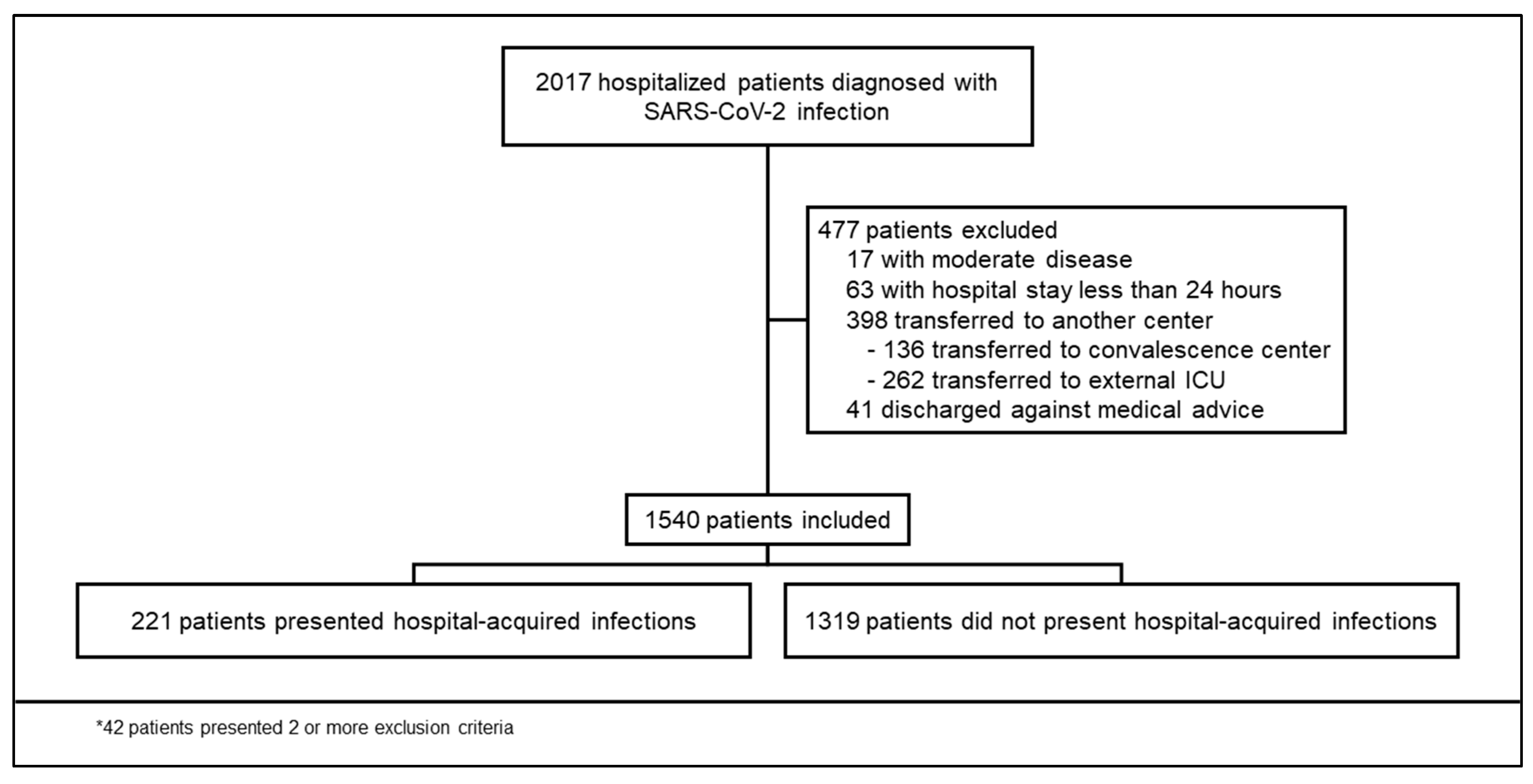
2. Results
3. Discussion
4. Materials and Methods
5. Conclusions
Supplementary Materials
Author Contributions
Funding
Institutional Review Board Statement
Informed Consent Statement
Data Availability Statement
Acknowledgments
Conflicts of Interest
References
- Behal, M.; Barlow, B.; Mefford, B.; Thompson Bastin, M.L.; Donaldson, J.C.; Laine, M.; Bissell, B.D. Pharmacotherapy in Coronavirus Disease 2019 and Risk of Secondary Infections: A Single-Center Case Series and Narrative Review. Crit. Care Explor. 2021, 3, e0492. [Google Scholar] [CrossRef] [PubMed]
- Osuchowski, M.F.; Winkler, M.S.; Skirecki, T.; Cajander, S.; Shankar-Hari, M.; Lachmann, G.; Monneret, G.; Venet, F.; Bauer, M.; Brunkhorst, F.M.; et al. The COVID-19 Puzzle: Deciphering Pathophysiology and Phenotypes of a New Disease Entity. Lancet Respir. Med. 2021, 9, 622–642. [Google Scholar] [CrossRef] [PubMed]
- Garcia-Vidal, C.; Sanjuan, G.; Moreno-García, E.; Puerta-Alcalde, P.; Garcia-Pouton, N.; Chumbita, M.; Fernandez-Pittol, M.; Pitart, C.; Inciarte, A.; Bodro, M.; et al. Incidence of Co-Infections and Superinfections in Hospitalized Patients with COVID-19: A Retrospective Cohort Study. Clin. Microbiol. Infect. 2021, 27, 83–88. [Google Scholar] [CrossRef] [PubMed]
- Giacobbe, D.R.; Battaglini, D.; Ball, L.; Brunetti, I.; Bruzzone, B.; Codda, G.; Crea, F.; de Maria, A.; Dentone, C.; di Biagio, A.; et al. Bloodstream Infections in Critically Ill Patients with COVID-19. Eur. J. Clin. Investig. 2020, 50, e13319. [Google Scholar] [CrossRef]
- Grasselli, G.; Scaravilli, V.; Mangioni, D.; Scudeller, L.; Alagna, L.; Bartoletti, M.; Bellani, G.; Biagioni, E.; Bonfanti, P.; Bottino, N.; et al. Hospital-Acquired Infections in Critically Ill Patients With COVID-19. Chest 2021, 160, 454–465. [Google Scholar] [CrossRef]
- Ramos, R.; de la Villa, S.; García-Ramos, S.; Padilla, B.; García-Olivares, P.; Piñero, P.; Garrido, A.; Hortal, J.; Muñoz, P.; Caamaño, E.; et al. COVID-19 Associated Infections in the ICU Setting: A Retrospective Analysis in a Tertiary-Care Hospital. Enferm. Infecc. Microbiol. Clin. 2023, 41, 278–283. [Google Scholar] [CrossRef]
- Rouzé, A.; Nseir, S. Hospital-Acquired Pneumonia/Ventilator-Associated Pneumonia and Ventilator-Associated Tracheobronchitis in COVID-19. Semin. Respir. Crit. Care Med. 2022, 43, 243–247. [Google Scholar] [CrossRef]
- Maslove, D.M.; Sibley, S.; Boyd, J.G.; Goligher, E.C.; Munshi, L.; Bogoch, I.I.; Rochwerg, B. Complications of Critical COVID-19. Chest 2022, 161, 989–998. [Google Scholar] [CrossRef]
- Zuglian, G.; Ripamonti, D.; Tebaldi, A.; Cuntrò, M.; Riva, I.; Farina, C.; Rizzi, M. The Changing Pattern of Bacterial and Fungal Respiratory Isolates in Patients with and without COVID-19 Admitted to Intensive Care Unit. BMC Infect. Dis. 2022, 22, 185. [Google Scholar] [CrossRef]
- Chan, X.H.S.; O’Connor, C.J.; Martyn, E.; Clegg, A.J.; Choy, B.J.K.; Soares, A.L.; Shulman, R.; Stone, N.R.H.; De, S.; Bitmead, J.; et al. Reducing Broad-Spectrum Antibiotic Use in Intensive Care Unit between First and Second Waves of COVID-19 Did Not Adversely Affect Mortality. J. Hosp. Infect. 2022, 124, 37–46. [Google Scholar] [CrossRef]
- RECOVERY Collaborative Group. Dexamethasone in Hospitalized Patients with COVID-19. N. Engl. J. Med. 2021, 384, 693–704. [CrossRef] [PubMed]
- Salama, C.; Han, J.; Yau, L.; Reiss, W.G.; Kramer, B.; Neidhart, J.D.; Criner, G.J.; Kaplan-Lewis, E.; Baden, R.; Pandit, L.; et al. Tocilizumab in Patients Hospitalized with COVID-19 Pneumonia. N. Engl. J. Med. 2021, 384, 20–30. [Google Scholar] [CrossRef]
- Shafran, N.; Shafran, I.; Ben-Zvi, H.; Sofer, S.; Sheena, L.; Krause, I.; Shlomai, A.; Goldberg, E.; Sklan, E.H. Secondary Bacterial Infection in COVID-19 Patients Is a Stronger Predictor for Death Compared to Influenza Patients. Sci. Rep. 2021, 11, 12703. [Google Scholar] [CrossRef] [PubMed]
- Vijay, S.; Bansal, N.; Rao, B.K.; Veeraraghavan, B.; Rodrigues, C.; Wattal, C.; Goyal, J.P.; Tadepalli, K.; Mathur, P.; Venkateswaran, R.; et al. Secondary Infections in Hospitalized COVID-19 Patients: Indian Experience. Infect. Drug. Resist. 2021, 14, 1893–1903. [Google Scholar] [CrossRef]
- Feng, Y.; Ling, Y.; Bai, T.; Xie, Y.; Huang, J.; Li, J.; Xiong, W.; Yang, D.; Chen, R.; Lu, F.; et al. COVID-19 with Different Severities: A Multicenter Study of Clinical Features. Am. J. Respir. Crit. Care Med. 2020, 201, 1380–1388. [Google Scholar] [CrossRef] [PubMed] [Green Version]
- Klein, S.L.; Flanagan, K.L. Sex Differences in Immune Responses. Nat. Rev. Immunol. 2016, 16, 626–638. [Google Scholar] [CrossRef]
- Dessie, Z.G.; Zewotir, T. Mortality-Related Risk Factors of COVID-19: A Systematic Review and Meta-Analysis of 42 Studies and 423,117 Patients. BMC Infect. Dis. 2021, 21, 855. [Google Scholar] [CrossRef]
- Cairns, S.; Reilly, J.; Stewart, S.; Tolson, D.; Godwin, J.; Knight, P. The Prevalence of Health Care–Associated Infection in Older People in Acute Care Hospitals. Infect. Control. Hosp. Epidemiol. 2011, 32, 763–767. [Google Scholar] [CrossRef]
- de Bruyn, A.; Verellen, S.; Bruckers, L.; Geebelen, L.; Callebaut, I.; de Pauw, I.; Stessel, B.; Dubois, J. Secondary Infection in COVID-19 Critically Ill Patients: A Retrospective Single-Center Evaluation. BMC Infect. Dis. 2022, 22, 207. [Google Scholar] [CrossRef]
- Mihai, S.; Codrici, E.; Popescu, I.D.; Enciu, A.-M.; Albulescu, L.; Necula, L.G.; Mambet, C.; Anton, G.; Tanase, C. Inflammation-Related Mechanisms in Chronic Kidney Disease Prediction, Progression, and Outcome. J. Immunol. Res. 2018, 2018, 2180373. [Google Scholar] [CrossRef] [Green Version]
- Arredondo, A.; Orozco, E.; Alcalde-Rabanal, J.; Navarro, J.; Azar, A. Retos Sobre La Carga Epidemiológica y Económica Para Diabetes e Hipertensión En México. Rev. Saude Publica 2018, 52, 23. [Google Scholar] [CrossRef] [PubMed] [Green Version]
- Curley, G.F.; Laffey, J.G.; Zhang, H.; Slutsky, A.S. Biotrauma and Ventilator-Induced Lung Injury: Clinical Implications. Chest 2016, 150, 1109–1117. [Google Scholar] [CrossRef] [PubMed]
- Moser, D.; Feuerecker, M.; Biere, K.; Han, B.; Hoerl, M.; Schelling, G.; Kaufmann, I.; Choukér, A.; Woehrle, T. SARS-CoV-2 Pneumonia and Bacterial Pneumonia Patients Differ in a Second Hit Immune Response Model. Sci. Rep. 2022, 12, 15485. [Google Scholar] [CrossRef]
- Martinez-Guerra, B.A.; Gonzalez-Lara, M.F.; De-Leon-Cividanes, N.A.; Tamez-Torres, K.M.; Roman-Montes, C.M.; Rajme-Lopez, S.; Villalobos-Zapata, G.I.; Lopez-Garcia, N.I.; Martínez-Gamboa, A.; Sifuentes-Osornio, J.; et al. Antimicrobial Resistance Patterns and Antibiotic Use during Hospital Conversion in the COVID-19 Pandemic. Antibiotics 2021, 10, 182. [Google Scholar] [CrossRef] [PubMed]
- Ayoub Moubareck, C.; Hammoudi Halat, D. The Collateral Effects of COVID-19 Pandemic on the Status of Carbapenemase-Producing Pathogens. Front. Cell. Infect. Microbiol. 2022, 12, 823626. [Google Scholar] [CrossRef] [PubMed]
- Habibzadeh, A.; Lankarani, K.B.; Farjam, M.; Akbari, M.; Kashani, S.M.A.; Karimimoghadam, Z.; Wang, K.; Imanieh, M.H.; Tabrizi, R.; Ahmadizar, F. Prevalence of Fungal Drug Resistance in COVID-19 Infection: A Global Meta-Analysis. Curr. Fungal Infect. Rep. 2022, 16, 154–164. [Google Scholar] [CrossRef]
- Peghin, M.; Vena, A.; Graziano, E.; Giacobbe, D.R.; Tascini, C.; Bassetti, M. Improving Management and Antimicrobial Stewardship for Bacterial and Fungal Infections in Hospitalized Patients with COVID-19. Ther. Adv. Infect. Dis. 2022, 9, 20499361221095732. [Google Scholar] [CrossRef]
- Brandi, N.; Ciccarese, F.; Rimondi, M.R.; Balacchi, C.; Modolon, C.; Sportoletti, C.; Renzulli, M.; Coppola, F.; Golfieri, R. An Imaging Overview of COVID-19 ARDS in ICU Patients and Its Complications: A Pictorial Review. Diagnostics 2022, 12, 846. [Google Scholar] [CrossRef]
- Corman, V.M.; Landt, O.; Kaiser, M.; Molenkamp, R.; Meijer, A.; Chu, D.K.; Bleicker, T.; Brünink, S.; Schneider, J.; Schmidt, M.L.; et al. Detection of 2019 Novel Coronavirus (2019-NCoV) by Real-Time RT-PCR. Eurosurveillance 2020, 25, 2000045. [Google Scholar] [CrossRef] [Green Version]
- Wu, Z.; McGoogan, J.M.; Wang, D.; Hu, B.; Hu, C.; Zhu, F.; Liu, X.; Zhang, J.; Wang, B.; Xiang, H.; et al. Characteristics of and Important Lessons from the Coronavirus Disease 2019 (COVID-19) Outbreak in China: Summary of a Report of 72314 Cases from the Chinese Center for Disease Control and Prevention. JAMA J. Am. Med. Assoc. 2020, 323, 1239–1242. [Google Scholar] [CrossRef]
- Koehler, P.; Bassetti, M.; Chakrabarti, A.; Chen, S.C.A.; Colombo, A.L.; Hoenigl, M.; Klimko, N.; Lass-Flörl, C.; Oladele, R.O.; Vinh, D.C.; et al. Defining and Managing COVID-19-Associated Pulmonary Aspergillosis: The 2020 ECMM/ISHAM Consensus Criteria for Research and Clinical Guidance. Lancet Infect. Dis. 2021, 21, e149–e162. [Google Scholar] [CrossRef]
- Mermel, L.A.; Allon, M.; Bouza, E.; Craven, D.E.; Flynn, P.; O’Grady, N.P.; Raad, I.I.; Rijnders, B.J.A.; Sherertz, R.J.; Warren, D.K. Clinical Practice Guidelines for the Diagnosis and Management of Intravascular Catheter-Related Infection: 2009 Update by the Infectious Diseases Society of America. Clin. Infect. Dis. 2009, 49, 1–45. [Google Scholar] [CrossRef] [PubMed]
- Pappas, P.G.; Kauffman, C.A.; Andes, D.R.; Clancy, C.J.; Marr, K.A.; Ostrosky-Zeichner, L.; Reboli, A.C.; Schuster, M.G.; Vazquez, J.A.; Walsh, T.J.; et al. Clinical Practice Guideline for the Management of Candidiasis: 2016 Update by the Infectious Diseases Society of America. Clin. Infect. Dis. 2016, 62, e1–e50. [Google Scholar] [CrossRef] [PubMed] [Green Version]
- Torres, A.; Niederman, M.S.; Chastre, J.; Ewig, S.; Fernandez-Vandellos, P.; Hanberger, H.; Kollef, M.; Li Bassi, G.; Luna, C.M.; Martin-Loeches, I.; et al. International ERS/ESICM/ESCMID/ALAT Guidelines for the Management of Hospital-Acquired Pneumonia and Ventilator-Associated Pneumonia. Eur. Respir. J. 2017, 50, 1700582. [Google Scholar] [CrossRef] [Green Version]
- Riley, R.D.; Ensor, J.; Snell, K.I.E.; Harrell, F.E.; Martin, G.P.; Reitsma, J.B.; Moons, K.G.M.; Collins, G.; van Smeden, M. Calculating the Sample Size Required for Developing a Clinical Prediction Model. BMJ 2020, 368, m441. [Google Scholar] [CrossRef] [PubMed] [Green Version]


| All Patients n = 1540 (100%) | Developed HAI n = 221 (14.35%) | Did Not Develop HAI n = 1319 (85.65%) | p | |
|---|---|---|---|---|
| Male sex, n (%) | 941 (61.1) | 159 (72.0) | 782 (59.3) | <0.001 |
| Age, years—median (IQR) | 55 (45–65) | 56 (46–65) | 54 (45–66) | 0.633 |
| Obesity—n (%) n = 1537 | 681 (44.3) | 120 (54.3) n = 221 | 561 (42.3) n = 1316 | 0.001 |
| Type 2 diabetes mellitus—n (%) n = 1539 | 440 (28.6) | 68 (30.8) n = 221 | 372 (28.2) n = 1318 | 0.438 |
| Hypertension—n (%) n = 1359 | 528 (34.3) | 69 (31.2) n = 221 | 459 (34.8) n = 1318 | 0.296 |
| Chronic obstructive pulmonary disease—n (%) n = 1539 | 22(1.4) | 1 (0.5) n = 221 | 21 (1.6) n = 1318 | 0.351 |
| Immunosuppression—n (%) n = 1538 | 87 (5.7) | 13 (5.9) n = 220 | 74 (5.6) n = 1318 | 0.861 |
| Cardiovascular disease—n (%) n = 1538 | 86 (5.6) | 14 (6.4) n = 220 | 72 (5.5) n = 1318 | 0.590 |
| Chronic kidney disease—n (%) n = 1539 | 51 (3.3) | 8 (3.6) n = 221 | 43 (3.3) n = 1318 | 0.838 |
| Liver cirrhosis—n (%) n = 1536 | 11 (0.7) | 1 (0.5) n = 221 | 10 (0.8) n = 1315 | 1.000 |
| Charlson comorbidity index >2—n (%) | 482 (31.3) | 70 (31.7) | 412 (31.2) | 0.897 |
| Time from symptom onset to hospital admission, days—median (IQR) | 7 (5–10) | 7 (5–9) | 7 (5–10) | 0.974 |
| Oxygen saturation <90%, n (%) n = 1510 | 1371 (90.8) | 206 (97.2) n = 212 | 1165 (89.8) n = 1298 | <0.001 |
| Lymphocyte count <800 cells/uL%—n (%) | 855 (55.9) | 142 (64.8) n = 219 | 713 (54.4) n = 1311 | 0.004 |
| C-reactive protein >10 mg/dL—n (%) n = 1498 | 1030 (68.8) | 187 (86.6), n = 216 | 843 (65.8) n = 1282 | <0.001 |
| Ferritin >500 ng/mL—n (%) n = 1487 | 828 (55.7) | 151 (72.3) n = 209 | 677 (53.0) n = 1278 | <0.001 |
| Lactate dehydrogenase ≥246 U/L—n (%) n = 1482 | 1285 (84.9) | 198 (93.0) n = 213 | 1060 (83.5) n = 1269 | <0.001 |
| D-dimer >500 ng/mL—n (%) n = 1500 | 555 (37.0) | 99 (46.1) n = 215 | 456 (35.5) n = 1285 | 0.003 |
| Multilobe involvement in CT—n (%) n = 1538 | 1530 (99.5) | 221 (100) n = 221 | 1309 (99.4) n = 1317 | 0.288 |
| Use of invasive mechanical ventilation in the first 24 h—n (%) | 279 (68.1) | 143 (64.7) | 136 (10.3) | <0.001 |
| Empiric antibiotic therapy—n (%) | 914 (59.4) | 136 (61.5) | 778 (59.0) | 0.474 |
| Treatment with steroids—n (%) | 688 (44.7) | 139 (62.9) | 549 (41.6) | <0.001 |
| Treatment with tocilizumab—n (%) | 97 (6.3) | 21 (9.5) | 76 (5.8) | 0.034 |
| Enrollment in a COVID-19 clinical trial—n (%) | 320 (20.8) | 26 (11.8) | 294 (22.3) | <0.001 |
| aOR (CI 95%) p * | |
|---|---|
| Use of IMV in the first 24 h after admission | 18.78 (12.56–28.07) p < 0.0001 |
| Chronic kidney disease | 3.41 (1.40–8.27) p = 0.007 |
| Corticosteroid treatment | 2.95 (1.92–4.53) p < 0.0001 |
| Tocilizumab treatment | 2.69 (1.38–5.22) p = 0.004 |
| Age > 60 years | 1.91 (1.27–2.88) p = 0.002 |
| Male sex | 1.52 (1.03–2.24) p = 0.031 |
| Obesity | 1.49 (1.03–2.15) p = 0.031 |
| Immunosuppression | 1.59 (0.75–3.35) p = 0.222 |
| Cardiovascular disease | 1.12 (0.52–2.39) p = 0.762 |
| Diabetes mellitus | 1.03 (0.68–1.54) p = 0.882 |
| Empirical antibiotic therapy | 1.00 (0.64–1.55) p = 0.993 |
| Enrollment in a clinical trial | 0.98 (0.59–1.64) p = 0.958 |
| Oxygen saturation <90% | 0.95 (0.33–2.69) p = 0.924 |
| Hypertension | 0.69 (0.46–1.05) p = 0.091 |
| Chronic obstructive pulmonary disease | 0.12 (0.01–1.05) p = 0.056 |
| * 1429 observations, PseudoR2 = 0.2937. | |
Disclaimer/Publisher’s Note: The statements, opinions and data contained in all publications are solely those of the individual author(s) and contributor(s) and not of MDPI and/or the editor(s). MDPI and/or the editor(s) disclaim responsibility for any injury to people or property resulting from any ideas, methods, instructions or products referred to in the content. |
© 2023 by the authors. Licensee MDPI, Basel, Switzerland. This article is an open access article distributed under the terms and conditions of the Creative Commons Attribution (CC BY) license (https://creativecommons.org/licenses/by/4.0/).
Share and Cite
Solís-Huerta, F.; Martinez-Guerra, B.A.; Roman-Montes, C.M.; Tamez-Torres, K.M.; Rajme-Lopez, S.; Ortíz-Conchi, N.; López-García, N.I.; Villalobos-Zapata, G.Y.; Rangel-Cordero, A.; Santiago-Cruz, J.; et al. Risk Factors Associated with the Development of Hospital-Acquired Infections in Hospitalized Patients with Severe COVID-19. Antibiotics 2023, 12, 1108. https://doi.org/10.3390/antibiotics12071108
Solís-Huerta F, Martinez-Guerra BA, Roman-Montes CM, Tamez-Torres KM, Rajme-Lopez S, Ortíz-Conchi N, López-García NI, Villalobos-Zapata GY, Rangel-Cordero A, Santiago-Cruz J, et al. Risk Factors Associated with the Development of Hospital-Acquired Infections in Hospitalized Patients with Severe COVID-19. Antibiotics. 2023; 12(7):1108. https://doi.org/10.3390/antibiotics12071108
Chicago/Turabian StyleSolís-Huerta, Fernando, Bernardo Alfonso Martinez-Guerra, Carla Marina Roman-Montes, Karla Maria Tamez-Torres, Sandra Rajme-Lopez, Narciso Ortíz-Conchi, Norma Irene López-García, Guadalupe Yvonne Villalobos-Zapata, Andrea Rangel-Cordero, Janet Santiago-Cruz, and et al. 2023. "Risk Factors Associated with the Development of Hospital-Acquired Infections in Hospitalized Patients with Severe COVID-19" Antibiotics 12, no. 7: 1108. https://doi.org/10.3390/antibiotics12071108