Article

Evidence of Antibiotic Resistance and Virulence Factors in Environmental Isolates of Vibrio Species

by
Rajkishor Pandey
1,2,*,
Simran Sharma
3 and
Kislay Kumar Sinha
1,*
1
Department of Biotechnology, National Institute of Pharmaceutical Education and Research, Hajipur 844102, Bihar, India
2
School of Medicine, University of Missouri, Columbia, MO 65211, USA
3
Department of Basic and Applied Sciences, National Institute of Food Technology Entrepreneurship & Management (NIFTEM), Kundli, Sonipat 131028, Haryana, India
*
Authors to whom correspondence should be addressed.
Antibiotics 2023, 12(6), 1062; https://doi.org/10.3390/antibiotics12061062
Submission received: 1 May 2023 / Revised: 3 June 2023 / Accepted: 15 June 2023 / Published: 16 June 2023

Abstract

:
The outbreak of waterborne diseases such as cholera and non-cholera (vibriosis) is continuously increasing in the environment due to fecal and sewage discharge in water sources. Cholera and vibriosis are caused by different species of Vibrio genus which are responsible for acute diarrheal disease and soft tissue damage. Although incidences of cholera and vibriosis have been reported from the Vaishali district of Bihar, India, clinical or environmental strains have not been characterized in this region. Out of fifty environmental water samples, twelve different biochemical test results confirmed the presence of twenty Vibrio isolates. The isolates were found to belong to five different Vibrio species, namely V. proteolyticus, V. campbellii, V. nereis, V. cincinnatiensis, and V. harveyi. From the identified isolates, 65% and 45% isolates were found to be resistant to ampicillin and cephalexin, respectively. Additionally, two isolates were found to be resistant against six and four separately selected antibiotics. Furthermore, virulent hlyA and ompW genes were detected by PCR in two different isolates. Additionally, phage induction was also noticed in two different isolates which carry lysogenic phage in their genome. Overall, the results reported the identification of five different Vibrio species in environmental water samples. The isolates showed multiple antibacterial resistance, phage induction, and virulence gene profile in their genome.

1. Introduction

In India, many people have limited access to safe drinking water, especially in rural areas where the water sources are often untreated and contaminated with sewage/feces. Contaminated water and food are major risk factors for the spread of waterborne infectious diseases such as cholera and vibriosis. Cholera is a severe acute watery diarrheal disease caused by the O1 and O139 serogroup of Vibrio cholerae [1]. Apart from V. cholerae, many non-cholera pathogenic species of the genus Vibrio which cause vibriosis have been discovered in America, Europe, Asia, and other low-income countries [2,3,4]. Vibrio species are naturally found in freshwater, brackish water, and marine water. Generally, vibriosis infection is acquired by the ingestion of contaminated water or seafood [5]. It has been observed that environmental factors affect both eukaryotic and prokaryotic life through direct or indirect interaction. Jamie et al. mentioned in their study that warm sea surface influences the growth of pathogenic Vibrio species which increases the occurrence of infections in humans as well as marine animals [6]. Tropical countries such as India and other Caribbean countries are the most favorable place for the growth of toxigenic Vibrio species [7].
The pathogenicity of Vibrio species is determined by the virulent factors encoded by virulent genes. Virulence factors influence the severity of infection and drug resistance [8,9]. It has been reported that Vibrio species acquire external genetic material from environmental sources or other bacteria by horizontal gene transfer (HGT) [10]. The shared genetic material might be encoding virulence factors that help to enhance Vibrio species’ adaptability in diverse environments [11]. Virulence-associated elements such as toxin production, quorum sensing (cell to cell communication), presence of lysogenic phage, hemolysin, proteases, etc., are major pathogenic factors involved in the pathogenicity of Vibrio species [12,13]. Therefore, assessment of virulence factors in environmental Vibrio isolates provides the role of pathogenesis and helps to begin a path for treatment.
It has been reported that resistance developed by Vibrio species against antibiotics is associated with virulent factors [8,9]. Over time, due to the immoderate use of antibiotics in human disease, agriculture, and aquaculture, the Vibrio species developed antimicrobial resistance [14]. In another study, it has been characterized that the unique genetic makeup and competency of Vibrio species help them in adapting to adverse environmental conditions and resisting the antibacterial agent [15]. Due to the resistance developed by Vibrio species, they adversely affect marine, terrestrial animals, as well as human life. As reported by the Centers for Disease Control and Prevention (CDC), in mild infection of Vibrio species, treatment is not compulsory, but a sufficient amount of liquid should be consumed by the patient to substitute the fluid that was lost in diarrhea. In the case of mild to moderate infection of V. cholerae, it can be reversed by the administration of an oral rehydration solution (ORS) that helps in rehydration. For severe cholera conditions, antibacterial agents such as tetracycline, fluoroquinolones, ceftriaxone, cefotaxime, and macrolides are most effective and are used for the treatment of the disease. However, in severe vibriosis conditions, no such evidence was found that antibiotics reduce the severity of illness [16,17,18].
In earlier studies on the hotspot of cholera, most of the Vibrio species have been characterized in marine or brackish water. In the present study, environmental water samples in the Vaishali district of Bihar, India, have been investigated for Vibrio strains. Therefore, taking this into consideration, this study was performed with the objective of evaluating the presence of virulence-associated factors and antibiotic resistance in the environmental isolates of different Vibrio species. Additionally, it involved the characterization of phage induction in the identified isolates with the lysogenic phage-inducing agent mitomycin C (MMC).

2. Results

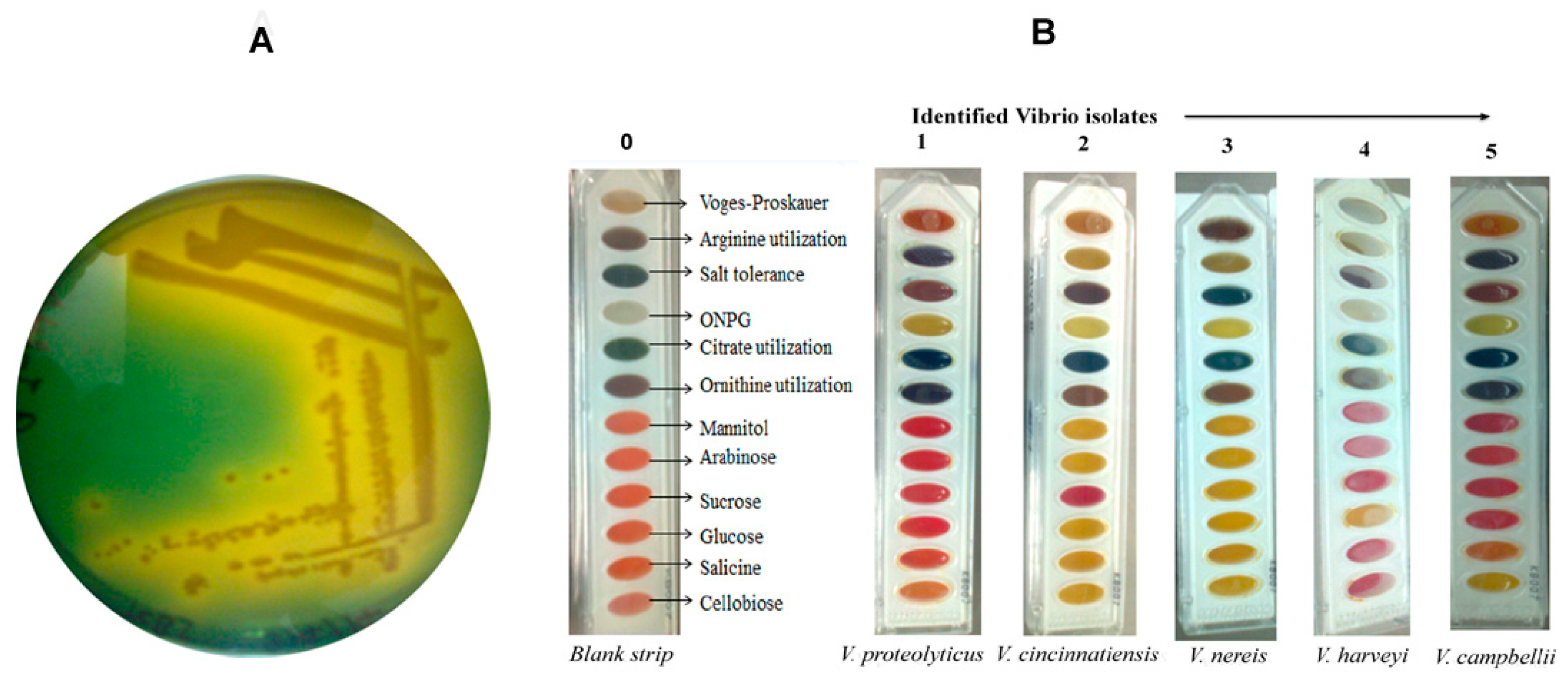
2.1. Isolation and Biochemical Identification of Vibrio Isolates

Out of 50 environmental water samples, 20 bacterial isolates colony (River-1, Pond-6, Stagnant water-5, Sevage-8) were picked from the TCBS plate (Figure 1A) and confirmed to be Vibrio positive by the 12 biochemical tests (Figure 1 and Supplementary Table S1). The biochemical test strip changed color after the addition of Vibrio culture, and as per the guidelines of manufacturer kit instruction (HiMedia, Mumbai, India), five different Vibrio species (V. proteolyticus, V. campbellii, V. harveyi, V. cincinnatiensis, V. nereis) were identified (Table 1).

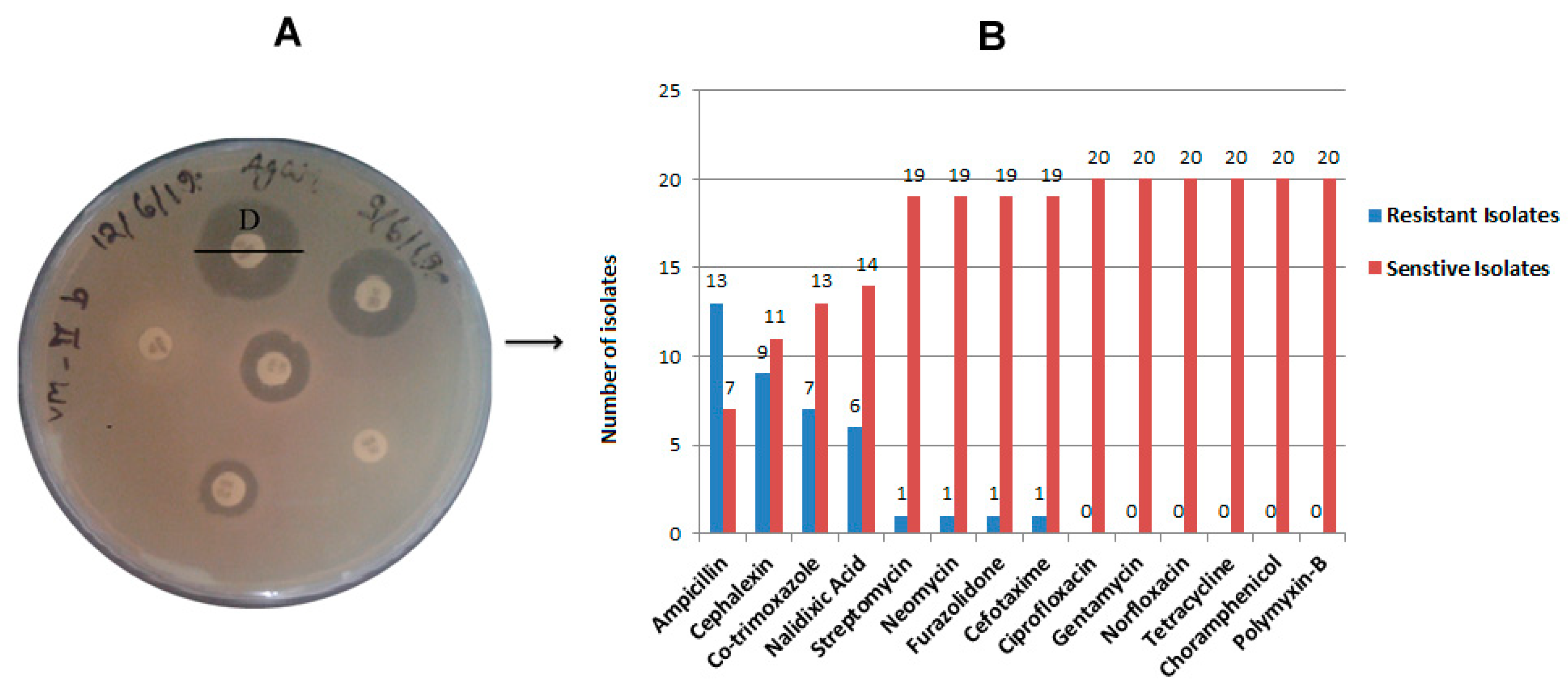
2.2. Antibiotic Resistance and Susceptibility Assay

The environmental Vibrio isolates were checked for resistance against 14 commonly used antibiotics by disk diffusion methods (Figure 2A). Out of twenty isolates, thirteen isolates (65%) were found ampicillin resistant, and nine isolates (45%) were cephalexin resistant (Figure 2B and Supplementary Table S2). Co-trimoxazole resistance was observed in seven isolates (35%) followed by nalidixic acid in six resistant isolates (30%), as depicted in Figure 2B. Resistance against streptomycin, neomycin, cefotaxime, and furazolidone was found in one isolate each (Figure 2 and Supplementary Table S2). Antibiotics such as ciprofloxacin, gentamycin, norfloxacin, tetracycline, chloramphenicol, and polymyxin-B showed sensitivity to all isolates. Isolate VHMC-D was found to be resistant against six antibiotics namely ampicillin, cephalexin, nalidixic acid, cefotaxime, co-trimoxazole, and furazolidone. Another isolate, VHMC-B was resistant to ampicillin, cephalexin, nalidixic acid, and co-trimoxazole (Supplementary Table S2). Another seven isolates were found to be resistant to more than one antibacterial agent. Two isolates were found to be sensitive to all the antibiotics checked in the study (Supplementary Table S2).

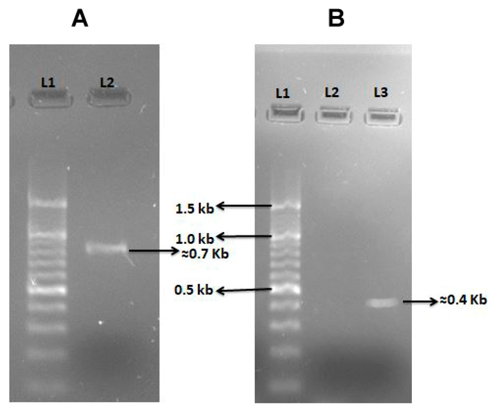
2.3. Virulence Profile of Vibrio Isolates

Out of twenty Vibrio isolates, six isolates were chosen for the amplification of Vibrio genes primers as mentioned in Table 2. All the mentioned genes primers were tested for amplification with the six selected isolates. The virulence gene primer hlyA El Tor was found to be amplified in isolate VH-I4 (V. proteolyticus) at 56 °C (Figure 3A), which represents hemolysin, which is an extracellular pore-forming hemolytic toxin [19]. Another ompW gene primer amplified in VHMC-A (V. campbellii) isolate at 56 °C (Figure 3B). ompW is a major outer membrane protein in Vibrio and is involved in salt tolerance as well as in the transferring of hydrophobic small molecules [20]. However, other selected Vibrio gene primers were not amplified in the selected isolates.

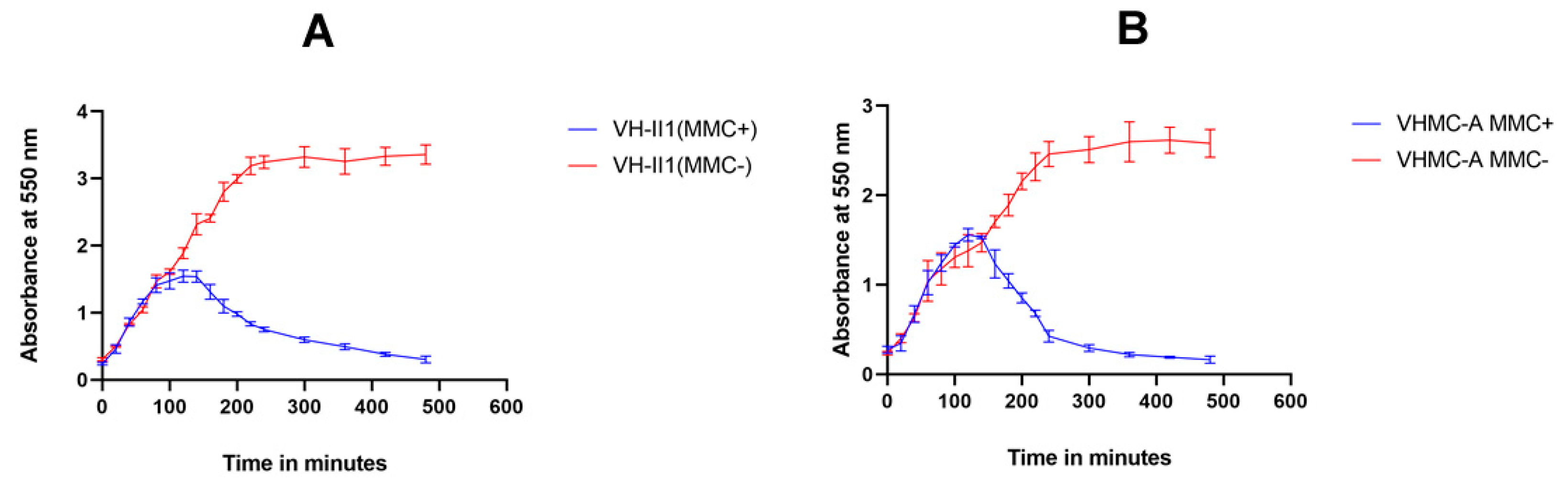
2.4. Phage Induction Assessment by Mitomycin C

In the present study, it was found that after induction most of the isolates (both mitomycin C treated as well as untreated control culture) grew exponentially at comparable rates. Approximately, 2.5 h after the addition of mitomycin C (MMC+), the OD550 value of the induced culture of Vibrio isolate VH-II1 (V. campbellii) drastically decreased from 1.45 to 0.3 (Figure 4A). Similarly, 1.5 h after the addition of mitomycin C, the OD550 value of the induced culture of Vibrio isolate VHMC-A (V. campbellii) dramatically decreased from 2.05 to 0.1 (Figure 4B), whereas the control sample without mitomycin C (MMC−) continued to grow in both of the isolates (Figure 4A,B). In both isolates, typical fibers such as particles were observed in the induced culture. The significant reduction in OD at 550 nm indicated that both isolates carried lysogenic phages in their genome.

3. Discussion

Organic waste such as foods, sewage, fertilizers, and animal and human feces contaminate water sources and play an important role in providing the most favorable place for the growth of microbes including Vibrio species [27]. Further climatic changes and the enormous use of antimicrobial agents potentiate prokaryotic population and their survival in harsh environmental conditions [28].
In the present study, five different Vibrio species have been isolated from fresh environmental water resources. The results indicated that out of twenty isolates, eight isolates (V-Gan, VR, VM-IIA, VP-IA, VP-IB, VH-I4, VH-II4, and VHVB-I) (40%) had characteristics of V. proteolyticus; four isolates (VHMC-A, VM-IB, VH-I3, and VH-II1) (20%) had characteristics of V. campbellii; three isolates (VHMC-C, VHVB-II, and VH-II2) (15%) had characteristics of V. cincinnatiensis; and another three isolates (VHMC-B, VHMC-D, and VM-IA) (15%) showed V. nereis characteristics. There was one isolate, VH-II3,identified as Vibrio harveyi. A fairly mixed population of different species of Vibrio was observed in this region (as shown in Figure 1, Table 1 and Supplementary Table S1). In the previous study, these Vibrio species have been characterized as potential pathogens for humans as well as aquatic animals [1,2,5]. In an in vitro study, it was found that V. proteolyticus produced virulent factors cytotoxic to eukaryotic cell lines such as macrophages and HeLa cells [29]. In the year of 2017, Paek, Jayoung et al. isolated V. cincinnatiensis from the clinical specimen sample of a patient having symptoms of Vibrio-associated disease, such as watery diarrhea and soft tissue injury [30]. Another Vibrio species mentioned in the Harveyi Clade was considered to be the most severely pathogenic Vibrio cluster for aquatic animals, capable of generating more than 50 different Vibrio diseases [31].
Globally, antimicrobial resistance is a big challenge for the health authority. Microbes develop anti-microbial resistant genes in their genome for their protection [32]. Antimicrobial susceptibility assay of Vibrio isolates revealed that out of twenty isolates, thirteen (65%) and nine (45%) isolates were found to be ampicillin-resistant and cephalexin-resistant, respectively (Figure 2B and Supplementary Table S2). Those thirteen isolates were from V. proteolyticus [6], V. campbellii [2], V. cincinnatiensis [3], and V. nereis [2]. The nine isolates were from V. proteolyticus [5], V. campbellii [2], V. cincinnatiensis [1], and V. nereis [1]. Co-trimoxazole resistance was observed in seven isolates (35%) from all the isolated Vibrio species except V. harveyi, followed by nalidixic acid for which six isolates (30%) of V. proteolyticus and V. nereis were found to be resistant. Resistance against streptomycin, neomycin, cefotaxime, and furazolidone was found in one isolate each (Supplementary Table S2). The identified Vibrio isolates exhibited a high incidence of resistance against selected antibacterial agents that are commonly used [33]. Additionally, two isolates, VHMC-D and VHMC-B, of V. nereis showed resistance against six (ampicillin, cephalexin, nalidixic acid, cefotaxime, co-trimoxazole, and furazolidone) and four (ampicillin, cephalexin, nalidixic acid, and co-trimoxazole) selected antibiotics, respectively. Another seven isolates from four different Vibrio species except V. harveyi were found to be moderate to highly resistant to more than one selected antibacterial agent (Figure 2 and Supplementary Table S2). It was noted that out of twenty isolates, sixteen isolates (80%) showed resistance against beta-lactam and its derivatives. In the previous study, it was also indicated that Vibrio isolates had resistance against beta-lactam antibiotics and their derivatives derived from China, Italy, and the U.S. [34,35,36]. However, it was observed that two isolates, VHMC-A (V. proteolyticus) and VH-II3 (V. harveyi), were sensitive to all the selected antibiotics used in this study. Further, it was found that all the isolates were sensitive to ciprofloxacin, gentamycin, norfloxacin, tetracycline, chloramphenicol, and polymyxin-B. This finding of antibiotic resistance of the Vibrio isolates indicates the emergence of multidrug resistance that could be a health threat.
In general, microbes show virulence by the secretion of virulent factors which are primarily infectious to host cells. It has been observed that virulent factors of Vibrio species are responsible for the pathogenesis and cause diarrhea, gastroenteritis, cholera, tissue injury, and blood infections [37]. In the present study, 10 different virulent genes primers were selected for the amplification of genes in Vibrio isolates (Table 2). As shown in Figure 3A, the isolate (VHI4) of V. proteolyticus showed amplification of hlyA gene (hemolysin) which indicates the presence of the pore-forming toxin in the genome of the isolate. Hemolysin is a well-known pore-forming hemolytic bacterial toxin, encoded by the hlyA gene. This toxin is present in the genome of various pathogenic Vibrio species. Hemolysin is a member of the class leucocidin superfamily, it alters the host cell membrane permeability and leads to cell death by the activation of the inflammasome pathway [38]. Further, another virulent gene primer, ompW was found to be amplified with the genomic DNA of VHMV-A isolate (V. campbellii) (Figure 3B). The bacterial outer membrane protein is a major bacterial protein which is expressed on the surface of the bacterial cell membrane. It participates in the regulation of salt stress, substance transport, and osmoregulation in many Vibrio species [39]. OmpW expression enhances the growth of V. cholerae in high saline water by carnitine channel [40]. In a previous study, an increase in the OmpW expression in V. alginolyticus was observed in the presence of high NaCl concentration [41].
In previous studies, horizontal transfer of virulent factors has been observed in Vibrio through phages and other mobile genetic elements [42,43,44]. In another study, it has been mentioned that virulent genes and anti-microbial resistant genes encoded by prophage-like elements get exchanged between pathogenic and nonpathogenic Vibrio species [45]. In our study, interestingly, two isolates (VHMC-A and VH-II1, both were V. campbellii) showed a drastic decrease in the OD550 after 1.5 and 2.5 h of the addition of mitomycin C (MMC+), respectively, which was a typical indication of the presence of lysogenic virulent phage (Figure 4A,B). Additionally, characteristic fibers such as particles were seen in mitomycin C induced cultures (MMC+). However, without mitomycin C added, bacterial culture (MMC−) was grown constantly, and, at one point, bacterial stationary phase was established. These observations strongly suggest that both these isolates, VHMC-A and VH-II1, carry lysogenic phages in their genome. Supernatants from the induced cultures were spotted on all the isolates in order to find a suitable host that can support the entry and growth of these putative phages, but no clearing was seen which indicated that these isolates could not be infected with these phages.

4. Materials and Methods

4.1. Sample Collection and Isolation of Bacterial Colonies

A total of 50 water samples were collected in sterile 500 mL bottles from various sources such as rivers, ponds, sewage, and stagnant rainwater from different locations in the Vishal district of Bihar, India (duration: October to June). After sample collection, 50 mL of water sample was filtered through a 0.22 μm membrane filter (Millipore, Bethesda, MD, USA). The retained content was suspended in phosphate-buffered saline (PBS, pH 8.4)and then it was enriched in alkaline peptone water (APW, pH 8.4) and incubated at 37 °C for 6–8 h in an incubator shaker (ThermoFisher Scientific, Berkeley, MO, USA). Further presumptive bacterial colonies were isolated according to the method described by Mishra et al. [46], with slight modification. Briefly, the enriched subculture was streaked using a sterile inoculation loop on a thiosulfate citrate bile salts sucrose (TCBS) agar plate (Sigma Aldrich, Kenilworth, NJ, USA) and incubated at 37 °C for 18–24 h. The promising isolated smooth yellow colonies were selected and subjected to biochemical tests for further confirmation of Vibrio species.

4.2. Identification of Vibrio Isolates by Biochemical Tests Assay

The preserved bacterial colonies on agar stabs (1.5% Agar, 5 g yeast extract, 10 g tryptone, and 10 g NaCl in 1000 mL of nuclease free water, pH = 7.2) were picked up for biochemical tests. A total of 12 biochemical tests were performed for each sample. These tests include Voges–Proskauer, arginine utilization, salt tolerance, ortho-nitrophenyl-b-D-galactopyranoside (ONPG), citrate utilization, ornithine utilization, and different carbohydrate tests as mentioned in manufacturer protocol (HiMedia, Mumbai, India).
The selected colony was inoculated in a conical flask containing 5 mL alkaline peptone water and incubated at 37 °C until the inoculum turbidity became ≥0.5 OD at 600 nm. At that point, the sample was ready to be used for biochemical tests. A test strip (HivibrioTM Identification kit, HiMedia, Mumbai, India) with 12 wells, each designated for a different biochemical test analysis, was used. The test strip was opened aseptically, then 50 µL of test sample was inoculated in each well of the test strip by surface inoculation method and it was incubated for 18–24 h at 37 °C. After incubation, the test strip was analyzed for different species of Vibrio on the basis of color changes as per the manufacturer chart description (HivibrioTM Identification kit, HiMedia, Mumbai, India).

4.3. Antibiotic Susceptibility Testing

Antibiotic susceptibility testing was implemented in accordance with the Kirby–Bauer disk diffusion method following the Clinical and Laboratory Standards Institute (CLSI) guidelines [47]. In brief, 150 µL of overnight Vibrio culture was spread aseptically on LB agar plates. E. coli DH5α was used as the negative control (usually E. coli strain K12 MG1655 is used as a negative control). The antibiotics which are most commonly used for the treatment of cholera were chosen for antibiotic susceptibility testing [15,33]. Different antibiotics discs [ampicillin (AMP, 10 μg), chloramphenicol (C, 30 μg), cefotaxime (Ce, 30 μg), ciprofloxacin (Cf, 5 μg), co-trimoxazole (Co, 25 μg), polymyxin-B (PB, 300 unit), furazolidone (FR, 100 μg), gentamycin (GEN, 10 μg), neomycin (N, 30 μg), norfloxacin (Nx, 10 μg), nalidixic acid (Na, 30 μg), tetracycline (T, 30 μg), cephalexin (CN, 30 μg), streptomycin (S, 10 μg), trimethoprim (Tr, 5 μg)] (HiMedia, Mumbai, India) were placed at certain distances on the surface of the agar plate. The plates were incubated at 37 °C for 24 h and then the diameter of the zone of inhibition of each antibiotic disc was measured.

4.4. Virulent Genes Amplification by Polymerase Chain Reaction (PCR)

The genomic DNA of different Vibrio isolates was extracted as per manufacturer protocol (QIAGEN, Germantown, MD, USA). The genomic DNA was used as a template for Vibrio virulent gene amplification. In the present study, specific Vibrio species gene primers [ctxA, ctxB, O1rfb, O139rfb, tcpA (El Tor and Classical), hlyA (El Tor and Classical), ompW, ompU, toxR, zot] were used for PCR amplification (Table 2). The PCR reaction was performed in 25 µL solution containing 10 µL PCR buffer including MgCl2, dNTP mix (10×) (Promega, Madison, WI, USA), 1 µL Taq DNA Polymerase (5 U/μL) (Promega, Madison, WI, USA), 1 µL each primer (10 µM), 1 µL of template DNA, and 11 µL of nuclease free water. The reaction mixture was set up on the thermal cycler (Bio-Rad, Hercules, CA, USA). The reaction conditions involved one cycle of initial denaturation at 95 °C for 5 min and 35 cycles of denaturation at 95 °C for 30 s, annealing at the annealing temperature for 1 min, extension at 72 °C for 30 s, and one cycle of final extension at 72 °C for 7 min. The PCR amplified product was run on 1% agarose gel electrophoresis for separation. The E. coli template DNA and their gene primer were used as a positive control for the PCR reaction.

4.5. Induction of Vibriophage by Mitomycin C

Induction of vibriophage by mitomycin C was performed according to the methodology mentioned by Castillo et al. [48], with slight modification. In brief, preserved Vibrio species in LB agar stab were aseptically transferred with a sterile inoculum loop to 10 mL of autoclaved LB media and incubated at 37 °C overnight at 200 rpm in an incubator shaker (ThermoFisher Scientific, Waltham, MA, USA). The overnight grown culture was sub-cultured in LB medium and incubated again at approximately 37 °C for 2 h at 200 rpm to reach the Vibrio culture optical density (OD550) of ≥0.2 at 550 nm. Phage induction was initiated by the addition of mitomycin C (at a final concentration of 0.5 µg/mL) (ThermoFisher Scientific, MA, USA) in grown culture (MMC+), except for the control (MMC−) without mitomycin C. Then, the optical density (OD600) of samples was measured at a time interval of 20 min for 8 h.

4.6. Statistics

Statistical analysis of Mitomycin-C (MMC+) induced and uninduced (MMC−) vibrio culture was expressed in mean ± SD (standard deviation). All statistically analyzed data were graphed using Graph Pad Prism version 8.0.2 and Microsoft 365 (2010).

5. Conclusions

The present study concluded that different environmental water resources of Vaishali district, Bihar, India have the presence of distinct Vibrio species. Most of the identified isolates showed potential drug resistance against different selected antibacterial agents which are commonly used for the treatment of cholera as well as other bacterial diseases. Additionally, the two identified isolates (VH-I4 and VHMC-A) showed virulence gene in their genome. In spite of these characterizations, two Vibrio isolates (VHMC-A and VH-II1) showed lysogenic phage induction as detected by the treatment of the inducing agent MMC. Furthermore, the characterization of more isolates would be helpful in identifying other virulent strains that could give more explanatory results regarding the virulent genes, phage induction, and other multidrug resistant Vibrio species. These findings suggest that this district might be the next hotspot of cholera and non-cholera associated diseases in the future.

Supplementary Materials

The following supporting information can be downloaded at: https://www.mdpi.com/article/10.3390/antibiotics12061062/s1, Figure S1: Result of biochemical test confirmation of each Vibrio isolates; Table S1: Results of biochemical test assay of environment water samples isolates; Table S2: Antibiotic resistance and susceptibility assessment of each isolate.

Author Contributions

R.P. collected the samples and performed all experiments, and wrote and revised the manuscript. S.S. helped in writing and revising this manuscript. K.K.S. devised the main conceptual idea, designed the research, and supervised this study as well as reviewed the manuscript. All authors have read and agreed to the published version of the manuscript.

Funding

This research received no external funding.

Institutional Review Board Statement

Not applicable.

Informed Consent Statement

Not applicable.

Data Availability Statement

All data generated or analyzed during this study are included in this published article in the main manuscript.

Acknowledgments

The authors are obligated to the National Institute of Pharmaceutical Education and Research (NIPER), Hajipur, Ministry of Chemical and Fertilizer, Govt. of India for the fellowship awarded to Rajkishor Pandey to pursue his research project work at NIPER, Hajipur.

Conflicts of Interest

The authors declare no conflict of interest.

References

  1. Muzembo, B.A.; Kitahara, K.; Debnath, A.; Ohno, A.; Okamoto, K.; Miyoshi, S.I. Cholera Outbreaks in India, 2011–2020: A Systematic Review. Int. J. Environ. Res. Public Health 2022, 19, 5738. [Google Scholar] [CrossRef] [PubMed]
  2. Zhang, X.H.; He, X.; Austin, B. Vibrio harveyi: A serious pathogen of fish and invertebrates in mariculture. Mar. Life Sci. Technol. 2020, 2, 231–245. [Google Scholar] [CrossRef] [PubMed] [Green Version]
  3. Le Roux, F.; Wegner, K.M.; Baker-Austin, C.; Vezzulli, L.; Osorio, C.R.; Amaro, C.; Ritchie, J.M.; Defoirdt, T.; Destoumieux-Garzón, D.; Blokesch, M. The emergence of Vibrio pathogens in Europe: Ecology, evolution, and pathogenesis (Paris, 11–12th March 2015). Front. Microbiol. 2015, 6, 830. [Google Scholar] [PubMed] [Green Version]
  4. Brehm, T.T.; Dupke, S.; Hauk, G.; Fickenscher, H.; Rohde, H.; Berneking, L. Non-cholera Vibrio species-currently still rare but growing danger of infection in the North Sea and the Baltic Sea. Internist 2021, 62, 876–886. [Google Scholar] [CrossRef] [PubMed]
  5. Baker-Austin, C.; Oliver, J.D.; Alam, M.; Ali, A.; Waldor, M.K.; Qadri, F.; Martinez-Urtaza, J. Vibrio spp. infections. Nat. Rev. Dis. Prim. 2018, 4, 8. [Google Scholar] [CrossRef] [PubMed]
  6. Harrison, J.; Nelson, K.; Morcrette, H.; Morcrette, C.; Preston, J.; Helmer, L.; Titball, R.W.; Butler, C.S.; Wagley, S. The increased prevalence of Vibrio species and the first reporting of Vibrio jasicida and Vibrio rotiferianus at UK shellfish sites. Water Res. 2022, 211, 117942. [Google Scholar] [CrossRef]
  7. Letchumanan, V.; Chan, K.-G.; Lee, L.-H. Vibrio parahaemolyticus: A review on the pathogenesis, prevalence, and advance molecular identification techniques. Front. Microbiol. 2014, 5, 705. [Google Scholar] [CrossRef] [Green Version]
  8. Gxalo, O.; Digban, T.O.; Igere, B.E.; Olapade, O.A.; Okoh, A.I.; Nwodo, U.U. Virulence and antibiotic resistance characteristics of Vibrio isolates from rustic environmental freshwaters. Front. Cell. Infect. Microbiol. 2021, 11, 765. [Google Scholar] [CrossRef]
  9. Schroeder, M.; Brooks, B.D.; Brooks, A.E. The complex relationship between virulence and antibiotic resistance. Genes 2017, 8, 39. [Google Scholar] [CrossRef] [Green Version]
  10. Soucy, S.M.; Huang, J.; Gogarten, J.P. Horizontal gene transfer: Building the web of life. Nat. Rev. Genet. 2015, 16, 472–482. [Google Scholar] [CrossRef]
  11. Hazen, T.H.; Pan, L.; Gu, J.-D.; Sobecky, P.A. The contribution of mobile genetic elements to the evolution and ecology of Vibrios. FEMS Microbiol. Ecol. 2010, 74, 485–499. [Google Scholar] [CrossRef] [Green Version]
  12. Ruwandeepika, H.; Defoirdt, T.; Bhowmick, P.; Shekar, M.; Bossier, P.; Karunasagar, I. Presence of typical and atypical virulence genes in vibrio isolates belonging to the Harveyi clade. J. Appl. Microbiol. 2010, 109, 888–899. [Google Scholar] [CrossRef] [PubMed]
  13. Xiao, Y.; Huang, Z.; Yu, K.; Wang, M.; Gao, H.; Bai, X.; Jiang, M.; Wang, D. Distribution and Molecular Characteristics of Vibrio Species Isolated from Aquatic Environments in China, 2020. Microorganisms 2022, 10, 2007. [Google Scholar] [CrossRef]
  14. Cabello, F.C.; Godfrey, H.; Tomova, A.; Ivanova, L.; Dölz, H.; Millanao, A.; Buschmann, A. Antimicrobial use in aquaculture re-examined: Its relevance to antimicrobial resistance and to animal and human health. Environ. Microbiol. 2013, 15, 1917–1942. [Google Scholar] [CrossRef] [PubMed]
  15. Das, B.; Verma, J.; Kumar, P.; Ghosh, A.; Ramamurthy, T. Antibiotic resistance in Vibrio cholerae: Understanding the ecology of resistance genes and mechanisms. Vaccine 2020, 38, A83–A92. [Google Scholar] [CrossRef] [PubMed]
  16. Centers for Disease Control and Prevention. Diagnosis and Treatment. 2023. Available online: https://www.cdc.gov/vibrio/diagnosis.html#:~:text=Treatment%20is%20not%20necessary%20in,in%20severe%20or%20prolonged%20illnesses (accessed on 8 January 2023).
  17. World Health Organization (WHO). Cholera. 2023. Available online: https://www.who.int/news-room/fact-sheets/detail/cholera?gclid=CjwKCAjwpuajBhBpEiwA_ZtfhRtJtsyyYxOn61c8unDkw_YvdjmCsJjbTBh_dC3OKsFrUUhC3CcFjhoCN-sQAvD_BwE (accessed on 8 January 2023).
  18. Yuan, X.H.; Li, Y.M.; Vaziri, A.Z.; Kaviar, V.H.; Jin, Y.; Jin, Y.; Maleki, A.; Omidi, N.; Kouhsari, E. Global status of antimicrobial resistance among environmental isolates of Vibrio cholerae O1/O139: A systematic review and meta-analysis. Antimicrob. Resist. Infect. Control 2022, 11, 62. [Google Scholar] [CrossRef]
  19. Gao, H.; Xu, J.; Lu, X.; Li, J.; Lou, J.; Zhao, H.; Diao, B.; Shi, Q.; Zhang, Y.; Kan, B. Expression of hemolysin is regulated under the collective actions of HapR, Fur, and HlyU in Vibrio cholerae El Tor serogroup O1. Front. Microbiol. 2018, 9, 1310. [Google Scholar] [CrossRef] [Green Version]
  20. Sharma, A.; Chaturvedi, A.N. Prevalence of virulence genes (ctxA, stn, OmpW and tcpA) among non-O1 Vibrio cholerae isolated from fresh water environment. Int. J. Hyg. Environ. Health 2006, 209, 521–526. [Google Scholar] [CrossRef]
  21. Keasler, S.P.; Hall, R.H. Detecting and biotyping Vibrio cholerae O1 with multiplex polymerase chain reaction. Lancet 1993, 341, 1661. [Google Scholar] [CrossRef]
  22. Ogawa, A.; Takeda, T. The gene encoding the heat-stable enterotoxin of Vibrio cholerae is flanked by 123-base pair direct repeats. Microbiol. Immunol. 1993, 37, 607–616. [Google Scholar] [CrossRef]
  23. Madhusudana, R.B.; Surendran, P.K. Detection of ctx gene positive non-O1/non-O139 V. cholerae in shrimp aquaculture environments. J. Food Sci. Technol. 2013, 50, 496–504. [Google Scholar] [CrossRef] [Green Version]
  24. Nandi, B.; Nandy, R.K.; Mukhopadhyay, S.; Nair, G.B.; Shimada, T.; Ghose, A.C. Rapid method for species-specific identification of Vibrio cholerae using primers targeted to the gene of outer membrane protein OmpW. J. Clin. Microbiol. 2000, 38, 4145–4151. [Google Scholar] [CrossRef] [Green Version]
  25. Rivera, I.N.; Chun, J.; Huq, A.; Sack, R.B.; Colwell, R.R. Genotypes associated with virulence in environmental isolates of Vibrio cholerae. Appl. Environ. Microbiol. 2001, 67, 2421–2429. [Google Scholar] [CrossRef] [Green Version]
  26. Singh, D.V.; Isac, S.R.; Colwell, R.R. Development of a hexaplex PCR assay for rapid detection of virulence and regulatory genes in Vibrio cholerae and Vibrio mimicus. J. Clin. Microbiol. 2002, 40, 4321–4324. [Google Scholar] [CrossRef] [PubMed] [Green Version]
  27. Adesiyan, I.M.; Bisi-Johnson, M.A.; Okoh, A.I. Incidence of antibiotic resistance genotypes of Vibrio species recovered from selected freshwaters in Southwest Nigeria. Sci. Rep. 2022, 12, 18912. [Google Scholar] [CrossRef] [PubMed]
  28. Li, W.; Liu, C.; Ho, H.C.; Shi, L.; Zeng, Y.; Yang, X.; Huang, Q.; Pei, Y.; Huang, C.; Yang, L. Association between antibiotic resistance and increasing ambient temperature in China: An ecological study with nationwide panel data. Lancet Reg. Health-West. Pac. 2023, 30, 100628. [Google Scholar] [CrossRef] [PubMed]
  29. Ray, A.; Kinch, L.N.; Santos, M.D.S.; Grishin, N.V.; Orth, K.; Salomon, D. Proteomics analysis reveals previously uncharacterized virulence factors in Vibrio proteolyticus. mBio 2016, 7, e01077-16. [Google Scholar] [CrossRef] [Green Version]
  30. Paek, J.; Shin, J.H.; Shin, Y.; Park, I.-S.; Kim, H.; Kook, J.-K.; Kang, S.-S.; Kim, D.-S.; Park, K.-H.; Chang, Y.-H. Vibrio injenensis sp. nov., isolated from human clinical specimens. Antonie Leeuwenhoek 2017, 110, 145–152. [Google Scholar] [CrossRef]
  31. Kang, H.; Yu, Y.; Liao, M.; Wang, Y.; Yang, G.; Zhang, Z.; Li, B.; Rong, X.; Wang, C. Physiology, metabolism, antibiotic resistance, and genetic diversity of Harveyi Glade bacteria isolated from coastal mariculture system in China in the last two decades. Front. Mar. Sci. 2022, 9, 932255. [Google Scholar] [CrossRef]
  32. Hernando-Amado, S.; Coque, T.M.; Baquero, F.; Martínez, J.L. Antibiotic resistance: Moving from individual health norms to social norms in one health and global health. Front. Microbiol. 2020, 11, 1914. [Google Scholar] [CrossRef]
  33. Heng, S.P.; Letchumanan, V.; Deng, C.Y.; Ab Mutalib, N.S.; Khan, T.M.; Chuah, L.H.; Chan, K.G.; Goh, B.H.; Pusparajah, P.; Lee, L.H. Vibrio vulnificus: An environmental and clinical burden. Front. Microbiol. 2017, 8, 997. [Google Scholar] [CrossRef] [PubMed] [Green Version]
  34. Zhu, Z.M.; Dong, C.F.; Weng, S.P.; He, J.G. The high prevalence of pathogenic Vibrio harveyi with multiple antibiotic resistance in scale drop and muscle necrosis disease of the hybrid grouper, Epinephelus fuscoguttatus (♀)× E. lanceolatus (♂), in China. J. Fish Dis. 2018, 41, 589–601. [Google Scholar] [CrossRef]
  35. Fernández-Delgado, M.; Suárez, P.; Giner, S.; Sanz, V.; Peña, J.; Sánchez, D.; García-Amado, M.A. Occurrence and virulence properties of Vibrio and Salinivibrio isolates from tropical lagoons of the southern Caribbean Sea. Antonie Leeuwenhoek 2017, 110, 833–841. [Google Scholar] [CrossRef]
  36. Zanetti, S.; Spanu, T.; Deriu, A.; Romano, L.; Sechi, L.; Fadda, G. In vitro susceptibility of Vibrio spp. isolated from the environment. Int. J. Antimicrob. Agents 2001, 17, 407–409. [Google Scholar] [CrossRef]
  37. Schirmeister, F.; Dieckmann, R.; Bechlars, S.; Bier, N.; Faruque, S.M.; Strauch, E. Genetic and phenotypic analysis of Vibrio cholerae non-O1, non-O139 isolated from German and Austrian patients. Eur. J. Clin. Microbiol. Infect. Dis. 2014, 33, 767–778. [Google Scholar] [CrossRef] [Green Version]
  38. Cohen, H.; Baram, N.; Edry-Botzer, L.; Munitz, A.; Salomon, D.; Gerlic, M. Vibrio pore-forming leukocidin activates pyroptotic cell death via the NLRP3 inflammasome. Emerg. Microbes Infect. 2020, 9, 278–290. [Google Scholar] [CrossRef] [PubMed] [Green Version]
  39. Jalajakumari, M.B.; Manning, P.A. Nucleotide sequence of the gene, ompW, encoding a 22kDa immunogenic outer membrane protein of Vibrio cholerae. Nucleic Acids Res. 1990, 18, 2180. [Google Scholar] [CrossRef] [PubMed] [Green Version]
  40. Fu, X.; Zhang, J.; Li, T.; Zhang, M.; Li, J.; Kan, B. The outer membrane protein OmpW enhanced V. cholerae growth in hypersaline conditions by transporting carnitine. Front. Microbiol. 2018, 8, 2703. [Google Scholar] [CrossRef] [Green Version]
  41. Xu, C.; Wang, S.; Ren, H.; Lin, X.; Wu, L.; Peng, X. Proteomic analysis on the expression of outer membrane proteins of Vibrio alginolyticus at different sodium concentrations. Proteomics 2005, 5, 3142–3152. [Google Scholar] [CrossRef]
  42. Deng, Y.; Xu, H.; Su, Y.; Liu, S.; Xu, L.; Guo, Z.; Wu, J.; Cheng, C.; Feng, J. Horizontal gene transfer contributes to virulence and antibiotic resistance of Vibrio harveyi 345 based on complete genome sequence analysis. BMC Genom. 2019, 20, 761. [Google Scholar] [CrossRef] [Green Version]
  43. Antonova, E.S.; Hammer, B.K. Genetics of natural competence in Vibrio cholerae and other vibrios. Microbiol. Spectr. 2015, 3, 3. [Google Scholar] [CrossRef] [Green Version]
  44. Lo Scrudato, M.; Blokesch, M. The regulatory network of natural competence and transformation of Vibrio cholerae. PLoS Genet. 2012, 8, e1002778. [Google Scholar] [CrossRef]
  45. Castillo, D.; Kauffman, K.; Hussain, F.; Kalatzis, P.; Rørbo, N.; Polz, M.F.; Middelboe, M. Widespread distribution of prophage-encoded virulence factors in marine Vibrio communities. Sci. Rep. 2018, 8, 9973. [Google Scholar] [CrossRef] [Green Version]
  46. Mishra, A.; Taneja, N.; Sharma, M. Environmental and epidemiological surveillance of Vibrio cholerae in a cholera-endemic region in India with freshwater environs. J. Appl. Microbiol. 2012, 112, 225–237. [Google Scholar] [CrossRef] [PubMed]
  47. Bauer, A.W. Antibiotic susceptibility testing by a standardized single disc method. Am. J. Clin. Pathol. 1966, 45, 149–158. [Google Scholar] [CrossRef]
  48. Castillo, D.; Andersen, N.; Kalatzis, P.G.; Middelboe, M. Large phenotypic and genetic diversity of prophages induced from the fish pathogen Vibrio anguillarum. Viruses 2019, 11, 983. [Google Scholar] [CrossRef] [PubMed] [Green Version]
Figure 1. Isolation and confirmation of Vibrio isolates: (A) Image of presumed grown Vibrio colonies on selective TCBS agar plate. (B) Biochemical identification of five different Vibrio species. Zero indicates blank strip (without the addition of Vibrio culture) of 12 different biochemical tests. Numbers one, two, three, four, and five represent five different Vibrio species namely, V. proteolyticus, V. cincinnatiensis, V. nereis, V. harveyi, and V. campbellii, respectively.
Antibiotics 12 01062 g001
Figure 2. Antimicrobial susceptibility testing of identified different Vibrio isolates: (A) A representative plate of antibiotic susceptibility test assay of different Vibrio isolates by diffusion disk method showing the diameter of zone of inhibition. (B) Antimicrobial resistance and susceptibility pattern in each confirmed Vibrio isolates against selected antimicrobials.
Antibiotics 12 01062 g002
Figure 3. Amplification of Vibrio isolates’ virulent genes: (A) Image of 1.5% agarose gel run where Lane 1 (L1) represents 1 Kb marker and Lane 2 (L2) represents virulent gene primer hly-A (El Tor) amplification by PCR with genomic DNA of VH-I4 isolate. (B) View of 1.5% agarose gel run showing Lane 1 (L1): 1 kb marker, Lane 2 (L2): unamplified gene primers with selected other isolates, and Lane 3 (L3) indicating virulent gene primer ompW amplification by PCR with genomic DNA of isolate VHMC-A.
Antibiotics 12 01062 g003
Figure 4. Vibriophage induction: Growth curve of vibriophage in (A) VH-II1 isolate and (B) VHMC-A isolate, after Mitomycin C induction (MMC+). The absorbance was monitored over time at 550 nm and confirmed the presence of vibriophage in VH-II1 and VHMC-A isolates.
Antibiotics 12 01062 g004
Table 1. Different identified Vibrio species from different environmental water resources.
S/NIsolate CodeIdentified Species
1V-GanV. proteolyticus
2VR
3VH-I4
4VH-II4
5VP-IA
6VP-IB
7VM-IIA
8VHVB-I
9VH-I3V. campbellii
10VH-II1
11VHMC-A
12VM-IB
13VHMC-CV. cincinnatiensis
14VH-II2
15VHVB-II
16VHMC-BV. nereis
17VHMC-D
18VM-IA
19VH-II3V. harveyi
20VM-IIBNot identifiable
Table 2. Primers for amplification of Vibrio virulent genes.
S/NPrimersNucleotide Sequences (5′−3′)Amplicon Size (bp) and Annealing Temperature in °CReference
1ctxA-FCTCAGACGGGATTTGTTAGGCACG302 (64 °C)[21]
ctxA-RTCTATCTCTGTAGCCCCTATTACG
2 ctxB-F GATACACATAATAGAATTAAGGATG 460 (60 °C)[22]
ctxB-R GGTTGCTTCTCATCATCGAACCAC
3O1 rfbFGTTTCACTGAACAGATGGG192 (57 °C)[23]
O1 rfbRGGTCATCTGTAAGTACAAC
4O139 rfbF AGCCTCTTTATTACGGGTGG449 (57 °C)[23]
O139 rfbRGTCAAACCCGATCGTAAAGG
5ompW-F CACCAAGAAGGTGACTTTATTGTG304 (56 °C)[24]
ompW-RGGTTTGTCGAATTAGCTTCACC
6 tcpA-F CACGATAAGAAAACCGGTCAAGAG 451 (El Tor) (60 °C)[25]
tcpA-R CGAAAGCACCTTCTTTCACACGTTG
tcpA-R TTACCAAATGCAACGCCGAATG 620 (Class)
7 zot-F TCGCTTAACGATGGCGCGTTTT 947 (60 °C)[24]
zot-R AACCCCGTTTCACTTCTACCCA
8 hlyA-F GGCAAACAGCGAAACAAATACC 481 (El Tor)[24]
hlyA-F GAGCCGGCATTCATCTGAAT 738/727 (ET and Class) (56 °C)
hlyA-R CTCAGCGGGCTAATACGGTTTA
9toxR-FCCTTCGATCCCCTAAGCAATAC779 (60 °C)[24]
toxR-RAGGGTTAGCAACGATGCGTAAG
10ompU-FACGCTGACGGAATCAACCAAA869 (62 °C)[26]
ompU-RGCGGAAGTTTGGCTTGAAGTAG
Disclaimer/Publisher’s Note: The statements, opinions and data contained in all publications are solely those of the individual author(s) and contributor(s) and not of MDPI and/or the editor(s). MDPI and/or the editor(s) disclaim responsibility for any injury to people or property resulting from any ideas, methods, instructions or products referred to in the content.

