Article

Efficacy and Safety of Antibiotic Impregnated Microporous Nanohydroxyapatite Beads for Chronic Osteomyelitis Treatment: A Multicenter, Open-Label, Prospective Cohort Study

by
Chittawee Jiamton
1,2,
Adinun Apivatgaroon
3,
Saree Aunaramwat
4,
Banchai Chawalitrujiwong
5,
Chaiwat Chuaychoosakoon
6,
Sitthiphong Suwannaphisit
6,
Choen Jirawison
7,
Chonlathan Iamsumang
8,
Pinkawas Kongmalai
9,
Pawaris Sukvanich
9,
Pongtep Na Nakorn
10,
Worawit Ongbumrungphan
11,
Pawin Rattanasumrit
11,
Suthee Tharakulphan
12,
Thanachai Thongtanworapat
13,
Faungchat Thammarakcharoen
14,
Autcharaporn Srion
14,
Jintamai Suwanprateeb
14,15,* and
Bancha Chernchujit
3,*
1
Institute of Orthopaedics, Lerdsin Hospital, Silom Road, Bang Rak, Bangkok 10500, Thailand
2
Queen Savang Vadhana Memorial Hospital, Jerm Jom Phon Road, Tambon Si Racha, Si Racha, Chonburi 20110, Thailand
3
Department of Orthopaedics, Faculty of Medicine, Thammasat University, Paholyothin Road, Klong Nueng, Klong Luang, Pathum Thani 12121, Thailand
4
Paholpolpayuhasena Hospital, Sangchuto Road, Pak Phraek, Mueang, Kanchanaburi 71000, Thailand
5
Suppasitthiprasong Hospital, Sappasit Road, Nai Mueang, Mueang, Ubon Ratchathani 34000, Thailand
6
Department of Orthopedics, Faculty of Medicine, Prince of Songkla University, Karnjanavanich Road, Kho Hong, Hat Yai, Songkhla 90110, Thailand
7
Bhudasothon Hospital, Marupong Road, Na Mueang, Mueang, Chachoengsao 24000, Thailand
8
Lampang Hospital, Phahonyothin Road, Hua Wiang, Mueang, Lampang 52000, Thailand
9
Department of Orthopedics, Faculty of Medicine, Srinakharinwirot University, Rangsit-Nakhon Nayok Road, Baan na, Ongkharak, Nakhon Nayok 26120, Thailand
10
Hatyai Hospital, Ratthakan, Tambon Hat Yai, Hat Yai, Songkhla 90110, Thailand
11
Bhumibol Adulyadej Hospital, Phahonyothin Road, Sai Mai, Bangkok 10220, Thailand
12
Khon Kaen Hospital, Sri Chant Road, Nai Mueang, Mueang, Khon Kaen 40000, Thailand
13
Pathumtani Hospital, Lat Lum Kaeo Road, Bang Prok, Muang, Pathum Thani 12000, Thailand
14
National Metal and Materials Technology Center (MTEC), National Science and Technology Development Agency (NSTDA), 111 Phahonyothin Road, Khlong Nueng, Khlong Luang, Pathum Thani 12120, Thailand
15
Thammasat University Center of Excellence in Computational Mechanics and Medical Engineering, Thammasat University, Phahonyothin Road, Khlong Nueng, Khlong Luang, Pathum Thani 12121, Thailand
*
Authors to whom correspondence should be addressed.
Antibiotics 2023, 12(6), 1049; https://doi.org/10.3390/antibiotics12061049
Submission received: 16 April 2023 / Revised: 2 June 2023 / Accepted: 5 June 2023 / Published: 15 June 2023
(This article belongs to the Special Issue Advances in Orthopedic Infection Management and Antibiotic Treatment)

Abstract

:
Chronic osteomyelitis is still a serious health problem that causes disabling conditions and has an impact on the quality of life. The objective of this study was to determine the clinical efficacy and safety of localized antibiotics delivery via impregnated microporous nanohydroxyapatite (nHA-ATB) beads for chronic osteomyelitis treatment. A total of 62 patients were enrolled in this study. After radical surgical debridement, the bone defect was filled with three types of antibiotics (vancomycin or gentamicin or fosfomycin) impregnated HA beads. The follow-up period was 48 weeks. It was found that the success rate was approximately 98% with a re-infection in only one patient. Quality of life of all patients after treatment improved significantly over time. Systemic exposure to vancomycin and gentamicin after beads implantation was limited and high local antibiotics concentrations were found in wound drainage fluid at 24, 48 and 72 h. Blood biochemistry measurements did not show any nephrotoxic or hepatotoxic effects. 20 adverse events were reported, but 90% of the events were resolved without having to remove the beads and the patients recovered. Satisfactory outcomes were observed in terms of success rate, quality of life and adverse effect. nHA-ATB beads impregnated by vancomycin or gentamicin or fosfomycin could potentially be employed as an alternative product of choice for localized antibiotics delivery in chronic osteomyelitis treatment.

1. Introduction

Chronic osteomyelitis is one of the common orthopedic problems that can cause disabling conditions and have an impact on the quality of life globally. The cost of infected bone treatment was estimated to be 1.2-fold to six-fold higher than those of non-infected cases [1]. The exact epidemiology of osteomyelitis is largely unknown and varied by several factors including counties, ages, locations, and risk factors. The incidence of pediatric osteomyelitis in high-income countries is 1.94–13 per 100,000 populations is lower than that of low-income countries which is 43–200 per 100,000 populations [2]. The annual prevalence and incidence of pediatric osteomyelitis were reported to be 20 and 9.2–13 cases per 100,000 individuals respectively [3,4,5,6]. The prevalence of osteomyelitis in Germany was 16.7 cases per 100,000 population in 2018 which increased by 10.44% from 2008 [7] while the incidence of osteomyelitis in the United States was reported to be 90 cases per 100,000 patients per year [8]. Osteomyelitis in diabetic patients could be as high as 10–20% [9,10].
Several factors, including hematogenous spread and direct contamination through an open wound, particularly open fractures, might contribute to this complication [11,12,13]. The patients usually present with recurrent sinus tract, pain, and delayed healing of the fracture. The general principles of chronic osteomyelitis treatment include adequate surgical debridement to remove all non-vitalized tissue and dead bone, adequate soft tissue coverage and administration of systemic antibiotics. Nevertheless, the limited blood circulation of bone tissue and the secretion of biofilm in the infected area results in poor antibiotic penetration at the infected bone resulting in the remnant of microorganisms in the necrotic bone tissue, especially in the area that has not undergone adequate surgical debridement. High dose and prolonged systemic antibiotics are thus often administrated for effective eradication of the bacteria which may induce systemic toxicity to the patients. To overcome these risks, local antibiotics delivery in the form of antibiotics loaded beads have been shown to be an effective solution for providing a sustained, local, high concentration of antibiotics at the site of infection and lower systemic side effects compared with intravenous antibiotics [14,15,16,17]. Although no clear agreement on the advantage of local antibiotic beads over intravenous antibiotics has been established, this localized treatment plays a vital role in treatment strategy especially in chronic osteomyelitis [18].
Polymethylmethacrylate (PMMA) has been long used as a local antibiotic released carrier and is commercially available as a ready-to-use antibiotic containing bead chain or antibiotic bone cement for manual bead preparation. It is a nonbiodegradable material that needs a second operation for bead removal and generates heat during the reaction which limits the use of only heat-stable antibiotics [19]. There was also a concern about the increase of bacterial antibiotic resistance of antibiotic-loaded PMMA, and a local substratum for bacterial inoculation on its surface although there is no strong clinical evidence for these concerns [20,21,22]. Moreover, the use of PMMA could induce foreign body reactions and there might be a need for bone graft to fill the defect. To overcome these disadvantages, various materials including calcium sulfate [23,24,25,26], polycaprolactone [27,28], polylactide/polyglycolide [29,30], chitosan [31], calcium sulfate/calcium carbonate [32,33] were extensively investigated as alternatives. Among these, pure calcium sulfate and a mixture with calcium carbonate or hydroxyapatite were commercially available and have been increasingly used in place of PMMA. However, there is still not a prominent level of clinical evidence and studies that can explicitly prove which materials are superior to other materials.
Concerning synthetic bone graft substitutes for bone defect reconstruction, calcium phosphates, particularly hydroxyapatite (HA), is probably the most popular due to its resembled chemical composition to the inorganic constituent of bone. It is a highly biocompatible material and possesses osteoconductive properties. It is typically used as a bone substitute to regenerate a new bone in the defect area. In recent years, HA has been investigated as a local antibiotic delivery system since it could provide a bone regeneration capability by providing the osteoconductive structure that helped filling dead space and promoted new tissue ingrowth which in turn led to the repair of osseous defects while releasing high concentration of antibiotics. This eliminated the need for subsequent removal of the antibiotics carrier and the use of additional grafting materials [34,35,36,37]. However, it was also noted that the antibiotic elution profile or biodegradation profile was not efficient and predictable for local drug release which might be partly due to the ability of antibiotics to bind on its dense surface only [38,39,40]. Typical HA was generally fabricated by high temperature sintering technique resulting in pore-less microstructure with high crystallinity and low resorption rate. Recently, low temperature phosphorization of calcium sulfate to HA was developed as a new technique to produce low crystalline nanostructure hydroxyapatite (nHA) which is closer to that of natural bone [41,42]. In comparison to the typical high-temperature sintering route, this low-temperature route produced microporous nano-hydroxyapatite with osteoconductivity and resorbability due to its nanostructure and low crystallinity. This material has been evaluated for its purity of composition, toxicity, biocompatibility with bone cells, microstructure, in vivo safety and clinical efficacy as bone graft and also its capability for antibiotics impregnation for using as locally delivered antibiotic bead [43,44]. Due to its numerous interconnected micropores, antibiotics could be absorbed throughout the matrix, retained and released more effectively compared to the surface-bound antibiotics as in the case of typical sintered hydroxyapatite. In medical device development, the clinical evaluation is an essential step to demonstrate the safety and performance of the medical device for either pre-marketing or post-marketing purposes. Due to their market availability, a vast number of literature reviewing or reporting the clinical evaluation of commercial PMMA and calcium sulfate for local antibiotics delivery was noted [19,23,24,25,26,32,33], but the clinical evaluation of using hydroxyapatite as a local antibiotic carrier was still scarce and performed in a small number of patients [45,46,47,48,49]. This work is aimed to expand the knowledge of using calcium phosphate as a local antibiotic carrier and; in particular, to determine the efficacy and safety of using developed nHA-ATB for chronic osteomyelitis treatment in a large group of patients and in a multicenter setting in terms of success rate, wound status, antibiotic side effect, quality of life of the patient and adverse event. The knowledge gained from this study can be useful for justifying the use of nHA-ATB, revealing its advantages and drawbacks, and elucidating the precautions that should be taken when this material is used in the clinical setting for osteomyelitis treatment.

2. Results

2.1. Demographics and Clinical Characteristics

Sixty-three patients were eligible for the study. One patient was excluded as the condition was not fulfilled to chronic osteomyelitis. Sixty-two patients were thus enrolled for treatment. Nine patients were discontinued from the study due to (1) Infected with multidrug-resistant gram-negative bacteria from an intra-operative culture (one patient), (2) discontinued by the decision of the investigator due to the presence of acute on-top chronic osteomyelitis and the clinical outcome was not improved (one patient), (3) withdrew their consent due to inconvenience in traveling (two patients), (4) loss to follow-up (four patients), and (5) died from other causes (one patient). Therefore, 53 patients participated in the whole 48 weeks follow-up in the study. Diagram of the study flow according to CONSORT guideline is shown in Figure 1. The demographic data including sex, age, wound location, Cierny-Mader (C-M) classification (anatomic type, physiological condition and risk factors), causes, and history of previous surgery for osteomyelitis are summarized in Table 1. Forty-nine patients (79%) were male. The mean age was 47.2 years. The major cause of osteomyelitis was post-traumatic injury in 48 patients (77.4%) while 14 patients (22.6%) were infected from hematogenous spreading. Fifteen patients (24.2%) never had a history of previous surgery for this condition while 47 patients (75.8%) had at least one operation. According to the Cierny-Mader classification for anatomic type which was categorized into 11 type I, 3 type II, 37 type III and 11 type IV, 47 patients (75.8%) had good immune systems (C-M Class A hosts), while 15 patients (24.2%) had compromised locally or systemically risk factors (C-M Class B hosts). Most of the patients had no risk factor (66.1%) according to the Cierny-Mader classification such as cellulitis or abscess formation, smoking history, diabetes mellitus, anemia, chronic lung disease, poor soft tissue requiring flap, adjacent joint stiff/arthritic, or heterotopic ossification. The tibia was the most common location in 34 patients (54.8%), followed by the femur (30.6%), the humerus (6.5%), the calcaneus (3.2), the clavicle (1%), the forearm (1%) and the fibula (1%). The mean operative time was 93 min. The median number of nHA-ATB beads used was 60 beads (range 6–130 beads). The minimum number of beads used was 6 beads and the maximum number was 180 beads. The ratio of each antibiotic impregnated bead used was 1:1:1 for each case. Staphylococcus was the most common organism (25.8%) and followed by Pseudomonas spp. (12.9%) while 17 patients (54.8%) had no significant growth. The operative details are shown in Table 2.

2.2. Blood Biochemistry and Therapeutic Drug Monitoring

Table 3 shows blood biochemistry of patients at each follow-up period. No significant difference in CBC (data not shown), liver function or renal function results between baseline values before surgery and values at each follow-up time was seen. Although some tests might display significant differences at some follow-up, but they were clinically insignificant. However, CRP and ESR decreased with follow-up times. The values at 8 weeks were not significantly different from baseline values, but CRP and ESR reached significant differences at longer periods of 12 weeks, 24 weeks and 48 weeks. Table 4 displays the therapeutic drug monitoring of antibiotics in serum and wound drainage fluid. The vancomycin level from wound drainage fluid was 128.74 µg/mL, 175.79 µg/mL and 125.43 µg/mL at 24, 48 and 72 h after surgery, respectively. The gentamicin level from wound drainage fluid was 412.04 µg/mL, 53.03 µg/mL and 58.41 µg/mL at 24, 48 and 72 h after surgery, respectively. In contrast, the serum vancomycin level was in the range of 2.67–6.94 µg/mL while serum gentamicin level was in the range of 0.36–0.40 µg/mL which were lower than the toxic levels of 50 and 6 µg/mL for both antibiotics, respectively.

2.3. Treatment Outcome

The success rate of treatment by using nHA-ATB beads at 12 weeks was 98.28% (57/58) since one patient was reported to remain infected and the clinical outcome did not improve at 12 weeks postoperatively. The surgeon decided to discontinue this patient from the study. There was no other patient who demonstrated evidence of re-infection until 48 weeks post-operatively and the overall success rate was 98.11% (52/53). Sequential radiographs by X-ray were also performed at 6, 12, 24, and 48 weeks to determine the treatment outcome (Figure 2). Progressive improvement with follow-up times was seen in most of them without osteolytic lesions, bone destruction or sign of infection excepting one patient that was discontinued from the study. Among 53 patients, 30 patients (56.60%) were evaluated for bone union at 24 weeks after treatment. No change in alignment was seen for all of them while bone union, consolidation and callus formation were seen in twenty-one patients (70%). At 48 weeks, nHA-ATB beads were still seen in all cases without displacement or migration and there was an incorporation of the nHA-ATB beads with the surrounding bone.

2.4. Quality of Life by SF-36 Questionnaire

The quality of life in both physical and mental categories generally increased with follow-up periods. At 6 weeks, all domains in physical health and mental health of the patients were significantly greater than the baseline values before surgery excepting only physical function, vitality and social function which did not differ significantly from the baseline values. At 12 weeks and 24 weeks, the data demonstrated that both the physical health and mental health of patients after surgery significantly improved in all domains compared to the baseline values as shown in Table 5.

2.5. Adverse Events

Twenty adverse events were reported comprising 1 definitely related event, 2 possibly related events and 17 unlikely or not related events as shown in Table 6. Three related events consisted of exposure to the nHA-ATB beads from wound dehiscence, acute on-top chronic osteomyelitis and wound complication. These patients were treated with a revised wound closure and the wound later healed uneventfully. In the case of acute on-top chronic osteomyelitis event, the patient was discontinued from the study since the clinical outcome was not improved. Concerning the consequences of 20 reported adverse events, 18 events were resolved. However, two events could not be resolved including one patient who died of causes unrelated to osteomyelitis or surgery and one patient who could not be contacted to obtain the information due to loss to follow-up and was discontinued from the study.

3. Discussion

The success rates of osteomyelitis treatment of long bones were reported to vary between 70 and 90% with a recurrence in 6–9% of the patients depending on the severity of the injury [50,51]. Localized antibiotic delivery has been shown to have a success rate of approximately 90%, but also varied depending on several factors [16,52,53]. Generally, localized antibiotics could be used alone or in combination with antibiotics administration via intravenous (IV) injection. Gauland demonstrated that the treatment of 323 patients with confirmed chronic osteomyelitis of the lower extremity with the combination of debridement and localized delivery beads without IV antibiotics administration had the encouraging result with a success rate of 86.4% [53]. Another study demonstrated the local application of gentamicin loaded calcium sulphate/carbonate beads in which half of the patient was not administrated with antibiotics injection showed an infection control rate of 80% without nephrotoxic or hepatotoxic effects [33]. The combined treatment using both IV antibiotic administration and localized antibiotic beads was also seen to provide a better outcome than IV antibiotics administration alone. Calhoun et al. demonstrated the success rate of IV antibiotic administration was 83.3% while the combined treatment of gentamicin loaded PMMA beads, and IV drug administration yielded a success rate of 89.3% [54]. In another study, the combined treatment of gentamicin loaded PMMA beads, and IV drug administration resulted in a 100% success rate while only 95% was achieved in the antibiotics IV administration group only [55]. The use of calcium sulphate carrier containing tobramycin for the treatment of chronic osteomyelitis showed a success rate of 90.8% with the recurrent infection (9.2%) occurring at a mean of 10.3 months post-operatively. McNally et al. reported the mid- to long-term result of single-stage surgery for patients with chronic osteomyelitis using a bioabsorbable gentamicin-loaded calcium sulphate/hydroxyapatite with 94% infection control [56]. No recurrence of infection was seen after using calcium hydroxyapatite block filled with antibiotic powder in the center during follow-up ranging from 24 to 75 months was reported [45] while the success rate of antibiotic impregnated hydroxyapatite blocks ranged 85–100% at the follow-up periods of 1–6 years [46,47,48]. In this study, the success rate of treatment by using nHA-ATB at 12 weeks was 98.28% while the overall success rate at 48 weeks was 98.11%. The recurrence rate at 48 weeks was 1.89% (1/53). Although it might be argued that a high success rate in this study may be due to our patients having good physiological status (75.8% was class A host) since it was reported previously that patients with class A host showed 96% success rate, while a success rate of 74% was seen for class B host [57]. Nevertheless, it was reported in another study that the re-infection rate was not significantly related to the physiological class of the host, microbiological culture, or the presence of an infected nonunion before surgery [56]. Therefore, the infection control rate by adjuvant use of nHA-ATB beads was not inferior and was comparable with other localized systems in previous studies.
After treatment, PMMA bead chain cannot be left in place since it could prevent bone ingrowth over time and will also present a risk of secondary infection on its surface [58]. The idea of the antibiotic carrier that could remain in the wound defect without the need for subsequent removal was; thus, foreseen. Calcium sulfate was later employed as a biodegradable antibiotic carrier that showed a resorption time in the range of 3 to 12 weeks as determined radiologically [59,60,61]. However, the bone regeneration capacity following the dissolution of the calcium sulfate beads varied and remained inconclusive. It was reported that either no bone filling, partial bone filling or complete bone filling was seen, and the mean percentage of bone filling ranged 37.5–49.7% [26,62,63]. This inconsistency might be related to the resorption rate which might vary for each patient. If the rate of dissolution was fast and did not match with the osteogenesis, limited new bone formation occurred during the resorption period and led to new bone cavities [26,63]. In addition, too fast resorption rate would also release high concentrations of antibiotics at short periods to the surrounding area which might be toxic to nearby stem cells or bone cells and prevent osteogenesis [64]. Calcium phosphates, particularly hydroxyapatite is typically used as a synthetic bone substitute to regenerate a new bone in the osseous defect area. Its resorption rate was relatively lower than that of calcium sulfate and provided bone regeneration capability through its osteoconductive scaffold for cells or tissues to ingrowth and integrate. Lowering the resorption rate of calcium sulfate by mixing with calcium phosphates to allow sufficient time for the bone to grow was found to show a significantly higher percentage of new bone formation than calcium sulfate alone at 1 and 6 months [63]. In this study, nHA-ATB beads were still radiologically seen at 48 weeks postoperatively without obvious evidence of resorption. However, follow-up radiographs showed that there was the incorporation of the nHA-ATB with surrounding bones as could be seen from the loss of margins definition and internal architecture of the beads [65]. This agrees with the previous study there was progress in the incorporation of bone and antibiotic-filled sintered hydroxyapatite blocks with times for up to five years without radiological evidence of resorption and some sclerotic areas were developed around the blocks [45]. In addition, some histological studies in those re-operated patients after healing revealed some degradation of the sintered hydroxyapatite blocks through a cell-mediated process.
Inflammation can occur as either an acute phase resulting from injury or infection or a chronic phase. Blood tests including ESR and CRP are simple, rapid, and economical means that are helpful in diagnosing and monitoring inflammation. Elevated ESR was found to be associated with chronic pain patients. CRP is a protein that emerged in plasma during infectious or inflammatory conditions and participated in the acute or first phase of the inflammatory process. In addition, these tests can be used as indicators for the presence of pain and inflammation and markers of treatment effectiveness. The osteomyelitis diagnosis by using ESR and CRP as biomarkers were also reported by many studies, but the correlation success and cut-off values varied depending on the locations and risk factors [66,67,68,69]. Generally, the combined use of both ESR and CRP was recommended to gain sufficient specificity and it was regarded as an adjunct tool for diagnosis or monitoring the treatment success rather than the definite diagnosis [70,71]. In this study, both CRP and ESR of chronic osteomyelitis patients were observed to continuously and significantly decreased during follow-up periods after surgical treatment. This corresponded well with the clinical and radiological evaluation that the number of patients who recovered from osteomyelitis increased from 4 at 8 weeks to 31 at 48 weeks. The overall number of patients whose conditions were either recovered or improved ranged from 51–58 patients at 48 weeks follow-up which accounted for 83.6–95.08% indicating the efficacy of the treatment. Only one patient was diagnosed as worsening but was not indicated as directly related to nHA-ATB beads used and might be caused by other complications or causes.
Psychosocial complications resulting from chronic osteomyelitis are common and it was mainly caused by loss of working ability, loss of medical coverage and loss of family support which in turn was significantly affected by the number of surgical procedures or unsuccessful antibiotic treatments [72]. It was observed that the quality of life of vertebral osteomyelitis patients improved significantly at 1 year post-operatively, but non-significantly changed from year 1 to year 2 [73]. In another study, the quality of life of patients in the treatment success group did not differ significantly from the treatment failure group [74]. In this study, the quality of life of patients after treatment improved significantly compared to the baseline before surgery in all domains including both physical health and mental health after 12 weeks and 24 weeks. However, some domains of physical function, vitality, and social function did not differ significantly at 6 weeks which was possibly caused by the intervention, which might limit the movement and activity of the patient in the early stage after surgery.
The pharmacokinetics of the treatment in this study were monitored by measuring the levels of vancomycin and gentamicin in serum and wound drainage fluid at 24 h, 48 h and 72 h after surgery. Fosfomycin was not determined in this study since no toxicity level was established. A high concentration of vancomycin and gentamicin in wound drainage fluid was detected at all periods whereas limited antibiotics concentration was found in serum even as high as 180 beads were used in one case and all the antibiotics levels were lower than the toxicity level of vancomycin and gentamicin which were 50 µg/mL and 6 µg/mL respectively. This demonstrated that high concentrations of antibiotics were locally released at the bone defect as intended, without systemic release into the bloodstream, and exposing the patient to antibiotic side effects. Correspondingly, no side effect of antibiotics was reported in all patients during the study. This was in accordance with previous studies that high concentrations of gentamicin which was 10–100 times greater than the MIC of bacteria were detected in the wound drainage fluid of the patients when being treated by gentamicin loaded polymethylmethacrylate beads [75,76]. In contrast, the low serum gentamicin level which was not greater than 0.5 µg/mL was found even as high as 80–180 gentamicin loaded polymethylmethacrylate beads were placed [75,77]. High tissue levels and low serum levels of antibiotics in patients after treatment were substantiated for calcium sulfate beads as well [78,79]. The pharmacokinetics of the treatment by using nHA-ATB beads correlated well with the blood chemistry test especially liver function and renal function which showed no significant difference at all follow-up indicating no nephrotoxic or hepatotoxic effects were found after infection treatment by using nHA-ATB beads.
Concerning the reports of complications and adverse events in relation to the use of local antibiotic beads. In the case of antibiotic loaded PMMA bead chain, mechanical complications including damage to the bowel or veins, inability to reduce a hip dislocation secondary and bead migration were reported [80,81,82]. There have also been reports of increased antibiotic resistance when PMMA bead chains were implanted for an extended period [21]. Regarding the calcium sulfate beads, three main complications which were related to the beads included prolonged wound drainage, transient hypercalcemia, and heterotopic ossification. The incidences rate of these complications was 3.2–51%, 5.4–20% and 1.7% respectively. These complications were hypothesized to occur due to the fast resorption of calcium sulfate beads which led to a calcium-rich fluid in the wound area, and it was found to directly correlate with the volume of implanted calcium sulfate beads especially for wound drainage and hypercalcemia [24,26,59,61,83,84,85,86]. It was even suggested that precautions against hypercalcemia which may cause convulsions, coma and cardiac arrest should be implemented and regulate the number of beads used (40 cc per intervention or 80 cc if used intramedullary) [24,87]. Other complications that could be found included experiencing pain and discomfort [88]. In this study, twenty adverse events were encountered for nHA-ATB. Among these, only one event (5%, 1/20) was reported to be definitely related to nHA-ATB beads, which was the exposure of beads, but the severity was low. Two possibly related events (10%, 2/20) including serum oozing from the surgical wound and acute on top chronic left tibia osteomyelitis were also observed. The overall incidence rate was thus 4.84%. Most of the events were resolved without having to remove the nHA-ATB beads and the patients recovered. No other complications as seen in the case of using PMMA or calcium sulfate were encountered. These all supported the safety of using nHA-ATB beads compared to other commercial products.
The advantages of using nHA-ATB beads as a local antibiotics carrier were; thus, their ability to provide a sustained, local, high concentration of antibiotics at the site of infection and lower systemic side effects compared with intravenous antibiotics. It could be left inside the body without the need for subsequent removal and act as a bone graft to provide bone regeneration capability in the bone defect. However, the drawbacks of using local antibiotics beads such as the nHA-ATB beads in this study compared to the standard intravenous antibiotics treatment would be the additional cost of the product and additional surgical time in filling the defects with the beads.
Limitations of this study were that the follow-up time is relatively short and no direct comparison with standard treatment or analysis of the risk of treatment failure was implemented. Osteomyelitis may recur years later, despite what appears to be a successful recovery. A longer follow-up period is needed to avoid overestimating the efficacy of the treatment. A future randomized controlled trial is considered to improve understanding of the efficacy and safety outcomes of using nHA-ATB beads in chronic osteomyelitis patients. In addition, the cost of the treatment analysis by using nHA-ATB beads in comparison to standard treatment should be performed to justify the financial benefit of using it as an alternative product of choice for chronic osteomyelitis treatment.

4. Materials and Methods

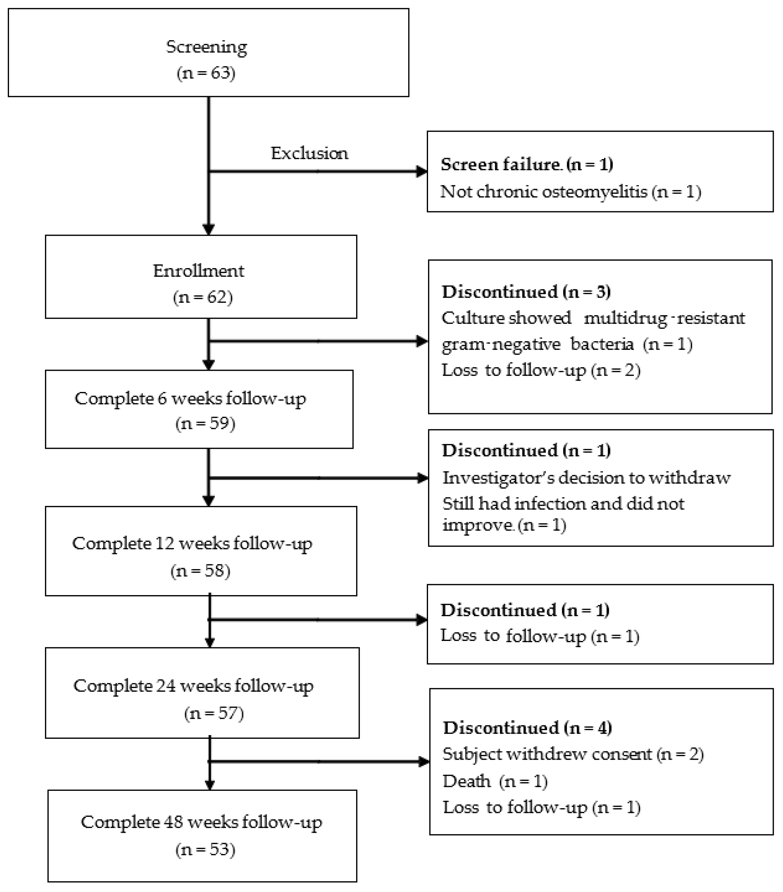
4.1. Patient Enrollment

This study was performed in accordance with the Declaration of Helsinki and the International Conference on Harmonization (ICH) for Good Clinical Practice (GCP). The protocol was registered in the Thai Clinical Trials Registry (TCTR20170123002) and approved by the human research ethical committee in each participated center. Patients with chronic osteomyelitis in 12 participating centers in Thailand including Thammasat university, Bhudasothon hospital, Lerdsin hospital, Srinakharinwirot university, Lampang hospital, Khon Kaen hospital, Pathumtani hospital, Suppasitthiprasong hospital, Bhumibol Adulyadej hospital, Paholpolpayuhasena hospital, Hatyai hospital and Prince of Songkla university were eligible to enroll in this multicenter, prospective cohort study. The sample size was calculated using a confidence interval for a proportion (http://www.sample-size.net/sample-size-conf-interval-proportion/, accessed on 16 March 2020) and 59 patients were necessary to provide a study power of 90% with an α of 0.10. Assuming a 5% loss to follow-up for 48 weeks period; hence, the number of participants was 62 patients.
The inclusion criteria were the age equal or above 18 years old and diagnosed as chronic osteomyelitis. A sinus, an abscess, intraoperative pus, supporting histology, or two or more microbiological cultures with identical organisms were all required for the diagnosis of chronic osteomyelitis, which was defined as the existence of symptoms for at least six weeks [89,90]. The exclusion criteria were the patient with a history of vancomycin, gentamicin or fosfomycin allergy, renal insufficiency, immunocompromise host, multiple active injuries or head injuries, malignancy at the site of infection, poor control of diabetes (HbA1C > 8 mg%), pregnancy, human immunodeficiency virus (HIV) infection, infection with antibiotic-resistant gram-negative bacteria. The participant will be discontinued from the study if they lose follow-up 2 times consecutively, the drug level is above the toxicity level (vancomycin > 50 µg/mL, gentamicin > 6 µg/mL), presence of drug allergy symptoms during the study and antibiotic-resistant gram-negative bacteria from intra-operative culture. The demographic data including sex, age, wound location, Cierny-Mader (C-M) (anatomic type, physiological condition and risk factors) [89], causes and history of previous surgery for osteomyelitis were collected.

4.2. Preparation of Antibiotic Impregnated Microporous Nanohydroxyapatite Beads

Antibiotic impregnated microporous nanohydroxyapatite (nHA-ATB) beads were prepared as described in the previous study [43]. Premixed calcium sulfate-based powder was loaded into a powder-based three-dimensional (3D) printing machine (Z400, Z Corporation, Burlington, MA, USA) to print spherical beads having a diameter of 7 mm by using water-based binder (Zb 7, Z Corporation, USA) as a jetting media and a layer thickness of 0.1 mm. The as-fabricated calcium sulfate beads were then transformed to hydroxyapatite by immersing them in 1 M of disodium hydrogen phosphate solution at 80 °C for 48 h. After reaching the period, the beads were taken out, cleaned by sonication in deionized water and oven dried. The compressive strength of nHA was 0.60 MPa and its porosity and mean pore size were 63.92% and 0.15 microns, respectively [43]. nHA beads were impregnated with one of three types of antibiotics intravenous injection solution including gentamicin, vancomycin or fosfomycin using a vacuum-assisted method under the same condition each time. After impregnation, the samples were taken out and dried in the room atmosphere for 48 h. The representative samples were randomly selected to measure antibiotic content in the beads to make sure that the preparation is consistent. They were then packed in a Tyvek heat-seal sterilization pouch, sterilized by ethylene oxide gas, and kept in dry conditions at room temperature until use.

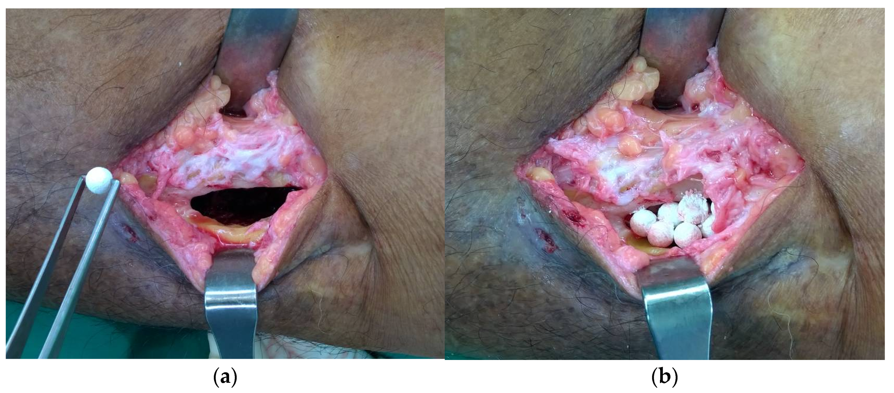
4.3. Operative Procedure

The patient was under general anesthesia or regional anesthesia depending on the location of the infected bone. The sterile preparation was done, and the sinus tract was excised. The debridement was down to the infected bone. Any dead bone and non-vitalized tissue were removed. The multiple deep tissue samplings were collected for microbiological study before the administration of intravenous antibiotics. The cultured isolates were identified by colony characteristics and biochemical reactions. Antibiotic sensitivity was done by Kirby Bauer disc diffusion method on Mueller Hinton agar as per Clinical Laboratory Standards Institutes (CLSI) guidelines. The infected bone was curetted with a mechanical burr until the healthy bleeding bone was reached and then was copiously irrigated with normal saline until no gross infection was present. The nHA-ATB beads were placed into the defect site at an equal ratio (1:1:1) of each antibiotic to completely fill the defect (Figure 3). The rationale of using three antibiotics in this study was to deliver broad-spectrum antibiotics locally to cover the most common organisms generally found in chronic osteomyelitis. Gel foam was applied to cover the beads securely within the intramedullary canal or defect. Hemostasis was achieved and the radivac drain was inserted. The subcutaneous and skin were closed, respectively. Flap was considered in case of inadequate soft tissue coverage. The intravenous cephalosporin was administered immediately after tissue sampling and the appropriate antibiotic was adjusted as soon as the culture result was obtained for 1 to 6 weeks or longer as advised by the infectious disease team. Therapeutic drug monitoring of gentamicin and vancomycin was performed on thirty-four patients (30% of the total number of patients) from Thammasat university or the Prince of Songkla university. Blood samples (10–15 mL) and wound drainage fluid via a radivac catheter were collected at 24 h, 48 h, and 72 h after surgery.
The follow-up evaluations were scheduled at 2 weeks, 4 weeks, 6 weeks, 12 weeks, 24 weeks, and 48 weeks postoperatively. The laboratory test including CBC, ESR, CRP, liver function test and renal function was done at 6 weeks, 12 weeks, 24 weeks, and 48 weeks. The radiographic examination of the treated site was performed to evaluate the radiographic union and the resorption of the materials. Adverse events and complications were recorded at all time points. All patients gave informed consent.
The primary endpoints of the study were the success rate at 12 weeks and the re-infection rate within 24 weeks period after the operation. The success of treatment was defined as no recurrent infection, no recurrence of sinus drainage, normal level of ESR and CRP or no requirement of antibiotics for persistent symptoms during the follow-up. The secondary endpoints were the quality of life of the participants at 0, 6, 12 and 24 weeks postoperatively which was evaluated by the Thai version of the SF-36 questionnaire [91] and the incidence of adverse effects which was graded according to National Cancer Institute criteria [92].

4.4. Statistical Analysis

All the data were analyzed using statistical analysis software (STATA version 14, StataCorp LLC, College Station, TX, USA). The data were described as the mean ± standard deviation. Blood biochemistry and quality of life were compared before and after surgery for each participant using a paired t-test. A p-value < 0.1 was considered statistically significant.

5. Conclusions

Microporous nanohydroxyapatite beads impregnated with vancomycin or gentamicin or fosfomycin could be employed as an alternative product of choice for chronic osteomyelitis treatment. Within the scope of this study, good outcomes in infection treatment were observed throughout the follow-up period and the material was safe and well tolerated while providing the enhancement in quality of life of the patients.

Author Contributions

Conceptualization, J.S. and B.C. (Bancha Chernchujit); methodology, J.S. and B.C (Bancha Chernchujit).; formal analysis, A.S., C.J. (Chittawee Jiamton), A.A, B.C. (Bancha Chernchujit) and J.S.; investigation, C.J. (Chittawee Jiamton), A.A., S.A., B.C. (Banchai Chawalitrujiwong), C.C., S.S., C.J. (Choen Jeraveesal), C.I., P.K., P.S., P.N.N., W.O., P.R., S.T., T.T., F.T., A.S., J.S. and B.C. (Bancha Chernchujit); writing—original draft preparation, C.J. (Chittawee Jiamton), A.A., and J.S.; writing—review and editing, C.J. (Chittawee Jiamton), A.A., S.A., B.C. (Banchai Chawalitrujiwong), C.C., S.S., C.J. (Choen Jeraveesal), C.I., P.K., P.S., P.N.N., W.O., P.R., S.T., T.T., F.T., A.S., J.S. and B.C. (Bancha Chernchujit); project administration, F.T., A.S. and J.S.; funding acquisition, J.S. and B.C. (Bancha Chernchujit). All authors have read and agreed to the published version of the manuscript.

Funding

This research was financially supported by the Health Systems Research Institute (HSRI), Thailand (Grant number HSRI 63-056).

Institutional Review Board Statement

The study was conducted in accordance with the Declaration of Helsinki. The protocol was registered in the Thai Clinical Trials Registry (TCTR20170123002) and approved by the human research ethical committee in each participated center including Thammasat university (099/2558, 3 July 2015), Bhudasothon hospital (BSH-IRB 014/2560, 18 May 2017, Lerdsin hospital (62046, 24 April 2019, Srinakharinwirot university hospital (SWUEC/E-459/2561, 14 May 2019), Lampang hospital (061/58, 20 August 2015), Khon Kaen hospital (KE60084, 24 August 2017), Pathumtani hospital (0032.203.3/23, 30 March 2016), Suppasitthiprasong hospital (026/2560, 3 May 2017), Bhumibol Adulyadej hospital (048/60, 20 July 2017), Paholpolpayuhasena hospital (2017-03, 21 April 2017), Hatyai hospital (22/2560, 5 April 2017) and Prince of Songkla university (REC.62-048-11-1, 22 June 2019).

Informed Consent Statement

Informed consent was obtained from all subjects involved in the study.

Data Availability Statement

The data presented in this study are available on request from the corresponding author. The data are not publicly available due to ethical restrictions.

Acknowledgments

MedResNet, Thailand was thanked for assistance in project management/coordinator and monitoring.

Conflicts of Interest

The authors declare no conflict of interest relevant to the content of this article. The funders had no role in the design of the study; in the collection, analyses, or interpretation of data; in the writing of the manuscript; or in the decision to publish the results.

References

  1. O’Connor, O.; Thahir, A.; Krkovic, M. How much does an infected fracture cost? Arch. Bone Jt. Surg. 2022, 10, 135–140. [Google Scholar] [CrossRef]
  2. Dartnell, J.; Ramachandran, M.; Katchburian, M. Haematogenous acute and subacute paediatric osteomyelitis. J. Bone Joint Surg. Br. 2012, 94-B, 584–595. [Google Scholar] [CrossRef] [PubMed]
  3. McCarthy, J.J.; Dormans, J.P.; Kozin, S.H.; Pizzutillo, P.D. Musculoskeletal infections in children. Basic treatment principles and recent advancements. J. Bone Joint Surg. 2004, 86A, 850–863. [Google Scholar] [CrossRef]
  4. De Boeck, H. Osteomyelitis and septic arthritis in children. Acta Orthop. Belg. 2005, 71, 505–515. [Google Scholar] [PubMed]
  5. Riise, O.R.; Kirkhus, E.; Handeland, K.S.; Flato, B.; Reiseter, T.; Cvancarova, M.; Nakstad, B.; Wathne, K.-O. Childhood osteomyelitis-Incidence and differentiation from other acute onset musculoskeletal features in a population-based study. BMC Pediatr. 2008, 8, 45. [Google Scholar] [CrossRef] [PubMed]
  6. Walter, N.; Bärtl, S.; Alt, V.; Rupp, M. The epidemiology of osteomyelitis in children. Children 2021, 8, 1000. [Google Scholar] [CrossRef]
  7. Walter, N.; Baertl, S.; Alt, V.; Rupp, M. What is the burden of osteomyelitis in Germany? An analysis of inpatient data from 2008 through 2018. BMC Infect. Dis. 2021, 21, 550. [Google Scholar] [CrossRef] [PubMed]
  8. Henke, P.K.; Blackburn, S.A.; Wainess, R.W.; Cowan, J.; Terando, A.; Proctor, M.; Wakefield, T.W.; Upchurch, G.R.; Stanley, J.C.; Greenfield, L.J. Osteomyelitis of the Foot and Toe in Adults Is a Surgical Disease. Ann. Surg. 2005, 241, 885–894. [Google Scholar] [CrossRef]
  9. Shone, A.; Burnside, J.; Chipchase, S.; Game, F.; Jeffcoate, W. Probing the validity of the probe-to bone test in the diagnosis of osteomyelitis of the foot in diabetes. Diabetes Care 2006, 29, 945. [Google Scholar] [CrossRef]
  10. Lavery, L.A.; Armstrong, D.G.; Peters, E.J.; Lipsky, B.A. Probe-to-bone test for diagnosing diabetic foot osteomyelitis: Reliable or relic? Diabetes Care 2007, 30, 270–274. [Google Scholar] [CrossRef]
  11. Masters, E.A.; Ricciardi, B.F.; Bentley, K.L.D.M.; Moriarty, T.F.; Schwarz, E.M.; Muthukrishnan, G. Skeletal infections: Microbial pathogenesis, immunity and clinical management. Nat. Rev. Microbiol. 2022, 20, 385–400. [Google Scholar] [CrossRef] [PubMed]
  12. Meroni, G.; Tsikopoulos, A.; Tsikopoulos, K.; Allemanno, F.; Martino, P.A.; Soares Filipe, J.F. A Journey into Animal Models of Human Osteomyelitis: A Review. Microorganisms 2022, 10, 1135. [Google Scholar] [CrossRef]
  13. Veis, D.J.; Cassat, J.E. Infectious osteomyelitis: Marrying bone biology and microbiology to shed new light on a persistent clinical challenge. J. Bone Miner. Res. 2021, 36, 636–643. [Google Scholar] [CrossRef] [PubMed]
  14. Wassif, R.K.; Elkayal, M.; Shamma, R.N.; Elkheshen, S.A. Recent advances in the local antibiotics delivery systems for management of osteomyelitis. Drug Deliv. 2021, 28, 2392–2414. [Google Scholar] [CrossRef] [PubMed]
  15. Cobb, L.H.; McCabe, E.M.; Priddy, L.B. Therapeutics and delivery vehicles for local treatment of osteomyelitis. J. Orthop. Res. 2020, 38, 2091–2103. [Google Scholar] [CrossRef] [PubMed]
  16. Lorentzen, A.K.; Engel, L.; Gottlieb, H.; Obinah, M.P.B. One-stage treatment of chronic osteomyelitis with an antibiotic-loaded biocomposite and a local or free flap. Eur. J. Plast. Surg. 2021, 44, 367–374. [Google Scholar] [CrossRef]
  17. Steadman, W.; Chapman, P.R.; Schuetz, M.; Schmutz, B.; Trampuz, A.; Tetsworth, K. Local Antibiotic Delivery Options in Prosthetic Joint Infection. Antibiotics 2023, 12, 752. [Google Scholar] [CrossRef]
  18. Roos, E.; Bartha, H.; Charles, V.; Hoepelman, A.I.M.; Peters, E.J.G. To bead or not to bead? Treatment of osteomyelitis and prosthetic joint-associated infections with gentamicin bead chains. Int. J. Antimicrob. Agents. 2011, 38, 371–375. [Google Scholar] [CrossRef]
  19. van Vugt, T.A.G.; Arts, J.J.; Geurts, J.A.P. Antibiotic-loaded polymethylmethacrylate beads and spacers in treatment of orthopedic infections and the role of biofilm formation. Front. Microbiol. 2019, 10, 1626. [Google Scholar] [CrossRef]
  20. Thomes, B.; Murray, P.; Bouchier-Hayes, D. Development of resistant strains of Staphylococcus epidermidis on gentamicin-loaded bone cement in vivo. J. Bone Joint Surg. Br. 2002, 84, 758–760. [Google Scholar] [CrossRef]
  21. Neut, D.; van de Belt, H.; Stokroos, I.; van Horn, J.R.; van der Mei, H.C.; Busscher, H.J. Biomaterial-associated infection of gentamicin-loaded PMMA beads in orthopaedic revision surgery. J. Antimicrob. Chemother. 2001, 47, 885–891. [Google Scholar] [CrossRef]
  22. Hansen, E.N.; Adeli, B.; Kenyon, R.; Parvizi, J. Routine use of antibiotic laden bone cement for primary total knee arthroplasty: Impact on infecting microbial patterns and resistance profiles. J. Arthroplast. 2014, 29, 1123–1127. [Google Scholar] [CrossRef] [PubMed]
  23. Ene, R.; Nica, M.; Ene, D.; Cursaru, A.; Cirstoiu, C. Review of calcium-sulphate-based ceramics and synthetic bone substitutes used for antibiotic delivery in PJI and osteomyelitis treatment. EFORT Open Rev. 2021, 6, 297–304. [Google Scholar] [CrossRef] [PubMed]
  24. Kallala, R.; Harris, W.E.; Ibrahim, M.; Dipane, M.; McPherson, E. Use of Stimulan absorbable calcium sulphate beads in revision lower limb arthroplasty. Bone Joint Res. 2018, 7, 570–579. [Google Scholar] [CrossRef] [PubMed]
  25. Thahir, A.; Lim, J.A.; West, C.; Krkovic, M. The use of calcium sulphate beads in the management of osteomyelitis of femur and tibia: A systematic review. Arch. Bone Jt. Surg. 2022, 10, 320–327. [Google Scholar] [CrossRef]
  26. Ferguson, J.Y.; Dudareva, M.; Riley, N.D.; Stubbs, D.; Atkins, B.L.; McNally, M.A. The use of a biodegradable antibiotic-loaded calcium sulphate carrier containing tobramycin for the treatment of chronic osteomyelitis. Bone Joint J. 2014, 96-B, 829–836. [Google Scholar] [CrossRef]
  27. Ali, T.; Mutalik, S.; Venkatesh, M.; Shenoy, D.B.; Acharya, L.; Udupa, U. Biodegradable implants of gentamicin sulfate for effective management of osteomyelitis. Saudi. Pharma. J. 2006, 14, 120–125. [Google Scholar]
  28. Chang, H.I.; Perrie, Y.; Coombes, A.G.A. Delivery of the antibiotic gentamicin sulphate from precipitation cast matrices of polycaprolactone. J. Control. Release 2006, 110, 414–421. [Google Scholar] [CrossRef]
  29. Naraharisetti, P.K.; Lee, H.C.G.; Fu, Y.C.; Lee, D.J.; Wang, C.H. In vitro and in vivo release of gentamicin from biodegradable discs. J. Biomed. Mater. Res. Part B Appl. Biomater. 2006, 77B, 329–337. [Google Scholar] [CrossRef]
  30. Schmidt, C.; Wenz, R.; Nies, B.; Moll, F. Antibiotic in vivo/in vitro release, histocompatibility and biodegradation of gentamicin implants based on lactic acid polymers and copolymers. J. Control. Release 1995, 37, 83–94. [Google Scholar] [CrossRef]
  31. Aimin, C.; Chunlin, H.; Juliang, B.; Tinyin, Z.; Zhichao, D. Antibiotic loaded chitosan bar. An in vitro, in vivo study of a possible treatment for osteomyelitis. Clin. Orthop. Relate Res. 1999, 366, 239–247. [Google Scholar] [CrossRef]
  32. Coraca-Huber, D.C.; Wurm, A.; Fille, M.; Hausdorfer, J.; Nogler, M.; Vogt, S.; Kuhn, K.D. Antibiotic-loaded calcium carbonate/calcium sulfate granules as co-adjuvant for bone grafting. J. Mater. Sci. Mater. Med. 2015, 26, 5344. [Google Scholar] [CrossRef]
  33. Fleiter, N.; Walter, G.; Bösebeck, H.; Vogt, S.; Büchner, H.; Hirschberger, W.; Hoffmann, R. Clinical use and safety of a novel gentamicin-releasing resorbable bone graft substitute in the treatment of osteomyelitis/osteitis. Bone Joint Res. 2014, 3, 223–229. [Google Scholar] [CrossRef] [PubMed]
  34. McKee, M.D.; Wild, L.M.; Schemitsch, E.H.; Waddell, J.P. The use of an antibiotic-impregnated, osteoconductive, bioabsorbable bone substitute in the treatment of infected long bone defects: Early results of a prospective trial. J. Orthop. Trauma 2002, 16, 622–627. [Google Scholar] [CrossRef]
  35. Prasanna, A.P.S.; Venkatasubbu, G.D. Sustained release of amoxicillin from hydroxyapatite nanocomposite for bone infections. Prog. Biomater. 2018, 7, 289–296. [Google Scholar] [CrossRef] [PubMed]
  36. Geuli, O.; Metoki, N.; Zada, T.; Reches, M.; Eliaz, N.; Mandler, D. Synthesis, coating, and drug-release of hydroxyapatite nanoparticles loaded with antibiotics. J. Mater. Chem. B 2017, 5, 7819–7830. [Google Scholar] [CrossRef]
  37. Kaya, M.; Şimşek-Kaya, G.; Gürsan, N.; Kireççi, E.; Dayi, E.; Gündoğdu, B. Local treatment of chronic osteomyelitis with surgical debridement and tigecycline-impregnated calcium hydroxyapatite: An experimental study. Oral Surg. Oral Med. Oral. Pathol. Oral Radiol. 2012, 113, 340–347. [Google Scholar] [CrossRef]
  38. Kattimani, V.S.; Kondaka, S.; Lingamaneni, K.P. Hydroxyapatite: Past, present, and future in bone regeneration. Bone Tissue Regen Insights 2016, 7, 9. [Google Scholar] [CrossRef]
  39. Barrère, F.; van Blitterswijk, C.A.; de Groot, K. Bone regeneration: Molecular and cellular interactions with calcium phosphate ceramics. Int. J. Nanomed. 2006, 1, 317–332. [Google Scholar]
  40. Ogose, A.; Kondo, N.; Umezu, H.; Hotta, T.; Kawashima, H.; Tokunaga, K.; Ito, T.; Kudo, N.; Hoshino, M.; Gu, W.; et al. Histological assessment in grafts of highly purified beta-tricalcium phosphate (OSferion) in human bones. Biomaterials 2006, 27, 1542–1549. [Google Scholar] [CrossRef]
  41. Suwanprateeb, J.; Suvannapruk, W.; Wasoontararat, K. Low temperature preparation of calcium phosphate structure via phosphorization of 3D-printed calcium sulfate hemihydrate based material. J. Mater. Sci. Mater. Med. 2010, 21, 419–429. [Google Scholar] [CrossRef]
  42. Suwanprateeb, J.; Thammarakcharoen, F.; Wasoontararat, K.; Suvannapruk, W. Influence of printing parameters on the transformation efficiency of 3D-printed plaster of paris to hydroxyapatite and its properties. Rapid Prototyp. J. 2012, 18, 490–499. [Google Scholar] [CrossRef]
  43. Suwanprateeb, J.; Thammarakcharoen, F.; Phanphiriya, P.; Chokevivat, W.; Suvannapruk, W.; Chernchujit, B. Preparation and characterizations of antibiotic impregnated microporous nano-hydroxyapatite for osteomyelitis treatment. Biomed. Eng.-Appl. Basis Commun. 2014, 26, 1450041. [Google Scholar] [CrossRef]
  44. Kijartorn, P.; Wongpairojpanich, J.; Thammarakcharoen, F.; Suwanprateeb, J.; Buranawat, B. Clinical evaluation of 3D printed nano-porous hydroxyapatite bone graft for alveolar ridge preservation: A randomized controlled trial. J. Dent. Sci. 2022, 17, 194–203. [Google Scholar] [CrossRef] [PubMed]
  45. Yamashita, Y.; Uchida, A.; Yamakawa, T.; Shinto, Y.; Araki, N.; Kato, K. Treatment of chronic osteomyelitis using calcium hydroxyapatite ceramic implants impregnated with antibiotic. Int. Orthop. 1998, 22, 247–251. [Google Scholar] [CrossRef] [PubMed]
  46. Itokazu, M.; Aoki, T.; Nonomura, H.; Nishimoto, Y.; Itoh, Y. Antibiotic-loaded porous hydroxyapatite blocks for the treatment of osteomyelitis and postoperative infection. A preliminary report. Bull. Hosp. Jt. Dis. 1998, 57, 125–129. [Google Scholar] [PubMed]
  47. Sudo, A.; Hasegawa, M.; Fukuda, A.; Uchida, A. Treatment of infected hip arthroplasty with antibiotic-impregnated calcium hydroxyapatite. J. Arthroplast. 2008, 23, 145–150. [Google Scholar] [CrossRef]
  48. Takigami, I.; Ito, Y.; Ishimaru, D.; Ogawa, H.; Mori, N.; Shimizu, T.; Terabayashi, N.; Shimizu, K. Two-stage revision surgery for hip prosthesis infection using antibiotic-loaded porous hydroxyapatite blocks. Arch. Orthop. Trauma Surg. 2010, 130, 1221–1226. [Google Scholar] [CrossRef] [PubMed]
  49. Matsumoto, K.; Itokazu, M.; Uemura, S.; Takigami, I.; Naganawa, T.; Shimizu, K. Successful joint arthroplasty after treatment of destructive MRSA arthritis of the knee using antibiotic-loaded hydroxyapatite blocks: A case report. Arch. Orthop. Trauma Surg. 2007, 127, 47–50. [Google Scholar] [CrossRef]
  50. Bose, D.; Kugan, R.; Stubbs, D.; McNally, M. Management of infected nonunion of the long bones by a multidisciplinary team. Bone Joint J. 2015, 97-B, 814–817. [Google Scholar] [CrossRef]
  51. Metsemakers, W.J.; Kuehl, R.; Moriarty, T.F.; Richards, R.G.; Verhofstad, M.H.J.; Borens, O.; Kates, S.; Morgenstern, M. Infection after fracture fixation: Current surgical and microbiological concepts. Injury 2018, 49, 511–522. [Google Scholar] [CrossRef] [PubMed]
  52. Klemn, K. The use of antibiotic-containing beads chain in the treatment of chronic bone infections. Clin. Microbial. Infect. 2001, 7, 28–31. [Google Scholar] [CrossRef] [PubMed]
  53. Gauland, C. Managing lower-extremity osteomyelitis locally with surgical debridement and synthetic calcium sulfate antibiotic tablets. Adv. Skin Wound Care 2011, 24, 515–523. [Google Scholar] [CrossRef] [PubMed]
  54. Calhoun, J.H.; Henry, S.L.; Anger, D.M.; Cobos, J.A.; Mader, J.T. The treatment of infected nonunions with gentamicin-polymethylmethacrylate antibiotic beads. Clin. Orthop. Relat. Res. 1993, 295, 23–27. [Google Scholar] [CrossRef]
  55. Patzakis, M.J.; Mazur, K.; Wilkins, J.; Sherman, R.; Holtom, P. Septopal beads and autogenous bone grafting for bone defects in patients with chronic osteomyelitis. Clin. Orthop. Relat. Res. 1993, 295, 112–118. [Google Scholar] [CrossRef]
  56. McNally, M.A.; Ferguson, J.Y.; Scarborough, M.; Ramsden, A.; Stubbs, D.A.; Atkins, B.L. Mid- to long-term results of single-stage surgery for patients with chronic osteomyelitis using a bioabsorbable gentamicin-loaded ceramic carrier. Bone Joint J. 2022, 104-B, 1095–1100. [Google Scholar] [CrossRef]
  57. Cierny, G., III. Surgical treatment of osteomyelitis. Plast. Reconstr. Surg. 2011, 127 (Suppl. S1), 190S–204S. [Google Scholar] [CrossRef] [PubMed]
  58. Walenkamp, G.H.; Kleijn, L.L.; de Leeuw, M. Osteomyelitis treated with gentamicin-PMMA beads: 100 patients followed for 1–12 years. Acta Orthop. Scand. 1998, 69, 518–522. [Google Scholar] [CrossRef]
  59. Kallala, R.; Haddad, F.S. Hypercalcaemia following the use of antibiotic-eluting absorbable calcium sulphate beads in revision arthroplasty for infection. Bone Joint J. 2015, 97-B, 1237–1241. [Google Scholar] [CrossRef]
  60. Lum, Z.C.; Pereira, G.C. Local bio-absorbable antibiotic delivery in calcium sulfate beads in hip and knee arthroplasty. J. Orthop. 2018, 15, 676–678. [Google Scholar] [CrossRef]
  61. McPherson, E.; Dipane, M.; Sherif, S. Dissolvable antibiotic beads in treatment of periprosthetic joint infection and revision arthroplasty—The use of synthetic pure calcium sulfate (Stimulan®) impregnated with vancomycin & tobramycin. Reconstr. Rev. 2013, 3, 32–43. [Google Scholar] [CrossRef]
  62. Chang, W.; Colangeli, M.; Colangeli, S.; Di Bella, C.; Gozzi, E.; Donati, D. Adult osteomyelitis: Debridement versus debridement plus Osteoset T pellets. Acta Orthop. Belg. 2007, 73, 238–243. [Google Scholar]
  63. Zhao, Z.; Wang, G.; Zhang, Y.; Luo, W.; Liu, S.; Liu, Y.; Zhou, Y.; Zhang, Y. The effect of calcium sulfate/calcium phosphate composite for the treatment of chronic osteomyelitis compared with calcium sulfate. Ann. Palliat. Med. 2020, 9, 1821–1833. [Google Scholar] [CrossRef] [PubMed]
  64. Kwong, F.N.; Porter, R.M.; Griffin, D.R.; Evans, C.H. Effect of tobramycin on the osteogenesis of stem cells derived from human bone marrow. J. Bone Joint Surg. [Br]. 2009, 91-B (Suppl. SI), 130–131. [Google Scholar]
  65. Francesca, D.B.; Bancroft, L.W.; Peterson, J.J.; Kransdorf, M.J.; Menke, D.M.; DeOrio, J.K. Imaging characteristics of bone graft materials. RadioGraphics. 2006, 26, 373–388. [Google Scholar] [CrossRef]
  66. Lavery, L.A.; Ahn, J.; Ryan, E.C.; Bhavan, K.; Oz, O.K.; La Fontaine, J.; Wukich, D.K. What are the optimal cutoff values for ESR and CRP to diagnose osteomyelitis in patients with diabetes-related foot infections? Clin. Orthop. Relat. Res. 2019, 477, 1594–1602. [Google Scholar] [CrossRef]
  67. Easton, C.R.; Junho, A.; Wukich, D.K.; Kim, P.J.; Fontaine, J.L.; Lavery, L.A. Diagnostic utility of erythrocyte sedimentation rate and C-reactive protein in osteomyelitis of the foot in persons without diabetes. J. Foot Ankle Surg. 2019, 58, 484–488. [Google Scholar] [CrossRef]
  68. Ghanem, E.; Antoci, V., Jr.; Pulido, L.; Joshi, A.; Hozack, W.; Parvizi, J. The use of receiver operating characteristics analysis in determining erythrocyte sedimentation rate and C-reactive protein levels in diagnosing periprosthetic infection prior to revision total hip arthroplasty. Int. J. Infect. Dis. 2009, 13, e444–e449. [Google Scholar] [CrossRef]
  69. Greidanus, N.V.; Masri, B.A.; Garbuz, D.S.; Wilson, S.D.; McAlinden, M.G.; Xu, M.; Duncan, C.P. Use of erythrocyte sedimentation rate and C-reactive protein level to diagnose infection before revision total knee arthroplasty. A prospective evaluation. J. Bone Joint Surg. Am. 2007, 89, 1409–1416. [Google Scholar] [CrossRef]
  70. Lammin, K.; Taylor, J.; Zenios, M. The usefulness of CRP and ESR In diagnosing long bone osteomyelitis in children—Are we being falsely reassured? Orthop. Procs. 2012, 94-B (Suppl. SXXXIX), 156. [Google Scholar]
  71. Chiang, H.Y.; Chung, C.W.; Kuo, C.C.; Lo, Y.C.; Chang, W.S.; Chi, C.Y. First-4-week erythrocyte sedimentation rate variability predicts erythrocyte sedimentation rate trajectories and clinical course among patients with pyogenic vertebral osteomyelitis. PLoS ONE 2019, 14, e0225969. [Google Scholar] [CrossRef]
  72. Cohen, G.; Lager, S.; Cece, D.; Rubel, I.F. The social impact associated with chronic osteomyelitis. A grading system for the most frequent complication. Osteosynth. Trauma Care 2004, 12, 74–76. [Google Scholar] [CrossRef]
  73. Yagdiran, A.; Otto-Lambertz, C.; Lingscheid, K.M.; Sircar, K.; Samel, C.; Scheyerer, M.J.; Zarghooni, K.; Eysel, P.; Sobottke, R.; Jung, N.; et al. Quality of life and mortality after surgical treatment for vertebral osteomyelitis (VO): A prospective study. Eur. Spine J. 2020, 30, 1721–1731. [Google Scholar] [CrossRef]
  74. Zayzan, K.R.; Yusof, N.M.; Sulong, A.F.; Zakaria, Z.; Rahman, J.A. Functional outcome and quality of life following treatment for post-traumatic osteomyelitis of long bones. Singap. Med. J. 2022, 63, 251–255. [Google Scholar] [CrossRef] [PubMed]
  75. Wahlig, H.; Dingeldein, E.; Bergmann, R.; Reuss, K. The release of gentamicin from polymethylmethacrylate beads. An experimental and pharmacokinetic study. J. Bone Joint Surg. Br. 1978, 60-B, 270–275. [Google Scholar] [CrossRef]
  76. Jenny, J.Y.; Jenny, G.; Lambert, J.; Gaudias, J.; Kempf, I. Utility of measurement of gentamicin release from PMMA beads in wound drainage fluid after in-vivo implantation. Acta Orthop. Belg. 1995, 61, 10–13. [Google Scholar]
  77. Salvati, E.A.; Callaghan, J.J.; Brause, B.D.; Klein, R.F.; Small, R.D. Reimplantation in infection. Elution of gentamicin from cement and beads. Clin. Orthop. 1986, 207, 83–93. [Google Scholar] [CrossRef]
  78. Livio, F.; Wahl, P.; Csajka, C.; Gautier, E.; Buclin, T. Tobramycin exposure from active calcium sulfate bone graft substitute. BMC Pharmacol Toxicol. 2014, 15, 12. [Google Scholar] [CrossRef]
  79. Shen, L.; Dong, Y.; Zhang, C.; Guo, Q. Chronic osteomyelitis treatment: A clinical and pharmaco-kinetic study of vancomycin impregnated calcium sulphate. J. Med. Imaging Health Inform. 2014, 5, 36–42. [Google Scholar] [CrossRef]
  80. Cresswell, T.; Shahane, S.; Stockley, I. Irreducible dislocation of a hip total prosthesis: The risk of gentamicin beads. Orthopedics 2000, 23, 1095–1096. [Google Scholar] [CrossRef]
  81. Fiddian, N.; Sudlow, R.; Browett, J. Ruptured femoral vein. A complication of the use of gentamicin beads in an infected excision arthroplasty of the hip. J. Bone Joint Surg. Br. 1984, 66, 493–494. [Google Scholar] [CrossRef] [PubMed]
  82. Clarke, J.; Shalhoub, J.; Das, S.K. Ceramic gentamicin beads in vascular graft infection—A cautionary note. Vasc. Endovasc. Surg. 2013, 47, 76–77. [Google Scholar] [CrossRef] [PubMed]
  83. Tarar, M.; Khalid, A.; Usman, M.; Javed, K.; Shah, N.; Abbas, M.W. Wound leakage with the use of calcium sulphate beads in prosthetic joint surgeries: A systematic review. Cureus 2021, 13, e19650. [Google Scholar] [CrossRef] [PubMed]
  84. Ziran, B.H.; Smith, W.R.; Morgan, S.J. Use of calcium-based demineralized bone matrix/allograft for nonunions and posttraumatic reconstruction of the appendicular skeleton: Preliminary results and complications. J. Trauma. 2007, 63, 1324–1328. [Google Scholar] [CrossRef] [PubMed]
  85. Kelly, C.M.; Wilkins, R.M.; Gitelis, S.; Hartjen, C.; Watson, J.T.; Kim, P.T. The use of a surgical grade calcium sulfate as a bone graft substitute: Results of a multicenter trial. Clin. Orthop. Relat. Res. 2001, 382, 42–50. [Google Scholar] [CrossRef]
  86. Borrelli, J., Jr.; Prickett, W.D.; Ricci, W.M. Treatment of nonunions and osseous defects with bone graft and calcium sulfate. Clin. Orthop. Relat. Res. 2003, 411, 245–254. [Google Scholar] [CrossRef]
  87. Smith, I. Convulsions and coma associated with iatrogenically elevated CSF calcium levels post spinal surgery: A case report. Crit. Care Resusc. 2005, 7, 173–176. [Google Scholar]
  88. Zhou, C.H.; Ren, Y.; Ali, A.; Meng, X.Q.; Zhang, H.A.; Fang, J.; Qin, C.H. Single-stage treatment of chronic localized tibial osteomyelitis with local debridement and antibiotic loaded calcium sulfate implantation: A retrospective study of 42 patients. J. Orthop. Surg. Res. 2020, 15, 201. [Google Scholar] [CrossRef]
  89. Panteli, M.; Giannoudis, P.V. Chronic osteomyelitis: What the surgeon needs to know. EFORT Open Rev. 2016, 1, 128–135. [Google Scholar] [CrossRef]
  90. Cunha, B.A. Osteomyelitis in Elderly Patients. Clin. Infect. Dis. 2002, 35, 287–293. [Google Scholar] [CrossRef]
  91. Leurmarnkul, W.; Meetam, P. Properties testing of the retranslated SF-36 (Thai version). Thai J. Pharm. Sci. 2005, 29, 69–88. [Google Scholar]
  92. Common Terminology Criteria for Adverse Events (CTCAE). Available online: https://ctep.cancer.gov/protocoldevelopment/electronic_applications/ctc.htm#ctc_60 (accessed on 15 April 2023).
Figure 1. CONSORT flowchart of the study.
Antibiotics 12 01049 g001
Figure 2. Representative radiographic images showing nHA-ATB beads in the bone cavity: (a) Immediate post-operatively; (b) 48 weeks post-operatively. The incorporation of nHA-ATB beads with surrounding bone and no translucency which might be suggestive of infection recurrence was observed.
Antibiotics 12 01049 g002
Figure 3. Intra-operative photographs showing the debridement and placement of nHA-ATB into the bone cavity: (a) The infected bone was curetted and was irrigated with copious normal saline; (b) The bone cavity was completely filled with nHA-ATB beads.
Antibiotics 12 01049 g003
Table 1. Demographic data.
Characteristics n = 62n (%)
Sex
 Male49 (79.0)
 Female13 (21.0)
Age (Mean ± SD)47.2 ± 14.7
Location
 Tibia34 (54.8)
 Femur19 (30.6)
 Humerus4 (6.5)
 Calcaneus2 (3.2)
 Clavicle1 (1.6)
 Forearm1 (1.6)
 Fibula1 (1.6)
Cierny-Mader Grade
 Type I Medullary osteomyelitis11 (17.7)
 Type II Superficial osteomyelitis3 (4.8)
 Type III Localized osteomyelitis37 (59.7)
 Type IV Diffuse osteomyelitis11 (17.7)
Physiological class
 Class A Good immune system and delivery47 (75.8)
 Class B Compromised locally or systemically risk factors15 (24.2)
 Class C requires suppressive or no treatment; minimal disability0 (0)
Causes
 Post-traumatic48 (77.4)
 Hematogones (non-trauma, sepsis)14 (22.6)
Previous surgery for this condition
 015 (24.2)
 119 (30.6)
 213 (21.0)
 38 (12.9)
 42 (3.2)
 52 (3.2)
 61 (1.6)
 >72 (3.2)
Risk factor
 No risk factor41 (66.1)
 Risk21 (33.9)
  Major risk factor: cellulitis or abscess formation5 (23.8)
  Minor systemic risk factor: subject had smoking history7 (33.3)
  Minor systemic risk factor: diabetes mellitus3 (14.3)
  Minor systemic risk factor: anemia2 (9.5)
  Minor systemic risk factor: chronic lung disease1 (4.8)
  Minor local risk factor: poor soft tissue requiring flap9 (42.9)
  Minor local risk factor: adjacent joint stiff/arthritic7 (33.3)
  Minor local risk factor: heterotopic ossification1 (4.8)
Table 2. Surgical details and cultured results.
Surgery DataQuantity
(Median, IQR (Min-Max))
d93, 57
Number of antibiotics loaded hydroxyapatite beads used60, 54 (6–180)
 Gentamicin beads20, 18 (2–60)
 Vancomycin beads20, 20 (2–60)
 Fosfomycin beads20, 20 (2–60)
Cultured organismn (%)
 No3 (4.8)
 Yes59 (95.2)
No growth28 (47.5)
Cultured organism (2 organisms found)7 (11.9)
Acinetobactor baumannii and E. coli (ESBL-Producing)1 (14.3)
Acinetobactor baumannii and Pseudomonas aeruginosa1 (14.3)
Morganella morganii and Klebsiella pneumoniae (MDR)1 (14.3)
Pseudomonas aeruginosa (MDR) and Staphylococcus haemolyticus (MR-CoNS)1 (14.3)
Pseudomonas aeruginosa and Enterococcus avium1 (14.3)
Pseudomonas aeruginosa and Shewanella putrefaciens1 (14.3)
Staphylococcus aureus and Staphylococcus epidermidis1 (14.3)
Cultured organism (1 organism found)24 (40.7)
Pseudomonas aeruginosa9 (37.5)
Staphylococcus aureus4 (16.7)
Staphylococcus aureus (MRSA)1 (4.2)
 Aerobic culture1 (4.2)
Aerococcus viridans1 (4.2)
Coagulase Negative Staphylococci2 (8.4)
Enterobacter cloacae1 (4.2)
Enterococcus faecalis1 (4.2)
Serratia marcescens1 (4.2)
Staphylococcus cohnii1 (4.2)
Staphylococcus haemolyticus1 (4.2)
Staphylococcus hominis1 (4.2)
Vancomycin sensitivity (n = 38)
 Sensitive8 (21.1)
 Intermediate0 (0)
 Resistant0 (0)
 Results not reported by hospital lab30 (78.9)
Gentamicin sensitivity (n = 38)
 Sensitive19 (50)
 Intermediate1 (2.6)
 Resistant4 (10.5)
 Results not reported by hospital lab14 (36.8)
Fosfomycin sensitivity (n = 38)
 Sensitive9 (23.7)
 Intermediate1 (2.6)
 Resistant2 (5.3)
 Results not reported by hospital lab26 (68.4)
IQR = Interquartile range.
Table 3. Blood biochemistry.
ResultsBaseline
Mean ± SD
8 Weeks
Mean ± SD
12 Weeks
Mean ± SD
24 Weeks
Mean ± SD
48 Weeks
Mean ± SD
Liver function test
AST (SGOT) (U/L)29.31 ± 13.8530.25 ± 16.0227.53 ± 12.0431.08 ± 14.8529.37 ± 14.75
ALT (SGPT) (U/L)32.78 ± 29.9534.5 ± 29.3228.81 ± 24.8631.67 ± 22.8027.07 ± 19.47
Alkaline Phosphatase (ALP) (U/L)103.36 ± 47.29103.81 ± 34.8497.96 ± 32.6094.37 ± 33.2583.44 ± 25.98
Renal function test
Serum Creatinine (mg/dL)0.81 ± 0.200.85 ± 0.251.13 ± 1.740.85 ± 0.210.88 ± 0.20 #
eGFR (ml/min/1.73 m2)103.11 ± 21.8497.67 ± 25.52 #98.55 ± 23.59 #100.46 ± 19.996.58 ± 20.86 #
BUN (mg/dL)14.03 ± 13.2812.24 ± 4.4711.65 ± 3.6413.22 ± 4.2812.90 ± 3.80
C-reactive protein (CRP) (mg/dL)12.30 ± 27.823.96 ± 4.863.12 ± 4.52 #2.7 ± 4.11 #2.45 ± 2.65 #
Erythrocyte sedimentation rate (ESR) (mm/hour)45.11 ± 32.8340.09 ± 26.5327.37 ± 21.68 #26.72 ± 27.67 #25.13 ± 26.70 #
# Denoted significantly different compared to the baseline values, p < 0.1.
Table 4. Therapeutic drug monitoring of antibiotics level.
AntibioticsDuration
(h)
Concentration
(µg/mL)
Mean ± SD
Wound drainage fluidVancomycin24128.64 ± 134.49
48175.79 ± 153.96
72125.43 ± 159.87
Gentamicin24412.04 ± 646.01
4853.03 ± 74.06
7258.41 ± 97.81
SerumVancomycin244.00 ± 0.00
482.67 ± 2.31
726.94 ± 4.16
Gentamicin240.38 ± 0.17
480.36 ± 0.12
720.40 ± 0.00
Table 5. Quality of life of participants at each follow-up as evaluated by SF-36 questionnaire.
ResultsBaseline
Mean ± SD
6 Weeks
Mean ± SD
12 Weeks
Mean ± SD
24 Weeks
Mean ± SD
Physical health37.14 ± 4.2347.94 ± 14.77 #55.51 ± 13.78 #60.63 ± 14.64 #
 Physical functioning46.86 ± 26.1050.49 ± 26.9163.98 ± 26.12 #72.93 ± 23.49 #
 Role-physical5.27 ± 8.1411.52 ± 10.71 #14.29 ± 10.67 #16.98 ± 9.65 #
 Bodily pain46.96 ± 26.5664.39 ± 20.34 #73.47 ± 18.29 #78.89 ± 19.95 #
 General health49.45 ± 19.5065.35 ± 17.69 #70.31 ± 16.84 #73.72 ± 17.99 #
Mental health47.44 ± 17.4858.99 ± 14.12 #64.81 ± 12.83 #67.21 ± 13.63 #
 Vitality66.79 ± 24.4880.76 ± 21.2487.88 ± 19.53 #91.44 ± 20.89 #
 Social function56.62 ± 25.5467.40 ± 20.7875.00 ± 19.09 #80.16 ± 19.82 #
 Role- emotional7.35 ± 9.6715.03 ± 10.41 #17.01 ± 10.06 #18.12 ± 10.14 #
 Mental health58.98 ± 21.9972.78 ± 14.24 #79.35 ± 12.09 #79.13 ± 13.38 #
# Denoted significantly different compared to the baseline values, p < 0.1.
Table 6. Adverse events and their relations to nHA-ATB beads.
Relations to nHA-ATB BeadsAdverse Events (n = 20)
SAE or Non-SAESeverityEvents
Definitely related
 - HA exposed from wound dehiscence Non-SAEGrade I1
Possibly related
 - Serum oozing from the surgical wound Non- SAEGrade I1
 - Acute on-top chronic left tibia osteomyelitis SAEGrade III1
Unlikely
 - Anterior shoulder dislocation Non-SAEGrade II1
 - Loosening of external fixator SAEGrade I1
 - Gastrointestinal hemorrhage, unspecified SAEGrade II1
Not related
 - Leg pain Non-SAEGrade I1
 - Wrist drop due to radial nerve palsy Non-SAEGrade II1
 - Acute pyelonephritis SAEGrade I1
 - Adjust external fixation SAEGrade I1
 - Bone fracture SAEGrade I1
 - Prolonged hospitalization of chronic osteomyelitis of left distal femur due to financial problem SAEGrade I1
 - Revised external fixation SAEGrade I1
 - Wound dehiscence SAEGrade I2
 - Surgical wound with drainage SAEGrade II1
 - Wound dehiscence after debridement and switch HA-ATB to cement ATB SAEGrade II1
 - Broken Steinman Pin SAEGrade III1
 - Nonunion of the fracture site SAEGrade IV1
 - Death from other causes SAEGrade V1
Disclaimer/Publisher’s Note: The statements, opinions and data contained in all publications are solely those of the individual author(s) and contributor(s) and not of MDPI and/or the editor(s). MDPI and/or the editor(s) disclaim responsibility for any injury to people or property resulting from any ideas, methods, instructions or products referred to in the content.

