Article

Antimicrobial Activity of Catechol-Containing Biopolymer Poly[3-(3,4-dihydroxyphenyl)glyceric Acid] from Different Medicinal Plants of Boraginaceae Family

1
TSMU I. Kutateladze Institute of Pharmacochemistry, Tbilisi 0159, Georgia
2
School of Pharmacy, Aristotle University, 54124 Thessaloniki, Greece
3
Mycological Laboratory, Institute for Biological Research “Siniša Stanković”–National Institute of Republic of Serbia, University of Belgrade, Blvd. Despot Stefan 142, 11000 Belgrade, Serbia
*
Author to whom correspondence should be addressed.
Antibiotics 2023, 12(2), 285; https://doi.org/10.3390/antibiotics12020285
Submission received: 21 November 2022 / Revised: 20 January 2023 / Accepted: 28 January 2023 / Published: 1 February 2023

Abstract

:
This study reports the antimicrobial activities of the biopolymers poly[3-(3,4-dihydoxyphenyl)glyceric acid] (PDHPGA) and poly[2-methoxycarbonyl-3-(3,4-dihydroxyphenyl)oxirane] (PMDHPO), extracted from the six plants of Boraginaceae family: Symphytum asperum (SA), S. caucasicum (SC), S. gr and iflorum (SG), Anchusa italica (AI), Cynoglosum officinale (CO), and Borago officinalis (BO) collected in various parts of Georgia. The study revealed that the antibacterial activities were moderate, and biopolymers from only three plants showed activities against all tested bacteria. Biopolymers from CO stems as well as SC and AI did not show any activity except low activity against a resistant P. aeruginosa strain, which was the most resistant among all three resistant strains. On the other hand, the antifungal activity was better compared to the antibacterial activity. Biopolymers from BO stems exhibited the best activities with MIC/MFC at 0.37–1.00 mg/mL and 0.75–1.5 mg/L, respectively, followed by those from SG stems. Biopolymers from SC and AI roots showed antifungal activities against all six fungi, in contrast to the antibacterial activity, while biopolymers from CO stems and SA roots had activities against four fungi and one fungus, respectively. The sugar-based catechol-containing biopolymers from BO stems demonstrated the best activities among all tested biopolymers against T. viride, P. funiculosum, P. cyclpoium var verucosum, and C. albicans (MIC 0.37 mg/mL). In addition, biopolymers from SG stems were half as active against A. fumigatus and T. viride as ketoconazole. Biopolymers from all plant materials except for CO stems showed higher potency than ketoconazole against T. viride. For the first time, it was shown that all plant materials exhibited better activity against C. albicans, one of the most dreadful fungal species.

1. Introduction

Biopolymers are chain-like molecules that consist of repeated chemical blocks derived from renewable resources that may decompose in the environment. The recent popular use of biomaterials is due to the fact that reducing the usage of non-renewable resources reduces the environmental pollution produced by synthetic materials. Furthermore, biopolymers have attracted significant attention as a class of polymer materials with a wide range of applications, especially in the medical [1] and pharmaceutical fields [2,3]. Due to their biodegradability and non-toxic nature, biopolymers play a positive role in the maintenance of a clean and safe environment.
Hydroxycinnamic acids have been extensively used in the preparation of polyesters, but there are also reports about their use in the synthesis of polyamides and poly(anhydride esters), among many others. These polymers have a wide spectrum of applications, ranging from industrial (e.g., (super) engineered plastics) to biomedical domains (e.g., drug delivery systems and shape memory materials) [4]. Plant-derived, adhesive polymers from natural sources such as 3,4-dihydroxycinnamic acid (caffeic acid) and 4-hydroxycinnamic acid (p-coumaric acid) modified by transesterification were found to show strong adhesive characteristics on metal surfaces, which were equivalent to conventional superglues from petroleum resources [5]. Lignin-mimetic polyesters prepared using an in-bulk polycondensation of bioavailable aromatic hydroxyl(carboxylic acids) can be applied as an environmentally degradable plastic with extremely high performance and can be used in automobiles, aircraft, electronic devices, and other materials [6].
Phenols, phenolic-rich plant extracts, including catechol and catechol-based polymers, exhibiting antibacterial and antifungal activities are of great interest to researchers and industry [7]. Antimicrobial polymers can slow or inhibit the growth of drug-resistant strains and present high antimicrobial efficacy due to the various antimicrobial modes and polymeric structures [8]. The phenolic-rich plant extracts of the Boraginaceae family have important therapeutic (antimicrobial, antitumor, antiviral, anti-inflammatory, cardiotonic, contraceptive, antiplatelet activity, etc.) and cosmetic applications [9]. Their pharmacological effects are related to the presence of various classes of compounds such as phenolic acids, phenols, naphthoquinones, flavonoids, terpenoids, and the purine derivative allantoin. However, plants of the Boraginaceae family are also rich in hepatotoxic pyrrolizidine alkaloids (which serve as chemical defense compounds, mainly against herbivores), and thus their consumption is restricted; nevertheless, the use of Boraginaceae plants as poultices for wounds does not raise any objection [9,10]. There are many references in the literature regarding the antimicrobial activity of phenolic plant extracts. Thus, Raphaelli et al. [11] reported an evaluation of the antimicrobial activities of apple phenolic extracts against Escherichia coli, Listeria monocytogenes, Salmonella Typhimurium, and Staphylococcus aureus. Hussain et al. [12] studied the composition and antimicrobial activities of different Emirati date (Phoenix dactylifera L.) pits against Staphylococcus aureus (ATCC 29123), Escherichia coli (ATCC 25922), and Candida albicans (ATCC 66027). Different extracts from these six date pits showed different antimicrobial activities. The only common finding was that none exhibited activity against C. albicans. Dikpınar et al. [13], in his review, mentioned the isolation of numerous phenolic compounds from plants/plant extracts that showed the ability to prevent the growth of bacteria and fungi. The review revealed that phenolic compounds from medicinal plants demonstrated different antimicrobial activities according to their molecular structures, such as the permeabilization and destabilization of the plasma membrane or the inhibition of extracellular enzymes. Salazar-Aranda [14] reported an antimicrobial activity evaluation of extracts from 17 plants from different regions of northeast Mexico against three Gram-negative bacterial strains (Pseudomonas aeruginosa, Klebsiella pneumoniae, and Acinetobacter baumannii), three Gram-positive bacterial strains (Enterococcus faecalis and two Staphylococcus aureus strains), and seven clinically isolated yeasts (Candida albicans, C. krusei, C. tropicalis, C. parapsilosis, and C. glabrata). It was shown that the antimicrobial activities from different plants and against different bacterial and fungal strains ranged from 31.25 to 250 mg/mL. In addition, plant phenolics and their extracts can be very good inhibitors of many foodborne pathogenic and spoilage bacteria [15,16]. In an extensive review, Taco et al. [17] reported the antimicrobial activities of plant extracts from several sources such as grape pomace, grape seed, apple, and various exotic fruit and medicinal plant samples. It was mentioned that red grape pomace exhibited a strong bactericidal effect against E. coli and S. aureus at 12 mg/mL [18]. Grape seed extracts also appeared to be active against bacteria such as S. Typhimurium, Listeria monocytogenes, Bacillus spp., Pseudomonas aeruginosa, and Campylobacter spp. [19,20], while red pitahaya (exotic fruits extracts) demonstrated a good antimicrobial spectrum against Gram-positive and Gram-negative bacteria, yeasts, and molds at concentrations from 7.8 μg/mL to 50 mg/mL [21,22].
Recently, the antimicrobial properties of some caffeic and 3-(3,4-dihydroxyphenyl)glyceric acid derivatives against Gram-positive and Gram-negative bacteria, a series of fungi [23], and three resistant bacterial strains (methicillin-resistant Staphylococcus aureus, resistant Escherichia coli, and resistant Pseudomonas aeruginosa) were evaluated in vitro [24]. The studies revealed that some caffeic and 3-(3,4-dihydroxyphenyl)glyceric acid derivatives appeared to be more active than the reference drugs ampicillin and streptomycin, whereas the antifungal activities of all compounds were greater than the reference drug ketoconazole, while some compounds appeared to be more active than bifonazole [23]. Kepa et al. [25] studied the antibacterial potential of caffeic acid (CA) alone and in an antibiotic–phytochemical combination against Staphylococcus aureus reference and clinical strains isolated from infected wounds. The effects of caffeic acid on the S. aureus strains were different, with variations in the minimum inhibitory concentration (MIC) from 256 μg/mL to 1024 μg/mL. The study revealed that the antimicrobial action of CA is probably strain-dependent. On the other hand, it was postulated that the antimicrobial activity of CA against S. aureus clinical isolates is higher than that of the reference drug. Furthermore, CA is capable of increasing the antimicrobial effect in combination with antibiotics.
In our previous studies, we reported the isolation of water-soluble mucilaginous high-molecular-weight (>1000 kDa or 500 kDa) fractions (HMFs) from plants of the Boraginaceae family: Symphytum asperum (SA), S. caucasicum (SC), S. gr and iflorum (SG), S. officinale (SO), Anchusa italica (AI), Cynoglosum officinale (CO), Borago officinalis (BO), and Paracynoglossum imeretinum (PI) through the ultrafiltration of hot-water extracts using membrane filters with a cut-off values of 1000 kDa or 500 kDa. The HMFs obtained using this procedure are free of pyrrolizidine alkaloids and allantoin. According to UV data , IR spectra, and different techniques of NMR spectroscopy (13C NMR, APT, 1H NMR, 1D NOE, 2D 1H/13C HSQC, and 2D DOSY) [26,27] (Supplementary materials, Figures S1–S8, Table S1), the main chemical constituent of the HMFs of the above-mentioned medicinal plants was found to be an exotic bioactive carbohydrate-based catechol-containing bio-polyether, namely poly[3-(3,4-dihydroxyphenyl)glyceric acid] (PDHPGA) (1, Figure 1), that is, poly[oxy-1-carboxy-2-(3,4-dihydroxyphenyl)ethylene] (POCDPE). The repeating unit of PDHPGA is a 3-(3,4-dihydroxyphenyl)glyceric acid residue. Most of the carboxyl groups of PDHPGA from AI, SG, and BO, unlike of biopolymers from SA, SC, SO, CO, and PI, are methyl-esterified and are named poly[2-methoxycarbonyl-3-(3,4-dihydroxyphenyl)oxirane] (PMDHPO) (2 in Figure 1) [26].
The polyoxyethylene (polyethylene glycol) chain is the backbone of polymeric molecule PDHPGA. 3,4-Dihydroxyphenyl (catechol) and carboxyl groups are regular substituents at two carbon atoms in the chain of PDHPGA [26]. On the other hand, a poly (2,3-glyceric acid ether) chain is the backbone of the polymeric molecule of PDHPGA. The basic moiety of poly (2,3-glyceric acid ether) is glyceric acid, a natural three-carbon sugar acid, which is an oxidative form of the simplest of all common aldoses, namely glyceraldehyde. In this case, a poly (2,3-glyceric acid ether) chain constitutes the backbone of the polymeric molecule of PDHPGA, with catechol groups as the regular substituents in the chain. PDHPGA, bearing a lot of catecholic groups along the polymeric chain, is a novel sugar-based multicatechol-containing biopolymer. Thus, we can consider PDHPGA as a representative of an exotic class of carbohydrate-based catechol-functionalized biopolymers with ether bonds. Such biopolymers, to our knowledge, are unknown in nature, as all above-mentioned synthetic and natural polymers are either polyesters or copolymers [4,5,6].
PDHPGA exhibits high immunomodulatory (anticomplementary), antioxidant, anti-inflammatory, wound and burn healing, and anticancer activities, both in vitro and in vivo [27].
Later, the antibacterial properties of natural polyethers from different species of the Boraginaceae family (PDHPGA-SA, PDHPGA-SC, PDHPGA-SG, PDHPGA-AI, and PDHPGA-CO), along with the synthetic polymers poly[2-methoxycarbonyl-3-(3,4-dimethoxyphenyl)oxirane (PMDMPO), poly[2-methoxycarbonyl-3-(3,4-dibenzyloxyphenyl)oxirane] (PMDBPO), and poly[2-methoxycarbonyl-3-(3,4-dihidroxphenyl)oxirane] (PMDHPO), were investigated against the pathogenic strains S. aureus ATCC 25923 and E. coli ATCC 25922. It was shown that only a synthetic oligomeric analogue of PMDHPO exhibited antimicrobial activity against these pathogenic strains at the concentrations that were used [27]. Interesting data obtained about the antimicrobial activities of synthetic compounds prompted us to continue research in this field and examine natural polyethers from the above-mentioned species of the Boraginaceae family against Gram-positive and Gram-negative bacteria, three resistant bacterial strains (MRSA, E. coli, and P. aeruginosa), and a panel of fungi.
The aim of this study was to examine and evaluate the antimicrobial activities of sugar-based catechol-containing biopolymers from SA, SC, SG, AI, CO, and BO.

2. Results and Discussion
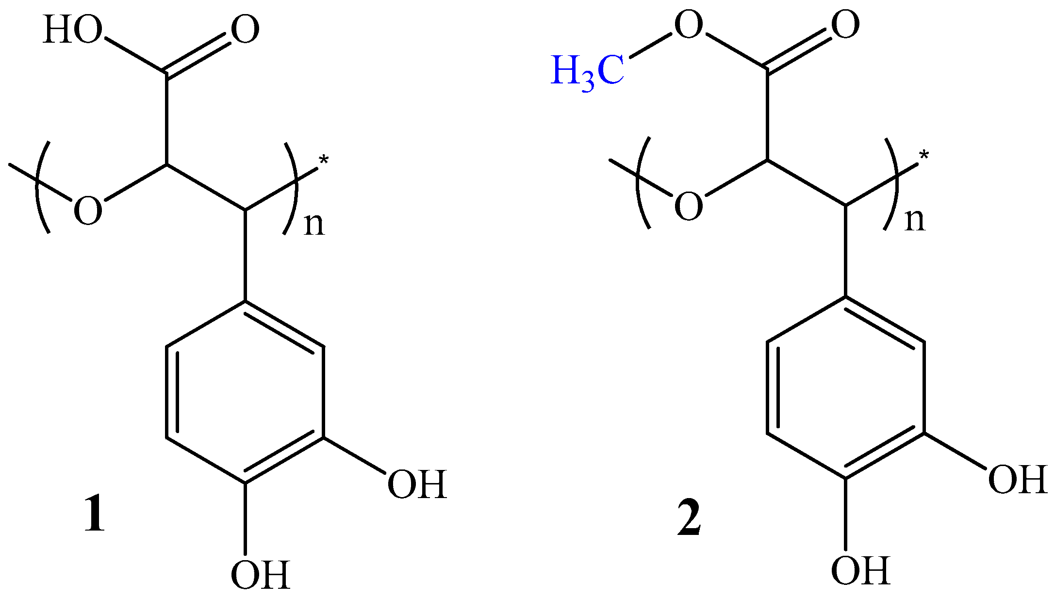
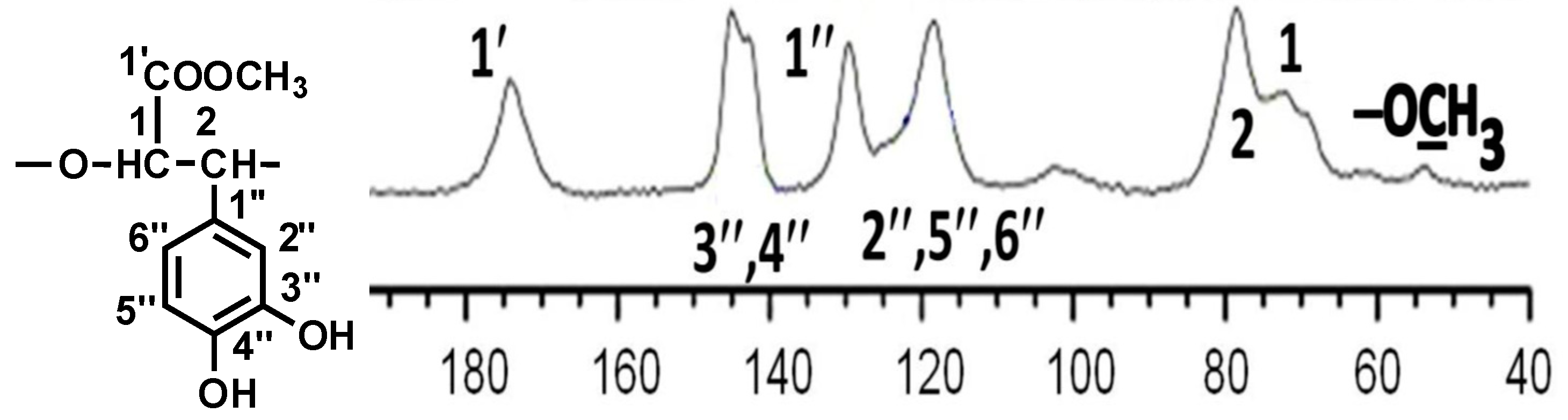
2.1. Chemistry

According to data from UV, IR, liquid-state 1H, 13C NMR, gCOSY, and 2D heteronuclear 1H/13C gHSQCED experiments, the main chemical constituent of the water-soluble high-molecular fractions from the stems and roots of SA, SC, SG, SO, AI, CO, and BO was found to be the biologically active caffeic-acid-derived polymer PDHPGA [26]. Most of the carboxyl groups of PDHPGA from AI, SG, and BO, unlike of biopolymers from SA, SC, SO, CO, and PI, are methyl-esterified [27].
Tests of the fractionation and molecular weight distribution of a biologically active PDHPGA preparation from SA were carried out. The molecular weights and contents of different fractions for the PDHPGA preparation were simultaneously determined using high-performance size-exclusion chromatography (HPSEC) coupled with multiangle laser light scattering (MALLS) and a refractive index detector (RID) with a refractive index increment (dn/dc) [28].
Absorption maxima at 212 nm, 236 nm (shoulder), 282 nm (shoulder), and 286 nm were observed in the UV spectra (H2O, λmax, nm) of PDHPGA-SA, PDHPGA-SC, PDHPGA-SG, PDHPGA-AI, PDHPGA-CO, and PDHPGA-BO, which could be attributable to substituted phenols (Figure S1). The IR spectra of PDHPGA-SA, PDHPGA-SC, PDHPGA-SG, PDHPGA-AI, PDHPGA-CO, and PDHPGA-BO contained absorption bands characteristic of phenol-carboxylic acids: 3425 (OH); 2924 (CH); 1605 (ionized carboxyl) and 1736 cm−1 for its ester form; 1512 and 1443 (aromatic C=C); 1404 and 1219 (phenols); 1265, 1080, and 1018 (R-O-R’); 872 (C-H in the aromatic ring with one isolated hydrogen atom); and 818 cm−1 (C-H in the aromatic ring with two neighboring hydrogen atoms (Figure S2). The signal assignments of the 13C and 1H NMR spectra of PDHPGA-SA, PDHPGA-SC, and PDHPGA-CO and of the methylated carboxylic groups of PDHPGA-SG, PDHPGA-AI, and PDHPGA-BO are given in Supplementary Materials Table S1.
The 2D DOSY experiment gave similar diffusion coefficients for the methylated and non-methylated signals of PDHPGA. Both sets of signals fell in the same horizontal range (Figure S8). This implies similar (same order of magnitude) molecular weights for methylated and non-methylated polymers. Thus, we concluded that the NMR signals of both the methylated and non-methylated carboxylic groups originated from the same PDHPGA polymer. In order to confirm the proposed structure of the repeating unit of non-methylated PDHPGA and the mostly methylated carboxylic groups of these poorly water-soluble high-molecular-weight PMDHPOs, the 13C solid-state NMR spectrum was recorded (Figure 2), and total assignments of the signals are given in Table 1. The total assignments of the signals of the solid state 13C NMR of a mixture of PDHPGA + PMDHPO were in good agreement with the results of the above-mentioned liquid-state 13C NMR classical spectra of the PDHPGA and PMDHPO samples in D2O.
Thus, according to data from different techniques of NMR spectroscopy, the main chemical constituent of the high-molecular-weight fractions from the medicinal plants SA, SC, SG, AI, CO, and BO is poly[3-(3,4-dihydroxyphenyl)glyceric acid] (PDHPGA), which is poly[oxy-1-carboxy-2-(3,4-dihydroxyphenyl)ethylene] (POCDPE).

2.2. Biological Evaluation

2.2.1. Antibacterial Activity Evaluation

The sugar-based catechol-containing biopolymers from SA, SC, SG, AI, CO, and BO were tested for their antibacterial activities against a panel of six bacterial species, including Staphylococcus aureus, Escherichia coli, and Pseudomonas aeruginosa, using a microdilution method for the determination of the minimal inhibitory (MIC) and minimal bactericidal concentrations (MBC). The activities of these polymers were moderate to low, with MICs in range of 0.75–6.00 mg/mL and MBCs of 1.00–9.00 mg/mL (Table 2). As can be seen in Table 2, the polymers from SG stems exhibited the best activities among all tested compounds, with MIC values of 0.75 mg/mL, while the polymer from SA roots showed the lowest activity (MIC 6.00 mg/mL) compared to ampicillin and streptomycin, which were used as reference drugs. Moderate activity was also observed against E. coli and P. aeruginosa for polymers from BO stems (MIC 1.00 mg/mL). On the other hand, the sugar-based catechol-containing biopolymers from SC and AI roots and CO stems did not show any activity against the bacteria in this experiment except for AI roots against E. coli. Thus, the order of activity can be presented as follows: SG stems > BO stems > SA roots. The bacteria most sensitive to these biopolymers appeared to be E. coli, while S. aureus was the most resistant.
These biopolymers were tested against resistant strains of the mentioned bacteria. The best activity was observed against res. P. aeruginosa, expressed by biopolymers from BO and SG stems (MIC/MBC at 0.75 mg/mL). The latter showed better activity against MRSA and res. E. coli, with MIC 1.00 mg/mL and 0.75 mg/mL, respectively, than the biopolymers from the materials of the two other plants. The order of activity against resistant strains can be presented as follows: MRSA > res. P. aeruginosa > res. E. coli.

2.2.2. Antifungal Activity Evaluation

These biopolymers were also evaluated for their antifungal activities against six fungal species, and the results are presented in Table 3. The sugar-based catechol-containing biopolymers exhibited better antifungal activities compared to antibacterial activities, with MICs in the range of 0.37–6.00 mg/mL and MFCs of 0.75–9.00 mg/mL. The best activity was observed for biopolymers from BO stems, with MIC/MFC at 0.37–1.00 mg/mL and 0.75–1.5 mg/L, respectively, followed by those from SG stems. It should be mentioned that biopolymers from SC and AI roots showed antifungal activities against all six fungi, while those from CO stems and SA showed antifungal activities against four fungi and one fungus, respectively, in contrast to the antibacterial activity. In general, the order of activity can be presented as: BO stems > SG stems > SC roots > AI roots > CO stems > SA roots. The best activity among all tested biopolymers was found for the biopolymer from BO stems against Trichoderma viride, Penicillium funicolosum, Penicillium verrucosum var. cyclopium, and Candida albicans (MIC 0.37 mg/mL). The same good activity was shown by biopolymers from SG stems against Aspergillus fumigatus and T. viride, which was half as active as ketoconazole, the reference drug. It should be mentioned that all sugar-based catechol-containing biopolymers from all plant material except for CO stems were more potent than ketoconazole against T. viride. The most sensitive fungus to these biopolymers appeared to be T. viride, while P. funiculosum was the most resistant, followed by P. verrucosum var. cyclopium. It should be noticed that, in general, sugar-based catechol-containing biopolymers demonstrated quite good activity against C. albicans in comparison to other strains except T. viride.
Antimicrobial studies revealed that PDHPGA from BO and SG stems showed the best antibacterial as well antifungal activities. The reason of this difference compared to PDHPGA from SA, SC, and CO may be the methylation of the carboxylic moieties of these polymers. As we reported in our previous studies, the same tendency was observed regarding the synthetic oligomeric analogue of PMDHPO, which was the only polymer showing antimicrobial activities against some pathogenic strains at the concentrations that were used [27].
It should be mentioned that, despite there being some references in the literature regarding the antimicrobial activities of catechol-based polymers [7,29], there are no publications concerning sugar-based catechol-containing biopolymers.

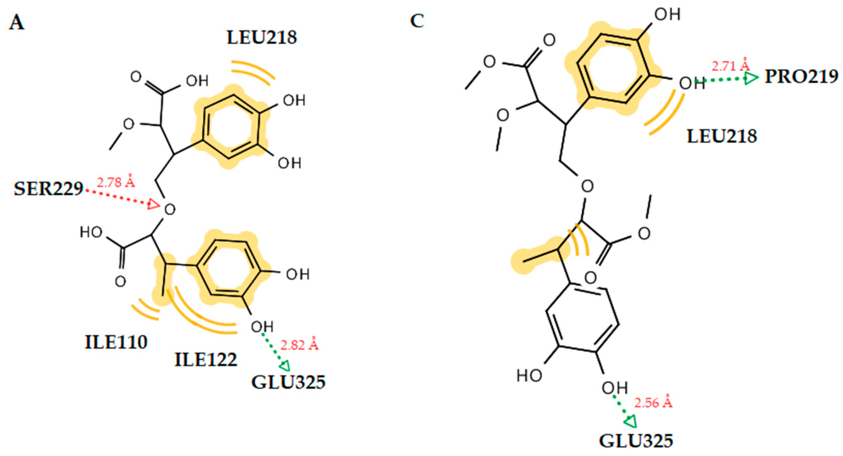
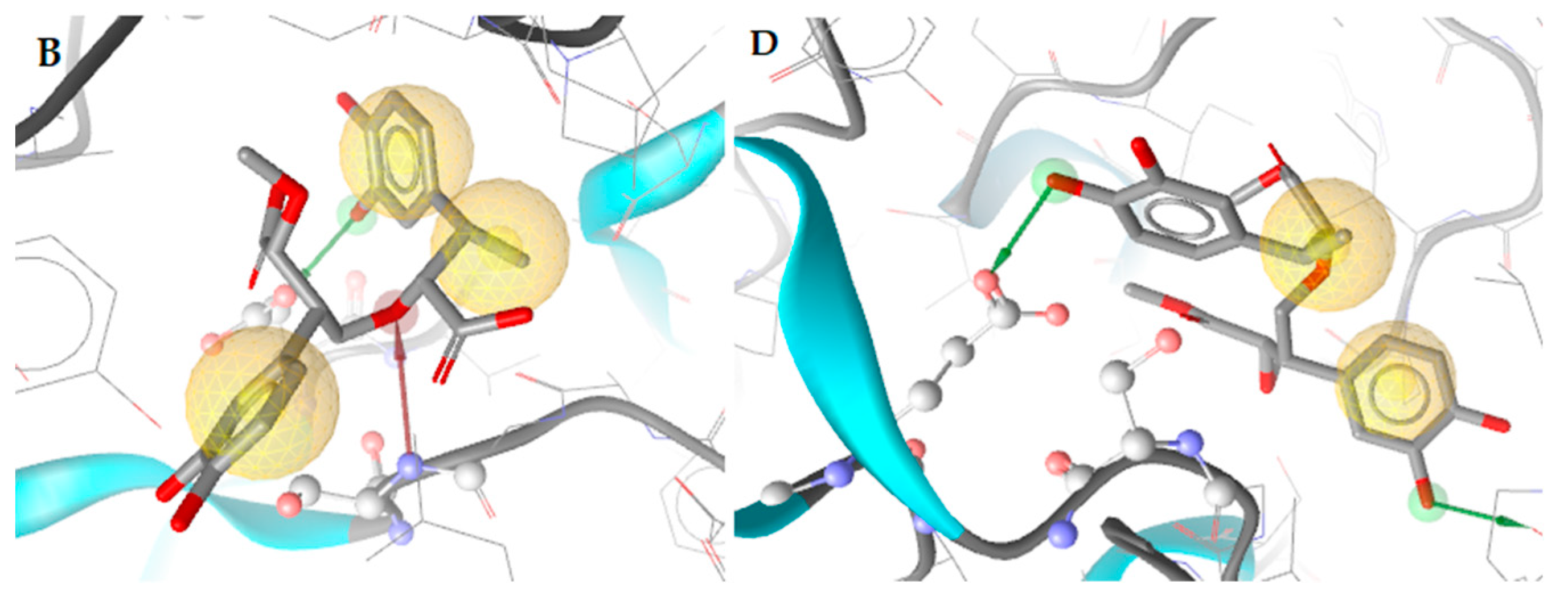
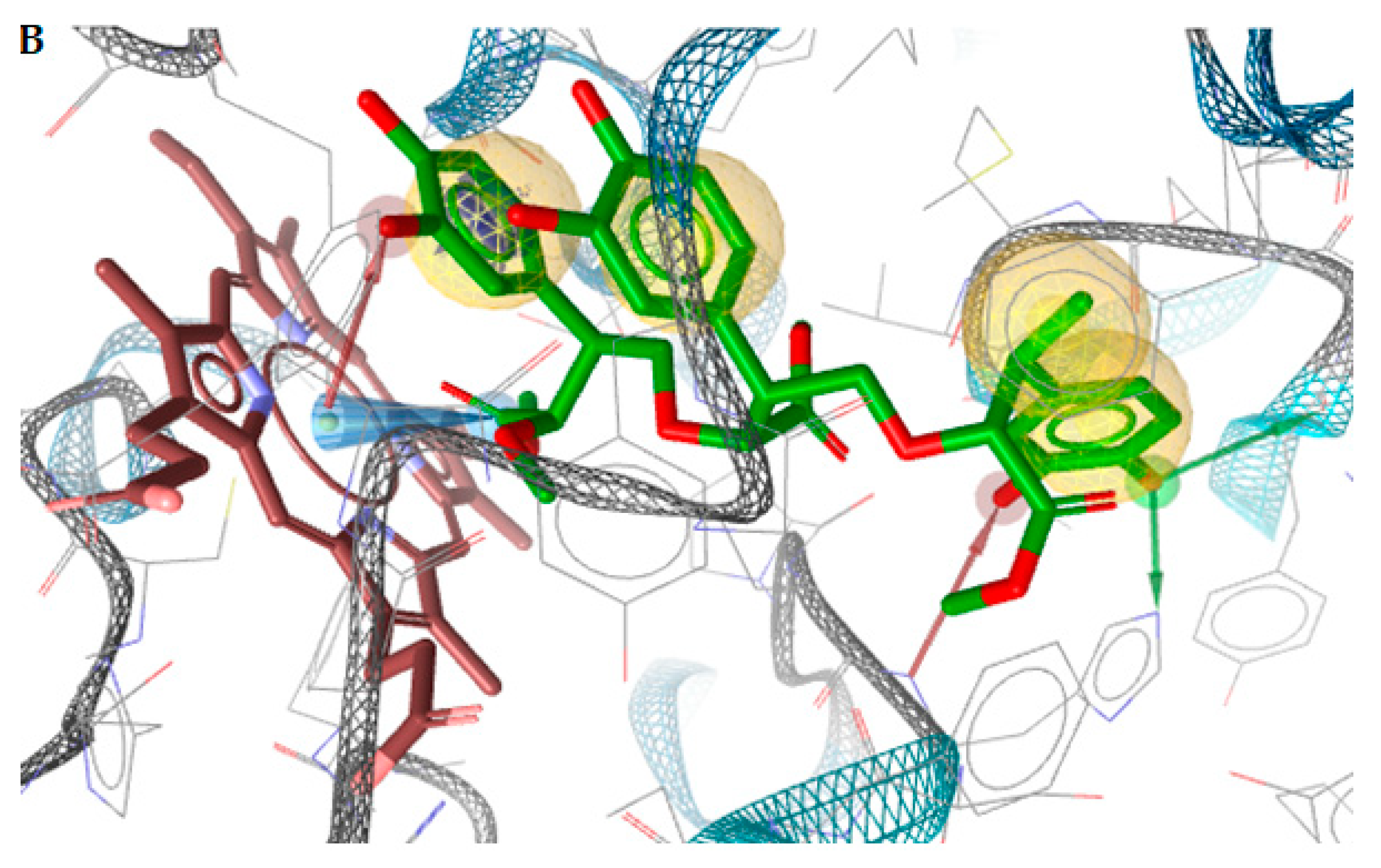
2.3. Docking Studies

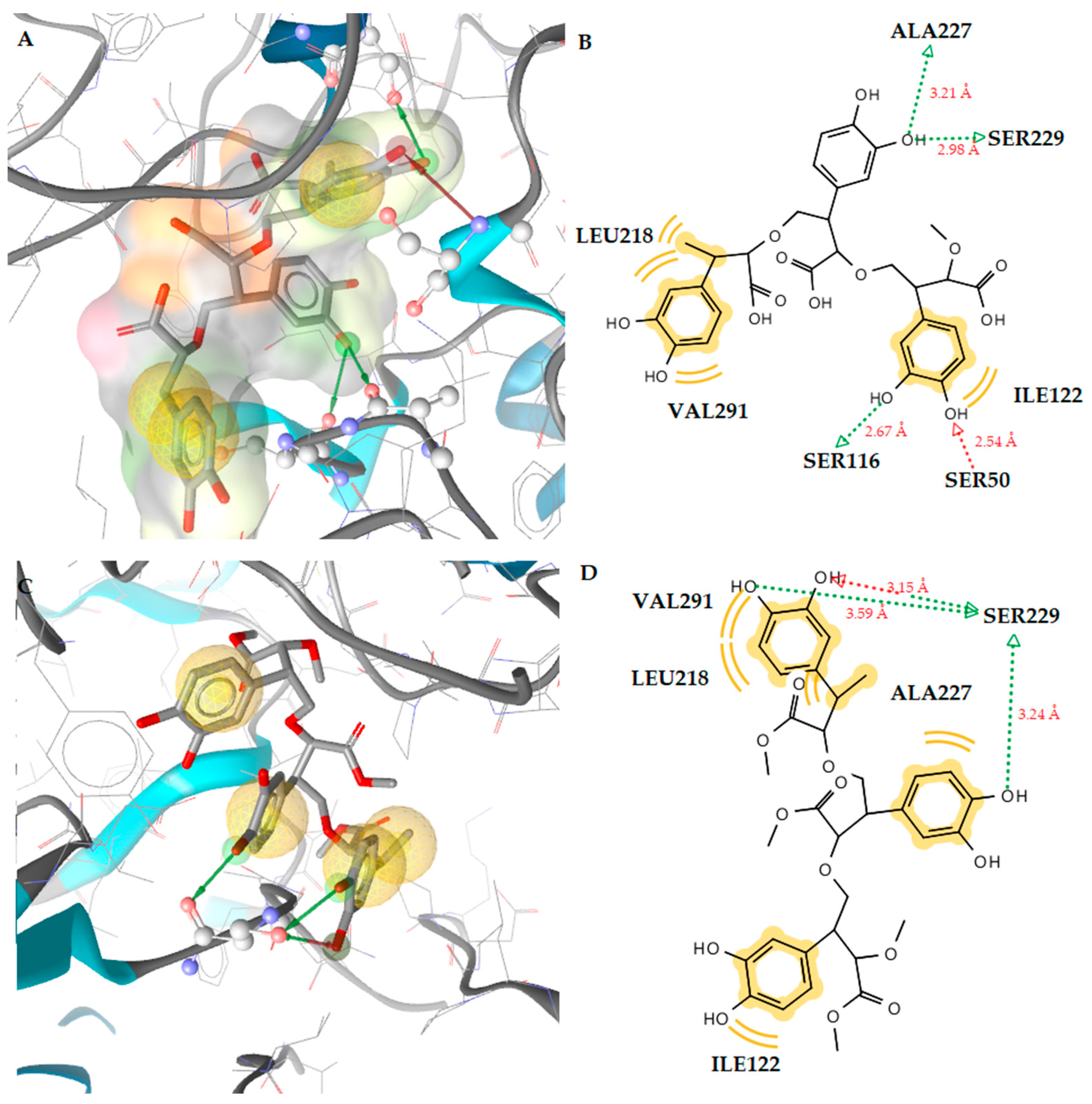
2.3.1. Docking to Antibacterial Targets

Docking studies of several antibacterial targets, such as E. coli DNA gyrase, S. aureus thymidylate kinase, E. coli primase, and E. coli MurB, were performed to give a structural insight into their mechanism of action. The docking studies showed that the assessments of the binding free energy to E. coli DNA gyrase, thymidylate kinase, E. coli primase, and E. coli MurA were higher than that to E. coli MurB. Therefore, it may be resolved that the inhibition of the E. coli MurB enzyme is the most probable mechanism of action of the molecules (Table 4).
Docking studies of the MurB enzyme revealed that the dimers of molecules 1 and 2 have close free energy of binding values, as do the trimers (Figure 3 and Figure 4). The best docking score was achieved by the trimer of molecule 1. This molecule showed four favorable hydrogen bond interactions between the hydroxyl substituents and the residues Ser50 (O···H, 2.54Å), Ser116 (O···H, 2.67Å), Ser229 (O···H, 2.98Å), and Ala227 (O···H, 3.21Å). Moreover, hydrophobic interactions were detected between the molecule and the residues Leu218, Ile122, and Val291 (Figure 2). These interactions stabilize the compound–enzyme complex and play a crucial role to the activity of the molecule. Furthermore, the hydrogen bond formation with the residue Ser229 is crucial for the inhibitory activity because this residue takes part in the proton transfer at the second stage of peptidoglycan synthesis [30].
Hydrogen bond interactions with the residue Ser229 were also observed for the trimers of both compounds.

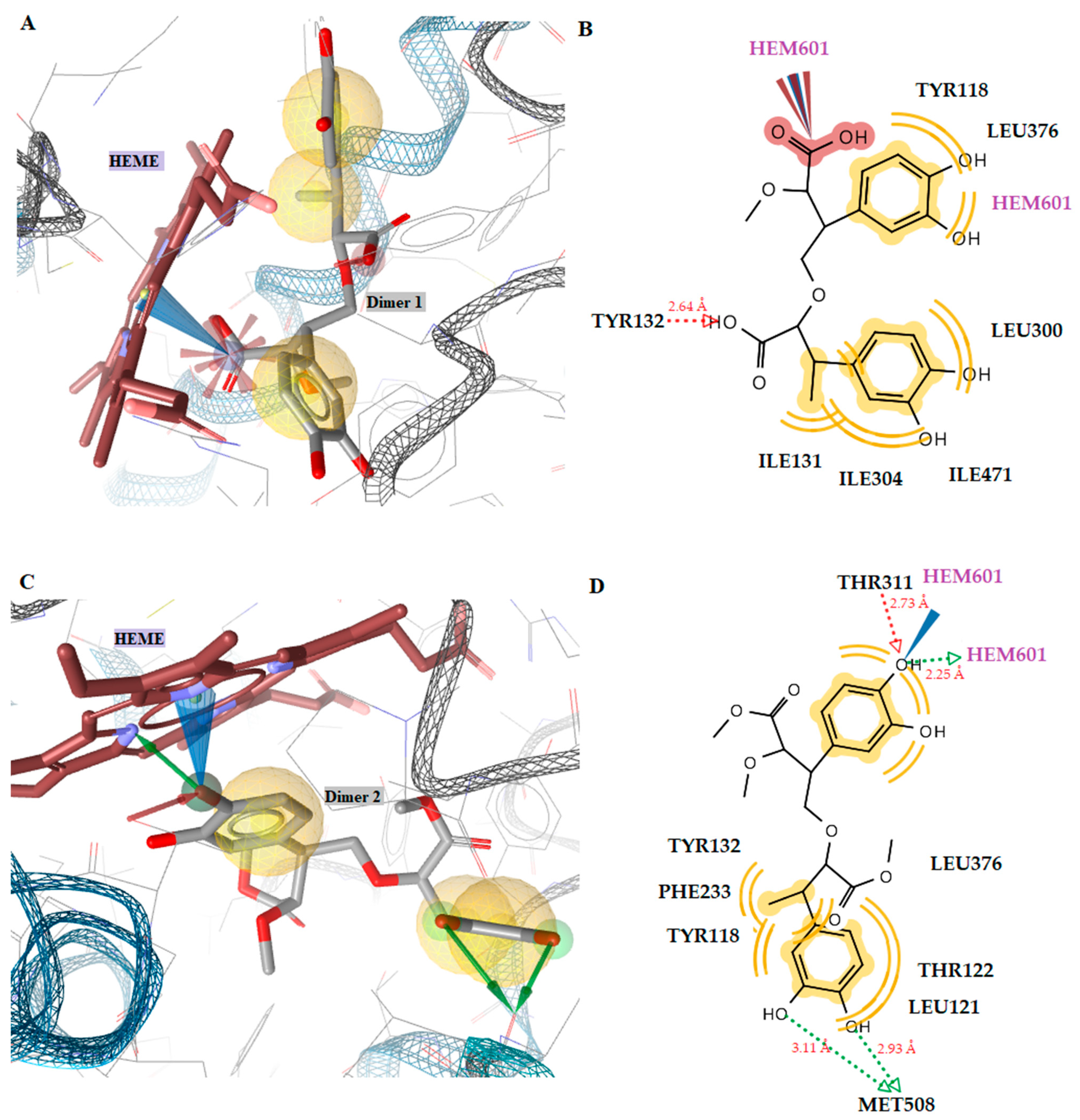
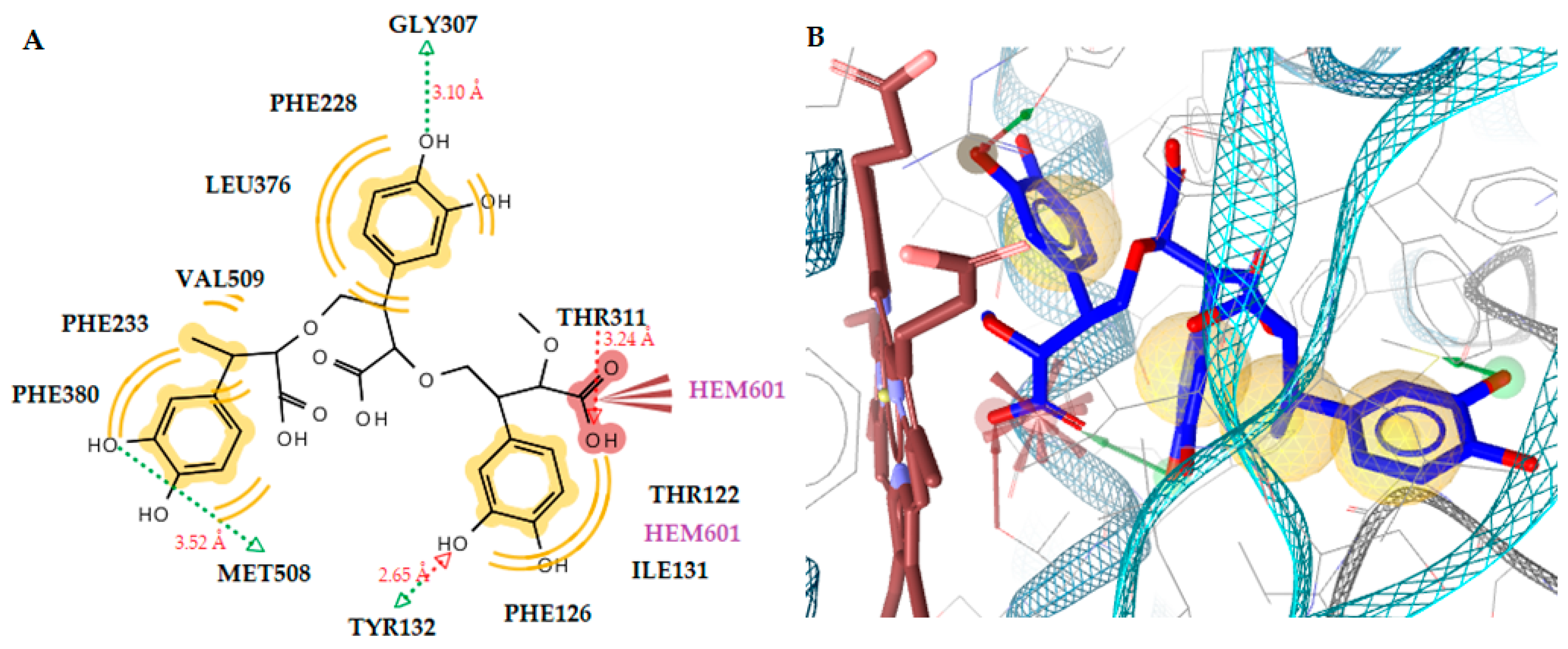
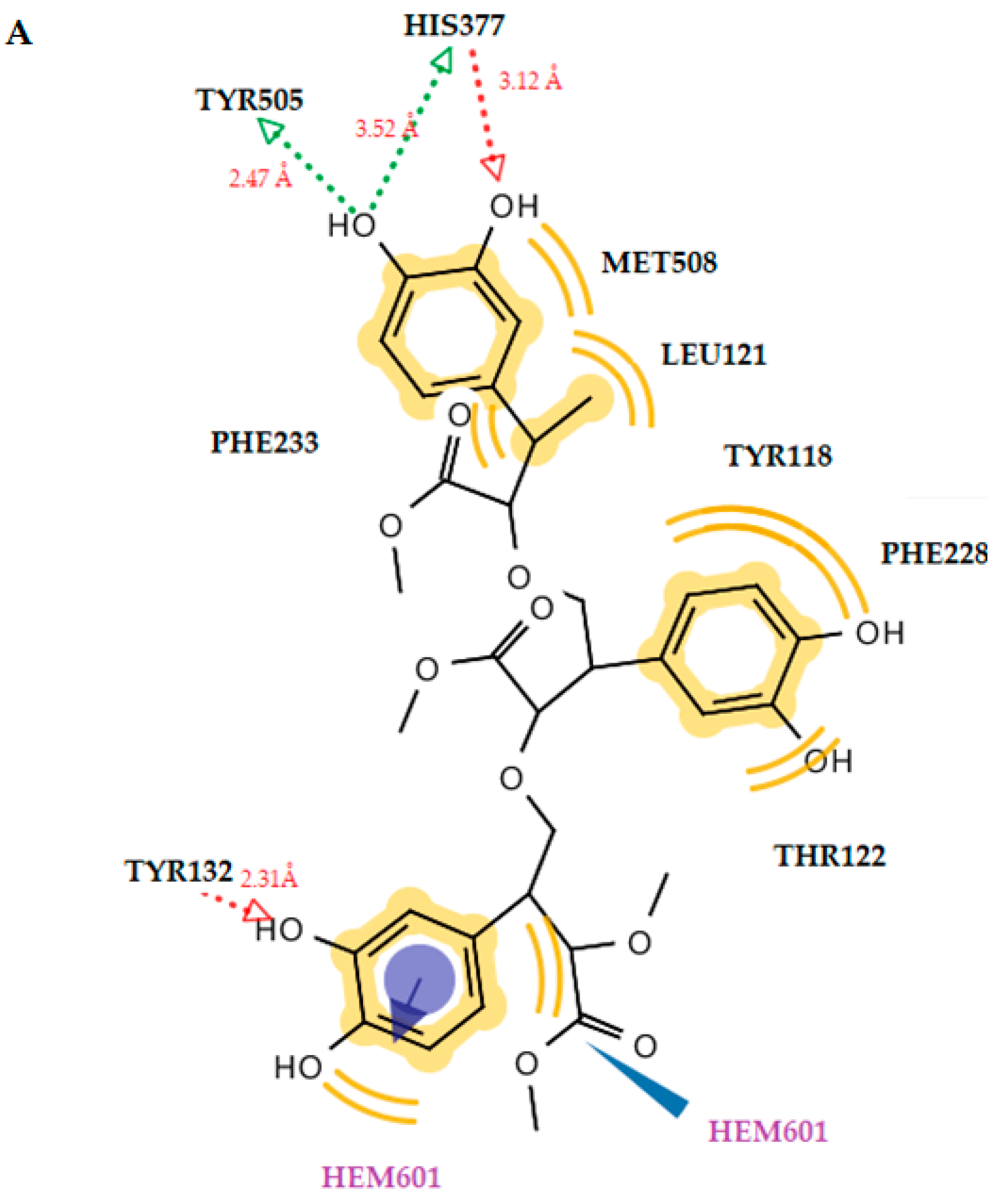
2.3.2. Docking to Antifungal Targets

All the molecules as dimers and trimers, as well as the reference drug ketoconazole, were docked to the lanosterol 14a-demethylase of C. albicans and DNA topoisomerase IV, and the results are presented in Table 5.
The docking results showed that molecules 1 and 2, whether are dimers or trimers, take place inside the active site of the enzyme interacting with the heme group of CYP51Ca. A detailed analysis of their binding showed that molecule 1, in the form of a dimer or trimer, interacts in the same way with the heme group, forming negative ionizable interactions throughout its carboxyl group (Figure 5, Figure 6 and Figure 7). It is noteworthy to mention that, in the case of the dimer of molecule 1, it interacts with the iron of the heme, making the molecule–enzyme complex more stable and justifying the increased free energy of binding of the trimer.
In the case of molecule 2, again the dimer suppresses the trimer because it interacts with the heme group, forming a stable hydrogen bond throughout its hydroxyl substituent (Figure 5, Figure 6 and Figure 7). The dimer form of molecule 2 also exhibited the best free energy of binding, with four hydrogen bonds between the hydroxyl substituents and the residues Thr311 (O···H, 2.73Å), Met508 (O···H, 3.93Å), Met508 (O···H, 3.11Å), and the heme group (O···H, 2.25Å). Hydrophobic interactions were also detected with the residues Tyr118, Leu121, Tyr122, Tyr132, Phe233, Thr311, and Leu376. An interaction was also observed between the heme group and the benzene ring of ketoconazole, which formed hydrophobic and aromatic interactions. However, molecule 2 in the dimer forms more and stronger interactions than ketoconazole and a more stable complex of ligand with enzyme. This is probably the reason why this molecule has better antifungal activity than ketoconazole.

2.4. Prediction of Toxicity

A prediction of the toxicity profile of the biopolymers was performed using the webserver ProTox-II.
This is a more rapid and less expensive process compared with in vitro testing in animals as well as in vitro testing in cell lines. On the other hand, it allows researchers to greatly reduce the number of animals used in experiments. Several online programs that use in silico methods to access toxicity are available. They predict the average lethal dose, carcinogenicity, mutagenicity, etc.
The PROTOX II program [31] predicts the average lethal dose (LD50) in rodents, classifying all chemical compounds into six GHS (Globally Harmonized System of Classification and Labeling of Chemicals) categories [32], depending on the toxicities of the compounds and the LD50 values. The LD50 values, given in mg/mL, are as follows:
  • Class I: fatal if swallowed (LD50 ≤ 5)
  • Class II: fatal if swallowed (5 < LD50 ≤ 50)
  • Class III: toxic if swallowed (50 < LD50 ≤ 300)
  • Class IV: harmful if swallowed (300 < LD50 ≤ 2000)
  • Class V: may be harmful if swallowed (2000 < LD50 ≤ 5000)
  • Class VI: non-toxic (LD50 > 5000)
The results of the prediction are presented in Table 6. It is worth noting that that the accuracy of the prediction increases as the confidence values increase. In particular, reliable estimates are considered to be more than 0.025.
According to the prediction using the Pro-Tox II webserver, the compounds were found to belong to categories IV and V, with LD50 values ranging from 2000 to 3500 mg/kg and 1000 mg/kg. They are safe to use (Table 6).

3. Materials and Methods

3.1. Plant Material

Plants were collected from various parts of Georgia province, namely Adjara, Kartli, the surroundings of Tbilisi, from June to September (2010–2020), depending on the climate. Each specimen was labeled, numbered, and annotated with the date of collection and the locality. The plants were identified by the TSMU I. Kutateladze Institute of Pharmacochemistry. Voucher specimens (whole plants) were deposited in the herbarium of the Department of Pharmacobotany of the TSMU Institute of Pharmacochemistry.

3.2. Methods

For extraction and isolation, plant material (roots or stems) was cut into small pieces, air-dried, and ground in a mill. Lipids, pigments, and low-molecular-weight compounds (monosaccharides, phenolics, etc.) were removed using Soxhlet extractions with chloroform, methanol, and acetone. The hot-water extraction of pretreated material was followed by dialysis, afforded by crude water-soluble polysaccharides. Further fractionation in a model 8200 stirred ultrafiltration cell (Millipore Corporation, Billerica, MA, USA) on a Biomax-500 ultrafiltration disc (500,000 NMWL) or a Filtron omega series membrane (1 M NMWL), as reported in [33], yielded water-soluble, high-molecular-weight (>500 kDa) preparations. The characterization of PDHPGA performed byUV, IR and NMR spectra of PDHPGA and are given in Supplementary materials Figures S1–S8.

3.3. Biological Evaluation

3.3.1. Evaluation of Antimicrobial Activity

Gram-negative bacteria (Escherichia coli (ATCC 35210) and Pseudomonas aeruginosa (ATCC 27853)) as well as Gram-positive bacteria (Staphylococcus aureus (ATCC 6538)) were used. The resistant strains used were methicillin-resistant S. aureus (IBRS MRSA 011), resistant E. coli (IBRS E003), and resistant P. aeruginosa (IBRS P001), which were obtained as described in Kartsev et al. [34]. The organisms were obtained from the Mycological Laboratory, Department of Plant Physiology, Institute for Biological Research ‘Siniša Stanković’, National Institute of Republic of Serbia, Belgrade, Serbia.

3.3.2. Evaluation of Antifungal Activity

The antifungal activities of all investigated samples were tested on strains obtained from the Mycological Laboratory, Department of Plant Physiology, Institute for Biological Research ‘Siniša Stanković’, National Institute of Republic of Serbia, Belgrade, Serbia. Aspergillus fumigatus (ATCC 1022), Aspergillus niger (ATCC 6275), Trichoderma viride (IAM 5061), Penicillium funiculosum (ATCC 36839), P. verrucosum var. cyclopium (P. v. c.) (food isolates), and Candida albicans (ATCC 10231) were used. The MIC and MBC/MFC concentrations were determined using the modified microdilution method, as previously reported [35,36]. The bacterial/fungal suspensions were adjusted with sterile saline to a concentration of 1.0 × 10⁵ CFU mL−1. The compounds were dissolved in a 30% EtOH solution (10 mg mL−1) and immediately added into a tryptic soy broth/malt broth medium (100 µL) with a bacterial/fungal inoculum. The microplates were incubated for 24 h at 37 ± 2 °C for bacterial species and for 72 h at 25 ± 2 °C (UE 500, Memmert) for fungi. The results are presented as MICs, MBCs, and MFCs. The MICs obtained from the susceptibility testing of various bacteria/fungi to the tested compounds were also determined using a colorimetric microbial viability assay based on the reduction in INT (p-iodonitrotetrazolium violet) [2-(4-iodophenyl)-3-(4-nitrphenyl)-5-phenyltetrazolium chloride], Merck KGaA, Darmstadt, Germany) color compared with a positive control for each microorganism strain. The MBCs and MFCs were determined using serial sub-cultivations of 2 µL into microtiter plates containing 100 µL of broth per well and further incubation for 24 h. The lowest concentration with no visible growth was defined as the MBC/MFC, indicating 99.5% killing of the original inoculum. The commercial antibiotics ampicillin and streptomycin and the fungicides bifonazole and ketoconazole were used as positive controls, while 30% EtOH was used as a negative control. All experiments were performed in duplicate.

3.4. Docking Studies

AutoDock 4.2® software (The Scripps Research Institute, La Jolla, CA, USA) was used for the docking simulation [37]. The binding energies (ΔG) of E. coli DNA GyrB, thymidylate kinase, E. coli primase, E. coli MurB, DNA topoIV, and CYP51 of C. albicans in complex with the inhibitors were generated using this molecular docking program. The X-ray crystal structure data of all enzymes that were used were obtained from the Protein Data Bank (PDB ID: 1KZN, AQGG, 1DDE, 2Q85, 1S16, and 5V5Z, respectively). All procedures were performed according to our previous paper [35].

4. Conclusions

In this work, six plants were collected from various parts of Georgia province, namely Adjara, Kartli, and the surroundings of Tbilisi, from June to September (2010–2020), depending on the climates from which the sugar-based catechol-containing biopolymers from SA, SC, SG, CO, BO, and AI were extracted. The antimicrobial activities of these biopolymers were evaluated. The study revealed that the antibacterial activities were moderate, and only biopolymers from three plants showed activities against all tested bacteria. Biopolymers from CO stems as well as SC and AI did not show any activities, except low activities against a resistant P. aeruginosa strain, which was the most resistant among the three resistant strains.
On the other hand, the antifungal activities were better compared to the antibacterial activities. Biopolymers from BO stems exhibited the best activities, with MIC/MFC values of 0.37–1.00 mg/mL and 0.75–1.5 mg/L, respectively, followed by those from SG stems. Biopolymers from SC and AI roots showed antifungal activities against all six fungi, in contrast to the antibacterial activities, while biopolymers from CO stems and SA roots showed antifungal activities against four fungi and one fungus, respectively. The sugar-based catechol-containing biopolymers from BO stems demonstrated the best activities among all tested biopolymers against T. viride, P. funiculosum, P. cyclpoium var verucosum, and C. albicans (MIC 0.37 mg/mL). In addition, biopolymers from SG stems were half as active against A. fumigatus and T. viride as ketoconazole. It is interesting to notice that biopolymers from all plant materials except for CO stems showed higher potency than ketoconazole against T. viride. The biopolymers from all plant materials showed better activities against C. albicans, one of the most dreadful fungi, compared to other fungal species. Thus, it can be concluded that plants from Georgia province can be good sources for natural antifungal agents. It should be noticed that, according to prediction results, these biopolymers are not toxic.

Supplementary Materials

The following supporting information can be downloaded at https://www.mdpi.com/article/10.3390/antibiotics12020285/s1, Table S1. Signals assignments of the 13C and 1H NMR spectra of PDHPGA-SA, PDHPGA-SC, PDHPGA-CO, PDHPGA-SG, PDHPGA-AI and PDHPGA-BO; Figure S1. UV spectra (H2O, λmax, nm) of PDHPGA-SA, PDHPGA-SC, PDHPGA-SG, PDHPGA-AI, PDHPGA-CO and PDHPGA-BO; Figure S2. The IR spectra of PDHPGA-SA, PDHPGA-SC, PDHPGA-SG, PDHPGA-AI, PDHPGA-CO and PDHPGA-BO; Figure S3. The 13C NMR spectrum of PDHPGA-SA, PDHPGA-SC and PDHPGA-CO; Figure S4. The 1H NMR spectrum of PDHPGA-SA, PDHPGA-SC and PDHPGA-CO; Figure S5. The HSQC spectrum of PDHPGA-SA, PDHPGA-SC and PDHPGA-CO; Figure S6. The 13C NMR spectrum methylated in carboxylic groups of PDHPGA-SG, PDHPGA-AI and PDHPGA-BO; Figure S7. The 1H NMR spectrum of methylated in carboxylic groups of PDHPGA-SG, PDHPGA-AI and PDHPGA-BO; Figure S8. The 2D DOSY spectrum of PDHPGA (R=H) and methylated in carboxylic groups of PDHPGA (R=CH3).

Author Contributions

Conceptualization, M.M. and V.B.; software, A.P.; methodology, V.B., L.G. and L.A.; investigation, A.Ć., M.S. and J.G.; data curation, M.M. and V.B.; visualization, A.P.; writing—original draft preparation, M.M. and A.G.; writing—review and editing, M.M. and A.G.; supervision, M.M.; project administration, V.B. All authors have read and agreed to the published version of the manuscript.

Funding

This research was funded by The Serbian Ministry of Education, Science, and Technological Development (project No. 451-03-68/2022-14/200007).

Institutional Review Board Statement

Not applicable.

Informed Consent Statement

Not applicable.

Data Availability Statement

Not applicable.

Conflicts of Interest

The authors declare no conflict of interest.

References

  1. Bibire, T.; Yilmaz, O.; Ghiciuc, C.M.; Bibire, N.; Danila, R. Biopolymers for Surgical Applications. Coatings 2022, 12, 211. [Google Scholar] [CrossRef]
  2. Varma, K.; Gopi, S. Biopolymers and their role in medicinal and pharmaceutical applications. In Biopolymers and Their Industrial Applications from Plant, Animal, and Marine Sources, to Functional Products; Elsevier: Amsterdam, The Netherlands, 2021; Chapter 7; pp. 175–191. [Google Scholar]
  3. Maghchiche, A. A Review: Application of Biopolymers in the Pharmaceutical Formulation. J. Adv. Bio-Pharm. Pharmacovigil. 2019, 1, 15–25. [Google Scholar]
  4. Fonseca, C.; Lima, M.; Sousa, A.F.; Silvestre, A.; Coelho, J.; Serra, A.C. Cinnamic acid derivatives as promising building blocks for advanced polymers: Synthesis, properties and applications. Polym. Chem. 2019, 10, 1696–1723. [Google Scholar] [CrossRef]
  5. Kaneko, D.; Kinugawa, S.; Matsumoto, K.; Kaneko, T. Terminally-catecholized hyper-branched polymers with high performance adhesive characteristics. Plant Biotechnol. 2010, 27, 293–296. [Google Scholar] [CrossRef]
  6. Kaneko, T.; Kaneko, D.; Wang, S. High-performance lignin-mimetic polyesters. Plant Biotechnol. 2010, 27, 243–250. [Google Scholar] [CrossRef]
  7. Razaviamri, S.; Wang, K.; Liu, B.; Lee, B.P. Catechol-Based Antimicrobial Polymers. Molecules 2021, 26, 559. [Google Scholar] [CrossRef]
  8. Chibane, L.B.; Degraeve, P.; Ferhout, H.; Bouajila, J.; Oulahal, N. Plant antimicrobial polyphenols as potential natural food preservatives. J. Sci. Food Agric. 2019, 99, 1457–1474. [Google Scholar] [CrossRef]
  9. Dresler, S.; Szymczak, G.; Wojcik, M. Comparison of some secondary metabolite content in the seventeen species of the Boraginaceae family. Pharm. Biol. 2017, 55, 691–695. [Google Scholar] [CrossRef]
  10. El-Shazly, A.; Wink, M. Diversity of Pyrrolizidine Alkaloids in the Boraginaceae Structures, Distribution, and Biological Properties. Diversity 2014, 6, 188–282. [Google Scholar] [CrossRef]
  11. Raphaelli, C.O.; Dannenberg, G.; Dalmazo, G.O.; Pereira, E.S.; Radünz, M.; Vizzotto, M.; Fiorentini, A.M.; Gandra, E.A.; Nora, L. Antibacterial and antioxidant properties of phenolic-rich extracts from apple (Malus domestica cv. Gala). Int. Food Res. J. 2019, 26, 1133–1142. [Google Scholar]
  12. Hussain, M.I.; Semreen, M.H.; Shanableh, A.; Khattak, M.N.K.; Ismail Saadoun, I.; Ahmady, I.M.; Muath Mousa, M.; Darwish, N.; Wameed Radeef, W.; Soliman, S.S.M. Phenolic Composition and Antimicrobial Activity of Different Emirati Date (Phoenix dactylifera L.) Pits: A Comparative Study. Plants 2019, 8, 497. [Google Scholar] [CrossRef] [PubMed]
  13. Dikpınar, T.; Süzgeç-Selçuk, S. Antimicrobial Activities of Medicinal Plants Containing Phenolic Compounds. J. Nat. Prod. 2020, 10, 514–534. [Google Scholar] [CrossRef]
  14. Salazar-Aranda, R.; Pérez-Lopez, L.A.; Lopez-Arroyo, J.; Alanic-Garza, B.A.; de Torres, N.W. Antimicrobial and Antioxidant Activities of Plants from Northeast of Mexico. Evid.-Based Complement. Altern. Med. 2011, 2011, 1–6. [Google Scholar]
  15. Gyawali, R.; Ibrahim, S.A. Natural products as antimicrobial agents. Food Control 2014, 46, 412–429. [Google Scholar] [CrossRef]
  16. Zambrano, C.; Kerekes, E.B.; Kotogán, A.; Papp, T.; Vágvölgyi, C.; Krisch, J.; Takó, M. Antimicrobial activity of grape, apple and pitahaya residue extracts after carbohydrase treatment against food-related bacteria. LWT Food Sci. Technol. 2019, 100, 416–425. [Google Scholar] [CrossRef]
  17. Takó, M.; Kerekes, E.B.; Zambrano, C.; Kotogán, A.; Papp, T.; Krisch, I.; Vágvölgyi, C. Plant Phenolics and Phenolic-Enriched Extracts as Antimicrobial Agents against Food-Contaminating Microorganisms. Antioxidants 2020, 9, 165. [Google Scholar] [CrossRef]
  18. Kabir, F.; Sultana, M.S.; Kurnianta, H. Antimicrobial activities of grape (Vitis vinifera L.) pomace polyphenols as a source of naturally occurring bioactive components. Afr. J. Biotechnol. 2015, 14, 2157–2161. [Google Scholar]
  19. Silván, J.M.; Mingo, E.; Hidalgo, M.; De Pascual-Teresa, S.; Carrascosa, A.V.; Martinez-Rodriguez, A.J. Antibacterial activity of a grape seed extract and its fractions against Campylobacter spp. Food Control 2013, 29, 25–31. [Google Scholar] [CrossRef]
  20. Levy, J.; Boyer, R.R.; Neilson, A.P.; O’Keefe, S.F.; Chu, H.S.S.; Williams, R.C.; Dorenkott, M.R.; Goodrich, K.M. Evaluation of peanut skin and grape seed extracts to inhibit growth of foodborne pathogens. Food Sci. Nutr. 2017, 5, 1130–1138. [Google Scholar] [CrossRef]
  21. Tenore, G.C.; Novellino, E.; Basile, A. Nutraceutical potential and antioxidant benefits of red pitaya (Hylocereus polyrhizus) extracts. J. Funct. Foods 2012, 4, 129–136. [Google Scholar] [CrossRef]
  22. Yong, Y.Y.; Dykes, G.; Lee, S.M.; Choo, W.S. Comparative study of betacyanin profile and antimicrobial activity of red pitahaya (Hylocereus polyrhizus) and red spinach (Amaranthus dubius). Plant Food. Hum. Nutr. 2017, 72, 41–47. [Google Scholar] [CrossRef]
  23. Merlani, M.; Barbakadze, V.; Amiranashvili, L.; Gogilashvili, L.; Poroikov, V.; Petrou, A.; Geronikaki, A.; Ćirić, A.; Glamočlija, J.; Soković, M. New Caffeic Acid Derivatives as Antimicrobial Agents: Design, Synthesis, Evaluation and Docking. Curr. Top. Med. Chem. 2019, 19, 292–304. [Google Scholar] [CrossRef] [PubMed]
  24. Merlani, M.; Barbakadze, V.; Amiranashvili, L.; Gogilashvili, L.; Petrou, A.; Geronikaki, A.; Ćirić, A.; Glamočlija, J.; Soković, M. Caffeic and 3-(3,4-dihydroxyphenyl)glyceric acid derivatives as antimicrobial agent: Biological evaluation and molecular docking studies. SAR QSAR Env. Res. 2022, 33, 307–321. [Google Scholar] [CrossRef] [PubMed]
  25. Kepa, M.; MiklasiNska-Majdanik, M.; Wojtyczka, R.D.; Idzik, D.; Korzeniowski, K.; Smolen-Dzirba, I.; Wdsik, T.J. Antimicrobial Potential of Caffeic Acid against Staphylococcus aureus Clinical Strains. BioMed Res. Inter. 2018, 2018, 7413504. [Google Scholar] [CrossRef] [PubMed] [Green Version]
  26. Barbakadze, V.; Gogilashvili, L.; Amiranashvili, L.; Merlani, M.; Getia, M.; Gogilashvili, A.; Salgado, A.; Chankvetadze, B. Biologically active sugar-based biopolyether poly [3-(3,4-dihydroxyphenyl)glyceric acid] from the stems and roots of Paracynoglossum imeretinum (Kusn.) M.Pop. Bull. Georg. Natl. Acad. Sci. 2022, 16, 110–115. [Google Scholar]
  27. Merlani, M.; Scheibel, D.M.; Barbakadze, V.; Gogilashvili, L.; Amiranashvili, L.; Geronikaki, A.; Catania, V.; Schillaci, D.; Gallo, G.; Gitsov, I. Enzymatic synthesis and antimicrobial activity of oligomer analogues of medicinal biopolymers from comfrey and their species of the Boraginaceae Family. Pharmaceutics 2022, 14, 115. [Google Scholar] [CrossRef]
  28. Barbakadze, V.; Gogilashvili, L.; Amiranashvili, L.; Merlani, M.; Li, S.P.; Chankvetadze, B. Fractionation of Biologically Active Poly[3-(3,4-Dihydroxyphenyl)Glyceric Acid] Preparation from Symphytum asperum, Simultaneous Determination of Molecular Weights and Contents of its Fractions Using HPSEC-MALLS-RID. Bull. Georg. Natl. Acad. Sci. 2021, 15, 69–75. [Google Scholar]
  29. Liu, B.; Zhou, C.; Zhang, Z.; Roland, J.D.; Lee, B.P. Antimicrobial property of halogenated catechols. Chem. Eng. J. 2021, 403, 126340. [Google Scholar] [CrossRef]
  30. Benson, T.E.; Walsh, C.T.; Massey, V. Kinetic characterization of wild-type and S229A mutant MurB: Evidence for the role of Ser 229 as a general acid. Biochemistry 1997, 36, 796–805. [Google Scholar] [CrossRef]
  31. Banerjee, P.; Eckert, A.O.; Schrey, A.K.; Preissner, R. ProTox-II: A webserver for the prediction of toxicity of chemicals. Nucleic Acids Res. 2018, 46, W257–W263. [Google Scholar] [CrossRef]
  32. GHS-UNECE. Available online: http://www.unece.org/trans/danger/publi/ghs/ghs_welcome_e.html (accessed on 10 November 2022).
  33. Barthomeuf, C.M.; Debiton, E.; Barbakadze, V.V.; Kemertelidze, E.P. Evaluation of the dietetic and therapeutic potential of a high molecular weight hydroxicinnamate-derived polymer from Symphytum asperum Lepech. regarding its antioxidant, antilipoperoxidant, antiinflammatory, and cytotoxic properties. J. Agric. Food Chem. 2001, 49, 3942–3946. [Google Scholar] [CrossRef]
  34. Kartsev, V.; Lichitsky, B.; Geronikaki, A.; Petrou, A.; Smiljkovic, M.; Kostic, M.; Radanovic, O.; Soković, M. Design, synthesis and antimicrobial activity of usnic acid derivatives. Med. Chem. Comm. 2018, 9, 870–882. [Google Scholar] [CrossRef]
  35. Fesatidou, M.; Zagaliotis, P.; Camoutsis, C.; Petrou, A.; Eleftheriou, P.; Tratrat, C.; Haroun, M.; Geronikaki, A.; Ciric, A.; Sokovic, M. 5-Adamantan thiadiazole-based thiazolidinones as antimicrobial agents. Design, synthesis, molecular docking and evaluation. Bioorg. Med. Chem. 2018, 26, 4664–4676. [Google Scholar] [PubMed]
  36. Zubenko, A.; Kartsev, V.; Petrou, A.; Geronikaki, A.; Ivanov, M.; Glamočlija, J.; Sokovic, M.; Divaeva, L.; Morkovnik, A.; Klimenko, A. Experimental and In Silico Evaluation of New Heteroaryl Benzothiazole Derivatives as Antimicrobial Agents. Antibiotics 2022, 11, 1654. [Google Scholar] [CrossRef] [PubMed]
  37. Morris, G.M.; Huey, R.; Lindstrom, W.; Sanner, M.F.; Belew, R.K.; Goodsell, D.S.; Olson, A.J. AutoDock4 and AutoDockTools4: Automated docking with selective receptor flexibility. J. Comput. Chem. 2009, 30, 2785–2791. [Google Scholar] [CrossRef] [PubMed] [Green Version]
Figure 1. Poly[3-(3,4-dihydroxyphenyl)glyceric acid] (PDHPGA) (1) and poly[2-methoxycarbonyl-3-(3,4-dihydroxyphenyl)oxirane] (PMDHPO) (2).
Antibiotics 12 00285 g001
Figure 2. Solid-state 13C NMR spectrum of PMDHPO. Solid-state 13C{1H} CP/MAS NMR spectrum, recorded at 9.4 T employing magic-angle spinning at 14.0 kHz and a CP time of 3.0 ms, showing the repeating unit δ (13C) of PMDHPO (ppm).
Antibiotics 12 00285 g002
Figure 3. (A) A 2D diagram of dimer molecule 1 in E. coli MurB. (B) Docked pose of dimer molecule 1 in E. coli MurB. (C) A 2D diagram of dimer molecule 2 in E. coli MurB. (D) Docked pose of dimer molecule 2 in E. coli MurB. Red and green dotted lines show the hydrogen bonds. Yellow spheres represent hydrophobic interactions.
Antibiotics 12 00285 g003aAntibiotics 12 00285 g003b
Figure 4. (A) Docked pose of trimer molecule 1 in E. coli MurB. (B) A 2D diagram of trimer molecule 1 in E. coli MurB. (C) Docked pose of trimer molecule 2 in E. coli MurB. (D) A 2D diagram of trimer molecule 2 in E. coli MurB. Red and green dotted lines show the hydrogen bonds. Yellow spheres represent hydrophobic interactions.
Antibiotics 12 00285 g004
Figure 5. (A) Docked pose of dimer molecule 1 in lanosterol 14alpha-demethylase of C. albicans (CYP51ca). (B) A 2D diagram of dimer molecule 1 in CYP51ca. (C) Docked pose of dimer molecule 2 in CYP51ca. (D) A 2D diagram of dimer molecule 2 in CYP51ca. Red and green dotted lines show the hydrogen bonds. Yellow spheres represent hydrophobic interactions.
Antibiotics 12 00285 g005
Figure 6. (A) A 2D diagram of trimer molecule 1 in CYP51ca. (B) Docked pose of trimer molecule 1 in CYP51ca. Red and green dotted lines show the hydrogen bonds. Yellow spheres represent hydrophobic interactions, and blue arrows represent aromatic interactions.
Antibiotics 12 00285 g006
Figure 7. (A) A 2D diagram of trimer molecule 2 in CYP51ca. (B) Docked pose of trimer molecule 2 in CYP51ca. Red and green dotted lines show the hydrogen bonds. Yellow spheres represent hydrophobic interactions, and blue arrows represent aromatic interactions.
Antibiotics 12 00285 g007aAntibiotics 12 00285 g007b
Table 1. The signal assignments of the solid-state 13C{1H} CP/MAS NMR spectrum of PMDHPGA (δ, ppm).
С Atom No.13C Chemical
Shift, δC, ppm
172
278
1′174
(−OCH3)54
1″130
2″, 5″, 6″118
3″, 4″143
Table 2. In vitro antibacterial evaluation of PDHPGA and PMDHPO from stems and roots of BO, AI, CO, SA, SC, and SG (mg/mL).
CompoundsS. a.MRSAE. coliRez. E. coliP. a.Rez. P. a.
PDHPGA and PMDHPO of BO (stems)MIC1.501.501.001.501.000.75
MBC3.003.001.503.001.501.50
PDHPGA and PMDHPO of AI (roots)MIC--3.00--3.00
MBC--6.00--6.00
PDHPGA of CO (stems)MIC-----3.00
MBC-----6.00
PDHPGA of SA (roots)MIC6.006.006.006.006.003.00
MBC9.009.009.009.0012.006.00
PDHPGA of
SC (roots)
MIC-----3.00
MBC-----6.00
PDHPGA and PMDHPO of SG (stems)MIC0.751.000.751.501.000.75
MBC1.501.501.503.001.501.50
AmpicillinMIC0.10-0.150.200.300.20
MBC0.15-0.20-0.50-
StreptomycinMIC0.100.100.100.050.100.10
MBC0.20-0.200.100.200.20
S. a.S. aureus, MRSA—methicillin-resistant S. aureus, E. c.E. coli, res. E. c.—resistant E. coli, P. a.P. aeruginosa, res. P. a.—resistant P. aeruginosa.
Table 3. In vitro antifungal evaluation of PDHPGA and PMDHPO from stems and roots of BO, AI, CO, SA, SC, and SG (mg/mL).
CompoundsA. fu.A. n.T. v.P. f.P. v. c.C. a.
PDHPGA and PMDHPO of BO (stems)MIC0.751.000.370.370.370.37
MFC1.501.500.750.750.750.75
PDHPGA and PMDHPO of AI
(roots)
MIC1.502.250.370.751.100.19
MFC3.003.000.751.503.000.37
PDHPGA of CO (stems) MIC0.750.560.190.370.750.37
MFC1.500.750.370.751.500.75
PDHPGA of SA
(roots)
MIC3.00-0.752.252.250.09
MFC4.50-1.504.504.500.19
PDHPGA of SC (roots)MIC0.370.370.190.750.750.19
MFC0.750.750.371.501.500.37
PDHPGA and PMDHPO of SG
(stems)
MIC0.370.560.090.560.750.14
MFC0.750.750.190.751.500.19
KetoconazoleMIC0.200.201.000.200.201.00
MFC0.500.501.500.500.302.00
BifonazoleMIC0.150.150.150.200.100.20
MFC0.200.200.200.250.200.30
A. f.A. fumigatus, A. n.A. niger, T. v.T. viride, P. f.P. funiculosum, P. v. c.P. cyclpoium var verucosum, C. a.Candida albicans.
Table 4. Molecular docking binding affinities to antibacterial targets.
NoEst. Binding Energy (kcal/mol)Residues Involved in Hydrogen BondsResidues Involved in Hydrophobic Interactions
E. coli Gyrase
1KZN
S. aureus Thymidylate Kinase
4QGG
E. coli Primase
1DDE
E. coli MurB 2Q85
1−4.55−2.56−2.41−5.16Ser229 (O···H, 2.65Å)Leu218, Ala227, Val291
1 (dimer)−4.31−2.67-−7.80Ser229 (O···H, 2.78Å), Glu325 (O···H, 2.82Å)Ile110, Leu218, Ile122
1 (trimer)−2.16−9.81Ser50 (O···H, 2.54Å), Ser116 (O···H, 2.67Å), Ser229 (O···H, 2.98Å), Ala227 (O···H, 3.21Å)Leu218, Ile122, Val291
2−4.12−2.46−2.57−5.89Ser229 (O···H, 2.55Å)Leu218, Ile122
2 (dimer)−3.57−1.28−1.06−6.73Pro219 (O···H, 2.71Å), Glu325 (O···H, 2.56Å)Leu218
2 (trimer)−1.67--−9.16Ser229 (O···H, 3.15Å), Ser229 (O···H, 3.59Å), Ser229 (O···H, 3.24Å)Leu218, Ile122, Ala227, Val291
Table 5. Molecular docking binding affinities to antifungal targets.
N/NEst. Binding Energy (kcal/mol)Residues Involved in Hydrogen BondsResidues
Involved in Hydrophobic Interactions
Interactions with HEM601
DNA TopoIV
1S16
CYP51 of C. albicans
5V5Z
1−1.52−7.96Tyr132 (O···H, 2.64Å)Tyr118, Ile131, Tyr132, Leu300, Leu376Negative ionizable, Hydrophobic
1 (dimer)-−9.55Tyr132 (O···H, 2.64Å),Tyr118, Ile131, Tyr132, Leu300, Leu304, Leu376, Ile407Negative ionizable, Hydrophobic, Fe-binding
1 (trimer)-−9.13Tyr132 (O···H, 2.65Å), Gly307 (O···H, 3.10Å),
Thr311 (O···H, 3.24Å),
Met508 (O···H, 3.52Å)
Ile131, Thr122, Phe126, Phe380, Phe233, Phe228, Leu376, Val509, Met508Negative ionizable, Hydrophobic
2−1.78−9.26Tyr132 (O···H, 2.67Å)Tyr118, Leu121, Tyr122, Tyr132, Phe233, Thr311, Leu376Negative ionizable, Hydrophobic
2 (dimer)−1.78−11.35Thr311 (O···H, 2.73Å), Met508 (O···H, 3.93Å),
Met508 (O···H, 3.11Å),
Hem601 (O···H, 2.25Å)
Tyr118, Leu121, Tyr122, Tyr132, Phe233, Thr311, Leu376Hydrophobic, H-Bond, Fe-binding
2 (trimer)-−11.10Tyr132 (O···H, 2.31Å), His377 (O···H, 3.12Å), His377 (O···H, 3.52Å),
Tyr505 (O···H, 2.47Å)
Met508, Leu121, Tyr118, Phe228, Thr122, Phe233Hydrophobic, aromatic, Fe-binding
ketoconazole-−8.23Tyr64 (O···H, 2.51Å)Tyr118, Ile131, Tyr132, Leu300, Ile304, Leu376, Met508Hydrophobic, aromatic
Table 6. Prediction of toxicity.
NoPredicted LD50 (mg/kg)Predicted Toxicity ClassHepatotoxicityCarcinogenicityImmunotoxicityMutagenicityCytotoxicity
12000IVInactive (0.6)Inactive (0.56)Inactive (0.54)Inactive (0.94)Inactive (0.95)
22000IVInactive (0.57)Inactive (0.56)Inactive (0.62)Inactive (0.91)Inactive (0.92)
1 (dimer)3500VInactive (0.79)Inactive (0.73)Inactive (0.58)Inactive (0.77)Inactive (0.86)
2 (dimer)3500VInactive (0.80)Inactive (0.74)Inactive (0.50)Inactive (0.78)Inactive (0.84)
Numbers in brackets indicate possibilities.
Disclaimer/Publisher’s Note: The statements, opinions and data contained in all publications are solely those of the individual author(s) and contributor(s) and not of MDPI and/or the editor(s). MDPI and/or the editor(s) disclaim responsibility for any injury to people or property resulting from any ideas, methods, instructions or products referred to in the content.

