Article

Antimicrobial Resistance and Pathogenicity of Aliarcobacter butzleri Isolated from Poultry Meat

by
Maria Gabriela Xavier de Oliveira
1,
Marcos Paulo Vieira Cunha
1,
Luisa Zanolli Moreno
1,
André Becker Simões Saidenberg
1,
Mônica Aparecida Midolli Vieira
2,
Tânia Aparecida Tardelli Gomes
2,
Andrea Micke Moreno
1 and
Terezinha Knöbl
1,*
1
Faculdade de Medicina Veterinária e Zootecnia, Universidade de São Paulo, USP, Butantã, São Paulo 05508-270, SP, Brazil
2
Departamento de Microbiologia, Imunologia e Parasitologia, Escola Paulista de Medicina–Universidade Federal Paulista UNIFESP, São Paulo 04023-062, SP, Brazil
*
Author to whom correspondence should be addressed.
Antibiotics 2023, 12(2), 282; https://doi.org/10.3390/antibiotics12020282
Submission received: 26 December 2022 / Revised: 17 January 2023 / Accepted: 25 January 2023 / Published: 1 February 2023
(This article belongs to the Special Issue Diversity of Antimicrobial Resistance Genes in Clinical Settings)

Abstract

:
Aliarcobacter butzleri (A. butzleri) is an emergent zoonotic food-related pathogen that can be transmitted through the consumption of poultry meat. Data regarding the pathogenicity and resistance of A. butzleri are still scarce, and the presence of virulent MDR strains of this zoonotic pathogen in poultry meat is an issue of particular concern to public health. This study aimed to characterize the pathogenicity and antimicrobial resistance profiles of A. butzleri strains isolated from poultry meat sold at retail markets in São Paulo, Brazil. The minimum inhibitory concentrations of 27 strains were determined using the broth microdilution method. The results showed that 77.7% of the isolates were resistant to clindamycin, 62.9% to florfenicol, 59.2% to nalidixic acid, 11.1% to azithromycin, 7.4% to ciprofloxacin and telithromycin, and 3.7% to erythromycin and tetracycline, although all were susceptible to gentamicin. Moreover, 55.5% of the virulent isolates were also multidrug-resistant (MDR). Three strains were selected for pathogenicity tests in vitro and in vivo. The tested strains expressed weak/moderate biofilm production and showed a diffuse adhesion pattern (3 h) in HeLa cells and toxicity in Vero cells (24 h). Experimental inoculation in 11-week-old chicks induced a transitory inflammatory enteritis. Intestinal hemorrhage and destruction of the intestinal crypts were observed in the rabbit ileal loop test. Considering the fact that Brazil is a major exporter of poultry meat, the data from this study point to the need of improvement of the diagnostic tools, as well as of the adoption of surveillance guidelines and more specific control strategies to ensure food safety, reducing the presence of pathogenic MDR strains in broilers.

1. Introduction

The genus Aliarcobacter is a member of the Campylobacteraceae family, and infection with the species A. butzleri is associated with abdominal cramps, gastroenteritis, severe diarrhea, blood in stools, and sepsis in humans, constituting serious hazards to human health [1,2]. Infection with these bacteria is commonly related to the ingestion of contaminated water, vegetables, seafood, meat, raw milk, and dairy products [3,4,5,6,7,8,9]. However, the main cause of food-born outbreaks has been attributed to the contamination of poultry meat by A. butzleri [6,10,11].
Arlicobacteriosis is a worldwide disease, and although there are few reports of human infection, the disease has already been described in several European countries, such as Italy, Spain, French, United Kingdom, Germany, Belgium, Denmark, as well South Africa, Japan, Thailand, and Hong Kong [1,12]. In Latin America, A. butzleri has been isolated from humans, animals, and food sources, mainly in Chile and Brazil [5,6,13].
The real incidence rate of A. butzleri is probably underestimated due the lack of diagnostics and misidentification, but some reports point this species as the fourth most frequent among Campylobacter-like isolates from human stool [2,12]. Besides that, the prevalence of MDR strains of Aliarcobacter is much higher than that of Campylobacter [14,15]. According to Son et al. [14], the resistance levels of A. butzleri range from 72.9 to 80.9%, and high rates of resistance to penicillins, aminopenicillins (ampicillin), and cephalosporins have been reported in bacterial isolates from humans and companion and production animals [14].
The pathogenicity of Aliarcobacter is still poorly understood, but the ability of some species to produce inflammatory reactions, enteritis, and septicemia have been investigated in vitro (cell cultures) and in vivo (laboratory animals) [1]. A. buztleri binds and invade Caco-2 cells and promotes cytotoxic effects on Vero cells. In vivo, A. buztleri may colonize the liver, kidney, ileum, and brain of rodents, which develop diarrhea and changes in hematological parameters, which are dose-dependent [16].
Nine putative virulence genes have been employed in molecular approaches for the characterization of the potentially pathogenic species of Aliarcobacter: cadF (fibronectin protein), ciaB (invasion), cj1349 (fibronectin protein), pldA (lysis of erythrocytes), mviN (peptidoglycan biosynthesis), tlyA (hemolysin), hecB (hemolysin protein activation), hecA (hemagglutinin filaments, which are involved in attack, aggregation, and cell death), and irgA (regulator of protein for iron acquisition) [6,16]. In a previous study, we identified A. butzleri in 11.66% of the poultry meat marketed in São Paulo, Brazil, and 100% of the strains were positive for ciaB, cj1349, mviN. The prevalence of pldA, tlyA, and cadF was 97.2%, 94.2%, and 74.2%, respectively. The prevalence of the genes hecA, irgA, and hecB ranged between 40 and 45.7% [6]. Brazil is a major global exporter of broilers. Studies focusing on the antimicrobial susceptibility profiles of Aliarcobacter spp. in chicken meat are rare, even though the presence of virulent and MDR strains of these bacteria represents an important public health issue [6]. Moreover, the different methodologies employed to determine the susceptibility to antimicrobials have impeded the interpretation and effective comparison of rates of resistance among different countries [1]. The classical disk diffusion method for assessing antimicrobial resistance is simple and commonly used, but the diameters of the inhibition zones are often difficult to discriminate with precision. In contrast, the broth microdilution method enables minimum inhibitory concentration (MIC) values to be determined accurately and reproducibly, and a standard protocol for antimicrobial susceptibility testing of Aliarcobacter spp. using this method has been proposed [15].
In light of the above, the present study aimed to establish the antimicrobial resistance profiles of A. butzleri strains isolated from poultry retail meat sold in markets in São Paulo city (Brazil), by determining the MIC values using the broth microdilution method. Additionally, three strains were selected for the analysis of pathogenicity in vitro and in vivo.

2. Results

The MIC values obtained for the 27 A. butzleri isolates revealed that 22 showed resistance to at least one antimicrobial, with 77.2% (21/27) being resistant to clindamycin, 62.9% (17/27) to florfenicol, 59.2% to nalidixic acid (16/27), 11.1% (3/27) to azithromycin, 7.4% (2/27) to ciprofloxacin, and 3.7% (1/27) to erythromycin, telithromycin, and tetracycline (Table 1). All the tested isolates were susceptible to gentamicin. The results of MIC50 and MIC90 are presented in Supplementary Material Table S1.
Ten different antimicrobial resistance profiles (R1–R10) were identified among the isolates of A. butzleri (Table 1). The antimicrobial resistance profiles revealed that 55.5% (15/27) of the virulent isolates of Aliarcobacter were MDR (profiles R6 to R10, Table 1).
The phenotypical tests of AB165 and AB170 showed a weak biofilm formation, with OD of 0.695 ± 0.055 and 0.679 ± 0.143, respectively. However, these strains presented moderate adherence to HeLa cells in 3 h. (Figure 1A). The strain AB167 presented a moderate biofilm formation, with OD of 0.926 ± 0.027, but did not adhere to HeLa cells in 3 h. The OD of the positive control, enteroaggregative E. coli 042, was 1.002 ± 0.076.
All strains were positive in the toxicity tests, with observable effects in the form of vacuolation and destruction of Vero cells (Figure 1B).
The chicken inoculation of A. butzleri promoted a mild diarrhea (Figure 2A) between the second and the fourth days after the challenge), with the presence of an orange content and gas in the cecum (Figure 2B) and hyperemia of the intestine. The strains were recovered from feces on the 2nd., 3rd,. and 4th. day after the challenge, but the culture was negative after the 5th. day. These changes were not observed in birds of the non-challenged group.
The microscopic changes showed a moderate inflammation of the intestinal crypts, with focal dilation and thickening of the villi and inflammatory infiltrate with lymphocytes, heterophils, plasmocytes, and macrophages in the lamina propria (Figure 3). Flattening of villi, with a decrease in length and tissue destruction, was also observed, as well as a slight increase in mitosis figures in the crypt. In the lumen, there were multiple bacterial colonies (bacilli).
The rabbit ileal loops model showed that the strains AB165, AB167, and AB170 induced intestinal hemorrhage (Figure 4A),12 hours after the inoculation. The microscopic analysis of these loops showed the presence of erythrocytes and cell debris in the lumen (Figure 5A). There were some submucosal changes, with discrete multifocal ectasia of the lymphatic vessels. Scanning electron microscopy showed the presence of some bacilli in the extracellular matrix, and the villi were destroyed (Figure 6A). None of these changes were observed in the negative control (Figure 4B, Figure 5B, Figure 6B).

3. Discussion

The unrestricted use of antibiotics in both human and animal populations has contributed to a global increase in infections by MDR pathogens. However, information on the antimicrobial resistance patterns of A. butzleri. is still limited [17,18,19,20,21]. In addition, the lack of standardization in the methodologies employed and in the interpretation of the applied criteria has generated challenges in analyzing the resistance data, resulting in misleading comparisons [1,15].
According to the antimicrobial susceptibility data reported herein, the studied Aliarcobacter isolates showed the highest resistance percentage (77.2%) to the lincosamide class of antibiotics (clindamycin) and a slightly lower resistance percentage to phenicol (florfenicol) and quinolone (nalidixic acid) antibiotics, corresponding to 62.9% and 59.2%, respectively. A percentage (81.5%; 22/27) of A. butzleri isolates were resistant to at least one antimicrobial, and a high percentage (55.5%) were MDR.
Treatment of aliarcobacteriosis with a course of antibiotics is recommended for patients presenting severe clinical signs but not for those with only mild symptoms [22]. Since different species of the genus Aliarcobacter are closely related to those of Campylobacter, the drugs of choice are fluoroquinolones (such as ciprofloxacin and enrofloxacin), macrolides, and, less commonly, tetracyclines and aminoglycosides [23]. In a study involving 174 isolates of Aliarcobacter obtained from broiler carcasses collected from a poultry processing plant in the United States, Son et al. [14] found that the A. butzleri isolates were highly resistant to clindamycin (90%), followed by azithromycin (81.4%) and nalidixic acid (23.6%). Our results also point to clindamycin as the least effective drug, with 77.2% of resistant strains, although the 59.2% resistance to nalidixic acid reported herein exceeds that established in the American study, while the resistance percentage to azithromycin (7.5%) is substantially lower.
According to Van den Abeele et al. [24], 13% of the A. butzleri strains isolated from Belgian patients with gastroenteritis showed resistance to ciprofloxacin, with MIC90 values >32 mg/L. Furthermore, Shah et al. [25] reported that strains of A. butzleri were resistant to ampicillin (56%), cefotaxime (33%), and ciprofloxacin (33%) but susceptible to gentamicin and enrofloxacin. In contrast, our data revealed that only 7.4% of Aliarcobacter isolates from broiler meat were resistant to ciprofloxacin, with MIC90 values of 0.5 µg/mL (Table S1). Unlike other countries, Brazil has never employed fluoroquinolones as growth promoters in poultry, limiting their use to the therapeutic treatment of specific diseases, for example, paratyphoid Salmonella. This restriction may account for the epidemiological differences found between the data obtained in the present study and those of reports from other countries.
There is evidence to support the hypothesis that resistance patterns in production animals are similar to those found in human isolates, and, for this reason, resistance to fluoroquinolones is of significant concern, since drugs of this class are used in therapies for both humans and animals. In this context, Van Boven et al. [26] collected cloacal swabs from broilers that had received enrofloxacin and observed that this treatment led to the rapid selection of resistant isolates of Campylobacter jejuni, all of which exhibited high frequencies of mutations in the gyrA gene. Interestingly, Van den Abeele et al. [24] carried out genomic analyses of ciprofloxacin-resistant strains of A. cryaerophilus isolated from stool samples of patients with gastroenteritis and established that all carried a mutation at position 254 of gyrA, thereby pointing to a mechanism of acquired resistance. In a recent study of A. butzleri strains isolated from a variety of animal, vegetable, dairy, and aquatic sources, Isidro et al. [9] confirmed that all ciprofloxacin- and levofloxacin-resistant isolates presented the same mutation in gyrA. These authors also reported the presence of the blaOXA-15-like gene in the strains [9].
Although the present study showed that the isolates of A. butzleri from poultry meat were sensitive to ciprofloxacin, a high resistance rate (62.9%) was recorded to florfenicol. This finding can probably be attributed to the use in Brazil of this antimicrobial for the prevention of respiratory diseases during the early stages of poultry breeding, thereby generating a selection pressure for resistant strains in the intestinal microbiome. To the best of our knowledge, no information is currently available that would allow a country-by-country comparison of the rates of resistance of Aliarcobacter spp. to florfenicol.
It is important to note that the use of macrolides for the management of avian mycoplasmosis can also result in the selection of resistant strains in the family Campylobacteraceae [17]. For example, Logue et al. [27] reported that the administration of tylosin to turkeys over a four-week period gave rise to an increase in macrolide-resistant strains of Campylobacter detected at slaughter. However, the results from the present study demonstrate that the rates of resistance to azithromycin, erythromycin, and telithromycin in broilers produced in Brazil are low (11.1, 3.7, and 3.7%, respectively).
Gentamicin and tetracycline are also considered effective drugs for the treatment of Aliarcobacter infections [28,29]. Van den Abeele et al. [24] evaluated the susceptibility of strains of A. butzleri isolated from human patients to these drugs and found that tetracycline presented the highest clinical efficiency. In addition, Isidro et al. [9] reported that all the studied strains of A. butzleri were susceptible to gentamicin. These findings agree with the data obtained in the present study, whereby the level of resistance of Aliarcobacter isolates to tetracycline was low (3.7%), and no strains exhibited resistance to gentamicin. According to Isidro et al. [9], the resistance of A. butzleri to tetracyclines is likely associated with the inactivation of a tetR gene repressor [9]. The resistance mechanisms of Aliarcobacter spp. are usually of a chromosomal nature, for example, related to mobilizable chromosomal genomic islands [30,31]. Isidro et al. [9] performed a comparative genomic study of 49 strains of A. butzleri and reported the presence of an array of efflux pump-related genes, some of which were associated with drug extrusion.
In our study, 10 different resistance profiles (R1–R10) were identified among the 27 isolates of A. butzleri (Table 1). Five of the isolates (18.5%) showed sensitivity to all antimicrobials tested (profile R1), while 11 isolates (40.7%) were resistant to clindamycin, nalidixic acid, and florfenicol (profile R6) (Table 1). Our findings differ from those of Son et al. [14], who found that 16.1% (28/174) of the strains tested presented a single MDR profile involving resistance to azithromycin, nalidixic acid, and clindamycin (profile R8 in the present study).
In addition to the multiple resistance profiles of A. butzleri, there is a concern about the pathogenicity of the agent. However, few experimental models have been employed to verify the risks associated with infections of humans and bird.. To assess this information, we selected three virulent strains for in vitro and in vivo assays.
Our study confirmed that all pathogenic strains (100%) could form a biofilm, as previously documented by Chaves et al. (2020), who reported that 67% of poultry meat strains of A. butzleri are biofilm producers [32]. Biofilm formation plays an important role in meat contamination in slaughterhouses, considering that A. butzleri are rare in fecal samples from health chickens, and the contamination of meat is frequently associated with the horizontal transference of pathogens by contaminated surfaces in the poultry industry [16,33].
AB165 and AB170 also presented adhesion to HeLa cells after 3 hours of infection (Figure 1A). Cell adherence and host colonization have been associated with the cadF and cj1349 genes in campylobacter-like microorganisms [33]. An in vitro study with two human intestinal cell lines (the mucus-producing HT-29-MTX and HT29 Caco-2 cells) demonstrated a high capacity of A. butzleri to colonize and adhere to HT29-MTX cells. Moreover, after 24 h of infection, A. butzleri crossed the Caco-2 epithelial barrier [34].
Attachment to the surface of epithelial cells and intestinal invasion are the first steps of gastrointestinal diseases, but toxin production also represents a step toward pathogenicity, due to the intense tissue damage, the occurrence of inflammatory reactions, and the increased risk of sepsis it causes. In our study, all strains were cytotoxic and induced cyto-distending and vacuolating effects in Vero cells (Figure 1B).
In order to better understand the pathogenicity of A. butzleri, we performed the inoculation of virulent strains in birds and mammals. The in vivo inoculation in SPF birds showed that the clinical signs were transient, and, despite the high dose, the 11-week-old chicks presented short-term inflammatory enteritis (Figure 3) with mild diarrhea (Figure 2A) between the third and the fourth days after the inoculation, recovering after this period. The fecal excretion of the agent was also limited to 2–4 days after infection. This clinical picture is compatible with the low frequency of Aliarcobacter isolation from intestinal content in broilers [35].
According to Ho et al. (2008), the prevalence of A. butzleri in fecal samples of chickens is low as a result of the avian body temperature (41 °C), as the strains grow at 18–37 °C. The authors also indicate that the pathogen may prefer the ileum over than anaerobic environmental of the cecum [36]. Here, we highlighted the change of color of the fecal cecum content four days after the inoculation of the AB170 strain (Figure 2B). New studies are necessary to investigate the patterns of susceptibility variations related to chicken age and infective doses. Here, we did not investigate crop colonization, but we believe that the colonization of the crop is very important, as it can influence excretion and the contamination of slaughterhouses during evisceration.
Although the infection in the chickens resulted in a mild clinical picture, the inoculation in a mammal model revealed a severe inflammatory and hemorrhagic illness (Figure 4A and Figure 5A). Ultrastructural microscopy revealed a severe tissue injury (Figure 6A), with villi destruction and the presence of bacilli in the extracellular matrix. Previous in vivo Aliarcobacter investigations were frequently based on models of rats, pigs, and zebrafish (Danio rerio) [37]. These studies evidenced the presence of an inflammatory disease, necrosis of organs, intestinal fluid accumulation, and risk of invasion and sepsis. Here, we used the rabbit ileal loop model, that is frequently employed in pathogenicity studies about diarrheagenic Escherichia coli. Our results confirmed the occurrence of hemorrhagic enteritis, compatible with the more severe pathology of bloody diarrhea in humans, reported in the literature [38]. In addition, we believe that the mammal model could be useful in next studies about A. butzleri.

4. Materials and Methods

4.1. Bacterial Isolates

In the present study, a total of 27 strains of A. butzleri (with virulence factors based on previous PCR screening) were selected for antimicrobial susceptibility profiling. These strains were obtained in a previous study from 231 samples of chicken meat collected from municipal markets and slaughterhouses in São Paulo state, Brazil [6]. In addition, the strains AB165, AB167, and AB170 were subjected to pathogenicity tests in vitro and in vivo. The strains were PCR-positive for the ciaB, aj1349, hecA, hecB, hecF, irgA, mviN, cadF, pldA, and tlyA genes. Isolation was carried out on JM selective agar [39] under aerobic conditions for 48 to 72 h at 30 °C. Species identification and detection of virulence genes were accomplished by polymerase chain reaction, as previously described [18,40,41,42].

4.2. Determination of MIC Values

The broth microdilution technique was employed to determine the MIC values according to the protocol described by the Clinical and Laboratory Standards Institute [43], utilizing an interpretive standard for Campylobacter spp. [1]. The TREK Diagnostic System (ThermoFisher Scientific, Waltham, MA, USA) Campylobacter Sensititre® MIC Plates employed in the assays contained the following panel of antimicrobials: azithromycin, ciprofloxacin, erythromycin, gentamicin, tetracycline, florfenicol, nalidixic acid, telithromycin, and clindamycin.
The inoculums (1–2 × 10⁸ CFU/mL) were prepared with pure overnight cultures (3–4 colonies) suspended in 4 mL of sterile saline, standardized in 0.5 MacFarland. The plates were inoculated with 100 µL per well, using a multichannel pipette. The plates were sealed and incubated at 36 °C in 10% CO2 for 48 h. Quality test controls were performed using the E. coli strain ATCC 25922 and the Staphylococcus aureus strain ATCC 29213.
The values of MIC50 and MIC90 were determined according to the definitions of Schwarz et al. [44], while BioNumerics version 7.6 software (Applied Maths, Saint-Martens-Latem, Belgium) was used to analyze the data and to generate a similarity matrix of antimicrobial resistance profiles. A bacterium was considered MDR when it presented resistance to at least one antimicrobial of three or more distinct classes [45].

4.3. Patogenicity In Vitro Tests

4.3.1. Biofilm

Biofilm formation was evaluated in triplicate by the crystal violet technique [46]. Overnight cultures were diluted to an optical density (OD) at 620 nm of 0.20 (~109 CFU/mL) and 0.02 (~108 CFU/mL), and 100 μL was inoculated into 96-well polystyrene plates, which were incubated for 48 h at 37°C, in microaerophile conditions. After the incubation, 25 µL of a 1% crystal violet (CV) solution in 100% ethanol was added to the wells, which were additionally incubated at room temperature for 15 min. The wells were rinsed five times with distilled water. Biofilm formation was quantified by dissolving the remaining CV with a solution composed of 30% methanol and 10% acetic acid and measuring the absorbance at 570 nm. The biofilm formation index (BIF) was calculated based on the optical density (OD) of attached and free bacteria, and biofilm formation was categorized into four categories: strong (≥1.10), moderate (0.70 to 1.09), weak (0.35 to 0.69), and none (<0.35) [47].

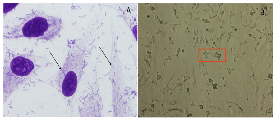
4.3.2. Bacterial Adherence and Toxicity

HeLa cells were purchased from the Adolfo Lutz Institute (São Paulo, Brazil) and were cultured in Dulbecco’s Modified Eagle Medium (DMEM) (GIBCO, Carlsbad, CA, USA), supplemented with 10% fetal bovine serum (FBS) (GIBCO, USA), 1% non-essential amino acids (GIBCO, USA), and penicillin–streptomycin (10,000 U/mL) (GIBCO, USA), in a 5% CO2 incubator. The adherent cells were subcultured every 2–3 days by treatment with a trypsin-EDTA (0.5%) solution (GIBCO, USA). For the adherence test, HeLa cells were subcultured in 24-well plates (105 cells/well) and incubated for 48 h in a 5% CO2 incubator.
The adherence test was performed as described by Vieira et al. (2001) [48]. The bacteria were cultured in Luria Bertani broth (Difco, USA) under aerobic conditions for 18 h at 37 °C. For the adherence assay, the bacteria (1 × 10⁸ CFU/well) were added to HeLa cells in 24-well plates, the medium was replaced with DMEM without antibiotics, and a 2% mannose solution was added, followed by incubation for 3 h at 37 °C. Each well was then washed three times with phosphate-buffered saline (PBS) to remove the non-bound bacteria, and DMEM was replaced. After 3 h of incubation in the same conditions, the cells were washed with PBS and fixed with methanol for 1 hour.
To analyze the bacterial adherence to Hela cells, cell staining was performed using May–Grünwald–Giemsa staining. Briefly, the cells were immersed in the May–Grünwald solution for 20 min, then transferred to a Giemsa solution for 5 min and washed three times with distilled water. The cells were visualized using a microscope (Nikon Eclipse E2000). For the adherence pattern control, the following strains were used: the EPEC prototype strain E2348/69 for localized adherence, the DAEC prototype strain C1845 for diffuse adherence, the EAEC prototype strain 042 for aggregative adherence.
The cytotoxicity assay in Vero cells (Monkey Kidney) was performed in triplicates as described by Martins et al. (2015) [49]. The strains were cultured in Luria Bertani broth (LB) (Difco—BBL, Detroit, MI, USA) at 37 °C for 18 h, in the presence of 5 ng/mL of ciprofloxacin (Sigma-Aldrich, St. Louis, MO, USA). The supernatants were obtained after centrifugation at 8800× g for 30 min and were filtered using a 0.22 Millipore filter. The test was conducted after the inoculation of 50 µL of supernatant into the microplate wells containing a Vero cells monolayer. E. coli O157:H7 (EDL933) and E. coli DH5α supernatants were used as positive and negative controls, respectively.

4.4. Patogenicity In Vivo Tests

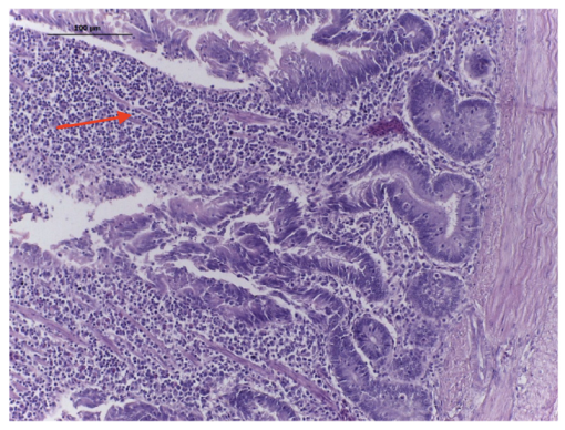
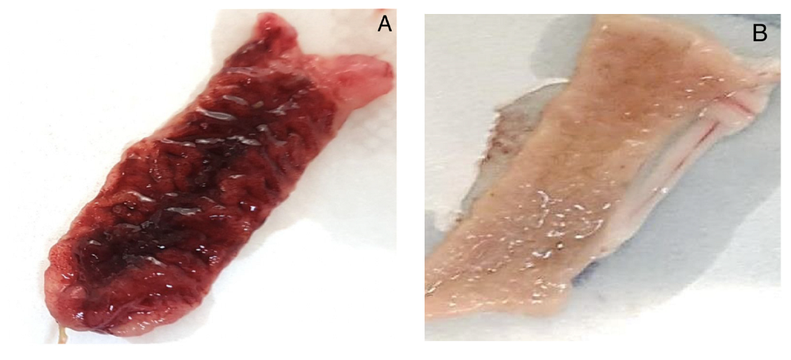
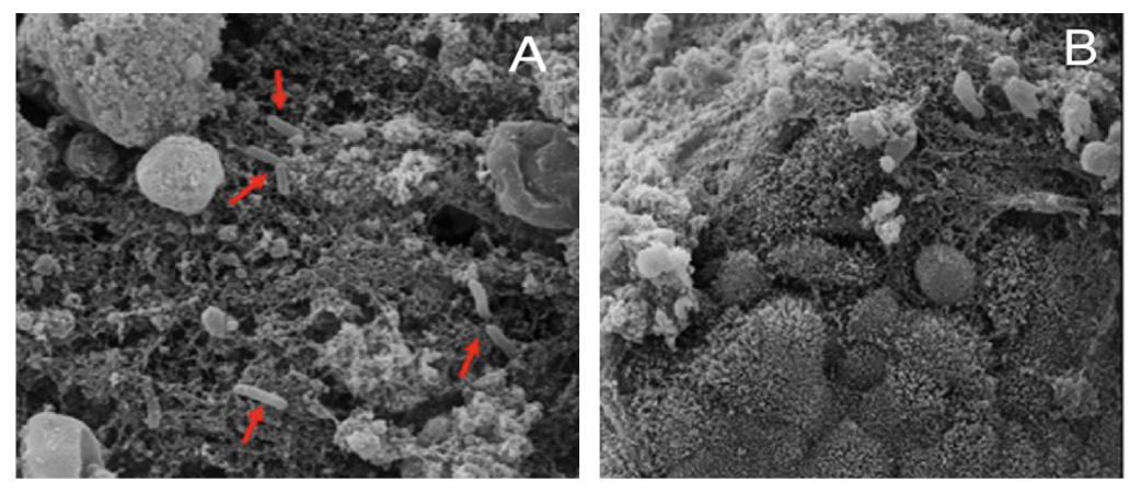
4.4.1. Experimental Inoculation of Chickens

A total of 24 specific pathogen-free chickens (11 weeks of age) were grouped, with 6 birds per cage. Three groups were inoculated with 0.1 mL of A. butzleri culture (1.0 × 10⁹ UFC/mL) by gavage (Day 0), and one group was kept as the negative control (non-inoculated). One bird was euthanized per day (day 1 to day 6), for the observation of gross lesions and to collect tissues for histopathology. Fecal samples were collected daily for 7 days. The fecal samples were diluted 1:9 in selective enrichment broth as described by Johnson, Murano (1999) and incubated in anaerobiosis at 30 °C for 48 h. Then, 10 μL of cultured broth was placed on a sterile cellulose membrane (0.45 μm) on Johnson and Murano agar. After one hour, the filters were removed, and the broth was seeded and incubated at 30 °C for 48–72 h.

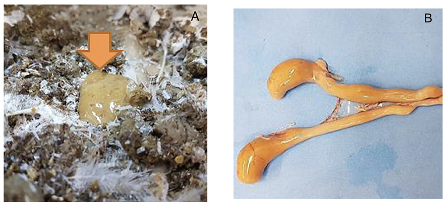
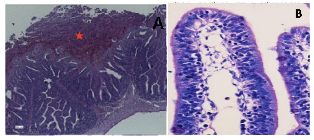
4.4.2. Rabbit Ileal Loop

One New Zealand white rabbit (female, 1.9 Kg) was subjected to laparotomy after inhalation anesthesia as described by Gioia-Di Chiacchio et al. (2018) [50]. The ileum was rinsed with sterile saline, and intestinal loops of 6 cm in length were ligated and separated by 3 cm inter-loops. These loops were inoculated with 1 mL of each strain (1 × 10⁶ CFU/mL), previously cultured in BHI plus 0.1% glucose, and incubated at 30 °C for 18 h at 200 rpm. Sterile PBS was inoculated as a negative control.
After 12 h, the animal was humanely euthanized for a post-mortem examination. Fragments of 0.5 mm of ileum tissue were collected and fixed in 10% buffered formalin and included in paraffin blocks for histopathology examination. The tissues stained with hematoxylin/eosin were examined by light microscopy (Eclipse NiU Nikon, with the camera DS-U3, Software Ni Elements; Nikon Corporation, Tokyo, Japan).
Scanning Electron Microscopy (SEM) was employed for the ultrastructural study, after fixation of 1 mm of tissue with 2.5% glutaraldehyde (v/v) in 0.1 M phosphate buffer (pH 7.2 at 0 °C). After fixation, fragments were rinsed with 0.1 M sodium cacodylate buffer, followed by 1% osmium tetroxide (OsO4) (v/v) and ethanol dehydrated solutions. The tissues were dried using the critical point method, mounted onto SEM stubs sputter-coated with gold, and examined using a Quanta 250 scanning electron microscope (FEI Company, Hillsboro, OR, USA) at 12.5 kV and working distance of 7 mm.

5. Conclusions

Poultry meat is a clearly underrated reservoir of Aliarcobacter strains resistant to fluoroquinolone, macrolide, and tetracycline. The resistance profile and pathogenicity of A. butzleri isolated from Brazilian poultry meat reveal a public health risk. During 2022, Brazil exported 4.822 million tons of poultry meat, but there is no regulation about the presence of Aliarcobacter spp. The data obtained in this study reinforce the need to improve the diagnostics and surveillance, as well as the adoption of preventive actions in the Brazilian poultry industry.

Supplementary Materials

The following are available online at https://www.mdpi.com/article/10.3390/antibiotics12020282/s1, Table S1: Minimum inhibitory concentration (MIC) values of antimicrobials assessed against 27 strains of Aliarcobacter butzleri isolated from commercial chicken meat from São Paulo, Brazil.

Author Contributions

Conceptualization, A.M.M. and T.K.; methodology, M.G.X.d.O., M.P.V.C., L.Z.M. and M.A.M.V.; investigation, M.G.X.d.O., M.P.V.C., L.Z.M. and M.A.M.V.; resources, A.M.M., T.A.T.G. and T.K.; data curation, A.B.S.S., A.M.M., M.A.M.V. and T.K. writing—original draft preparation, M.G.X.d.O. and A.B.S.S.; writing—review and editing, A.M.M., M.A.M.V., T.A.T.G. and T.K.; supervision, A.M.M., L.Z.M., M.A.M.V., T.A.T.G. and T.K.; project administration, T.K.; funding acquisition, T.K. All authors have read and agreed to the published version of the manuscript.

Funding

This research was funded by the Fundação de Amparo à Pesquisa do Estado de São Paulo (grant number 2014/07837-6) and the Coordenação de Aperfeiçoamento de Pessoal de Nível Superior Brazil (finance code 001). A.M.M. and T.K. are Conselho Nacional de Desenvolvimento Científico e Tecnológico (CNPq) fellows (grant numbers 310736/2018-8 and 306396/2020).

Institutional Review Board Statement

The study was conducted in accordance with the Committee on Ethics and Animal Experimentation of the School of Veterinary Medicine and Animal Science of THE University of São Paulo (CEUA 4686280114).

Informed Consent Statement

Not applicable.

Data Availability Statement

The data is available at https://www.teses.usp.br/teses/disponiveis/10/10133/tde-09042020-103046/pt-br.php (accessed on 26 December 2022).

Conflicts of Interest

The authors state that they have no conflicts of interest to declare.

References

  1. Chieffi, D.; Fonelli, F.; Fusco, U. Arcobacter butzleri: Up-to-date taxonomy, ecology and pathogenicity of an emerging pathogen. Compr. Rev. Food Sci. Food Saf. 2020, 19, 2071–2109. [Google Scholar] [CrossRef] [PubMed]
  2. Prouzet-Mauléon, V.; Labardi, L.; Bouges, N.; Ménard, A.; Mégraud, F. Arcobacter butzleri: Underestimated enteropathogen. Emerg. Infect. Dis. 2006, 12, 307–309. [Google Scholar] [CrossRef] [PubMed]
  3. Ertas, N.; Dogruer, Y.; Gonulalan, Z.; Guner, A.; Ulger, I. Prevalence of Arcobacter species in drinking water, spring water, and raw milk as determined by multiplex PCR. J. Food Prot. 2010, 73, 2099–2102. [Google Scholar] [CrossRef]
  4. Nieva-Echevarria, B.; Martinez-Malaxetxebarria, I.; Girbau, C.; Alonso, R.; Fernandez-Astorga, A. Prevalence and genetic diversity of Arcobacter in food products in the north of Spain. J. Food Prot. 2013, 76, 1447–1450. [Google Scholar] [CrossRef] [PubMed]
  5. Gobb, D.D.S.; Spindola, M.G.; Moreno, L.Z.; Matajira, C.E.C.; Oliveira, M.G.X.; Paixão, R.; Ferreira, T.S.P.; Moreno, A.M. Isolation and molecular characterization of Arcobacter butzleri and Arcobacter cryaerophilus from pork production chain in Brazil. Pesqui. Vet. Bras. 2018, 38, 393–399. [Google Scholar] [CrossRef]
  6. Oliveira, M.G.X.; Gomes, V.T.D.M.; Cunha, M.P.V.; Moreno, L.Z.; Moreno, A.M.; Knöbl, T. Genotypic Characterization of Arcobacter spp. Isolated from Chicken Meat in Brazil. Foodborne Pathog. Dis. 2018, 15, 293–299. [Google Scholar] [CrossRef]
  7. Vicente-Martins, S.; Oleastro, M.; Domingues, F.C.; Ferreira, S. Arcobacter spp. at retail food from Portugal: Prevalence, genotyping and antibiotics resistance. Food Control 2018, 85, 107–112. [Google Scholar] [CrossRef]
  8. Cruzado-Bravo, M.L.M.; Barancelli, G.V.; Dini Andreote, A.P.; Saldana, E.; Vidal-Veuthey, B.; Collado, L.; Contreras-Castillo, C.J. Occurrence of Arcobacter spp. in Brazilian Minas frescal cheese and raw cow milk and its association with microbiological and physicochemical parameters. Food Control 2020, 109, 106904. [Google Scholar] [CrossRef]
  9. Isidro, J.; Ferreira, S.; Pinto, M.; Domingues, F.; Oleastro, M.; Gomes, J.; Borges, V. Virulence and antibiotic resistance plasticity of Arcobacter butzleri: Insights on the genomic diversity of an emerging human pathogen. Infection. Genet. Evol. 2020, 80, 104213. [Google Scholar] [CrossRef]
  10. Shange, N.; Gouws, P.; Hoffman, L.C. Campylobacter and Arcobacter species in food-producing animals: Prevalence at primary production and during slaughter. World J. Microbiol. Biotechnol. 2019, 35, 146. [Google Scholar] [CrossRef]
  11. Ferreira, S.; Queiroz, J.A.; Oleastro, M.; Domingues, F.C. Insights in the pathogenesis and resistance of Arcobacter: A review. Crit. Rev. Microbiol. 2016, 42, 364–383. [Google Scholar] [CrossRef] [PubMed]
  12. Jiménez-Guerra, G.; Casanovas MorenoTorres, I.; Moldovan, T.D.; Navarro-Marí, J.M.; Gutiérrez-Fernández, J. Arcobacter butzleri and intestinal colonization. Rev. Espanõla De Quimioter. 2020, 33, 73–75. [Google Scholar] [CrossRef] [PubMed]
  13. Fernandez, H.; Villanueva, M.P.; Mansilla, I.; Gonzalez, M.; Latif, F. Arcobacter butzleri and A. cryaerophilus in human, animals and food sources, in southern Chile. Braz. J. Microbiol. 2015, 46, 145–147. [Google Scholar] [CrossRef] [PubMed]
  14. Son, I.; Englen, M.D.; Berrang, M.E.; Fedorka-Cray, P.J.; Harrison, M.A. Antimicrobial resistance of Arcobacter and Campylobacter from broiler carcasses. Int. J. Antimicrob. Agents 2007, 29, 451–455. [Google Scholar] [CrossRef]
  15. Riesenberg, A.; Fromke, C.; Stingl, K.; Fesler, A.T.; Golz, G.; Glocker, E.O.; Kreienbrock, L.; Klarmann, D.; Werckenthin, C.; Schwarz, S. Antimicrobial susceptibility testing of Arcobacter butzleri: Development and application of a new protocol for broth microdilution. J. Antimicrob. Chemother. 2017, 72, 2769–2774. [Google Scholar] [CrossRef]
  16. Çelik, C.; Pinar, O.; Sipari, N. The prevalence of Alicobacter species in the fecal microbiota of farm animals and potential effective agents for their treatment: A review of past decades. Microorganisms 2022, 10, 2430. [Google Scholar] [CrossRef]
  17. Houf, K.; Tutenel, A.; De Zutter, L.; Van Hoof, J.; Vandamme, P. Development of a multiplex PCR assay for the simultaneous detection and identification of Arcobacter butzleri, Arcobacter cryaerophilus and Arcobacter skirrowii. FEMS Microbiol. Lett. 2000, 193, 89–94. [Google Scholar] [CrossRef]
  18. Atabay, H.I.; Aydin, F. Susceptibility of Arcobacter butzleri isolates to 23 antimicrobial agents. Lett. Appl. Microbiol. 2001, 33, 430–433. [Google Scholar] [CrossRef]
  19. Houf, K.; Devriese, L.A.; Van Hoof, J.; Vandamme, P. Susceptibility of Arcobacter butzleri, Arcobacter cryaerophilus, and Arcobacter skirrowii to antimicrobial agents used in selective media. J. Clin. Microbiol. 2001, 39, 1654–1656. [Google Scholar] [CrossRef]
  20. Fera, M.T.; Maugeri, T.L.; Giannone, M.; Gugliandolo, C.; La Camera, E.; Blandino, G.; Carbone, M. In vitro susceptibility of Arcobacter butzleri and Arcobacter cryaerophilus to different antimicrobial agents. Int. J. Antimicrob. Agents 2003, 21, 488–491. [Google Scholar] [CrossRef]
  21. Kabeya, H.; Maruyama, S.; Morita, Y.; Ohsuga, T.; Ozawa, S.; Kobayashi, Y.; Abe, M.; Katsube, Y.; Mikami, T. Prevalence of Arcobacter species in retail meats and antimicrobial susceptibility of the isolates in Japan. Int. J. Food Microbiol. 2004, 90, 303–308. [Google Scholar] [CrossRef] [PubMed]
  22. Debruyne, L.; Gevers, D.; Vandamme, P. Taxonomy of the family Campylobacteraceae. In Campylobacter, 3rd ed.; Nachamkin, I., Szymanski, C.M., Blaser, M.J., Eds.; American Society of Microbiology: Washington, DC, USA, 2008; p. 3-25-121. [Google Scholar]
  23. Centers for Disease Control and Prevention. National Antimicrobial Resistance Monitoring System for Enteric Bacteria (NARMS): Human Isolates Final Report. Available online: https://www.cdc.gov/narms/faq.html (accessed on 5 February 2019).
  24. Van den Abeele, A.M.; Vogelaers, D.; Vanlaere, E.; Houf, K. Antimicrobial susceptibility testing of Arcobacter butzleri and Arcobacter cryaerophilus strains isolated from Belgian patients. J. Antimicrob. Chemother. 2016, 71, 124. [Google Scholar] [CrossRef] [PubMed]
  25. Shah, A.H.; Saleha, A.A.; Zunita, Z.; Murugaiyah, M. Arcobacter–An emerging threat to animals and animal origin food products? Trends Food Sci. Technol. 2011, 22, 225–236. [Google Scholar] [CrossRef]
  26. Van Boven, M.; Veldman, K.T.; de Jong, M.C.; Mevius, D.J. Rapid selection of quinolone resistance in Campylobacter jejuni but not in Escherichia coli in individually housed broilers. J. Antimicrob. Chemother. 2003, 52, 719–723. [Google Scholar] [CrossRef]
  27. Logue, C.M.; Danzeisen, G.T.; Sherwood, J.S.; Thorsness, J.L.; Mercier, B.M.; Axtman, J.E. Repeated therapeutic dosing selects macrolide-resistant Campylobacter spp. in a turkey facility. J. Appl. Microbiol. 2010, 109, 1379–1388. [Google Scholar] [CrossRef] [PubMed]
  28. Acheson, D.; Allos, B.M. Campylobacter jejuni infections: Update on emerging issues and trends. Clin. Infect. Dis. 2001, 32, 1201–1206. [Google Scholar] [CrossRef]
  29. Blaser, M.J.; Engberg, J. Clinical aspects of Campylobacter jejuni and Campylobacter coli infections. In Campylobacter, 3rd ed.; Nachamkin, I., Szymanski, C.M., Blaser, M.J., Eds.; American Society of Microbiology: Washington, DC, USA, 2008; pp. 99–121. [Google Scholar]
  30. Douidah, L.; De Zutter, L.; Van Nieuwerburgh, F.; Deforce, D.; Ingmer, H.; Vandenberg, O.; Vanden Abeele, A.M.; Houf, K. Presence and analysis of plasmids in human and animal associated arcobacter species. PLoS ONE 2014, 9, e85487. [Google Scholar] [CrossRef]
  31. Abdelbaqi, K.; Buissonniere, A.; Prouzet-Mauleon, V.; Gresser, J.; Wesley, I.; Mégraud, F.; Ménard, A. Development of a real-time fluorescence resonance energy transfer PCR to detect Arcobacter species. J. Clin. Microbiol. 2007, 45, 3015–3021. [Google Scholar] [CrossRef]
  32. Chaves, M.; Vazquez-Valverde, D.; Fernández-Jaramillo, H.; Arias-Echandi, M.L. The ability of Aliarcobacter butzleri strains isolates from foods of animal origin in Costa Rica to form biofilm. Ital. J. Food Saf. 2021, 10, 9020. [Google Scholar] [CrossRef]
  33. Flanagan, R.C.; Neal-Mckinney, J.M.; Dhillon, A.S.; Miller, W.G.; Konkel, M.E. Examination of Campylobacter jejuni putative adhesins leads to the identification of a new protein, designated FlpA, required for chicken colonization. Infect. Immun. 2009, 77, 2399–2407. [Google Scholar] [CrossRef] [Green Version]
  34. Buzzanca, D.; Botta, C.; Cocolin, l.S.; Alessandria, V.; Rantsiou, K. Exploring the Virulence Gene Expression of Arcobacter butzleri during Simulated Infection of Human Gut Models. IAFP’s European Symposium on Food Safety: Nantes, France, 2019; p. 102. [Google Scholar]
  35. Heimesaat, M.M.; Karadas, G.; Alutis, M.; Fischer, A.; Kühl, A.A.; Breithaupt, A.; Göbel, U.B.; Alter, T.; Bereswill, S.; Gölz, G. Survey of small intestinal and systemic immune responses following murine Arcobacter butzleri infection. Gut Pathog. 2015, 7, 28. [Google Scholar] [CrossRef]
  36. Ho, H.T.K.; Lipman, L.J.A.; Gaastra, W. The Introduction of Arcobacter Spp. in Poultry Slaughterhouses. Int. J. Food Microbiol. 2008, 125, 223–229. [Google Scholar] [CrossRef] [PubMed]
  37. Açik, M.N.; Yüksel, H.; Ulucan, A.; Çetinkaya, B. The first experimental research on the pathogenicity of Arcobacter butzleri in zebrafish. Vet. Microbiol. 2016, 189, 32–38. [Google Scholar] [CrossRef] [PubMed]
  38. Vandenberg, O.; Dediste, A.; Houf, K.; Ibekwem, S.; Souayah, H.; Cadranel, S.; Douat, N.; Zissis, G.; Butzler, J.-P.; Vandamme, P. Arcobacter species in humans. Emerg. Infect. Dis. 2004, 10, 1863–1867. [Google Scholar] [CrossRef]
  39. Johnson, L.G.; Murano, E.A. Development of a new medium for the isolation of Arcobacter spp. J. Food Prot. 1999, 62, 456–462. [Google Scholar] [CrossRef]
  40. Bastyns, K.; Chapelle, S.; Vandamme, P.; Goossens, H.; De Wachter, R. Species-specific detection of campylobacters important in veterinary medicine by PCR amplification of 23S rDNA areas. Syst. Appl. Microbiol. 1995, 17, 563–568. [Google Scholar] [CrossRef]
  41. Douidah, L.; De Zutter, L.; Vandamme, P.; Houf, K. Identification of five human and mammal associated Arcobacter species by a novel multiplex-PCR assay. J. Microbiol. Methods 2010, 80, 281–286. [Google Scholar] [CrossRef] [PubMed]
  42. Douidah, L.; De Zutter, L.; Baré, J.; De Vos, P.; Vandamme, P.; Vandenberg, O.; Van Den Abeele, A.M.; Houf, K. Occurrence of putative virulence genes in Arcobacter species isolated from humans and animals. J. Clin. Microbiol. 2012, 50, 735–741. [Google Scholar] [CrossRef]
  43. Clinical and Laboratory Standards Institute. Performance Standards for Antimicrobial Disk Susceptibility Tests, 13th ed.; Clinical and Laboratory Standards Institute: Wayne, PA, USA, 2018. [Google Scholar]
  44. Schwarz, S.; Silley, P.; Simjee, S.; Woodford, N.; van Duijkeren, E.; Johnson, A.P.; Gaastra, W. Assessing the antimicrobial susceptibility of bacteria obtained from animals. J. Antimicrob. Chemother. 2010, 65, 601–604. [Google Scholar] [CrossRef]
  45. Magiorakos, A.P.; Srinivasan, A.; Carey, R.B.; Carmeli, Y.; Falagas, M.E.; Giske, C.G.; Harbath, S.; Hindler, J.F.; Kahlmeter, G.; Olsson-Lijequist, B.; et al. Multidrug-resistant, extensively drug-resistant and pandrug-resistant bacteria: An international expert proposal for interim standard definitions for acquired resistance. Clin. Microbiol. Infect. 2012, 18, 268–281. [Google Scholar] [CrossRef] [Green Version]
  46. Mclennan, M.K.; Ringoir, D.D.; Frirdich, E.; Svensson, S.L.; Wells, D.H.; Jarrel, H.; Szymanski, C.M.; Gaynor, E.C. Campylobacter jejuni biofilms up-regulated in the absence of the stringent response utilize a calcofluor white-reactive polysaccharide. J. Bacteriol. 2008, 190, 1097–1107. [Google Scholar] [CrossRef]
  47. Naves, P.; Del Prado, G.; Huelves, L.; Gracia, M.; Ruiz, V.; Blanco, J.; Rodríguez-Cerrato, V.; Ponte, M.C.; Soriano, F. Measurement of biofilm formation by clinical isolates of Escherichia coli is method-dependent. J. Appl. Microbiol. 2008, 105, 585–590. [Google Scholar] [CrossRef] [PubMed]
  48. Vieira, M.A.M.; Andrade, J.R.C.; Trabulsi, L.R.; Rosa, A.C.P.; Dias, A.M.G.; Ramos, S.R.T.S.; Frankel, G.; Gomes, T.A.T. Phenotypic and Genotypic Characteristics of Escherichia coli strains of non-Enteropathogenic E. coli (EPEC) Serogroups that carry eae and lack the EPEC adherence factor and Shiga Toxin DNA probe sequences. J. Infect. Dis. 2001, 183, 762–772. [Google Scholar] [CrossRef] [PubMed]
  49. Martins, F.H.; Guth, B.E.C.; Piazza, R.M.; Leão, S.C.; Ludovico, A.; Ludovico, M.S.; Dahbi, G.; Marzoa, J.; Mora, A.; Blanco, J.; et al. Diversity of Shiga toxin-producing Escherichia coli in sheep flocks of Paraná State, southern Brazil. Vet. Microbiol. 2015, 175, 150–156. [Google Scholar] [CrossRef] [PubMed]
  50. Gioia-Di Chiacchio, R.M.; Cunha, M.P.V.; de Sá, L.R.M.; Davies, Y.M.; Pereira, C.B.P.; Martins, F.H.; Munhoz, D.D.; Abe, C.M.; Franzolin, M.R.; Dos Santos, L.F.; et al. Novel Hybrid of Typical Enteropathogenic Escherichia coli and Shiga-Toxin-Producing E. coli (tEPEC/STEC) Emerging from Pet Birds. Front. Microbiol. 2018, 9, 2975. [Google Scholar] [CrossRef] [PubMed] [Green Version]
Figure 1. (A) Adherence of Arcobacter butzleri to HeLa cells. (B) VERO cells after the addition of A. butzleri supernatant, presenting elongated cells with rounded vacuoles (Red box).
Antibiotics 12 00282 g001
Figure 2. Feces and cecum of birds, four days after the inoculation of A. butzleri AB170 strain. (A) The orange arrow shows the diarrheagenic aspect of poultry feces. (B) Bird’s ceca with an orange content.
Antibiotics 12 00282 g002
Figure 3. Micrograph of a chicken intestine, four days after the inoculation of A. butzleri AB170 strain, presenting inflammatory infiltrate cells (Red arrow). HE × 100.
Antibiotics 12 00282 g003
Figure 4. Macroscopic evaluation of rabbit ileal loops, 12 h after the inoculation of the AB170 strain. (A) Macroscopic hemorrhagic aspect of the intestine 12 h after the inoculation of A. butzleri. (B) Negative control. Intestinal tissue inoculated with PBS.
Antibiotics 12 00282 g004
Figure 5. Ileal histopathology of an experimentally inoculated rabbit, 12 h after the inoculation of the AB170 strain. (A) Ileal loop showing erythrocytes and debris in the lumen (Red star). HE × 100. (B) Negative control. Intestinal tissue is preserved, HE × 200.
Antibiotics 12 00282 g005
Figure 6. Scanning Electron Microscopy (SEM) of the small intestine in the inoculated rabbit ileal loops model, 12 h post inoculation of the AB170 strain. (A) Destroyed villi and presence of bacilli in the extracellular matrix (Red arrows). (B) Negative control. Intestinal crypts are preserved.
Antibiotics 12 00282 g006
Table 1. Resistance profiles of 27 strains of Aliarcobacter butzleri isolated from commercial poultry meat from São Paulo city, Brazil.
Resistant Profiles Number of Strains
R1Susceptible strains 5
R2CLIN2
R3NAL/CIP1
R4NAL/CIP/CLIN1
R5FLOR/CLIN3
R6NAL/FLOR/CLIN11
R7TET/NAL/FLOR/CLIN1
R8NAL/AZI/CLIN1
R9NAL/AZI/FLOR/CLIN1
R10AZI/ERI/TELI/FLOR/CLIN1
CLIN—clindamycin; NAL—nalidixic acid; CIP—ciprofloxacin; FLOR—florfenicol; TELI—telithromycin; TET—tetracycline; AZI—azithromycin; ERI—erythromycin.
Disclaimer/Publisher’s Note: The statements, opinions and data contained in all publications are solely those of the individual author(s) and contributor(s) and not of MDPI and/or the editor(s). MDPI and/or the editor(s) disclaim responsibility for any injury to people or property resulting from any ideas, methods, instructions or products referred to in the content.

