Article

Exploring the Accessory Genome of Multidrug-Resistant Rhodococcus equi Clone 2287

by
Sonsiray Alvarez Narvaez
* and
Susan Sanchez
*
Department of Infectious Diseases, College of Veterinary Medicine, University of Georgia, Athens, GA 30602, USA
*
Authors to whom correspondence should be addressed.
Antibiotics 2023, 12(11), 1631; https://doi.org/10.3390/antibiotics12111631
Submission received: 1 November 2023 / Revised: 15 November 2023 / Accepted: 16 November 2023 / Published: 17 November 2023
(This article belongs to the Special Issue Antimicrobial Resistance in Veterinary Science)

Abstract

:
Decades of antimicrobial overuse to treat respiratory disease in foals have promoted the emergence and spread of zoonotic multidrug-resistant (MDR) Rhodococcus equi worldwide. Three main R. equi MDR clonal populations—2287, G2106, and G2017—have been identified so far. However, only clones 2287 and G2016 have been isolated from sick animals, with clone 2287 being the main MDR R. equi recovered. The genetic mechanisms that make this MDR clone superior to the others at infecting foals are still unknown. Here, we performed a deep genetic characterization of the accessory genomes of 207 R. equi isolates, and we describe IME2287, a novel genetic element in the accessory genome of clone 2287, potentially involved in the maintenance and spread of this MDR population over time. IME2287 is a putative self-replicative integrative mobilizable element (IME) carrying a DNA replication and partitioning operon and genes encoding its excision and integration from the R. equi genome via a serine recombinase. Additionally, IME2287 encodes a protein containing a Toll/interleukin-1 receptor (TIR) domain that may inhibit TLR-mediated NF-kB signaling in the host and a toxin–antitoxin (TA) system, whose orthologs have been associated with antibiotic resistance/tolerance, virulence, pathogenicity islands, bacterial persistence, and pathogen trafficking. This new set of genes may explain the success of clone 2287 over the other MDR R. equi clones.

1. Introduction

Rhodococcus equi is an animal and human pathogen mainly known for being the most common cause of severe pneumonia in foals [1,2]. R. equi is a soil saprotroph that becomes pathogenic upon acquiring a virulence plasmid (pVAP) that allows R. equi to infect and proliferate in macrophages [3]. So far, three different pVAPs have been reported, presenting different host tropisms: equine pVAPA, porcine pVAPB, and ruminant pVAPN [4,5,6]. R. equi is endemic on many horse-breeding farms [2,7,8]. It mainly produces disease in foals between 1 and 4 months of age, typically in the form of a multifocal purulent pneumonia [9,10]. The pulmonary infection is contracted during the neonatal period via inhalation of contaminated aerosolized dust [11,12,13]. No commercial vaccines or other consistently efficient immunoprophylactic strategies are available [14,15]. The elevated costs resulting from veterinary care, long-term therapy, and mortality of foals on endemic farms have forced many breeders to adopt practices such as chemoprophylaxis for all foals during the first weeks following birth [16] or to treat foals presenting ultrasonographic lung lesions with antimicrobials before the onset of clinical signs [17,18]. Only a few antibiotics have been seen to be clinically effective in horses to treat R. equi [19,20], and the combination of a macrolide and rifampin has been the treatment of choice since the 1980s [21,22,23]. Before the early 2000s, R. equi resistant to macrolides and rifampin were rarely found [24]. Then, the practice of thoracic screening plus subclinical treatment was implemented, resulting in a considerable increase in the use of macrolides and rifampin on endemic farms and the subsequent emergence of MDR R. equi isolates resistant to these two antibiotics [25].
Over time, the constant antimicrobial selective pressure exerted by the continuous prophylactic antimicrobial treatment has promoted the spread of multidrug-resistant (MDR) R. equi [24,25,26]. To date, isolates resistant to macrolides and/or rifampin have been reported in 16 US states [24], China [6,27], Ireland [28,29], France [30], and Poland [31]. Our previous work revealed that in the US, R. equi isolates resistant to macrolides and rifampin are mainly clustered in three clonal populations: clone 2287, clone G2016, and clone G2017 [26,32,33]. Clone 2287 harbors pRErm46, a 90 kb conjugative plasmid that carries antimicrobial resistance genes (ARGs) for macrolides, lincosamide, streptogramin B (MLSB), tetracycline, and sulfamethoxazole [34] and a chromosomal rpoBS531F mutation conferring resistance to rifampin. pRErm46 horizontal gene transfer (HGT) into a different R. equi genetic background gave rise to clone G2016, associated with a different rifampin resistance mutation (rpoBS531Y). These two clones are the only resistant ones recovered so far, despite an intense search of sick horses, with clone 2287 accounting for most of the clinical cases [26]. Clone G2017, carrying MLSB resistance plasmids pRErm51 and pMobErm51, has been found exclusively in the environment [26,33]. Although the presence of pVAPA (a conjugative plasmid that carries the virulence factors required to colonize the equine host efficiently) has been reported in members of the three MDR clonal populations, clone 2287 is the most-recovered MDR isolate from sick animals [35,36]. Here, we investigated the accessory genome of 207 R. equi (environmental and clinical isolates) to elucidate the genetic mechanisms that make clone 2287 superior at infecting foals.

2. Materials and Methods

2.1. Bioinformatic Analysis

BLAST+ v2.9.0 [37] was used to align the contigs of 207 R. equi isolates characterized in our earlier studies [32,33,35] to known R. equi genetic elements (R. equi chromosome, pVAPA, pRErm46, and erm(51)) at >95% identity and >80% coverage. Similarly, BLASTn [37] was used to align the unknown contigs from sample 156 to the other novel contigs and the nucleotide NCBI database. IME2287 annotation was performed using Prokka V1.14.5 [38] and InterProScan V86.0 [39] and subsequently manually inspected and curated based on BLASTx [37] analysis.

2.2. Bacterial Strains and Culture Conditions

Two R. equi strains were used in this study: R. equi PAM2287 (NCBI BioSample database no. SAMN04880532), which is a macrolide- and rifampin-resistant clinical isolate carrying virulence plasmid pVAPA, macrolide resistance plasmid pRErm46, rifampin mutation rpoBS531, and mobilizable element IME2287 [35], and R. equi 103-ApraR, which is a plasmidless derivative strain of reference strain R. equi 103 containing the aac(3)IV apramycin resistance cassette integrated on the chromosome [40]. R. equi isolates were routinely cultured in brain heart infusion medium (BHI; Difco Laboratories-BD, Franklin Lakes, NJ, USA) at 30 °C and 200 rpm unless otherwise stated. Agar media were prepared by adding 1.6% of bacteriological agar (Oxoid, Basingstoke, United Kingdom). Media were supplemented with antibiotics (erythromycin, 8 μg/mL; apramycin, 50 μg/mL; rifampin, 25 μg/mL; Sigma, Saint Louis, MO, USA) when required. All in vitro bacterial work (including the bacterial conjugation and the plasmid loss assays) were carried out in our laboratory at the Athens Veterinary Diagnostic Laboratory from the University of Georgia (Athens, GA, USA).

2.3. Bacterial Conjugation

Conjugation assays were carried out as described previously by Alvarez-Narvaez et al. [41]. Briefly, R. equi donor (MDR R. equi clone 2287) and recipient (macrolide-susceptible 103-ApraR) strains were grown overnight in BHI and in the presence of the corresponding antibiotic. Then, donor and recipient bacteria were mixed 1:1 in 5 μL of BHI and spotted in a thick drop onto a BHI plate. After 72 h of incubation at 30 °C, the bacterial mixture was scraped and resuspended in PBS. Serial dilutions were plated onto BHI agar supplemented with apramycin (recipient selection) or apramycin plus erythromycin (transconjugant selection). Conjugation ratios were calculated using the following formula: conjugation ratio = no. of transconjugant cells/no of recipient cells.

2.4. Plasmid Loss Assay

R. equi PAM2287 was inoculated into 10 mL of BHI, or donor horse serum (DHS), and grown at room temperature (RT, ~22 °C) or 37 °C with shaking (200 rpm) for 24 h. After that, bacteria were subcultured in 10 mL of fresh BHI or DHS at an initial optical density at 600 nm wavelength (OD600) of 0.02. OD600 was measured using a Thermo Scientific™ Multiskan™ FC Microplate Photometer (Thermo Fisher Scientific, Waltham, MA, USA). Every subculture was considered a passage, and 45 passages were performed in this experiment. At passages 0, 15, 30, and 45, cultures were serially diluted 1:10 in PBS and plated onto BHI plates. In parallel, R. equi PAM2287 was inoculated into 5 mL test tubes (n = 10) containing 1 g of autoclaved soil (121 °C for 20 min; Ref. [42]) from the gardens of the University of Georgia School of Veterinary Medicine at a concentration of 5 × 10⁴ CFU/g. Inoculated tubes were mixed well (shaking at ~1800 rpm in a vortex genie 2 bench mixer (Scientific Industries, Bohemia, NY, USA) for 30 min) and incubated statically at RT (n = 5) and 37 °C (n = 5) for up to 45 days. On days 0, 15, 30, and 45, the soil of 1 tube per temperature condition was resuspended in 2 mL phosphate-buffered saline (PBS) (by shaking at ~1800 rpm in a vortex genie 2 bench mixer (Scientific Industries) for 30 min) and quantitatively cultured through serial 10-fold dilutions in BHI. One hundred colonies per medium (BHI, DHS, and soil) and temperature (RT and 37 °C) were randomly selected and re-plated onto BHI, BHI-Ery, and BHI-Rmp plates. The presence of pRErm46 IME2287 and pVAPA was tested using PCR.

2.5. PCR

Transconjugants were confirmed using PCR for the macrolide-resistant gene erm(46) and apramycin-resistant gene aac(3)IV. pVAPA, pRErm46, and IME2287 mobilization were also tested using PCR. Table 1 contains the list of oligos used in this study. PCRs were carried out using a T100 thermocycler (Bio-Rad, Hercules, CA, USA) and GoTaq Flexi DNA polymerase (Promega, Madison, WI, USA) under the conditions specified in Alvarez-Narvaez et al. [41]. Briefly, we used an initial denaturation step for 5 min at 95 °C, followed by 30 amplification cycles (involving 30 s at 95 °C of denaturation, 30 s of oligonucleotide hybridization at the appropriate melting temperature, and 2 min of elongation at 72 °C) and a final elongation of 10 min at 72 °C.

3. Results

3.1. The Search for Genetic Elements in MDR R. equi Clone 2287 Selected and Maintained over Time

PacBio and Illumina whole-genome assemblies from 62 R. equi clinical isolates (40 MDR clone 2287 and 22 susceptible) collected between 2002 and 2017 in different US states and characterized in our earlier study [32] were used to identify potential new genetic elements associated exclusively with MDR R. equi clone 2287 (Table S1). Four thousand and one contigs from the 40 members of clonal population 2287 were first aligned to known R. equi genetic elements (chromosome, virulence plasmid pVAPA, and MDR plasmid pRErm46). In total, 2669, 85, and 168 contigs matched with the R. equi chromosome (accession number NC_014659), pVAPA1037 (accession number NC_011151), and pRErm46 (accession number KY494640), respectively, with >95% similarity and >80% coverage, and 1079 contigs were identified as unknown.
Strain 156 showed fewer unknown contigs, only two: contig_221 and contig_249. These two contigs were aligned to all the unknown contigs from the other resistant strains and to the genomes of the 18 susceptible isolates. Only contig_249 was common in all resistant strains and was not present in the assemblies of any susceptible isolates. Contig_249 was 22,578 bp long and comprised a novel 14,317 bp genetic element (Figure 1) that we noted, IME2287 (integrative mobilizable element from clone 2287), and a subsequent partial IME2287 duplication of 8260 bp in length. Further analysis of the contigs carrying this novel piece of DNA showed that in most of the cases, IME2287 was the only genetic element that appeared in the contigs, repeated (and sometimes inverted) in tandem (Figure 2). Further analysis showed that only one isolate (sample 169) presented with IME2287 randomly inserted in the bacterial chromosome (Figure 2).

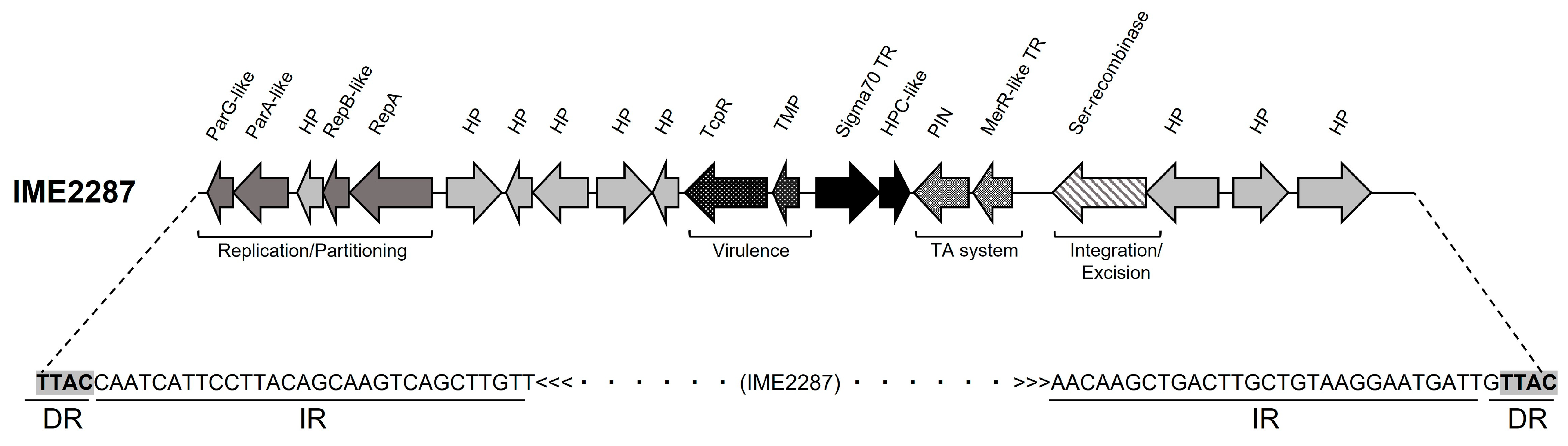
3.2. Molecular Characterization of IME2287, a Putative Mobilizable Element Associated with Macrolide Resistance in MDR R. equi

The in silico annotation of IME2287 identified 20 ORFs (Figure 1), of which 11 were predicted to have a biological function or a functional domain and 9 were classified as hypothetical proteins. Homology searches showed that the novel set of genes carried by IME2287 share high homology with genes previously found in the genus Rhodococcus and other Actinobacteria. Table 2 lists the 20 ORFs with the in silico functional predictions and corresponding homologies. Of interest, four genes are clustered together and code for proteins involved in DNA replication and partitioning. In this order, ORF IME2287_0010 was predicted to be a ParG-like DNA binding protein with a ribbon–helix–helix domain in its C-terminal region, ORF IME2287_0020 was identified as an AAA-ATPase ParA partitioning protein, ORF IME2287_0030 was classified as a RepB-like RNA polymerase with a DNA binding domain, and ORF IME2287_0050 was found to be a RepA plasmid replicase. These genes indicate that IME2287 is most likely a self-replicative genetic element.
ORF IME2287_0170 encodes a serine recombinase that would allow IME2287 to excise and reintegrate into the R. equi genome. A maximum identity value of 87% with its closest homolog unclassified ISBli29 from Brevibacterium linens (Actinobacteria) indicates that the serine recombinase is a novel orphan transposase, which we named ISRe2287. The presence of this gene and the replication and partitioning machinery suggested that IME2287 could be an integrative conjugative element (ICE). However, no conjugation genes were predicted in IME2287, and we found the inverted repeat (IR) sequence 5′-CAATCATTCCTTACAGCAAGTCAGCTTGTT-3′ invariably followed by the directly repeated (DR) tetranucleotide TTAC, supporting that IME2287 could be a transposable element. Additionally, IME2287 carries a gene that encodes a protein containing a Toll/interleukin-1 receptor (TIR) domain (TcpR, IME2287_0140) that acts by suppressing the innate immune system in other bacteria species [45,46,47,48] and a putative toxin–antitoxin system composed of a PIN domain-containing protein (toxin, IME2287_0150) and a MerR-like DNA binding protein (antitoxin, IME2287_0160).

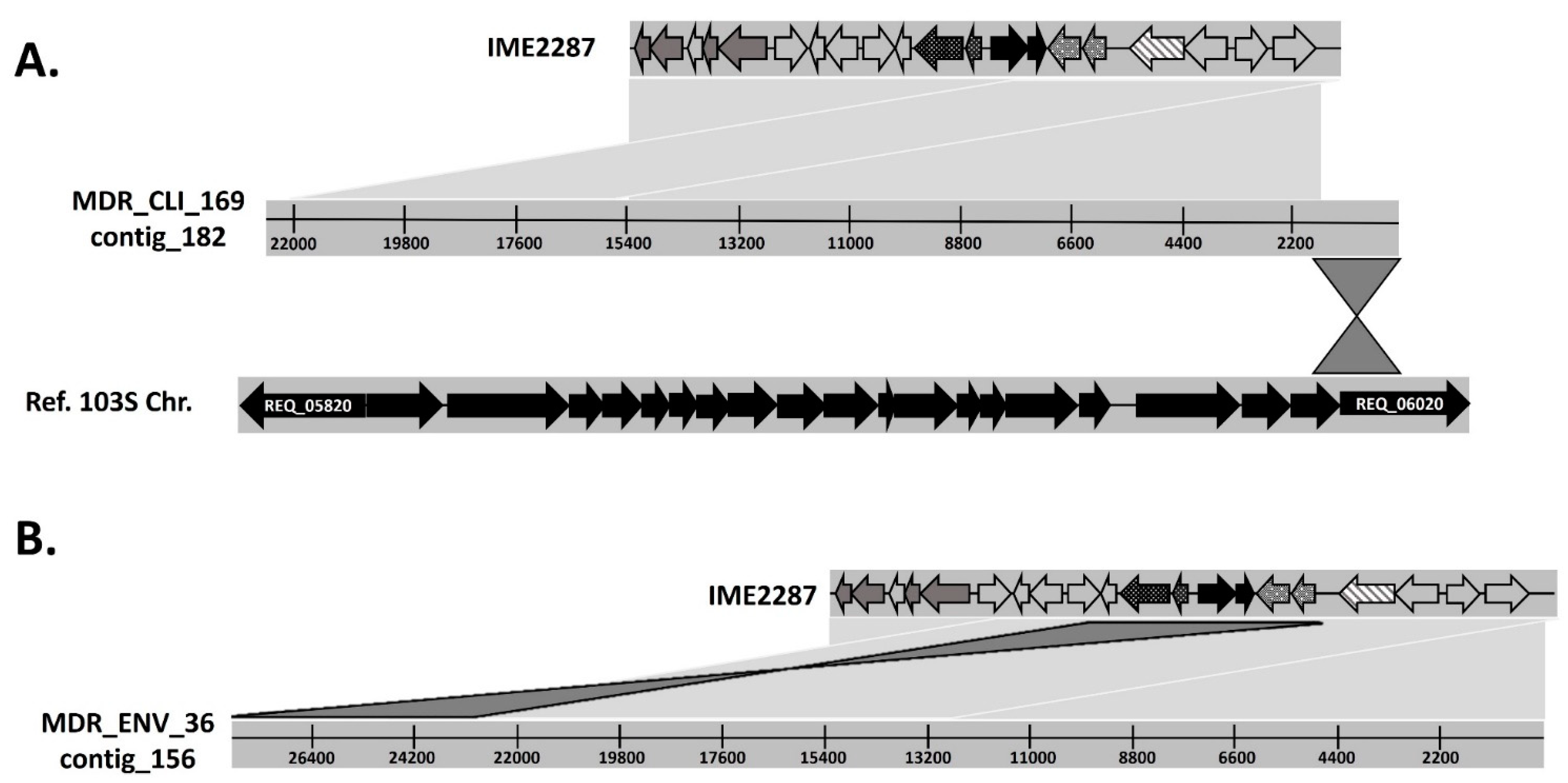
3.3. IME2287 Moved from MDR R. equi Clone 2287 to other R. equi Genetic Backgrounds Associated with Macrolide Resistance

To explore if IME2287 was a mobile genetic element, the whole-genome assemblies of 10 additional MDR R. equi clinical isolates harboring pRErm46 but with genetic backgrounds different from clone 2287 were analyzed. Two isolates were members of the new MDR R. equi clonal population G2016 (Table 3), and eight were classified as singletons (an MDR R. equi isolate that shows a unique genetic background and therefore does not belong to any of the known R. equi clonal populations) in previous phylogenetic analysis [26,32]. We observed the presence of IME2287 in one of the R. equi G2016 clones and two of the singletons, indicating that this element moved from R. equi clone 2287 into other R. equi isolates through horizontal gene transfer (HGT).
Furthermore, we looked for evidence of IME2287 in the PacBio whole-genome assemblies from 135 R. equi environmental isolates collected from 100 farms in central Kentucky for a previous investigation [26,33]. The 45 environmental MDR R. equi that previous phylogenetic analysis [26,33] classified as part of clonal population 2287 were shown to harbor both pRErm46 and IME2287 in their genome, while none of the 38 susceptible isolates did (Table 3). R. equi clone G2016 (sample 52), the only MDR environmental isolate of this clone, carried pRErm46 but was not shown to have IME2287. Similarly, only 7 of the 39 MDR R. equi environmental isolates phylogenetically classified as members of the clonal population G2017 were shown to carry pRErm46/tnRErm46. Of these seven, three also carried IME2287, and four did not. Regarding the environmental singleton-resistant isolates, nine out of twelve were seen to carry pRErm46 or tnRErm46, and from those nine, seven isolates also carried IME2287 (Table 3).
As previously seen in the clinical isolates, IME2287 always appears in an individual contig that exclusively contains copies of these elements (Figure 2). Interestingly, we did not find any clinical or environmental isolates that presented IME2287 without any genetic elements (pRErm46 or tnRErm46) associated with the erm(46) resistance gene, suggesting that IME2287 could be hijacking pRErm46 conjugation machinery to move.

3.4. IME2287 Mobilization Is Independent of Macrolide Resistance Plasmid pRErm46

We previously reported that macrolide resistance plasmid pRErm46 was mobilizable at a high frequency through horizontal gene transfer via bacterial conjugation [35,41]. The findings above lead to the following question: is IME2287 mobilizable simultaneously with pRErm46? To answer this question, conjugation assays were carried out using MDR R. equi clone 2287 as a donor and susceptible (pRERrm46- negative) avirulent R. equi 103- with an apramycin resistance aac(3)IV cassette (103-ApraR) [40] as the recipient in a ratio 1:1. Transfer of pRErm46 to 103-ApraR was observed at a frequency of 2.06 ± 0.70 × 10⁻³ transconjugants/recipient cells. In total, 90 CFUs were PCR-tested for virulence plasmid pVAPA and novel element IME2287. Five out of the ninety colonies tested were shown to have acquired pVAPA together with pRErm46, but no colonies were carrying IME2287, indicating that its mobilization is most likely independent from pRErm46 conjugal transfer.
Next, we investigated if IME2287 is lost over time in the same fashion pRErm46 is. We monitored changes in the antimicrobial resistance phenotype and genotype of MDR R. equi clone 2287 culture-passed daily in BHI and DHS at RT (~22 °C) or 37 °C for 45 passages. In parallel, we looked for changes in the antimicrobial resistance phenotype and genotype of MDR R. equi clone 2287 incubated in autoclaved soil at RT or 37 °C for 45 passages (Table 4). We observed that the percentage of macrolide- and rifampin-resistant colonies of clone 2287 was maintained for the entire experiment duration (45 passages) in all conditions, except for bacteria incubated in DHS at 37 °C, where macrolide resistance declined progressively over time. This indicates that, regardless of the temperature in BHI and soil, macrolide and rifampin resistance expression do not represent a fitness cost for R. equi clone 2287.
We PCR-tested the 46 CFUs that were shown to have lost macrolide resistance for the presence of pRErm46 and IME2287 (Table 5). As expected, all susceptible colonies had lost the macrolide resistance plasmid, but only half of those (n = 23) also lost IME2287, indicating again that the mobilization of these two elements is independent. Interestingly, looking at IME2287 loss over time, we realized that up to passage 30, the colonies that lost pRErm46 still maintained IME2287. In passage 45, 23 out of 36 (~64%) appeared to have lost both elements. This indicates that the loss of IME2287 is most likely subsequent to the loss of the pRErm46 plasmid.

4. Discussion

The emergence and spread of MDR bacterial clones represent a significant threat to animal and human health. The success of these dominant bacterial clades relies on the acquisition of virulence and antimicrobial resistance (AMR) genes associated with mobile genetic elements [49]. Although we currently know more about resistance mechanisms and their mobilization dynamics, there is still much to learn to help us prevent their selection and spread. Here, we explored the accessory genome of MDR R. equi clone 2287, looking for genetic elements that could explain its superiority over other MDR R. equi clones. Considering that bacterial genomes are subject to genetic drift that deletes superfluous sequences [50], we hypothesized that any genetic element that would provide an advantage in the adaptation of MDR clone 2287 would be exclusively present in all clone members and maintained over time. Based on this, we first performed a preliminary screening of clone 2287’s accessory genome, trying to identify genetic elements present in all the genomes of the clonal population and not in susceptible strains. We only identified one genomic sequence under this threshold, and its genomic annotation indicated that we were dealing with a putative mobile genetic element. Consequently, we named the novel element IME2287.
The oldest member of clone 2287, in which we detected the presence of IME2287, is an isolate collected in Florida in 2002. Since then, IME2287 has been found in all the R. equi phylogenetically classified as clone 2287 that were analyzed (n = 85, over a period of 15 years in five different US states); the most recent isolate was found in Kentucky in 2017. It is not surprising to find the R. equi genome harboring new genetic elements because this bacteria species is a unique example of plasmid-driven full virulence, antimicrobial resistance, and host adaptation [10]. However, R. equi tends to lose its virulence host-adapted plasmids (pVAPs) when living as a saprotroph in the soil as they introduce a fitness cost due to the expression of the virulence genes [5,51]. The fact that IME2287 has been maintained for more than 15 years in this clonal population is intriguing and suggests that instead, IME2287 may not impact R. equi fitness, or if it does, the role that IME2287 plays for the bacterium overcomes that fitness cost.
We performed a deep genetic characterization of IME2287, looking for genes that could explain its persistence over time. We found that IME2287 carries a potential new virulence factor. The tcpR gene encodes a putative TIR domain protein that we designated as TcpR (Figure 1, Table 2). In eukaryotes, TIR domains are part of Toll-like receptors (TLRs), a family of proteins that recognize pathogens and initiate the innate immune response [52]. TIR domain proteins are also found in plants, where they mediate disease resistance, and in bacteria, associated with virulence and bacterial metabolic regulation [47]. TIR domain proteins have been reported to block TLR signaling, inhibiting innate immune responses in important animal and human pathogens such as Brucella spp. [53], uropathogenic Escherichia coli (UPEC) [54], nosocomial strains of Enterococcus faecium [55], and Staphylococcus aureus [48]. TIR domain proteins are frequently carried on mobile genetic elements and, in some cases, are associated with ARGs [48]. The innate immune system is the principal response against intracellular R. equi [56]. Therefore, an extra virulence factor, such as TcpR, would help R. equi circumvent innate host immunity during infection, which is extremely important for the bacteria. It would explain why IME2287 is still within all MDR R. equi clinical isolates 15 years after being first reported. Furthermore, this extra virulence factor could be the key to clone 2287’s superiority at infecting animals over the other genomic backgrounds.
Another interesting operon also found in IME2287 consists of two genes that encode an MerR transcriptional regulator and a PIN domain ribonuclease (MerR-PIN). In silico work has classified MerR-PIN operons as putative Type 2 toxin–antitoxin (TA) systems [57], in which the PIN ribonuclease is toxic to the cell and stable. At the same time, the MerR antitoxin is unstable and requires continuous transcription to inhibit the produced toxin [58]. MerR-PIN and other Type II TA systems are often found in plasmids or, as appreciated in IME2287, inserted in chromosomes in association with mobile genetic elements [57,59]. TA systems have been associated with antibiotic resistance, virulence factors, and pathogenicity islands in pathogenic bacteria [58]. One of the first functions assigned to TA systems was plasmid stabilization through a process known as “post-segregational killing”, a suicide mechanism for those cells that do not carry the TA plasmid after cell division [57]. More recently, Type II TA systems have been described as involved in bacterial permanence under stress conditions such as nutrient starvation or antibiotic exposure [60]. Under stress conditions, the antitoxins are selectively degraded, leaving toxins to promote growth arrest and dormancy. Once the stressor is removed, dormant persistent cells revert to the actively growing state and repopulate the original population [60]. Based on the current information regarding Type II TA systems, we hypothesized that the MerR-PIN system could have a double function for MDR R. equi, (i) avoiding the loss of IME2287 during cell division for over 15 years and (ii) mediating the permanence of MDR R. equi in the soil even in the absence of antimicrobial selective pressure. Future in vitro experimentation to determine MerR-PIN functionality is guaranteed.
IME2287 has been observed in several different genetic backgrounds, indicating that this genetic element can move through horizontal gene transfer. The IME2287 genetic characterization revealed that IME2287 carries a replication and partitioning operon (Figure 1, Table 2) and an integration/excision capability via a serine recombinase. This is consistent with the core modules of integrative conjugative elements (ICEs) previously identified in Actinomycetes [61]. However, IME2287 lacks the conjugation module, often consisting of a relaxase, a coupling protein, and a mating pair formation system [61]. We also identified IR and DR flanking IME2287, suggesting that this genetic element is likely a mobilizable transposon or a non-canonical IME [62]. The distinction between mobilizable transposons and IMEs is unclear and is considered irrelevant for some [62]. Various non-canonical IMEs (that do not encode any relaxase) have been identified in the Firmicutes and Proteobacteria phyla, sometimes with several copies integrated in tandem, like in the case of IME2287 [63]. Regardless of the nature of IME2287 (transposon, or IME), the in silico data obtained in this project reflect that at some point, IME2287 jumped from clone 2287, its native genomic background, to other R. equi. IME2287 was found in 50% (13/27) of pRErm46 containing non-clone 2287 R. equi, suggesting that its mobilization could be tied to the antimicrobial resistance plasmid mobilization. Hence, we hypothesized that IME2287 (lacking its conjugal machinery) could hijack the pRErm46 conjugation apparatus to spread. We explored this possibility using mating experiments, and we found that none of the transconjugants that acquired pRErm46 also received IME2287. This indicates that IME2287 mobilization is likely independent of pRErm46. However, we cannot discard that IME2287 could indeed be using pRErm46 mobilization machinery but with a very low mobilization rate, or maybe IME2287 transfer is not triggered or selected by exposure to macrolides.
Given the lack of success of our IME2287 transfer attempt, we decided to test if the loss of this novel genetic element was tied to the loss of pRErm46. Our experiments subculturing clone 2287 in the absence of antimicrobials and under different media and temperature conditions showed that the loss of IME2287 is independent of pRErm46 and that the recently discovered genetic element would get lost, most probably after the loss of the macrolide-resistant plasmid. Additionally, this last experiment allowed us to study the persistence of clone 2287 resistant phenotypes without selective pressure. We observed that clone 2287 maintained a rifampin resistance phenotype during the entire duration of the experiment, independent of the incubation conditions. Willingham-Lane and collaborators studied the stability of three lab-generated rifampin-resistant rpoB mutations in the R. equi 103S genetic background by passing bacteria in BHI at 37 °C [64]. They reported that two of the three rpoB mutations reverted to the wild-type form after 20 passages (>90% revert) and that these mutations resulted in decreased growth in vitro and in soil. This, together with the fact that the mutation present in MDR clone 2287 (rpoBS531F) has been impossible to recreate in the laboratory (Willingham-Lane personal communication), suggests that rpoBS531F has been naturally selected over other potential rpoB mutations because it does not impact R. equi fitness either in vitro (soil and BHI at RT) or in vivo (DHS and 37 °C). In our experiment, clone 2287 maintained its macrolide resistance phenotype when incubated at room temperature independently of the medium. We only found a significant reduction (of ~35%) in macrolide resistance when clone 2287 was subcultured in horse serum at 37 °C. Willingham-Lane et al. also investigated the stability of macrolide resistance in R. equi 103S subcultured in BHI at 37 °C and the performance of the macrolide-resistant strain in vitro and soil [64]. Under their conditions, macrolide resistance was lost over time (decrease ~50% after 40 passages), and the growth of the macrolide-resistant strains was affected in vitro and in soil. We did not observe a decrease in the macrolide-resistant phenotype in BHI over time, but the duration of our experiment was shorter. Additionally, our previous work did not detect a fitness cost associated with the presence of pRErm46 in R. equi 103S [35]. We decided to subculture bacteria in DHS and soil at different temperatures to recreate the closest conditions R. equi will face inside the host (DHS at 37 °C) and in the environment (soil at RT). Taking this into consideration, our results indicate that in the environment (i.e., bacteria in soil), the presence of pRErm46 does not have an impact on the growth of clone 2287, which will be able to keep the macrolide-resistant plasmid for long periods in the absence of antimicrobial selective pressure. However, inside the host, when antimicrobials are not administered (DHS at 37 °C), the expression of the virulence genes is essential for R. equi survival at the expense of reduced growth [3], which will prompt the bacteria to lose the non-essential MGEs such as pRErm46 and IME2287.

5. Conclusions

The work presented herein studied a clonal competitive event in the zoonotic human pathogen R. equi. We characterized an accessory genetic element, IME2287, in the genome of MDR R. equi clone 2287 that could explain its superiority over other MDR R. equi clones. More specifically, this study elucidated at least two molecular traits in IME2287, a toxin–antitoxin (TA) system and a host NF-kB signaling inhibitor, that successful MDR R. equi clones may acquire to their advantage when infecting and colonizing the host. Unfortunately, the factors that trigger IME2287 mobilization and the genetic mechanisms required for this task are still unknown.

Supplementary Materials

The following supporting information can be downloaded at: https://www.mdpi.com/article/10.3390/antibiotics12111631/s1, Table S1: Genomes of the 72 R. equi clinical isolates used in this study.

Author Contributions

Conceptualization, S.A.N. and S.S.; methodology, S.A.N.; software, S.A.N.; formal analysis, S.A.N.; investigation, S.A.N. and S.S.; resources, S.S.; data curation, S.A.N.; writing—original draft preparation, S.A.N.; writing—review and editing, S.A.N. and S.S.; visualization, S.A.N.; supervision, S.S.; project administration, S.A.N. and S.S.; funding acquisition, S.S. All authors have read and agreed to the published version of the manuscript.

Funding

This research was funded by the US Department of Agriculture (USDA), grant number 1U18FD006157-04.

Institutional Review Board Statement

Not applicable.

Informed Consent Statement

Not applicable.

Data Availability Statement

All genomic data used in this study have been previously used in other studies and are available in the GenBank database (https://www.ncbi.nlm.nih.gov/genbank/).

Acknowledgments

We thank L. Berghaus, K. Hart, and the Large Animal Medicine Department from the University of Georgia for facilitating the R. equi strains PAM2287 and 103-ApraR.

Conflicts of Interest

The authors declare no conflict of interest.

References

  1. Prescott, J.F. Rhodococcus equi: An animal and human pathogen. Clin. Microbiol. Rev. 1991, 4, 20–34. [Google Scholar] [CrossRef] [PubMed]
  2. Bordin, A.I.; Huber, L.; Sanz, M.G.; Cohen, N.D. Rhodococcus equi foal pneumonia: Update on epidemiology, immunity, treatment and prevention. Equine Vet. J. 2022, 54, 481–494. [Google Scholar] [CrossRef] [PubMed]
  3. Giguere, S.; Hondalus, M.K.; Yager, J.A.; Darrah, P.; Mosser, D.M.; Prescott, J.F. Role of the 85-kilobase plasmid and plasmid-encoded virulence-associated protein A in intracellular survival and virulence of Rhodococcus equi. Infect. Immun. 1999, 67, 3548–3557. [Google Scholar] [CrossRef] [PubMed]
  4. Valero-Rello, A.; Hapeshi, A.; Anastasi, E.; Alvarez, S.; Scortti, M.; Meijer, W.G.; MacArthur, I.; Vazquez-Boland, J.A. An Invertron-Like Linear Plasmid Mediates Intracellular Survival and Virulence in Bovine Isolates of Rhodococcus equi. Infect. Immun. 2015, 83, 2725–2737. [Google Scholar] [CrossRef] [PubMed]
  5. Ocampo-Sosa, A.A.; Lewis, D.A.; Navas, J.; Quigley, F.; Callejo, R.; Scortti, M.; Leadon, D.P.; Fogarty, U.; Vazquez-Boland, J.A. Molecular epidemiology of Rhodococcus equi based on traA, vapA, and vapB virulence plasmid markers. J. Infect. Dis. 2007, 196, 763–769. [Google Scholar] [CrossRef]
  6. Song, Y.; Xu, X.; Huang, Z.; Xiao, Y.; Yu, K.; Jiang, M.; Yin, S.; Zheng, M.; Meng, H.; Han, Y.; et al. Genomic Characteristics Revealed Plasmid-Mediated Pathogenicity and Ubiquitous Rifamycin Resistance of Rhodococcus equi. Front. Cell. Infect. Microbiol. 2022, 12, 807610. [Google Scholar]
  7. Chaffin, M.K.; Cohen, N.D.; Martens, R.J.; Edwards, R.F.; Nevill, M. Foal-related risk factors associated with development of Rhodococcus equi pneumonia on farms with endemic infection. J. Am. Vet. Med. Assoc. 2003, 223, 1791–1799. [Google Scholar] [CrossRef]
  8. Chaffin, M.K.; Cohen, N.D.; Martens, R.J.; Edwards, R.F.; Nevill, M.; Smith, R., 3rd. Hematologic and immunophenotypic factors associated with development of Rhodococcus equi pneumonia of foals at equine breeding farms with endemic infection. Vet. Immunol. Immunopathol. 2004, 100, 33–48. [Google Scholar] [CrossRef]
  9. Giguere, S.; Prescott, J.F. Clinical manifestations, diagnosis, treatment, and prevention of Rhodococcus equi infections in foals. Vet. Microbiol. 1997, 56, 313–334. [Google Scholar] [CrossRef]
  10. Vazquez-Boland, J.A.; Giguere, S.; Hapeshi, A.; MacArthur, I.; Anastasi, E.; Valero-Rello, A. Rhodococcus equi: The many facets of a pathogenic actinomycete. Vet. Microbiol. 2013, 167, 9–33. [Google Scholar] [CrossRef]
  11. Muscatello, G.; Gilkerson, J.R.; Browning, G.F. Detection of virulent Rhodococcus equi in exhaled air samples from naturally infected foals. J. Clin. Microbiol. 2009, 47, 734–737. [Google Scholar] [CrossRef] [PubMed]
  12. Muscatello, G.; Leadon, D.P.; Klayt, M.; Ocampo-Sosa, A.; Lewis, D.A.; Fogarty, U.; Buckley, T.; Gilkerson, J.R.; Meijer, W.G.; Vazquez-Boland, J.A. Rhodococcus equi infection in foals: The science of ‘rattles’. Equine Vet. J. 2007, 39, 470–478. [Google Scholar] [CrossRef] [PubMed]
  13. Takai, S.; Yoda, A.; Sasaki, Y.; Kakuda, T.; Suzuki, Y.; Oikawa, M. Birth month associated with tracheal colonization of Rhodococcus equi in newborn foals on horse-breeding farms with sporadic rhodococcosis in Japan. Vet. Microbiol. 2022, 267, 109373. [Google Scholar] [CrossRef] [PubMed]
  14. Giles, C.; Vanniasinkam, T.; Ndi, S.; Barton, M.D. Rhodococcus equi (Prescottella equi) vaccines; the future of vaccine development. Equine Vet. J. 2015, 47, 510–518. [Google Scholar] [CrossRef]
  15. Kahn, S.K.; Cohen, N.D.; Bordin, A.I.; Coleman, M.C.; Heird, J.C.; Welsh, T.H., Jr. Transfusion of hyperimmune plasma for protecting foals against Rhodococcus equi pneumonia. Equine Vet. J. 2022, 55, 376–388. [Google Scholar] [CrossRef]
  16. Perkins, G.A.; Yeager, A.; Erb, H.N.; Nydam, D.V.; Divers, T.J.; Bowman, J.L. Survival of foals with experimentally induced Rhodococcus equi infection given either hyperimmune plasma containing R. equi antibody or normal equine plasma. Vet. Ther. 2002, 3, 334–346. [Google Scholar]
  17. Higuchi, T.; Arakawa, T.; Hashikura, S.; Inui, T.; Senba, H.; Takai, S. Effect of prophylactic administration of hyperimmune plasma to prevent Rhodococcus equi infection on foals from endemically affected farms. Zentralbl Vet. B 1999, 46, 641–648. [Google Scholar] [CrossRef]
  18. Cohen, N. Control and prevention of Rhodococcus equi foal pneumonia. AAEP Proc. 2010, 56, 129–131. [Google Scholar]
  19. Giguere, S. Treatment of Infections Caused by Rhodococcus equi. Vet. Clin. N. Am. Equine Pract. 2017, 33, 67–85. [Google Scholar] [CrossRef]
  20. Erol, E.; Scortti, M.; Fortner, J.; Patel, M.; Vazquez-Boland, J.A. Antimicrobial Resistance Spectrum Conferred by pRErm46 of Emerging Macrolide (Multidrug)-Resistant Rhodococcus equi. J. Clin. Microbiol. 2021, 59, e0114921. [Google Scholar] [CrossRef]
  21. Hillidge, C.J. Use of erythromycin-rifampin combination in treatment of Rhodococcus equi pneumonia. Vet. Microbiol. 1987, 14, 337–342. [Google Scholar] [CrossRef] [PubMed]
  22. Sweeney, C.R.; Sweeney, R.W.; Divers, T.J. Rhodococcus equi pneumonia in 48 foals: Response to antimicrobial therapy. Vet. Microbiol. 1987, 14, 329–336. [Google Scholar] [CrossRef]
  23. Erol, E.; Shaffer, C.L.; Lubbers, B.V. Synergistic combinations of clarithromycin with doxycycline or minocycline reduce the emergence of antimicrobial resistance in Rhodococcus equi. Equine Vet. J. 2021, 54, 799–806. [Google Scholar] [CrossRef]
  24. Giguere, S.; Lee, E.; Williams, E.; Cohen, N.D.; Chaffin, M.K.; Halbert, N.; Martens, R.J.; Franklin, R.P.; Clark, C.C.; Slovis, N.M. Determination of the prevalence of antimicrobial resistance to macrolide antimicrobials or rifampin in Rhodococcus equi isolates and treatment outcome in foals infected with antimicrobial-resistant isolates of R. equi. J. Am. Vet. Med. Assoc. 2010, 237, 74–81. [Google Scholar] [CrossRef] [PubMed]
  25. Huber, L.; Giguere, S.; Slovis, N.M.; Carter, C.N.; Barr, B.S.; Cohen, N.D.; Elam, J.; Erol, E.; Locke, S.J.; Phillips, E.D.; et al. Emergence of resistance to macrolides and rifampicin in clinical isolates of Rhodococcus equi from foals in central Kentucky, USA: 1995 to 2017. Antimicrob. Agents Chemother. 2018, 63, e01714-18. [Google Scholar] [PubMed]
  26. Alvarez-Narvaez, S.; Huber, L.; Giguere, S.; Hart, K.A.; Berghaus, R.D.; Sanchez, S.; Cohen, N.D. Epidemiology and Molecular Basis of Multidrug Resistance in Rhodococcus equi. Microbiol. Mol. Biol. Rev. 2021, 85, e00011-21. [Google Scholar] [CrossRef]
  27. Liu, H.; Wang, Y.; Yan, J.; Wang, C.; He, H. Appearance of multidrug-resistant virulent Rhodococcus equi clinical isolates obtained in China. J. Clin. Microbiol. 2014, 52, 703. [Google Scholar] [CrossRef]
  28. Buckley, T.; McManamon, E.; Stanbridge, S. Resistance studies of erythromycin and rifampin for Rhodococcus equi over a 10-year period. Ir. Vet. J. 2007, 60, 728–731. [Google Scholar] [CrossRef]
  29. Val-Calvo, J.; Darcy, J.; Gibbons, J.; Creighton, A.; Egan, C.; Buckley, T.; Schmalenberger, A.; Fogarty, U.; Scortti, M.; Vazquez-Boland, J.A. International Spread of Multidrug-Resistant Rhodococcus equi. Emerg. Infect. Dis. 2022, 28, 1899–1903. [Google Scholar] [CrossRef]
  30. Duchesne, R.; Castagnet, S.; Maillard, K.; Petry, S.; Cattoir, V.; Giard, J.C.; Leon, A. In vitro antimicrobial susceptibility of equine clinical isolates from France, 2006–2016. J. Glob. Antimicrob. Resist. 2019, 19, 144–153. [Google Scholar] [CrossRef]
  31. Kalinowski, M.; Jarosz, L.; Gradzki, Z. Assessment of Antimicrobial Susceptibility of Virulent Strains of Rhodococcus equi Isolated From Foals and Soil of Horse Breeding Farms With and Without Endemic Infections. J. Equine Vet. Sci. 2020, 91, 103114. [Google Scholar] [CrossRef]
  32. Alvarez-Narvaez, S.; Giguère, S.; Cohen, N.; Slovis, N.M.; Vazquez-Boland, J. Spread of emerging multidrug-resistant Rhodococcus equi in USA. Emerg. Infect. Dis. 2021, 27, 529. [Google Scholar] [CrossRef] [PubMed]
  33. Huber, L.; Giguere, S.; Slovis, N.M.; Alvarez-Narvaez, S.; Hart, K.A.; Greiter, M.; Morris, E.R.A.; Cohen, N.D. The novel and transferable erm(51) gene confers macrolides, lincosamides and streptogramins B (MLSB) resistance to clonal Rhodococcus equi in the environment. Environ. Microbiol. 2020, 22, 2858–2869. [Google Scholar] [CrossRef]
  34. Anastasi, E.; Giguere, S.; Berghaus, L.J.; Hondalus, M.K.; Willingham-Lane, J.M.; MacArthur, I.; Cohen, N.D.; Roberts, M.C.; Vazquez-Boland, J.A. Novel transferable erm(46) determinant responsible for emerging macrolide resistance in Rhodococcus equi. J. Antimicrob. Chemother. 2015, 70, 3184–3190. [Google Scholar] [PubMed]
  35. Alvarez-Narvaez, S.; Giguere, S.; Anastasi, E.; Hearn, J.; Scortti, M.; Vazquez-Boland, J.A. Clonal Confinement of a Highly Mobile Resistance Element Driven by Combination Therapy in Rhodococcus equi. mBio 2019, 10, e02260-19. [Google Scholar] [CrossRef] [PubMed]
  36. Alvarez-Narvaez, S.; Berghaus, L.J.; Morris, E.R.A.; Willingham-Lane, J.M.; Slovis, N.M.; Giguere, S.; Cohen, N.D. A Common Practice of Widespread Antimicrobial Use in Horse Production Promotes Multi-Drug Resistance. Sci. Rep. 2020, 10, 911. [Google Scholar] [CrossRef]
  37. Altschul, S.F.; Gish, W.; Miller, W.; Myers, E.W.; Lipman, D.J. Basic local alignment search tool. J. Mol. Biol. 1990, 215, 403–410. [Google Scholar] [CrossRef]
  38. Seemann, T. Prokka: Rapid prokaryotic genome annotation. Bioinformatics 2014, 30, 2068–2069. [Google Scholar] [CrossRef]
  39. Blum, M.; Chang, H.Y.; Chuguransky, S.; Grego, T.; Kandasaamy, S.; Mitchell, A.; Nuka, G.; Paysan-Lafosse, T.; Qureshi, M.; Raj, S.; et al. The InterPro protein families and domains database: 20 years on. Nucleic Acids Res. 2021, 49, D344–D354. [Google Scholar] [CrossRef]
  40. Tripathi, V.N.; Harding, W.C.; Willingham-Lane, J.M.; Hondalus, M.K. Conjugal transfer of a virulence plasmid in the opportunistic intracellular actinomycete Rhodococcus equi. J. Bacteriol. 2012, 194, 6790–6801. [Google Scholar] [CrossRef]
  41. Alvarez-Narvaez, S.; Giguere, S.; Berghaus, L.J.; Dailey, C.; Vazquez-Boland, J.A. Horizontal Spread of Rhodococcus equi Macrolide Resistance Plasmid pRErm46 across Environmental Actinobacteria. Appl. Environ. Microbiol. 2020, 86, e00108-20. [Google Scholar] [CrossRef]
  42. Berns, A.E.; Philipp, H.; Narres, H.D.; Burauel, P.; Vereecken, H.; Tappe, W. Effect of gamma-sterilization and autoclaving on soil organic matter structure as studied by solid state NMR, UV and fluorescence spectroscopy. Eur. J. Soil. Sci. 2008, 59, 540–550. [Google Scholar] [CrossRef]
  43. Alvarez-Narvaez, S. Conjugal Transfer of Host-Adaptive Determinants in the Pathogenic Actinobacterium Rhodococcus equi. Ph.D. Thesis, Edinburgh Research Archive, University of Edinburgh, Edinburgh, Scotland, 2017. [Google Scholar]
  44. Carver, T.J.; Rutherford, K.M.; Berriman, M.; Rajandream, M.-A.; Barrell, B.G.; Parkhill, J. ACT: The Artemis comparison tool. Bioinformatics 2005, 21, 3422–3423. [Google Scholar] [CrossRef]
  45. Askarian, F.; van Sorge, N.M.; Sangvik, M.; Beasley, F.C.; Henriksen, J.R.; Sollid, J.U.; van Strijp, J.A.; Nizet, V.; Johannessen, M. A Staphylococcus aureus TIR domain protein virulence factor blocks TLR2-mediated NF-kappaB signaling. J. Innate Immun. 2014, 6, 485–498. [Google Scholar] [CrossRef]
  46. Cirl, C.; Wieser, A.; Yadav, M.; Duerr, S.; Schubert, S.; Fischer, H.; Stappert, D.; Wantia, N.; Rodriguez, N.; Wagner, H.; et al. Subversion of Toll-like receptor signaling by a unique family of bacterial Toll/interleukin-1 receptor domain-containing proteins. Nat. Med. 2008, 14, 399–406. [Google Scholar] [CrossRef]
  47. Essuman, K.; Summers, D.W.; Sasaki, Y.; Mao, X.; Yim, A.K.Y.; DiAntonio, A.; Milbrandt, J. TIR Domain Proteins Are an Ancient Family of NAD(+)-Consuming Enzymes. Curr. Biol. 2018, 28, 421–430 e424. [Google Scholar] [CrossRef]
  48. Patot, S.; Imbert, P.R.; Baude, J.; Martins Simoes, P.; Campergue, J.B.; Louche, A.; Nijland, R.; Bes, M.; Tristan, A.; Laurent, F.; et al. The TIR Homologue Lies near Resistance Genes in Staphylococcus aureus, Coupling Modulation of Virulence and Antimicrobial Susceptibility. PLoS Pathog. 2017, 13, e1006092. [Google Scholar]
  49. Klemm, E.J.; Wong, V.K.; Dougan, G. Emergence of dominant multidrug-resistant bacterial clades: Lessons from history and whole-genome sequencing. Proc. Natl. Acad. Sci. USA 2018, 115, 12872–12877. [Google Scholar] [CrossRef]
  50. Bobay, L.M.; Ochman, H. The Evolution of Bacterial Genome Architecture. Front. Genet. 2017, 8, 72. [Google Scholar] [CrossRef]
  51. Takai, S.; Sugawara, T.; Watanabe, Y.; Sasaki, Y.; Tsubaki, S.; Sekizaki, T. Effect of growth temperature on maintenance of virulent Rhodococcus equi. Vet. Microbiol. 1994, 39, 187–192. [Google Scholar] [CrossRef]
  52. Kawasaki, T.; Kawai, T. Toll-like receptor signaling pathways. Front. Immunol. 2014, 5, 461. [Google Scholar] [CrossRef] [PubMed]
  53. Coronas-Serna, J.M.; Louche, A.; Rodriguez-Escudero, M.; Roussin, M.; Imbert, P.R.C.; Rodriguez-Escudero, I.; Terradot, L.; Molina, M.; Gorvel, J.P.; Cid, V.J.; et al. The TIR-domain containing effectors BtpA and BtpB from Brucella abortus impact NAD metabolism. PLoS Pathog. 2020, 16, e1007979. [Google Scholar] [CrossRef] [PubMed]
  54. Yadav, M.; Zhang, J.; Fischer, H.; Huang, W.; Lutay, N.; Cirl, C.; Lum, J.; Miethke, T.; Svanborg, C. Inhibition of TIR domain signaling by TcpC: MyD88-dependent and independent effects on Escherichia coli virulence. PLoS Pathog. 2010, 6, e1001120. [Google Scholar] [CrossRef] [PubMed]
  55. Wagner, T.M.; Janice, J.; Paganelli, F.L.; Willems, R.J.; Askarian, F.; Pedersen, T.; Top, J.; de Haas, C.; van Strijp, J.A.; Johannessen, M.; et al. Enterococcus faecium TIR-Domain Genes Are Part of a Gene Cluster Which Promotes Bacterial Survival in Blood. Int. J. Microbiol. 2018, 2018, 1435820. [Google Scholar] [CrossRef] [PubMed]
  56. Dawson, T.; Horohov, D.W.; Meijer, W.G.; Muscatello, G. Current understanding of the equine immune response to Rhodococcus equi. An immunological review of R. equi pneumonia. Vet. Immunol. Immunopathol. 2010, 135, 1–11. [Google Scholar] [CrossRef]
  57. Makarova, K.S.; Wolf, Y.I.; Koonin, E.V. Comprehensive comparative-genomic analysis of type 2 toxin-antitoxin systems and related mobile stress response systems in prokaryotes. Biol. Direct 2009, 4, 19. [Google Scholar] [CrossRef]
  58. Arcus, V.L.; McKenzie, J.L.; Robson, J.; Cook, G.M. The PIN-domain ribonucleases and the prokaryotic VapBC toxin-antitoxin array. Protein Eng. Des. Sel. 2011, 24, 33–40. [Google Scholar] [CrossRef]
  59. Fraikin, N.; Goormaghtigh, F.; Van Melderen, L. Type II Toxin-Antitoxin Systems: Evolution and Revolutions. J. Bacteriol. 2020, 202, e00763-19. [Google Scholar] [CrossRef]
  60. Page, R.; Peti, W. Toxin-antitoxin systems in bacterial growth arrest and persistence. Nat. Chem. Biol. 2016, 12, 208–214. [Google Scholar] [CrossRef]
  61. Ghinet, M.G.; Bordeleau, E.; Beaudin, J.; Brzezinski, R.; Roy, S.; Burrus, V. Uncovering the prevalence and diversity of integrating conjugative elements in actinobacteria. PLoS ONE 2011, 6, e27846. [Google Scholar] [CrossRef]
  62. Guedon, G.; Libante, V.; Coluzzi, C.; Payot, S.; Leblond-Bourget, N. The Obscure World of Integrative and Mobilizable Elements, Highly Widespread Elements that Pirate Bacterial Conjugative Systems. Genes 2017, 8, 337. [Google Scholar] [CrossRef] [PubMed]
  63. Puymege, A.; Bertin, S.; Guedon, G.; Payot, S. Analysis of Streptococcus agalactiae pan-genome for prevalence, diversity and functionality of integrative and conjugative or mobilizable elements integrated in the tRNA(Lys CTT) gene. Mol. Genet. Genom. 2015, 290, 1727–1740. [Google Scholar] [CrossRef] [PubMed]
  64. Willingham-Lane, J.M.; Berghaus, L.J.; Berghaus, R.D.; Hart, K.A.; Giguere, S. Effect of Macrolide and Rifampin Resistance on the Fitness of Rhodococcus equi. Appl. Environ. Microbiol. 2019, 85, e02665-18. [Google Scholar] [CrossRef] [PubMed]
Figure 1. Genetic structure of IME2287. IME2287 carries its own replication and partitioning machinery (dark grey) comprised of ParG-like, ParA-like, RepB-like, and a RepA proteins in the same operon and excision/integration capacities via a serine recombinase (white with grey lines). Other exciting genes in IME2287 include the following: a putative virulence operon (black with white polka dots) composed of a Toll/interleukin-1 receptor protein (TcpR) and a transmembrane protein (TMP) and a putative toxin–antitoxin system containing a PIN domain-containing protein (toxin) and a MerR-like DNA binding protein (antitoxin). See text and Table 2 for other IME2287 components. DRs, direct repeats, (shaded) at the junction with genomic DNA and adjacent inverted repeats (IRs) comprise the TTAC sequence targeted by IME2287. This figure was produced using SnapGene Viewer v7.0.3 (www.snapgene.com) and manually edited with Microsoft PowerPoint for Mac v16.78.3.
Antibiotics 12 01631 g001
Figure 2. (A) IME2287 insertion in R. equi chromosome. Above, IME2287 with ORFs are represented by arrows. In the middle, contig 182 from MDR clinical isolate 169 (GenBank accession no. SAMN13392202). Below, a portion of the Rhodococcus equi 103S chromosome (ORFs are represented by black arrows; GenBank accession no. FN563149.1). (B) Example of IME2287 duplications. Above, IME2287 with ORFs represented by arrows. Below, the environmental MDR isolate 36 (GenBank accession no. WVCU00000000). Regions with significant similarity (nBlast, Score matrix Blosum62) are connected by colored lines (light grey, sequences in direct orientation; dark grey, sequences in reverse orientation). All connecting lines between sequences represent a percentage of identity > 97%. This figure was produced using the Artemis Comparison Tool (ACT) [44] and manually edited with Microsoft PowerPoint.
Antibiotics 12 01631 g002
Table 1. List of oligos used in this study.
NameSequence 5′–3′Amplicon Size (bp)PurposeSource
IME2287_repAB_F GGAGCACTACTACTGGACG1746IME2287 backbone marker[43]
IME2287_repAB_RGTTGACTGTGAACTCGGTGT
IME2287_sigma70_F CTTGCGAGTAGGACATGAAG1752IME2287 backbone marker[43]
IME2287_sigma70_RGACCTTCGTCAGGGAGTAAG
IME2287_tnpR- helix_F TCTACGTCGACAAGAAGTCC1745IME2287 backbone marker[43]
IME2287_tnpR- helix_RGTATGTGAACCGACCTTGTG
ChoE_F AGTTGTCGATTCCCATCGTC672choE gene, chromosomal marker[5]
ChoE_RAAGCGCAACTACTTCGAGGAG
TraA-F1 AGAGTTCATGCGTGACAACG959traA gene, pVAPA backbone marker[5]
TraA-R1GTCCACAGGTCACCGTTCTT
erm(46)F TATGGAGTCGATCTGCAACG1098macrolide resistance gene erm(46)[34]
erm(46)RGAGATCGGACGAGTCTGACA
pRErm46_traG_FACCGTCGTAGCAGTAGCC1533traG gene, pRErm46 backbone marker[35]
pRErm46_traG_RCCTCAGCGAGTGTCTTCTC
ApraFGGCCACTTGGACTGATCGAG937apramycin cassette aac(3)IV inserted in chromosome[40]
ApraRGCATGACCGACTGGACCTTC
Table 2. Annotation of IME2287 from R. equi sample 156.
IME2287
Locus Tag
Location
(nt Position)
Size (bp) Product
(BlastX)
Coverage (AA Level)Identity
(AA Level)
Species
direct_repeat143----
inverted_repeat53429----
IME2287_001046303257ParG>90%>80%Conserved in Actinobacteria
IME2287_00203061067761ParA>50%>50%Conserved in Actinobacteria
IME2287_003011651488323Conserved hypothetical protein>90%>80%Conserved in Rhodococcus spp.
IME2287_004014781765287RepB>70%>80%Conserved in Rhodococcus spp.
IME2287_005017652651886RepA>70%>70%Conserved in Rhodococcus spp.
IME2287_006028733520647Conserved hypothetical protein>70%>50%Conserved in Actinobacteria
IME2287_007035313854323Conserved hypothetical protein>50%>50%Conserved in Rhodococcus spp.
IME2287_008038144413599Conserved hypothetical protein>50%>50%Conserved in Rhodococcus spp.
IME2287_009045285208680Hypothetical proteinno matchno matchNo match
IME2287_010051675409242Conserved hypothetical protein>70%>50%Conserved in Rhodococcus spp.
IME2287_011055036471968TIR domain-containing protein>90%>40%Conserved bacterial protein
IME2287_012065436731188Transmembrane proteinno matchno matchNo match
IME2287_013070107903893Sigma70>70%>35%Conserved in Actinobacteria
IME2287_014079008241341TPR-like domian-containing proteinno matchno matchNo match
IME2287_015083929024632PIN domain-containing protein >85%>40%Conserved in Actinobacteria
IME2287_016090739543470MerR-like DNA binding protein>60%60%Conserved in Actinobacteria
IME2287_017010,01411,2671253Serine recombinase>40%>70%Conserved in Rhodococcus spp.
IME2287_018011,26412,223959Conserved hypothetical protein>85%>25%Conserved in Actinobacteria
IME2287_019012,47012,886416Conserved hypothetical protein>80%>50%Conserved in Actinobacteria
IME2287_020013,03813,931893Conserved hypothetical protein>90%>40%Conserved in Actinobacteria
inverted_repeat14,28414,31329----
direct_repeat14,31414,3173----
See Figure 1 for the genetic structure of the element.
Table 3. Association between genetic background, susceptibility, and the presence of pRErm46/tnErm46 and IME2287.
SourceGenetic BackgroundpRErm46/tnRErm46 and IME2287pRErm46/
tnRErm46 Only
No pRErm46/tnRErm46 or IME2287Total
ClinicalClone 2287400040
Clone G20161102
Clone G20170000
Singletons2608
Susceptible002222
EnvironmentalClone 2287450045
Clone G20160101
Clone G2017343239
Singletons72312
Susceptible003838
Total981495207
Numbers correspond to the number of isolates found in each clonal population and carrying each genetic element combination.
Table 4. Maintenance of resistant phenotype to macrolides or rifampin in soil and during subculturing in DHS and BHI. The table exclusively presents conditions with a decrease in antimicrobial resistance phenotype.
CFU with Macrolide-Resistant Phenotype
DHSSoil
PassageRT37 °CRT37 °C
0100%100%100%100%
15100%99%100%100%
3099%97%100%100%
45100%64%100%95%
Numbers reflect the proportion of colony forming units (CFUs) that presented a macrolide-resistant phenotype after a particular number of passages under different temperature conditions (room temperature (RT) or 37 °C) and in different media (commercial donor horse serum (DHS) or autoclaved soil (Soil)). In bold are the conditions where we have seen a decrease in the resistance phenotype.
Table 5. pRErm46 and IME2287 stability in R. equi 2287 genetic background.
MediaTemp.PassageLoss of pRErm46 OnlyLoss of pRErm46 and IME2287
Soil37 °C4550
DHS37 °C3010
RT1510
3030
451323
Total2323
In the plasmid loss experiment, 46 colonies lost the macrolide resistance phenotype. The numbers in the table reflect the number of macrolide-susceptible colonies that lost pRErm46 only or pRErm46 and IME2287 under different temperature conditions (room temperature (RT) or 37 °C) and in different media (brain heart infusion (BHI), commercial donor horse serum (DHS) or autoclaved soil (Soil)). The passage in which each colony was retrieved is also indicated in the table. PCR was used to determine the presence/absence of pRErm46 and IME2287 (see materials and methods).
Disclaimer/Publisher’s Note: The statements, opinions and data contained in all publications are solely those of the individual author(s) and contributor(s) and not of MDPI and/or the editor(s). MDPI and/or the editor(s) disclaim responsibility for any injury to people or property resulting from any ideas, methods, instructions or products referred to in the content.

