Communication

Hiding in Plain Sight: Characterization of Aeromonas Species Isolated from a Recreational Estuary Reveals the Carriage and Putative Dissemination of Resistance Genes

by
Anna Luiza Bauer Canellas
1,
Bruno Francesco Rodrigues de Oliveira
2 and
Marinella Silva Laport
1,*
1
Laboratório de Bacteriologia Molecular e Marinha, Instituto de Microbiologia Paulo de Góes, Universidade Federal do Rio de Janeiro (UFRJ), Av. Carlos Chagas Filho, 373, Cidade Universitária, Rio de Janeiro 21941-902, Brazil
2
Departamento de Microbiologia e Parasitologia, Instituto Biomédico, Universidade Federal Fluminense, Niterói 24210-130, Brazil
*
Author to whom correspondence should be addressed.
Antibiotics 2023, 12(1), 84; https://doi.org/10.3390/antibiotics12010084
Submission received: 28 October 2022 / Revised: 20 December 2022 / Accepted: 21 December 2022 / Published: 4 January 2023

Abstract

:
Antimicrobial resistance (AMR) has become one of the greatest challenges worldwide, hampering the treatment of a plethora of infections. Indeed, the AMR crisis poses a threat to the achievement of the United Nations’ Sustainable Development Goals and, due to its multisectoral character, a holistic approach is needed to tackle this issue. Thus, the investigation of environments beyond the clinic is of utmost importance. Here, we investigated thirteen strains of antimicrobial-resistant Aeromonas isolated from an urban estuary in Brazil. Most strains carried at least one antimicrobial resistance gene and 11 carried at least one heavy metal resistance gene. Noteworthy, four (30.7%) strains carried the blaKPC gene, coding for a carbapenemase. In particular, the whole-genome sequence of Aeromonas hydrophila strain 34SFC-3 was determined, revealing not only the presence of antimicrobial and heavy metal resistance genes but also a versatile virulome repertoire. Mobile genetic elements, including insertion sequences, transposons, integrative conjugative elements, and an IncQ1 plasmid were also detected. Considering the ubiquity of Aeromonas species, their genetic promiscuity, pathogenicity, and intrinsic features to endure environmental stress, our findings reinforce the concept that A. hydrophila truly is a “Jack of all trades’’ that should not be overlooked under the One Health perspective.

1. Introduction

Antimicrobial resistance (AMR) has quickly evolved into one of the biggest challenges in the global health sector, involving not merely the transfer of antimicrobial resistance genes (ARGs) between bacteria typically associated with humans, but also between bacteria widely distributed in animals and natural environments [1]. Hence, a multidisciplinary approach, such as One Health, is urgently needed to better comprehend the origins of ARGs and to monitor their spread. In particular, the growing acknowledgment of aquatic environments as “reservoirs” of ARGs indicates the importance of considering these habitats under the One Health perspective [2].
When considering natural environments, the genus Aeromonas stands out for its ubiquity and resilience. These bacteria can be found in a wide range of habitats and are able to thrive even under adverse conditions, such as in highly polluted waters [3]. The genus Aeromonas currently comprises 31 species [4], from which Aeromonas caviae, Aeromonas dhakensis, Aeromonas veronii, and Aeromonas hydrophila are most frequently described in cases of human infections [5]. In recent years, several studies have described potentially pathogenic and antimicrobial-resistant Aeromonas spp. in water matrices [6,7,8]. Of particular interest are the reports of such bacteria in recreational areas, where they come into close contact with humans, including immunocompromised individuals [9].
In this study, we investigated a total of 13 strains of Aeromonas spp. previously isolated on Thiosulfate Citrate Bile Salts Sucrose (TCBS) agar supplemented with ceftriaxone (2 µg/mL). These strains were isolated from Guanabara Bay (GB) [8], Rio de Janeiro, Brazil, and had their resistome features primarily investigated under a molecular and genomic perspective. GB is a tropical urban estuary located in the metropolitan area of Rio de Janeiro, and it is deeply impacted by anthropogenic pollution, especially by the disposal of untreated sewage [10]. Although often used as a recreational and fishing area, little is known about the risks involved with exposure to these waters, and studies of this nature could help to shed some light on the problem in GB and in other polluted areas. Since certain species of Aeromonas could be used as indicators of antimicrobial resistance in aquatic environments [11], our aim was to investigate strains of antimicrobial-resistant Aeromonas spp. from GB and characterize them regarding the presence of ARGs and heavy metal resistance genes (HMRGs). Apart from the presence of several resistance determinants, whole genome sequencing of a strain of A. hydrophila revealed key features that promote its survival in a deeply contaminated site, thus, expanding our understanding of the mechanisms employed by Aeromonas spp. to endure environmental stressors.

2. Results

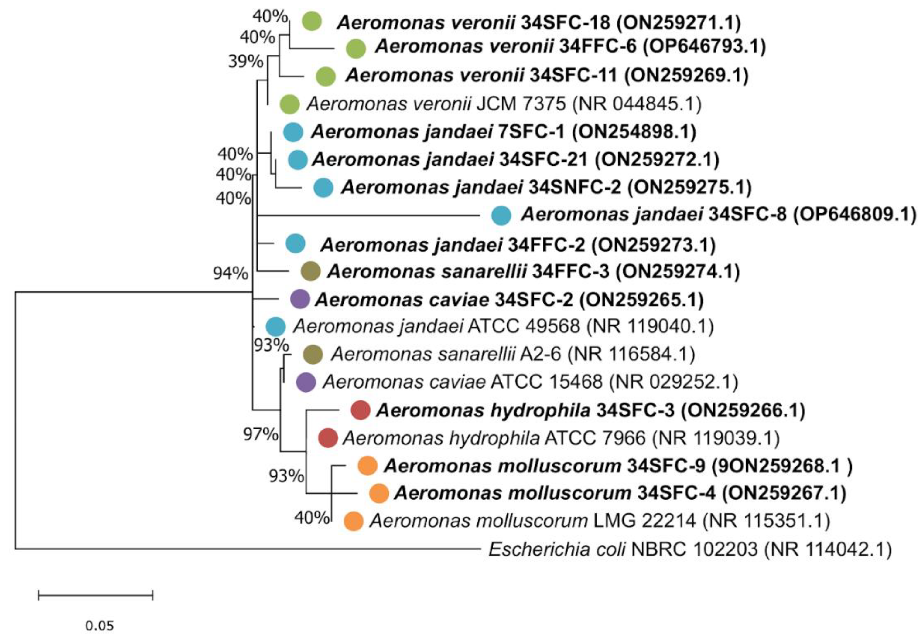
2.1. Identification of Aeromonas Strains

Among the 13 antimicrobial-resistant strains of Aeromonas investigated, five (7SFC-1, 34SFC-21, 34SNFC-2, 34SFC-8, and 34FFC-2) were identified as Aeromonas jandaei (38.4%), three (34SFC-11, 34FFC-6, 34SFC-18) as Aeromonas veronii (23%), two (34SFC-4, 34SFC-9) as Aeromonas molluscorum, one (34SFC-2) as Aeromonas caviae, one (34FFC-3) as Aeromonas sanarellii, and one (34SFC-3) as Aeromonas hydrophila subsp. hydrophila. All strains were identified at the species level with 16S rRNA sequence identities higher than 98.75%, except for strain A. jandaei 34SFC-8, which showed an identity of 94.96%.
Phylogenetic analysis revealed a clustering of strains identified as A. veronii, A. hydrophila, and A. molluscorum with their corresponding reference sequence (A. veronii JCM 7375, A. hydrophila ATCC 7966, and A. molluscorum LMG 22214, respectively). The strains of A. caviae and A. sanarelli did not cluster with their reference sequence. Although somewhat distant from their reference sequence, strains of A. jandaei tended to cluster together (Figure 1).

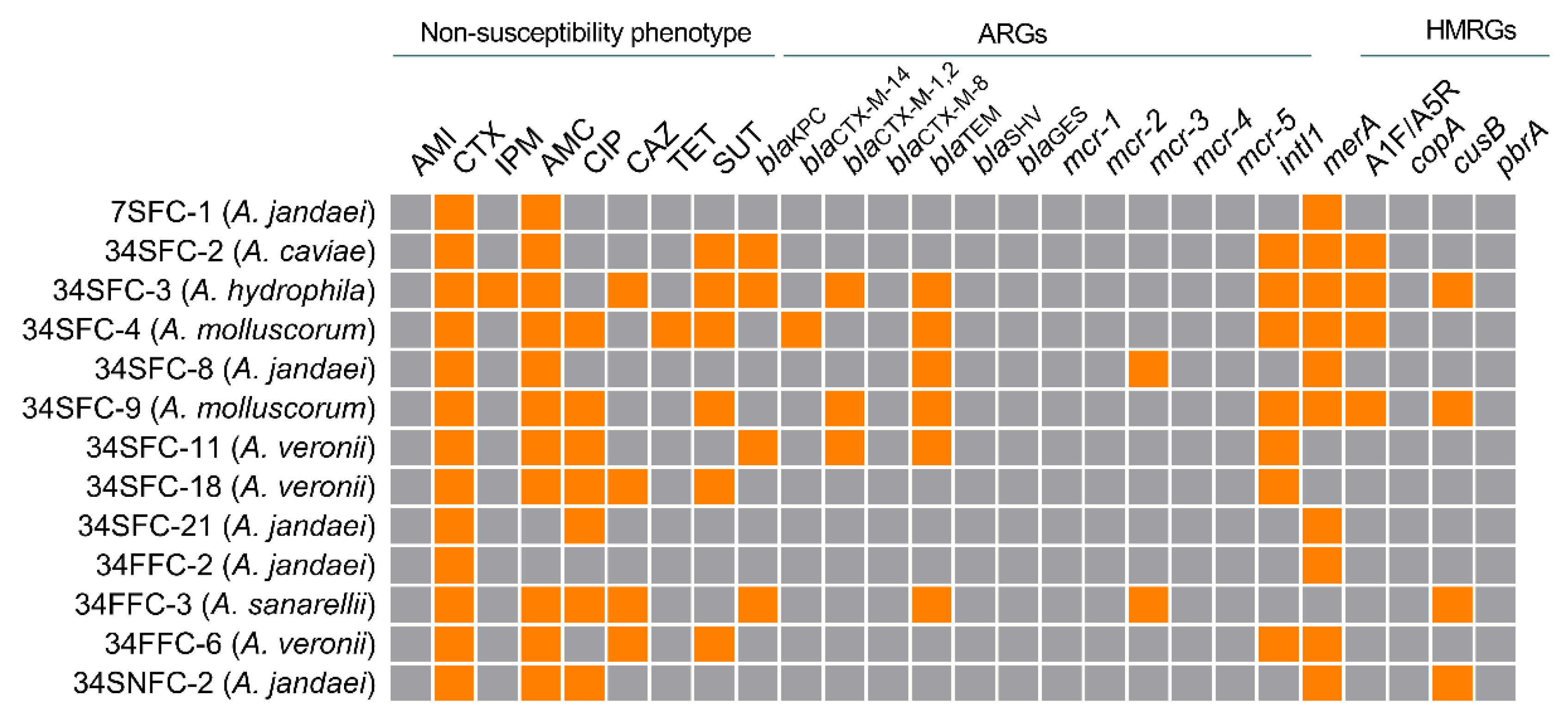
2.2. Detection of intI1, Antimicrobial and Heavy Metal Resistance Genes

Antimicrobial-resistant strains were further characterized to assess the presence of class 1 integron-integrase, and antimicrobial and heavy metal resistance genes. Among these, six (46.1%) were blaTEM positive. The second most frequently detected gene was blaKPC, identified in four (30.7%) strains. The mcr-3 gene was detected in two (15.4%) and the intI1 gene was detected in seven (53.8%) of the tested strains (Figure 2). On the other hand, merA, coding for mercury resistance, was the most often detected HMRG (10, 76.9%) and, out of a total of 11 strains positive for at least one HMRG, six (54.5%) strains also carried at least one ARG. The previously performed phenotypic antimicrobial susceptibility test results [8] are also shown in Figure 2. For the detection of heavy metal resistance, we relied on PCR assays, a more direct and eco-friendly approach.

2.3. Genomic Analyses

On account of its non-susceptibility to imipenem, the detection of ARGs and HMRGs, and the presence of the intI1 gene, A. hydrophila strain 34SFC-3 was selected for whole genome sequencing. Genome-based identification confirmed the strain’s identification as A. hydrophila, and its complete genome consists of a single circular 4,961,387-bp chromosome with 60.9% GC content that encodes 4352 predicted coding sequences (CDSs). A total of 67 tRNA and five rRNA genes were predicted in the genome. Multilocus sequence typing assigned the strain to the ST378.
Upon investigation of the strain’s complete genome sequence, several ARGs were detected (Table 1), conferring resistance to ten different antimicrobial classes: beta-lactams, macrolides, sulfonamides, aminoglycosides, tetracyclines, colistin, fluoroquinolones, chloramphenicol, virginiamycin, and elfamycin. Noteworthy, A. hydrophila 34SFC-3 carries seven beta-lactamases, namely blaTEM-1, blaCTX-M-2, blaKPC-2, blaOXA-726, imiH, blaOXA-18, and blaTOHO-1. Interestingly, several mobile genetic elements were also detected, including an IncQ1 plasmid with 87.4% identity to the Escherichia coli plasmid RSF1010 (accession number: M28829.1). However, ARGs were not found in this plasmid, letting us infer that it may not play a role in the transfer of the detected genes in our survey. Five different insertion sequences (IS) were found, namely ISAdh2, ISAhy1, ISAeme10, and ISAs23, which belong to the IS1595 family, and ISPsy43, which belongs to the IS66 family. For the detected ISs, different bacterial origins were designated, such as A. dhakensis, A. hydrophila, Pseudomonas syringae, Aeromonas media, and Aeromonas salmonicida.
Furthermore, we detected an oriT region, accompanied by a relaxase gene (traI) and two clusters of a type IV secretion system cluster in relative proximity (within a range of 300-kb upstream and 300-kb downstream from the oriT site) of six ARGs [blaKPC, blaCTX-M-2, mphA, sul1, aac(6’)-Ib-cr6, and aac(3)-IId]. The search for integrative conjugative elements (ICEs) revealed a putative ICE associated with a T4SS (31,665 bp) and a putative integrative and mobilizable element (IME; 9157 bp) (Figure 3).
Along with ARGs, heavy metal resistance genes were found during the genomic analyses of strain 34SFC-3 (Table 2). Genes typically found in the mer operon, conferring resistance to mercury, were detected. In addition, other genes related to copper, silver, arsenic, cadmium, cobalt, zinc, and lead resistance were also identified.
Among the main virulence factors described for the genus Aeromonas, A. hydrophila strain 34SFC-3 harbors several genes encompassing different mechanisms, which are presented in Table 3.

2.4. Adaptation to a Polluted Environment

A. hydrophila 34SFC-3 harbors several features that could contribute to its survival in a deeply polluted environment. For instance, the genes involved in pyomelanin production, phhA, phhB, and tyrR, are present in its genome. The gene hpd, which codes for a 4-hydroxyphenylpyruvate dioxygenase and is essential for melanin biosynthesis, as well as the hmgA gene, coding for a homogentisate 1,2-dioxygenase, were also detected during genome annotation.
Sulfo-related reductase enzymes have also been identified in A. hydrophila 34SFC-3′s genome, which play a role in dealing with oxidative stress. For example, the strain harbors glutathione S-transferases (gstB), peptide methionine sulfoxide reductases (msrA and msrB), glutaredoxins (grxC), a zinc-type alcohol dehydrogenase-like protein, and 3-ketoacyl-CoA thiolases (fadI and fadA). Further, a thiopurine S-methyltransferase (tpm; involved in selenium cycling), a putative NAD(P)H nitroreductase YdjA (ydjA; associated with the reduction of nitroaromatic compounds) and an FMN-dependent NADH-azoreductase (azoR; associated with the reduction of azo groups) were detected. Enzymes involved in phosphonates degradation, such as 2-aminoethylphosphonate-pyruvate transaminase (phnW), and phosphonoacetaldehyde hydrolase (phnX) were also identified.

3. Discussion

Under the One Health perspective, investigating antimicrobial resistance and bacterial virulence beyond the clinic is essential to better understand the mechanisms behind the origin and spread of clinically-relevant determinants. In the current study, we focused on strains previously classified as antimicrobial-resistant [8] and expanded their characterization to also encompass their heavy metal resistance profile and to determine the carriage of class 1 integron-integrases. The intI1 gene plays a pivotal role in bacterial adaptation and antimicrobial resistance acquisition and dissemination [12]. More than 50% of the tested strains were positive for the detection of intI1, which has already been described in other environmental strains of Aeromonas, such as those isolated from zebrafish [13] and water and wastewater samples [14]. Also, the presence of the ARGs and HMRGs investigated in this study agrees with what is described in the genus Aeromonas [15,16,17,18].
All strains investigated were phenotypically resistant to at least two antimicrobials [8], raising the possibility that these strains are under selective pressure in the environment. Even though Guanabara Bay is a recreational and fishing area, it receives substantial input from untreated sewage, and agricultural and industrial effluents. Indeed, several studies have already reported the transport of ARGs, antimicrobial residues, heavy metals, and other pollutants in this kind of effluent [10,19]. Hence, the isolation of antimicrobial and heavy metal-resistant Aeromonas strains in a recreational environment raises attention to the potential risks associated with human exposure to these waters or the consumption of seafood from this region [20,21]. Although cases of Aeromonas spp. infections are not as frequent as other water-borne pathogens, they are considered emerging pathogens, and the arsenal of ARGs, virulence factors, and mobile genetic elements they carry may indeed pose a challenge to public health [5,22].
Regarding the antimicrobial resistance profile of the selected strains, it is of special interest to note the detection of the mobile colistin resistance gene mcr-3 and the carbapenemase-encoding blaKPC gene. The presence of mcr-3 is well established in Aeromonas spp., and these bacteria may act as reservoirs of this gene in the environment [23,24]. The detection of blaKPC-harboring Aeromonas spp. has increased over the past years and they are usually described in hospital effluents and wastewater treatment plants [6,25,26]. Further genomic analyses confirmed not only the presence of the blaKPC and mcr-3 genes in strain 34SFC-3 but also unveiled several other ARGs, which could also pose a risk to public health. Some beta-lactamases are intrinsic to Aeromonas spp., such as cphA, which was detected in this study and confers carbapenem resistance [27]. Also, considering the arsenal of mobile genetic elements usually detected in this bacterial genus, Aeromonas spp. are possibly key disseminators of ARGs and likely mediate their transfer between typical clinical isolates and environmental bacteria [22]. For example, a strain of A. caviae isolated from a patient with pneumonia harbored IMEs associated with ARGs conferring resistance to different antimicrobial classes. Multidrug-resistant strains of Aeromonas isolated from zebrafish have also been shown to harbor class 1 integrons associated with ARGs, namely the qacG-aadA6-qacG array and drfA1 [13]. Indeed, a highly mobilizable region was detected in the A. hydrophila 34SFC-3 genome, thus reinforcing its genetic promiscuity.
Apart from the antimicrobial susceptibility profile of the strains, the pathogenic potential was further investigated in A. hydrophila 34SFC-3. Several virulence-related genes were detected, mostly encompassing mechanisms of adhesion and secretion systems. The strain also harbors the aerA (aerolysin) and the hlyA (hemolysin) genes, suggesting its pathogenic potential. The occurrence of virulence genes in environmental strains is not unusual and they likely emerge as a consequence of exaptation, that is, factors primarily involved in environmental adaptation may also enable bacteria to become opportunistic pathogens, particularly in aquatic animals and, also, in humans [28].The strain was also assigned to ST378 which, according to the PubMLST database, had only been described once in China in 2014. Thus, its epidemiological relevance remains unclear.
With the exception of strain A. jandaei 7SFC-1, isolated from site 7, which is intermediately impacted by anthropogenic pollution in GB, all strains were isolated from site 34, considered one of the most polluted areas of GB [8,29]. In order to survive in environments with harsh conditions, bacteria must develop mechanisms to ensure their adaptation. For instance, nitroaromatic and azocompounds are often present in polluted environments. Nitroaromatic compounds usually originate from pharmaceutical, agrochemical, and explosives industries, while azocompounds are typically found in dyes used in textile industries [30]. Genomic analysis of A. hydrophila 34SFC-3 revealed the presence of a putative nitroreductase (ydjA) and an azoreductase (azoR), which have been previously investigated for their bioremediation potential. For example, a thermostable azoreductase from Geobacillus stearothermophilus exhibited broad substrate specificity and was considered a good candidate for whole-cell wastewater treatment [31]. Further, the several sulfo-related reductases here described have already been identified in strains of Aeromonas spp. isolated from effluents originating from a wastewater treatment plant, where they might offer protection against oxidative stress [6].
The detection of a thiopurine S-methyltransferase (tpm) also hints at the pollution levels in the waters where strain 34SFC-3 was isolated. This enzyme has already been described in the genus Aeromonas and is involved in the detoxification of metalloid-containing oxyanions and xenobiotics [32,33]. The 2-aminoethylphosphonate-pyruvate transaminase (phnW) and phosphonoacetaldehyde hydrolase (phnX) have already been reported in the A. hydrophila strain ATCC 7966T and are involved in the degradation of phosphonates [34]. Another feature of some species of Aeromonas is the production of melanin and multiple enzymes previously reported in the synthesis pathways of this pigment were found. Melanin plays an important role in the protection against environmental stress, such as radiation, oxidative damage, and heavy metals, among others, and may also contribute to bacterial virulence [35]. For instance, a strain of A. media isolated from a lake in China was able to synthesize high levels of melanin, which was also proposed to be explored from a biotechnological perspective, namely in the photoprotection of bioinsecticides [36].

4. Materials and Methods

4.1. Water Sampling and Bacterial Strains

Aeromonas strains were isolated from three different sampling sites (1, 7, and 34; Figure S1) in Guanabara Bay (Rio de Janeiro, Brazil) in a previous report [8]. Briefly, filtered (membrane pore size 0.22 µm) and non-filtered 100 µL aliquots of water were spread onto TCBS agar supplemented with ceftriaxone (2 µg/mL). The cultures were incubated overnight at 25 °C and the colonies were isolated, purified, and cryopreserved as 20% glycerol stocks at −20 °C for further analyses. Thirteen strains were selected according to their phenotypic antimicrobial susceptibility [8].

4.2. Bacterial Identification and Phylogenetic Analysis

Bacterial identification was carried out by amplification and sequencing of the 16S rRNA gene. Briefly, genomic DNA was obtained using Chelex 100 resin [37]. Polymerase chain reaction (PCR) conditions were conducted as previously described [38] using the 16S rRNA universal primers, 27F (5′-GAGTTTGATCMTGGCTCAG-3′) and 1492R (5′-TACGGYTACCTTGTTACGACTT3′) [39]. PCR products were purified and directly sequenced using the 338F (5′-ACTCCTACGGGAGGCAGC-3′) at Biotecnologia, Pesquisa e Inovação (BPI, SP, Brazil). The resulting 16S rRNA sequences were quality-checked using Chromas 2.0 and taxonomic classification was performed with the EzBioCloud Database. An identity cut-off of 98.7% was considered for species delimitation [40]. All gene sequences were deposited in the NCBI GenBank database under the accession numbers: ON254898.1 (7SFC-1); ON259265.1 (34SFC-2); ON259266.1 (34SFC-3); ON259267.1 (34SFC-4); OP646809.1 (34SFC-8); ON259268.1 (34SFC-9); ON259269.1 (34SFC-11); ON259271.1 (34SFC-18); ON259272.1 (34SFC-21); ON259273.1 (34FFC-2); ON259274.1 (34FFC-3); OP646793.1 (34FFC-6); ON259275.1 (34SNFC-2).
Phylogenetic analysis of the thirteen strains was conducted by multiple sequence alignment with the Muscle algorithm in the MEGA software v.7. Reference sequences were downloaded from NCBI and a maximum likelihood (ML) tree was constructed using the Tamura-Nei model (Tn93+G+I) as the best-fit nucleotide substitution model with a bootstrap of 100 replications. E. coli NBRC 102203 (accession number: NR_114042.1) was used as an outgroup during the phylogeny inference.

4.3. Molecular Detection of Antimicrobial and Heavy Metal Resistance Genes

PCR reactions were performed for the following genes encoding resistance to beta-lactams: blaCTX-M-8, blaCTX-M-1,2, blaCTX-M-14, blaGES, blaTEM, blaSHV, and blaKPC. Genes coding for mobile colistin resistance mcr-1, mcr-2, mcr-3, mcr-4, and mcr-5, as well as class 1 integron-integrase (intI1), were assessed in our survey. Heavy metal resistance genes merA, merB, cusB, copA, and pbrA were also investigated. Amplification reactions were carried out in a total volume of 25 µL using 10 ng of genomic DNA, 1x buffer GO TAQ Green Master Mix (Promega, Madison, WI, USA), and 20 pmol/L of each primer. All primer sequences and specific amplification conditions are detailed in Tables S1 and S2.

4.4. Whole Genome Sequencing, Assembly, and Annotation

Total DNA was extracted by the phenol-chloroform method [41]. Library preparation was performed using the Nextera DNA Flex Library Prep (Illumina) kit. DNA concentration and quantification were assessed using QuBit 4 with the DNA High Sensitivity test. Fragment size was visualized using Tapestation (Agilent Technologies) with the D1000 ScreenTape System. Genomic DNA was sequenced with the Illumina NextSeq 550. The quality of the read libraries was assessed using the FastQC v0.11.9 tool [42]. Pre-processing was performed with the Trim Galore v0.6.7 [43]. Genome assembly was conducted with SPAdes v. 3.15.4 [44]. Gene prediction was performed with Prokka v1.14.15 [45] and the genome completeness was calculated with the CheckM v1.1.11 tool [46]. The complete genome of strain 34SFC-3 is deposited at NCBI under the BioProject PRJNA894989.

4.5. Functional Genomic Analyses

The GTDB-Tool Kit 2.1.0 (GTDB-Tk) was used to taxonomically classify all strains based on the presence of 120 single-copy marker genes in their draft assemblies and the placement of their genomes in the Genome Taxonomy Database (GTDB) reference tree using the available database (GTDB version 1.4.5) [47]. Multilocus sequence typing of the whole genome sequence was performed with the web-based MLST sequence database [48] using the Aeromonas spp. MLST scheme (https://pubmlst.org/organisms/aeromonas-spp; accessed on 14 October 2022). Acquired antimicrobial resistance genes (ARGs) and/or chromosomal mutations were verified with the Resistance Gene Identifier tool (RGI 5.1.0) from the Comprehensive Antibiotic Resistance Database (CARD 3.0.7) [49], narrowing the criteria for “perfect and strict hits only” and the sequence quality for “high quality/coverage”. Additional ARGs were also searched manually. Virulence factors were investigated with the Virulence Factors Database (VFDB) [50]. Plasmids and insertion sequences (IS) were screened using PlasmidFinder [51,52] and ISFinder [53] (cut-off: 1 × 10⁻⁵), respectively. Integrative mobilizable elements (IMEs) and integrative conjugative elements (ICEs) were investigated with the ICEfinder web-based tool in ICEberg v.2.0 [54]. OriTfinder was used to detect the origins of transfer and three other transfer-associated modules, namely relaxase, type IV coupling proteins (T4CP), and type IV secretion system (T4SS) [55]. To predict integrons, the sequencing data were uploaded to the Galaxy web platform and the public server at https://galaxy.pasteur.fr/ (accessed on 24 October 2022) was used for the analysis to analyze the data with Integron Finder version 2.0.2 [56].

5. Conclusions

All in all, we demonstrated that Aeromonas strains isolated from a recreational estuary can harbor several ARGs, HMRGs, and virulence factors, suggesting that these bacteria should not be overlooked. Hence, our findings reinforce the importance of environmental assessment of the resistance and virulence determinants, thus providing valuable insights to tackle the emerging issue of antimicrobial resistance having Aeromonas as a paramount model in that framework.

Supplementary Materials

The following supporting information can be downloaded at: https://www.mdpi.com/article/10.3390/antibiotics12010084/s1, Figure S1: Guanabara Bay sampling sites; Table S1: Primer sequences, cycling conditions and amplicon sizes of the antimicrobial resistance genes and the class 1 integron-integrase gene investigated in this study; Table S2: Primer sequences, cycling conditions and amplicon sizes of the heavy metal resistance genes and the class 1 integron-integrase gene investigated in this study.

Author Contributions

Conceptualization, A.L.B.C.; B.F.R.d.O. and M.S.L.; formal analysis, A.L.B.C.; B.F.R.d.O.; writing—original draft preparation, A.L.B.C.; B.F.R.d.O. and M.S.L.; writing—review and editing, A.L.B.C.; B.F.R.d.O. and M.S.L.; supervision, M.S.L.; funding acquisition, M.S.L. All authors have read and agreed to the published version of the manuscript.

Funding

This work was supported by Conselho Nacional de Desenvolvimento Científico e Tecnológico (CNPq) under Grant number 306395/2020-7; Coordenação de Aperfeiçoamento de Pessoal de Nível Superior (CAPES) under Grant number 88887.613830/2021-00, Financial Code 001; Fundação Carlos Chagas Filho de Amparo à Pesquisa do Estado do Rio de Janeiro under Grant numbers E26/211.554/2019, E-26/200.948/2021, E-26/211.284/2021, E-26/202.405/2022.

Acknowledgments

The authors thank Walter Oelemann for his contributions during the preparation of the manuscript and acknowledge Servier Medical Art (smart.servier.com) for providing some of the elements within the graphical abstract.

Conflicts of Interest

The authors declare no conflict of interest.

References

  1. Larsson, D.G.; Flach, C.F. Antibiotic resistance in the environment. Antibiotic resistance in the environment. Nat. Rev. Microbiol. 2022, 20, 257–269. [Google Scholar] [CrossRef] [PubMed]
  2. Marti, E.; Variatza, E.; Balcazar, J.L. The role of aquatic ecosystems as reservoirs of antibiotic resistance. Trends Microbiol. 2014, 22, 36–41. [Google Scholar] [CrossRef] [PubMed]
  3. Canellas, A.L.B.; Laport, M.S. The biotechnological potential of Aeromonas: A bird’s eye view. Crit. Rev. Microbiol. 2022, 1–13. [Google Scholar] [CrossRef] [PubMed]
  4. List of Prokaryotic Names with Standard Nomenclature (LPSN). Aeromonas. Available online: https://lpsn.dsmz.de/genus/aeromonas (accessed on 22 October 2022).
  5. Fernández-Bravo, A.; Figueras, M.J. An update on the genus Aeromonas: Taxonomy, epidemiology, and pathogenicity. Microorganisms 2020, 8, 129. [Google Scholar] [CrossRef] [PubMed] [Green Version]
  6. Sekizuka, T.; Inamine, Y.; Segawa, T.; Hashino, M.; Yatsu, K.; Kuroda, M. Potential KPC-2 carbapenemase reservoir of environmental Aeromonas hydrophila and Aeromonas caviae isolates from the effluent of an urban wastewater treatment plant in Japan. Environ. Microbiol. Rep. 2019, 11, 589–597. [Google Scholar] [CrossRef] [Green Version]
  7. Conte, D.; Palmeiro, J.K.; Bavaroski, A.A.; Rodrigues, L.S.; Cardozo, D.; Tomaz, A.P.; Camargo, J.O.; Dalla-Costa, L.M. Antimicrobial resistance in Aeromonas species isolated from aquatic environments in Brazil. J. Appl. Microbiol. 2021, 131, 169–181. [Google Scholar] [CrossRef]
  8. Canellas, A.L.B.; da Costa, W.F.; Paranhos, R.; Laport, M.S. Diving into the unknown: Identification of antimicrobial resistance hotspots in a tropical urban estuary. Lett. Appl. Microbiol. 2021, 73, 270–279. [Google Scholar] [CrossRef]
  9. Leonard, A.F.; Zhang, L.; Balfour, A.J.; Garside, R.; Hawkey, P.M.; Murray, A.K.; Ukoumunne, O.C.; Gaze, W.H. Exposure to and colonisation by antibiotic-resistant E. coli in UK coastal water users: Environmental surveillance, exposure assessment, and epidemiological study (Beach Bum Survey). Environ. Int. 2018, 114, 326–333. [Google Scholar] [CrossRef]
  10. Fistarol, G.O.; Coutinho, F.H.; Moreira, A.P.B.; Venas, T.; Cánovas, A.; de Paula, S.E., Jr.; Coutinho, R.; de Moura, R.L.; Valentin, J.V.; Tenenbaum, D.R.; et al. Environmental and sanitary conditions of Guanabara Bay, Rio de Janeiro. Front. Microbiol. 2015, 6, 1232. [Google Scholar] [CrossRef] [Green Version]
  11. Grilo, M.L.; Sousa-Santos, C.; Robalo, J.; Oliveira, M. The potential of Aeromonas spp. from wildlife as antimicrobial resistance indicators in aquatic environments. Ecol. Indic. 2020, 115, 106396. [Google Scholar] [CrossRef]
  12. Ghaly, T.M.; Gillings, M.R.; Penesyan, A.; Qi, Q.; Rajabal, V.; Tetu, S.G. The natural history of integrons. Microorganisms 2021, 9, 2212. [Google Scholar] [CrossRef] [PubMed]
  13. Hossain, S.; Dahanayake, P.S.; De Silva, B.C.J.; Wickramanayake, M.V.K.S.; Wimalasena, S.H.M.P.; Heo, G.J. Multidrug resistant Aeromonas spp. isolated from zebrafish (Danio rerio): Antibiogram, antimicrobial resistance genes and class 1 integron gene cassettes. Lett. Appl. Microbiol. 2019, 68, 370–377. [Google Scholar] [CrossRef] [PubMed]
  14. Harnisz, M.; Korzeniewska, E. The prevalence of multidrug-resistant Aeromonas spp. in the municipal wastewater system and their dissemination in the environment. Sci. Total Environ. 2018, 626, 377–383. [Google Scholar] [CrossRef] [PubMed]
  15. Piotrowska, M.; Przygodzińska, D.; Matyjewicz, K.; Popowska, M. Occurrence and variety of β-lactamase genes among Aeromonas spp. isolated from urban wastewater treatment plant. Front. Microbiol. 2017, 8, 863. [Google Scholar] [CrossRef] [Green Version]
  16. Dahanayake, P.S.; Hossain, S.; Wickramanayake, M.V.K.S.; Heo, G.J. Antibiotic and heavy metal resistance genes in Aeromonas spp. isolated from marketed Manila Clam (Ruditapes philippinarum) in Korea. J. Appl. Microbiol. 2019, 127, 941–952. [Google Scholar] [CrossRef]
  17. De Silva, B.C.; Hossain, S.; Dahanayake, P.S.; Heo, G.J. Frozen white-leg shrimp (Litopenaeus vannamei) in Korean markets as a source of Aeromonas spp. harboring antibiotic and heavy metal resistance genes. Microb. Drug Resist. 2018, 24, 1587–1598. [Google Scholar] [CrossRef]
  18. Hossain, S.; Heo, G.J. Detection of Antimicrobial and Heavy-Metal Resistance Genes in Aeromonas spp. Isolated from Hard-Shelled Mussel (Mytilus coruscus). Microb. Drug Resist. 2022, 28, 127–135. [Google Scholar] [CrossRef]
  19. Xu, J.; Xu, Y.; Wang, H.; Guo, C.; Qiu, H.; He, Y.; Zhang, Y.; Li, X.; Meng, W. Occurrence of antibiotics and antibiotic resistance genes in a sewage treatment plant and its effluent-receiving river. Chemosphere 2015, 119, 1379–1385. [Google Scholar] [CrossRef]
  20. Dong, P.; Cui, Q.; Fang, T.; Huang, Y.; Wang, H. Occurrence of antibiotic resistance genes and bacterial pathogens in water and sediment in urban recreational water. J. Environ. Sci. 2019, 77, 65–74. [Google Scholar] [CrossRef]
  21. de Melo Rodrigues Sobral, M.; Barreto, C.; Bianco, K.; de Oliveira, S.S.A.; Clementino, M.M. Virulence determinants in genetically heterogeneous populations of Aeromonads recovered from an urban lagoon. J. Water Health. 2019, 17, 380–392. [Google Scholar] [CrossRef]
  22. Lamy, B.; Baron, S.; Barraud, O. Aeromonas: The multifaceted middleman in the One Health world. Curr. Opin. Microbiol. 2022, 65, 24–32. [Google Scholar] [CrossRef]
  23. Shen, Y.; Xu, C.; Sun, Q.; Schwarz, S.; Ou, Y.; Yang, L.; Huang, Z.; Eichhorn, I.; Walsh, T.R.; Wang, Y.; et al. Prevalence and genetic analysis of mcr-3-positive Aeromonas species from humans, retail meat, and environmental water samples. Antimicrob. Agents Chemother. 2018, 62, e00404-18. [Google Scholar] [CrossRef] [Green Version]
  24. Li, L.; Yao, R.; Olsen, R.H.; Zhang, Y.; Meng, H. Antibiotic resistance and polymyxin B resistance mechanism of Aeromonas spp. isolated from yellow catfish, hybrid snakeheads and associated water from intensive fish farms in Southern China. LWT 2022, 166, 113802. [Google Scholar] [CrossRef]
  25. Yang, C.; Guo, M.; Yang, H.; Wen, Y.; Zhu, Z.; Wang, T.; Zhu, J.; Chen, L.; Du, H. blaKPC-24-Harboring Aeromonas veronii from the Hospital Sewage Samples in China. Microbiol. Spectr. 2022, 10, e00555-22. [Google Scholar] [PubMed]
  26. Li, Y.; Qiu, Y.; Fang, C.; Dai, X.; Zhang, L. Genomic Characterization of a Multidrug-Resistant Aeromonas caviae Isolate Carrying a Novel blaKPC-2-Harbouring Plasmid and an IMP-4-Encoding Phage-like Plasmid. Microbiol. Spectr. 2022, 10, e00840-22. [Google Scholar] [PubMed]
  27. Lim, S.R.; Lee, D.H.; Park, S.Y.; Lee, S.; Kim, H.Y.; Lee, M.S.; Han, J.E.; Kim, H.K.; Kim, J.H. Wild nutria (Myocastor coypus) is a potential reservoir of carbapenem-resistant and zoonotic Aeromonas spp. in Korea. Microorganisms 2019, 7, 224. [Google Scholar] [CrossRef] [Green Version]
  28. Talagrand-Reboul, E.; Colston, S.M.; Graf, J.; Lamy, B.; Jumas-Bilak, E. Comparative and evolutionary genomics of isolates provide insight into the pathoadaptation of Aeromonas. Genome Biol. Evol. 2020, 12, 535–552. [Google Scholar] [CrossRef] [Green Version]
  29. Canellas, A.L.B.; Lopes, I.R.; Mello, M.P.; Paranhos, R.; de Oliveira, B.F.R.; Laport, M.S. Vibrio species in an urban tropical estuary: Antimicrobial susceptibility, interaction with environmental parameters, and possible public health outcomes. Microorganisms 2021, 9, 1007. [Google Scholar] [CrossRef]
  30. Mercier, C.; Chalansonnet, V.; Orenga, S.; Gilbert, C. Characteristics of major Escherichia coli reductases involved in aerobic nitro and azo reduction. J. Appl. Microbiol. 2013, 115, 1012–1022. [Google Scholar] [CrossRef]
  31. Matsumoto, K.I.; Mukai, Y.; Ogata, D.; Shozui, F.; Nduko, J.M.; Taguchi, S.; Ooi, T. Characterization of thermostable FMN-dependent NADH azoreductase from the moderate thermophile Geobacillus stearothermophilus. Appl. Microbiol. Biotechnol. 2010, 86, 1431–1438. [Google Scholar] [CrossRef]
  32. Rael, R.M.; Frankenberger, W.T., Jr. Influence of pH, salinity, and selenium on the growth of Aeromonas veronii in evaporation agricultural drainage water. Water Res. 1996, 30, 422–430. [Google Scholar] [CrossRef]
  33. Aigle, A.; Colin, Y.; Bouchali, R.; Bourgeois, E.; Marti, R.; Ribun, S.; Marjolet, L.; Pozzi, A.C.M.; Misery, B.; Colinon, C.; et al. Spatio-temporal variations in chemical pollutants found among urban deposits match changes in thiopurine S-methyltransferase-harboring bacteria tracked by the tpm metabarcoding approach. Sci. Total Environ. 2021, 767, 145425. [Google Scholar] [CrossRef] [PubMed]
  34. Seshadri, R.; Joseph, S.W.; Chopra, A.K.; Sha, J.; Shaw, J.; Graf, J.; Haft, D.; Wu, M.; Ren, Q.; Rosovitz, M.J.; et al. Genome sequence of Aeromonas hydrophila ATCC 7966T: Jack of all trades. J. Bacteriol. 2006, 188, 8272–8282. [Google Scholar] [CrossRef] [PubMed] [Green Version]
  35. Qiao, Y.; Wang, J.; Wang, H.; Chai, B.; Rao, C.; Chen, X.; Du, S. 4-Hydroxyphenylpyruvate dioxygenase thermolability is responsible for temperature-dependent melanogenesis in Aeromonas salmonicida subsp. salmonicida. Appl. Environ. Microbiol. 2019, 85, e01926-18. [Google Scholar] [CrossRef] [Green Version]
  36. Wan, X.; Liu, H.M.; Liao, Y.; Su, Y.; Geng, J.; Yang, M.Y.; Chen, X.D.; Shen, P. Isolation of a novel strain of Aeromonas media producing high levels of DOPA-melanin and assessment of the photoprotective role of the melanin in bioinsecticide applications. J. Appl. Microbiol. 2007, 103, 2533–2541. [Google Scholar] [CrossRef]
  37. Walsh, P.S.; Metzger, D.A.; Higuchi, R. Chelex 100 as a medium for simple extraction of DNA for PCR-based typing from forensic material. Biotechniques 1991, 10, 506–513. [Google Scholar] [CrossRef] [PubMed] [Green Version]
  38. Laport, M.S.; Bauwens, M.; Oliveira, S.N.; Willenz, P.; George, I.; Muricy, G. Culturable bacterial communities associated to Brazilian Oscarella species (Porifera: Homoscleromorpha) and their antagonistic interactions. Antonie Van Leeuwenhoek 2017, 110, 489–499. [Google Scholar] [CrossRef]
  39. Weisburg, W.G.; Barns, S.M.; Pelletier, D.A.; Lane, D.J. 16S ribosomal DNA amplifcation for phylogenetic study. J. Bacteriol. 1991, 173, 697–703. [Google Scholar] [CrossRef] [Green Version]
  40. Stackebrandt, E.; Ebers, J. Taxonomic parameters revisited: Tarnished gold standards. Microbiol. Today 2006, 33, 152–155. [Google Scholar]
  41. Jackson, S.A.; Crossman, L.; Almeida, E.L.; Margassery, L.M.; Kennedy, J.; Dobson, A.D. Diverse and abundant secondary metabolism biosynthetic gene clusters in the genomes of marine sponge derived Streptomyces spp. isolates. Mar. Drugs 2018, 16, 67. [Google Scholar] [CrossRef] [Green Version]
  42. Andrews, S. FastQC: A Quality Control Tool for High Throughput Sequence Data. Available online: http://www.bioinformatics.babraham.ac.uk/projects/fastqc/ (accessed on 10 April 2022).
  43. Krueger, F.; James, F.; Ewels, P.; Afyounian, E.; Schuster-Boeckler, B. FelixKrueger/TrimGalore: v0.6.7-DOI via Zenodo (0.6.7), version 0.6.7; Available online: https://zenodo.org/record/5127899 (accessed on 10 April 2022).
  44. Bankevich, A.; Nurk, S.; Antipov, D.; Gurevich, A.A.; Dvorkin, M.; Kulikov, A.S.; Lesin, V.M.; Nikolenko, S.I.; Pham, S.; Prjibelski, A.D.; et al. SPAdes: A new genome assembly algorithm and its applications to single-cell sequencing. J. Comput. Biol. 2012, 19, 455–477. [Google Scholar] [CrossRef]
  45. Seemann, T. Prokka: Rapid prokaryotic genome annotation. Bioinformatics 2014, 30, 2068–2069. [Google Scholar] [CrossRef] [PubMed] [Green Version]
  46. Parks, D.H.; Imelfort, M.; Skennerton, C.T.; Hugenholtz, P.; Tyson, G.W. CheckM: Assessing the quality of microbial genomes recovered from isolates, single cells, and metagenomes. Genome Res. 2015, 25, 1043–1055. [Google Scholar] [CrossRef] [PubMed] [Green Version]
  47. Chaumeil, P.A.; Mussig, A.J.; Hugenholtz, P.; Parks, D.H. GTDB-Tk: A toolkit to classify genomes with the Genome Taxonomy Database. Bioinformatics 2019, 36, 1925–1927. [Google Scholar] [PubMed]
  48. Jolley, K.A.; Bray, J.E.; Maiden, M. Open-access bacterial population genomics: BIGSdb software, the PubMLST.org website and their applications. Wellcome Open Res. 2018, 3, 124. [Google Scholar] [CrossRef] [PubMed]
  49. Alcock, B.P.; Raphenya, A.R.; Lau, T.T.; Tsang, K.K.; Bouchard, M.; Edalatmand, A.; Huynh, W.; Nguyen, A.L.V.; Cheng, A.A.; Liu, S.; et al. CARD 2020: Antibiotic resistome surveillance with the comprehensive antibiotic resistance database. Nucleic Acids Res. 2020, 48, D517–D525. [Google Scholar] [CrossRef] [PubMed]
  50. Liu, B.; Zheng, D.; Zhou, S.; Chen, L.; Yang, J. VFDB 2022: A general classification scheme for bacterial virulence factors. Nucleic Acids Res. 2022, 50, D912–D917. [Google Scholar] [CrossRef] [PubMed]
  51. Camacho, C.; Coulouris, G.; Avagyan, V.; Ma, N.; Papadopoulos, J.; Bealer, K.; Madden, T.L. BLAST+: Architecture and applications. BMC Bioinform. 2009, 10, 421. [Google Scholar] [CrossRef] [Green Version]
  52. Carattoli, A.; Zankari, E.; Garcia-Fernandez, A.; Larsen, M.V.; Lund, O.; Villa, L.; Aarestrup, F.M.; Hasman, H. PlasmidFinder and pMLST: In silico detection and typing of plasmids. Antimicrob. Agents Chemother. 2014, 58, 3895–3903. [Google Scholar] [CrossRef] [Green Version]
  53. Siguier, P.; Perochon, J.; Lestrade, L.; Mahillon, J.; Chandler, M. ISfinder: The reference centre for bacterial insertion sequences. Nucleic Acids Res. 2006, 34, D32–D36. [Google Scholar] [CrossRef] [Green Version]
  54. Liu, M.; Li, X.; Xie, Y.; Bi, D.; Sun, J.; Li, J.; Tai, C.; Deng, Z.; Ou, H.Y. ICEberg 2.0: An updated database of bacterial integrative and conjugative elements. Nucleic Acids Res. 2019, 47, D660–D665. [Google Scholar] [CrossRef] [PubMed]
  55. Li, X.; Xie, Y.; Liu, M.; Tai, C.; Sun, J.; Deng, Z.; Ou, H.Y. oriTfinder: A web-based tool for the identification of origin of transfers in DNA sequences of bacterial mobile genetic elements. Nucleic Acids Res. 2018, 46, W229–W234. [Google Scholar] [CrossRef] [PubMed] [Green Version]
  56. Néron, B.; Littner, E.; Haudiquet, M.; Perrin, A.; Cury, J.; Rocha, E. IntegronFinder 2.0: Identification and Analysis of Integrons across Bacteria, with a Focus on Antibiotic Resistance in Klebsiella. Microorganisms 2022, 10, 700. [Google Scholar] [CrossRef] [PubMed]
Figure 1. Maximum likelihood (ML) phylogenetic tree based on the 16S rRNA of Aeromonas spp. isolated from GB waters (in bold) and from reference sequences downloaded from NCBI. The colored dots represent different species, and the accession numbers are also depicted. Brown: A. sanarellii; purple: A. caviae; blue: A. jandaei; green: A. veronii; red: A. hydrophila, and orange A. molluscorum. Escherichia coli strain NRBC 102203 was used as an outgroup.
Antibiotics 12 00084 g001
Figure 2. Antimicrobial susceptibility profile of the thirteen strains of antimicrobial-resistant Aeromonas from GB waters (Rio de Janeiro, Brazil), as well as the results of the detection of antimicrobial and heavy metal resistance genes (ARGs and HMRGs) and the integron-integrase gene (intI1). AMI: amikacin; CTX: cefotaxime; IPM: imipenem; AMC: amoxicillin-clavulanic acid; CIP: ciprofloxacin; CAZ: ceftazidime; TET: tetracycline; SUT: trimethoprim-sulfamethoxazole. Orange squares indicate positive PCR results, while gray squares indicate negative results.
Antibiotics 12 00084 g002
Figure 3. A highly mobilizable region in A. hydrophila 34SFC-3 genome. All genes are represented by arrows which indicate their genomic orientation. Genes encoding the respective mobile elements and ARGs are differently colored following the legend. Three key areas are zoomed in for greater comprehension of the genetic contexts surrounding ARGs and mobile elements.
Antibiotics 12 00084 g003
Table 1. Antimicrobial resistance genes detected in the A. hydrophila 34SFC-3 genome, their corresponding gene family, and antimicrobials to which they confer resistance. The first part of the table refers to the results obtained in the CARD database, and the second, to the results obtained by manual curation.
GeneGene FamilyAntimicrobials
TEM-1TEM beta-lactamaseMonobactam, cephalosporin, penam, penem
mphAMacrolide phosphotransferase (MPH)Macrolide
sul1_1; sul1_2Sulfonamide resistant sulSulfonamide
qacE∆1_1; qacE∆1_2Major facilitator superfamily (MFS) antibiotic efflux pumpDisinfecting agents and antiseptics
CTX-M-2CTX-M beta-lactamaseCephalosporin
arr-3Rifampin ADP-ribosyltransferase (Arr)Rifamycin
aac(3)-IIdAAC (3)Aminoglycoside
KPC-2KPC beta-lactamaseMonobactam, carbapenem, cephalosporin, penam
qacJSmall multidrug resistance (SMR) antibiotic efflux pumpDisinfecting agents and antiseptics
adeFResistance-nodulation-cell division (RND) antibiotic efflux pumpFluoroquinolone antibiotic, tetracycline
OXA-726OXA beta-lactamaseCarbapenem, cephalosporin, penam
rsmAResistance-nodulation-cell division (RND) antibiotic efflux pumpFluoroquinolone antibiotic, diaminopyrimidine antibiotic, phenicol
imiHcphA beta-lactamaseCarbapenem
adeFResistance-nodulation-cell division (RND) antibiotic efflux pumpFluoroquinolone antibiotic, tetracycline
aac(6’)-Ib-cr6AAC (6’)-Ib-crFluoroquinolone antibiotic, aminoglycoside
Escherichia coli EF-Tu mutants conferring resistance to PulvomycinElfamycin resistant EF-TuElfamycin
tetATetracycline resistance protein, class BTetracycline
catChloramphenicol acetyltransferaseChloramphenicol
emrAColistin resistance protein EmrAColistin
emrBColistin resistance protein EmrBColistin
bla_3Beta-lactamase Toho-1Penicillin G, ampicillin, oxacillin, carbenicillin, piperacillin, cephalothin, cefoxitin, Cefotaxime, ceftazidime, and aztreonam
bla_1Beta-lactamase OXA-18amoxicillin, ticarcillin, cephalothin, Ceftazidime, cefotaxime, and aztreonam
yokDSPbeta prophage-derived aminoglycoside N (3’)-acetyltransferase-like protein YokDAminoglycoside
vatVirginiamycin A acetyltransferaseVirginiamycin
qacCQuaternary ammonium compound-resistance protein QacCQuaternary ammonium compounds, antiseptics
Table 2. Heavy metal resistance genes detected in A. hydrophila 34SFC-3 genome. The table shows the heavy metal target, the corresponding heavy metal resistance genes detected, and their functional assignment, according to Prokka annotation.
Heavy MetalGeneFunctional Assignment
MercurymerA, merA2Mercuric reductase
merC, merC2Mercuric transport protein MerC
merP, merP2Mercuric transport protein periplasmic component
merT, merT2Mercuric transport protein MerT
merR, merR1Mercuric resistance operon regulatory protein
Copper and silver resistancecopAPutative copper-importing P-type ATPase A
copA2Copper-exporting P-type ATPase
copA3Copper resistance protein A
copA4Putative copper-importing P-type ATPase A
copBCopper resistance protein B
copRTranscriptional activator protein CopR
cusA1, cusA2Cation efflux system protein CusA
cusBCation efflux system protein CusB
cusSSensor histidine kinase CusS
cueOBlue copper oxidase CueO
cueRHTH-type transcriptional regulator CueR
Arsenic resistancearsAArsenical pump-driving ATPase
arsCArsenate reductase
arsDArsenical resistance operon trans-acting repressor ArsD
acr3Arsenical-resistance protein Acr3
Molybdate resistance and homeostasismodAMolybdate-binding protein ModA
moaEMolybdopterin synthase catalytic subunit
moaDMolybdopterin synthase sulfur carrier subunit
moaAGTP 3’,8-cyclase
moaBMolybdenum cofactor biosynthesis protein B
moaCCyclic pyranopterin monophosphate synthase
Heavy metal efflux pumps and transportersczcDCadmium, cobalt and zinc/H(+)-K(+) antiporter
corAMagnesium transport protein CorA
zntRHTH-type transcriptional regulator ZntR
zntBZinc transport protein ZntB
zntAZinc/cadmium/lead-transporting P-type ATPase
fieF_1Ferrous-iron efflux pump FieF
cusBCation efflux system protein CusB
acrAMultidrug efflux pump subunit AcrA
Table 3. Virulence genes detected in A. hydrophila 34SFC-3 genome, their corresponding functional assignment, and major virulence mechanism involved.
GeneFunctional AssignmentMechanism
flab1, flab2Flagellin BAdhesion
flgBFlagellar basal body rod protein FlgB
flgCFlagellar basal-body rod protein FlgC
flgDBasal-body rod modification protein FlgD
flgEFlagellar hook protein FlgE
flgFFlagellar basal-body rod protein FlgF
flgGFlagellar basal-body rod protein FlgG
flgHFlagellar L-ring protein
flgIFlagellar P-ring protein
flgJPeptidoglycan hydrolase FlgJ
ecpDFimbria adhesin EcpD
pileFimbrial protein
pilQType IV pilus biogenesis and competence protein PilQ
ecpACommon pilus major fimbrillin subunit EcpA
pilT1, pilT2, pilT3Twitching mobility protein
tcpEToxin coregulated pilus biosynthesis protein E
chew1,2Chemotaxis protein CheW
pomA1,2,3Chemotaxis protein PomA
tabAToxin-antitoxin biofilm protein TabA
aerAAerolysinHemolysins
hlyAHemolysin
hlyBAlpha-hemolysin translocation ATP-binding Protein HlyB
hlyD1, 2Hemolysin secretion protein D, chromosomal
vgrG1,2,3Actin cross-linking toxin VgrG1
apxIB1,2Toxin RTX-I translocation ATP-binding protein
bvg1, 2Virulence sensor protein BvgSVirulence sensors
phoQVirulence sensor histidine kinase PhoQ
entEEnterobactin synthase component ESiderophore
entBEnterobactin synthase component B
entDEnterobactin synthase component D
FurFerric uptake regulation protein
epsCType II secretion system protein CSecretion systems
epsEType II secretion system protein E
epsF1,2Type II secretion system protein F
epsGType II secretion system protein G
epsHType II secretion system protein H
xcpWType II secretion system protein J
epsLType II secretion system protein L
epsMType II secretion system protein M
outNType II secretion system protein N
sctCType 3 secretion system secretin
Disclaimer/Publisher’s Note: The statements, opinions and data contained in all publications are solely those of the individual author(s) and contributor(s) and not of MDPI and/or the editor(s). MDPI and/or the editor(s) disclaim responsibility for any injury to people or property resulting from any ideas, methods, instructions or products referred to in the content.

