Review

Coumarin Triazoles as Potential Antimicrobial Agents

by
Siddappa A. Patil
1,
Aravind R. Nesaragi
1,
Raúl R. Rodríguez-Berrios
2,
Sydney M. Hampton
3,
Alejandro Bugarin
3,* and
Shivaputra A. Patil
4,*
1
Centre for Nano and Material Sciences, Jain University, Jain Global Campus, Bangalore 562112, Karnataka, India
2
Department of Chemistry, University of Puerto Rico, Rio Piedras Campus, P.O. Box 23346, San Juan 00931-3346, Puerto Rico
3
Department of Chemistry and Physics, Florida Gulf Coast University, 10501 FGCU Boulevard South, Fort Myers, FL 33965, USA
4
Pharmaceutical Sciences Department, College of Pharmacy, Rosalind Franklin University of Medicine and Science, 3333 Green Bay Road, North Chicago, IL 60064, USA
*
Authors to whom correspondence should be addressed.
Antibiotics 2023, 12(1), 160; https://doi.org/10.3390/antibiotics12010160
Submission received: 18 December 2022 / Revised: 8 January 2023 / Accepted: 9 January 2023 / Published: 12 January 2023

Abstract

:
Currently, in hospitals and community health centers, microbial infections are highly common diseases and are a leading cause of death worldwide. Antibiotics are generally used to fight microbial infections; however, because of the abuse of antibiotics, microbes have become increasingly more resistant to most of them. Therefore, medicinal chemists are constantly searching for new or improved alternatives to combat microbial infections. Coumarin triazole derivatives displayed a variety of therapeutic applications, such as antimicrobial, antioxidant, and anticancer activities. This review summarizes the advances of coumarin triazole derivatives as potential antimicrobial agents covering articles published from 2006 to 2022.

1. Introduction

In the modern drug discovery era, the design and development of new antimicrobial drugs are receiving great attention from the research community due to the emergence of multidrug-resistant strains (MDRs) in recent years [1,2,3,4]. MDRs pose a serious health threat to the global population and are frequently associated with increased healthcare costs and prolonged hospital stays [5]. Even though recent advances have improved our understanding of the pathogenesis of antimicrobial infection, scientists have become increasingly focused on discovering novel, more effective, and safe drug Candidates to overcome MDRs. In recent years our research lab has been actively involved in the design and development of new bioactive molecules to tackle MDR strains [6,7,8,9,10,11,12,13,14].
Coumarin pharmacophore has been considered the most ideal small-molecule scaffold for the development of new drugs because of its drug-like properties and, more significantly, its association with innumerable pharmacological activities. Coumarin pharmacophore is part of several clinically used drug Candidates, including some well-known antibiotic drugs (Figure 1A). Our lab recently comprehensively reviewed the medicinal applications of pharmacologically important coumarins [15,16].
Triazole, also recognized as pyrrodiazole, is a five-membered nitrogen heterocycle with two carbon and three nitrogen atoms. Triazole exists in two isomeric forms—1,2,3-triazole (II) and 1,2,4-triazole (III)—based on the positions of the nitrogen atoms in the five-membered ring system (Figure 2). Triazole analogs have greatly attracted biologists and chemists alike due to their wide applications in medicinal chemistry with numerous biological activities [17,18,19,20]. Triazole moiety is part of several clinically used drugs for the treatment of various illnesses such as cancer, diabetes, etc. Some notable antimicrobial drugs have been listed in Figure 1B.
The combination of two or more clinical drugs to achieve higher efficacy and greater clinical benefits is becoming the new normal in clinical trials. Thus, combinatorial therapies are becoming a very important part of the clinical trial process to achieve success in patient well-being. Keeping this in mind, drug discovery researchers are planning to combine two or more drug functionalities in a single molecule to obtain synergistic effects or to enhance the particular pharmacological effects of drug Candidates. Considering the pharmacological importance of both coumarins and triazoles, medicinal chemists have worked to develop new small-molecule drugs by combining coumarin (I) and triazole moieties (II or III) to generate more effective drugs (IV and V) (Figure 2).
From the literature, we observed increased antimicrobial activities by the insertion of a triazole ring into the various organic core molecules. Most of the existing antimicrobial drugs hold triazole pharmacophore in their elemental structures, which proves the antimicrobial potencies of the triazole template so that it expresses significant antimicrobial activity. From the in silico studies, it is evident that the enzyme forms hydrogen bonding interactions with the triazole ring along with coumarin moiety. Since both lactone (coumarin) and triazole are bioactive pharmacophores, the new hybrid molecule with these two bioactive species will be with increased effects evaluated in comparison to the parent drug.
The present article covers the antimicrobial activities of the combined coumarin and triazole analogs published to date and serves to further advance the drug design and development process of coumarin-bearing triazoles as possible new drug Candidates to overcome the effects of the MDR strains.

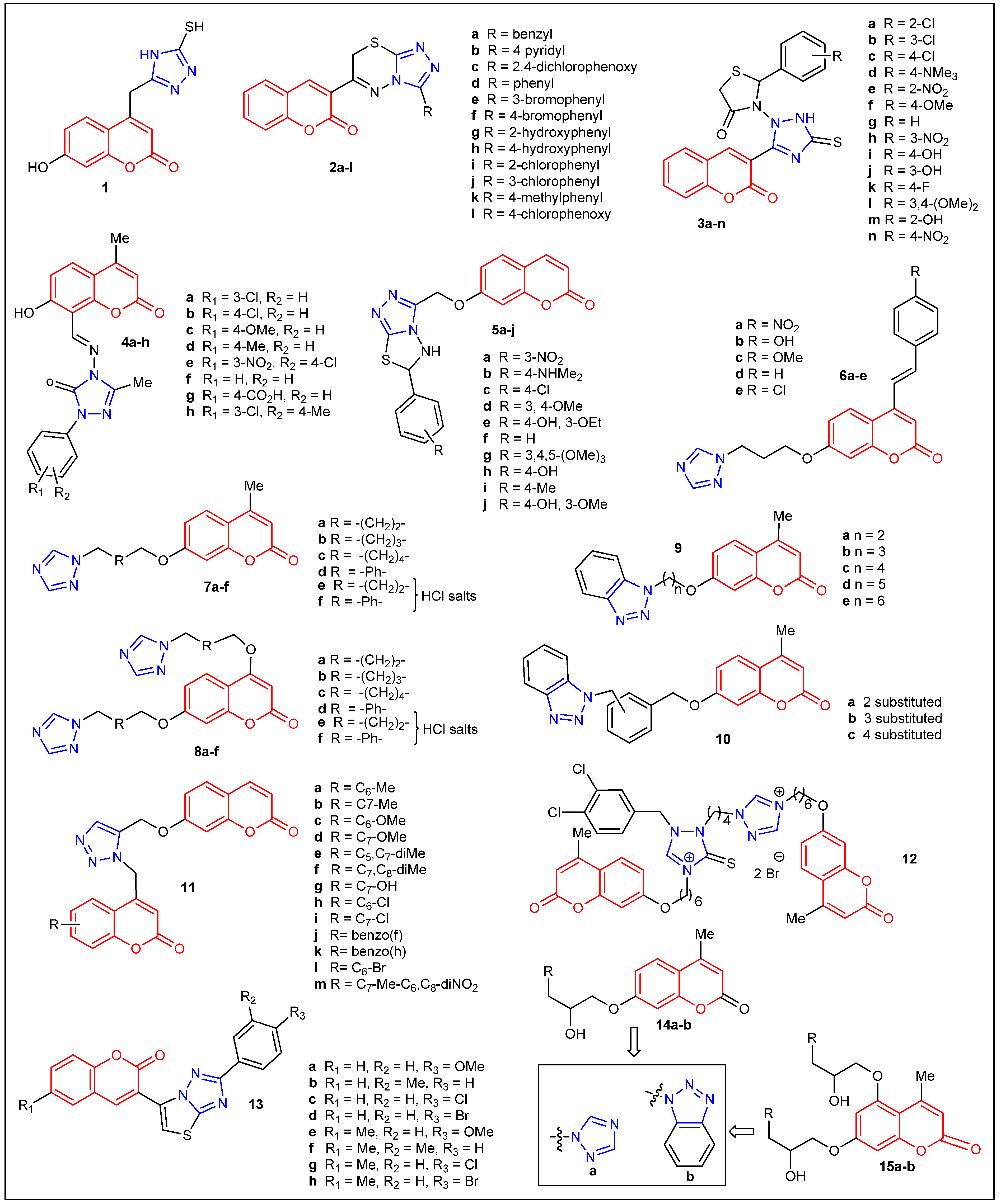
2. Antibacterial and Antifungal Activities of Coumarin Triazole Derivatives

In 2006, M. Cacic et al. reported the first example of a C4-triazole-substituted coumarin 1 (Figure 3) together with its antibacterial activity [21]. Examination of the antimicrobial activity of 1 indicated high antimicrobial activity against S. pneumoniae, and it was slightly less active against P. aeruginosa, B. subtilis, B. cereus, and S. panama. The authors did not report the exact values of antimicrobial activity data and concluded their results with a generalized viewpoint. Furthermore, they noted that the research was in progress. A year later, Jayashree et al. reported the synthesis, characterization, and antimicrobial activity of twelve C-3-substituted triazolo-thiadiazinyl coumarin derivatives 2al from salicylaldehyde as a starting material (Figure 3) (Table 1) [22]. The antibacterial screening demonstrated that compounds 2a, 2b, and 2c had a comparable activity with the standard antibiotics (amoxicillin and gentamycin) against two species of Gram-positive bacteria (B. subtilis and S. aureus) and three species of Gram-negative bacteria (E. coli, K. pneumoniae, and P. aeruginosa). Overall, aryl substitution has improved the antimicrobial activity compared to their corresponding heteroaryl analogs. Compound 2a displayed a 38 mm zone of inhibition (ZoI) toward B. subtilis, 35 mm (K. pneumoniae), and 32 mm (S. aureus and E. coli). Their most active compound, 2b, exhibited the ZoI toward S. aureus (43 mm), B. subtilis, K. pneumoniae, P. aeruginosa (42 mm), and E. coli (40 mm).
In 2009, the synthesis and characterization of fourteen C-3-substituted triazolothiazolidinone derivatives of coumarin 3an were reported by Mashooq A. Bhat et al. (Figure 3) (Table 1) [23]. Compounds with Cl substitution, 3b and 3c, showed the highest activity against S. aureus (ZoI = ~20 mm). In addition, analogs with N(Me)2 (3d), NO2 (3e), OMe (3f), and Cl (3a, 3b, and 3c) substitutions displayed the highest activity against C. albicans (ZoI = ~18 mm). Interestingly, the compound without substitutions 3g showed broad growth inhibition against S. aureus, E. coli, and C. albicans. Although all the adducts exhibited modest to good inhibition, none of them were superior to the standards ciprofloxacin (ZoI = 25 mm) or ketoconazole (ZoI = 20 mm). In addition, in 2019, Kotresh et al. reported the synthesis and antimicrobial properties of eight coumarin C-8-substituted Schiff Bases Triazole Derivatives (Figure 3) (Table 1) [24]. The highest antibacterial activity against B. subtilis and E. coli was obtained by compounds 4a and 4b (ZoI = ~18 mm), but less so than the reference drug norfloxacin (ZoI = 22 mm). Adducts 4a, 4c, 4d, and 4e showed good antifungal activity toward A. niger and C. albicans (ZoI = 18–22 mm) but lower than the standard griseofulvin (ZoI = 26 mm). Both electron-withdrawing groups (chloro, nitro) and electron-donating groups (methoxy, methyl) on the aryl ring might have contributed to the effectiveness of the particular strains. In general, the results indicated that the majority of the C-8-substituted coumarin compounds might serve as better fungicides than bactericides. In 2010, P. M. Kumar et al. employed microwave irradiation to synthesize ten coumarinyl-triazolothiadiazoles derivatives (5aj) in high-yield and short-reaction times [25]. These compounds (Figure 3) (Table 1) were screened in vitro for their antibacterial and antioxidant activity. Particularly, compound 5a (R = 3-nitrophenyl) showed the greatest antibacterial activity against S. aureus (10–15 mm inhibition diameter) and E. coli (16–22 mm inhibition diameter), while compounds 5b (4-dimethylaminophenyl) and 5c (4-chlorophenyl) showed moderate activity (10–15 mm inhibition diameter). Compounds 5a (R = 3-nitrophenyl), 5d (3,4-dimethoxyphenyl), and 5e (4-hydroxy-3-ethoxyphenyl) displayed moderate antifungal activity toward C. albicans (10–15 mm inhibition diameter). Unfortunately, none of them showed superior activity when compared to the standard ciprofloxacin and fluconazole. G. R. Kokil et al. attached to 7-hydroxy-4-methylcoumarin a triazole moiety and a substituted aromatic ring at the C-7 and C-4 positions, respectively (Figure 3) (Table 1) [26]. The resulting 1,2,4-triazole coumarin derivatives were screened for their in vitro antifungal activity against C. albicans ATCC 24433. Compound 6a (R = 4-NO2) showed good antifungal activity (MIC = 12.5 μg/mL), which was comparable with the standard drug ketoconazole (MIC = 12.5 μg/mL). The other compounds, such as 6b (R = 4-OH) and 6c (R = 4-OCH3), showed moderate antifungal activity.
In 2011, the synthesis and in vitro antimicrobial evaluation of two series of coumarin-mono- and bis-triazoles derivatives 7af and 8af were reported by Y. Shi and C. H. Zhou (Figure 3) (Table 1) [27]. Particularly, bis-triazole 8a and its hydrochloride 8e gave the most potent antimicrobial efficacy (MIC = 1–4 μg/mL) against four Gram-positive bacteria (S. aureus ATCC 25923, (MRSA), B. subtilis ATCC 6633, and M. luteus ATCC 4698), four Gram-negative bacteria (E. coli ATCC 25922, P. vulgaris ATCC 6896, S. typhi ATCC 9484 and S. dysenteriae ATCC 49550); as well as three fungi (C. albicans ATCC 76615, S. cerevisiae ATCC 9763, and A. fumigatus ATCC 96918). Other mono-triazole compounds 7ac, bis-triazole 8ac, hydrochloride mono-triazole 7ef, and hydrochloride bis-triazole 7ef showed comparable or superior anti-MRSA activity than the clinical antibacterial drugs enoxacin (MIC = 1–4 μg/mL) and chloromycin (MIC = 4–16 μg/mL). Compounds 7a, 8a, and 8e exhibited comparable antifungal potency against C. albicans and S. cerevisiae (MIC = 2–4 μg/mL) than the positive control fluconazole (MIC = 1–2 μg/mL) and showed strongest inhibition toward A. fumigatus (MIC = 2–48 μg/mL), whereas fluconazole gave MIC = 128 μg/mL. In conclusion, the alkyl linker has provided better activity compared to the phenyl linker in both monomers as well as dimear triazolo-coumarins. In general, coumarin-bis-triazoles 7 exhibit stronger antimicrobial efficiency compared to their corresponding mono-triazole derivatives 8. The authors pointed out that water-soluble hydrochloride salts have shown stronger antibacterial and antifungal efficacy in comparison with their corresponding poor water-soluble triazole precursors. They postulated that the conversion of triazoles into their hydrochlorides could modulate the lipid/water partition coefficient, affect their diffusion in bacterial cells, as well as interact with bacterial cells and tissues. Thus, water-soluble salts might improve the pharmacological properties of these new triazole analogs. They assume that further studies will help to understand the mechanism of actions of these derivatives.
Shi Yuan et al. also reported the synthesis of two series of coumarin-based benzotriazole derivatives (9 and 10) via a multi-step sequence (Figure 3) (Table 1) and studied the in vitro antimicrobial activities against four Gram-positive bacteria, four Gram-negative bacteria, and three fungi [28]. Compounds 9ae and 10ac were more active (MIC = 4–8 μg/mL) than chloromycin (MIC = 16 μg/mL) on P. vulgaris ATCC 6896. Coumarin benzotriazoles 9a (n = 2; CH2-CH2) and 10b (3-substituted) displayed comparable antibacterial efficacy against S. aureus ATCC 25923 and M. luteus ATCC 4698 in comparison with the reference drug chloromycin (MIC = 4 μg/mL). Compared to fluconazole (MIC = 128 μg/mL), compounds 10ad showed stronger inhibition against A. fumigatus ATCC 96918 (MIC = 64 μg/mL). More importantly, fluconazole-insensitive A. fumigatus and methicillin-resistant S. aureus N 315 (MRSA) were sensitive to the new adducts.
In 2012, Naik et al. employed click chemistry as a means to synthesize thirteen 1,4-disubstituted bis-chromenyl triazole coumarin derivatives 11am and studied their antimicrobial activity (Figure 3) (Table 1) [29]. Only three compounds 11hj showed antitubercular activity against M. tuberculosis, equivalent to the activity of streptomycin, with a MIC value of 6.25 μg/mL. Compound 11c (C6-Methoxy) showed higher antifungal activity (MIC = 6.25 μg/mL) than fluconazole (MIC = 8 μg/mL) against A. niger. In summary, all the compounds were better antitubercular agents than antimicrobial agents. However, they showed modest activity against Gram-positive bacteria [S. faecalis (MTCC 3382) and S. aureus (MTCC 3160)] and Gram-negative bacteria [P. aeruginosa (MTCC 1034) and E. coli (MTCC 1089)]. The synthesis of thio-triazole derivative 12 (Figure 3) and its in vitro antibacterial and antifungal activities were reported by Wang and coworkers [30]. This coumarin thio-triazole salt showed good antimicrobial activities (MIC = 8–32 μg/mL) against MRSA (N315), S. aureus (ATCC25923), B. subtilis and M. luteus (ATCC4698), E. coli (DH52), E. typhosa, and C. albicans (ATCC76615) and low efficiency (MIC = 128 μg/mL) toward S. dysenteriae, P. aeruginosa, and C. mycoderma. A green synthesis of 2-aryl-5-(coumarin-3-yl)-thiazolo [3,2-b][1,2,4]-triazoles 13ah (Figure 3), using microwave irradiations under solvent-free conditions, was reported by K. Jakhar and J. K. Makrandi (Table 1) [31]. All compounds displayed low to good inhibition (ZoI = 9–16 mm) against Gram-negative bacteria; E. coli, P. aeruginosa, K. pneumoniae, and S. typhi. Only compounds 13a, 13b, 13c, and 13h exhibited activity against the tested Gram-positive bacteria S. aureus (ZoI = 9–12 mm). It seems as if both methoxy and halogen substitution on the phenyl ring with methyl substitution on the coumarin ring showed the best activity.
Table 1. Antimicrobial activity data of reported coumarin triazole derivatives.
CompoundActivity ObservedBacteria/FungalRef.CompoundActivity ObservedBacteria/FungalRef.
2a9 (nm)B. subtilis and S. aureus[22]6a200 (µg/mL)C. albicans[26]
2b35 (nm)K. pneumoniae[22]6b25 (µg/mL)C. albicans[26]
2c12 (nm)B. subtilis and E. coli[22]6c12.5 (µg/mL)C. albicans[26]
2d19 (nm) B. subtilis[22]6d75 (µg/mL)C. albicans[26]
2g8 (nm)S. aureus[22]6e37.5 (µg/mL)C. albicans[26]
2h16 (nm)B. subtilis and E. coli[22]Ketoconazole12.5 (µg/mL)C. albicans[26]
2i10 (nm)B. subtilis[22]7a16 (µg/mL)P. vulgaris, S. typhi, S. dysenteriae, and A. fumigatus[27]
2j43 (nm) S. aureus[22]7b32 (µg/mL)E. coli, P. vulgaris, and S. dysenteriae[27]
2k26 (nm)B. subtilis[22]7c32 (µg/mL)E. coli, P. vulgaris, S. typhi, S. dysenteriae, S. cerevisiae, and A. fumigatus[27]
2l34 (nm) P. aeruginosa[22]7d64 (µg/mL)MRSA, E. coli, P. vulgaris, S. typhi, S. dysenteriae, S. cerevisiae typhi S, and A. fumigatus[27]
Amoxicillin40 (nm)P. aeruginosa[22]7e64 (µg/mL)S. dysenteriae[27]
Gentamycin41 (nm)P. aeruginosa[22]7f64 (µg/mL)E. coli, P. vulgaris, S. typhi, S. dysenteriae, S. cerevisiae, and A. fumigatus[27]
3a16 (nm)C. albicans[23]8a4 (µg/mL)A. fumigatus[27]
3b18 (nm)C. albicans[23]8b32 (µg/mL)A. fumigatus[27]
3c16 (nm)C. albicans[23]8c32 (µg/mL)A. fumigatus[27]
3d14 (nm)C. albicans[23]8d64 (µg/mL)MRSA B. subtilis, M. luteus, E. coli, S. dysenteriae, and A. fumigatus[27]
3e17 (nm)C. albicans[23]8e2 (µg/mL)MRSA, P. vulgaris, S. cerevisiae, and A. fumigatus[27]
3f16 (nm)S. aureus[23]8f16 (µg/mL)MRSA B. subtilis, M. luteus, P. vulgaris, S. typhi, S. dysenteriae, and S. cerevisiae A. fumigatus[27]
3g16 (nm)E. coli[23]Enoxacin4 (µg/mL)MRSA[27]
3h14 (nm)S. aureus and C. albicans[23]Chloromycin16 (µg/mL)MRSA[27]
3i17 (nm)E. coli and C. albicans[23]Fluconazole128 (µg/mL)A. fumigatus[27]
3j18 (nm)S. aureus[23]11a>100 (µg/mL)S. faecalis, P. aeruginosa, and E. coli,[29]
3j18 (nm)C. albicans[23]11b>100 (µg/mL)P. aeruginosa and E. coli,[29]
3k19 (nm)S. aureus[23]11c50 (µg/mL)S. faureus, S. aureus, and C. albicans[29]
3l20 (nm)S. aureus[23]11d>100 (µg/mL)P. aeruginosa[29]
3m23 (nm)S. aureus[23]11e>100 (µg/mL)P. aeruginosa[29]
3n17 (nm)S. aureus, E. coli[23]11f>100 (µg/mL)P. aeruginosa[29]
Ciprofloxacin25 (nm)S. aureus[23]11g>100 (µg/mL)P. aeruginosa[28]
Ciprofloxacin25 (nm)E. coli[23]11h50 (µg/mL)S. faureus, E. coli, and C. albicans[29]
4a17 (nm)A. niger[24]11i50 (µg/mL)S. faureus, P. aeruginosa, E. coli, C. albicans, and A. niger[29]
4b23 (nm)C. albicans[24]11j50 (µg/mL)S. faureus, P. aeruginosa, E. coli, C. albicans, and A. niger[29]
4c18 (nm)C. albicans[24]11k>100 (µg/mL)P. aeruginosa,[29]
4d22 (nm)A. niger[24]11l>100 (µg/mL)P. aeruginosa,[29]
4e18 (nm)A. niger[24]11m>100 (µg/mL)P. aeruginosa,[29]
4f18 (nm)A. niger[24]Ciprofloxacin1 (µg/mL)S. faureus, S. aureus, P. aeruginosa, and E. coli[29]
4g18 (nm)C. albicans[24]Fluconazole16 (µg/mL)C. albicans[29]
4h21 (nm)C. albicans[24]12128 (µg/mL)S. dysenteriae,
P. aeruginosa, and C. mycoderma
[30]
Norfloxacin22 (nm)E. coli[24]Chloromycin16 (µg/mL)P. aeruginosa[30]
Norfloxacin22 (nm)B. subtilis[24]Norfloxacin4 (µg/mL)MRSA and E. typhosa[30]
Griseofulvin26 (nm)A. niger[24]Fluconazole4 (µg/mL)C. mycoderma[30]
Griseofulvin26 (nm)C. albicans[24]14a4 (µg/mL)C. utilis, C. albicans, and P. aeruginosa[32]
5b16 (nm)E. coli[25]14b4 (µg/mL)C. albicans[32]
5c10 (nm)E. coli[25]15a1 (µg/mL)C. albicans and E. coli[32]
5d7 (nm)E. coli[25]15b8 (µg/mL)C. albicans[32]
5e7 (nm)S. aureus and E. coli[25]Fluconazole1 (µg/mL)C. albicans[32]
5h10 (nm)C. albicans[25]Chloromycin8 (µg/mL)M. luteus[32]
5j10 (nm)C. albicans[25]Norfloxacin1 (µg/mL)P. aeruginosa[32]
In 2014, two series of coumarin triazoles 14a,b and 15a,b (Figure 3) were prepared and characterized by IR, NMR, MS, and HRMS spectra, and their in vitro biological activity with six bacteria and five fungi was evaluated (Table 1) [32]. Bis-triazole coumarin derivative 15a showed the same anti-C. utilis activity (MIC = 4 μg/mL) to mono-triazole derivative 14a, which makes those two adducts more potent than Fluconazole (MIC = 8 μg/mL). In addition, compound 14a exhibited better activity against MRSA (MIC = 8 μg/mL) than 14b (MIC = 32 μg/mL) and norfloxacin (MIC = 16 μg/mL). Compound 14b showed very good activity (MIC = 4 μg/mL) toward C. albicans, and modest MIC values (16 μg/mL) were obtained for C. utilis, C. mycoderma, MRSA N315, B. subtilis, and E. coli JM109. Finally, 15b showed lower or comparable antimicrobial activities than 14b and the reference drugs mentioned above. Overall, mono-triazole substitution favors antimicrobial activity compared to bis-triazole coumarin analogs.
K. Kushwaha et al. reported the design and synthesis of coumarin-1,2,3-triazole derivatives 16ad and 17ah to study their antimicrobial properties (Figure 4) (Table 2) [33]. The majority of the compounds displayed similar antifungal activity toward A. fumigatus MTCC 343, A. flavus MTCC 277, and C. albicans MTCC 227 (ZoI = 12–23 mm). Remarkably, 16d was the most active (ZoI = 23 mm) against A. fumigatus, and 16a (n = 1; CH2) was the most active against C. albicans (ZoI = 20 mm), which was slightly better than the reference miconazole (ZoI = 15–19 mm). All the derivatives presented modest to good antibacterial activity against all the seven tested bacteria, albeit lower (ZoI = 10.5–15.7 nm) than the standard drug ciprofloxacin (ZoI = 18–20 nm). In general, compounds 17ae were selected as the best Candidates for further investigations due to their lower toxicity, high drug score values, and good oral bioavailability. Furthermore, in 2014, a group of C-7-triazole-substituted coumarins 18ae were synthesized with good yields and short reaction times using both microwave irradiation and grinding techniques (Figure 4) (Table 2) [34]. Compounds 18ce showed good antibacterial activity against K. pneumonia (ZoI = 16 mm), whereas adducts 18ae displayed moderate to good antimicrobial activity against E. coli, A. niger, A. fumigates, and A. terrus (ZoI = 6–12 mm).
Dongamanti et al. also reported a microwave-assisted synthesis of a series of hybrid compounds containing coumarin, 1,2,3-triazole, and chalcone substructures 19ai (Figure 4) which were screened for antimicrobial activity (Table 2) [35]. Derivatives 19c and 19d exhibit excellent activities against Gram-positive bacteria (S. aureus and B. subtilis) (ZoI = 32–35 mm) and Gram-negative bacteria (E. coli and P. aeruginosa) (ZoI = 31–33 mm) that are superior to the activities of the reference antibiotic amoxicillin (ZoI = 10–30 mm). Compounds 19b, 19e, and 19h displayed good antibacterial activity, products 19f and 19g were moderately active, and derivatives 19a and 19i were weakly active in the antibacterial assay (ZoI = 4–17 mm). In regard to antifungal activity, adducts 19cd were more bioactive against A. niger, F. oxysporum, and P. italicum (ZoI = 13–30 mm) than the reference drug mycostatin (ZoI = 12–25 mm), while the other compounds were good to moderately active. In conclusion, dimethoxy and trimethoxy substitution yielded the highest activity toward several strains. Joshi et al. reported the synthesis and characterization of two series (20ad and 21ad) of s-triazine-1,2,3-triazoles-coumarin dendrimers using click-chemistry (Figure 4) [36]. Compounds tris-(coumarin-1,2,3-triazole)s-triazines 20ad and bis-(coumarin-1,2,3-triazole)s-triazin-anilines 21ad were screened for antimicrobial activity against Gram-positive bacteria [S. aureus (MTCC96), S. epidermidis (MTCC435)], Gram-negative bacteria [E. coli DH5a and P. aeruginosa (MTCC434)] and fungal strains [G. candidum, C. galbrata, and C. albicans]. Adduct 20a exhibited modest antifungal activities (% killing of 83) at a high concentration (250 μM) but displayed modest activity against all bacterial strains tested (values not shown) (Table 2).
In 2015, A. M. Hayallah et al. documented the synthesis and antimicrobial activity of coumarin triazoles 22 and 23al (Figure 4) (Table 2) [37]. The in vitro antibacterial activity was determined using S. aureus (AUMC B71) and E. coli (AUMC B69). In general, most of the newly-synthesized compounds exhibited moderate to good antibacterial activities compared to that of ciprofloxacin (20–30 vs. 40 ZoI). Specifically, compounds 22, 23d, and 23h exhibited the same antibacterial activity against E. coli (MIC = 12.5 μmol/mL); however, 23d was the most active against S. aureus (MIC = 25 μmol/mL), but lower than ciprofloxacin (MIC = 1.75 μmol/mL). Derivatives 22 and 23a, 23e, and 23fj were tested against C. albicans using fluconazole as a reference drug (MIC = 1.85 μmol/mL) and showed poor to null activity. Only compounds 23b, 23c, and 23d showed antifungal activities (MIC = 25–50 μmol/mL). Kalwania et al. reported the synthesis, characterization, and antimicrobial activities of a 1,2,4-triazole-coumarin Schiff Bases 24ae and their Mn (II) and Co (II) complexes 25aj (Figure 4) (Table 2) [38]. Compounds 24ae and metal complexes 25aj were evaluated in vitro against five bacterial strains; E. coli, P. aeruginosa, S. typhi, S. aureus, and B. subtilis, using the standard drug gentamycin. Furthermore, the antifungal activities were evaluated against A. niger and C. albicans using fluconazole as the standard drug. All the Schiff bases 24ae demonstrated inferior antimicrobial activities with ZoI in the range of 45.21 mm to 78.32 mm toward all five bacterial and two fungal strains. However, their corresponding metal complexes 25aj showed higher antibacterial activity against selected bacteria, especially against S. typhi (ZoI: 25c—79.36 mm; 25d—76.44 mm; 25e—82.05 mm; 25j—80.00 mm). The metal complexes 25e (ZoI: 76.09 mm and 79.23 mm) and 25j (ZoI: 73.84 mm and 77.62 mm) have confirmed the antifungal activity toward A. niger and C. albicans, respectively. None of their compounds are comparable or superior to the standard drugs tested. In summary, metal complexes (25) have shown very good antimicrobial activity compared to their corresponding ligands (24).
Table 2. Antimicrobial activity data of reported coumarin triazole derivatives.
CompoundActivity ObservedBacteria/FungalRef.CompoundActivity ObservedBacteria/FungalRef.
16a20.2 (±1.69) mmC. albicans[33]Mycostatin20 mmP. italicum[35]
16b21.3 (±1.90) (±) mmA. fumigatus[33]2230 mmE. coli[37]
16c18.9 (±1.34) mmA. fumigatus[33]23a20 mmE. coli[37]
16d23.4 (±1.97) mmA. fumigatus[33]23b20 mmCandida[37]
17a18.5 (±0.70) mmA. fumigatus[33]23c19 mmCandida[37]
17b18.8 (±1.13) mmA. fumigatus[33]23d30 mmE. coli[37]
17c16.9 (±1.17) mmA. fumigatus[33]23e25 mmS. aureus[37]
17d18.4 (±0.63) mmA. fumigatus[33]23f20 mmS. aureus[37]
17e18.2 (±1.76) mmA. fumigatus[33]23g24 mmE. coli[37]
17f20.6 (±0.91) mmA. fumigatus[33]23h28 mmE. coli[37]
17g19.0 (±1.41) mmA. fumigatus[33]23i26 mmE. coli[37]
17h18.5 (±0.70) mmA. fumigatus[33]23j20 mmE. coli[37]
Ciprofloxacin20 mmS. epidermis[33]Ciprofloxcin40 mmS. aureus and E. coli[37]
Miconazole19 mmC. albicans Fluconazole40 mmCandida[37]
18a12 mmK. pneumonia[34]24a64.73 mmS. typhi[38]
18b12 mmK. pneumonia and Aspergillus terrs[34]24b70.31 mmC. albicans[38]
18c16 mmK. pneumonia[34]24c76.44 mmS. typhi[38]
18d16 mmK. pneumonia[34]24d72.96 mmS. typhi[38]
18e16 mmK. pneumonia[34]24e78.32 mmS. typhi[38]
Gentamycin18 mmK. pneumonia[34]25a68.13 mmS. typhi[38]
Fluconazole13 mmA. niger and Aspergillus terrs[34]25b72.00 mmS. typhi[38]
19a17 mmF. oxysporum[35]25c79.36 mmS. typhi[38]
19b28 mmS. aureus and E. coli[35]25d76.44 mmS. typhi[38]
19c32 mmS. aureus[35]25e82.05 mmS. typhi[38]
19d35 mmS. aureus[35]25f65.00 mmS. typhi[38]
19e27 mmS. aureus and E. coli[35]25g71.32 mmC. albicans[38]
19f22 mmS. aureus[35]25h75.66 mmS. typhi[38]
19g22 mmE. coli[35]25i72.22 mmS. typhi[38]
19h25 mmS. aureus[35]25j80.00 mmS. typhi[38]
19i18 mmF. oxysporum[35]Gentamycine100 mmE. coli, P. aeruginosa, S. typhi, S. aureus, and B. subtilis[38]
Amoxicillin30 mmS. aureus and E. coli[35]Fluconazole100 mmA. niger and C. albicans[38]
In 2016, Shingate et al. reported the synthesis of two sets of coumarin triazole derivatives [26af (7-subsituted) and 27ae (4-subsituted)], Figure 5 (Table 3). These new compounds were subjected to in vitro antimicrobial activity against three Gram-positive bacteria (S. aureus, M. luteus, and B. cereus), three Gram-negative bacteria (E. coli, P. fluorescens, and F. devorans), and three fungal strains (A. niger, P. chrysogenum, and C. lunata) [39]. All compounds showed modest to good antibacterial activity, but adduct 27a was the most bioactive, with MIC values of 2 μg/mL against the three tested Gram-negative bacteria. Those were the same MIC values (2 μg/mL) obtained from the three standards used (ampicillin, kanamycin, and chloramphenicol). Similar results were observed from the antifungal study. However, this time adduct 26d was the most superior compound among all, with MIC values of 4–8 μg/mL, which are comparable to those of the standards [miconazole (16 μg/mL), amphotericin B (2–16 μg/mL), and Fluconazole (2–4 μg/mL)]. The same year, Shingate et al. described the synthesis and antifungal activity of eight coumarin triazole derivatives [28ah (7-substituted)], Figure 5 (Table 3) [40]. This time, the following five fungal stains were evaluated: C. albicans, F. oxysporum, A. flavus, A. niger, and C. neoformans. Compound 28c, 28d, 28e (chloro-substituted), and 28h were as potent as the standard drug miconazole against C. albicans (MIC = 25 μg/mL), while adduct 28f showed twofold bioactivity when compared with miconazole and equally potent to fluconazole (MIC = 12.5 μg/mL). In order to identify the mechanism of action of these compounds, authors performed molecular docking studies with the active site of fungal C. albicans enzyme P450 cytochrome lanosterol 14α-demethylase. The highly effective compound 28f exhibited the lowest interaction energy (−72.29 kcal/mol), and the standard drugs fluconazole and miconazole also showed good interaction energy that is −69.76 and −71.90 kcal/mol, respectively. Similarly, Raić-Malić et al. reported a straightforward synthesis (using click-chemistry to form the 1,2,3-triazole moiety) that produced 31 new coumarin triazole derivatives [29a-z2 (4-substituted, 7-hydroxycoumarins) and 30ad (4-substituted, 7-methylcoumarins)], Figure 5 (Table 3) [41]. The relatively large library of compounds was screened against three Gram-positive bacteria [S. aureus (ATCC 25923), E. faecalis, vancomycin-resistant E. faecium (VRE)], and four Gram-negative bacteria [P. aeruginosa (ATCC 27853), E. coli (ATCC 25925), A. baumannii (ATCC 19606), and extended-spectrum β-lactamase (ESBL)-producing K. pneumoniae]. Unfortunately, none of the 31 adducts exhibited any bioactivity against the Gram-negative bacteria tested. In addition, among the 31 tested compounds, only 13 showed activity against two of the three Gram-positive bacteria examined [E. faecalis and vancomycin-resistant E. faecium (VRE)]. Nonetheless, coumarin 1,2,3-triazole hybrids 29n (p-pentylphenyl), 29t (2-chloro-4-fluorobenzenesulfonamide), and 29x (dithiocarbamate) showed selective anti-Enterococcus species activities. For instance, those three compounds displayed MIC values of 64 μg/mL against vancomycin-resistant E. faecium, whereas the reference antibiotics ceftazidime and ciprofloxacin didn’t exhibit bioactivity (MICs were >256 mg/mL). Furthermore, adduct 29n demonstrated superior inhibitory against E. faecium (MIC value of 8 μg/mL). Among this large pool of compounds, aryl and heteroaryl substitution on triazole moiety demonstrated greater activity, implying that the substitution on triazole is vital for obtaining better antimicrobial activity.
In 2017, the synthesis of twelve 1,2,4-triazolo-1,3,4-thiadiazepino-fused coumarins, together with their antimicrobial activity, was presented by Patel and co-workers [42]. To produce those 12 adducts (31al), Figure 5, the authors simply reacted three 4-chloro-3-formylcoumarins with four 4-amino-5-substituted-3-mercapto-1,2,4-triazoles in the presence of a base. All the adducts (31al) were evaluated against two Gram-positive bacteria, S. aureus (MTCC 96) and B. subtilis (MTCC 441), two Gram-negative bacteria, E. coli (MTCC 443) and S. typhi (MTCC 98), and two fungal strains, C. albicans (MTCC 227) and A. niger (MTCC 282) (Table 3). All compounds were inactive against all fungal strains (griseofulvin and nystatin were used as standard antifungal drugs). Only a few adducts (31a, 31e, 31j, and 31k) showed antibacterial activity comparable to the standard drug ampicillin (MIC values around 100 μg/mL) but lower activity compared with chloramphenicol (MIC = 50 μg/mL) and Norfloxacin (MIC = 10 μg/mL) (Table 3). The adducts 31a, 31e, and 31j exhibited the MIC 62.5 μg/mL toward E. coli, whereas 31k showed the MIC 62.5 μg/mL against S. aureus. Only the methyl substitution on coumarin and (thio)phenyl substitution on triazole moiety have produced the desired antimicrobial activity comparable to the standards used. In the same year, Pal et al. prepared 17 new coumarin triazole derivatives (32af and 33ak), Figure 5 [43]. All synthesized adducts were evaluated against one Gram-positive bacteria, S. aureus, and three Gram-negative bacteria, E. coli, P. aeruginosa, and K. pneumonia (MTCC 441). Although all compounds showed some inhibition at 11–18 mm (ZoI at 0.5 mg/100 μL) for all the tested bacterial strains, these values were lower than the reference drug pefloxacin (28–36 mm). Jin et al. published the synthesis and antimicrobial evaluation of 10 different triazole-tethered isatin–coumarin hybrids (34aj), Figure 5 (Table 4) [44]. The following four Gram-positive bacterial strains: methicillin-sensitive S. epidermidis, methicillin-resistant S. epidermidis, methicillin-sensitive S. aureus, and methicillin-resistant S. aureus (ATCC) and four Gram-negative bacterial strains: extended-spectrum beta-lactamases (ESBLs)-producing E. coli ESBLs (−), E. coli ESBLs (+), K. pneumoniae ESBLs (+), and K. pneumoniae ESBLs (−) were used to evaluate the newly synthesized adduct. All adducts displayed poor to modest activity across the board (MIC range 16 to >200 μg/mL), whereas the reference drug ciprofloxacin showed a MIC range of 0.015 to 64 μg/mL. It is worth noting that those bacterial strains are resistant and adduct 34e (n = 1; R1 = OMe; R2 = NOMe) showed 16 μg/mL (MIC) against methicillin-resistant S. epidermidis, which had a higher inhibition when compared to ciprofloxacin (MIC = 64 μg/mL).
The microwave-aided synthesis of dimers of ten distinct coumarin-1,2,3-triazoles containing an alkyl spacer (35aj) was reported by Ashok et al. in 2018 [45] (Figure 6). The synthesized compounds were screened for their antimicrobial activity against two Gram-positive strains, B. subtilis (ATCC 6633) and S. aureus (ATCC 6538), two Gram-negative strains, E. coli (ATCC 11229) and P. vulgaris (ATCC 29213), and two fungal strains, A. niger (ATCC 9029) and C. albicans (ATCC 10231). The compound 35j showed MIC values of 3.125–6.25 µg/mL and 12.5 µg/mL, four bacterial and two fungal strains, respectively. The compound 35j was discovered to be more effective than the other investigated compounds against the tested bacterial and fungal strains. Except for compound 35j, compounds 35e and 35i demonstrated modest activity against bacterial strains with MIC values of 6.25–12.5 µg/mL. Compounds 35d, 35e, and 35i displayed better antifungal activity with MIC values of 12.5–25 µg/mL. Coumarin–triazoles with alkyl linker (n = 6 and 8) have produced comparable antibacterial (35e and 35i) as well as antifungal activity, indicating that the long linker could have played a role in getting the desired activity. López- Rojas et al. [46] reported a series of coumarin-1,2,3-triazole derivatives with diverse alkyl, phenyl, and heterocycles at C-4 of the triazole nucleus via copper(I)-catalyzed Huisgen 1,3-dipolar cycloaddition reaction (36am and 37am) (Figure 6) (Table 4). The antibacterial activity of each molecule was evaluated against Gram-positive bacteria, B. subtilis, S. aureus, and E. faecalis, Gram-negative bacteria, E. coli, P. vulgaris, K. pneumonia, P. aeruginosa, and the fungus C. albicans for antifungal activity. Compounds 36a, 36b, 36f, 37h, and 37k exhibited potential activity against E. faecalis at MICs ranging from 2.5 to 50.0 µg/mL. The most effective compound was found to be 36b, with the 2-OMe-Ph group linked to the triazole nucleus and an OCH2 linker. In contrast, the comparable isoster 37b (-NHCH2-) was found to be 64-fold less active than 36b. Subsequently, compounds 36c (3-OMe-Ph) and 36d (4-OMe-Ph) had 8- and 16-fold less antibacterial activity than 36b, respectively. The location of the OMe group on the phenyl ring also plays a significant influence on the activity. In order to be a successful antimicrobial drug Candidate, it should display the least toxicity toward normal cells. The authors evaluated the active compounds 36a, 36b, 36f, 37h, and 37k for toxicity (hemolytic activity) against human erythrocytes, and all tested compounds demonstrated low toxicity toward human erythrocytes.
Table 3. Antimicrobial activity data of reported coumarin triazole derivatives.
CompoundActivity ObservedBacteria/FungalRef.CompoundActivity ObservedBacteria/FungalRef.
26a2 µg/mLE. coli and P. fluorescens[39]29t32 µg/mLE. faecalis[41]
26b2 µg/mLP. fluorescens[39]29u256 µg/mLE. faecalis[41]
26c2 µg/mLF. devorans[39]29v32 µg/mLE. faecalis[41]
26d2 µg/mLF. devorans[39]29x16 µg/mLE. faecalis[41]
26e4 µg/mLB. cereus, E. coli, and F. devorans[39]Ceftazidime0.5 µg/mLE. coli[41]
26f4 µg/mLM. luteus, E. coli, and F. devorans[39]Ciprofloxacin<0.125 µg/mLP. aeurigonsa, E. coli, and A. baumanni[41]
27a2 µg/mLE. coli, P. fluorescens, and F. devorans[39]31a62.5 µg/mLE. coli[42]
27b4 µg/mLM. luteus, B. cereus, E. coli, and P. fluorescens[39]31b100 µg/mLS. aureus[42]
27c4 µg/mLM. luteus, E. coli, F. devorans, and A. niger[39]31c100 µg/mLE. coli[42]
27d4 µg/mLM. luteus[39]31d100 µg/mLE. coli[42]
27e4 µg/mLM. luteus, E. coli, and F. devorans[39]31e62.5 µg/mLE. coli[42]
Ampicillin2 µg/mLB. cereus, and P. fluorescens[39]31f125 µg/mLE. coli[42]
Kanamycin2 µg/mLS. aureus, M. luteus, B. cereus, E. coli, P. fluorescens, and F. devorans[39]31g125 µg/mLB. subtilis and S. aureus[42]
Chloramphenicol2 µg/mLS. aureus, M. luteus, B. cereus, E. coli, P. fluorescens, and F. devorans[39]31h250 µg/mLB. subtilis, S. aureus, and E. coli[42]
Miconazole16 µg/mLA. niger, P. chrysogenum, and C. lunata[39]31i250 µg/mLE. coli[42]
Amphotericin B2 µg/mLA. niger[39]31j62.5 µg/mLE. coli[42]
Fluconazole2 µg/mLA. niger and P. chrysogenum[39]31k62.5 µg/mLS. aureus[42]
28a50 µg/mLC. albicans and A. niger[40]31l200 µg/mLB. subtilis[42]
28b50 µg/mLC. albicans[40]Ampicillin100 µg/mLE. coli and S. typhi[42]
28c25 µg/mLC. albicans and A. flavus[40]Chloramphenicol50 µg/mLB. subtilis, S. aureus, E. coli, and S. typhi[42]
28d25 µg/mLC. albicans and F. oxysporum[40]Norfloxacin10 µg/mLS. aureus, E. coli, and S. typhi[42]
28e12.5 µg/mLF. oxysporum[40]Griseofulvin100 µg/mLA. niger[42]
28f12.5 µg/mLC. albicans[40]Nystatin100 µg/mLA. niger and C. albicans[42]
28g50 µg/mLC. albicans and F. oxysporum[40]32a18 mmP. aeruginosa[43]
28h25 µg/mLC. albicans[40]32b14 mmS. aureus and K. pneumoniae[43]
Miconazole12.5 µg/mLA. flavus[40]32c15 mmS. aureus[43]
Fluconazole6.25 µg/mLF. oxysporum and A. flavus[40]32e13 mmE. coli[43]
29g128 µg/mLE. faecalis[41]32f13 mmK. pneumoniae[43]
29i256 µg/mLE. faecalis[41]33a15 mmS. aureus[43]
29l256 µg/mLE. faecalis[41]33e15 mmK. pneumoniae[43]
29m64 µg/mLE. faecalis[41]33g17 mmE. coli[43]
29n8 µg/mLE. faecalis[41]33h13 mmS. aureus[43]
29o16 µg/mLE. faecalis[41]33j16 mmP. aeruginosa[43]
29p64 µg/mLE. faecalis[41]33k15 mmE. coli[43]
29q64 µg/mLE. faecalis[41]Pefloxacin36 mmS. aureus[43]
29s64 µg/mLE. faecalis[41]
In 2018, Savanur et al. [47] established new series of coumarin, quinolinone, and benzyl-linked 1,2,3-triazole derivatives (38ab, 39ak, 40ag, 41af) via click chemistry, as portrayed in Figure 6, and subjected the molecules to antimicrobial studies. Synthesized coumarin–triazole compounds were screened for antibacterial studies against Gram-positive bacteria, E. coli (NCIM 5346), P. aeruginosa (NCIM 5514), and B. bronchiseptica (NCIM 5346), and Gram-negative bacteria, S. aureus (NCIM 5345), B. subtilis (NCIM 2920), and (NCIM 5346) (Table 4). With a MIC of 1.0 µg/mL, compound 39j with chloro and methoxy substitution on coumarin was extremely effective against S. aureus and P. aeruginosa. Additionally, compound 39j exhibited excellent activity with MICs of 8.0 µg/mL, 16 µg/mL, and 16 µg/mL against B. subtilis, B. cereus, and B. bronchiseptica, respectively. Apart from compound 39j, compounds 40g (chloro substitution at C-6 on coumarin and 1-azacoumarin) and 41f (chloro-substituted triazoles with benzyl group) demonstrated excellent activity against S. aureus with MICs of 1.0 µg/mL, which is comparable to the standard dose of ciprofloxacin (1.0 µg/mL). Further, the molecules tested for their antifungal assay against eight Candida fungal strain species (yeast specimens), included C. albicans, C. tropicalis, C. utilis, C. krusei, and Aspergillus species (filamentous fungi), such as A. fumigatus, A. niger, R. oryzae, and R. bataticola. Of all the compounds tested, 39i and 39j (with chloro and methoxy substitution) were highly active with MIC 1.0 µg/mL against Candida species. Compound 39e was excellent with MICs of 1.0 µg/mL and MIC of 2.0 µg/mL against C. krusei and C. albicans, respectively. Furthermore, 40f, a quinolinone analog with methyl substitution, was found to be a highly-active compound against C. albicans, C. utilis, and C. krusei with MICs 1.0 µg/mL, 2.0 µg/mL, and 4.0 µg/mL, respectively. Additionally, the same compound (40f) was also found to be very active against A. niger with MIC of 1.0 µg/mL. The in silico analysis showed that the active compounds (39f and 39h) bind to the active sites of the two antifungal target proteins (1FI4 and 3LD6). Interestingly, compound 39h showed the highest binding affinity (−11.0 kcal/mol) toward 1FI4, whereas 39f displayed favorable interaction (−12.5 kcal/mol) toward 3LD6. The authors believe that these compounds represent a new platform for antimicrobial activity and could be further optimized therapeutically.
Table 4. Antimicrobial activity data of reported coumarin triazole derivatives.
CompoundActivity ObservedBacteria/FungalRef.CompoundActivity ObservedBacteria/FungalRef.
35a25 (10) µg/mLB. subtilis and E. coli[45]42f12 µg/mLS. aureus[48]
35b25 (13) µg/mLS. aureus and E. coli[45]42g11 µg/mLS. aureus[48]
35c12.5 (12) µg/mLB. subtilis[45]42h9 µg/mLS. aureus[48]
35d6.25 (15) µg/mLS. aureus[45]42i12 µg/mLE. coli[48]
35e6.25(15) µg/mLB. subtilis, S. aureus, and P. vulgaris[45]42j7 µg/mLS. aureus[48]
35f25(12) µg/mLS. aureus and E. coli[45]42k11 µg/mLS. aureus and E. coli[48]
35g12.5 (12) µg/mLB. subtilis and S. aureus[45]42l18 µg/mLE. coli[48]
35h6.25 (15) µg/mLS. aureus and E. coli[45]43a7.5 µg/mLE. coli and P. aeruginosa[49]
35i6.25 (15) µg/mLB. subtilis, S. aureus, and E. coli[45]43b5.5 µg/mLE. coli[49]
35j3.125 (19) µg/mLB. subtilis, S. aureus, and E. coli[45]43c6.5 µg/mLE. coli[49]
Gentamicin1.56 (31) µg/mLB. subtilis, S. aureus, and E. coli[45]Ciprofloxacin4.5 µg/mLK. pneumoniae[49]
Fluconazole3.125 (25) µg/mL A. niger and C. albicans[45]44a0.8 µg/mLM. tuberculosis[50]
36a50 µg/mLE. faecalis[46]44b1.6 µg/mLM. tuberculosis[50]
36b12.5 µg/mLE. faecalis[46]44c1.6 µg/mLM. tuberculosis[50]
36c100 µg/mLE. faecalis[46]44d1.6 µg/mLM. tuberculosis[50]
36d200 µg/mLE. faecalis[46]44e1.6 µg/mLM. tuberculosis[50]
36e100 µg/mLE. faecalis[46]44f3.12 µg/mLM. tuberculosis[50]
36f50 µg/mLE. faecalis[46]44g6.25 µg/mLM. tuberculosis[50]
36g100 µg/mLE. faecalis[46]44h1.6 µg/mLM. tuberculosis[50]
36h400 µg/mLS. aureus and E. faecalis [46]44i12.5 µg/mLM. tuberculosis[50]
36i200 µg/mLE. faecalis[46]Pyrazinamide3.12 µg/mLM. tuberculosis[50]
36j800 µg/mLS. aureus and E. faecalis [46]Streptomycin6.25 µg/mLM. tuberculosis[50]
36k400 µg/mLE. faecalis[46]Ciprofloxacin3.12 µg/mLM. tuberculosis[50]
36l400 µg/mLE. faecalis[46]45a2.5 ± 0.2 cmPenicillium sp. [51]
37a400 µg/mLE. faecalis[46]45b2.5 ± 0.5 cmS. aureus[51]
37b800 µg/mLE. faecalis and K. pneumoniae[46]45c2.1 ± 0.4 cmS. aureus[51]
37c400 µg/mLE. faecalis[46]45d1.7 ± 0.6 cmS. aureus[51]
37d100 µg/mLE. faecalis[46]45e1.8 ± 0.4 cmPenicillium sp. [51]
37e100 µg/mLE. faecalis[46]45f1.4 ± 0.3 cmPenicillium sp. [51]
37f200 µg/mLC. albicans[46]45g1.2 ± 0.6 cmPenicillium sp. [51]
37g200 µg/mLS. aureus[46]46a1.7 ± 0.4 cmPenicillium sp. [51]
37h50 µg/mLE. faecalis[46]46b1.3 ± 0.6 cmPenicillium sp. [51]
37i100 µg/mLE. faecalis[46]46c1.5 ± 0.4 cmPenicillium sp. [51]
37j800 µg/mLS. aureus and E. faecalis[46]46d1.0 ± 0.4 cmPenicillium sp. [51]
37k50 µg/mLE. faecalis[46]46e1.1 ± 0.3 cmS. enterica[51]
37l800 µg/mLE. faecalis[46]46f0.7 ± 0.1 cmS. enterica[51]
Chloramphenicol1.2 µg/mLE. coli[46]46g0.5 ± 0.1 cmE. coli[51]
Ketoconazole8 µg/mLC. albicans[46]47a1.1 ± 0.2 cmS. enterica[51]
38a31.25 µg/mLS. aureus and B. subtilis[47]47b0.6 ± 0.1 cmS. aureus[51]
38b16 µg/mLS. aureus[47]47c0.5 ± 0.2 cmS. aureus[51]
39a16 µg/mLB. subtilis and B. cereus[47]47d1.1 ± 0.1 cmS. enterica[51]
39b31.25 µg/mLB. subtilis[47]47e0.7 ± 0.2 cmF. oxysporum[51]
39c8 µg/mLS. aureus[47]47f0.6 ± 0.1 cmM. smegmatis[51]
39d8 µg/mLB. subtilis[47]47g0.5 ± 0.1 cmE. coli[51]
39e4 µg/mLS. aureus[47]48a>1000 µg/mLS. aureus[52]
39f31.25 µg/mLS. aureus[47]48b416.7 ± 60.09 µg/mLS. aureus[52]
39g8 µg/mLS. aureus and B. subtilis[47]48c0.16 ± 0.08 µg/mLS. aureus[52]
39h4 µg/mLS. aureus[47]Ceftriaxonum0.97 ± 0.02 µg/mLS. aureus[52]
39i8 µg/mLS. aureus[47]Streptomycin1.89 ± 0.08 µg/mLS. aureus[52]
39j1 µg/mLS. aureus and P. aeruginosa[47]62a250 ± 20.41 µg/mLS. aureus[52]
39k16 µg/mLS. aureus[47]62b425 ± 47.87 µg/mLS. aureus[52]
40a16 µg/mLP. aeruginosa[47]62c51.25 ± 3.15 µg/mLS. aureus[52]
40b16 µg/mLP. aeruginosa[47]63a>1000 µg/mLS. aureus[52]
40c16 µg/mLS. aureus[47]63b>1000 µg/mLS. aureus[52]
40d8 µg/mLS. aureus and B. subtilis[47]63c0.31 ± 0.23 µg/mLS. aureus[52]
40e8 µg/mLS. aureus and P. aeruginosa[47]64a0.03 µg/mLC. albicans[53]
40f4 µg/mLS. aureus and P. aeruginosa[47]64b0.015 µg/mLC. albicans and C. parapsilosis[53]
Kolichala et al. [48] reported the regioselective synthesis and antibacterial activity of 6-[(l-ethyl-lH-l,2,3-triazol-4-yl)methoxy]-4-methyl-2H-chromen-2-ones (42al), as depicted in Figure 6 (Table 4). The disclosed compounds were examined using the paper disc technique against the bacterial strains E. coli (Gram-negative) and S. aureus (Gram-positive). According to the authors, each analog exhibited good to moderate activity. The compounds 42b, 42e, 42f, 42g, 42i, 42h, and 42l among the studied compounds showed relatively moderate to exceptional activity (MIC range 8–32 µg/mL), but they did not compare standard drugs in this study. Chityala et al. [49] reported the synthesis and antibacterial activity of coumarin-1,2,3-triazoles (43ac) (Figure 6) (Table 4). The compounds were evaluated for antibacterial assay against bacterial strains E. coli, K. pneumonia, P. aeruginosa, S. aureus, and S. pyogenes. Compounds 43ac portrayed excellent results, as confirmed by their MIC values ranging from 5.5–17.5µg/mL. PEG-400 was used as an environmentally acceptable catalyst by Shaikh et al. [50] to explain the synthesis and antibacterial activity of a series of substituted coumarin-1,2,4-triazolidine-3-thiones 44ai (Figure 6). Gram-positive (S. aureus, B. subtilis), Gram-negative (E. coli, P. aeruginosa), and four fungus strains (C. albicans, A. niger, A. flavus, and A. fumigatus) were used to assess the antibacterial activity of all the adducts. Excellent antibacterial activity was revealed by compounds 44a, 44b, 44c, 44h, 44i, 44a, and 44b against S. aureus, B. subtilis, and E. coli strains with MICs ranging from 0.8 to 1.6 µg/mL. All the tested substances had a mediocre effect on the P. aeruginosa bacterial strain. To elucidate the interaction mechanism of these compounds with target proteins, authors performed molecular docking studies and identified the target protein of E. coli FabH (Fatty acid biosynthesis, enzyme H). The compound 44d docked well, and three important hydrogen bonding interactions were shown (PDB ID 1HNJ) in this study.
Bhagat et al. [51] synthesized a library of indolinedione–coumarin hybrids 45ag, 46ag, and 47ag (Figure 6) (Table 4). All the synthesized hybrid molecules were screened for antibacterial assay against two Gram-positive bacteria (S. aureus, M. smegmatis) and two Gram-negative bacteria (E. coli, S. enteric). Among these tested microorganisms, S. aureus was the most sensitive, and E. coli was the most resistant one. Among all the compounds (45ag) tested, 45b arose as the most potent one with ZoI of 2.5 and 1.3 cm for bacterial strains, S. aureus and S. enteric, respectively. Additionally, compounds 45ag were tested for antifungal studies against four fungal strains (C. albicans, A. mali, Penicillium sp., and F. oxysporum). Of all the molecules, 45a (ZoI 2.5 cm) and 45b (ZoI 1.3 cm) exhibited excellent antifungal activity for the fungal strain Penicillium sp. The molecular docking studies revealed the probable mechanism of action of these analogs. The docking studies displayed binding interactions of 45b within the catalytic active site of S. aureus DHFR. This potent indolinedione–coumarin hybrid 45b could be further developed as an antimicrobial agent.
In 2019 Lipeeva et al. [52] reported the synthesis of 1,2,3-triazoles-linked coumarin and 1,2,3-triazolyl or 1,2,3-triazolylalk-1-inyl-linked coumarin–2,3-furocoumarin hybrids (48ac, 4961, 62ac, and 63ac) (Figure 6 and Figure 7) and evaluated for their in vitro antibacterial activity against the strains S. aureus, B. subtilis, A. viscosus, and E. coli. Coumarin-benzoic acid hybrids 48c (MIC 0.16 µg/mL), 63c (MIC 0.31 µg/mL), and compound 57, non-triazole-coumarin analog (MIC 0.41 µg/mL), showed promising inhibition against S. aureus. Furthermore, 1,2,3-triazolyloct-1-inyl-linked coumarin–2,3-furocoumarin hybrid 62c (MIC 0.02 μg/mL) demonstrated excellent activity toward B. subtilis. In the same year, Elias et al. reported coumarin and quinoline-based antifungal azole derivatives (64an), as depicted in Figure 7 (Table 4). All molecules were screened against a series of Candida pathogens: C. albicans 90028, C. albicans P-87, C. albicans SN152, C. glabrata 66032, C. glabrata 2001, C. glabrata 192, C. parapsilosis 90018, C. parapsilosis 22019, C. guilliermondii T-47, C. dubliniensis T-99. The newly prepared imidazole or triazole-bearing coumarins have shown MIC 0.03 to 63 μg/mL toward tested fungal strains. The biological findings revealed that imidazole-bearing antifungals were more efficient than analogs derived from triazoles in reducing the lagging proliferation linked to the retention and/or recurrence of fungal infections [53].
From copper(I)-catalyzed click reaction between various substituted terminal alkynes and arylazides, coumarin-based 1,4-disubstituted 1,2,3-triazoles [65al] (Figure 8) were synthesized through microwave irradiation [54]. All the prepared compounds were screened for their antibacterial potential against S. aureus, E. coli, B. subtilis, and K. pneumonia at concentrations of 10 µg mL−1 and 20 µg mL−1, respectively. Amongst all the newly prepared coumarin triazoles, 65a (32 mm), 65d (32 mm), 65g (34 mm), and 65j (34 mm) were highly active toward E. coli because of the presence of the methoxy group in the triazole ring. Furthermore, compounds 65k (26 mm) and 65l (27 mm) have demonstrated nearly similar activity to that of the standard drug gatifloxacin (30 mm). Synthesized compounds [65al] were also screened for their in vitro antifungal potential through three fungal organisms such as A. flavus, F. sporum, and A. niger, at a concentration of 50 µg mL−1, and the results with ZoI range from 10.3mm to 18.8mm and have been mostly comparable to the standard drug Clotrimazole (Table 5). It was noticed that among all the prepared compounds, 65a, 65b, 65c, 65j, 65k, and 65l exhibited good activity through three pathogenic fungi due to the presence of fluorine and methoxy groups on coumarin and triazole rings. The remaining compounds displayed comparable activity to Clotrimazole as a standard drug. In this series of compounds, the chloro and bromo halogens, along with the methoxy substitutions on both phenyl rings, seem to be important for obtaining comparable antimicrobial activity. Singh et al. reported the synthesis and antimicrobial evaluation of a series of new coumarin-tagged β-lactam triazole hybrids [66ao] [55] (Figure 8). Antimicrobial activity studies concluded that compounds containing chloro and methyl groups (66c and 66i) exhibited moderate antimicrobial activity toward P. aeruginosa (18.97% inhibition at 32 µg/mL) and C. albicans (21.65% inhibition at 32 µg/mL) strains, respectively. Conversely, all the screened compounds were found to be less active than the standard drugs, such as Colistin and Vancomycin for bacterial and Fluconazole for fungal strains (Table 5).
Table 5. Antimicrobial activity data of reported coumarin triazole derivatives.
CompoundActivity ObservedBacteria/FungalRef.CompoundActivity ObservedBacteria/FungalRef.
65a23 mmB. subtilis[54]67i50 μg/mLP. aeruginosa[56]
65b16 mmB. subtilis[54]67k5 μg/mLS. aureus[56]
65c18 mmS. aureus[54]67l25 μg/mLP. aeruginosa[56]
65d23 mmS. aureus[54]67m10 μg/mLP. aeruginosa[56]
65e16 mmS. aureus[54]67p50 μg/mLB. subtilis[56]
65f19 mmS. aureus[54]67s50 μg/mLB. subtilis[56]
65g24 mmS. aureus[54]67t75 μg/mLP. aeruginosa[56]
65h16 mmB. subtilis[54]Ciprofloxacin0.2 μg/mLS. aureus[56]
65i19 mmB. subtilis[54]Fluconazole10 μg/mLA. flavus[56]
65j27 mmB. subtilis[54]68a12.5 μg/mLA. niger[57]
65k19 mmS. aureus[54]68b12.5 μg/mLA. niger and C. neoformans[57]
65l19 mmS. aureus[54]68c12.5 μg/mLC. albicans[57]
Gatifloxacin20 mmS. aureus and B. subtilis[54]68d12.5 μg/mLA. flavus and A. niger [57]
66b10.44 mmP. aeruginosa[55]68e12.5 μg/mLC. albicans and A. niger[57]
66c18.97 mmP. aeruginosa[55]68f25 μg/mLA. niger and C. neoformans[57]
66d14.96 mmC. albicans[55]68g25 μg/mLC. albicans and F. oxysporum[57]
66e4.35 mmC. albicans[55]69a25 μg/mLF. oxysporum, A. flavus, and C. neoformans[57]
66f17.78 mmP. aeruginosa[55]69b12.5 μg/mLC. albicans, A. flavus, A. niger, and C. neoformans[57]
66g11.11 mmP. aeruginosa[55]69c12.5 μg/mLF. oxysporum and A. niger[57]
66h12.11 mmP. aeruginosa[55]69d12.5 μg/mLA. flavus[57]
66i21.65 mmC. albicans[55]69e12.5 μg/mLC. albicans, F. oxysporum, A. flavus, and A. niger[57]
66j9.42 mmC. albicans[55]69f12.5 μg/mLF. oxysporum, A. flavus, and A. niger[57]
66k7.32 mmP. aeruginosa[55]69g12.5 μg/mLC. neoformans[57]
66l16.37 mmP. aeruginosa[55]70a16 μg/mLS. aureus[58]
66m7.74 mmP. aeruginosa[55]70b31.25 μg/mLS. aureus and E. coli[58]
66n6.66 mmP. aeruginosa[55]70c4 μg/mLS. aureus[58]
66o8.47 mmP. aeruginosa[55]70d4 μg/mLS. aureus[58]
67a50 μg/mLB. subtilis[56]70e8 μg/mLS. aureus and P. aeruginosa[58]
67f10 μg/mLE. coli, S. aureus, and P. aeruginosa[56]70f16 μg/mLS. aureus[58]
67g10 μg/mLE. coli, S. aureus, P. aeruginosa and B. subtilis[56]70g16 μg/mLS. aureus[58]
Joy et al. synthesized coumarins linked with 1,2,3-triazoles [67at] (Figure 8) under microwave irradiation and evaluated their antimicrobial activity (Table 5) [56]. The coumarins linked with 1,2,3-triazoles (67k) (5 μg/mL MIC) and (67g) (10 μg/mL MIC) revealed good antibacterial activity compared with the standard drug Ciprofloxacin (0.2 μg/mL MIC) against all the tested bacteria. Additionally, 67n (150 μg/mL MIC) displayed better antifungal activity compared to other prepared coumarins linked with 1,2,3-triazoles but was not promising when compared with the standard drug fluconazole (20 μg/mL MIC). A series of new 1,2,3-triazole-tethered coumarin conjugates [68ag and 69ag] (Figure 8) (Table 5) were prepared via the click chemistry approach in excellent yields and screened for their antifungal activity toward five fungal strains such as C. albicans, F. oxysporum, A. flavus, A. niger and C. neoformans [57]. Furthermore, 1,2,3-triazole-tethered coumarin conjugates 68b, 68d, 68e, 69b, and 69e demonstrated excellent antifungal activity with MIC values ranging from 12.5 to 25 μg/mL compared with the standard drug miconazole with lower MIC values. The molecular docking studies of novel triazole–coumarin conjugates disclosed that they have a high affinity toward the active site of enzyme P450 cytochrome lanosterol 14α-demethylase. This docking study offers a new platform for the structure-based drug design development for antimicrobial agents. Kalkhambkar et al. reported the antimicrobial activity of coumarin- and 1-azacoumarin-linked triazoles against four bacterial and six fungal microorganisms [58]. Among them, chloro-substituted coumarin (70c) (4 μg/mL MIC) and azacoumarin (70b) (16 μg/mL MIC) compounds exhibited the highest antibacterial activity toward S. aureus. On the other hand, methyl (71b) (4 μg/mL MIC) and bromo-substituted coumarin (70g) (6 μg/mL MIC) demonstrated better antifungal activity against C. utills and C. krusei, whereas dimethyl-substituted azacoumarins (70f and 71g) (1.0 μg/mL MIC) exhibited comparable antifungal activity toward C. albicans compared to standard drugs Itraconazole and Miconazole. The design and synthesis of three new 3-arylcoumarin derivatives (72a-b and 73) (Figure 8) were reported by Pavic et al. [59]. In addition, antibacterial activity studies were done against Gram-positive bacteria, three S. aureus strains, including methicillin-resistant S. aureus (MRSA), E. faecium, and L. monocytogenes, Gram-negative bacterial strain P. aeruginosa, and four Candida species including C. albicans, C. glabrata, C. krusei and C. parapsilosis. Unfortunately, all three new 3-arylcoumarin derivatives (72a,b, and 73) are virtually inactive against the pathogens.
Uracil–coumarin hybrids (74ag) (Figure 9) were screened for their antibacterial activities against a panel of drug-susceptible and drug-resistant Gram-negative and Gram-positive pathogens (Table 6). Antibacterial activities resulted in two lead molecules, 74b, the fluoro substitution on a pyrimidine-dione ring (MIC = 11.7 μg/mL) and 74c, the chloro substitution on a pyrimidine-dione ring (MIC = 7.23 μg/mL), which were found comparable to that of standard drug Levofloxacin’s MIC value of 3.12 μg/mL [60]. A series of new benzoxazole–coumarin-linked 1,2,3-triazoles (75ap) (Figure 9) (Table 6) were prepared from conventional as well as microwave irradiation methods in good purity and yields and were studied for their antibacterial activity toward panel of Gram-positive and Gram-negative bacteria [61]. The benzoxazole–coumarin-linked 1,2,3-triazoles 75m and 75o displayed excellent antimicrobial results for all tested microorganisms at MICs ranging from 3.12 to 6.25 μg/mL in comparison with the marketed drugs. The antimicrobial activity results demonstrated that the compounds 75m and 75o highlighted the importance of the presence as well as the position of the methyl group. The antimicrobial activity of coumarin-tethered 1,2,3-triazoles (76ai) was evaluated toward a panel of pathogenic microorganisms, including the bacterial pathogens E. coli, B. subtilis, S. aureus, and fungal stains A. niger, A. flavus and C. albicans by Kariyappa et al. Antimicrobial results indicate that the prepared coumarin-tethered 1,2,3-triazoles (76ai) (Figure 9) showed medium to good antimicrobial activities with MIC values of 6.5–75.0 μg/mL toward bacteria and 12.5–100.0 μg/mL against fungal species. The results, which were comparable with the standard drugs, employed ciprofloxacin (12.5–25.0 μg/mL) against bacteria and nystatin (25.0–50.0 μg/mL) toward fungi [62]. Narkhede et al. reported the preparation and antimicrobial activity of coumarin triazole derivatives (77ae) (Figure 9) (Table 6). All coumarin triazole derivatives (77ae) displayed around 44–51% inhibition against E. coli and S. aureus, whereas they did not show any activity toward S. typhi. It should be noted that antifungal data revealed that compounds 77c and 77d established the broadest spectrum of inhibitory activity (74.07% and 66.66%) toward A. flavus. The remaining coumarin triazole derivatives 77c, 77d, and 77e are inactive against C. albicans; 77a and 77b were inactive against A. flavus [63].
Synthesis of new hybrids of 3-(1,2,3)-trazolyl-coumarin derivatives [78aw] (Figure 9) was reported by Kraljevic et al. [64]. All hybrids of 3-(1,2,3)-trazolyl-coumarin derivatives [78aw] were tested toward Gram-positive S. aureus, S. aureus MRSA, E. faecali, E. faecium VRE, and Gram-negative E. coli, K. pneumoniae, K. pneumoniae ESBL, P. and A. baumannii. Undesirably, all 3-(1,2,3)-trazolyl-coumarin derivatives [78aw] had MICs higher than 128 μg/mL against all tested bacterial species (Table 6). In 2022, Kamble et al. reported the synthesis of a series of new triazolothiadiazine–coumarin hybrid derivatives (79an) (Figure 9) through a green and versatile synthetic route using agro waste extract WELPSA catalyzed cyclocondensation [65]. All the synthesized compounds were screened in vitro for their antifungal activity against three pathogenic fungi strains viz., A. niger, C. albicans, and P. citranum. New triazolothiadiazine–coumarin hybrid derivatives 79a (14 mm), 79d (12 mm), 79f (16 mm), 79j (15 mm), and 79m (11 mm) are good inhibitors for A. niger, whereas 79a (16 mm), 79g (14 mm), and 79m (14 mm) are respective inhibitors for C. albicans, and compounds 79b (10 mm), 79d (12m m), and 79e (11 mm) are decent inhibitors for P. citranum (Table 6). The remaining compounds have displayed hopeful results suggesting that triazolothiadiazine–coumarin hybrid analogs could be further developed as promising drug Candidates.
Table 6. Antimicrobial activity data of reported coumarin triazole derivatives.
CompoundActivity ObservedBacteria/FungalRef.CompoundActivity ObservedBacteria/FungalRef.
74a10 ± 0.3 mmS. aureus[60]79b60 μg/mLP. citranum[65]
74b26 ± 0.9 mmS. aureus[60]79c60 μg/mLA. niger[65]
74c28 ± 1.2 mmS. aureus[60]79d40 μg/mLA. niger and P. citranum[65]
74d24 ± 1.1 mmS. aureus[60]79e60 μg/mLP. citranum[65]
74e25 ± 1.0 mmS. aureus[60]79f40 μg/mLA. niger[65]
74f16 ± 0.7 mmS. aureus[60]79g60 μg/mLC. albicans[65]
74g20 ± 0.9 mmS. aureus[60]79h80 μg/mLC. albicans[65]
76a25.0 ± 0.50 μg/mLE. coli[62]79i60 μg/mLA. niger and P. citranum[65]
76b12.5 ± 0.45 μg/mLS. aureus[62]79j40 μg/mLA. niger[65]
76c>100.0 μg/mLS. aureus, E. coli, P. aeruginosa, and C. albicans[62]79k60 μg/mLP. citranum[65]
76d37.5 ± 0.80 μg/mLS. aureus[62]79l60 μg/mLP. citranum[65]
76e25.0 ± 0.85 μg/mLP. aeruginosa[62]79m40 μg/mLA. niger[65]
76f37.5 ± 1.60 μg/mLE. coli[62]79n60 μg/mLA. niger[65]
76g6.5 ± 0.40 μg/mLP. aeruginosa[62]Fluconazole40 μg/mLA. niger, C. albicans, and P. citranum[65]
76h>100.0 μg/mLS. aureus, E. coli, P. aeruginosa, and C. albicans[62]80a18.75 μg/mLB. subtilis[66]
76i>100.0 μg/mLS. aureus, E. coli, and P. aeruginosa[62]80b18.75 μg/mLS. aureus[66]
Ciprofloxacin12.5 ± 0.35 μg/mLP. aeruginosa 80c>75 μg/mLS. aureus, B. subtilis, and K. pneumonia[66]
Nystatin25.0 ± 0.45C. albicans 80d>75 μg/mLS. aureus, B. subtilis, and K. pneumonia[66]
77a44.00 (11) mmC. albicans[63]80e>75 μg/mLS. aureus[66]
77b32.00 (08) mmC. albicans[63]80f9.3 μg/mLS. aureus, B. subtilis, and E. coli[66]
77c44.00 (11) mmP. aeruginosa[63]80g9.3 μg/mLB. subtilis and M. luteus[66]
77d38.46 (10) mmE. coli[63]80h9.3 μg/mLB. subtilis and M. luteus[66]
77e44.44 (12) mmS. aureus and A. flavus[63]80i>75 μg/mLB. subtilis[66]
Strepto-mycin100 (25) mmP. aeruginosa and S. typhi[63]80j>75 μg/mLB. subtilis[66]
Greseo-fulvin100 (25) mmC. albicans[63]Ampicillin4.6 μg/mLS. aureus, B. subtilis, M. luteus, and K. pneumonia[66]
79a40 μg/mLA. niger[65]
In the same year, synthesis and antimicrobial activity of a novel class of 4-[(40-hydroxymethylphenyl)-1H-10,20,30-triazol-1-yl-methyl]-2H-chromen-2-ones (80aj) (Figure 9) were reported from Suresh et al. [66]. The investigation of the antimicrobial activities of the prepared coumarinyl-derivatives (80aj) toward three Gram-positive bacterial strains, S. aureus, B. subtilis, M. luteus, and three Gram-negative bacterial strains, E. coli, K. pneumonia, P. aeruginosa, were carried out (Table 6). Few of the coumarin derivatives exhibited medium to good activity with MIC values ranging from 9.3–37.50 μg/mL in DMSO. However, compounds 80f (9.3 mm, 9.3 mm, 18.75 mm, and 9.3), 80g (18.75 mm, 9.3 mm, 9.3 mm, and 18.75 mm), and 80h (18.75 mm, 9.3 mm, 9.3 mm, and 18.75) displayed great activity against S. aureus, B. subtilis, M. luteus, and E. coli, respectively. This could be due to the existence of the t-butyl group/aromatic rings in the compounds 80f, 80g, and 80h. The prepared compounds (80aj) were also subjected to antifungal activity to determine their zone of inhibition. The antifungal activities have been completed with A. fumigatus, T. vivide, C. lipolytic, and A. niger. The coumarinyl derivatives 80f (18 mm, and 18 mm), 80g (20 mm, and 19 mm), and 80h (20 mm, and 18 mm) are highly active toward the fungal strains A. fumigatus and T. vivide, respectively. However, medium activity was observed toward the other strains, C. lipolytica and A. niger. The antifungal potential trends are as follows: 80g80h > 80f > 80c > 80b > 80a80i80j > 80d > 80e. In summary, antifungal properties follow the same pattern as discussed for the antibacterial properties [66]. The molecular docking studies using the most potent compounds 80f, 80g, and 80h with N-terminal domain of DNA binding protein of S. aureus (4PQL), a long-chain secondary alcohol dehydrogenase protein of M. luteus (6QKN), and lipase of B. subtilis (1ISP) revealed their mechanism of action and produced improved activity. High binding affinity with target proteins confirms that these analogs are extremely active antibacterial agents.

3. Conclusions

The MDR strains are posing serious health threats, especially in developing countries. Therefore, there is a great need to develop novel antibiotics to overcome MDR microbial strains. The coumarin- and triazole-based compounds are potential structural motifs because of their drug-like properties and high therapeutic indexes. Both pharmacophores have been extensively utilized in the development of several clinical drugs. Medicinal chemists are now actively engaged in combining both coumarin and triazole moieties to obtain novel and highly effective single-molecule antibiotic drug Candidates. Our review abridges the known reports of various coumarin triazoles or triazole–coumarin derivatives and their antimicrobial activities. As summarized in the above sections, the presence of both coumarin and triazole functionalities in a single molecule has enhanced the efficacy of antimicrobial activities. The above information aims to aid the medical research community in developing novel, potent, and safe antimicrobial drug Candidates to combat the MDR in microbial diseases.

Author Contributions

Conceptualization, S.A.P. (Siddappa A. Patil), A.B. and S.A.P. (Shivaputra A. Patil); Methodology, S.A.P. (Siddappa A. Patil), A.R.N., R.R.R.-B. and S.M.H.; Validation, A.B. and S.A.P. (Shivaputra A. Patil); Data curation, S.A.P. (Siddappa A. Patil), A.R.N., R.R.R.-B. and S.M.H.; Writing—original draft preparation, S.A.P. (Siddappa A. Patil), A.B., S.A.P. (Shivaputra A. Patil), A.R.N., R.R.R.-B. and S.M.H.; Writing—review and editing, S.A.P. (Siddappa A. Patil), A.B., S.A.P. (Shivaputra A. Patil), A.R.N., R.R.R.-B. and S.M.H.; Funding acquisition, S.A.P. (Siddappa A. Patil) and A.B. All authors have read and agreed to the published version of the manuscript.

Funding

The authors thank Jain University (JU/MRP/CNMS/5/2022), India, for financial support.

Institutional Review Board Statement

Not applicable.

Informed Consent Statement

Not applicable.

Data Availability Statement

The data are contained within the article.

Acknowledgments

The authors thank Florida Gulf University (USA) and Jain University (India) for partially supporting this work.

Conflicts of Interest

The authors declare no conflict of interest.

References

  1. Padiyara, P.; Inoue, H.; Sprenger, M. Global Governance Mechanisms to Address Antimicrobial Resistance. Infect. Dis. Res. Treat. 2018, 11, 1178633718767887. [Google Scholar] [CrossRef] [PubMed] [Green Version]
  2. Chattopadhyay, M.K.; Chakraborty, R.; Grossart, H.P.; Reddy, G.S.; Jagannadham, M.V. Antibiotic resistance of bacteria. BioMed Res. Int. 2015, 2015, 501658. [Google Scholar] [CrossRef] [Green Version]
  3. Blair, J.M.A.; Webber, M.A.; Baylay, A.J.; Ogbolu, D.O.; Piddock, L.J.V. Molecular mechanisms of antibiotic resistance. Nat. Rev. Microbiol. 2015, 13, 42–51. [Google Scholar] [CrossRef]
  4. Schillaci, D.; Spanò, V.; Parrino, B.; Carbone, A.; Montalbano, A.; Barraja, P.; Diana, P.; Cirrincione, G.; Cascioferro, S. Pharmaceutical Approaches to Target Antibiotic Resistance Mechanisms. J. Med. Chem. 2017, 60, 8268–8297. [Google Scholar] [CrossRef] [PubMed]
  5. Dadgostar, P. Antimicrobial Resistance: Implications and Costs. Infect. Drug Resist. 2019, 12, 3903–3910. [Google Scholar] [CrossRef] [Green Version]
  6. Patil, S.; Clafey, J.; Deally, A.; Hogan, M.; Gleeson, B.; Menéndez Méndez, L.M.; Müller-Bunz, H.; Paradisi, F.; Tacke, M. Synthesis, cytotoxicity and antibacterial studies of p-methoxybenzyl-substituted and benzyl-substituted N-heterocyclic carbene-silver complexes. Eur. J. Inorg. Chem. 2010, 2010, 1020–1031. [Google Scholar] [CrossRef]
  7. Patil, S.; Deally, A.; Gleeson, B.; Muller-Bunz, H.; Paradisi, F.; Tacke, M. Novel benzyl-substituted N-heterocyclic carbene-silver acetate complexes: Synthesis, cytotoxicity and antibacterial studies. Metallomics 2011, 3, 74–88. [Google Scholar] [CrossRef]
  8. Patil, S.A.; Patil, S.A.; Patil, R.; Keri, R.S.; Budagumpi, S.; Balakrishna, G.R.; Tacke, M. N-heterocyclic carbene metal complexes as bio-organometallic antimicrobial and anticancer drugs. Future Med. Chem. 2015, 7, 1305–1333. [Google Scholar] [CrossRef] [PubMed]
  9. Shahini, C.R.; Achar, G.; Budagumpi, S.; Müller–Bunz, H.; Tacke, M.; Patil, S.A. Benzoxazole and dioxolane substituted benzimidazole–based N–heterocyclic carbene–silver(I) complexes: Synthesis, structural characterization and in vitro antimicrobial activity. J. Organomet. Chem. 2018, 868, 1–13. [Google Scholar] [CrossRef]
  10. Shahini, C.R.; Achar, G.; Budagumpi, S.; Tacke, M.; Patil, S.A. Non-symmetrically p-nitrobenzyl-substituted N-heterocyclic carbene-silver(I) complexes as metallopharmaceutical agents. Appl. Organomet. Chem. 2017, 31, e3819. [Google Scholar] [CrossRef]
  11. Shahini, C.R.; Achar, G.; Budagumpi, S.; Tacke, M.; Patil, S.A. Synthesis, structural investigation and antibacterial studies of non–symmetrically p–nitrobenzyl substituted benzimidazole N–heterocyclic carbene–silver(I) complexes. Inorg. Chim. Acta 2017, 466, 432–441. [Google Scholar] [CrossRef]
  12. Sharkey, M.A.; O’Gara, J.P.; Gordon, S.V.; Hackenberg, F.; Healy, C.; Paradisi, F.; Patil, S.; Schaible, B.; Tacke, M. Investigations into the Antibacterial Activity of the Silver-Based Antibiotic Drug Candidate SBC3. Antibiotics 2012, 1, 25–28. [Google Scholar] [CrossRef] [Green Version]
  13. Subramanya Prasad, T.V.; Shahini, C.R.; Patil, S.A.; Huang, X.; Bugarin, A.; Patil, S.A. Non-symmetrically p-nitrobenzyl- and p-cyanobenzyl-substituted N-heterocyclic carbene-silver(I) complexes: Synthesis, characterization and antibacterial studies. J. Coord. Chem. 2017, 70, 600–614. [Google Scholar] [CrossRef]
  14. Patil, M.; Noonikara-Poyil, A.; Joshi, S.D.; Patil, S.A.; Patil, S.A.; Bugarin, A. New Urea Derivatives as Potential Antimicrobial Agents: Synthesis, Biological Evaluation, and Molecular Docking Studies. Antibiotics 2019, 8, 178. [Google Scholar] [CrossRef] [PubMed] [Green Version]
  15. Patil, S.A.; Patil, S.A.; Fariyike, T.; Marichev, K.O.; Martinez, H.M.H.; Bugarin, A. Medicinal applications of coumarins bearing azetidinone and thiazolidinone moieties. Future Med. Chem. 2021, 13, 1907–1934. [Google Scholar] [CrossRef]
  16. Patil, S.A.; Kandathil, V.; Sobha, A.; Somappa, S.B.; Feldman, M.R.; Bugarin, A.; Patil, S.A. Comprehensive Review on Medicinal Applications of Coumarin-Derived Imine–Metal Complexes. Molecules 2022, 27, 5220. [Google Scholar] [CrossRef]
  17. Kharb, R.; Sharma, P.C.; Yar, M.S. Pharmacological significance of triazole scaffold. J. Enzym. Inhib. Med. Chem. 2011, 26, 1–21. [Google Scholar] [CrossRef]
  18. Strzelecka, M.; Świątek, P. 1,2,4-Triazoles as Important Antibacterial Agents. Pharmaceuticals 2021, 14, 224. [Google Scholar] [CrossRef]
  19. Zoidis, G.; Kritsi, E.; Lecinska, P.; Ivanov, M.; Zoumpoulakis, P.; Sokovic, M.; Catto, M. The Triazole Ring as a Privileged Scaffold for Putative Antifungals: Synthesis and Evaluation of a Series of New Analogues. ChemMedChem 2020, 16, 134–144. [Google Scholar] [CrossRef]
  20. Kumari, M.; Tahlan, S.; Narasimhan, B.; Ramasamy, K.; Lim, S.M.; Shah, S.A.A.; Mani, V.; Kakkar, S. Synthesis and biological evaluation of heterocyclic 1,2,4-triazole scaffolds as promising pharmacological agents. BMC Chem. 2021, 15, 5. [Google Scholar] [CrossRef]
  21. Cacic, M.; Trkovnik, M.; Cacic, F.; Has-Schon, E. Synthesis and Antimicrobial Activity of Some Derivatives on the Basis (7-hydroxy-2-oxo-2H-chromen-4-yl)-acetic Acid Hydrazide. Molecules 2006, 11, 134–147. [Google Scholar] [CrossRef] [PubMed] [Green Version]
  22. Jayashree, B.S.; Sahu, A.R.; Murthy, M.S.; Venugopala, K.N. Synthesis, characterization and determination of partition coefficient of some triazole derivatives of coumarins for their antimicrobial activity. Asian J. Chem. 2007, 19, 73–78. [Google Scholar]
  23. Bhat, M.A.; Siddiqui, N.; Khan, S.A.; Mohamed, M.I. Synthesis of triazolothiazolidinone derivatives of coumarin with antimicrobial activity. Acta Pol. Pharm.-Drug Res. 2010, 66, 625–632. [Google Scholar]
  24. Lamani, K.S.S.; Kotresh, O.; Phaniband, M.S.A.; Kadakol, J.C. Synthesis, Characterization and Antimicrobial Properties of Schiff Bases Derived from Condensation of 8-Formyl-7-hydroxy-4-methylcoumarin and Substituted Triazole Derivatives. E-J. Chem. 2009, 6, S239–S246. [Google Scholar] [CrossRef] [Green Version]
  25. Kumar, P.; Ravi, T.; Chawla, R.; Bhuvana, S.; Sonia, G.; Gopalakrishnan, S. Microwave assisted synthesis and biological activity of novel coumarinyltriazolothiadiazoles. Indian J. Pharm. Sci. 2010, 72, 357–360. [Google Scholar] [CrossRef] [PubMed] [Green Version]
  26. Kokil, G.R.; Rewatkar, P.V.; Gosain, S.; Aggarwal, S.; Verma, A.; Kalra, A.; Thareja, S. Synthesis and in vitro evaluation of novel 1, 2, 4-triazole derivatives as antifungal agents. Lett. Drug Des. Discov. 2010, 7, 46–49. [Google Scholar] [CrossRef]
  27. Shi, Y.; Zhou, C.-H. Synthesis and evaluation of a class of new coumarin triazole derivatives as potential antimicrobial agents. Bioorg. Med. Chem. Lett. 2011, 21, 956–960. [Google Scholar] [CrossRef]
  28. Shi, Y.; Zhou, C.-H.; Zhou, X.-D.; Geng, R.-X.; Ji, Q.-G. Synthesis and antimicrobial evaluation of coumarin-based benzotriazoles and their synergistic effects with chloromycin and fluconazole. Yao Xue Xue Bao = Acta Pharm. Sin. 2011, 46, 798–810. [Google Scholar]
  29. Naik, R.J.; Kulkarni, M.V.; Pai, K.S.R.; Nayak, P.G. Click Chemistry Approach for Bis-Chromenyl Triazole Hybrids and Their Antitubercular Activity. Chem. Biol. Drug Des. 2012, 80, 516–523. [Google Scholar] [CrossRef]
  30. Wang, Q.; Zhang, J.; Damu, G.L.V.; Wan, K.; Zhang, H.; Zhou, C. Synthesis and biological activities of thio-triazole derivatives as novel potential antibacterial and antifungal agents. Sci. China Chem. 2012, 55, 2134–2153. [Google Scholar] [CrossRef]
  31. Jakhar, K.; Makrandi, J.K. A green synthesis and antibacterial activity of 2-aryl-5-(coumarin-3-Yl)-thiazolo [3, 2-b][1, 2, 4] triazoles. Indian J. Chem. Sect. B 2012, 51, 1511–1516. [Google Scholar]
  32. Damu, G.L.; Cui, S.-F.; Peng, X.-M.; Wen, Q.-M.; Cai, G.-X.; Zhou, C.-H. Synthesis and bioactive evaluation of a novel series of coumarinazoles. Bioorg. Med. Chem. Lett. 2014, 24, 3605–3608. [Google Scholar] [CrossRef] [PubMed]
  33. Kushwaha, K.; Kaushik, N.; Lata; Jain, S.C. Design and synthesis of novel 2H-chromen-2-one derivatives bearing 1,2,3-triazole moiety as lead antimicrobials. Bioorg. Med. Chem. Lett. 2014, 24, 1795–1801. [Google Scholar] [CrossRef] [PubMed]
  34. Reddy, K.R.; Mamatha, R.; Babu, M.S.S.; Kumar, K.S.; Jayaveera, K.N.; Narayanaswamy, G. Synthesis and Antimicrobial Activities of Some Triazole, Thiadiazole, and Oxadiazole Substituted Coumarins. J. Heterocycl. Chem. 2013, 51, 132–137. [Google Scholar] [CrossRef]
  35. Dongamanti, A.; Bommidi, V.L.; Arram, G.; Sidda, R. Microwave-assisted synthesis of (e)-7-[(1-benzyl-1h-1, 2, 3-triazol-4-Yl) methoxy]-8-(3-arylacryloyl)-4-methyl-2h-chromen-2-ones and their antimicrobial activity. Heterocycl. Commun. 2014, 20, 293–298. [Google Scholar] [CrossRef]
  36. Joshi, P.; Tripathi, M.; Rawat, D.S. Synthesis and characterization of novel 1, 2, 3-triazole-linked theophylline and coumarin s-triazines. Indian J. Chem. Sect. B 2014, 53, 311–318. [Google Scholar]
  37. Elbastawesy, M.; Youssif, B.; Abdelrahman, M.H.; Hayallah, A. Synthesis and biological evaluation of some new coumarin derivatives as potential antimicrobial, analgesic and anti-inflammatory agents. Der Pharma Chem. 2015, 7, 337–349. [Google Scholar]
  38. Kalwania, G.S.; Bajroliya, S. Synthesis, characterization and antimicrobial activities of 1, 2, 4-triazole-coumarin schiff bases and their Mn (II), Co (II) complexes. Asian J. Chem. 2015, 27, 3956. [Google Scholar] [CrossRef]
  39. Shaikh, M.H.; Subhedar, D.D.; Shingate, B.B.; Khan, F.A.K.; Sangshetti, J.N.; Khedkar, V.M.; Nawale, L.; Sarkar, D.; Navale, G.R.; Shinde, S.S. Synthesis, biological evaluation and molecular docking of novel coumarin incorporated triazoles as antitubercular, antioxidant and antimicrobial agents. Med. Chem. Res. 2016, 25, 790–804. [Google Scholar] [CrossRef]
  40. Shaikh, M.H.; Subhedar, D.D.; Khan, F.A.K.; Sangshetti, J.N.; Shingate, B.B. 1,2,3-Triazole incorporated coumarin derivatives as potential antifungal and antioxidant agents. Chin. Chem. Lett. 2016, 27, 295–301. [Google Scholar] [CrossRef]
  41. Kraljević, T.G.; Harej, A.; Sedić, M.; Pavelić, S.K.; Stepanić, V.; Drenjančević, D.; Talapko, J.; Raić-Malić, S. Synthesis, in vitro anticancer and antibacterial activities and in silico studies of new 4-substituted 1,2,3-triazole–coumarin hybrids. Eur. J. Med. Chem. 2016, 124, 794–808. [Google Scholar] [CrossRef] [PubMed]
  42. Patel, D.S.; Patel, N.J.; Shaikh, P.V.; Brahmbhatt, D. A novel one pot facile synthesis of 1,2,4-triazolo-1,3,4-thiadiazepino fused coumarins and their antimicrobial and antituberculosis activity studies. Der Pharma Chem. 2017, 9, 10–15. [Google Scholar]
  43. Pal, S.; Kuntala, N.; Telu, J.R.; Banothu, V.; Anireddy, J.S. Synthesis, characterization, antibacterial and anticancer activity of some novel triazolyl chromenone derivatives. Der Pharma Chem. 2017, 9, 1–7. [Google Scholar]
  44. Jin, X.; Xu, Y.; Yang, X.; Chen, X.; Wu, M.; Guan, J.; Feng, L. Design, Synthesis and In Vitro Anti-microbial Evaluation of Ethylene/ Propylene-1H-1,2,3-Triazole-4-Methylene-tethered Isatin-coumarin Hybrids. Curr. Top. Med. Chem. 2018, 17, 3213–3218. [Google Scholar] [CrossRef] [PubMed]
  45. Ashok, D.; Gundu, S.; Aamate, V.K.; Devulapally, M.G.; Bathini, R.; Manga, V. Dimers of coumarin-1,2,3-triazole hybrids bearing alkyl spacer: Design, microwave-assisted synthesis, molecular docking and evaluation as antimycobacterial and antimicrobial agents. J. Mol. Struct. 2018, 1157, 312–321. [Google Scholar] [CrossRef]
  46. Lopez-Rojas, P.; Janeczko, M.; Kubinski, K.; Amesty, A.; Maslyk, M.; Estevez-Braun, A. Synthesis and antimicrobial activity of 4-substituted 1,2,3-triazole-coumarin derivatives. Molecules 2018, 23, 199. [Google Scholar] [CrossRef] [Green Version]
  47. Savanur, H.M.; Naik, K.N.; Ganapathi, S.M.; Kim, K.M.; Kalkhambkar, R.G. Click Chemistry Inspired Design, Synthesis and Molecular Docking Studies of Coumarin, Quinolinone Linked 1,2,3-Triazoles as Promising Anti-Microbial Agents. Chemistryselect 2018, 3, 5296–5303. [Google Scholar] [CrossRef]
  48. Kolichala, N.; Thummala, B.; Karkala, V.K.P. Regioselective Synthesis and Antibacterial Activity Studies of 1,2,3-Triazol-4-YL]-4-methyl-2H-chromen-2-ones. J. Heterocycl. Chem. 2018, 55, 1398–1402. [Google Scholar] [CrossRef]
  49. Chityala, Y.; Mesram, M.K.; Reddy, A.R. Synthesis, antibacterial and antioxidant activity of 1, 4-disubstituted naphthyloxymethyl-N-alkyl naphthimido and coumarine-1, 2, 3-triazoles. J. Appl. Chem. 2018, 7, 790–805. [Google Scholar]
  50. Shaikh, F.; Shastri, S.L.; Naik, N.S.; Kulkarni, R.; Madar, J.M.; Shastri, L.A.; Joshi, S.D.; Sunagar, V. Synthesis, Antitubercular and Antimicrobial Activity of 1,2,4-Triazolidine-3-thione Functionalized Coumarin and Phenyl Derivatives and Molecular Docking Studies. Chemistryselect 2019, 4, 105–115. [Google Scholar] [CrossRef]
  51. Bhagat, K.; Bhagat, J.; Gupta, M.K.; Singh, J.V.; Gulati, H.K.; Singh, A.; Kaur, K.; Kaur, G.; Sharma, S.; Rana, A.; et al. Design, synthesis, antimicrobial evaluation, and molecular modeling studies of novel indolinedione-coumarin molecular hybrids. ACS Omega 2019, 4, 8720–8730. [Google Scholar] [CrossRef] [PubMed] [Green Version]
  52. Lipeeva, A.V.; Zakharov, D.O.; Burova, L.G.; Frolova, T.S.; Baev, D.S.; Shirokikh, I.V.; Evstropov, A.N.; Sinitsyna, O.I.; Tolsikova, T.G.; Shults, E.E. Design, Synthesis and Antibacterial Activity of Coumarin-1,2,3-triazole Hybrids Obtained from Natural Furocoumarin Peucedanin. Molecules 2019, 24, 2126. [Google Scholar] [CrossRef] [PubMed] [Green Version]
  53. Elias, R.; Benhamou, R.I.; Jaber, Q.Z.; Dorot, O.; Zada, S.L.; Oved, K.; Pichinuk, E.; Fridman, M. Antifungal activity, mode of action variability, and subcellular distribution of coumarin-based antifungal azoles. Eur. J. Med. Chem. 2019, 179, 779–790. [Google Scholar] [CrossRef] [PubMed]
  54. Dharavath, R.; Nagaraju, N.; Ram Reddy, M.; Ashok, D.; Sarasija, M.; Vijjulatha, M.; Vani, T.K.; Prashanthi, G. Microwave-assisted synthesis, biological evaluation and molecular docking studies of new coumarinbased 1,2,3-triazoles. RSC Adv. 2020, 10, 11615. [Google Scholar] [CrossRef] [Green Version]
  55. Dhawan, S.; Awolade, P.; Kisten, P.; Cele, N.; Pillay, A.-S.; Saha, S.T.; Kaur, M.; Jonnalagadda, S.B.; Singh, P. Synthesis, cytotoxicity and antimicrobial evaluation of new coumarin-tagged β-lactam triazole hybrid. Chem. Biodivers. 2020, 17, e1900462. [Google Scholar] [CrossRef] [Green Version]
  56. Joy, M.N.; Bodke, Y.D.; Telkar, S.; Bakulev, V.A. Synthesis of coumarins linked with 1,2,3-triazoles under microwave irradiation and evaluation of their antimicrobial and antioxidant activity. J. Mex. Chem. Soc. 2020, 64, 53–73. [Google Scholar]
  57. Akolkar, S.V.; Nagargoje, A.A.; Shaikh, M.H.; Warshagha, M.Z.A.; Sangshetti, J.N.; Damale, M.G.; Shingate, B.B. New N-phenylacetamide-linked 1,2,3-triazole-tethered coumarin conjugates: Synthesis, bioevaluation, and molecular docking study. Arch. Pharm. 2020, 353, e2000164. [Google Scholar] [CrossRef]
  58. Sutar, S.M.; Savanur, H.M.; Patil, C.; Pawashe, G.M.; Aridoss, G.; Kim, K.M.; Kalkhambkar, R.G. Synthesis, molecular modelling studies and antimicrobial activity of Coumarin and 1-Azacoumarin linked 1,2,3-Triazole. Chem. Data Collect. 2020, 28, 100480. [Google Scholar] [CrossRef]
  59. Sovari, S.N.; Vojnovic, S.; Bogojevic, S.S.; Crochet, A.; Pavic, A.; Nikodinovic-Runic, J.; Zobi, F. Design, synthesis and in vivo evaluation of 3-arylcoumarin derivatives of rhenium(I) tricarbonyl complexes as potent antibacterial agents against methicillin-resistant Staphylococcus aureus (MRSA). Eur. J. Med. Chem. 2020, 205, 112533. [Google Scholar] [CrossRef]
  60. Sanduja, M.; Gupta, J.; Singh, H.; Pagare, P.P.; Rana, A. Uracil-coumarin based hybrid molecules as potent anti-cancer and anti-bacterial agents. J. Saudi Chem. Soc. 2019, 24, 251–266. [Google Scholar] [CrossRef]
  61. Nesaragi, A.R.; Kamble, R.R.; Bayannavar, P.K.; Metre, T.V.; Kariduraganavar, M.Y.; Margankop, S.B.; Joshi, S.D.; Kumbar, V.M. Microwave facilitated one-pot three component synthesis of coumarin-benzoxazole clubbed 1,2,3-triazoles: Antimicrobial evaluation, molecular docking and in silico ADME studies. Synth. Commun. 2021, 51, 3460–3472. [Google Scholar] [CrossRef]
  62. Channabasappa, V.; Kumara, K.; Kariyappa, A.K. Design, synthesis of coumarin tethered 1,2,3-triazoles analogues, evaluation of their antimicrobial and α-amylase inhibition activities. J. Chem. Sci. 2021, 133, 130. [Google Scholar] [CrossRef]
  63. Bhirud, J.D.; More, Y.B.; Baviskar, P.D.; Narkhede, H.P. Synthesis and biological activity of 7-(2-(1H-1,2,4-triazol-1-yl)ethoxy)-4-(styryl/4-substituted styryl)-2H-chromen-2-one. Indian J. Chem. B 2021, 60, 1097–1102. [Google Scholar]
  64. Sokol, I.; Toma, M.; Krnić, M.; Macan, A.M.; Drenjančević, D.; Liekens, S.; Raić-Malić, S.; Gazivoda Kraljević, T. Transition metal-catalyzed synthesis of new 3-substituted coumarin derivatives as antibacterial and cytostatic agents. Future Med. Chem. 2021, 13, 1865–1884. [Google Scholar] [CrossRef] [PubMed]
  65. Hoolageri, S.R.; Nesaragi, A.R.; Kamble, R.R.; Dixit, S.; Vootla, S.; Joshi, S.D.; Shaikh, S.J. Green Synthesis of Novel Triazolothiadiazine-Coumarins Catalyzed by Agro Waste Extract: An Approach towards In-Silico and In-Vitro Anti-Fungal Activity. Chemistryselect 2022, 7, e202200077. [Google Scholar] [CrossRef]
  66. Yesudass, S.C.; Ranjan, P.; Suresh, H.P. Synthesis, antimicrobial/radical scavenging, and in-silico investigations of a novel class of 4-[(4′-hydroxymethylphenyl)-1H-1′,2′,3′-triazol-1-yl-methyl]-2H-chromen-2-ones. J. Heterocycl. Chem. 2022, 59, 309–318. [Google Scholar] [CrossRef]
Figure 1. Clinically used important (A) coumarin and (B) triazole-based antibiotic drugs.
Antibiotics 12 00160 g001
Figure 2. Combination of coumarin and triazole moieties to obtain a more effective single-drug molecule.
Antibiotics 12 00160 g002
Figure 3. Structures of the reported coumarin triazole derivatives from 20062014.
Antibiotics 12 00160 g003
Figure 4. Structures of the reported coumarin triazole derivatives from 20142015.
Antibiotics 12 00160 g004
Figure 5. Structures of the reported coumarin triazole derivatives from 20162017.
Antibiotics 12 00160 g005
Figure 6. Structures of the reported coumarin triazole derivatives from 20182019.
Antibiotics 12 00160 g006
Figure 7. Structures of the reported coumarin triazole derivatives from 20182019.
Antibiotics 12 00160 g007
Figure 8. Structures of the reported coumarin triazole derivatives from 20202021.
Antibiotics 12 00160 g008
Figure 9. Structures of the reported coumarin triazole derivatives from 20212022.
Antibiotics 12 00160 g009
Disclaimer/Publisher’s Note: The statements, opinions and data contained in all publications are solely those of the individual author(s) and contributor(s) and not of MDPI and/or the editor(s). MDPI and/or the editor(s) disclaim responsibility for any injury to people or property resulting from any ideas, methods, instructions or products referred to in the content.

