Brief Report

A Universal LC-MS/MS Method for Simultaneous Detection of Antibiotic Residues in Animal and Environmental Samples

1
School of Public Health, Li Ka Shing Faculty of Medicine, The University of Hong Kong, Hong Kong SAR, China
2
HKU-Pasteur Research Pole, School of Public Health, Li Ka Shing Faculty of Medicine, The University of Hong Kong, Hong Kong SAR, China
3
The Jockey Club School of Public Health and Primary Care, The Chinese University of Hong Kong, Hong Kong SAR, China
4
Microbiota I-Center (MagIC), The Chinese University of Hong Kong, Hong Kong SAR, China
*
Author to whom correspondence should be addressed.
These authors contributed equally to this work.
Antibiotics 2022, 11(7), 845; https://doi.org/10.3390/antibiotics11070845
Submission received: 24 May 2022 / Revised: 12 June 2022 / Accepted: 20 June 2022 / Published: 24 June 2022

Abstract

:
Detecting and monitoring the usage of antibiotics is a critical aspect of efforts to combat antimicrobial resistance. Antibiotic residue testing with existing LC-MS/MS methods is limited in detection range. Current methods also lack the capacity to detect multiple antibiotic residues in different samples simultaneously. In this study, we demonstrate a methodology that permits simultaneous extraction and detection of antibiotic residues in animal and environmental samples. A total of 30 different antibiotics from 13 classes could be qualitatively detected with our methodology. Further study to reduce analytes’ matrix effect would allow for quantification of antibiotic residues.

1. Introduction

Antibiotics have been in use for nearly a century and have been an important means to treat and prevent bacterial infection in both humans and animals [1]. However, the misuse and overuse of antibiotics have driven the rapid development of drug-resistant bacteria [2]. This has contributed to the bigger problem of antimicrobial resistance (AMR), which is currently a severe global public health issue. In order to tackle the issue of AMR, a One Health holistic approach, which covers human, animal, and environmental sectors, is necessary, due to inter-sectoral transmission [3].
Under the One Health framework, governments and organizations have taken a primary approach to mitigating AMR by reducing antimicrobial use (AMU) in human and animal sectors [4,5]. However, AMU and antimicrobial consumption have been difficult to measure. AMU survey or antimicrobial procurement data serve as a proxy to measure AMU but are limited by data availability and reliability. Thus, testing of antibiotic residues would be a complementary or alternative option.
In antibiotic residue testing, most of the fundamental laboratory detection methods involve an initial extraction followed by liquid chromatography tandem mass spectrometry (LC-MS/MS) (Figure 1). Although the fine details of the methodology differ between different studies, this fundamental approach has been used to detect the chemical composition of and antibiotic residues in different samples, including porcine muscle [6], duck meat [7], aquaculture products [8], bovine milk [9], milk [10], honey [11], natural water [12,13], swine manure [14], and distiller grains [15].
However, the common limitations of antibiotic residue testing in these studies are the lack of proven applicability of the testing protocol to detect different sample types and the wide number of antibiotic classes used in human and animals. For example, effectively extracting target chemicals from solid samples is different from that of water samples, and the classes of antibiotic that can be detected vary. Moreover, antibiotics and their residues can be unstable. For example, these substances may be unstable in water [16] and could be unstable based on their storage conditions [17,18]. To address these shortcomings, we demonstrate how a single extraction and detection method can be used in AMU surveillance to simultaneously detect multiple antibiotics in both animal and environmental samples.

2. Materials and Methods

2.1. Chemicals and Reagents

Acetonitrile, ammonia solution, citric acid monohydrate, dibasic sodium hydrogen phosphate, and LC-MS grade methanol were purchased from VWR (Radnor, PA, USA) for chemical extraction. The methanol that was used as the solvent in LC-MS was procured from Wako Chemicals (Osaka, Japan). Ethylenediaminetetraacetic acid disodium salt (Na2EDTA) was purchased from BDH (Radnor, PA, USA), while formic acid was purchased from Fisher Scientific (Waltham, MA, USA). A total of 43 antibiotics belonging to 16 different antibiotic classes were used for this study (Table 1). Amoxicillin, ceftazidime, cefuroxime sodium salt, chloramphenicol, ciprofloxacin hydrochloride, colistin sulfate, doxycycline monohydrate, levofloxacin, florfenicol, metronidazole, oxytetracycline, spectinomycin hydrochloride pentahydrate, and sulfadiazine were purchased from Abcam (Cambridge, UK). Mequindox was ordered from Huawen Chemical (Henan, China), while tylvalosin was purchased from Santa Cruz Biotechnology (Dallas, TX, USA). Erythromycin, gentamicin sulfate, tylosin tartrate salt, and trimethoprim were purchased from MP Biomedicals (Irvine, CA, USA). Ampicillin, caffeine-(trimethyl-13C3) solution (used as internal standard), cefalexin, cefquinome sulfate, ceftiofur sodium, chlortetracycline hydrochloride, clindamycin phosphate, enrofloxacin, kanamycin sulfate, lincomycin hydrochloride, meropenem, neomycin trisulfate salt hydrate, norfloxacin, ofloxacin, penicillin G sodium salt, streptomycin sulfate salt, sulfachloropyridazine, sulfadimidine, sulfamethoxazole, sulfamonomethoxine, tetracycline, tiamulin, tilmicosin, and vancomycin were purchased from Sigma-Aldrich (Darmstadt, Germany).

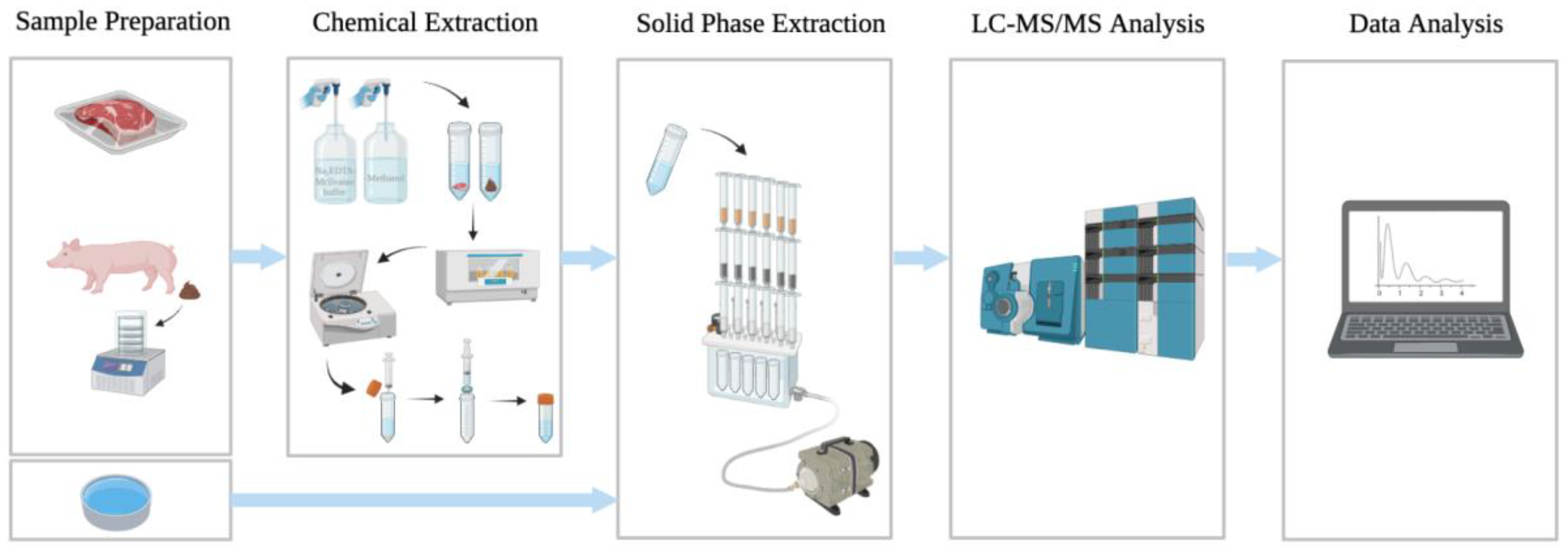
2.2. Sample Preparation

Swine feces were collected from local farms and freeze-dried before processing. Pork was purchased from local markets. River water, animal drinking water, and Milli-Q water were used as water samples. An antibiotic mixture (~0.01 mg/mL concentration) containing 43 different antibiotics was used for the spike-in. For solid samples, the antibiotic mixture was directly spiked into 1 g of solid sample (i.e., fecal sample and meat sample) by adding the aqueous antibiotic mixture into a Falcon tube containing the solid sample and then mixing by vortex. The sample was allowed to stand for 1 h to allow for the antibiotic mixture to be absorbed into the solid sample. For liquid samples, the antibiotic mixture was spiked into 100 mL of liquid sample at three volumes: 1 mL of 0.01 mg/mL (i.e., 10 μg), 0.5 mL of 0.01 mg/mL (i.e., 5 μg), and 0.1 mL of 0.01 mg/mL (i.e., 1 μg). All samples were stored at −20 °C and extracted within 1 week.

2.3. Chemical Extraction

Chemical extraction was carried out according to the literature [14] with modifications. The chemical extraction buffer that was used was a mixture of Na2EDTA-McIlvaine buffer solution (10 mL), which contains 10.93 mg/mL anhydrous dibasic sodium phosphate, 12.93 mg/mL citric acid monohydrate, and 37.22 mg/mL Na2EDTA, and 100% methanol (10 mL). For extraction, 20 mL of this chemical extraction buffer was added into 1 g of sample (fecal sample or meat sample). Extraction was carried out in a shaking incubator at 300 rpm for 30 min at room temperature. Supernatants were collected after centrifugation at 3200 g for 5 min at 4 °C. Residues were further extracted two more times with the same protocol. Supernatants of the same sample were then combined and stored at −20 °C until solid-phase extraction (SPE) was performed. For liquid samples, chemical extraction was not performed. All samples were filtered through a 0.2 μm PES syringe filter, purchased from Sartorius (Göttingen, Germany), prior to solid-phase extraction.

2.4. Solid-Phase Extraction (SPE)

An Oasis MAX 6cc (150 mg) cartridge (cat. no.: 186000369), Oasis PRiME HLB 6cc (200 mg) cartridge (cat. no.: 186008057), and Oasis PRiME MCX 6cc (150 mg) cartridge (cat. no.: 186008919) were purchased from Waters (Milford, MA, USA) and used to form a MAX-HLB-MCX combined cartridge for solid-phase extraction. All cartridges were pre-conditioned with acetonitrile and Milli-Q water separately. To investigate the retention of antibiotics in different SPE cartridges, 10 mL of the antibiotic mixture containing 43 targeted antibiotics was loaded to the MAX-HLB-MCX combined cartridge. For solid samples, supernatants collected from chemical extractions were diluted with Milli-Q water to reduce the methanol content to less than 5% (v/v). All of the diluted supernatant flowed through the MAX-HLB-MCX combined cartridge at a flow rate of 3 mL/min. For liquid samples, all of the samples were passed through the MAX-HLB-MCX combined cartridge directly at a flow rate of 3 mL/min. Analytes were eluted from the MAX cartridge, HLB cartridge, and MCX cartridge separately. To elute the analytes, 4 mL of 2% formic acid in methanol, acetonitrile/methanol (60%/40%; v/v), and 5% ammonia solution in methanol were used, respectively. The elution process was repeated three times. The ratio in which elutes were mixed from the MAX, HLB, and MCX cartridges was 1:1:1. Elutes from the same sample were combined for LC-MS/MS analysis.

2.5. LC-MS/MS Analysis

The Acquity I-Class ultra-high-performance liquid chromatographic system by Waters (Milford, MA, USA), coupled with the QTRAP® 6500+ MS system from AB Sciex (Framingham, MA, USA), was used for LC-MS/MS analysis. A Phenomenex Synergi 4 μm Fusion-RP 80 Å (2 mm × 50 mm) column was used for separation, and the column oven temperature was set at 40 °C. The elution gradient (solvent A: 0.1% aqueous formic acid, solvent B: acetonitrile) was set up as follows: 0 min, 0% B; 0.1 min, 0% B; 1 min, 10% B; 6.5 min, 50% B; 7 min, 100% B; 8 min, 100% B. This gradient was re-equilibrated to 0% B for 2 min after each run. The flow rate was 0.5 mL/min, and the injection volume was 5 μL. In terms of electrospray ionization, the parameters were the following: (CUR), nitrogen, 12; collision gas (CAD), high; electrospray voltage, +5500 V; ion source temperature, 550 °C; curtain gas of 25, CAD gas medium, and gas 1 and 2 of 45 and 50 psi, respectively. Retention time and transitions are shown in Table 2.
For transition ranges, each pure antibiotic compound purchased commercially was first injected into the LC-MS/MS instrument for preliminary testing. From this preliminary test, information about transition ranges was obtained. Two transitions of each antibiotic with sharp peaks shown were chosen as references to identify the antibiotic.

2.6. Data Analysis

Data analysis for LC-MS/MS was performed using the SciEX OS-Q Analysis Software (Framingham, MA, USA). Analytes were confirmed by comparing the retention time and the ratio of characteristic transitions between the sample and the standard.

3. Results

3.1. Limit of Detection

This study began with the determination of the detection limit (i.e., limit of detection, LOD) of the MS system. The LOD was determined by injecting a low concentration of antibiotic standard into the mass spectrometer directly and then reviewing the peak generated. If the signal was three times higher than the background base noise level, then we accepted the peak as an actual peak. Infusion was only used for optimizing the MRM parameter before LC-MS/MS. The results of LOD are shown in Table 2. Generally, the detection limit varied. Most of the 43 antibiotics were detectable at levels lower than 70 ppb, with only six antibiotics having a level of detection higher than 70 ppb. These six antibiotics were colistin A, colistin B, neomycin trisulfate, gentamicin, and streptomycin sulfate. Nevertheless, all 43 targeted antibiotics were detectable.

3.2. Solid-Phase Extraction

The percentage of recovery was calculated by comparing the concentration of antibiotic recovered after passing through SPE and without passing through SPE. In other words, the percentage of recovery = total concentration of antibiotic in elutes from SPE/concentration of antibiotic in antibiotic mixture before passing through SPE. The recoveries of amoxicillin, ampicillin, cefquinome sulfate, meropenem, and tiamulin from SPE were poor, with loss being >75% (Table 3). Apart from those which had a poor recovery, 11 antibiotics were retained in the MAX cartridge; five antibiotics were retained in the HLB cartridge; and 17 antibiotics were retained in the MCX cartridge. For chlortetracycline hydrochloride, doxycycline, and mequindox, the MAX cartridge could not completely retain all of the residues, and a significant portion flowed through the MAX cartridge and were retained in the HLB cartridge. For sulfadiazine and sulfadimidine, they could be detected in the elutes of all three cartridges in the MAX-HLB-MCX tandem. Overall, 38 out of the 43 antibiotics had a recovery that was satisfactory or good after SPE.

3.3. Chemical Extraction

The integrated recovery of antibiotic residues (i.e., including the limitation of chemical extraction, SPE, and LOD) is shown in Table 4. Using our methodology, 30 out of 43 targeted antibiotics could be detected (Table 4). The 13 antibiotics that could not be detected were: amoxicillin, cefquinome sulfate, ceftazidime, cefuroxime, ciprofloxacin, colistin (A and B), gentamicin, kanamycin sulfate, meropenem, neomycin trisulfate, norfloxacin, and vancomycin. Among these 13 undetectable antibiotics, amoxicillin, cefquinome sulfate, and meropenem were found to have a poor recovery from SPE.

3.4. Sensitivity

The sensitivities of antibiotic classes were calculated by taking the raw data and then summing the data for all of the tests performed for all antibiotics in a class. Overall, the sensitivity of the protocol in this study was high (i.e., >60%) for 30 out of the 43 antibiotic residues (Table 5). When the spike-in concentration was high, i.e., 10 μg, our approach showed a sensitivity of 100% in most of the antibiotics in all three types of sample. For ampicillin, ceftiofur, chloramphenicol, chlortetracycline, doxycycline, erythromycin, metronidazole, penicillin, spectinomycin, streptomycin, tetracycline, tilmicosin, tylosin tartrate, and tylvalosin, the sensitivities of the protocol were only reduced when the spiked-in antibiotic content was reduced to 1 μg (Table S1).
Out of 16 classes of antibiotic tested, antibiotics from 13 classes could be detected (Table 6). The sensitivity of detecting antibiotics from the antifolate, lincosamide, pleuromutilin, quinoxaline 1,4-di-N-oxide (QdNO), and sulfonamide classes was relatively high in all three types of sample at three different concentrations. The sensitivity of detection of antibiotics from the amphenicol, macrolide, nitroimidazole, and tetracycline classes was high (around 100%) when the spiked-in content was 10 μg and 5 μg, but it was less sensitive when the spiked-in content was reduced to 1 μg. The sensitivity of our detection method was relatively lower for aminoglycosides, cephalosporins, fluoroquinolones, and penicillins, while carbapenems, glycopeptides, and polymyxins could not be detected.

4. Discussion

In this study, we have developed a LC-MS/MS-based working protocol that could detect residues of 30 antibiotics from 13 classes in animal meat and environmental samples. Although using LC-MS/MS to detect antibiotic residues in food and water samples is not a novel technique, our protocol has two significant improvements. The first improvement is that we have developed a methodology that can cover different sample types. Previous developed methods can detect 34 veterinary drugs from six distinct groups in porcine muscle [6]; 75 antibiotics from six groups in meat and aquaculture products [8]; 63 pharmaceuticals in natural water [12]; 58 antibiotics from eight groups in milk [10]; and 20 antibiotics from three different groups in honey [11]. Most of these methods targeted food samples and are not sufficient in monitoring antibiotic contaminations, particularly in various types of environmental sample.
The second improvement is that we have developed a methodology that can cover a wider range of different antibiotics. Different antibiotics have different chemical properties, and they may require different extraction and detection methods [19,20]. In our study, we used a single extraction and detection protocol to cover 30 antibiotics from 13 families, which makes it an improvement over existing methodologies in terms of efficiency and convenience [7,9,13,14,15]. To provide an insight into why we could make the aforementioned two improvements in detecting antibiotic residues, we provide an interpretation of the results after SPE and chemical extraction were conducted in our protocol.
In theory, SPE is a common procedure performed to clean up the samples before LC-MS/MS analysis [21,22]. However, the major purpose of using SPE in this study was to concentrate the analytes from large-volume but low-concentration samples, such as environmental water and wastewater with diluted concentrations of antibiotic residues. Thus, in order to increase the sensitivity of detection, SPE was an essential step of our developed method. The chemical nature of the antibiotics and the type of SPE cartridge could influence the final recovery of antibiotic residues for detection. Considering that 43 antibiotics with varying properties were targeted in this study, we used a MAX-HLB-MCX tandem consisting of the MAX cartridge, the MCX cartridge, and the HLB cartridge to retain as much of the antibiotic residues as possible. The rationale behind this tandem formation was that the different cartridges would target compounds with different pH properties: the MAX cartridge targets acidic compounds; the MCX cartridge targets basic compounds; and the HLB cartridge targets relatively neutral compounds (i.e., those that are neither basic nor acidic).
Our SPE method was able to recover a majority of the antibiotics (38 out of 43 antibiotics) with satisfactory or good performance, indicating its ability to successfully detect antibiotics of different pH properties. For those five antibiotics that had a poor recovery after SPE (i.e., >75% loss), we hypothesized that the chemical structure of these antibiotics may not be compatible with the tandem formation that we designed. Most of these belonged to the β-lactam class of antibiotics, of which amoxicillin, ampicillin, cefquinome sulfate, and meropenem possess a chemically unstable β-lactam ring that spontaneously undergoes hydrolysis [23]. Thus, this is a plausible reason explaining why it was very difficult to recover these antibiotics in water samples.
Generally, the extraction method of using Na2EDTA-McIlvaine buffer solution/methanol (1:1; v/v) provided a detection of 30 different antibiotics. There were three antibiotics that were found to have a poor SPE recovery and could not be detected in spiked-in samples: amoxicillin, cefquinome sulfate, and meropenem. It would not be possible to determine the reason of zero sensitivity for these three antibiotics, since it could be related to the chemical extraction method, poor SPE recovery, or both. Thus, it is difficult to determine whether the three antibiotics could be extracted using our chemical extraction protocol. For the rest of the nine antibiotics that could not be detected, it seems that the extraction method was insufficient in recovering these antibiotics. We arrived at this conclusion because we observed a satisfactory recovery rate from SPE for these antibiotics, and the spike-in concentrations were higher than the LOD. Based on these results, the extraction method may not be suitable for extracting the residues of ceftazidime, cefuroxime, ciprofloxacin, colistin, gentamicin, kanamycin sulfate, neomycin trisulfate, norfloxacin, and vancomycin from samples. Further study for modifying the chemical extraction method to further increase the range of recovery may be necessary.
The proposed method has notable limitations, such as the inability to conduct precise quantitative analysis and a decline in sensitivity with lowering the spiked-in concentration. Although SPE is commonly performed to remove impurities from analytes in order to reduce a potential matrix effect, the use of the MAX-HLB-MCX tandem resulted in strong matrix effects. Moreover, it is difficult to obtain corresponding isotopes for all tested antibiotics to correct the matrix effect of each antibiotic during quantification. However, an estimation of the antibiotic concentrations may still be made by performing calculations. From LC-MS/MS results, one will have the information of the total amount of antibiotics in the elute of each sample. The concentration of an antibiotic can be estimated by the following equation: (concentration of antibiotic in sample = amount of antibiotic/sample weight or sample volume). Thus, the difference in volume between solid and liquid samples during the whole process does not affect the estimation of the antibiotic concentration in samples after calculation.
Only 15 out of the 30 detectable antibiotics could maintain a sensitivity of 100% when the spiked-in content was reduced from 10 μg to 5 μg and 1 μg for all three types of sample. It seems that, when the spiked-in concentrations were gradually lowered, the sensitivity of the protocol decreased. From the results in Table 3 and Table 4, the loss of antibiotic residues during the extraction process and SPE was expected. The explanation is that the likelihood of a false negative result increased when the residue content decreased. Having stated these limitations, our method could be useful in AMU surveillance in livestock farms as a first-line qualitative assessment tool—especially for detecting residues in farm waste.

5. Conclusions

In conclusion, 30 different antibiotics from 13 classes could be detected with high sensitivity with our sample processing method when the residue content was 10 ppm or above. When the residue content was reduced to 1 ppm, 27 different antibiotics could still be detected, and 21 of them had a sensitivity higher than 50%. The developed chemical extraction method, together with SPE, allowed us to detect at least 30 antibiotic residues from 13 families qualitatively in foods and environmental samples at the same time. Nevertheless, further study to reduce the matrix effect of analytes is necessary so that quantification of antibiotic residues could be possible.

Supplementary Materials

The following supporting information can be downloaded at: https://www.mdpi.com/article/10.3390/antibiotics11070845/s1, Table S1. Reduction in sensitivity of overall protocol for 43 antibiotics at spike-in concentrations of 10 μg, 5 μg, and 1 μg.

Author Contributions

Conceptualization, H.M.T.; Methodology, C.-L.C. and H.K.-F.W.; Formal Analysis, C.-L.C. and H.K.-F.W.; Writing—Original Draft Preparation, C.-L.C. and H.K.-F.W.; Writing—Review & Editing, P.W., S.-W.L. and O.S.-K.C.; Supervision, H.M.T.; Funding Acquisition, H.M.T., C.-L.C. and H.K.-F.W. shared co-first authorship. All authors have read and agreed to the published version of the manuscript.

Funding

This study was supported by the Research Impact Fund, University Grants Committee (project no. R7033-18F) RGC Theme-based Research Scheme (project no. T21-705/20-N), the Health and Medical Research Fund Fellowship Scheme (project no. 04180077) tegic Public Policy Research Fund, Policy Innovation, and Co-ordination Office (project no. S2017.A8.005.17S) of the Government of Hong Kong Special Administrative Region, the Ntional Natural Science Foundation of China (NSFC)–Research Grant Council (RGC) Joint Research Scheme (project no. N_HKU740/19), and Welcome Trust Grant Funding (project no. 219622/Z/19/Z).

Institutional Review Board Statement

Not applicable.

Informed Consent Statement

Not applicable.

Acknowledgments

The authors acknowledge the assistance of The Proteomics and Metabolomics Core, Centre for PanorOmic Sciences, Li Ka Shing Faculty of Medicine, The University of Hong Kong, Hong Kong SAR, China.

Conflicts of Interest

The authors declare no conflict of interest.

References

  1. World Health Organization. Antimicrobial Resistance. 2020. Available online: https://www.who.int/news-room/fact-sheets/detail/antimicrobial-resistance (accessed on 31 March 2021).
  2. Ventola, C.L. The antibiotic resistance crisis: Part 1: Causes and threats. Pharm. Ther. 2015, 40, 277–283. [Google Scholar]
  3. World Health Organization. WHO, FAO, and OIE Unite in the Fight Against Antimicrobial Resistance; World Health Organization: Geneva, Switzerland, 2015. [Google Scholar]
  4. European Commission. A European One Health Action Plan against Antimicrobial Resistance (AMR); European Commission: Brussels, Belgium, 2017. [Google Scholar]
  5. World Health Organization. Antimicrobial Resistance: Global Report on Surveillance; World Health Organization: Geneva, Switzerland, 2014. [Google Scholar]
  6. Lopes, R.; Augusti, D.; Oliveira, A.; Oliveira, F.; Vargas, E.; Augusti, R. Development and validation of a methodology to qualitatively screening veterinary drugs in porcine muscle via an innovative extraction/clean-up procedure and LC-MS/MS analysis. Food Addit. Contam. Part A Chem. Anal. Control Expo. Risk Assess. 2011, 28, 1667–1676. [Google Scholar] [CrossRef] [PubMed]
  7. Zheng, W.; Abd El-Aty, A.M.; Kim, S.K.; Choi, J.M.; Park, D.H.; Yoo, K.H.; Kang, Y.S.; Jeon, J.S.; Hacimuftuoglu, A.; Shim, J.H.; et al. Development and validation of a solid-phase extraction method coupled with LC-MS/MS for the simultaneous determination of 16 antibiotic residues in duck meat. Biomed. Chromatogr. 2019, 33, e4501. [Google Scholar] [CrossRef] [PubMed]
  8. Dubreil, E.; Gautier, S.; Fourmond, M.-P.; Bessiral, M.; Gaugain, M.; Verdon, E.; Pessel, D. Validation approach for a fast and simple targeted screening method for 75 antibiotics in meat and aquaculture products using LC-MS/MS. Food Addit. Contam. Part A 2017, 34, 453–468. [Google Scholar] [CrossRef] [PubMed]
  9. Alija, G.; Hajrulai-Musliu, Z.; Uzunov, R. Development and validation of confirmatory LC–MS/MS method for multi-residue analysis of antibiotic drugs in bovine milk. SN Appl. Sci. 2020, 2, 1563. [Google Scholar] [CrossRef]
  10. Gaugain-Juhel, M.; Delepine, B.; Gautier, S.; Fourmond, M.P.; Gaudin, V.; Hurtaud-Pessel, D.; Verdon, E.; Sanders, P. Validation of a liquid chromatography-tandem mass spectrometry screening method to monitor 58 antibiotics in milk: A qualitative approach. Food Addit. Contam. Part A Chem. Anal. Control Expo. Risk Assess. 2009, 26, 1459–1471. [Google Scholar] [CrossRef] [PubMed] [Green Version]
  11. Kaufmann, A.; Roth, S.; Ryser, B.; Widmer, M.; Guggisberg, D. Quantitative LC/MS-MS determination of sulfonamides and some other antibiotics in honey. J. AOAC Int. 2002, 85, 853–860. [Google Scholar] [CrossRef] [PubMed] [Green Version]
  12. Mokh, S.; El Khatib, M.; Koubar, M.; Daher, Z.; Al Iskandarani, M. Innovative SPE-LC-MS/MS technique for the assessment of 63 pharmaceuticals and the detection of antibiotic-resistant-bacteria: A case study natural water sources in Lebanon. Sci. Total Environ. 2017, 609, 830–841. [Google Scholar] [CrossRef] [PubMed]
  13. Yang, Y.; Song, W.; Lin, H.; Wang, W.; Du, L.; Xing, W. Antibiotics and antibiotic resistance genes in global lakes: A review and meta-analysis. Environ. Int. 2018, 116, 60–73. [Google Scholar] [CrossRef] [PubMed] [Green Version]
  14. Chen, Y.; Zhang, H.; Luo, Y.; Song, J. Occurrence and assessment of veterinary antibiotics in swine manures: A case study in East China. Chin. Sci. Bull. 2012, 57, 606–614. [Google Scholar] [CrossRef] [Green Version]
  15. Duelge, K.J.; Nishshanka, U.; De Alwis, H.G. An LC-MS/MS method for the determination of antibiotic residues in distillers grains. J. Chromatogr. B Anal. Technol. Biomed. Life Sci. 2017, 1053, 81–86. [Google Scholar] [CrossRef] [PubMed]
  16. Barreca, S.; Forni, C.; Colzani, L.; Clerici, L.; Daverio, D.; Dellavedova, P. Study on the stability of antibiotics, pesticides and drugs in water by using a straightforward procedure applying HPLC-mass spectrometric determination for analytical purposes. Separations 2021, 8, 179. [Google Scholar] [CrossRef]
  17. Okerman, L.; Van Hende, J.; De Zutter, L. Stability of frozen stock solutions of beta-lactam antibiotics, cephalosporins, tetracyclines and quinolones used in antibiotic residue screening and antibiotic susceptibility testing. Anal. Chim. Acta 2007, 586, 284–288. [Google Scholar] [CrossRef] [PubMed]
  18. Berendsen, B.J.; Elbers, I.J.; Stolker, A.A. Determination of the stability of antibiotics in matrix and reference solutions using a straightforward procedure applying mass spectrometric detection. Food Addit. Contam. Part A Chem. Anal. Control Expo. Risk Assess. 2011, 28, 1657–1666. [Google Scholar] [CrossRef] [PubMed]
  19. Rossi, R.; Saluti, G.; Moretti, S.; Diamanti, I.; Giusepponi, D.; Galarini, R. Multiclass methods for the analysis of antibiotic residues in milk by liquid chromatography coupled to mass spectrometry: A review. Food Addit. Contam. Part A Chem. Anal. Control Expo. Risk Assess. 2018, 35, 241–257. [Google Scholar] [CrossRef] [PubMed]
  20. Zhang, Y.; Li, X.Q.; Li, H.M.; Zhang, Q.H.; Gao, Y.; Li, X.J. Antibiotic residues in honey: A review on analytical methods by liquid chromatography tandem mass spectrometry. TrAC Trends Anal. Chem. 2019, 110, 344–356. [Google Scholar] [CrossRef]
  21. Andrade-Eiroa, A.; Canle, M.; Leroy-Cancellieri, V.; Cerdà, V. Solid-phase extraction of organic compounds: A critical review. part ii. TrAC Trends Anal. Chem. 2016, 80, 655–667. [Google Scholar] [CrossRef]
  22. Pico, Y.; Fernandez, M.; Ruiz, M.J.; Font, G. Current trends in solid-phase-based extraction techniques for the determination of pesticides in food and environment. J. Biochem. Biophys. Methods 2007, 70, 117–131. [Google Scholar] [CrossRef] [PubMed]
  23. Seifrtova, M.; Novakova, L.; Lino, C.; Pena, A.; Solich, P. An overview of analytical methodologies for the determination of antibiotics in environmental waters. Anal. Chim. Acta 2009, 649, 158–179. [Google Scholar] [CrossRef] [PubMed]
Figure 1. Flowchart showing the key steps in antibiotic residue testing. The figure was created with BioRender.com.
Antibiotics 11 00845 g001
Table 1. List of antibiotics tested.
ClassesAntibiotics
AminoglycosidesGentamicin
Kanamycin sulfate
Neomycin trisulfate salt hydrate
Spectinomycin hydrochloride pentahydrate
Streptomycin sulfate salt
AmphenicolsChloramphenicol
Florfenicol
AntifolateTrimethoprim
CarbapenemsMeropenem
CephalosporinsCefalexin
Cefquinome sulfate
Ceftazidime
Ceftiofur sodium
Cefuroxime
FluoroquinolonesCiprofloxacin
Enrofloxacin
Levofloxacin
Norfloxacin
Ofloxacin
GlycopeptidesVancomycin
LincosamidesClindamycin phosphate
Lincomycin hydrochloride
MacrolidesErythromycin
Tilmicosin
Tylosin tartrate salt
Tylvalosin
NitroimidazoleMetronidazole
PenicillinsAmoxicillin
Ampicillin
Penicillin G sodium salt
PleuromutilinsTiamulin
PolymyxinsColistin A
Colistin B
Quinoxaline 1,4-di-N-oxides (QdNOs)Mequindox
SulfonamidesSulfachloropyridazine
Sulfadiazine
Sulfadimidine
Sulfamethoxazole
Sulfamonomethoxine
TetracyclinesChlortetracycline hydrochloride
Doxycycline
Oxytetracycline
Tetracycline
Table 2. Liquid chromatography and mass spectrometry of 43 antibiotics.
AntibioticsRetention Time (min)Transition 1 (m/z)Transition 2 (m/z)Limit of Detection (ppb)
Amoxicillin0.83366.1 > 348.9366.1 > 2088.51
Ampicillin1.78350 > 191.9350 > 1600.49
Cefalexin1.79348 > 158348 > 1740.9
Cefquinome sulfate2.33529 > 396529 > 1341.11
Ceftazidime2.14547.1 > 467.8547.1 > 3963.44
Ceftiofur sodium3.53524 > 241524 > 2850.31
Cefuroxime2.57447 > 385.7447 > 3424.1
Chloramphenicol2.56323.1 > 274.9323.1 > 304.85.41
Chlortetracycline hydrochloride2.66479 > 444479 > 4624.06
Ciprofloxacin2.35332.1 > 313.9332.1 > 231.10.4
Clindamycin phosphate3.16505.1 > 457505.1 > 487.10.55
Colistin A1.95585.6 > 535.5585.6 > 576.4862.53
Colistin B1.77578.5 > 528.4578.5 > 569.5793.3
Doxycycline2.94445.1 > 428445.1 > 2670.49
Enrofloxacin2.58360 > 316360 > 2450.4
Erythromycin3.83734.3 > 576.3734.3 > 157.914.52
Florfenicol2.26358 > 340358 > 24113.21
Gentamicin2.79500.1 > 456500.1 > 227.1320
Kanamycin sulfate0.27485 > 324485 > 1636.54
Levofloxacin2.36362.1 > 318.2362.1 > 261.10.47
Lincomycin hydrochloride1.43407 > 126407 > 3590.45
Mequindox2.18219 > 143219 > 1851.7
Meropenem1.79384.1 > 340.1384.1 > 297.71.76
Metronidazole1.06172 > 128.2172 > 82.10.46
Neomycin trisulfate salt hydrate0.26615 > 293615 > 161345.18
Norfloxacin2.3320 > 302320 > 231.20.54
Ofloxacin2.35362 > 318362 > 2610.43
Oxytetracycline2.02461 > 426461 > 4441.98
Penicillin G sodium salt2.36335 > 160335 > 17610.79
Spectinomycin hydrochloride pentahydrate0.3333 > 189333 > 1402.52
Streptomycin sulfate salt4.13582 > 174582 > 156425.98
Sulfachloropyridazine2.3285 > 156285 > 1080.6
Sulfadiazine1.45251 > 156251 > 920.45
Sulfadimidine2.08279 > 186279 > 1560.36
Sulfamethoxazole2.4254.1 > 155.8254.1 > 108.20.43
Sulfamonomethoxine2.38281 > 156281 > 1260.62
Tetracycline2.14445 > 410445 > 2690.49
Tiamulin4.12494 > 192494 > 1190.59
Tilmicosin3.38869.4 > 696869.4 > 1745.73
Trimethoprim1.92291.1 > 230291.1 > 260.90.41
Tylosin tartrate salt4.13916.3 > 772916.3 > 17411.63
Tylvalosin5.21042.3 > 8141042.3 > 17464.27
Vancomycin1.95726 > 144725 > 14426.63
Table 3. Solid-phase extraction of 43 antibiotics mixture.
AntibioticsSPE RecoveryMainly Retained
AmoxicillinPoorMAX
AmpicillinPoorMAX
CefalexinGoodMCX
Cefquinome sulfatePoorMAX/HLB
CeftazidimeGoodMAX
Ceftiofur sodiumGoodMAX
CefuroximeGoodMAX
ChloramphenicolGoodMAX
Chlortetracycline hydrochlorideSatisfactoryMAX/HLB
CiprofloxacinGoodMCX
Clindamycin phosphateGoodMAX
Colistin ASatisfactoryMCX
Colistin BSatisfactoryMCX
DoxycyclineSatisfactoryMAX/HLB
EnrofloxacinSatisfactoryMCX
ErythromycinGoodHLB
FlorfenicolGoodMAX
GentamicinSatisfactoryMAX
Kanamycin sulfateGoodMCX
LevofloxacinGoodMCX
Lincomycin hydrochlorideGoodMCX
MequindoxSatisfactoryMAX/HLB
MeropenemPoorMAX/HLB
MetronidazoleGoodMCX
Neomycin trisulfate salt hydrateGoodMCX
NorfloxacinGoodMCX
OfloxacinGoodMCX
OxytetracyclineSatisfactoryMCX
Penicillin G sodium saltGoodMAX
Spectinomycin hydrochloride pentahydrateSatisfactoryMCX
Streptomycin sulfate saltGoodHLB
SulfachloropyridazineGoodMAX
SulfadiazineGoodMAX/HLB/MCX
SulfadimidineGoodMAX/HLB/MCX
SulfamethoxazoleGoodMAX
SulfamonomethoxineGoodMAX
TetracyclineGoodMCX
TiamulinPoorMCX
TilmicosinSatisfactoryHLB
TrimethoprimGoodMCX
Tylosin tartrate saltGoodHLB
TylvalosinGoodHLB
VancomycinGoodMCX
Concentration of antibiotics mixture: 10 mL, 0.01 mg/mL. Recovery < 25% is considered to be “Poor”; recovery ≥25% and ≤60% is considered to be “Satisfactory”; and recovery >60% is considered to be “Good”.
Table 4. Detection of antibiotic mixture for spike-in of water, fecal, and meat sample.
AntibioticsWater SampleFecal SampleMeat Sample
AmoxicillinN.D.N.D.N.D.
AmpicillinDetectedDetectedDetected
CefalexinDetectedDetectedDetected
Cefquinome sulfateN.D.N.D.N.D.
CeftazidimeN.D.N.D.N.D.
Ceftiofur sodiumDetectedDetectedDetected
CefuroximeN.D.N.D.N.D.
ChloramphenicolDetectedDetectedDetected
Chlortetracycline hydrochlorideDetectedDetectedDetected
CiprofloxacinN.D.N.D.N.D.
Clindamycin phosphateDetectedDetectedDetected
Colistin AN.D.N.D.N.D.
Colistin BN.D.N.D.N.D.
DoxycyclineDetectedDetectedDetected
EnrofloxacinDetectedDetectedDetected
ErythromycinDetectedDetectedDetected
FlorfenicolDetectedDetectedDetected
GentamicinN.D.N.D.N.D.
Kanamycin sulfate mixture of kanamycin A (main component) and kanamycin B and CN.D.N.D.N.D.
LevofloxacinDetectedDetectedDetected
Lincomycin hydrochlorideDetectedDetectedDetected
MequindoxDetectedDetectedDetected
MeropenemN.D.N.D.N.D.
MetronidazoleDetectedDetectedDetected
Neomycin trisulfate salt hydrateN.D.N.D.N.D.
NorfloxacinN.D.N.D.N.D.
OfloxacinDetectedDetectedDetected
OxytetracyclineDetectedDetectedDetected
Penicillin G sodium saltDetectedDetectedDetected
Spectinomycin hydrochloride pentahydrateDetectedDetectedDetected
Streptomycin sulfate saltDetectedDetectedDetected
SulfachloropyridazineDetectedDetectedDetected
SulfadiazineDetectedDetectedDetected
SulfadimidineDetectedDetectedDetected
SulfamethoxazoleDetectedDetectedDetected
SulfamonomethoxineDetectedDetectedDetected
TetracyclineDetectedDetectedDetected
TiamulinDetectedDetectedDetected
TilmicosinDetectedDetectedDetected
TrimethoprimDetectedDetectedDetected
Tylosin tartrate saltDetectedDetectedDetected
TylvalosinDetectedDetectedDetected
VancomycinN.D.N.D.N.D.
1 mL of antibiotic mixture (0.01 mg/mL) was spiked into 1 g of solid sample or 100 mL of liquid sample. N.D. represents “not detected”.
Table 5. Sensitivity of overall protocol for 43 antibiotics at spike-in concentrations of 10 μg, 5 μg, and 1 μg.
AntibioticsSensitivity
10 μg Spiked-in5 μg Spiked-in1 μg Spiked-in
Water SampleFecal SampleMeat SampleWater SampleFecal SampleMeat SampleWater SampleFecal SampleMeat Sample
Amoxicillin000000000
Ampicillin10010010010010010066.7033.3
Cefalexin100100100100100100100100100
Cefquinome sulfate000000000
Ceftazidime000000000
Ceftiofur sodium10010010010010010066.766.70
Cefuroxime000000000
Chloramphenicol1001001001001001000066.7
Chlortetracycline hydrochloride10010010010010010010010066.7
Ciprofloxacin000000000
Clindamycin phosphate100100100100100100100100100
Colistin A000000000
Colistin B000000000
Doxycycline100100100100100100033.30
Enrofloxacin100100100100100100100100100
Erythromycin10010010010033.310033.3033.3
Florfenicol100100100100100100100100100
Gentamicin000000000
Kanamycin sulfate000000000
Levofloxacin100100100100100100100100100
Lincomycin hydrochloride100100100100100100100100100
Mequindox100100100100100100100100100
Meropenem000000000
Metronidazole10010010010010010010033.366.7
Neomycin trisulfate salt hydrate000000000
Norfloxacin000000000
Ofloxacin100100100100100100100100100
Oxytetracycline100100100100100100100100100
Penicillin G sodium salt10010010010033.3100000
Spectinomycin hydrochloride pentahydrate10066.710066.766.7100000
Streptomycin sulfate salt66.710010033.3066.7000
Sulfachloropyridazine100100100100100100100100100
Sulfadiazine100100100100100100100100100
Sulfadimidine100100100100100100100100100
Sulfamethoxazole100100100100100100100100100
Sulfamonomethoxine10010066.7100100100100100100
Tetracycline10010010010010010010010066.7
Tiamulin100100100100100100100100100
Tilmicosin66.710010066.7100100033.333.3
Trimethoprim100100100100100100100100100
Tylosin tartrate salt10010010010010010010010066.7
Tylvalosin10010010010010010033.3100100
Vancomycin000000000
Sensitivity = [number of true positives/(number of true positives + number of false negatives)] × 100%. Note: the number of tests performed to calculate detection sensitivity was 3.
Table 6. Sensitivity of overall protocol for 16 groups of antibiotics at spike-in concentrations of 10 μg, 5 μg, and 1 μg.
Antibiotics GroupSensitivity
10 μg Spiked-in5 μg Spiked-in1 μg Spiked-in
Water SampleFecal SampleMeat SampleWater SampleFecal SampleMeat SampleWater SampleFecal SampleMeat Sample
Aminoglycosides33.333.3402013.333.3000
Amphenicols100100100100100100505083.3
Antifolate100100100100100100100100100
Carbapenems000000000
Cephalosporins40404040404033.333.320
Fluoroquinolones606060606060606060
Glycopeptides000000000
Lincosamides100100100100100100100100100
Macrolides91.710010091.783.310041.758.358.3
Nitroimidazole10010010010010010010033.366.7
Penicillins66.766.766.766.744.466.722.2011.1
Pleuromutilins100100100100100100100100100
Polymyxins000000000
Quinoxaline 1,4-di-N-oxides (QdNOs)100100100100100100100100100
Sulfonamides10010093.3100100100100100100
Tetracyclines1001001001001001007583.358.3
Sensitivity = [number of true positives/(number of true positives + number of false negatives)] × 100%.
Publisher’s Note: MDPI stays neutral with regard to jurisdictional claims in published maps and institutional affiliations.

