Article

The Prevalence and Molecular Biology of Staphylococcus aureus Isolated from Healthy and Diseased Equine Eyes in Egypt

1
Department of Animal Medicine, Faculty of Veterinary Medicine, Kafrelshkh University, Kafrelsheikh 33511, Egypt
2
Department of Internal Medicine and Infectious Diseases, Faculty of Veterinary Medicine, Mansoura University, Mansoura 35516, Egypt
3
Department of Poultry and Rabbit Diseases, Faculty of Veterinary Medicine, Kafrelsheikh University, Kafrelsheikh 33511, Egypt
4
Department of Bacteriology, Mycology and Immunology, Faculty of Veterinary Medicine, Benha University, Benha 13518, Egypt
5
Department of Biotechnology, College of Science, Taif University, P.O. Box 11099, Taif 21944, Saudi Arabia
*
Author to whom correspondence should be addressed.
Antibiotics 2022, 11(2), 221; https://doi.org/10.3390/antibiotics11020221
Submission received: 26 November 2021 / Revised: 5 February 2022 / Accepted: 8 February 2022 / Published: 10 February 2022

Abstract

:
This work aimed to characterize S. aureus isolates from the eyes of healthy and clinically affected equines in the Kafrelsheikh Governorate, Egypt. A total of 110 animals were examined for the presence of S. aureus, which was isolated from 33 animals with ophthalmic lesions and 77 healthy animals. We also investigated the antimicrobial resistance profile, oxacillin resistance mechanism, and the major virulence factors implicated in many studies of the ocular pathology of pathogenic S. aureus. The association between S. aureus eye infections and potential risk factors was also investigated. The frequency of S. aureus isolates from clinically affected equine eyes was significantly higher than in clinically healthy equids. A significant association was found between the frequency of S. aureus isolation from clinically affected equine eyes and risk factors including age and season but not with sex or breed factors. Antimicrobial resistance to common antibiotics used to treat equine eyes was also tested. Overall, the isolates showed the highest sensitivity to sulfamethoxazole (100%) and the highest resistance to cephalosporin (90.67%) and oxacillin (90.48%). PCR was used to demonstrate that mecA was present in 100% of oxacillin- and β-lactam-resistant S. aureus strains. The virulence factor genes Spa (x region), nuc, and hlg were identified in 62.5%, 100%, and 56%, of isolates, respectively, from clinically affected equines eyes. The severity of the eye lesions increased in the presence of γ-toxin-positive S. aureus. The phylogenetic tree of the Spa (x region) gene indicated a relationship with human reference strains isolated from Egypt as well as isolates from equines in Iran and Japan. This study provides insight into the prevalence, potential risk factors, clinical pictures, zoonotic potential, antimicrobial resistance, and β-lactam resistance mechanism of S. aureus strains that cause eye infection in equines from Egypt.

1. Introduction

Bacterial infection of the eye causes diseases, including conjunctivitis, keratitis, endophthalmitis, and corneal ulcers, of which the bacteria Staphylococcus aureus is considered a major causative agent [1]. The bacterial community that inhabits the corneal and conjunctival spaces of healthy horses includes non-pathogenic and opportunistic Gram-positive (G +ve) bacteria [2]. The robust immune system of the horse cornea reduces the possibility of infection; however, opportunistic bacteria such as S. aureus exploit antecedent trauma or pre-existing disease to overcome the host immune defense [3]. Whenever the natural barriers of the host are lowered, S. aureus produces a wide range of purulent and/or toxin-mediated diseases [4]. The development of methicillin resistance in some S. aureus lineages and its fast spread in recent decades is a challenge to human and animal healthcare practitioners [5].
Horse eye diseases and their associated complications are serious health threats to the animal, as they affect its quality of life, its value, and reduce its use for athletic and show purposes. Threatening eye infections that may disrupt horse training and potentially disqualify the animal from competition may necessitate long and expensive treatment courses [6]. Such infections result in a huge economic loss to the horse sports and trading industry, which is estimated at a million dollars a year in the USA [7]. Since S. aureus is able to accommodate various environments and develop diverse new antibiotic resistance mechanisms, it is a predominant bacterial cause of endemic human and animal diseases worldwide [8]. Additionally, due to the close relationship between horse and human, especially in sports, there is a high risk for zoonotic transmission to human caregivers. The bacterial community that inhabits the cornea and conjunctiva of healthy horses includes nonpathogenic and opportunistic Gram-positive (G +ve) bacteria [2]. Furthermore, another study identified methicillin-resistant S. aureus (MRSA) from a donkey conjunctival swab [9].
The probable presence of MRSA in the normal ocular microbiota of healthy horses increases the chance of opportunistic MRSA infections and increases the likelihood of horse eye destruction if it is not diagnosed and treated quickly, which is complicated by the lack of treatment options for MRSA infections. MRSA has been repeatedly isolated from individuals in close contact with infected animals [10]. Therefore, horses, in addition to other pets, including dogs and cats, are often regarded as possible MRSA reservoirs [11,12]. This assumption was strengthened with the detection of genetically related MRSA isolates from horses and their human companions [13]. A large new body of evidence indicates an increase in MRSA infections in animals, including horses, that have close contact with human companions [14,15,16]. Moreover, humans can also be a source of MRSA infection for horses, as MRSA strains can also be transferred from humans to animals [14].
The prevalence of MRSA infection is increasing worldwide with high morbidity and mortality, and since it has multidrug resistance, there are narrow therapeutic options, which makes it a significant health concern [1,17]. Geographical differences between MRSA lineages worldwide have been proposed due to its ability to colonize and infect a wide host range and then circulate within distinct ecological environments [18,19]. This may weaken the efficacy of a general protocol for management. A spatiotemporal relationship between the isolates was also assumed [18]. Therefore, it is highly probable that the similarity between MRSA strains infecting humans and animals is high within each locality. Recently, an S. aureus lineage belonging to sequence type (ST) 398 was indicted as the predominant S. aureus strain infecting animals in Europe; due to its continuous acquisition of new antibiotic-resistance and virulence determinants, it was described as a serious public health concern [20]. The study of S. aureus strains in our country to characterize the virulence factors, drug-resistance patterns, and prevalence of regional strains may be required to enable the construction of region-specific plans for the proper management, control, and treatment of S. aureus.
To the best of our knowledge, a major study has not been conducted to characterize the predominant MRSA strains across Egypt. This study was designed to investigate the epidemiological, microbial, and molecular patterns of S. aureus isolated from clinically healthy and clinically affected equine eyes in the Kafrelsheikh governorates of Egypt. We report the resistance profiles of pathogenic S. aureus isolates to antibiotics and their associated mechanism(s). We also conducted a phylogenetic analysis of the Spa x region of pathogenic S. aureus strains to describe the zoonotic potential of these isolates within the region based on their relationship to previous isolates.

2. Material and Methods

2.1. Study Design

This survey targeted equines from December 2019 to June 2020 in the Kafrelsheikh province of northern Egypt. The survey effort was applied proportionally across the province of Kafrelsheikh based on the number of horses and donkeys in each town or city. The total number of equines in the study was 13,000, with a sample size of 0.85% using survey design and analysis software (survey toolbox program), which was used for the random selection of 220 eye swap samples from 110 equines, and several animals were sampled depending on the size of the herd. A total of 110 animals, 33 with ophthalmic lesions and 77 healthy, were selected for the study. The animals were randomly selected from 14 village herds. The herd effect was considered in logistic regression analysis.
All horses were fed a concentrated diet with berseem in the winter and hay in the summer. S. aureus were isolated from clinically healthy and clinically affected equine eyes.
Ophthalmic examination procedures included a general inspection of the eyes as well as detailed ocular examination [21]. All experiments and animal procedures were conducted in accordance with local guidelines following the approval of all experimental procedures by the local ethical committee of infectious diseases at the Faculty of Veterinary Medicine, Kafrelsheikh University, Egypt (ethical approval number KFS-2019/1). The samples were collected after obtaining permission from the farm owners, farm managers, or herdsmen.

2.2. Sample Collection

A total of 220 swabs were collected from 110 equines from several locations in the Kafrelsheikh province. A swab from the ocular conjunctiva of each was collected from the inferior conjunctival fornix of both eyes, without touching the eyelashes or eyelids. All samples were collected before the animals received any antibiotics or anesthetics [21]. The collected swabs were placed directly into sterile test tubes containing tryptone soya broth (Oxoid, Altrincham, UK) as an enrichment medium. The samples were kept on ice in power-cooled boxes until they were transported to the laboratory, within 5 h of collection, for further bacteriological examination.

2.3. Isolation and Identification of Staphylococcus Species

The samples inoculated in enrichment broth were incubated overnight at 37 °C and then a loopful was streaked on mannitol salt agar medium (Lab. Lemco 400, Worthing, UK). A colony from each plate suspected of being S. aureus, based on agar appearance, was picked and streaked on Baird Parker agar plates then incubated for 16 to 18 h at 37 °C. All S. aureus isolates were identified based on culture characteristics on Baird Parker agar with mannitol salt. The coagulase test was also performed to confirm S. aureus isolation. All S. aureus isolates were biochemically confirmed using the API 20NE system (BioMerieux, Marcy-l’E’ toile, France) and molecularly confirmed by PCR with staphylococcal 16s RNA primers to confirm the Staphylococcal genus and with the nuc gene to confirm the species S. aureus (Table 1).

2.4. Antibiotic Susceptibility Testing

The antibiotic sensitivity test was performed for all pathogenic S. aureus isolates using the Kirby–Bauer disk diffusion method [21]. Each isolate was inoculated in Mueller–Hinton broth and incubated for 24 h. The bacterial suspension was adjusted to match the 0.5 McFarland standard (approximately 1–2 × 10⁸ CFU/mL for American Type Culture Collection 25922 E. coli) using sterile saline solution. Each saline suspension was spread onto the surface of Mueller–Hinton agar plates with a sterile swab, and paper disks impregnated with antibiotics were dispensed onto the surfaces of Mueller–Hinton agar plates that were at least 24 mm apart from the center of each other using a multi-disk dispenser. The used antibiotic disks were 30 μg tetracycline, 1 μg oxacillin, 5 μg rifampicin, 10 μg ampicillin, 25 μg sulfamethoxazole/trimethoprim, 10 μg gentamicin, 25 μg streptomycin, 30 μg chloramphenicol, and 10 μg cephalosporin (Oxoid, UK). The plates were then incubated at 37 °C for 24 h. The diameters of the inhibition zones were measured using a caliper and interpreted using standard break points according to the European Committee on Antimicrobial Susceptibility Testing [22] to classify antibiotics as susceptible, intermediate, and resistant. The antimicrobial disks were selected based on the active principles most frequently used in the ophthalmic treatment routine of horses. Topical gentamicin and chloramphenicol are antibiotics indicated for the preventive treatment of corneal ulcers in horses [23,24]; chloramphenicol is still effective against MRSA strains [25].

2.5. DNA Extraction and PCR

Colonies of S. aureus were grown in 5 mL of tryptone soya broth at 37 °C with shaking for 16 h. DNA was extracted from these samples using the QIAamp DNA Mini kit (Qiagen, Germantown, MD, USA) following the manufacturer’s protocol. The extracted DNA was used for PCR amplification of the Staphylococcal 16s RNA region for the genus as well as the S. aureus spa (x region), nuc, mecA, and hlg genes. Cycling conditions and the gene-related primers are described in Table 1.

2.6. Sequencing and Phylogenetic Analysis

Three PCR samples were selected from S. aureus spa (x region) gene-positive samples (one sample from each animal species: Arabian horse, draft horse, and donkey) and their nucleotide sequences were determined to confirm the accuracy of the amplified gene. In this regard, the PCR products were purified using the QIAquick PCR product extraction kit (Qiagen). The Bigdye Terminator V3.1 cycle sequencing kit (Perkin-Elmer, Applied Biosystems, Foster City, CA, USA) was used for the sequence reaction and purified using a Centri-Sep spin column. DNA sequences were obtained with an Applied Biosystems3130 genetic analyzer (HITACHI, Tokyo, Japan). A BLAST® analysis (Basic Local Alignment Search Tool) [26] was initially performed to establish sequence identity with GenBank accessions. The phylogenetic tree was created with the MegAlign module of LasergeneDNAStar version 12.1 [27], and the phylogenetic analyses were performed using maximum likelihood, neighbor joining, and maximum parsimony in MEGA6 [28]. The nucleotide sequences of the S. aureus Spa (x region) gene fragment from the three selected samples were then deposited in GenBank (NCBI) with accession numbers MZ005310/Arab horse/Egypt/SA-AH/2019, MZ005311/Draft horse/Egypt/SA-DH/2019, and MZ005312/Donkey/Egypt/SA-D/2019.
Table 1. Primer sequences, target genes, amplicon sizes, and cycling conditions.
Target GenePrimer SequencesAmplified Segment (bp)Primary
Denaturation
Amplification (35 Cycles)Final ExtensionReference
Secondary DenaturationAnnealingExtension
spa (x region)For CAA GCA CCA AAA GAG GAAVariable94 °C
5 min.
94 °C
30 s.
60 °C
40 s.
72 °C
40 s.
72 °C
7 min.
[29]
Rev CAC CAG GTT TAA CGA CAT
nucFor ATATGTATGGCAATCGTTTCAAT39594 °C
5 min.
94 °C
30 s.
55 °C
40 s.
72 °C
40 s.
72 °C
7 min.
[30]
Rev GTAAATGCACTTGCTTCAGGAC
mecAFor GTA GAA ATG ACT GAA CGT CCG ATA A310 94 °C
5 min.
94 °C
30 s.
50 °C
30 s.
72 °C
30 s.
72 °C
7 min.
[31]
Rev CCA ATT CCA CAT TGT TTC GGT CTA A
hlgFor GCCAATCCGTTATTAGAAAATGC93794 °C
5 min.
94 °C
30 s.
55 °C
40 s.
72 °C
1 min.
72 °C
10 min.
[32]
Rev CCATAGACGTAGCAACGGAT
Staph 16sRNAFor AAC TCT GTT ATT AGG GAA GAA CA75694 °C
5 min.
94 °C
30 s.
60 °C
40 s.
72 °C
1 min
72 °C
10 min
[33]
Rev CCA CCT TCC TCC GGT TTG TCA CC

2.7. Statistical Analysis

One hundred and ten equines were randomly selected from the study area using survey design and analysis software (survey toolbox program). The total number of equines in study were 13,000, with sample size of 0.85%. The statistical analyses were performed with SPSS 23 (IBM, Armonk, NY, USA). Numerical data are expressed as medians (ranges), while categorical data are expressed as numbers (%). For the assessment of risk factors, all hypothesized factors were categorized. Firstly, Chi-square tests and Fisher’s exact test for small sample sizes were conducted to assess the associations between various risk factors, including age, sex, breed, and season, and the frequency of isolation (percentage) of S. aureus isolated from ocular swabs. Furthermore, multivariate logistic regression analysis with forward conditional was applied. Fisher’s Exact Test was applied for breed and sex due to small sample size. The p-value, odds ratio (OR), and 95% confidence interval (CI 95%) were recorded to detect the associated risk factors of presence of S. aureus. For all statistical analyzes, variables at p-value < 0.05 were considered significant.

3. Results

3.1. Identification and Epidemiology of S. aureus in Healthy and Diseased Equine Eyes

The age range of examined equines in this study was 6 months to 22 years. In total, 47 putative S. aureus strains were isolated from individual animal eyes (one isolate per eye). Of them, 24 isolates were from 19 diseased animals and 23 isolates were from 23 healthy equines. Overall, the prevalence of S. aureus in eyes was 53.4%. The putative S. aureus strains were identified first by their characteristic appearance as yellow colonies with yellow zones on mannitol-phenol red agar due to the fermentation of mannitol with an acidic by-product. S. aureus was confirmed biochemically and by nuc gene PCR.
S. aureus was isolated in a higher proportion, 19 out of 33 (57.6%), from equines with diseased eyes as compared to those with healthy eyes (23 out of 77 (29.9%)) (Table 2). The frequency of S. aureus isolation also increased with age and season. The highest proportion of S. aureus-positive isolates was 31 out of the 42 (73.8%) isolates from 5- to 20-year-old horses, as compared to only 11 out of 42 (26.2%) S. aureus-positive isolates from horses that were less than 5 years old (Table 3). Regarding sex, out of the 42 S. aureus-positive cases, 37 (88.1%) were female, compared to 5 (11.9%) male horses. The multivariate logistic regression model revealed that season is a potential risk factor for prevalence of MRSA in equine eyes (p, 0.031; odds ratio: 2.5; confidence interval at 95%: 1.8–9.2). In terms of the effect of season, the highest proportion of isolates was the 32 (76.2%) recorded in the winter as compared to 10 (23.8%) isolates obtained in the spring (Table 3). With regard to antibiotic sensitivity, the S. aureus isolates from infected equine eyes were 100% sensitive to sulfamethoxazole, 75% to tetracycline, 75% to chloramphenicol, 70.83% to ampicillin, 33.33% to gentamycin, 25% to rifampicin, 8.33% to oxacillin, and 4.16% to cephalosporin. Meanwhile, these isolates were 90.48% resistant to oxacillin, 90.67% to cephalosporin, and 70.83% to rifampicin (Table 4). Clinical symptoms included watery-to-mucopurulent lacrimation, blepharospasm, conjunctival congestion, eye lid edema, corneal edema, and corneal opacity (Table 5).
Since S. aureus infection severity depends on some virulence factors, we used PCR to detect the presence of virulence and antibiotic resistance genes. PCR revealed that mecA was present in 100% of the oxacillin- and β-lactam-resistant strains. About 62.5% of S. aureus isolates possessed the spa (x region) gene, 100% had the nuc gene, and 56% contained the hlg gene. Moreover, 41.67% of the S. aureus isolates were found in both eyes of the same animal, while 58.33% were isolated from only one infected eye per animal (Table 5).

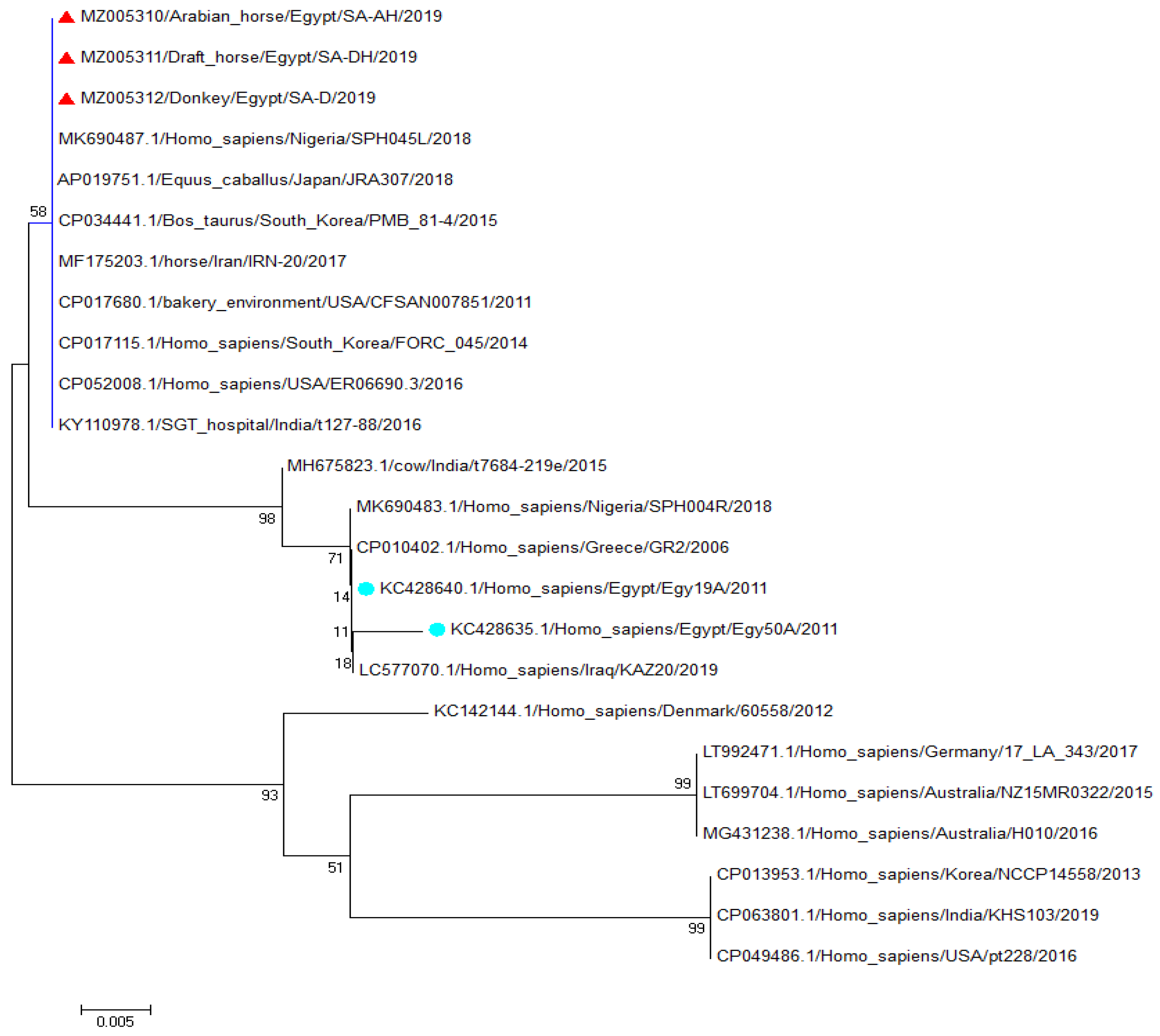
3.2. Sequencing and Phylogenetic Analysis of the Spa (x Region) Gene Fragment

Three samples were randomly selected from S. aureus Spa (x region)-gene-positive samples (one from each equine species). Sequences were analyzed against reference S. aureus spa (x region)-gene sequences in GenBank (Figure 1). The three identified strains of S. aureus strains (MZ005310/Arab horse/Egypt/SA-AH/2019, MZ005311/draft horse /Egypt/SA-DH/2019, and Z005312/Donkey/Egypt/SA-D/2019) identified in the present study were aligned with the Egyptian strain KC428640.1/Homo_sapiens/Egypt/Egy19A/2011 and KC428635.1/Homo_ sapiens/Egypt/Egy50A/2011, which were isolated from humans in Egypt (Figure 1). The S. aureus Spa (x region) from the Arabian horse, draft horse, and donkey were related to each other with 100% identity and shared a 97% identity with the KC428640.1/Homo_sapiens/Egypt/Egy19A/2011 S. aureus strain isolated from humans in Egypt. The identified S. aureus Arabian horse, draft horse, and donkey isolate Spa (x-region) genes had a 97.5% identity with KC428635.1/Homo_sapiens/Egypt/Egy50A/2011 (Supplemental Figure S1). These S. aureus isolates also had 100% identity with AP019751.1 /Equus_caballus /Japan/JRA307/2018, which was isolated from an equine in Japan, and MF175203.1 /horse/Iran/IRN-20/2017, which was isolated from a horse in Iran.

4. Discussion

Among the total number (110) of equine samples, the prevalence of ocular disease was 30%, which is consistent with a previous study in India [34] but higher than previously reported in Ethiopia (23.5%) [35]. The close rates in these countries could be due to similar windy and dusty environmental conditions, especially considering that the majority of ocular infections occurred in the winter in the current study. The rate of S. aureus isolation in the current study was much higher than a previous study in Poland with a rate of 9.8% [6]; this could be explained again by environmental differences and the increasing prevalence of S. aureus [36]. Differences in weather, environment, management, the presence of other infectious diseases, insect populations, and horse breeds are additional factors that may alter S. aureus prevalence. The higher percent (73.8%) of positive S. aureus isolates from horses aged 5–20 years is consistent with a previous study that demonstrated a high rate of S. aureus infection (87.8%) in horses older than 15 years [37].
The isolation of S. aureus from 19 out of 33 (57.6%) equines suffering ocular infection indicates that S. aureus is a major cause of ocular infection in equines. Similarly, S. aureus is reported to be a major cause of human eye infections [38], albeit at a lower rate (25%). This may be related to the unique structure of the equine eye structure, which is larger, more ovoid, and prominent from the sides, all of which make it more prone to damage by grass or dust that results in more ocular infection [39]. The S. aureus isolation rate of 29.9% from apparently healthy equine eyes might be the result of subclinical infections [37] or the presence of S. aureus as a member of the eye microbiota [4].
S. aureus has gained structural changes to the β-lactam target site through acquisition of the mecA gene, which enables resistance to oxacillin and methicillin. The expression of mecA in S. aureus enables it to synthesize a protein called the penicillin-binding protein, PBP2a, which decreases the binding affinity of β-lactams to MRSA strains [40]. The mecA gene is carried on the mec staphylococcal cassette chromosome (SCCmec), a mobile genetic element that is wide-spread in S. aureus due to extensive use of β-lactam antibiotics and related selective pressures. PBP2 is the main factor in the survival and success of S. aureus as a major pathogen in recent decades [41]. Oxacillin-resistant S. aureus is classified as MRSA according to the Institute of Clinical and Laboratory Standards (2005). In this study, the high resistance rate (91.67%) of isolated S. aureus to oxacillin indicates that most equine eye infections are probably MRSA. This assumption is boosted by the detection of the mecA gene in 100% of β-lactam-resistant S. aureus isolates [42]. Interestingly, the rate of MRSA among equine ocular infection in this study is higher than a previous study in India that reported an MRSA prevalence of 49% [43] and another study undertaken in China with a 52.8% MRSA prevalence [1]. In the region in which this study was conducted, the high rate of MRSA strains among ocular infections is serious and alarming and may reflect antibiotic overuse.
Of note, this study found a low resistance of S. aureus to tetracycline (25%), which contrasts with a previously reported resistance of 58% to tetracycline, especially among MRSA strains, and the suggested association of the tet gene with the SCCmec [44]. This difference can be attributed to in the geographical variation of MRSA in its prevalence and pattern of antibiotic sensitivity to antibiotics [45], and may also indicate the difference in the association between the mecA gene and the tet gene in the currently isolated S. aureus, in contrast to the previously assumed association between the oxacillin resistance genes and tetracycline resistance genes [44]. Importantly, the higher sensitivity of the MRSA strains isolated in this study to chloramphenicol (75%) and ampicillin (70.83%) as compared to those in other studies may reflect changes in S. aureus-antibiogram trends due to their lower rate of use in recent decades. Similarly, a recent report described changes in Salmonella antibiograms, including 60–80% sensitivity to chloramphenicol [46]. Additionally, another study in Egypt on Salmonella reported 92% sensitivity to chloramphenicol [47].
The detection of the Spa gene in 62.5% of S. aureus isolates indicates the highly pathogenic nature of these isolates, as the expression of this gene and its encoded protein A allow S. aureus to escape host immune system opsonization and phagocytosis [48] as well as inhibit host immune cell proliferation [49]. The S. aureus Spa (x region) is polymorphic and varies worldwide; its sequence is used as a typing method for clinical isolates of S. aureus [50]. This analysis revealed spa (x region) sequence conformity between S. aureus isolates from an Arabian horse, a draft horse, and a donkey, which indicates the local circulation of these isolates and its transmission among animals in the study region. This is consistent with the recently reported prevalence of Spa in some countries [51]. This may reflect the mandatory need for molecular typing of S. aureus, including Spa, across Egypt to better describe S. aureus type distribution. The variety of S. aureus clones in clinical settings of a cross-border region between the Netherlands and Germany was previously reported [52]. The current study also conducted a sequence analysis that revealed a close identity of the spa (x region) between S. aureus isolates from equine ocular infection and human isolates, which confirms its zoonotic nature. The phylogenic analysis also revealed that they were aligned together in the same clade (Figure 1) with at least 97% nucleotide similarity to the three equine samples (2.5–3% diversity, Supplemental Figure S1).
The presence of the nuc gene in 100% of the study isolates is also noteworthy as it encodes the thermostable nuclease enzyme that hydrolyzes host cell DNA and RNA, which devastates host tissue and facilitates pathogen dissemination, partially by avoiding neutrophil extracellular traps [53] and suppressing biofilm formation [54,55]. This gene confirmed the identification of our isolates as S. aureus since nuc is considered a useful marker for S. aureus detection [33]. The high rate of mecA gene expression and its associated high correlation with MRSA strains compared to the usual low rate of nuc detection in S. aureus isolates should be a reminder not to depend on one gene, especially nuc, to identify S. aureus. This observation agrees with the assumed probability of S. aureus misidentification if it only requires nuc gene detection, due to its variation [56]. The high expression rate (56%) of gamma-hemolysin (hlg) genes in S. aureus reflect their high virulence in equine ocular infections in Egypt. This rate is lower than the detection rate of hlg (87.5%) in community-associated MRSA encountered in children [57]. In particular, this study detected the hlg gene in most MR and four methicillin-sensitive S. aureus isolates, which supports previously reported data [58].

5. Conclusions

The results of this study demonstrated that most S. aureus isolates from clinically affected equine eyes are MRSA, as indicated by antibiograms and the detection of relevant genes. This research highlighted the local distribution and circulation of MRSA among equine ocular lesions in the studied area. This study also identified risk factors associated with the MRSA clinically affected equine eyes. Of these, the age and season have a significant effect while the breed and sex have non-significant effects. The phylogenetic analysis of S. aureus causing eye lesions in equines is of crucial importance as it indicates its zoonotic potential. In light of the current study, bacterial culture and antibiotic profiles and the molecular detection of MRSA isolates should be conducted in all locations within each country to generate procedures to limit the spread of multidrug-resistant genes. Future work on the zoonotic potential of equine ocular S. aureus should address whether the distribution of methicillin-resistant isolates from the same geographic region share the same sequences between equines and humans to confirm the zoonotic potential of this organism.

Supplementary Materials

The following supporting information can be downloaded at: https://www.mdpi.com/article/10.3390/antibiotics11020221/s1, Figure S1: Diversity percent between S. aureus strains identified in this study and other strains from GenBank. Diversity was calculated by MEGA X software.

Author Contributions

A.T. and S.E.-K. designed the study protocol and conception of the research idea. A.T., H.K.E., H.E.-S., A.M.R. and H.M.M.I. participated in the design of the methodology and sampling, and performed the laboratory work. A.T., H.M.M.I., A.M.R., H.E.-S., M.A. and A.M.E.-S. performed data analysis and interpretation. A.T., H.M.M.I., M.A.Y. and S.E.-K. participated in the data analysis. A.T., H.K.E. and H.E.-S. wrote and prepared the manuscript for publication and revision. H.K.E., H.M.M.I., M.A.Y. and A.T. contributed their scientific advice. All authors have read and agreed to the published version of the manuscript.

Funding

The current work was funded by the Taif University Researchers Supporting Project number (T URSP 2020/310), Taif University, Taif, Saudi Arabia.

Institutional Review Board Statement

Not applicable.

Informed Consent Statement

Not applicable.

Data Availability Statement

The data presented in this study are openly available on the PubMed database.

Acknowledgments

The authors also thank the veterinarians and medical specialists for their support and help in providing data and collecting samples throughout the study.

Conflicts of Interest

The authors declare that they have no conflict of interest.

References

  1. Hsiao, C.-H.; Chuang, C.-C.; Tan, H.Y.; Ma, D.H.K.; Lin, K.-K.; Chang, C.-J.; Huang, Y.C. Methicillin-resistant Staphylococcus aureus ocular infection: A 10-year hospital-based study. Ophthalmology 2012, 119, 522–527. [Google Scholar] [CrossRef]
  2. Parhizgari, N.; Khoramrooz, S.S.; Hosseini, S.A.A.M.; Marashifard, M.; Yazdanpanah, M.; Emaneini, M.; Gharibpour, F.; Mirzaii, M.; Darban-Sarokhalil, D.; Moein, M. High frequency of multidrug-resistant Staphylococcus aureus with Scc, Mec, Type III, and Spa types T037 and T631 isolated from burn patients in southwest Iran. Apmis 2016, 124, 221–228. [Google Scholar] [CrossRef] [PubMed]
  3. Paschalis-Trela, K.; Cywinska, A. The prevalence of ocular diseases in Polish Arabian horses. BMC Vet. Res. 2017, 13, 319. [Google Scholar] [CrossRef]
  4. Gilger, B.C.; Deeg, C. Equine recurrent uveitis. In Equine Ophthalmology; Elsevier: Amsterdam, The Netherlands, 2011; pp. 317–349. [Google Scholar]
  5. Johns, I.C.; Baxter, K.; Booler, H.; Hicks, C.; Menzies-Gow, N. Conjunctival bacterial and fungal flora in healthy horses in the UK. Vet. Ophthalmol. 2011, 14, 195–199. [Google Scholar] [CrossRef]
  6. Foti, M.; Fisichella, V.; Giacopello, C. Detection of methicillin-resistant Staphylococcus aureus (MRSA) in the microbial flora from the conjunctiva of healthy donkeys from Sicily (Italy). Vet. Ophthalmol. 2013, 16, 89–92. [Google Scholar] [CrossRef]
  7. Camargo, I.L.B.C.; Gilmore, M.S. Staphylococcus aureus—Probing for host weakness? J. Bacteriol. 2008, 190, 2253–2256. [Google Scholar] [CrossRef] [Green Version]
  8. Wieler, L.H.; Ewers, C.; Guenther, S.; Walther, B.; Lübke-Becker, A. Methicillin-resistant staphylococci (MRS) and extended-spectrum beta-lactamases (ESBL)-producing enterobacteriaceae in companion animals: Nosocomial infections as one reason for the rising prevalence of these potential zoonotic pathogens in clinical samples. Int. J. Med. Microbiol. 2011, 301, 635–641. [Google Scholar] [PubMed]
  9. Walther, B.; Tedin, K.; Lübke-Becker, A. Multidrug-resistant opportunistic pathogens challenging veterinary infection control. Vet. Microbiol. 2017, 200, 71–78. [Google Scholar] [CrossRef]
  10. Baptiste, K.E.; Williams, K.; Willams, N.J.; Wattret, A.; Clegg, P.D.; Dawson, S.; Corkill, J.E.; O’Neill, T.; Hart, C.A. Methicillin-resistant staphylococci in companion animals. Emerg. Infect. Dis. 2005, 11, 1942. [Google Scholar] [CrossRef]
  11. Vengust, M.; Anderson, M.E.C.; Rousseau, J.; Weese, J.S. Methicillin-resistant staphylococcal colonization in clinically normal dogs and horses in the community. Lett. Appl. Microbiol. 2006, 43, 602–606. [Google Scholar] [CrossRef]
  12. Weese, J.S.; Rousseau, J.; Traub-Dargatz, J.L.; Willey, B.M.; McGeer, A.J.; Low, D.E. Community-associated methicillin-resistant Staphylococcus aureus in horses and humans who work with horses. J. Am. Vet. Med. Assoc. 2005, 226, 580–583. [Google Scholar] [CrossRef]
  13. Leonard, F.C.; Markey, B.K. Methicillin-resistant Staphylococcus aureus in animals: A review. Vet. J. 2008, 175, 27–36. [Google Scholar] [CrossRef] [PubMed]
  14. Strommenger, B.; Kehrenberg, C.; Kettlitz, C.; Cuny, C.; Verspohl, J.; Witte, W.; Schwarz, S. Molecular characterization of methicillin-resistant Staphylococcus aureus strains from pet animals and their relationship to human isolates. J. Antimicrob. Chemother. 2006, 57, 461–465. [Google Scholar] [CrossRef] [PubMed] [Green Version]
  15. van Duijkeren, E.; Moleman, M.; van Oldruitenborgh-Oosterbaan, M.M.S.; Multem, J.; Troelstra, A.; Fluit, A.C.; Van Wamel, W.B.J.; Houwers, D.J.; De Neeling, A.J.; Wagenaar, J.A. Methicillin-resistant Staphylococcus aureus in horses and horse personnel: An investigation of several outbreaks. Vet. Microbiol. 2010, 141, 96–102. [Google Scholar] [CrossRef]
  16. Waqar, N.; Amin, Q.; Munir, T.; Ikram, M.S.; Shahzad, N.; Mirza, A.; Ali, A.; Arshad, M.I. A cross-sectional study of methicillin-resistant Staphylococcus aureus at the equine-human interface. Trop. Anim. Health Prod. 2019, 51, 1927–1933. [Google Scholar] [CrossRef]
  17. Plummer, C.E. Corneal response to injury and infection in the horse. Vet. Clin. Equine Pract. 2017, 33, 439–463. [Google Scholar] [CrossRef] [PubMed]
  18. Walther, B.; Wieler, L.H.; Friedrich, A.W.; Hanssen, A.M.; Kohn, B.; Brunnberg, L.; Lübke-Becker, L. Methicillin-resistant Staphylococcus aureus (MRSA) isolated from small and exotic animals at a university hospital during routine microbiological examinations. Vet. Microbiol. 2008, 127, 171–178. [Google Scholar] [CrossRef]
  19. Vincze, S.; Stamm, I.; Kopp, P.A.; Hermes, J.; Adlhoch, C.; Semmler, T.; Wieler, L.H.; Lübke-Becker, A.; Walther, B. Alarming proportions of methicillin-resistant Staphylococcus aureus (MRSA) in wound samples from companion animals, Germany 2010–2012. PLoS ONE 2014, 9, e85656. [Google Scholar] [CrossRef] [Green Version]
  20. Diene, S.M.; Corvaglia, A.R.; François, F.; van der Mee-Marquet, N. Prophages and adaptation of Staphylococcus aureus St398 to the human clinic. BMC Genom. 2017, 18, 133. [Google Scholar] [CrossRef] [Green Version]
  21. Ferreira, A.R.d.A.; Santana, A.F.; Almeida, A.C.d.V.R.; Sousa, R.F.; Perecmanis, S.; Galera, P.D. Bacterial culture and antibiotic sensitivity from the ocular conjunctiva of horses. Ciência Rural 2017, 47, 19. [Google Scholar] [CrossRef]
  22. Kahlmeter, G.; Brown, D.F.; Goldstein, F.W.; MacGowan, A.P.; Mouton, J.W.; Odenholt, I.; Rodloff, A.; Soussy, C.J.; Steinbakk, M.; Soriano, F.; et al. European Committee on Antimicrobial Susceptibility Testing (EUCAST) technical notes on antimicrobial susceptibility testing. Clin. Microbiol. Infect. 2006, 12, 501–503. [Google Scholar] [CrossRef] [PubMed] [Green Version]
  23. Sauer, P.; Andrew, S.E.; Lassaline, M.; Gelatt, K.N.; Denis, H.M. Changes in antibiotic resistance in equine bacterial ulcerative keratitis (1991–2000): 65 horses. Vet. Ophthalmol. 2003, 6, 309–313. [Google Scholar] [CrossRef]
  24. Wada, S.; Hobo, S.; Niwa, H. Ulcerative keratitis in thoroughbred racehorses in Japan from 1997 to 2008. Vet. Ophthalmol. 2010, 13, 99–105. [Google Scholar] [CrossRef]
  25. Kuroda, T.; Kinoshita, Y.; Niwa, H.; Mizobe, F.; Ueno, T.; Kuwano, A.; Hatazoe, T.; Hobo, S. Methicillin-resistant Staphylococcus aureus ulcerative keratitis in a thoroughbred racehorse. J. Equine Sci. 2015, 26, 95–98. [Google Scholar] [CrossRef] [PubMed] [Green Version]
  26. Altschul, S.F.; Gish, W.; Miller, W.; Myers, E.W.; Lipman, D.J. Basic local alignment search tool. J. Mol. Biol. 1990, 215, 403–410. [Google Scholar] [CrossRef]
  27. Thompson, J.D.; Higgins, D.G.; Gibson, T.J. Clustal W: Improving the sensitivity of progressive multiple sequence alignment through sequence weighting, position-specific gap penalties and weight matrix choice. Nucleic Acids Res. 1994, 22, 4673–4680. [Google Scholar] [CrossRef] [PubMed] [Green Version]
  28. Tamura, K.; Stecher, G.; Peterson, D.; Filipski, A.; Kumar, S. Mega6: Molecular evolutionary genetics analysis version 6.0. Mol. Biol. Evol. 2013, 30, 2725–2729. [Google Scholar] [CrossRef] [Green Version]
  29. Momtaz, H.; Dehkordi, F.S.; Rahimi, E.; Asgarifar, A.; Momeni, M. Virulence genes and antimicrobial resistance profiles of Staphylococcus aureus isolated from chicken meat in Isfahan Province, Iran. J. Appl. Poult. Res. 2013, 22, 913–921. [Google Scholar] [CrossRef]
  30. Gao, J.; Ferreri, M.; Liu, X.Q.; Chen, L.B.; Su, J.L.; Han, B. Development of multiplex polymerase chain reaction assay for rapid detection of Staphylococcus aureus and selected antibiotic resistance genes in bovine mastitic milk samples. J. Vet. Diagn. Investig. 2011, 23, 894–901. [Google Scholar] [CrossRef] [Green Version]
  31. McClure, J.A.; Conly, J.M.; Lau, V.; Elsayed, S.; Louie, T.; Hutchins, W.; Zhang, K. Novel multiplex PCR assay for detection of the staphylococcal virulence marker Panton-Valentine leukocidin genes and simultaneous discrimination of methicillin-susceptible from resistant staphylococci. J. Clin. Microbiol. 2006, 44, 1141–1144. [Google Scholar] [CrossRef] [Green Version]
  32. Kumar, J.; Dinesh, Y.K.N.; Gaur, A.; Khanna, D. Detection of virulence genes in Staphylococcus aureus isolated from paper currency. Int. J. Infect. Dis. 2009, 13, e450–e455. [Google Scholar] [CrossRef] [Green Version]
  33. Zhang, K.; Sparling, J.; Chow, B.L.; Elsayed, S.; Hussain, Z.; Church, D.L.; Gregson, D.B.; Louie, T.; Conly, J.M. New quadriplex PCR assay for detection of methicillin and mupirocin resistance and simultaneous discrimination of Staphylococcus aureus from coagulase-negative staphylococci. J. Clin. Microbiol. 2004, 42, 4947–4955. [Google Scholar] [CrossRef] [PubMed] [Green Version]
  34. Tamilmahan, P.; Zama, M.M.S.; Pathak, R.; Muneeswaran, N.S.; Karthik, K. A retrospective study of ocular occurrence in domestic animals: 799 cases. Vet. World 2013, 6, 274–276. [Google Scholar] [CrossRef]
  35. Scantlebury, C.E.; Aklilu, N.; Reed, K.; Knottenbelt, D.C.; Gebreab, F.; Pinchbeck, G.L. Ocular disease in working horses in Ethiopia: A cross-sectional study. Vet. Rec. 2013, 172, 99. [Google Scholar] [CrossRef] [PubMed] [Green Version]
  36. Vola, M.E.; Moriyama, A.S.; Lisboa, R.; Vola, M.M.; Hirai, F.E.; Bispo, P.J.M.; Höfling-Lima, A.L. Prevalence and antibiotic susceptibility of methicillin-resistant Staphylococcus aureus in ocular infections. Arq. Bras. Oftalmol. 2013, 76, 350–353. [Google Scholar] [CrossRef] [Green Version]
  37. Malalana, F.; McGowan, T.W.; Ireland, J.L.; Pinchbeck, G.L.; McGowan, C.M. Prevalence of owner-reported ocular problems and veterinary ocular findings in a population of horses aged ≥15 years. Equine Vet. J. 2019, 51, 212–217. [Google Scholar] [CrossRef] [PubMed] [Green Version]
  38. Ramesh, S.; Ramakrishnan, R.; Bharathi, M.J.; Amuthan, M.; Viswanathan, S. Prevalence of bacterial pathogens causing ocular infections in South India. Indian J. Pathol. Microbiol. 2010, 53, 281. [Google Scholar] [PubMed]
  39. Timney, B.; Macuda, T. Vision and hearing in horses. J. Am. Vet. Med. Assoc. 2001, 218, 1567–1574. [Google Scholar] [CrossRef]
  40. Plata, K.; Rosato, A.E.; Wegrzyn, G. Staphylococcus aureus as an infectious agent: Overview of biochemistry and molecular genetics of its pathogenicity. Acta Biochim. Pol. 2009, 56, 597–612. [Google Scholar] [CrossRef]
  41. Chambers, H.F.; DeLeo, F.R. Waves of resistance: Staphylococcus aureus in the antibiotic era. Nat. Rev. Microbiol. 2009, 7, 629–641. [Google Scholar] [CrossRef]
  42. Soimala, T.; Lübke-Becker, A.; Schwarz, S.; Feßler, A.T.; Huber, C.; Semmler, T.; Merle, R.; Gehlen, H.; Eule, J.C.; Walther, B. Occurrence and molecular composition of methicillin-resistant Staphylococcus aureus isolated from ocular surfaces of horses presented with ophthalmologic disease. Vet. Microbiol. 2018, 222, 1–6. [Google Scholar] [CrossRef]
  43. Bagga, B.; Reddy, A.K.; Garg, P. Decreased susceptibility to quinolones in methicillin-resistant Staphylococcus aureus isolated from ocular infections at a tertiary eye care centre. Br. J. Ophthalmol. 2010, 94, 1407–1408. [Google Scholar] [CrossRef]
  44. Schmitz, F.J.; Krey, A.; Sadurski, R.; Verhoef, J.; Milatovic, D.; Fluit, A.C. European Sentry Participants. Resistance to tetracycline and distribution of tetracycline resistance genes in European Staphylococcus aureus isolates. J. Antimicrob. Chemother 2001, 47, 239–240. [Google Scholar] [CrossRef] [Green Version]
  45. Zilberberg, M.D.; Shorr, A.F.; Kollef, M.F. Growth and geographic variation in hospitalizations with resistant infections, United States, 2000–2005. Emerg. Infect. Dis. 2008, 14, 1756. [Google Scholar] [CrossRef]
  46. Lakshmi, V.; Ashok, R.; Susmita, J.; Shailaja, V.V. Changing trends in the antibiograms of Salmonella isolates at a tertiary care hospital in Hyderabad. Indian J. Med. Microbiol. 2006, 24, 45–48. [Google Scholar] [CrossRef]
  47. Hammad, O.M.; Hifnawy, T.; Omran, D.; Tantawi, M.A.E.; Girgis, N.I. Ceftriaxone versus chloramphenicol for treatment of acute typhoid fever. Life Sci. J. 2011, 8, 100–105. [Google Scholar]
  48. Graille, M.; Stura, E.A.; Corper, A.L.; Sutton, B.J.; Taussig, M.J.; Charbonnier, J.-B.; Silverman, G.J. Crystal structure of a Staphylococcus aureus protein A domain complexed with the Fab fragment of a human IgM antibody: Structural basis for recognition of B-cell receptors and superantigen activity. Proc. Natl. Acad. Sci. USA 2000, 97, 5399–5404. [Google Scholar] [CrossRef] [Green Version]
  49. Keener, A.B.; Thurlow, L.T.; Kang, S.A.; Spidale, N.A.; Clarke, S.H.; Cunnion, K.M.; Tisch, R.; Richardson, A.R.; Vilen, B.J. Staphylococcus aureus protein A disrupts immunity mediated by long-lived plasma cells. J. Immunol. 2017, 198, 1263–1273. [Google Scholar] [CrossRef] [PubMed] [Green Version]
  50. Shopsin, B.; Gomez, M.; Montgomery, S.O.; Smith, D.H.; Waddington, M.; Dodge, D.E.; Bost, D.A.; Riehman, M.; Naidich, S.; Kreiswirth, B.N. Evaluation of protein A gene polymorphic region DNA sequencing for typing of Staphylococcus aureus strains. J. Clin. Microbiol. 1999, 37, 3556–3563. [Google Scholar] [CrossRef] [PubMed] [Green Version]
  51. Asadollahi, P.; Farahani, N.N.; Mirzaii, M.; Khoramrooz, S.S.; Van Belkum, A.; Asadollahi, K.; Dadashi, M.; Darban-Sarokhalil, D. Distribution of the most prevalent Spa types among clinical isolates of methicillin-resistant and-susceptible Staphylococcus aureus around the world: A review. Front. Microbiol. 2018, 9, 163. [Google Scholar] [CrossRef] [PubMed] [Green Version]
  52. Van der Donk, C.F.M.; Schols, J.M.G.A.; Schneiders, V.; Grimm, K.-H.; Stobberingh, E.E. Antibiotic resistance, population structure and spread of Staphylococcus aureus in nursing homes in the Euregion Meuse-Rhine. Eur. J. Clin. Microbiol. Infect. Dis. 2013, 32, 1483–1489. [Google Scholar] [CrossRef] [PubMed]
  53. Berends, E.T.M.; Horswill, A.R.; Haste, N.M.; Monestier, M.; Nizet, V.; von Köckritz-Blickwede, M. Nuclease expression by Staphylococcus aureus facilitates escape from neutrophil extracellular traps. J. Innate Immun. 2010, 2, 576–586. [Google Scholar] [CrossRef] [PubMed] [Green Version]
  54. Mann, E.E.; Rice, K.C.; Boles, B.R.; Endres, J.L.; Ranjit, D.; Chandramohan, L.; Tsang, L.H.; Smeltzer, M.S.; Horswill, A.R.; Bayles, K.W. Modulation of eDNA release and degradation affects Staphylococcus aureus biofilm maturation. PLoS ONE 2009, 4, e5822. [Google Scholar] [CrossRef] [PubMed] [Green Version]
  55. Kiedrowski, M.R.; Kavanaugh, J.S.; Malone, C.L.; Mootz, J.M.; Voyich, J.M.; Smeltzer, M.S.; Bayles, K.W.; Horswill, A.R. Nuclease modulates biofilm formation in community-associated methicillin-resistant Staphylococcus aureus. PLoS ONE 2011, 6, e26714. [Google Scholar] [CrossRef] [PubMed] [Green Version]
  56. Hoegh, S.V.; Skov, M.N.; Boye, K.; Worning, P.; Jensen, T.G.; Kemp, M. Variations in the Staphylococcus aureus-specific nuc gene can potentially lead to misidentification of methicillin-susceptible and-resistant S. aureus. J. Med. Microbiol. 2014, 63, 1020–1022. [Google Scholar] [CrossRef] [PubMed] [Green Version]
  57. Wang, X.; Li, X.; Liu, W.; Huang, W.; Fu, Q.; Li, M. Molecular characteristic and virulence gene profiles of community-associated methicillin-resistant Staphylococcus aureus isolates from pediatric patients in Shanghai, China. Front. Microbiol. 2016, 7, 1818. [Google Scholar] [CrossRef] [Green Version]
  58. Zarizal, S.; Yeo, C.C.; Faizal, G.M.; Hoong, C.; Chew, Z.A.Z.; Al-Obaidi, M.M.J.; Amin, N.S.; Nasir, M.D.M. Nasal colonisation, antimicrobial susceptibility and genotypic pattern of Staphylococcus aureus among agricultural biotechnology students in Besut, Terengganu, east coast of Malaysia. Trop. Med. Int. Health 2018, 23, 905–913. [Google Scholar] [CrossRef] [Green Version]
Figure 1. The phylogenetic neighbor-joining tree of three S. aureus strains. The alignment of nucleotides sequences for three selected samples shows that the strains identified in the present study (red triangles) are closely related to each other and to other S. aureus strains isolated from humans in Egypt (cyan circles).
Antibiotics 11 00221 g001
Table 2. Frequency of S. aureus isolates from healthy and diseased equine eyes.
S. aureusHealthy
(n = 77)
Diseased
(n = 33)
ORp-Value95% CI
0.3090.0090.125–0.763
Positive23 (29.9%)19 (57.6%)
Negative54 (70.1%)14 (42.4%)
Table 3. Risk factors of S. aureus isolation from healthy and diseased equine eyes.
Risk FactorS. aureusORp-Value95% CI
NegativePositive
Age 3.3680.0031.412–7.960
<537 (54.4%)11(26.2%)
5–1031 (45.6%)31 (73.8%)
Breed 7.0530.060.07–0.6
Arabian67 (98.5%)38 (90.5%)
Others1 (1.5%)4 (9.5%)
Sex 2.8690.051.0–1.798
Male19 (27.9%)5 (11.9%)
Female49 (72.1%)37 (88.1%)
Season 2.9830.0301.017–8.234
Winter61 (89.7%)32 (76.2%)
Spring7 (10.3%)10 (23.8%)
Table 4. Antimicrobial susceptibility for S. aureus isolates from diseased equine eyes.
Antimicrobial Sensitivity for Staphylococcus aureusAntimicrobial Agent
SensitiveIntermediateResistant
24 (100%)0 (0%)0 (0%)Sulfamethoxazole/trimethoprim (25 μg)
2 (8.33%)0 (0%)22 (91. 67%)Oxacillin (1 μg)
18 (75%)0 (0%)6 (25%)Tetracycline (30 μg)
6 (25%)1 (4.16%)17 (70.83%)Rifamycin (5 μg)
17 (70.83%)2 (8.33%)5 (20.83%)Ampicillin (10 μg)
8 (33.33%)5 (20.83%)11 (45.83%)Gentamycin (10 μg)
1 (4.16%)1 (4.16%)22 (91.67%)Cephalosporin (10 μg)
18 (75%)1 (4.16%)5 (20.83%)Chloramphenicol (30 μg)
Table 5. Clinical signs, antibiotic resistance markers and virulence genes from S. aureus isolates of diseased equine eyes.
Case No.Sample
(Eye)
Equine SourceAge
(in Years)
Clinical Signsspa (x Region)nucmecAhlg
11(Right)Arabian 0.5corneal edema, eyelid edema, keratitis+++
22 (Left)Arabian 6mucopurulent discharge, corneal edema, eyelid edema, keratitis+++
33 (Right)Arabian 8conjunctivitis, watery lacrimation++
44 Right)Arabian 1.5conjunctivitis+
55 (Left)Draft 1mucopurulent discharge, corneal edema, eyelid edema, keratitis e++++
56 (Right)Draft 1mucopurulent discharge+++
67 (Left)Arabian 2.5 corneal opacity+++
78 (Left)Draft 4conjunctivitis, watery lacrimation+++
79 (Right)Draft 4conjunctivitis, watery lacrimation+++
810 (Left)Donkey5eyelid edema, mucopurulent discharge+++
911 (Right)Arabian 9conjunctivitis, watery lacrimation+++
912 (Left)Arabian 9conjunctivitis, watery lacrimation+++
1013 (Left)Arabian 5conjunctivitis, watery lacrimation+++
1014 (Right)Arabian 5conjunctivitis, watery lacrimation+++
1115 (Left)Arabian 11corneal opacity++++
1216 (Right)Arabian 14conjunctivitis, watery lacrimation+++
1317 (Right)Donkey13eyelid edema, keratitis, mucopurulent discharge++++
1418 (Left)Arabian 6corneal edema, eyelid edema, keratitis++++
1519 (Right)Donkey9eyelid edema, keratitis, mucopurulent discharge++++
1620 (Left)Arabian 12lacrimation, conjunctivitis++
1721 (Left)Arabian 7corneal edema, eyelid edema, keratitis, mucopurulent discharge+++
1822 (Left)Donkey7eyelid edema, keratitis, mucopurulent discharge++++
1923 (Left)Arabian 9eyelid edema, keratitis, mucopurulent discharge+++
1924 (Right)Arabian 9eyelid edema, keratitis, mucopurulent discharge+++
Publisher’s Note: MDPI stays neutral with regard to jurisdictional claims in published maps and institutional affiliations.

