Article

Protective Effects of Glycyrrhiza Total Flavones on Liver Injury Induced by Streptococcus agalactiae in Tilapia (Oreochromis niloticus)

1
Key Laboratory of Integrated Rice-Fish Farming Ecology, Ministry of Agriculture and Rural Affairs, Freshwater Fisheries Research Center, Chinese Academy of Fishery Sciences, Wuxi 214081, China
2
College of Veterinary Medicine, Agricultural University of Hebei, Baoding 071001, China
3
Wuxi Fisheries College, Nanjing Agricultural University, Wuxi 214081, China
*
Authors to whom correspondence should be addressed.
Antibiotics 2022, 11(11), 1648; https://doi.org/10.3390/antibiotics11111648
Submission received: 21 October 2022 / Revised: 15 November 2022 / Accepted: 15 November 2022 / Published: 18 November 2022
(This article belongs to the Special Issue Antioxidant and Antibacterial Activity of Plant Extracts)

Abstract

:
Clinical studies have confirmed that Glycyrrhiza total flavones (GTFs) have good anti-hepatic injury, but whether they have a good protective effect on anti-hepatic injury activity induced by Streptococcus agalactiae in tilapia (Oreochromis niloticus) is unknown. The aims of this study were to investigate the protective effects of Glycyrrhiza total flavones on liver injury induced by S. agalactiae (SA) and its underlying mechanism in fish. A total of 150 tilapia were randomly divided into five groups, each with three replicates containing 10 fish: normal control group, S. agalactiae infection group, and three Glycyrrhiza total flavone treatment groups (addition of 0.1, 0.5, or 1.0 g of GTF to 1 kg of feed). The normal control group was only fed with basic diet, after 60 d of feeding, and intraperitoneal injection of the same volume of normal saline (0.05 mL/10 g body weight); the S. agalactiae infection group was fed with basic diet, and the S. agalactiae solution was intraperitoneally injected after 60 d of feeding (0.05 mL/10 g body weight); the three GTF treatment groups were fed with a diet containing 0.1, 0.5, or 1.0 g/kg of GTF, and the S. agalactiae solution was intraperitoneally injected after 60 d of feeding (0.05 mL/10 g body weight). After 48 h injection, blood and liver tissues were collected to measure biochemical parameters and mRNA levels to evaluate the liver protection of GTFs. Compared with the control group, the serum levels of glutamic oxaloacetic transaminase (GOT), glutamic pyruvic transaminase (GPT), alkaline phosphatase (AKP) and glucose (GLU) in the streptococcal infection group increased significantly, while the levels of total antioxidant capacity (T-AOC), superoxide dismutase (SOD), catalase (CAT) and reduced glutathione (GSH) decreased significantly; observations of pathological sections showed obvious damage to the liver tissue structure in response to streptococcal infection. S. agalactiae can also cause fatty liver injury, affecting the function of fatty acid β-oxidation and biosynthesis in the liver of tilapia, and also causing damage to function of the immune system. The addition of GTFs to the diet could improve oxidative stress injury caused by S. agalactiae in tilapia liver tissue to different degrees, promote the β-oxidation of fatty acids in the liver, accelerate the lipid metabolism in the liver, and repair the damaged liver tissue. GTFs have a good protective effect on liver injury caused by streptococcus.

1. Introduction

Streptococcal disease is a common bacterial disease in fish farming, which mainly occurs in the seasons with a temperature above 20 degrees, and summer is the peak season for this disease. The incidence of the disease can reach 20–50%, even higher in some areas, and the mortality rate can reach 90% in some regions. The disease can affect many kinds of fish, especially tilapia. Researchers have found that a variety of bacteria can cause liver injury diseases, such as Escherichia coli, Streptococcus, Brucella, Staphylococcus aureus and so on [1,2,3,4]. The most harmful bacterial disease in tilapia is streptococcal disease. Fish bacteria are mainly from Streptococcus or Lactococcus genera and include several pathogenic species such as Streptococcus iniae, Streptococcus agalactiae, Streptococcus paramammary, and Lactococcus grigneri according to the original disease [5]. S. agalactiae was the main pathogenic bacteria affecting tilapia. Zhu et al. found that S. agalactiae caused serious liver damage to tilapia liver tissue, with disordered arrangement of hepatocytes and unclear cord-like structure [6]. Luo et al. studied the pathological study of S. agalactiae on tilapia niloticus, and the results showed that the hepatocytes of tilapia infected with S. agalactiae were severely denaturated and necrotic, the nuclei disappeared, and a large area of necrosis appeared [7]. Ao et al. studied the effects of S. agalactiae infection on the blood and hepatopancreas biochemical parameters of tilapia; the results showed that biochemical indexes of AKP, SOD and T-AOC in liver tissues were significantly changed, and the anatomy of tilapia hepatopanthus showed symptoms of swelling and necrosis [8].
At present, the infection route and pathogenesis of Streptococcus infection in fish are still unclear. Chen et al. believe that the pathogenesis process of Streptococcus infection in fish is mainly divided into four steps: colonization of host cells, evasion of host cell defense, proliferation in vivo and diffusion in vivo, which causes damage to fish tissues and organs. Capsular polysaccharide, β-hemolysin, Cyclic adenosine 3, 5′-monophosphate (cAMP) factor and C5α peptidase are important virulence factors in the process of infection [9]. It has been reported that a series of neurological symptoms, such as circuses in tilapia after infection, are mainly regulated by serotonin [10]. Another study reported that S. agalactiae can escape the immune defense of phagocytic cells and migrate with macrophages in the body to infect other tissues [11]. Li et al. also pointed out that Streptococcus can colonize the gastrointestinal lumen of tilapia and invade the internal body to infect the host [12].
Regarding the treatment of tilapia streptococcosis, antibiotic therapy is a commonly used method in aquaculture. Although antibiotics have a good therapeutic effect on Streptococcus, the excessive use of antibiotics can easily cause bacterial resistance and drug residues in tilapia. With the proposal of the concept of green ecological breeding, Chinese herbal medicine has been more and more widely used in the breeding process, and there have been a large number of reports on the antibacterial effect of Chinese herbal medicine. Licorice (Glycyrrhiza uralensis Fisch.) is a plant in the leguminous, licorice family. The root of licorice is usually the medicinal part, which has been used in many traditional Chinese prescriptions. Flavonoids are the main active components isolated from roots and rhizomes of licorice. It mainly contains six flavonoids, 5-(1,1-dimethylallyl)-3,4,4′-trihydroxy-2-methoxychalcone, licochalcone B, licochalcone A, echinatin, glycycoumarin and glyurallin B [13,14,15]. Wang et al. found that crude extract of licorice has a good antibacterial effect on Streptococcus suis [16]. Liu studied the antibacterial activity of licorice extract, and found that oleic acid, dehydrocyrrhizin D, glycyrrhetinic acid had a good inhibitory effect on Streptococcus mutans, Escherichia coli, Bacillus subtilis and other bacteria [17]. Recent studies have found that GTF have good anti-inflammatory, antibacterial and antiviral effects [18,19,20]. However, there has been no relevant report on whether GTFs have an antibacterial effect on tilapia Streptococcus.
In this study, Nile tilapia (Oreochromis niloticus) were used as the experimental object, and the tilapia were infected by intraperitoneal injection of S. agalactiae. The effective ingredient of Chinese herbal medicine, Glycyrrhiza total flavones, was added to the diet. The serum biochemical indicators, histopathological observation and expression changes of related genes were used to evaluate whether GTFs had an antibacterial effect on Streptococcus tilapia. In order to provide a theoretical basis for the development of anti-streptococcal drugs.

2. Materials and Methods

2.1. Test Materials

2.1.1. Ethics Statement

This experiment was approved by the Institutional Animal Care and Use Committee (IACUC) of the Chinese Academy of Fishery Science.

2.1.2. Fish

Tilapia (Oreochromis niloticus) were obtained from the Freshwater Fisheries Research Center of the Chinese Academy of Fishery Science, Wuxi, China. The fish were healthy with an intact body surface and normal swimming posture, and the average body weight was 20 ± 1 g. After being retrieved from the fishery, the fish were kept in a circulating water system for 1 week prior to the experiment [21,22,23], the water temperature was set at 28 ± 1 °C; pH 6.7–7.3; dissolved oxygen, >6 mg/L, and fish were fed on a commercial diet purchased from Tongwei Group Co., Ltd. (Chengdu, China) twice each day.

2.1.3. Reagents and Drugs

Glycyrrhiza total flavones (S25400, UV ≥ 98%) were purchased from Shanghai Yuanye Biotechnology Co., Ltd. (Shanghai, China). Glutamateruvate transaminase (GPT), glutamate oxalate transaminase (GOT), alkaline phosphatase (AKP), malondialdehyde (MDA), total antioxidant capacity (T-AOC), reduced glutathione (GSH) and superoxide dismutase (SOD) were purchased from Nanjing Jiancheng Haihao Bioengineering Co., Ltd. (Jiangsu, China). The reagents required for quantitative real-time PCR (qRT-PCR) were purchased from Takara (Dalian, China).

2.1.4. Resuscitation and Culture of S. agalactiae

S. agalactiae (No. 619) was used in this study, which was collected by Dr. Xi from Laboratory of Aquatic Diseases and Feed, Freshwater Fisheries Research Center, Chinese Academy of Fishery Sciences. The S. agalactiae was resuscitated in brain-heart infusion via incubating overnight at 28 °C under 150 rpm. A single colony was selected for Gram staining to detect the purity of the culture. After identification, a single colony was inoculated into brain-heart liquid media and incubated for 24 h, then the cultured bacterial liquid was centrifuged at 6000 r/min for 5 min, the supernatant was discarded and the bacterial precipitate was added to sterilized physiological saline, and the obtained bacteria solution could be used for subsequent tests.

2.2. Experimental Design and Parameter Measurement

2.2.1. Test Groups

A total of 150 healthy tilapia with body weight of about 20 g were randomly divided into 5 treatment groups: a normal control group, a streptococcal injection group and 3 GTF treatment groups (addition of 0.1, 0.5, or 1.0 g/kg of total flavones to the diet), with three replicates in each group and 10 fish in each replicate. The fish were fed twice a day at 4% of their body weight for 60 days. The injection concentration was based on our preliminary results (LD50 1.69 × 10⁸ CFU/mL). In the treatment group, the tilapia were injected with S. agalactiae at the concentration of 60% 96 h LD50. The normal control group and Streptococcal injection group were fed with the basal diet (31.6% crude protein, 4.6% crude lipid, 6.4% crude ash), and the three GTF-treated groups were fed with the basal diet supplemented with GTF at doses of 0.1, 0.5, and 1.0 g/kg diet, respectively. After 60 days feeding, the fish in the Streptococcal injection treatment group and GTF-treated groups were injected intraperitoneally (i.p.) with S. agalactiae (0.05 mL/10 g body weight), while the fish in the normal control group were administrated with the same volume saline (0.05 mL/10 g body weight).

2.2.2. Sample Collection and Processing

At 48 h after injection, the tilapia were anesthetized with 3-Aminobenzoic acid ethyl ester methanesulfonate (MS-222) [24,25,26]. The blood and liver tissues were collected from each treatment (10 fish/group) to investigate the protective effects of GTF against oxidative damage. The blood was collected by tail vein syringe, centrifuged at 5000 rpm for 10 min in a 4-degree centrifuge, and the separated serum was transferred to a 1.5 mL centrifuge tube. The serum and liver tissues were placed in a −80 degree refrigerator for storage.

2.2.3. Measurement of Biochemical Indexes

The levels of glutamic oxaloacetic transaminase (GOT), glutamic pyruvic transaminase (GPT), alkaline phosphatase (AKP) and glucose (GLU) in the streptococcal infection group increased significantly, while the levels of total antioxidant capacity (T-AOC), superoxide dismutase (SOD), catalase (CAT) and reduced glutathione (GSH) in the serum were determined by kits from the Nanjing Jiancheng Bioengineering Institute (Nanjing, China). GPT and GOT were expressed as IU/L, AKP as U/L, SOD as U/mL, T-AOC as units/mL, CAT as U/L, GLU as mmol/L, and GSH as μmol/gprot.

2.2.4. Growth Performance Indicators

Feeding was stopped the day before the end of the experiment, and each fish was weighed after anesthesia (MS-222). The calculation formulas are as follows:
Weight gain rate, WGR, % = (W1 − W0)/W1 × 100%;
Specific growth rate, SGR, % = (lnW1 − lnW0)/T × 100%.
where W0 is the initial average body weight (g), W1 is the final average body weight (g), T is the farming time (d) of the experiment.

2.2.5. Preparation and Observation of Liver Slices

The collected liver tissues of each group were fixed in Bouin’s fixative solution for 48 h, and paraffin tissue sections were prepared. The effect of GTF against S. agalactiae on liver tissue injury of tilapia was observed using HE staining.

2.2.6. Real-Time Quantitative RT-PCR

An appropriate amount of tilapia liver tissue was taken, and total RNA was extracted from liver tissue using RNAiso Plus reagent. The operation process was carried out according to the instructions. The purity of RNA was analyzed by absorbance analysis, and the quality of total RNA extraction was evaluated by the OD260/OD280 ratio between 1.7 and 2.1. At the end of the RNA concentration determination, cDNA was synthesized by reverse transcription reaction using a reverse transcription kit and stored in a −80 degree refrigerator for subsequent experiments.
Real-time PCR was performed using TB Green® Premix Ex Taq™ II kit (Takara Bio Inc., Dalian, China). The reaction system was 25 μL, TB Green Premix Ex Taq II (2X) 12.5 μL, 1 μL each of upstream and downstream primers, and 2 μL of cDNA template. Sterilized water 8.5 μL. A two-step PCR amplification procedure was used with specific reaction conditions as follows: one cycle at 95 °C for 30 s, followed by 40 cycles of 95 °C for 5 s and 60 °C for 30 s. The relative expression of target genes was determined by 2−ΔΔCt method [27]. The primer of the target gene is shown in the figure below (Table 1).

2.2.7. Statistical Processing

SPSS22.0 software was used to analyze all data, and the final results were expressed as mean ± standard error of the mean (SEM). One-Way ANOVA test was used to analyze the significance of differences between the data. * p < 0.05 was considered as significant difference, and ** p < 0.01 was considered as extremely significant difference.

3. Results

3.1. Effects of GTF on Growth Performance of Tilapia

As shown in Table 2, after 60 d of feeding, the relative weight gain rate and specific growth rate of tilapia were not significantly affected by the dietary GTF level compared with the control group (0 g/kg) (p > 0.05).

3.2. Effect of GTF on Liver Morphology of Tilapia

As shown in Figure 1, the nucleus of the liver tissue in the normal group was clearly visible, and the thickness of the cords was uniform; in the SA infection group, some nuclei dissolved and disappeared, and there were round vacuoles with different sizes and uneven distribution in hepatocyte plasma, which showed reticular or transparent shape, and the staining became light. In addition, 1.0 g/kg GTF had a good protective effect against liver tissue damage caused by S. agalactiae, and could repair damaged hepatocytes.

3.3. Effects of GTF on Serum Biochemical Indices of Tilapia

As shown in Figure 2, at 48 h after S. agalactiae infection, the activities of GOT, GPT, and AKP in the serum increased significantly (p < 0.01 or p < 0.05) compared to the normal control group. The addition of 1.0 g/kg of GTF significantly decreased the activities of GOT, GPT, and AKP (p < 0.01).

3.4. Effects of GTF on Antioxidant System of Tilapia

As shown in Figure 3, after 48 h of S. agalactiae infection, the level of T-AOC, SOD, CAT, and GSH in serum were significantly decreased compared to the normal control group (p < 0.05 or p < 0.01). The addition of 1.0 g/kg of GTF could significantly increase the levels of antioxidant enzymes in serum, and alleviated the damage of streptococcus to tilapia.

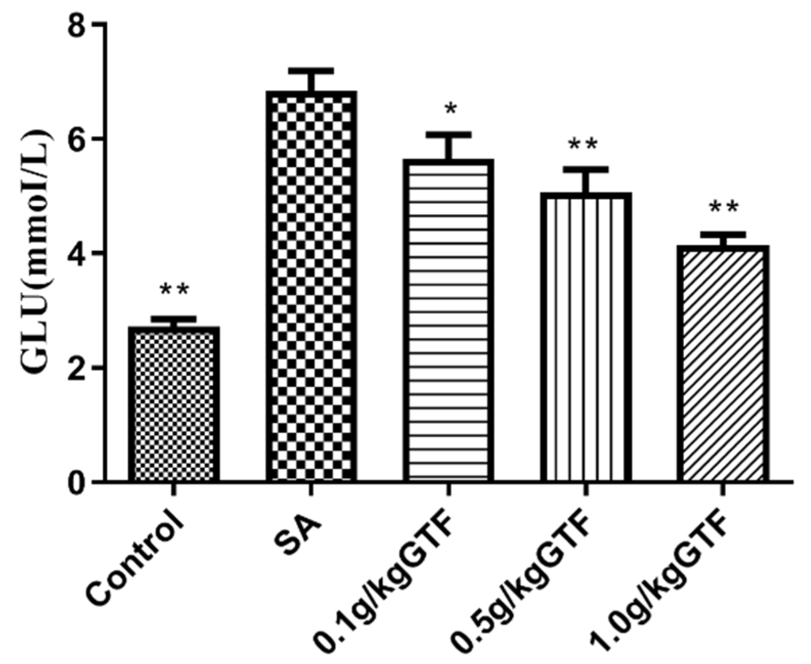
3.5. Effects of GTF on Glucose Metabolism Indexes of Tilapia

After 48 h, S. agalactiae infection significantly increased the content of GLU in serum (Figure 4; p < 0.01). However, 0.5 and 1.0 g/kg of GTF could significantly decrease the content of GLU in serum; the results showed that GTF could significantly improve the abnormal glucose metabolism caused by S. agalactiae.

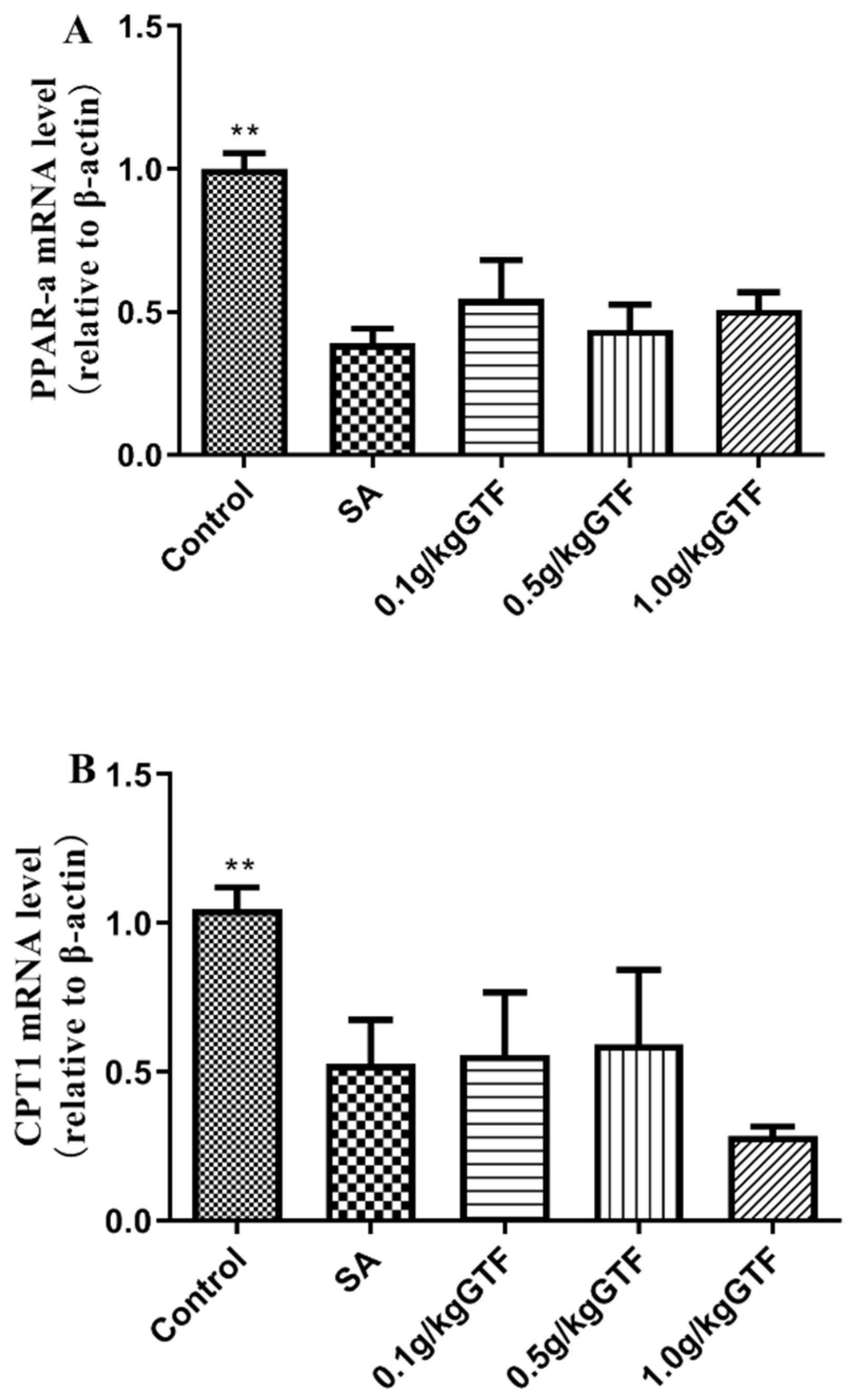
3.6. Effects of GTF on β-Oxidation-Related Gene Rxpression in Tilapia

As shown in Figure 5, the mRNA levels of carnitine palmitoyl transferase 1(CPT1) and peroxisome proliferator-activated receptor ɑ (PPARa) in the liver were significantly down-regulated with prolonged S. agalactiae infection time (p < 0.01); the expression of ACC oxidase gene (ACO1) gene was down-regulated, but the difference was not significant (p > 0.05). The addition of 0.1, 0.5, or 1.0 g/kg of GTF to the diet, the expression levels of the three genes in each concentration group could be increased to varying degrees, indicating that GTF had a certain alleviation effect on the oxidative damage of tilapia liver tissue caused by S. agalactiae.

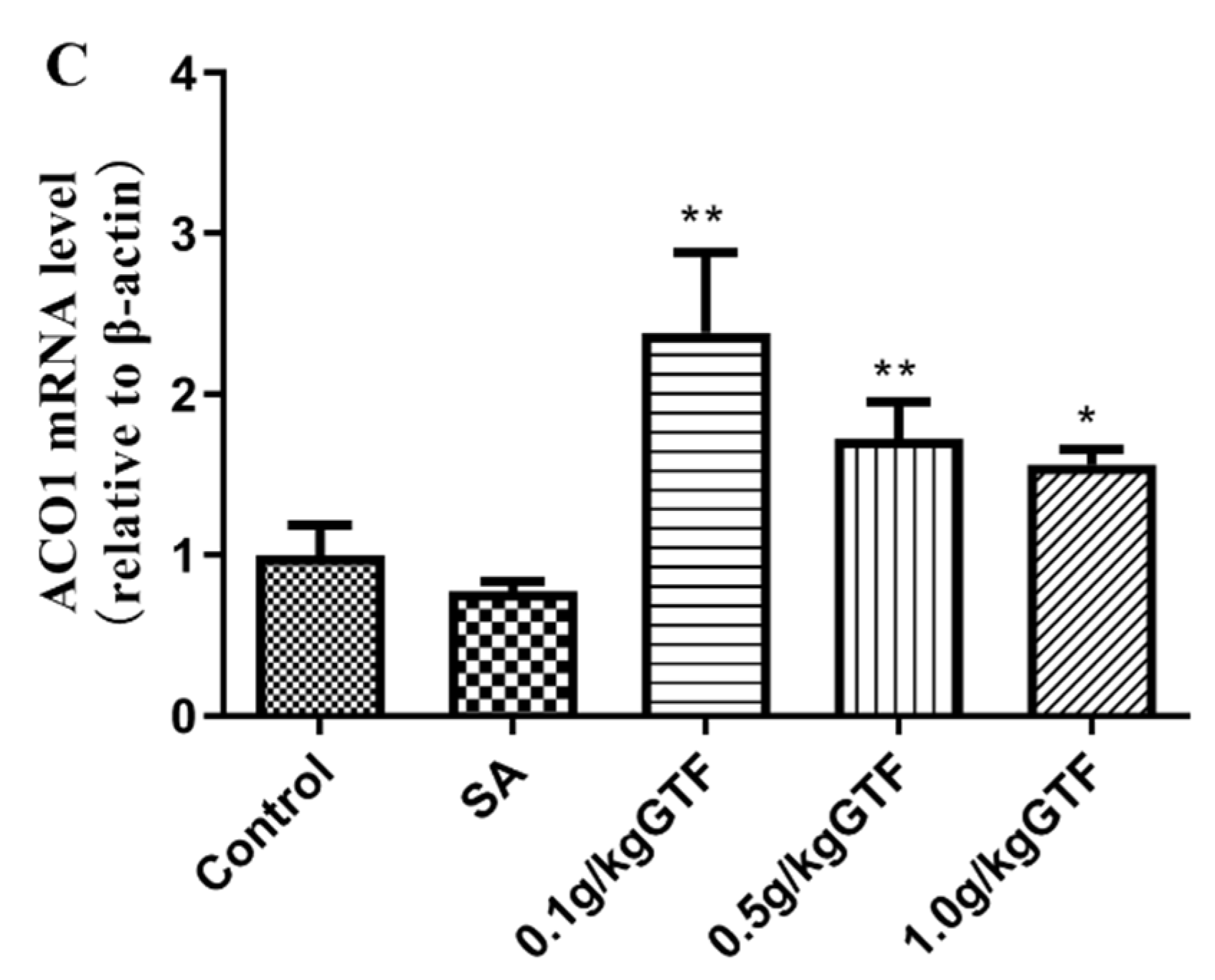
3.7. Effects of GTF on Expression of Genes Related to Lipid Droplet Formation in Tilapia

As shown in Figure 6, the expression levels of cell-death-induced DFFA-like effector B(Cideb) and cell-death-induced DFFA-like effector C (Cidec) genes were significantly down-regulated in the livers of tilapia infected with S. agalactiae compared to the normal control group (p < 0.01). The addition of 0.1 g/kg of GTF could significantly up-regulate the expression level of Cidec mRNA (p < 0.01) and the addition of 0.1 or 1.0 g/kg of GTF could significantly up-regulate the expression level of Cideb mRNA (p < 0.01). This indicated that GTF could promote lipid metabolism in the liver.

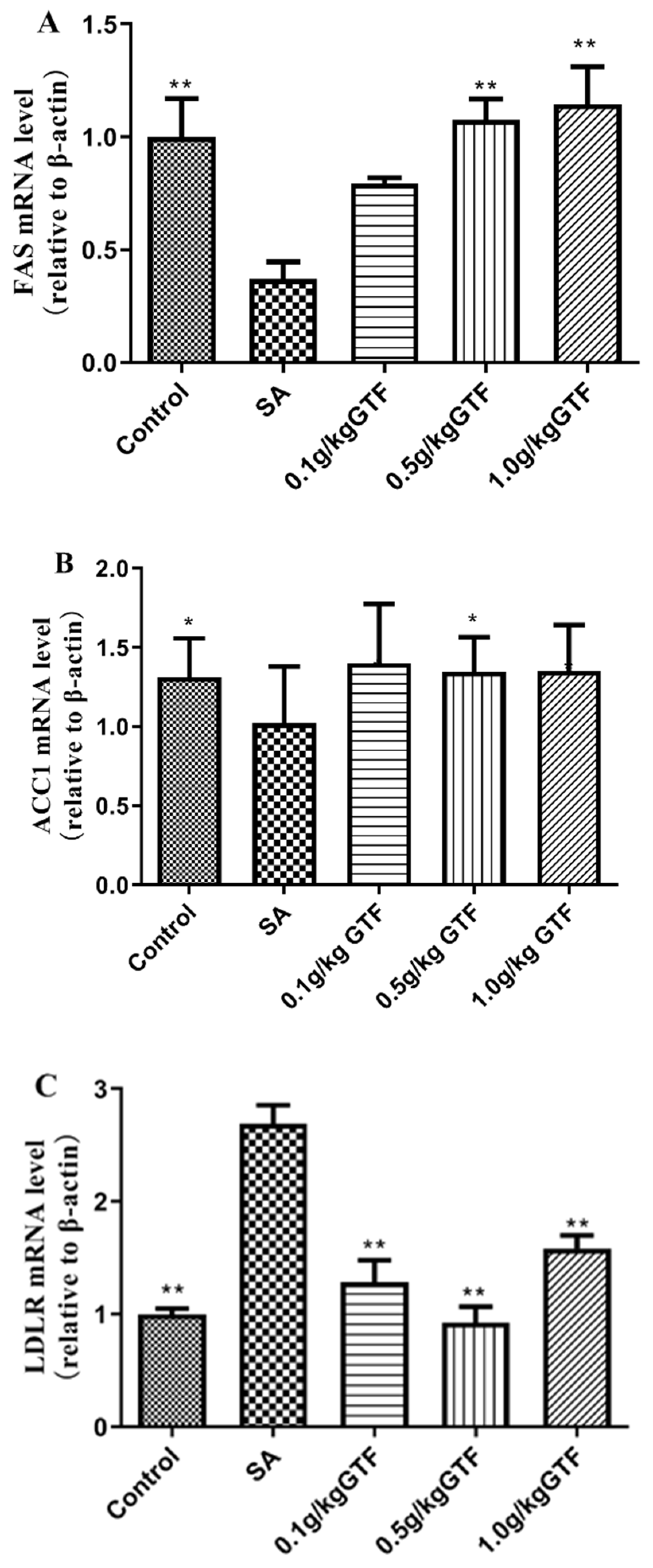
3.8. Effects of GTF on Genes Related to Fatty Acid Formation in Tilapia

As shown in Figure 7, the mRNA levels of acetylCoA carboxylase (ACC1) and fatty acid synthetase (FAS) in tilapia infected with S. agalactiae were significantly down-regulated (p < 0.01); the expression level of low-density lipoprotein receptor (LDLR) gene was significantly up-regulated compared to the normal control group (p < 0.01). In terms of the addition of 0.1, 0.5, or 1.0 g/kg of GTF to the diet, 0.5 g/kg GTF significantly up-regulated the expression levels of ACC1 and FAS genes in liver tissue, and addition of the 0.1 g/kg, 0.5 g/kg and 1.0 g/kg concentrations could significantly inhibit the expression levels of LDLR gene in liver tissue.

3.9. Effects of GTF on the Expression of HSP70 and IgM Genes in Tilapia Liver Tissue

As shown in Figure 8, the expression levels of heat stress protein 70 (HSP70) and immunoglobulin M (IgM) genes were significantly up-regulated compared to the normal control group (p < 0.05 or p < 0.01). Addition of 0.1, 0.5, or 1.0 g/kg of GTF to the diet significantly down-regulated the HSP70 gene expression level (p < 0.01), and the addition of 0.1 g/kg of GTF could significantly down-regulate the IgM gene expression level (p < 0.05).

4. Discussion

Streptococcal disease is one of the main diseases that threaten tilapia culture, which has the characteristics of rapid infection, strong pathogenicity and high mortality. The disease occurs mostly from March to November, and can be transmitted through fish contact, feed or water environment. As previously reported, the most effective treatment of Staphylococcus diseases are connected by the use of such antibiotics as neomycin, enrofloxacin and florfenicol [28,29]. However, a large number of experimental studies have shown that antibiotics only have certain therapeutic significance for tilapia in the early stages of disease. Long-term use of antibiotics can not only cause the emergence of drug-resistant strains, but also can easily cause drug residues [30]. As a natural medicine, Chinese herbal medicine has the advantages of green environmental protection and no residue, and has been widely used in human beings and mammals, but the application research of aquatic animals is relatively small. In this study, the GTF were mainly used as feed additives to study whether it has a therapeutic effect on tilapia S. agalactiae, explore its specific anti-infection mechanism, and provide a new idea and method for the development of anti-Streptococcus infection drugs.
GOT, GPT and AKP are commonly used indicators to evaluate liver function [31]. After liver damage, the increased permeability of liver cell membrane will increase the release of GOT, GPT and AKP into the blood [32]. In the present study, the increased activities of GOT, GPT, and AKP enzymes confirmed that Streptococcus can cause liver tissue injury in tilapia. Qiang et al. studied the effects of S. iniae on biochemical indexes of different tilapia species, finding that S. iniae caused an increase in the activities of GOT, GPT, AKP and other enzymes in serum [33], which was consistent with the results of this study. After the intervention of GTF, 1.0 g/kg GTF could effectively reduce the enzyme activities of GPT, GSH and AKP in serum, which indicated that GTF could alleviate the liver tissue damage of tilapia caused by Streptococcus.
T-AOC, CAT, SOD and GSH are important components of the body’s antioxidant defense system, which play a crucial role in maintaining the dynamic balance between oxidation and antioxidant [34]. Studies have shown that the GTF is a natural free radical scavenger with good antioxidant effect [35,36]. Du et al. found that GTF had a good protective effect on tilapia liver injury caused by a high-fat diet, and could significantly improve GSH activity, SOD activity and T-AOC level in liver tissue [37]. Liu et al. found that the addition of licorice flavonoids in diets has a good therapeutic effect on the oxidative damage caused by ethanol in mice, which can improve the antioxidant capacity of the body, and increase the T-AOC and glutathione peroxidase (GSH-Px) levels in blood and liver tissues of mice [38]. Li et al. found that GTF could increase CAT levels in mice with myocardial injury caused by strenuous exercise [39]. The results of this experiment were similar to previous studies. Compared with the blank control group, the levels of T-AOC, CAT, SOD and GSH in serum of tilapia were significantly increased after adding GTF to the diet, indicating that GTF could improve the antioxidant capacity of tilapia and alleviate the damage of streptococcus to the antioxidant enzyme system of tilapia.
Peroxisome proliferator-activated receptor ɑ (PPARa) is a nuclear receptor protein that is abundant in the liver [40]. Studies have shown that PPARa is widely involved in energy metabolism, oxidative stress, inflammation and other physiological reactions in the body [41]. Carnitine palmitoyl transferase 1 (CPT1) exists in the outer membrane of mitochondria and is a rate-limiting enzyme of fatty acid oxidation, which plays an important role in energy metabolism [42,43,44]. Existing studies have proved that inhibition of fatty acid oxidation can effectively control blood glucose levels in the body [45,46]. Acyl-coenzyme A oxidase 1(ACO1) is a key enzyme in the oxidation of fatty acid β, which plays an important role in the decomposition of fatty acids [47]. The results of this study showed that the expression levels of PPARa, CPT1, and ACO1 genes were all down-regulated, indicating that streptococcus caused an impairment in the process of fatty acid β oxidation in tilapia, which affected the catabolism of fatty acids and resulted in an increase of glucose content in serum. After adding GTF to the diet, the expression levels of PPARa, CPT1 and ACO1 genes were increased to different degrees, and the glucose content in serum was decreased, indicating that GTF could promote the β-oxidation of fatty acids in the liver tissue of tilapia, protect hepatocytes, and alleviate the liver tissue damage caused by streptococcus.
A large number of experimental studies have confirmed that the cell-death-inducing DNA-fragmentation-factor (DFF45)-like effector (cide) family is involved in intracellular energy metabolism and is an important regulator in the body [48,49]. The cide family consists of three family members: cidea, cideb and cidec. Cideb mainly exists in liver tissue, and cideb plays an important role in cell apoptosis [50]; Chen et al. have also reported that cideb is involved in the process of fat metabolism in the liver [51]. Cidec mainly exists in adipose tissue, heart, intestine and other tissues, and can also be expressed in liver tissue. Li et al. have reported that cidec plays an important role in lipid metabolism, and abnormal cidec expression can lead to lipid metabolism disorders [52]. The function of cidec was also reported by foreign scholars, and it was found that cidec expression induced the apoptosis process of cells [53]. In this study, the expression of cidec and cideb genes was significantly down-regulated, indicating that Streptococcus affected the expression of the cide family in liver tissue, resulting in abnormal liver metabolic function and further liver tissue injury. The addition of GTF to the diet could increase the expression levels of cidec and cideb mRNA in liver tissue to different degrees, indicating that GTF could repair damaged liver tissues and promote liver lipid metabolism.
FAS mainly exists in cytoplasm and is a fat metabolism enzyme with high expression in liver tissue. The amount of fatty acid synthetase plays an important role in the process of body fat deposition in animals. Some studies have reported that fatty acid synthetase is associated with the occurrence and development of diseases. Jia Rui et al. found that feeding tilapia with a high-fat diet for 60 days resulted in a significant decrease in FAS gene expression in the liver tissue of tilapia [54]. Zheng et al. found that the combination of theophylline and tea polyphenols could significantly reduce the expression of FAS gene in mouse liver tissue [55]. Acetyl-coa carboxylase (ACC) plays a major regulatory role in the process of fatty acid synthesis, which can regulate the oxidation of fatty acids [56]. Acc is highly expressed in the liver, and its activity can directly affect the fat metabolism of liver tissue. Qian et al. found that starvation stress could cause a significant decrease in the expression of ACC gene in the liver and muscle tissues of large yellow croaker [57]. Low-density lipoprotein receptor (LDLR) is a kind of membrane glycoprotein, which widely exists in various kinds of cells and tissues, plays an important role in lipoprotein metabolism. A large number of studies have shown that the abnormal blood lipid level in the body is closely related to the occurrence of many diseases. When studying the effect of bacterial lipopolysaccharides on LDLR gene expression in macrophages, Ye Qiang et al. found that with the increase of lipopolysaccharide concentration, LDLR gene expression level in macrophages was up-regulated in a concentration-dependent manner [58]. In this study, after streptococcal infection, the expression level of fatty acid synthetase gene in liver tissue was significantly down-regulated, while the expression level of low-density lipoprotein receptor gene in liver tissue was significantly up-regulated. The results of this experiment were similar to those of previous studies, indicating that S. agalactiae affected the normal lipid metabolism in the liver tissue of tilapia, causing lipid substances to accumulate in the liver tissue of tilapia, and then led to damage to the liver tissue. After adding GTF to the diet, three different concentrations of GTF can improve the damage of liver tissue of tilapia by S. agalactiae to different degrees, indicating that Chinese herbal medicine can regulate lipid metabolism and correct the disorder of lipid metabolism in liver tissue.
Heat stress protein 70 (HSP70) is one of the important members of the heat shock protein family, which plays a role in synergistic immunity and widely exists in animals, plants and microorganisms [59]. The level of HSP70 in normal cells is relatively low. When the cells are stimulated by external factors, the level of HSP70 will increase significantly, bind to the denatured proteins in the cells and degrade, thus playing a role in protecting the cells. Liu et al. studied the mechanism of HSP70 in carbon tetrachloride induced acute liver tissue injury in mice and found that the expression of HSP70 protein was significantly increased [60]; Ma et al. found that the expression of HSP70 in the liver tissue of mice induced by acetaminophen was significantly increased at 24 h [61]. IgM is an important component of the fish immune system, which plays an important role in resisting the stimulation of external pathogens such as pathogenic bacteria and other pathogens. Liu et al. studied the changes of immunoglobulin when carbon tetrachloride damaged the liver cells of Jian-carp and found that the content of IgM in the cells increased significantly [62]; Guo et al. found that lead exposure caused damage to the immune system of crucian carp (Carassius auratus), resulting in a significant increase in serum IgM level [63]. The results of this study were similar to previous studies; the expression levels of HSP70 and IgM genes in the streptococcal infection group were significantly increased compared to the blank control group. The expression levels of HSP70 and IgM genes decreased significantly after the addition of total licorice flavonoids to the diet. We speculated that S. agalactiae infection stimulated the synthesis of IgM and HSP70 to conduct immune responses. The total flavonoids of licorice can relieve the damaged liver tissue caused by Streptococcus.
In summary, the current study demonstrated that S. agalactiae can cause liver injury, which may be related to oxidative stress and abnormal lipid metabolism in tilapia liver tissue. GTFs have a good protective effect on tilapia liver injury caused by S. agalactiae, and can repair the damaged antioxidant enzyme system and immune system, alleviating liver tissue damage caused by S. agalactiae. Our results highlight the potential for using a Chinese herbal medicine to streptococcal disease in tilapia, and the specific protective mechanism of GTF needs to be further studied.

Author Contributions

Conceptualization, J.D. and L.C.; methodology, J.G.; software, H.Z. and Z.N.; validation and formal analysis, R.J.; investigation, L.C.; resources, B.X.; data curation, R.J. and J.G.; writing—original draft preparation, J.D.; writing—review and editing, J.D., L.C. and R.J.; supervision, Y.M.; project administration, G.Y.; funding acquisition, G.X. All authors have read and agreed to the published version of the manuscript.

Funding

This research was funded by the earmarked fund for CARS-46, and the Jiangsu Provincial Natural Science Foundation of China (NO.BK20201143).

Institutional Review Board Statement

This experiment was approved by the Institutional Animal Care and Use Committee (IACUC) of the Chinese Academy of Fishery Science.

Informed Consent Statement

Not applicable.

Data Availability Statement

Data is contained within the article.

Acknowledgments

This work was supported by the earmarked fund for CARS-46, and the Jiangsu Provincial Natural Science Foundation of China (NO.BK20201143).

Conflicts of Interest

The authors declare no conflict of interest.

References

  1. Salman, K.H.; Ali, F.A.Z.; Elhanbaly, R. Effect of cultured white soft cheese on the histopathological changes in the kidneys and liver of albino rats. Sci. Rep. 2022, 12, 2564. [Google Scholar] [CrossRef]
  2. Medvedeva, N.; Lazaruc, S.C.; Mirzaei, S.; Mikosch, P. Abnormal liver findings as the first manifestation in a patient with brucellosis: Case report. Wien. Med. Wochenschr. 2022, 172, 274–279. [Google Scholar] [CrossRef] [PubMed]
  3. Hamamoto, H.; Panthee, S. Transcriptome change of Staphylococcus aureus in infected mouse liver. Commun. Biol. 2022, 5, 721. [Google Scholar] [CrossRef]
  4. Shi, C.; Wang, J.; Zhang, R.; Ishfaq, M.; Li, Y.; Zhang, R.; Si, C.; Li, R.; Li, C.; Liu, F. Dihydromyricetin alleviates Escherichia coli lipopolysaccharide-induced hepatic injury in chickens by inhibiting the NLRP3 inflammasome. Vet. Res. 2022, 53, 6. [Google Scholar] [CrossRef] [PubMed]
  5. Nur-Nazifah, M.; Sabri, M.Y.; Siti-Zahrah, A. Development and efficacy of feed-based recombinant vaccine encoding the cell wall surface anchor family protein of Streptococcus agalactiae against streptococcosis in Oreochromis sp. Fish Shellfish. Immunol. 2014, 37, 193–200. [Google Scholar] [CrossRef] [PubMed]
  6. Zhu, J.L.; Li, D.L.; Zhou, Z.Y.; Xiao, W.; Yu, J.; Yang, H.; Xue, L.Y. The physiological response of hybrid tilapia (Oreochromis niloticus ♀ × Oreochromis aures ♂) and its parents infected with Streptococcus agalactiae. Acta Hydrobiol. Sin. 2021, 45, 1190–1200. [Google Scholar] [CrossRef]
  7. Luo, W.; Gan, X.; Zhu, J.J.; Ao, Q.G.; Tan, Y.; Chen, M.; Luo, Y.J. Pathological research of GIFT tilapia infected by Streptococcus agalactiae. J. South. Agric. 2018, 49, 375–382. [Google Scholar]
  8. Ao, Q.Z.; Luo, Y.J.; Lv, M.; Zhu, J.J. Effects of Streptococcus agalactiae Infection on Blood and Hepatopancreatic Tissue Biochemical Indices in Different Species of Tilapia. Prog. Fish. Sci. 2020, 41, 167–173. [Google Scholar] [CrossRef]
  9. Chen, R.R. Isolation, Identification and Pathogenic Biology of Streptococcus agalactiae from Tilapia in Guangzhou. Master’s Thesis, Nanjing Agricultural University, Nanjing, China, 2012. [Google Scholar]
  10. Yi, M.M.; Wang, M.; Li, Z.W.; Zhang, D.F.; Liu, Z.G.; Gao, F.Y.; Lu, M.X. The impacts and mechanism of Streptococcus agalactiae on behavior of GIFT Tilapia (Oreochromis niloticus). J. Agric. Biotechnol. 2019, 27, 799–808. [Google Scholar]
  11. Su, Y.L.; Liu, C.; Deng, Y.Q.; Guo, Z.X.; Feng, J. Research on Streptococcus agalactiae disease in tilapia: A review. J. Dalian Fish. Univ. 2019, 34, 757–766. [Google Scholar] [CrossRef]
  12. Li, Y.; Wang, B.; Wang, Z.W.; Cai, X.H.; Tang, J.F.; lu, Y.S.; Jian, J.C. Transmission routes of Streptococcus agalactiae and the strategy of immune evasion in tilapia. Guangdong Agric. Sci. 2017, 44, 132–140. [Google Scholar] [CrossRef]
  13. Fu, Y.; Chen, J.; Li, Y.J.; Zheng, Y.F.; Li, P. Antioxidant and anti-inflammatory activities of six flavonoids separated from licorice. Food Chem. 2013, 141, 1063–1071. [Google Scholar] [CrossRef] [PubMed]
  14. Li, T.; Hua, S.; Ma, J.; Dong, L.; Xu, F. Spectrum-Effect Relationships of Flavonoids in Glycyrrhiza uralensis Fisch. J. Anal. Methods Chem. 2020, 2020, 8838290. [Google Scholar] [CrossRef] [PubMed]
  15. Zhang, Z.; Yang, L.; Hou, J.; Tian, S.; Liu, Y. Molecular mechanisms underlying the anticancer activities of licorice flavonoids. J. Ethnopharmacol. 2021, 267, 113635. [Google Scholar] [CrossRef]
  16. Wang, C.H.; Wang, H.J. The antibacterial action of five traditional Chinese herbs including radix glycyrrhiza and rhizomacoptidis. Chin. J. Vet. Med. 2014, 50, 44–46. [Google Scholar]
  17. Liu, Y. Study on Antibacterial Activity of Extracts and Active Constituents of Licorice. Master’s Thesis, Yanbian University, Yanbian, China, 2013. [Google Scholar]
  18. Zhou, H.; Dai, C.; Cui, X.; Zhang, T.; Che, Y.; Duan, K.; Yi, L.; Nguyen, A.D.; Li, N.; De Souza, C.; et al. Immunomodulatory and antioxidant effects of Glycyrrhiza uralensis polysaccharide in Lohmann Brown chickens. Front. Vet. Sci. 2022, 9, 959449. [Google Scholar] [CrossRef]
  19. Chang, H.J.; Kim, Y.H.; Kang, Y.H.; Choi, M.H.; Lee, J.H. Antioxidant and antibacterial effects of medicinal plants and their stick-type medicinal concentrated beverages. Food Sci. Biotechnol. 2020, 29, 1413–1423. [Google Scholar] [CrossRef]
  20. Wang, J.; Chen, X.; Wang, W.; Zhang, Y.; Yang, Z.; Jin, Y.; Ge, H.M.; Li, E.; Yang, G. Glycyrrhizic acid as the antiviral component of Glycyrrhiza uralensis Fisch. against coxsackievirus A16 and enterovirus 71 of hand foot and mouth disease. J. Ethnopharmacol. 2013, 147, 114–121. [Google Scholar] [CrossRef]
  21. Cao, L.P.; Du, J.L.; Jia, R.; Ding, W.D.; Xu, P.; Yin, G.J.; Zhang, T. Effects of cyclophosphamide on antioxidative and immune functions of Nile tilapia (Oreochromis niloticus) via the TLR-NF-κB signaling pathway. Aquat. Toxicol. 2021, 239, 105956. [Google Scholar] [CrossRef]
  22. Zhang, C.Y.; Cao, L.P.; Du, J.L.; Jia, R.; Gu, Z.Y.; Yin, G.J.; Jeney, G. Nrf2 mediate protective effects of curcumin against carbon tetrachloride (CCl4 )-induced liver injury in Carassius auratus. Jiangsu J. Agr. Sci. 2018, 34, 114–121. [Google Scholar] [CrossRef]
  23. Cao, L.; Du, J.; Nie, Z.; Jia, R.; Yin, G.; Xu, P.; Ding, W.; Xu, G. Alteration of endoplasmic reticulum stress, inflammation and anti-oxidative status in cyclophosphamide-damaged liver of Nile tilapia (Oreochromis niloticus). Comp. Biochem. Physiol. Part C Toxicol. Pharmacol. 2022, 254, 109271. [Google Scholar] [CrossRef] [PubMed]
  24. Pecoraro, R.; Marino, F.; Salvaggio, A.; Capparucci, F.; Di Caro, G.; Iaria, C.; Salvo, A.; Rotondo, A.; Tibullo, D.; Guerriero, G.; et al. Evaluation of Chronic Nanosilver Toxicity to Adult Zebrafish. Front. Physiol. 2017, 8, 1011. [Google Scholar] [CrossRef] [PubMed] [Green Version]
  25. Rairat, T.; Chi, Y.; Hsieh, C.Y.; Liu, Y.K.; Chuchird, N.; Chou, C.C. Determination of Optimal Doses and Minimum Effective Concentrations of Tricaine Methanesulfonate, 2-Phenoxyethanol and Eugenol for Laboratory Managements in Nile Tilapia (Oreochromis niloticus). Animals 2021, 11, 1521. [Google Scholar] [CrossRef] [PubMed]
  26. Jia, R.; Gu, Z.; He, Q.; Du, J.; Cao, L.; Jeney, G.; Xu, P.; Yin, G. Anti-oxidative, anti-inflammatory and hepatoprotective effects of Radix Bupleuri extract against oxidative damage in tilapia (Oreochromis niloticus) via Nrf2 and TLRs signaling pathway. Fish Shellfish. Immunol. 2019, 93, 395–405. [Google Scholar] [CrossRef] [PubMed]
  27. Livak, K.J.; Schmittgen, T.D. Analysis of relative gene expression data using real-time quantitative PCR and the 2−ΔΔCT Method. Methods 2001, 25, 402–408. [Google Scholar] [CrossRef]
  28. Lu, Y.J.; Lai, D.G.; Tian, X.J.; Yu, H. Isolation, identification and drug sensitivity test of Tilapia Streptococcus iniae. Guangdong Agric. Sci. 2014, 41, 110–113. [Google Scholar] [CrossRef]
  29. Huang, Y.H.; Ma, S.; Han, S.Y.; Huang, W.D.; Wei, M.L.; Meng, L.L.; Lv, X.L.; Tan, H.M.; Li, D.Z.; Liang, J.Z.; et al. Antibiogram types of Streptococcus agalactiae strains isolated from Oreochromis spp. in Guangxi during 2011–2016. J. South. Agric. 2018, 49, 1023–1031. [Google Scholar] [CrossRef]
  30. Zhou, M.; Yu, S.; Hong, B.; Li, J.; Han, H.; Qie, G. Antibiotics control in aquaculture requires more than antibiotic-free feeds: A tilapia farming case. Environ. Pollut. 2021, 268, 115854. [Google Scholar] [CrossRef]
  31. Batool, R.; Khan, M.R.; Majid, M. Euphorbia dracunculoides L. abrogates carbon tetrachloride induced liver and DNA damage in rats. BMC Complement. Altern. Med. 2017, 17, 223. [Google Scholar] [CrossRef] [Green Version]
  32. Yang, Y.; Yang, L.; Han, Y.; Wu, Z.; Chen, P.; Zhang, H.; Zhou, J. Protective effects of hepatocyte-specific glycyrrhetic derivatives against carbon tetrachloride-induced liver damage in mice. Bioorg. Chem. 2017, 72, 42–50. [Google Scholar] [CrossRef]
  33. Qiang, J.; Yang, H.; Wang, H.; Xu, P.; Qi, S.L.; He, J. Studies on blood biochemical indices and expression of hepatic HSP70 mRNA of different tilapia strains artificially challenged with Streptococcus iniae. J. Fish. China 2012, 36, 958–968. [Google Scholar] [CrossRef]
  34. Rajput, S.A.; Liang, S.J.; Wang, X.Q.; Yan, H.C. Lycopene Protects Intestinal Epithelium from Deoxynivalenol-Induced Oxidative Damage via Regulating Keap1/Nrf2 Signaling. Antioxidants 2021, 10, 1493. [Google Scholar] [CrossRef] [PubMed]
  35. Wu, B.H.; Long, C.G.; Wang, X.M.; Li, Q.R.; Zheng, Q.X.; Hu, C.L. The scavenging effect of flavoniods of glycyrrhiza on hydroxyl radical studied in vitro. J. North Sichuan Med. Coll. 2001, 16, 3–5. [Google Scholar] [CrossRef]
  36. Gou, S.H.; He, M.; Li, B.B.; Zhu, N.Y.; Ni, J.M. Hepatoprotective effect of total flavonoids from Glycyrrhiza uralensis Fisch in liver injury mice. Nat. Prod. Res. 2021, 35, 6083–6087. [Google Scholar] [CrossRef]
  37. Du, J.L.; Cao, L.P.; Jia, R.; Gu, Z.Y.; He, Q.; Xu, P.; Jeney, G.; Ma, Y.Z.; Yin, G.J. Protective effects of Glycyrrhiza total flavones on liver injury of tilapia (Oreochromis niloticus) under high fat condition. Acta Agriculturae Zhejiangensis 2021, 33, 1826–1835. [Google Scholar] [CrossRef]
  38. Liu, J.F.; Guo, X.F. Effects of Glycyrrhiza flavone on the activity of GSH-Px and T-AOC in mice induced by ethanol oxidation. Heilongjiang Anim. Sci. Vet. Med. 2015, 176–178. [Google Scholar] [CrossRef]
  39. Li, M.H.; Tang, J.; Wang, C.L. Protective Effect of Glycyrrhizic Flavone on Myocardial Injury Induced by Long-term Exhaustive Exercised Rats. Genom. Appl. Biol. 2015, 34, 290–295. [Google Scholar] [CrossRef]
  40. Zhao, Q.; Tang, P.; Zhang, T.; Huang, J.F.; Xiao, X.R.; Zhu, W.F.; Gonzalez, F.J.; Li, F. Celastrol ameliorates acute liver injury through modulation of PPARα. Biochem. Pharmacol. 2020, 178, 114058. [Google Scholar] [CrossRef]
  41. Xu, W.J. PPARa involved in lipid metabolism pathway and nonalcoholic fatty liver disease. Chin. Hepatol. 2020, 25, 894–896. [Google Scholar] [CrossRef]
  42. Li, Y.J.; Hou, W.; Chen, W.H.; Long, Y.F.; Wu, J.Z. Effects of Anthraquinone Glycosides from Juemingzion the Expression of Fatty Acid β-Oxidation Related Enzyme Protein in Rats with Nonalcoholic Fatty Liver Disease. West. J. Tradit. Chin. Med. 2021, 34, 6–10. [Google Scholar] [CrossRef]
  43. Misheva, M.; Kotzamanis, K. Oxylipin metabolism is controlled by mitochondrial β-oxidation during bacterial inflammation. Nat. Commun. 2022, 13, 139. [Google Scholar] [CrossRef] [PubMed]
  44. Jiang, W.; Xu, M.; Qin, M.; Zhang, D.; Wu, S.; Liu, X.; Zhang, Y. Study on the role and mechanism of lncRNA in the remodeling of atrial energy metabolism in rabbits with atrial fibrillation based on nano sensor technology. Bioengineered 2022, 13, 863–875. [Google Scholar] [CrossRef] [PubMed]
  45. Zhang, F.L.; Luo, M. Research progress of carnitine palmitoyl transferase-I. Int. J. Endocrinol. Metab. 2002, 22, 166–169. [Google Scholar] [CrossRef]
  46. Liao, W.Y.; Xu, D. Study on the improvement effect of Ginsenoside Rg1 on nonalcoholic fatty liver phenotype by regulation of β-oxidation. Chongqing Med. 2016, 45, 1179–1182. [Google Scholar] [CrossRef]
  47. Li, P.; Zhang, R.; Wang, M.; Chen, Y.; Chen, Z.; Ke, X.; Zuo, L.; Wang, J. Baicalein Prevents Fructose-Induced Hepatic Steatosis in Rats: In the Regulation of Fatty Acid De Novo Synthesis, Fatty Acid Elongation and Fatty Acid Oxidation. Front Pharmacol. 2022, 13, 917329. [Google Scholar] [CrossRef]
  48. Chen, F.J.; Yin, Y.; Chua, B.T.; Li, P. CIDE family proteins control lipid homeostasis and the development of metabolic diseases. Traffic 2020, 21, 94–105. [Google Scholar] [CrossRef]
  49. Xu, W.; Wu, L.; Yu, M.; Chen, F.J.; Arshad, M.; Xia, X.; Ren, H.; Yu, J.; Xu, L.; Xu, D.; et al. Differential Roles of Cell Death-inducing DNA Fragmentation Factor-α-like Effector (CIDE) Proteins in Promoting Lipid Droplet Fusion and Growth in Subpopulations of Hepatocytes. J. Biol. Chem. 2016, 291, 4282–4293. [Google Scholar] [CrossRef] [Green Version]
  50. Ye, J.; Li, J.Z.; Liu, Y.; Li, X.; Yang, T.; Ma, X.; Li, Q.; Yao, Z.; Li, P. Cideb, an ER- and lipid droplet-associated protein, mediates VLDL lipidation and maturation by interacting with apolipoprotein B. Cell Metab. 2009, 9, 177–190. [Google Scholar] [CrossRef] [Green Version]
  51. Chen, Z.; Guo, K.; Toh, S.Y.; Zhou, Z.; Li, P. Mitochondria localization and dimerization are required for CIDE-B to induce apoptosis. J. Biol. Chem. 2000, 275, 22619–22622. [Google Scholar] [CrossRef] [Green Version]
  52. Li, Y.; Kang, H.; Chu, Y.; Jin, Y.; Zhang, L.; Yang, R.; Zhang, Z.; Zhao, S.; Zhou, L. Cidec differentially regulates lipid deposition and secretion through two tissue-specific isoforms. Gene 2018, 641, 265–271. [Google Scholar] [CrossRef]
  53. Zheng, G.S.; Tan, Y.M.; Shang, Y.Y.; Liu, Y.P.; Hu, B.A.; Wang, D.; Han, L.; Wang, Z.H.; Zhang, W.; Ti, Y.; et al. CIDEC silencing attenuates diabetic nephropathy via inhibiting apoptosis and promoting autophagy. J. Diabetes Investig. 2021, 12, 1336–1345. [Google Scholar] [CrossRef] [PubMed]
  54. Jia, R.; Cao, L.P.; Du, J.L.; He, Q.; Yin, G.J. Effects of High-Fat Diet on Steatosis, Endoplasmic Reticulum Stress and Autophagy in Liver of Tilapia (Oreochromis niloticus). Front. Mar. Sci. 2020, 7, 363. [Google Scholar] [CrossRef]
  55. Zheng, G.D.; Yang, L.C.; Lin, L.Z.; Zhang, Q.F. Effects of caffeine and catechins combination on gene and protein expression levels of hepatic fatty acid synthase in mice. J. Chin. Inst. Food Sci. Technol. 2016, 16, 21–25. [Google Scholar] [CrossRef]
  56. Brownsey, R.W.; Boone, A.N.; Elliott, J.E.; Kulpa, J.E.; Lee, W.M. Regulation of acetyl-CoA carboxylase. Biochem. Soc. Trans. 2006, 34, 223–227. [Google Scholar] [CrossRef] [PubMed]
  57. Qian, B.Y.; Huang, H.L.; Li, J.; Cao, M.Y.; Zhang, Y.; Zhao, Y.Y.; Xue, L.Y. Effects of fasting on the mRNA levels and enzymatic activities of hormone sensitive lipase and acetyl-CoA carboxylase in the large yellow croaker Larimichthys crocea. Acta Oceanol. Sin. 2018, 40, 31–39. [Google Scholar] [CrossRef]
  58. Ye, Q.; Lei, H.; Zheng, W.B.; Zheng, S.Z.; Chen, Y.X. Macrophages converted into foam cells via LPS-induced LDLr: Another important pathway of foam-cell formation under inflammatory stress. Chin. Pharmacol. Bull. 2013, 29, 1064–1070. [Google Scholar] [CrossRef]
  59. Tashiro, S.; Miyake, H.; Rokutan, K. Role of geranylgeranylacetone as non-toxic HSP70 inducer in liver surgery: Clinical application. J. Hepato-Biliary-Pancreat. Sci. 2018, 25, 269–274. [Google Scholar] [CrossRef]
  60. Liu, X.Z.; Li, S.Q.; Ren, J.T.; Xi, S.M.; Xu, Z.S.; Sun, Y.J. Expression and significance of HSP70, IL-6 and TNF-α in the tissue of acute liver enjury induced by CCl 4. Shandong Med. J. 2011, 51, 17–19. [Google Scholar]
  61. Ma, Y.J.; Li, S.Q. Expression of HSP70 in Acetaminophen Induced Acute Liver Injury Tissue. J. Henan Univ. Sci. Technol. (Med. Sci.) 2012, 30, 8–9. [Google Scholar] [CrossRef]
  62. Liu, Y.J.; Du, J.L.; Jia, R.; Cao, L.P.; Wang, J.H.; Yin, G.J. Hepatoprotective effects of Lycium barbarum polysaccharides on carbon tetrachloride induced primary hepatocyte injury in Jian Carp. J. Shanghai Ocean. Univ. 2014, 23, 718–725. [Google Scholar]
  63. Guo, X.Y.; Yin, Y.W.; Mao, A.T.; Dai, J.; Zhang, L.; Zhang, P.J.; Li, Y.H. Effects of lead exposure on antioxidation and immunity of Carassius aurous. Chin. Vet. Sci. 2018, 48, 222–227. [Google Scholar] [CrossRef]
Figure 1. Effects of GTF on morphological changes of liver tissue of tilapia at different concentrations (bar = 30 μm). (A): Control group; (B): SA infection group; (C): 0.1 g/kg GTF treatment group; (D): 0.5 g/kg GTF treatment group; (E): 0.1 g/kg GTF treatment group.
Antibiotics 11 01648 g001
Figure 2. Effects of GTF on GOT (A), GPT (B) and AKP (C) activities in the serum of tilapia. Values are expressed as the mean ± SEM (n = 10). * p < 0.05; ** p < 0.01, compared with the control group.
Antibiotics 11 01648 g002
Figure 3. Effects of GTF on the activities of T-AOC (A), CAT (B), SOD (C) and GSH (D) in the serum of tilapia. Values are expressed as the mean ± SEM (n = 10). * p < 0.05; ** p < 0.01, compared with the streptococcal injection group.
Antibiotics 11 01648 g003
Figure 4. Effects of GTF on GLU level in the serum of tilapia. Values are expressed as the mean ± SEM (n = 10). * p < 0.05; ** p < 0.01, compared with the control group.
Antibiotics 11 01648 g004
Figure 5. Effects of GTF on the mRNA levels of PPARa (A), CPT1 (B) and ACO1 (C) in the liver of tilapia. Values are expressed as mean ± SEM (n = 10). * p < 0.05 and ** p < 0.01 compared with streptococcal injection group.
Antibiotics 11 01648 g005aAntibiotics 11 01648 g005b
Figure 6. Effects of GTF on the mRNA levels of Cidec (A) and Cideb (B) in the liver of tilapia. Values are expressed as mean ± SEM (n = 10). ** p < 0.01 compared with streptococcal injection group.
Antibiotics 11 01648 g006
Figure 7. Effects of GTF on the mRNA levels of FAS (A), ACC1 (B) and LDLR (C) in the liver of tilapia. Values are expressed as mean ± SEM (n = 10). * p < 0.05 and ** p < 0.01 compared with streptococcal injection group.
Antibiotics 11 01648 g007
Figure 8. Effects of GTF on the mRNA levels of HSP70 (A) and IgM (B) in the liver of tilapia. Values are expressed as mean ± SEM (n = 10). * p < 0.05 and ** p < 0.01 compared with streptococcal injection group.
Antibiotics 11 01648 g008
Table 1. The primer sequences used in the present study.
TypeGenePrimer Sequence (5′–3′)GenBank Number/References
Fatty acid β-oxidationPPAR-αF: CTGATAAAGCTTCGGGCTTCCANM_001290066.1
CPT-1R: CGCTCACACTTATCATACTCCAGCT
F: TTTCCAGGCCTCCTTACCCA
XM_013268638.3
R: TGTACTGCTCATTGTCCAGCAGA
ACO-1F: GGTCAAAGGCAACAATCAGGAGNM_001290199.1
R: GACTCTGCCAAAGGCAACCA
Lipid droplet formationCidebF: CTACCCGACAACGTCATGCTXM_013266878.3
R: CCTTGAAATGTGGCCTGCAC
CidecF: TGGAGCCCACATCCTTACAAXM_005450237.4
R: TTTTTGGCAGCATAACAGCG
Fatty acid synthesisACC1F: GCGGTGTTCGGCTTGTTTTTXM_025910662.1
R: CAAGTCCACCTTCCCTTGGT
FASF: TTTGAGATGTGCTCACAGCTGCAXM_003454056.5
R: TCAGCCAGTGAGCTGTGGATGAT
Fatty acid uptakeLDLRF: TACGGCTTACCAGTCCTCCAXM_003443172.5
R: AGGTGACTGGAGCTTGTGTG
DetoxificationHSP70F:ATTTCAGACGGAGGGAAGCCXM_019357557.1
R:CAGCGTTGGACACCTTTTGG
ImmuneIgMF: ACCGAATCGAAAAATGCGGCKJ676389.1
R: AACACAACCAGGACATTGGTTC
Internal referenceβ-actinF:CCTGAGCGTAAATACTCCGTCTGKJ126772.1
R:AAGCACTTGCGGTGGACGAT
Table 2. Effects of dietary GTF level on growth indices of tilapia.
IndexDietary GTF Level/(g/kg)
00.10.51.0
Initial weight/g 20.00 ± 0.5719.33 ± 0.8820.67 ± 0.3319.67 ± 0.67
Final weight/g 80.19 ± 1.7982.34 ± 2.1285.16 ± 2.0585.90 ± 2.59
WGR, %3.02 ± 0.213.29 ± 0.303.12 ± 0.053.38 ± 0.20
SGR/(%/d)1.00 ± 0.041.05 ± 0.051.07 ± 0.031.10 ± 0.04
Publisher’s Note: MDPI stays neutral with regard to jurisdictional claims in published maps and institutional affiliations.

