Article

Concordance between Antimicrobial Resistance Phenotype and Genotype of Staphylococcus pseudintermedius from Healthy Dogs

1
Vetgenomics, Edifici EUREKA, PRUAB, Campus de la Universitat Autònoma de Barcelona (UAB), Bellaterra, 08193 Barcelona, Spain
2
Molecular Genetics Veterinary Service (SVGM), Universitat Autònoma de Barcelona (UAB), Bellaterra, 08193 Barcelona, Spain
3
Department of Animal Medicine and Surgery, Universitat Autònoma de Barcelona (UAB), Bellaterra, 08193 Barcelona, Spain
4
Institute of Science and Technology for Brain-Inspired Intelligence, Fudan University, Shanghai 200433, China
5
Unitat mixta d’Investigació IRTA-UAB en Sanitat Animal, Centre de Recerca en Sanitat Animal (CReSA), Campus de la Universitat Autònoma de Barcelona (UAB), Bellaterra, 08193 Barcelona, Spain
6
Institut de Recerca i Tecnologia Agroalimentàries (IRTA), Programa de Sanitat Animal, Centre de Recerca en Sanitat Animal (CReSA), Campus de la Universitat Autònoma de Barcelona (UAB), Bellaterra, 08193 Barcelona, Spain
*
Author to whom correspondence should be addressed.
Current address: Servei de Microbiologia-CDB, Hospital Clínic de Barcelona, c/ Villarroel 170, 08036 Barcelona, Spain.
Current address: ISGlobal, Institut for Global Health, c/ Rosselló 132, 08036 Barcelona, Spain.
Antibiotics 2022, 11(11), 1625; https://doi.org/10.3390/antibiotics11111625
Submission received: 21 October 2022 / Revised: 9 November 2022 / Accepted: 11 November 2022 / Published: 15 November 2022

Abstract

:
Staphylococcus pseudintermedius, a common commensal canine bacterium, is the main cause of skin infections in dogs and is a potential zoonotic pathogen. The emergence of methicillin-resistant S. pseudintermedius (MRSP) has compromised the treatment of infections caused by these bacteria. In this study, we compared the phenotypic results obtained by minimum inhibitory concentration (MICs) for 67 S. pseudintermedius isolates from the skin of nine healthy dogs versus the genotypic data obtained with Nanopore sequencing. A total of 17 antibiotic resistance genes (ARGs) were detected among the isolates. A good correlation between phenotype and genotype was observed for some antimicrobial classes, such as ciprofloxacin (fluoroquinolone), macrolides, or tetracycline. However, for oxacillin (beta-lactam) or aminoglycosides the correlation was low. Two antibiotic resistance genes were located on plasmids integrated in the chromosome, and a third one was in a circular plasmid. To our knowledge, this is the first study assessing the correlation between phenotype and genotype regarding antimicrobial resistance of S. pseudintermedius from healthy dogs using Nanopore sequencing technology.

1. Introduction

Staphylococcus pseudintermedius is a common bacteria found in the mucous membranes and skin of dogs. Carriage of S. pseudintermedius in healthy dog populations can range between 46% and 92% [1,2,3]. However, it is also the primary pathogen isolated from clinical canine specimens, especially from pyoderma, otitis externa, and superficial folliculitis. In recent decades, the emergence of methicillin-resistant S. pseudintermedius (MRSP) has compromised the treatment of these infections in veterinary medicine. Furthermore, in recent years, several studies have reported community-acquired infections with S. pseudintermedius transmitted from dogs to humans directly or by contamination of the household environment [4,5], highlighting their zoonotic potential. As a result, S. pseudintermedius has been identified among the most relevant antimicrobial-resistant (AMR) bacteria in the European Union for dogs and cats, and its eligibility to enter the list of animal diseases within the framework of the Animal Health Law has been recently discussed [6].
To date, the vast majority of studies have focused on S. pseudintermedius isolated from diseased dogs and have evidenced a high level of resistance to clinically important antimicrobials, such as beta-lactams, lincosamides, and fluoroquinolones [7]. In many cases, the presence of the mecA gene, or its homolog, mecC producing a modified penicillin-binding protein (PBP), provides them with phenotypic resistance to all beta-lactam antimicrobials registered in veterinary medicine, limiting the therapeutic options. Additionally, the global distribution of some sequence types (ST) associated with MRSP causing infections, such as ST71 in Europe and ST68 in North America, have contributed to unravel the epidemiology of this pathogen and identifying lineages of S. pseudintermedius with pathogenic and zoonotic potential [8].
However, to have a broader knowledge of the epidemiology of this opportunistic pathogen and to help veterinarians guide antimicrobial stewardship, it is necessary to study not only isolates associated with clinical cases but also the commensal population inhabiting the skin of healthy dogs. Furthermore, strain diversity within each individual animal has been studied by traditional methods, such as phenotypic tests, pulsed-field gel electrophoresis, or PCR, describing high genotypic diversity between different anatomical sites of the animal [9] but not many differences in antimicrobial resistant profiles. Hence, high-throughput sequencing technologies have the advantage of studying complete genomes. In particular, long-read sequencing technologies, such as Oxford Nanopore Technology (ONT), offer the advantages of closing genomes and plasmids, enabling establishment of the location and the genetic context of the different antimicrobial resistance genes [10,11]. This facilitates the assessment of the risk of transmission being higher when it is plasmid-borne and has the potential of being horizontally transferred intra and inter-species.
To our knowledge, whole genome sequencing techniques have not been fully applied to determine the prevalence and antimicrobial resistance determinants within S. pseudintermedius of healthy dogs. Therefore, the aim of this study was to characterize genotypically and phenotypically the population of S. pseudintermedius from the skin of different anatomical areas of healthy dogs and to correlate the phenotype with the genotype. Additionally, the location of the different antimicrobial resistant genes and their genetic context have been identified.

2. Results

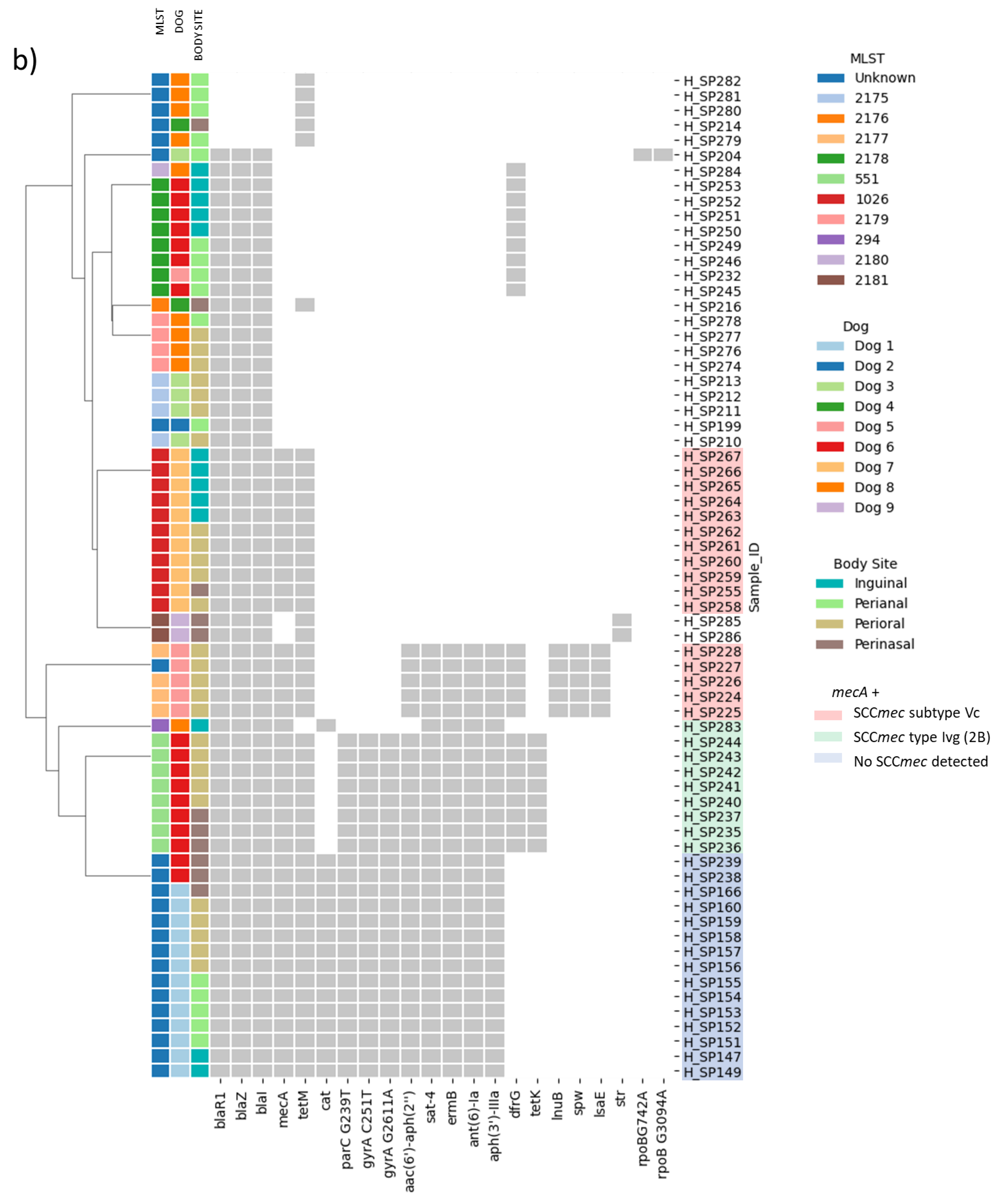
2.1. Distribution of MRSP and MDR Strains

Sixty-seven S. pseudintermedius were recovered from the skin of nine healthy dogs. The isolates were obtained from four different anatomical areas (Table 1): 27 from the perioral (six dogs), 16 from the perianal (six dogs), 13 from the inguinal (four dogs), and 11 from the nasal region (five dogs).
As seen in Figure 1a, minimal inhibitory concentration assays (MIC) described resistance to penicillin in 82.1% of the isolates, to tetracycline in 71.6%, to ampicillin in 59.7%, to oxacillin in 50.8%, to erythromycin and clindamycin in 43.3%, to ceftriaxone and ciprofloxacin in 37.3%, to co-trimoxazole in 32.8%, to gentamicin in 10.5%, to vancomycin in 1.5%, and no isolates were resistant to rifampicin. Multidrug resistance was described in 43.3% of the isolates. The most common combination of resistances among multidrug resistant isolates (MDR) was the resistance to erythromycin, penicillin, ampicillin, ceftriaxone, oxacillin, tetracycline, ciprofloxacin, and clindamycin in 20.9% of the isolates, followed by those isolates that were resistant to the same antimicrobials as above plus co-trimoxazole (13.4%).

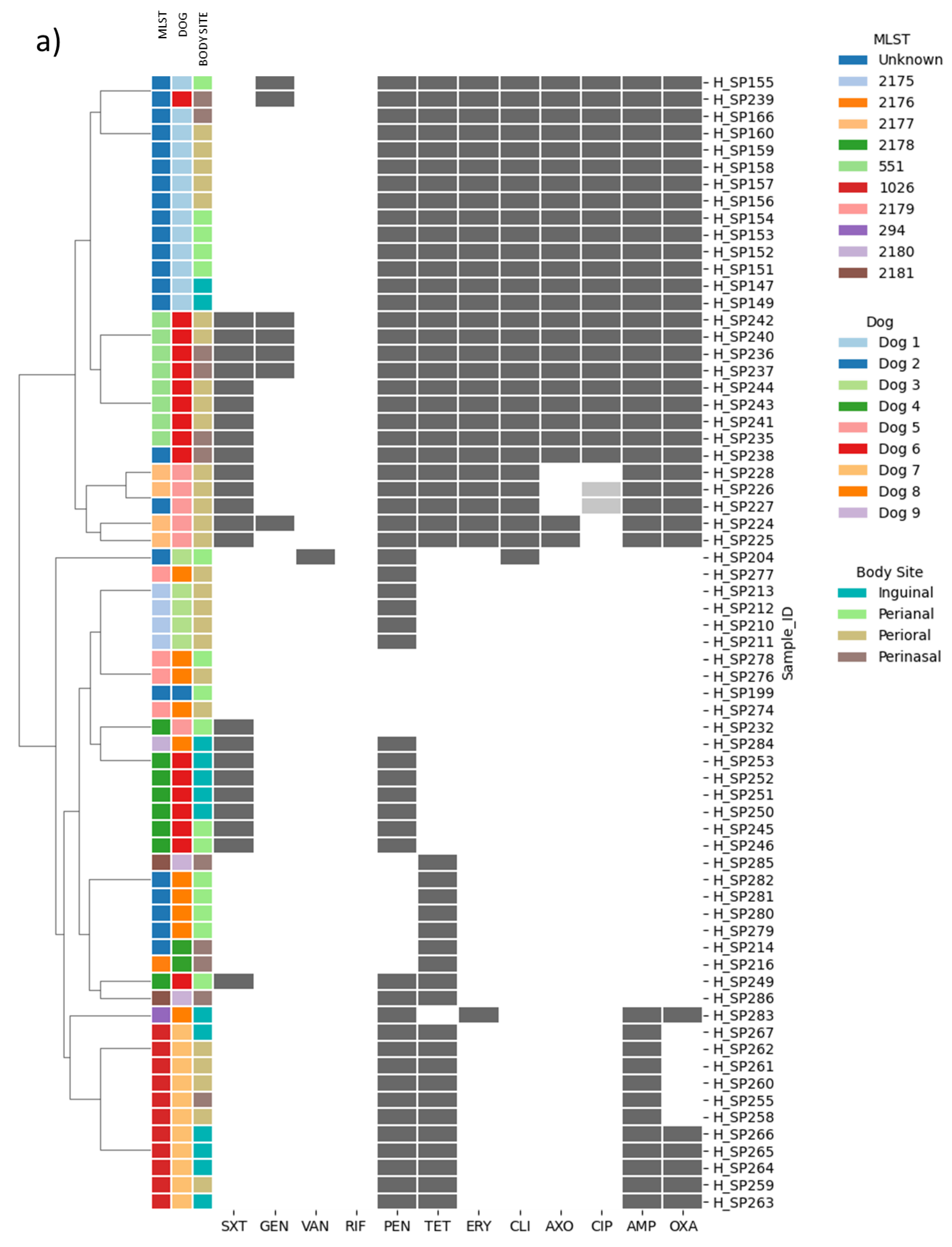
2.2. Genotype and Phenotype Concordance Varied Depending on the Antibiotic Family

A total of 17 antibiotic resistance genes (ARGs) (Figure 1b, see Supplementary Table S1 to check the MICs) were detected among the isolates, with 43.3% being phenotypically and genotypically MDR. The bla operon (blaZ, blaI, blaR1) co-occurred with all the ARGs retrieved in all isolates except for tetM. Additionally, the presence of the mecA gene was associated with the presence of aac(6′)-aph(2″), ant(6)-Ia, aph(3′)-IIIa, spw, tetM, tetK, lsaE, lnuB, ermB and sat-4 (Figure 2).
In general, a good correlation was observed between phenotypic and genotypic results. The concordance between the genotype and the phenotype was higher than 90% for lincosamides, trimethroprim, tetracyclines, macrolides, fluoroquinolones, and ampicillin (Supplementary Table S3). Penicillin and oxacillin showed a concordance of 88.8% and 85%, respectively. For ceftriaxon this value was 60%. However, aminoglycosides showed only 25% concordance. Moreover, when correlating phenotype and genotype, some inconsistencies were observed (Supplementary Table S3). One isolate (H_SP249, from Dog 6) was phenotypically resistant to tetracycline, but none of the published genes were detected. Another isolate (H_SP283, from Dog 8) exhibited resistance to two antimicrobial families, macrolides and beta-lactams, but in addition to presenting ermB and mecA, some genes conferring resistance to aminoglycosides were also present (aad(6) and aph(3′)-IIIa). Finally, six isolates (H_SP255, H_SP258, H_SP260 to H_SP262, and H_SP267) phenotypically susceptible to methicillin harbored the mecA gene.

2.3. Beta-Lactams and Cephalosporin Resistance

MIC values were evaluated for four beta-lactams, including penicillin, ampicillin, ceftriaxone, and oxacillin, and resistance was observed in 55, 40, 25 and 34 isolates, respectively (Supplementary Table S1).
Bioinformatic analyses detected the presence of two genes that can explain the resistance to these antibiotics: blaZ and mecA. All isolates resistant to penicillin (n = 55) harbored blaZ gene. However, seven extra isolates yielded the whole bla operon, including blaZ, balrR1, and blaI but were phenotypically susceptible to penicillin. The mecA gene was present in 40 isolates (Supplementary Table S4). All of them were resistant to ampicillin; 34 to oxacillin, and 25 to ceftriaxone. Interestingly, all the isolates showing the mecA gene and that were susceptible to both ceftriaxone (n = 15) and oxacillin (n = 6) presented the SCCmec type IVg (2B). When comparing the oxacillin and ceftriaxone MIC values between different SCCmec types, isolates (n = 17) with the type IVg (2B) exhibited MICs ≤1 mg/L and ≤8–16 mg/L, respectively, while the strains harboring the subtype Vc (5C2&5) (n = 8) or with no SCCmec cassette (n = 15) exhibited MIC values for oxacillin and ceftriaxone of >8 mg/L and >64 mg/L, respectively (Supplementary Table S4).

2.4. Fluoroquinolone Resistance

Phenotypic resistance to ciprofloxacin was observed in 23 isolates (13 from Dog 1 and 10 from Dog 6) and intermediate in two (Dog 2) (Supplementary Table S1). Mutations in gyrA and parC (also known as grlA) genes have been previously related to fluoroquinolone resistance [12]. Among the 67 S. pseudintermedius isolates, 38-point mutations in the gyrA gene were identified (Supplementary Table S5). Within these point mutations, 17 have been previously described [12], and 21 are described herein. Two of the point mutations described among our strains were specific for all the phenotypic antibiotic-resistant isolates: C251T (Ser84Leu) and G2611A (Glu871Lys). For parC gene, 36 point mutations were found among the 67 isolates, including 11 previously described [12] and 25 that are described herein (Supplementary Table S6). One mutation was specific for all the phenotypic-resistant isolates, G239T (Ser80Ile); however, we described six-point mutations present in 15 of the 23 resistant isolates, and three-point mutations present in three of the resistant strains.

2.5. Vancomycin Resistance

One isolate (H_SP204) exhibited resistance to vancomycin (MIC 16 μg/mL), and the MIC for teicoplanin was 16 μg/mL (Supplementary Table S1). None of the van genes conferring resistance to vancomycin were detected. However, mutations in rpoB and clpP genes were also assessed (Supplementary Tables S7 and S8, respectively). Two different mutations were observed for the rpoB gene, encoding the β subunit of RNA polymerase, G742A (Val248Ile) and G3094A (Val1032Ile). Additionally, one mutation C17T (Thr6Ile) in the clpP encoding for an ATP-dependent protease involved in cell wall synthesis was found.

2.6. Macrolide and Lincosamide Resistance

A total of 28 isolates were phenotypically co-resistant to erythromycin and clindamycin. However, one isolate was resistant only to erythromycin (H_SP283) and another isolate to clindamycin (H_SP204) (Supplementary Table S1).
All the isolates resistant to erythromycin and clindamycin harbored the ermB gene, except for the clindamycin susceptible isolate. Additionally, five isolates also yielded the lsaE gene along with lnuB. lsaE, and lnuB were located next to each other, with one site-specific recombinase upstream and downstream (Figure 3).

2.7. Tetracycline Resistance

A total of 48 isolates were phenotypically resistant to tetracycline (47 with MICs ≥ 16 µg/mL, and one with MIC 4 µg/mL) (Supplementary Table S1). Genotypically only the isolates with MIC ≥ 16 µg/mL presented a gene related to tetracycline resistance, tetM. Moreover, eight isolates from the same dog presented tetM together with tetK; this last one located in a potential plasmid inserted in the chromosome. tetK was accompanied by the other two genes present in the pSP-G3C4 (MN612109.1) plasmid harboring tetK gene: repC (coverage 71.4% and identity 90.1%) and repC polypeptide A (coverage 99.5% and identity 99.4%).

2.8. Trimethoprim-Sulfamethoxazole (Cotrimoxazole) Resistance

A total of 23 isolates were phenotypically resistant to cotrimoxazole (trimethoprim/sulfamethoxazole) (Supplementary Table S1). In agreement, the acquired dfrG gene was present in 22 of the 23 resistant isolates (Supplementary Table S3). H_SP238 exhibited resistance to cotrimoxazole, but no related ARG was found.
Mutations in the constitutive gene dhfr were assessed in all isolates (resistant and susceptible), but no specific mutations were detected (Supplementary Table S9). Additionally, point mutations in the folP gene were further investigated. Interestingly, all the resistant strains and half of the susceptible strains (24/44, 54.6%) presented an insertion of 12 nucleotides (AGAGGAAGTCAC, from position 177 to 188), translated into four extra amino acids in the protein sequence (Arg-Gly-Ser-His) (Supplementary Table S10).

2.9. Aminoglycoside Resistance

For aminoglycosides, it was difficult to establish a clear correlation between genotypic and phenotypic results (Supplementary Table S3). Additionally, clinical breakpoints are only described for gentamicin. However, applying CARD and NCBI databases, five genes conferring resistance to aminoglycosides were retrieved: aac(6′)-Ie-aph(2″)-Ia (aac(6′)-aph(2″)), ant(6)-Ia (also known as aad(6)), aph(3′)-IIIa, spw, and str. Within 31 isolates, four different combinations of aminoglycoside resistance genes were identified (Supplementary Table S2).
The genes aac(6′)-aph(2″), ant(6)-Ia, aph(3′)-IIIa, and spw cooccurred in 96.6% to 100% of the isolates that also present the bla operon, tetM, ermB, and sat-4 (Figure 2). The spw gene was also present in 100% of the isolates with lsaE, lnuB, and dfrG. str gene only cooccured with the bla operon and tetM.
Isolates H_SP285 and H_SP286 (both from the same dog) presented the str gene in a small contig independent from the chromosomal contig. The contig size was 4329 bp for H_SP285 and 4336 bp H_SP286; both were circular, with similar gene content (replicon, mobilization, and hypothetical proteins), and yielded a replicon rep7a_4_repD(pK214), indicating they were potential plasmids (Figure 4).

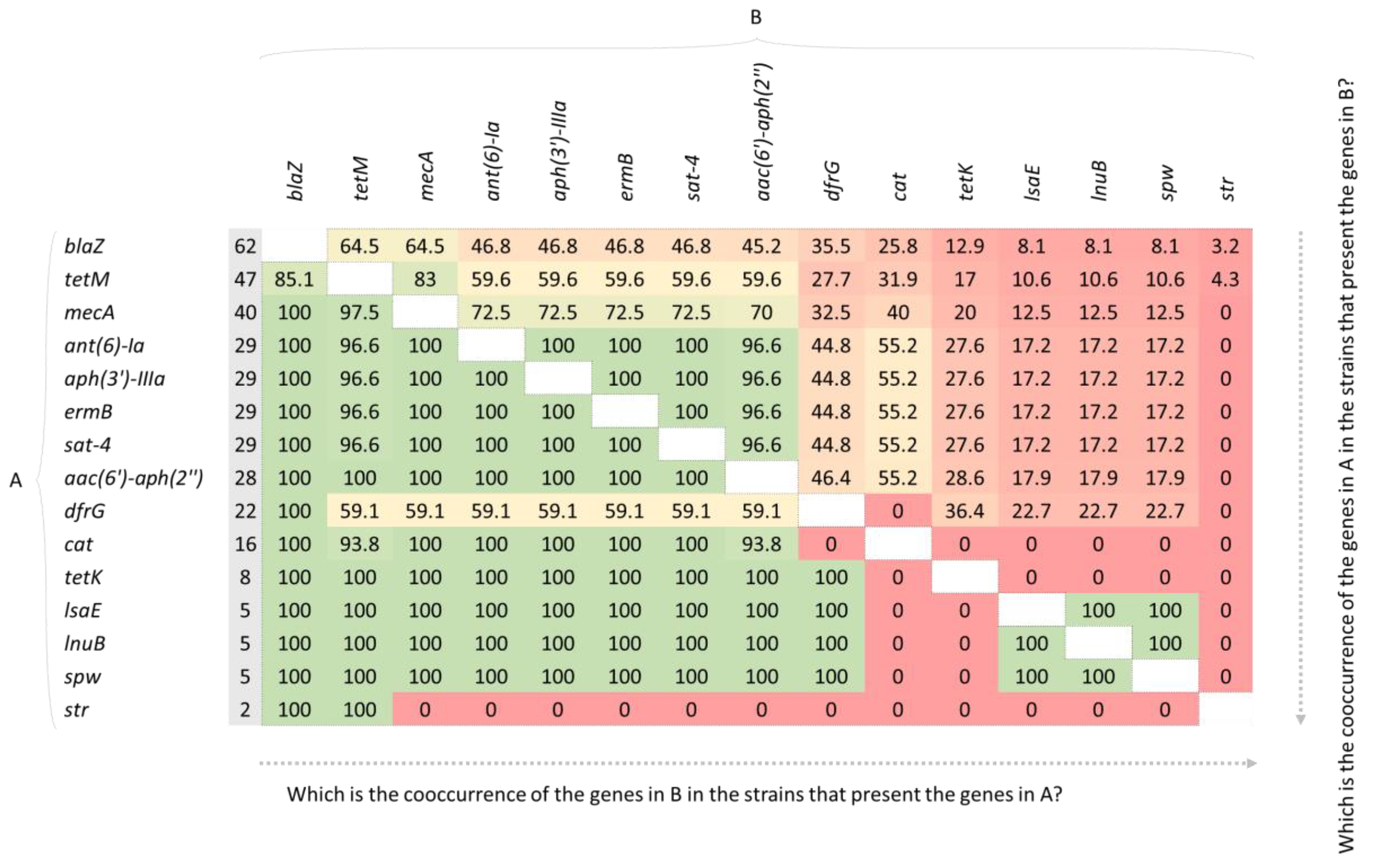
2.10. Genes Not Tested Phenotypically: sat-4 and cat

As shown in Supplementary Table S2, a total of 24 isolates presented the gene sat-4 (encoding for a streptothricin acetyltransferase [13]) with coverage and identity values equal or higher than 99% However, five isolates from dog 5 only presented a fragment of this gene (289 bp of 543 bp) located in the negative strand of the chromosome. Analyzing the genetic context of these five isolates (H_SP224 to H_SP228) (Figure 3) and comparing it to five isolates that presented the complete gene, we observed that in all cases sat-4 is flanked upstream and downstream by aph(3′)-III and ant(6)-I, respectively.
Sixteen isolates harbored a cat gene encoding for a chloramphenicol o-acetyltransferase in the chromosome (Supplementary Table S2). The gene was surrounded by several elements involved in recombination, mobilization, integration, and replication (Figure 3).

3. Discussion

In this study, we carried out a comprehensive correlation study between the genotypic and the phenotypic data of 67 S. pseudintermedius isolates obtained from four anatomical regions of nine healthy dogs [14]. Long-read Oxford Nanopore whole genome sequencing was performed with the MinION Mk1C followed by de novo assembling and polishing steps. Different authors have previously studied phenotype and genotype correlations in S. pseudintermedius using Illumina sequencing, [15,16] describing difficulties with regards to assembling the genetic context of the antibiotic resistance genes [15]. Here, we demonstrate that this avantgarde sequencing technology is an excellent tool not only to study the presence of resistance genes but also their genetic context, including insertion sequences (IS) and repeated regions.
The average number of resistances to different antibiotic families varied among the S. pseudintermedius isolates from one dog compared to the other dogs, but generally, the same profile was observed for the same sampled area. The nasal region should be considered a hot spot for the transmission of antibiotic resistance to other specimens since the colonization of the nasal region of a dog owner by the same S. pseudintermedius colonizing its dog’s nose has already been described [17].
In our previous study [18], all the S. pseudintermedius isolated from the skin of six healthy dogs (n = 22) were methicillin-susceptible S. pseudintermedius (MSSP), and only two of them were MDR. In contraposition, the present work identified 27 MSSP (40%) and 40 MRSP (60%), 29 of the latter also MDR, identified in five out of the nine healthy dogs. Although the number of dogs is quite limited for performing any statistical test, it appears that the presence of the mecA gene in S. pseudintermedius isolates from healthy dogs is more prevalent than previously thought. Furthermore, a complete mecA gene was confirmed in isolates susceptible to oxacillin and ceftriaxone, which can hinder the therapeutic outcome in case of infection. In this regard, isolates that harbored the SCCmec IVg exhibited lower MIC than those with SCCmec_subtype-Vc(5C2&5) or lacking the cassette, as described by Worthing et al. [19] Therefore, our results confirmed that oxacillin and ceftriaxone susceptibility appears to be associated with the type of SCCmec harbored by the bacterium.
The emergence of methicillin-resistant Staphylococcus spp has led to the use of vancomycin as one of the first line antimicrobials to combat Gram positive infections [20,21]. Fortunately, we could not detect any of the vancomycin resistant genes described in the literature. However, mutations in rpoB and clpP can cause reduced susceptibility against vancomycin [15,22,23,24,25]. We described herein two possible mutations associated with these genes that should be confirmed by more precise sequencing technology. Further analysis, including morphometric studies using electron microscopy and doubling time assays, should be performed as described by Cui et al. [26] to check the cell wall thickness and multiplication rate among this resistant isolate.
One hundred percent concordance between phenotype and genotype was observed for ciprofloxacin. While C251T (Ser84Leu) has been consistently described as a characteristic mutation in the gyrA gene conferring resistance to fluoroquinolones in different Staphylococcus species [12,27,28,29,30], here we report a novel second mutation that was only present in resistant isolates, G2611A (Glu871Lys). For parC gene, the G239T (Ser80Ile) mutation was only present in resistant isolates.
For erythromycin, 100% concordance was observed between phenotype and genotype, with two discrepancies for clindamycin resistance. Isolate H_SP204 was clindamycin resistant and erythromycin susceptible, and no antibiotic resistance gene was described. On the other hand, isolate H_SP283 presented phenotypic resistance to erythromycin but was susceptible to clindamycin. It is possible that this isolate presented an inducible clindamycin resistance, unlike all the other strains that presented the ermB gene and that were resistant to both erythromycin and clindamycin. It has been described in S. pseudintermedius that the ermB gene can be constitutively or induced expressed [31]. Thus, further experiments with a double-disk diffusion testing both antibiotics should be performed to ensure clindamycin resistance, since clindamycin is one of the predilected antibiotics to use in treatment when resistance to methicillin is present. Interestingly, lsaE and lnuB genes were flanked by one site-specific recombinase (Figure 3), which could denote a previous event of mobilization and integration in the chromosome. In fact, Yan et al. described these genes in a conjugative plasmid in Enterococcus faecium, potentially transmissible to Staphylococcus aureus [32]. These analyses also identified that both genes, lsaE and lnuB were located next to an antibiotic resistance region with ant(6)-I-sat-4-aph(3′)-III clusters [33], as described herein.
As Tyson et al. reported in their study [15] as a limitation, no concordance between phenotype and the presence of resistance genes was observed for aminoglycosides in this study. Moreover, Wegener et al. [16] described that the most usual discrepancies relied on the aac(6′)-Ie-aph(2″)-Ia gene. Additionally, the lack of breakpoints for some of the drugs belonging to this antimicrobial family also hampered any possible correlations between genotype and phenotype. Furthermore, several discrepancies between CARD and NCBI databases regarding coverage and gene nomenclature for this antimicrobial class made it very difficult to compare genotypic results between isolates. Still, a cluster of aminoglycoside resistance genes was detected in our isolates, comprising ant(6)-I (also known as aadE), sat-4, and aph(3′)-III with the presence of other genes such as ermB and dfrG and cat [34,35,36]. These clusters are known as Tn5405-like elements and can be found in Enterococcus faecalis plasmid pRE25 [37]. Loeffler and Lloyd [38] showed the rapid evolution of S. pseudintermedius to multidrug resistance through the acquisition of mecA on a staphylococcal cassette chromosome (SCC), a large transposon (Tn5405-like element) carrying up to five resistance genes, and genome point mutations for fluoroquinolones (gyrA/grlA) and sulfonamide resistance.
Genotypically, only the isolates exhibiting MIC ≥ 16 µg/mL for tetracycline harbored tetM or tetK. On the other hand, isolates with MIC 4 µg/mL (also categorized as resistant) did not present any known genes, although the presence of efflux pumps cannot be disregarded. In previous studies, tetK has been described in small size plasmids [18,39] in contraposition to our isolates that present this gene in the chromosome. According to the genetic context, it is plausible that a pSP-G3C4-like plasmid has been integrated into the chromosome, as previously described for tetK bearing S. aureus pT181 plasmid [40,41]. Similarly, we observed that the cat gene was surrounded by elements associated to plasmids, including a recombinase, a replication initiation protein, and a toxin-antitoxin system (zeta, ζ, and epsilon, ε, respectively). Therefore, it appears that this plasmid has also been integrated into the chromosome. The presence of plasmids carrying a cat gene integrated into the chromosome of methicillin-resistant S. aureus MRSA isolated from both dogs and humans has been previously reported [34].
Finally, the genetic background encoding resistance for trimethoprim or sulfonamides could not be determined since no correlation could be detected between mutations in the constitutive dihydrofolate reductase (dhfr) conferring resistance to trimethoprim [42,43] or dihydropteroate synthase (folP) target of sulfonamides [44].

4. Materials and Methods

4.1. Bacterial Cultures

The dogs came to the Hospital Clínic Veterinari (Universitat Autònoma de Barcelona, UAB, Spain) for routine preventive medical interventions (vaccinations, deworming). Prior to vaccination, they were subjected to a general physical examination by a veterinary surgeon, in which no clinical signs or lesions were detected. A total of nine healthy dogs, belonging to different breeds, and without previous records of antimicrobial treatment were selected during a period spanning one month. Samples were obtained by rubbing sterile swabs on four different skin anatomical areas: perinasal, perioral, inguinal, and perianal. These four different skin anatomical sites were chosen to represent different types of microbial habitat within the dog: from a region with fur and mostly dry like the groin (inguinal samples) to mucocutaneous areas like the muzzle (nasal and perioral samples), and the perianal region, close to the gastrointestinal tract [45,46]. Swabs were cultured in blood agar at 37 °C for 24 h. Up to five colonies morphologically compatible with S. pseudintermedius (small silver colonies) were selected and sub-cultured in 3 mL of Brain Heart Infusion (BHI) media at 37 °C for 16 h. A total of 67 S. pseudintermedius isolates were included in this study, as shown in Table 1.

4.2. Antibiotic Susceptibility

Minimal inhibitory concentration (MIC) assays were carried out for 18 antimicrobial compounds, using Sensititre plates (Sensititre, Trek diagnostic Systems Inc., East Grinstead, UK) (Supplementary Table S1). For confirmation of the vancomycin resistant profile, a different Sensititre MIC plate containing vancomycin and teicoplanin was used. Antimicrobial breakpoints were those recommended by the Clinical and Laboratory Standards Institute (CLSI) for the Staphylococcus species [47,48]. Multidrug resistant (MDR) isolates were defined for those isolates showing phenotypic resistance to at least three antimicrobials from different classes [49].

4.3. DNA Extraction and Nanopore Sequencing Library Preparation

DNA was extracted with ZymoBIOMICSTM DNA Miniprep Kit (Zymo Research, Irvine, CA, USA). DNA quality and quantity were determined using NanoDrop 2000 Spectrophotometer and QubitTM dsDNA BR Assay Kit (Fisher Scientific SL, Madrid, Spain). The sequencing libraries prepared approximately 200 to 400ng of DNA, which were subjected to transposase fragmentation using the Rapid Barcoding Sequencing kit (SQK-RBK004; Oxford Nanopore Technologies, ONT, Oxford, UK). Up to twelve barcoded samples were loaded in a MinION FLO-MIN106 v9.4.1 flow cell (ONT, Oxford, UK) and sequenced in a MinION Mk1B or Mk1C (ONT, Oxford, UK).

4.4. Bioinformatic Genome Sequencing Analysis

The fast5 files were basecalled, demultiplexed, and adapters trimmed with Guppy (v5.0.11) [50] (--dna_r9.4.1_450bps_sup.cfg) (--config configuration.cfg --barcode_kits SQK-RBK004 --trim_barcodes; min_score threshold default 60). Reads with a quality score lower than 10 were discarded. Run summary statistics were obtained with Nanoplot (v1.38.1) [51] (--N50 --fastq).
Isolates were confirmed as S. pseudintermedius by taxonomy assignment using the EPI2ME What’s in my pot (WIMP) workflow [52]. Genomes were de novo assembled using Flye (v2.8.3) [53] (--nano-raw --plasmids --trestle), except for HSP279 and HSP281 samples, which were de novo assembled using Flye (v2.9) (--nano-hq). Contigs were polished with Medaka (v1.4.3) [54] (medaka_consensus; -m r941_min_sup_g507). Genome completeness and contamination were assessed with CheckM (v1.1.3) (lineage_wf) [55]. Circlator (v1.5.5) was used to rotate the genomes fixing the start with the dnaA gene [56] (fixstart --min_id 70), when possible.
Multilocus sequence types (MLSTs) were determined on the basis of the S. pseudintermedius PubMLST database (https://pubmlst.org/organisms/staphylococcus-pseudintermedius, accessed on 21 October 2022) [57] and the MLST (v2.0) software (https://cge.cbs.dtu.dk/services/MLST/, accessed on 21 October 2022) [58].
Cluster maps were plotted using the seaborn [59] library in Python. The input files were the presence/absence matrix of antimicrobial resistant genes and antimicrobial resistance.

4.5. Antibiotic Resistance Bioinformatic Analysis

Genomes were analysed with Abricate [60] (v1.0.1) using the Comprehensive Antibiotic Resistance Database (CARD) and the National Center for Biotechnology Information (NCBI) databases (both updated on 21 December 2021) to determine the presence of antibiotic resistance genes (ARGs), applying a threshold of identity and coverage ≥95%. Antibiotic resistance associated with point mutations of some specific genes were identified by analyzing individual files, extracting the sequences, and aligning them with BioEdit’s [61] (v7.2.5) ClustalW Multiple Alignment tool.
To determine the genetic context of some ARGs, genome sequences were uploaded to the PAThosystems Resource Integration Center (PATRIC) [62] (v3.6.12) and genome annotation was performed online with Rapid Annotation using the Subsystem Technology tool kit (RASTtk) [63] tool. SnapGene Viewer (v5.2.4) (from Insightful Science; available at https://www.snapgene.com/, accessed on 21 October 2022) was used to visualize the annotated GenBank file and to customize the output. MDR isolates were defined as those isolates showing the presence of ARG from at least three different antimicrobial families.
When the mecA gene was present, the Staphylococcal chromosome cassette mec (SCCmec) was described using SCCmecFinder (v1.2) (from the Center for Genomic Epidemiology of the Technical University of Denmark; available at https://cge.cbs.dtu.dk/services/SCCmecFinder/, accessed on 21 October 2022).

5. Conclusions

To our knowledge, this is the first study assessing the correlation between phenotype and genotype regarding antimicrobial resistance of S. pseudintermedius from healthy dogs using Nanopore long-read whole genome sequencing technology. Even though we were limited by the antimicrobial cutoffs for our species, a good correlation was observed for some antimicrobials, such as ciprofloxacin (fluoroquinolone), macrolides, or tetracycline. However, for oxacillin (beta-lactam) or aminoglycosides the correlation was quite low. It is noteworthy that some isolates harbored the mecA gene and were susceptible to oxacillin and/or ceftriaxone, highlighting the importance of using sequencing technology for confirmation of phenotypic results. The presence of the mecA gene and the number of MDR isolates with integrated plasmids in their chromosome emphasizes the importance of monitoring S. pseudintermedius for its zoonotic potential. Since we enrolled nine dogs in our study, future projects will include a broader number of individuals and a broader number of antimicrobials tested. Moreover, we will not only focus on S. pseudintermedius, but also on other Staphylococcus species isolated by culture that were discarded during the isolation steps. Finally, more analyses of the origin of the discrepancies between the phenotype and the genotype will be included.

Supplementary Materials

The following supporting information can be downloaded at: https://www.mdpi.com/article/10.3390/antibiotics11111625/s1, Supplementary Table S1: Minimum inhibitory concentration (MIC) values for the 18 antibiotics tested. There are also represented the MIC values applied in this paper. The numeric values represent the mg/L. Co-trimoxazole is the combination of trimethoprim and sulfamethoxazole. We could not apply MIC values for six antibiotics: SYN, DAP, LEVO, LZD, STR, GAT. S, susceptible; I, intermediate susceptibility; R, resistant; IN, inguinal; PA, perianal; PO, perioral; NA, nasal. Supplementary Table S2: Cooccurrence of the antibiotic resistance genes retrieved by Abricate. Antibiotic resistance genes described by Abricate with CARD and NCBI databases. aac(6’)-ah(2″), aac(6’)-Ie-aph(2″)-Ia; IN, inguinal; PA, perianal; PO, perioral; NA, nasal; spw gene belongs to the ant(9) family of aminoglycoside resistance genes. Twenty-four isolates presented a full-length copy of the sat-4 gene, but we also included five isolates from Dog 5 (in red) that did harbor half of the gene in the same context as the isolates with the full-length gene. In the las row there are the percentages of presence of each gene among the 67 isolates. Supplementary Table S3: Congruences and discrepancies between the genotype and the phenotype. In the last row are calculated the concordances in percentages. * C251T and G2611A described in gyrA gene and G239T in parC gene; ** from all the antibiotic resistance genes for aminoglycosides described only aac(6)-aph(2) confers resistance to gentamicin. Supplementary Table S4: All the strains that presented de mecA gene. Here we describe which SCCmec are presented and their MICs for oxacillin (OXA) and ceftriaxone (AXO). The SCCmec percentage is given by the tool we used to determine the presence of SCCmec, the SCCmecFinder. Supplementary Table S5: Mutations found in gyrA gene. Mutations in this gene are related to fluoroquinolone resistance. The sequences were aligned with BioEdit. R/S, resistant/susceptible. The numbers in bold correspond to the nucleotide position. Supplementary Table S6: Mutations found in parC gene (also known as grlA). Mutations in this gene are related to fluoroquinolone resistance. The sequences were aligned with BioEdit. R/S, resistant/susceptible. The numbers in bold correspond to the nucleotide position. Supplementary Table S7: Mutations found in rpoB gene. Mutations in this gene are related to vancomycin resistance. The sequences were aligned with BioEdit. R/S, resistant/susceptible. The numbers in bold correspond to the nucleotide position. Supplementary Table S8: Mutations found in clpP gene. Mutations in this gene are related to vancomycin resistance. The sequences were aligned with BioEdit. R/S, resistant/susceptible. The numbers in bold correspond to the nucleotide position. Supplementary Table S9: Mutations found in dhfr gene. Mutations in this gene are related to co-trimoxazole resistance. The sequences were aligned with BioEdit. R/S, resistant/susceptible. The numbers in bold correspond to the nucleotide position. Supplementary Table S10: Mutations found in folP gene. Mutations in this gene are related to co-trimoxazole resistance. The sequences were aligned with BioEdit. R/S, resistant/susceptible. The numbers in bold correspond to the nucleotide position.

Author Contributions

Conceptualization, J.V., L.F., O.F.; methodology, D.P., J.V., L.F., L.M.-G., N.F., R.F.; software, J.V.; validation, J.V., L.M.-G.; formal analysis, J.V., L.M.-G.; data curation, J.V., D.P., N.F.; writing—original draft preparation, J.V., L.M.-G.; writing—review and editing, A.C., D.P., J.V., L.M.-G., N.F., O.F.; visualization, J.V.; supervision, A.C., L.F., O.F., L.M.-G.; project administration, L.F., O.F.; funding acquisition, J.V., L.F., O.F. All authors have read and agreed to the published version of the manuscript.

Funding

Funding sources were as follows: Research Project RTI2018-101991-B-I00 (“From whole genome sequencing to clinical metagenomics: investigations on the pathogenesis of Staphylococcus pseudintermedius pyoderma in the dog”), Spanish Ministry of Science, Innovation and Universities. Torres Quevedo Project, PTQ2018-009961, co-financed by the European Social Fund and the Spanish Ministry of Science and Innovation. Industrial Doctorate Program Grant 2017DI037 AGAUR, Generalitat de Catalunya, Spain.

Institutional Review Board Statement

Not applicable.

Informed Consent Statement

Not applicable.

Data Availability Statement

The genome assemblies and genomic data [14] are publicly available in GeneBank, BioProject PRJNA685966, genome accession numbers CP083183 to CP083231; CP085723, CP085724 and JAJEKF000000000 to JAJEKU000000000. The raw data are available from the Sequence Read Archive (SRA), BioProject PRJNA685966.

Acknowledgments

J.V. would like to thank Cristina Pitart from the Hospital Clinic de Barcelona for her advice regarding some discrepancies between genotypes and phenotypes.

Conflicts of Interest

L.F. has received unrelated honoraria for lecturing from Zoetis, Bayer, LETI, and Affinity Petcare. O.F. and A.C. have received unrelated honoraria for lecturing from Oxford Nanopore Technologies.

References

  1. Bannoehr, J.; Guardabassi, L. Staphylococcus pseudintermedius in the dog: Taxonomy, diagnostics, ecology, epidemiology and pathogenicity. Vet. Dermatol. 2012, 23, 253-e52. [Google Scholar] [CrossRef] [PubMed]
  2. Paul, N.C.; Bärgman, S.C.; Moodley, A.; Nielsen, S.S.; Guardabassi, L. Staphylococcus pseudintermedius colonization patterns and strain diversity in healthy dogs: A cross-sectional and longitudinal study. Vet. Microbiol. 2012, 160, 420–427. [Google Scholar] [CrossRef] [PubMed] [Green Version]
  3. Rubin, J.E.; Chirino-Trejo, M. Prevalence, Sites of Colonization, and Antimicrobial Resistance Among Staphylococcus pseudintermedius Isolated from Healthy Dogs in Saskatoon, Canada. J. Vet. Diagn. Investig. 2011, 23, 351–354. [Google Scholar] [CrossRef] [PubMed] [Green Version]
  4. Somayaji, R.; Priyantha, M.A.R.; Rubin, J.E.; Church, D. Human infections due to Staphylococcus pseudintermedius, an emerging zoonosis of canine origin: Report of 24 cases. Diagn. Microbiol. Infect. Dis. 2016, 85, 471–476. [Google Scholar] [CrossRef]
  5. Hackmann, C.; Gastmeier, P.; Schwarz, S.; Lübke-Becker, A.; Bischoff, P.; Leistner, R. Pet husbandry as a risk factor for colonization or infection with MDR organisms: A systematic meta-analysis. J. Antimicrob. Chemother. 2021, 76, 1392–1405. [Google Scholar] [CrossRef]
  6. Nielsen, S.S.; Bicout, D.J.; Calistri, P.; Canali, E.; Drewe, J.A.; Garin-Bastuji, B.; Gonzales Rojas, J.L.; Gortázar, C.; Herskin, M.; Michel, V.; et al. Assessment of listing and categorisation of animal diseases within the framework of the Animal Health Law (Regulation (EU) No 2016/429): Antimicrobial-resistant Staphylococcus pseudintermedius in dogs and cats. EFSA J. 2022, 20, e07080. [Google Scholar] [CrossRef]
  7. Nielsen, S.S.; Bicout, D.J.; Calistri, P.; Canali, E.; Drewe, J.A.; Garin-Bastuji, B.; Gonzales Rojas, J.L.; Gortazar Schmidt, C.; Herskin, M.; Michel, V.; et al. Assessment of animal diseases caused by bacteria resistant to antimicrobials: Dogs and cats. EFSA J. 2021, 19, e06680. [Google Scholar] [CrossRef]
  8. Bannoehr, J.; Ben Zakour, N.L.; Waller, A.S.; Guardabassi, L.; Thoday, K.L.; Van Den Broek, A.H.M.; Fitzgerald, J.R. Population Genetic Structure of the Staphylococcus intermedius Group: Insights into agr Diversification and the Emergence of Methicillin-Resistant Strains. J. Bacteriol. 2007, 189, 8685–8692. [Google Scholar] [CrossRef] [Green Version]
  9. Hartmann, F.A.; White, D.G.; West, S.E.H.; Walker, R.D.; DeBoer, D.J. Molecular characterization of Staphylococcus intermedius carriage by healthy dogs and comparison of antimicrobial susceptibility patterns to isolates from dogs with pyoderma. Vet. Microbiol. 2005, 108, 119–131. [Google Scholar] [CrossRef]
  10. Marí-Almirall, M.; Cosgaya, C.; Pitart, C.; Viñes, J.; Muñoz, L.; Campo, I.; Cuscó, A.; Rodríguez-Serna, L.; Santana, G.; Del Río, A.; et al. Dissemination of NDM-producing Klebsiella pneumoniae and Escherichia coli high-risk clones in Catalan healthcare institutions. J. Antimicrob. Chemother. 2020, 76, 345–354. [Google Scholar] [CrossRef]
  11. Viñes, J.; Cuscó, A.; Napp, S.; Alvarez, J.; Saez-Llorente, J.L.; Rosàs-Rodoreda, M.; Francino, O.; Migura-Garcia, L. Transmission of Similar Mcr-1 Carrying Plasmids among Different Escherichia coli Lineages Isolated from Livestock and the Farmer. Antibiotics 2021, 10, 313. [Google Scholar] [CrossRef]
  12. Descloux, S.; Rossano, A.; Perreten, V. Characterization of new staphylococcal cassette chromosome mec (SCCmec) and topoisomerase genes in fluoroquinolone- and methicillin-resistant Staphylococcus pseudintermedius. J. Clin. Microbiol. 2008, 46, 1818–1823. [Google Scholar] [CrossRef] [PubMed] [Green Version]
  13. Jacob, J.; Evers, S.; Bischoff, K.; Carlier, C.; Courvalin, P. Characterization of the sat 4 gene encoding a streptothricin acetyltransferase in Campylobacter coli BE/G4. FEMS Microbiol. Lett. 1994, 120, 13–17. [Google Scholar] [CrossRef] [PubMed] [Green Version]
  14. Fàbregas, N.; Pérez, D.; Viñes, J.; Fonticoba, R.; Cuscó, A.; Migura-García, L.; Ferrer, L.; Francino, O. Whole-Genome Sequencing and De Novo Assembly of 67 Staphylococcus pseudintermedius Strains Isolated from the Skin of Healthy Dogs. Microbiol. Resour. Announc. 2022, 11, e00039-22. [Google Scholar] [CrossRef] [PubMed]
  15. Tyson, G.H.; Ceric, O.; Guag, J.; Nemser, S.; Borenstein, S.; Slavic, D.; Lippert, S.; McDowell, R.; Krishnamurthy, A.; Korosec, S.; et al. Genomics accurately predicts antimicrobial resistance in Staphylococcus pseudintermedius collected as part of Vet-LIRN resistance monitoring. Vet. Microbiol. 2021, 254, 109006. [Google Scholar] [CrossRef]
  16. Wegener, A.; Broens, E.M.; Zomer, A.; Spaninks, M.; Wagenaar, J.A.; Duim, B. Comparative genomics of phenotypic antimicrobial resistances in methicillin-resistant Staphylococcus pseudintermedius of canine origin. Vet. Microbiol. 2018, 225, 125–131. [Google Scholar] [CrossRef]
  17. Walther, B.; Hermes, J.; Cuny, C.; Wieler, L.H.; Vincze, S.; Abou Elnaga, Y.; Stamm, I.; Kopp, P.A.; Kohn, B.; Witte, W.; et al. Sharing More than Friendship—Nasal Colonization with Coagulase-Positive Staphylococci (CPS) and Co-Habitation Aspects of Dogs and Their Owners. PLoS ONE 2012, 7, e35197. [Google Scholar] [CrossRef]
  18. Ferrer, L.; García-Fonticoba, R.; Pérez, D.; Viñes, J.; Fàbregas, N.; Madroñero, S.; Meroni, G.; Martino, P.A.; Martínez, S.; Maté, M.L.; et al. Whole genome sequencing and de novo assembly of Staphylococcus pseudintermedius: A pangenome approach to unravelling pathogenesis of canine pyoderma. Vet. Dermatol. 2021, 32, 654–663. [Google Scholar] [CrossRef]
  19. Worthing, K.A.; Schwendener, S.; Perreten, V.; Saputra, S.; Coombs, G.W.; Pang, S.; Davies, M.R.; Abraham, S.; Trott, D.J.; Norris, J.M. Characterization of Staphylococcal Cassette Chromosome mec Elements from Methicillin-Resistant Staphylococcus pseudintermedius Infections in Australian Animals. mSphere 2018, 3, e00491-18. [Google Scholar] [CrossRef] [Green Version]
  20. Foglia Manzillo, V.; Paola Nocera, F.; Gizzarelli, M.; Oliva, G. A Successful Vancomycin Treatment Of Multidrug-resistant MRSA-associated Canine Pyoderma. J. Dermatol. Res. Ther. 2015, 1, 19–25. [Google Scholar] [CrossRef]
  21. Aizawa, J.; de Souza-Filho, A.F.; Cortez, A.; Vasconcelos, C.G.C.; Biotto, J.; Heinemann, M.B. Staphylococcus pseudintermedius multirresistente isolado do cão: Relato de caso. Braz. J. Vet. Res. Anim. Sci. 2017, 54, 430–433. [Google Scholar] [CrossRef] [Green Version]
  22. Novick, R.P.; Morse, S.I. In vivo transmission of drug resistance factors between strains of Staphylococcus aureus. J. Exp. Med. 1967, 125, 45–59. [Google Scholar] [CrossRef] [PubMed]
  23. Wang, Y.; Li, X.; Jiang, L.; Han, W.; Xie, X.; Jin, Y.; He, X.; Wu, R. Novel Mutation Sites in the Development of Vancomycin- Intermediate Resistance in Staphylococcus aureus. Front. Microbiol. 2017, 7, 2163. [Google Scholar] [CrossRef] [PubMed] [Green Version]
  24. Shoji, M.; Cui, L.; Iizuka, R.; Komoto, A.; Neoh, H.; Watanabe, Y.; Hishinuma, T.; Hiramatsu, K. walK and clpP Mutations Confer Reduced Vancomycin Susceptibility in Staphylococcus aureus. Antimicrob. Agents Chemother. 2011, 55, 3870–3881. [Google Scholar] [CrossRef] [PubMed] [Green Version]
  25. Watanabe, Y.; Cui, L.; Katayama, Y.; Kozue, K.; Hiramatsu, K. Impact of rpoB Mutations on Reduced Vancomycin Susceptibility in Staphylococcus aureus. J. Clin. Microbiol. 2011, 49, 2680–2684. [Google Scholar] [CrossRef] [PubMed] [Green Version]
  26. Cui, L.; Ma, X.; Sato, K.; Okuma, K.; Tenover, F.C.; Mamizuka, E.M.; Gemmell, C.G.; Kim, M.-N.; Ploy, M.-C.; El Solh, N.; et al. Cell Wall Thickening Is a Common Feature of Vancomycin Resistance in Staphylococcus aureus. J. Clin. Microbiol. 2003, 41, 5–14. [Google Scholar] [CrossRef] [Green Version]
  27. Vitali, L.A.; Daniela, B.; Massimo, B.; Dezemona, P. Draft genome of an extremely-drug resistant st551 Staphylococcus pseudintermedius from an Italian dog with otitis externa. J. Glob. Antimicrob. Resist. 2021, 25, 107–109. [Google Scholar] [CrossRef]
  28. Loiacono, M.; Martino, P.A.; Albonico, F.; Dell’Orco, F.; Ferretti, M.; Zanzani, S.; Mortarino, M. High-resolution melting analysis of gyrA codon 84 and grlA codon 80 mutations conferring resistance to fluoroquinolones in Staphylococcus pseudintermedius isolates from canine clinical samples. J. Vet. Diagn. Investig. 2017, 29, 711–715. [Google Scholar] [CrossRef] [Green Version]
  29. Sanfilippo, C.M.; Hesje, C.K.; Haas, W.; Morris, T.W. Topoisomerase Mutations That Are Associated with High-Level Resistance to Earlier Fluoroquinolones in Staphylococcus aureus Have Less Effect on the Antibacterial Activity of Besifloxacin. Chemotherapy 2011, 57, 363–371. [Google Scholar] [CrossRef]
  30. Yamada, M.; Yoshida, J.; Hatou, S.; Yoshida, T.; Minagawa, Y. Mutations in the quinolone resistance determining region in Staphylococcus epidermidis recovered from conjunctiva and their association with susceptibility to various fluoroquinolones. Br. J. Ophthalmol. 2008, 92, 848–851. [Google Scholar] [CrossRef]
  31. Gold, R.M.; Lawhon, S.D. Incidence of Inducible Clindamycin Resistance in Staphylococcus pseudintermedius from Dogs. J. Clin. Microbiol. 2013, 51, 4196–4199. [Google Scholar] [CrossRef] [PubMed] [Green Version]
  32. Yan, X.-M.; Wang, J.; Tao, X.-X.; Jia, H.-B.; Meng, F.-L.; Yang, H.; You, Y.-H.; Zheng, B.; Hu, Y.; Bu, X.-X.; et al. A Conjugative MDR pMG1-Like Plasmid Carrying the lsa(E) Gene of Enterococcus faecium with Potential Transmission to Staphylococcus aureus. Front. Microbiol. 2021, 12, 667415. [Google Scholar] [CrossRef] [PubMed]
  33. Berbel, D.; Càmara, J.; García, E.; Tubau, F.; Guérin, F.; Giard, J.-C.; Domínguez, M.Á.; Cattoir, V.; Ardanuy, C. A novel genomic island harbouring lsa(E) and lnu(B) genes and a defective prophage in a Streptococcus pyogenes isolate resistant to lincosamide, streptogramin A and pleuromutilin antibiotics. Int. J. Antimicrob. Agents 2019, 54, 647–651. [Google Scholar] [CrossRef] [PubMed]
  34. Phumthanakorn, N.; Schwendener, S.; Donà, V.; Chanchaithong, P.; Perreten, V.; Prapasarakul, N. Genomic insights into methicillin-resistant Staphylococcus pseudintermedius isolates from dogs and humans of the same sequence types reveals diversity in prophages and pathogenicity islands. PLoS ONE 2021, 16, e0254382. [Google Scholar] [CrossRef] [PubMed]
  35. Wegener, A.; Broens, E.M.; van der Graaf-van Bloois, L.; Zomer, A.L.; Visser, C.E.; van Zeijl, J.; van der Meer, C.; Kusters, J.G.; Friedrich, A.W.; Kampinga, G.A.; et al. Absence of Host-Specific Genes in Canine and Human Staphylococcus pseudintermedius as Inferred from Comparative Genomics. Antibiotics 2021, 10, 854. [Google Scholar] [CrossRef]
  36. Gómez-Sanz, E.; Torres, C.; Lozano, C.; Sáenz, Y.; Zarazaga, M. Detection and characterization of methicillin-resistant Staphylococcus pseudintermedius in healthy dogs in La Rioja, Spain. Comp. Immunol. Microbiol. Infect. Dis. 2011, 34, 447–453. [Google Scholar] [CrossRef]
  37. Kang, J.-H.; Hwang, C.-Y. First detection of multiresistance pRE25-like elements from Enterococcus spp. in Staphylococcus pseudintermedius isolated from canine pyoderma. J. Glob. Antimicrob. Resist. 2020, 20, 304–308. [Google Scholar] [CrossRef]
  38. Loeffler, A.; Lloyd, D.H. What has changed in canine pyoderma? A narrative review. Vet. J. 2018, 235, 73–82. [Google Scholar] [CrossRef] [Green Version]
  39. Viñes, J.; Cuscó, A.; Francino, O. Hybrid assembly from a pathogenic Methicillin and Multi-Drug Resistant Staphylococcus pseudintermedius isolated from a canine otitis, Spain. Microbiol. Resour. Announc. 2019, 9, e01121-19. [Google Scholar]
  40. Khan, S.A.; Novick, R.P. Complete nucleotide sequence of pT181, a tetracycline-resistance plasmid from Staphylococcus aureus. Plasmid 1983, 10, 251–259. [Google Scholar] [CrossRef]
  41. Côrtes, M.F.; Botelho, A.M.N.; Almeida, L.G.P.; Souza, R.C.; Cunha, O.d.L.; Nicolás, M.F.; Vasconcelos, A.T.R.; Figueiredo, A.M.S. Community-acquired methicillin-resistant Staphylococcus aureus from ST1 lineage harboring a new SCCmec IV subtype (SCCmec IVm) containing the tetK gene. Infect. Drug Resist. 2018, 11, 2583–2592. [Google Scholar] [CrossRef] [PubMed]
  42. Dale, G.E.; Broger, C.; D’Arcy, A.; Hartman, P.G.; DeHoogt, R.; Jolidon, S.; Kompis, I.; Labhardt, A.M.; Langen, H.; Locher, H.; et al. A single amino acid substitution in Staphylococcus aureus dihydrofolate reductase determines trimethoprim resistance. J. Mol. Biol. 1997, 266, 23–30. [Google Scholar] [CrossRef] [PubMed] [Green Version]
  43. Fowler, P.W.; Cole, K.; Gordon, N.C.; Kearns, A.M.; Llewelyn, M.J.; Peto, T.E.A.; Crook, D.W.; Walker, A.S. Robust Prediction of Resistance to Trimethoprim in Staphylococcus aureus. Cell Chem. Biol. 2018, 25, 339–349.e4. [Google Scholar] [CrossRef] [Green Version]
  44. Griffith, E.C.; Wallace, M.J.; Wu, Y.; Kumar, G.; Gajewski, S.; Jackson, P.; Phelps, G.A.; Zheng, Z.; Rock, C.O.; Lee, R.E.; et al. The Structural and Functional Basis for Recurring Sulfa Drug Resistance Mutations in Staphylococcus aureus Dihydropteroate Synthase. Front. Microbiol. 2018, 9, 1369. [Google Scholar] [CrossRef] [PubMed] [Green Version]
  45. Rodrigues Hoffmann, A.; Patterson, A.P.; Diesel, A.; Lawhon, S.D.; Ly, H.J.; Stephenson, C.E.; Mansell, J.; Steiner, J.M.; Dowd, S.E.; Olivry, T.; et al. The Skin Microbiome in Healthy and Allergic Dogs. PLoS ONE 2014, 9, e83197. [Google Scholar] [CrossRef] [PubMed] [Green Version]
  46. Cuscó Martí, A.; Sanchez Bonastre, A.; Altet, L.; Ferrer, L.; Francino, O. Individual Signatures Define Canine Skin Microbiota Composition and Variability. Front. Vet. Sci. 2017, 4, 6. [Google Scholar] [CrossRef] [Green Version]
  47. CLSI Clinical and Laboratory Standards Institute. Performance Standards for Antimicrobial Disk and Dilution Susceptibility Tests for Bacteria Isolated from Animals, 4th ed.; CLSI Supplement VET08; Clinical and Laboratory Standards Institute: Wayne, PA, USA, 2018. [Google Scholar]
  48. CLSI Clinical and Laboratory Standards Institute. Performance Standards for Antimicrobial Susceptibility Testing, 28th ed.; CLSI Supplement M100; Clinical and Laboratory Standards Institute: Wayne, PA, USA, 2018. [Google Scholar]
  49. Schwarz, S.; Silley, P.; Simjee, S.; Woodford, N.; van Duijkeren, E.; Johnson, A.P.; Gaastra, W. Assessing the antimicrobial susceptibility of bacteria obtained from animals. Vet. Microbiol. 2010, 141, 1–4. [Google Scholar] [CrossRef]
  50. Wick, R.R.; Judd, L.M.; Holt, K.E. Performance of neural network basecalling tools for Oxford Nanopore sequencing. Genome Biol. 2019, 20, 129. [Google Scholar] [CrossRef] [Green Version]
  51. De Coster, W.; D’Hert, S.; Schultz, D.T.; Cruts, M.; Van Broeckhoven, C. NanoPack: Visualizing and processing long-read sequencing data. Bioinformatics 2018, 34, 2666–2669. [Google Scholar] [CrossRef] [Green Version]
  52. Juul, S.; Izquierdo, F.; Hurst, A.; Dai, X.; Wright, A.; Kulesha, E.; Pettett, R.; Turner, D. What’s in my pot? Real-time species identification on the MinION. bioRxiv 2015, 030742. [Google Scholar] [CrossRef] [Green Version]
  53. Kolmogorov, M.; Yuan, J.; Lin, Y.; Pevzner, P.A. Assembly of long, error-prone reads using repeat graphs. Nat. Biotechnol. 2019, 37, 540–546. [Google Scholar] [CrossRef] [PubMed]
  54. Oxford Nanopore Technologies Medaka. Available online: https://github.com/nanoporetech/medaka (accessed on 21 October 2022).
  55. Parks, D.H.; Imelfort, M.; Skennerton, C.T.; Hugenholtz, P.; Tyson, G.W. CheckM: Assessing the quality of microbial genomes recovered from isolates, single cells, and metagenomes. Genome Res. 2015, 25, 1043–1055. [Google Scholar] [CrossRef] [PubMed] [Green Version]
  56. Hunt, M.; De Silva, N.; Otto, T.D.; Parkhill, J.; Keane, J.A.; Harris, S.R. Circlator: Automated circularization of genome assemblies using long sequencing reads. Genome Biol. 2015, 16, 294. [Google Scholar] [CrossRef] [PubMed] [Green Version]
  57. Jolley, K.A.; Bray, J.E.; Maiden, M.C.J. Open-access bacterial population genomics: BIGSdb software, the PubMLST.org website and their applications. Wellcome Open Res. 2018, 3, 124. [Google Scholar] [CrossRef]
  58. Larsen, M.V.; Cosentino, S.; Rasmussen, S.; Friis, C.; Hasman, H.; Marvig, R.L.; Jelsbak, L.; Sicheritz-Pontén, T.; Ussery, D.W.; Aarestrup, F.M.; et al. Multilocus sequence typing of total-genome-sequenced bacteria. J. Clin. Microbiol. 2012, 50, 1355–1361. [Google Scholar] [CrossRef] [Green Version]
  59. Waskom, M. Seaborn: Statistical data visualization. J. Open Source Softw. 2021, 6, 3021. [Google Scholar] [CrossRef]
  60. Seemann, T. Abricate. Available online: https://github.com/tseemann/abricate (accessed on 21 October 2022).
  61. Hall, T.A. BioEdit: A User-Friendly Biological Sequence Alignment Editor and Analysis Program for Windows 95/98/NT. Nucleic Acids Symp. Ser. 1999, 41, 95–98. [Google Scholar] [CrossRef]
  62. Davis, J.J.; Wattam, A.R.; Aziz, R.K.; Brettin, T.; Butler, R.; Butler, R.M.; Chlenski, P.; Conrad, N.; Dickerman, A.; Dietrich, E.M.; et al. The PATRIC Bioinformatics Resource Center: Expanding data and analysis capabilities. Nucleic Acids Res. 2020, 48, D606–D612. [Google Scholar] [CrossRef] [Green Version]
  63. Brettin, T.; Davis, J.J.; Disz, T.; Edwards, R.A.; Gerdes, S.; Olsen, G.J.; Olson, R.; Overbeek, R.; Parrello, B.; Pusch, G.D.; et al. RASTtk: A modular and extensible implementation of the RAST algorithm for building custom annotation pipelines and annotating batches of genomes. Sci. Rep. 2015, 5, 8365. [Google Scholar] [CrossRef]
Figure 1. Continuation. Clustermaps for the (a) phenotype and (b) genotype results. For both clustermaps, the first three columns correspond to Multilocus Sequence type (MLST), dog number, and body site, respectively. For (a) phenotype clustermap, dark gray represents resistance and light gray represents intermediate susceptibility. In the (b) genotype clustermap, the absence/presence of antibiotic resistance determinants (genes and point mutations described in this study) are represented. SCCmec types are highlighted in the right side of the panel: light red for SCCmec subtype Vc; light green for SCCmec type Ivg (2B); and light blue for those strains that harbored the mecA gene, but no SCCmec was detected by https://cge.food.dtu.dk/services/SCCmecFinder/ (accessed on 21 October 2022).
Antibiotics 11 01625 g001aAntibiotics 11 01625 g001b
Figure 2. Cooccurrences (%) of the antibiotic resistance genes (ARGs). Seventeen genes were detected within the 67 S. pseudintermedius isolates. blaI and blaR1 are not shown in the figure since they share the same presence as blaZ. The second column (in gray) indicates the number of isolates harboring the gene. The % values are calculated by dividing the number of cooccurrences of two given genes by the number of times a gene is present in our isolates. As an example, all the isolates presenting mecA (40), also present the gene blaZ (100% of cooccurrence); however, only 40 out of the 62 isolates presenting blaZ also present the mecA gene (64.5% of cooccurrence). For all the data and how it is calculated, see Supplementary Table S2.
Antibiotics 11 01625 g002
Figure 3. Genomic context of antibiotic resistance genes. In red, the antibiotic resistance genes; in orange, elements related to recombination, mobility, etc.; in pink, hypothetical proteins.
Antibiotics 11 01625 g003
Figure 4. Visualization of the contig harboring str in H_SP286. The red arrow indicates the antibiotic resistance gene str; in pink, hypothetical proteins; in orange, genes related to mobilization and replication.
Antibiotics 11 01625 g004
Table 1. Numbers and distribution of S. pseudintermedius isolates from four different sites in nine dogs.
DogInguinalPerianalPerioralNasalTotal
1255113
201001
301405
400022
501506
6435517
7505111
8253010
900022
Total13162711
Publisher’s Note: MDPI stays neutral with regard to jurisdictional claims in published maps and institutional affiliations.

