Article

Synergy between Mecillinam and Ceftazidime/Avibactam or Avibactam against Multi-Drug-Resistant Carbapenemase-Producing Escherichia coli and Klebsiella pneumoniae

by
Karoline Knudsen List
1,
Mette Kolpen
1,
Kasper Nørskov Kragh
2,
Godefroid Charbon
1,3,*,
Stine Radmer
1,
Frank Hansen
4,
Anders Løbner-Olesen
3,
Niels Frimodt-Møller
1 and
Frederik Boetius Hertz
1,*
1
Department of Clinical Microbiology, Rigshospitalet, DK-2100 Copenhagen, Denmark
2
Costerton Biofilm Center, Department of Immunology and Microbiology, University of Copenhagen, DK-2200 Copenhagen, Denmark
3
Department of Biology, University of Copenhagen, DK-2100 Copenhagen, Denmark
4
Statens Serum Institut, DK-2300 Copenhagen, Denmark
*
Authors to whom correspondence should be addressed.
Antibiotics 2022, 11(10), 1280; https://doi.org/10.3390/antibiotics11101280
Submission received: 17 August 2022 / Revised: 15 September 2022 / Accepted: 16 September 2022 / Published: 20 September 2022

Abstract

:
Background: Carbapenemase-producing Klebsiella pneumoniae and Escherichia coli have become a significant global health challenge. This has created an urgent need for new treatment modalities. We evaluated the efficacy of mecillinam in combination with either avibactam or ceftazidime/avibactam against carbapenemase-producing clinical isolates. Materials and methods: Nineteen MDR clinical isolates of K. pneumoniae and E. coli were selected for the presence of blaKPC, blaNDM, blaOXA or blaIMP based on whole-genome sequencing and phenotypic susceptibility testing. We tested the synergy between mecillinam and avibactam or ceftazidime/avibactam. We used time–kill studies in vitro and a mouse peritonitis/sepsis model to confirm the synergistic effect. We investigated avibactam’s impact on mecillinam´s affinity for penicillin-binding proteins with a Bocillin assay, and cell changes with phase-contrast and confocal laser scanning microscopy. Results: Mecillinam combined with ceftazidime/avibactam or avibactam substantially reduced MICs (from up to >256 µg/mL to <0.0016 µg/mL) for 17/18 strains. Significant log-CFU reductions were confirmed in time–kill and in vivo experiments. The Bocillin assay did not reveal changes. Conclusion: Mecillinam in combination with avibactam or ceftazidime/avibactam has a notable effect on most types of CPEs, both in vitro and in vivo. The mecillinam/avibactam combination treatment could be a new efficient antibiotic treatment against multi-drug-resistant carbapenemase-producing Gram-negative pathogens.

1. Introduction

The rapid spread of multi-drug-resistant Gram-negative bacteria (MDR-GNB), particularly Escherichia coli and Klebsiella pneumoniae, is of major clinical concern due to the lack of effective antibiotics, with new treatment modalities being an urgent unmet medical need [1,2,3,4]. Carbapenemases are enzymes capable of hydrolyzing almost all β-lactam antibiotics, including carbapenems. In Enterobacterales, carbapenemases are often plasmid-acquired. These plasmids commonly carry other resistance genes such as extended-spectrum β-lactamases (ESBL) and genes causing resistance to other classes of antibiotics (e.g., aminoglycosides and quinolones). Carbapenemases, such as Klebsiella pneumoniae carbapenemase (KPC)-type enzymes (class A), are not inhibited by older β-lactamase inhibitors such as clavulanate or tazobactam. Still, they may be inhibited by new inhibitors such as avibactam and vaborbactam. However, metallo-β-lactamases (MBLs) are not inhibited by any approved β-lactamase inhibitor (BLI), and as they hydrolyze most β-lactams, including carbapenems, they pose a substantial clinical threat. Interestingly, Marrs et al. noted that Enterobacterales with NDM-1 are commonly susceptible to mecillinam [5]. Additionally, Zykov et al. showed the effect of mecillinam against an NDM-1-producing E. coli strain in an experimental urinary tract infection model, and others found mecillinam-susceptible isolates among a large collection of carbapenemase-producing Enterobacterales, producing OXA-48, OXA-48-like, IMI and NDM-1 carbapenemases [6,7,8]. Thus, mecillinam may constitute a treatment option for these MDR-GNB. The purpose of our study was to evaluate the efficacy of mecillinam in combination with either avibactam or ceftazidime/avibactam against 18 carbapenemase-producing MDR clinical isolates of K. pneumoniae and E. coli, in vitro and in vivo.

2. Results

2.1. A Synergistic Effect between Mecillinam and Ceftazidime/Avibactam or Avibactam on Carbapenemase-Producing Strains

Supplementary Table S1 and Table 1 show the β-lactamase genes present in the strains and the MIC determination for mecillinam, ceftazidime and ceftazidime/avibactam acquired by using the gradient test for all strains of K. pneumoniae and E. coli. As expected, all KPC carbapenemase (class A)-producing strains were susceptible to ceftazidime/avibactam, with MICs in the range of 1–3 mg/L, while all NDM (class B)-producing K. pneumoniae strains were resistant to ceftazidime/avibactam (>256 mg/L). Mecillinam MICs of NDM- and KPC-producing strains were in the ranges of 1–>256 mg/L and 16–>256 mg/L, respectively. All five KPC-producing K. pneumoniae (KP1-5) strains had mecillinam MIC > 8 mg/L, which is classified as resistant (EUCAST Clinical Breakpoint Tables v. 12.0). However, three K. pneumoniae strains, KP6, KP8 and KP9, that possess NDM carbapenemase had mecillinam MIC ≤ 8 mg/L. Furthermore, all six E. coli (EC2–7) isolates producing either NDM or OXA had mecillinam MIC ≤ 8 mg/L, while one isolate producing IMP (class B) was resistant to mecillinam (KP12). Of note, KP7 (NDM-1, OXA-48) had MICs of >256 mg/L for mecillinam, ceftazidime/avibactam and ceftazidime, respectively.
Ceftazidime/avibactam Etest strips combined with 1 or 2 mg/L mecillinam led to a ceftazidime/avibactam MIC decrease in almost all strains up to ≤0.016 mg/L, except those of KP9 (NDM-7, OXA-181), EC3 (NDM-5) and KP12 (IMP-1). The most remarkable result was KP7 (NDM-1, OXA-48), for which ceftazidime/avibactam combined with mecillinam led to the decrease in ceftazidime/avibactam MIC of up to ≤0.016 mg/L, despite the MICs for mecillinam, ceftazidime/avibactam and ceftazidime, respectively, being >256 mg/L.
Table 1 shows the susceptibility patterns of the isolates, while Table 2 shows the results of the synergistic tests of mecillinam combined with either ceftazidime/avibactam or avibactam. Interestingly, 1 or 4 mg/L avibactam combined with mecillinam Etests decreased the mecillinam MIC of KP7 to 2 and 1 mg/L, respectively. A similar decrease in the mecillinam MIC was seen in combination with 4 and 16 mg/L ceftazidime/avibactam (Table 2). All ceftazidime/avibactam-resistant K. pneumoniae strains showed a similar MIC decrease when mecillinam Etests were combined with avibactam or ceftazidime/avibactam (Table 2).

2.2. Mecillinam and Avibactam Combination Treatment in Time–Kill Assays of K. pneumoniae Indicates Synergism

All the time–kill assays showed that the combination treatment of 1 mg/l avibactam and 4 mg/L mecillinam effectively killed several K. pneumoniae isolates (KP2, KP5, KP6, KP7, KP8 and K. pneumoniae ATCC 13883) (Supplementary Materials: Figure S1, Table S2).

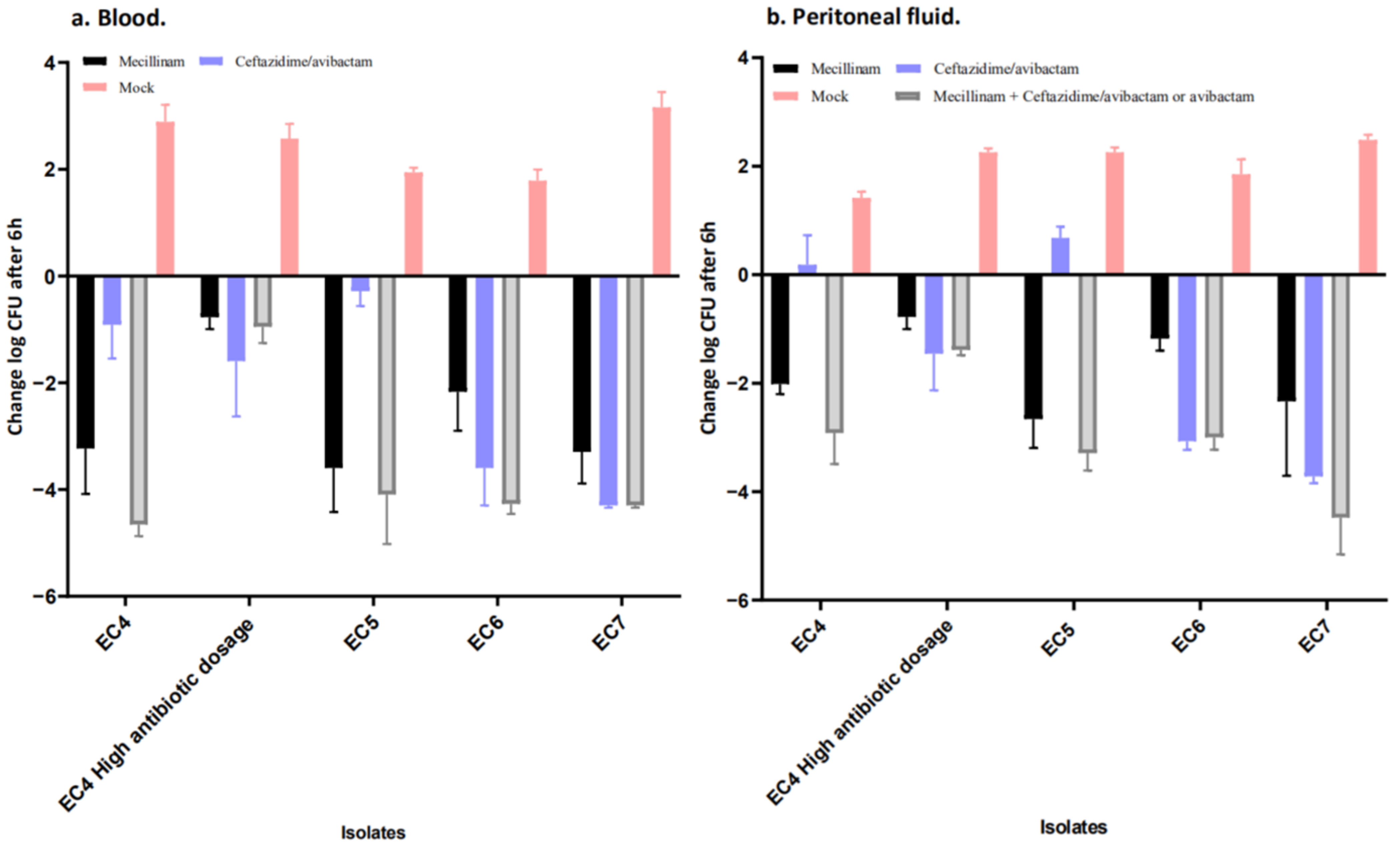
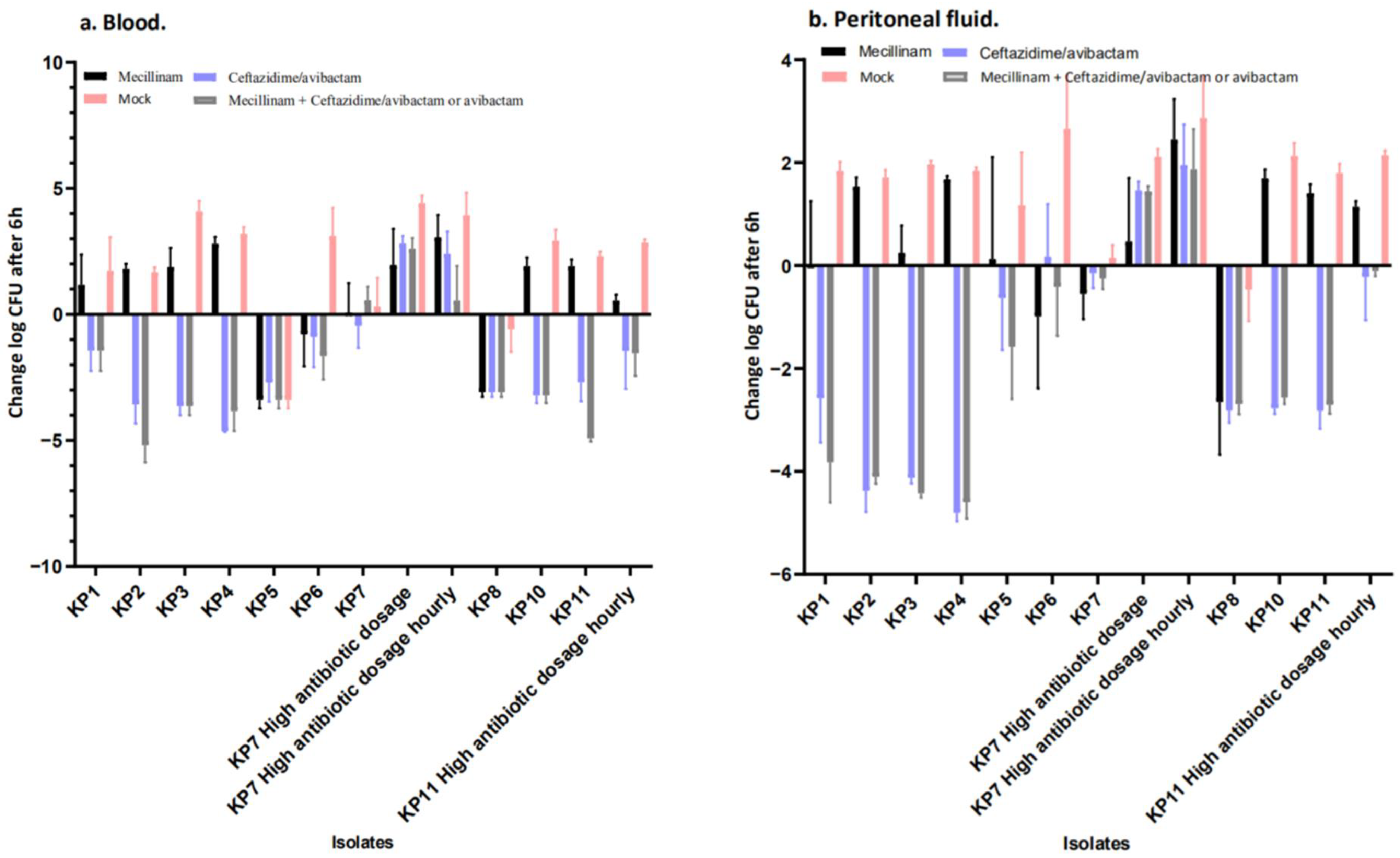
2.3. Mecillinam Combination Treatment with Avibactam or Ceftazidime/Avibactam In Vivo

Figure 1 and Figure 2 compare the Log CFU/mL counts in peritoneal fluids and blood at the end of the antibiotic treatment to mock treated controls at the same time point (i.e., at 5 h). The mock group depicted the increase in bacterial burden in saline-treated animals during the treatment period. Thus, the figures compared all Log CFU/mL counts at the end of the treatment period with animals sacrificed at 0 h, i.e., immediately before antibiotic treatment was initiated. The reductions in Log CFU/mL counts caused by the antibiotic treatment appeared smaller, as the difference was calculated relative to controls at baseline, instead of comparing to 5 h mock-treated controls. The efficacy of mecillinam, ceftazidime and mecillinam + ceftazidime/avibactam was evaluated as the reduction in CFU/mL in peritoneal fluid and blood collected 5 h after the initiation of the antibiotic treatment compared to Log CFU/mL counts obtained from control mice at the time of the initiation of therapy.
A reduction in Log CFU/mL was observed for the tested E. coli regardless of antibiotic treatment. Yet, ceftazidime/avibactam monotherapy failed to produce a log reduction in the peritoneal fluid for EC4 (NDM-5 and OXA-181) and EC5 (NDM7) (Figure 2a,b).
For all K. pneumoniae isolates with a KPC (KP1, KP2, KP3, KP4 and KP5) or an OXA (KP10 and KP11), ceftazidime/avibactam monotherapy was sufficient to produce a reduction in Log CFU/mL, while mecillinam failed to reduce Log CFU/mL. Of note, for KP5, there was a reduction in Log CFU/mL for all groups, including the mock-treated mice, indicating that the infection was not successful. For the K. pneumoniae isolate KP6 carrying the NDM carbapenemase, there was a reduction in the mean Log CFU/mL for all treatment groups, while Log CFU/mL reductions for KP7 (NDM-1 and OXA-48) were lower for mecillinam + ceftazidime/avibactam than for the other groups (Figure 3). As seen in “Supplementary data from in vivo studies_log_change”, the KP7 combinational treatment with 200 mg/kg mecillinam combined with 100 mg/kg ceftazidime/avibactam in the mouse peritonitis/sepsis model led to a 3.4-log reduction (p = 0.001) in blood samples and 1.0-log reduction in peritoneal samples. However, this was seen for the treatment regimen containing the high doses and hourly administration only. Of note, for KP8 (NDM-5), the situation was like that mentioned above for KP5.

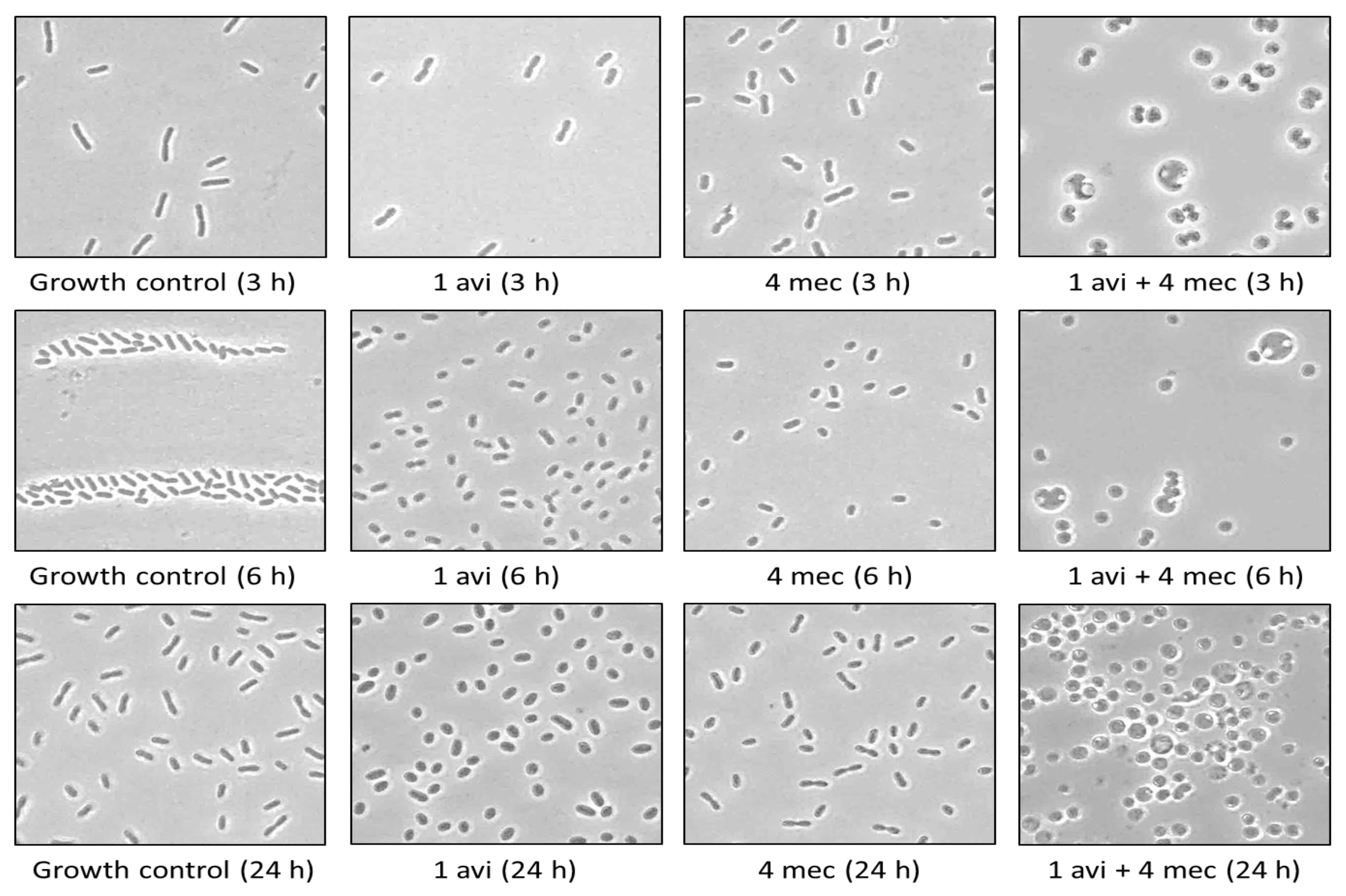
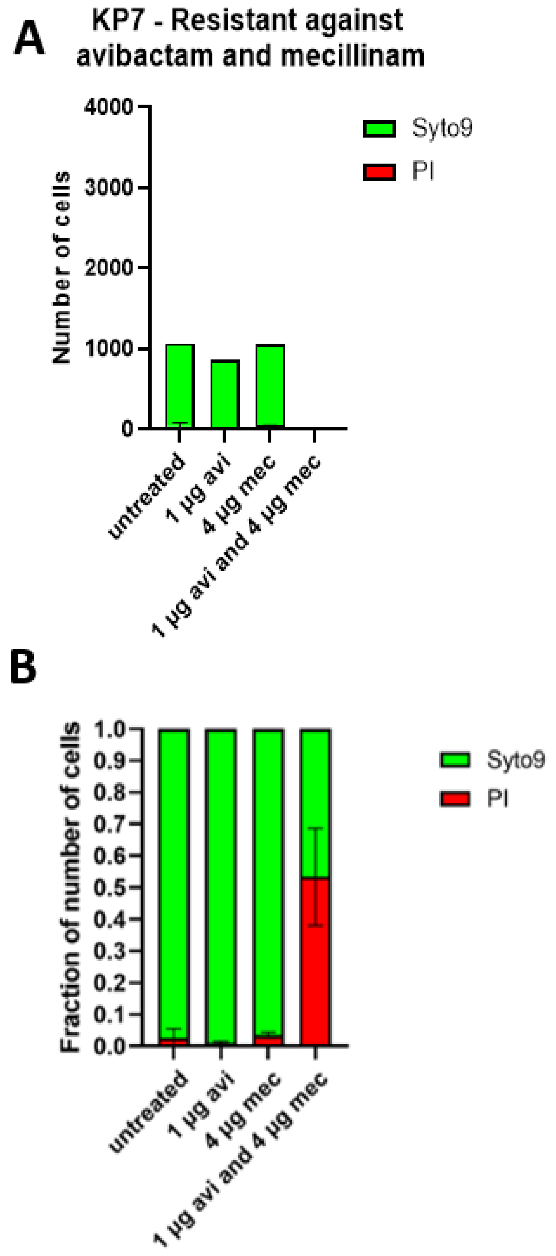
2.4. The Cell Shapes of KP7 and KP11 Are Strongly Affected by the Mecillinam and Avibactam Combination In Vitro and Ex Vivo

In vitro, bacterial suspensions were treated with increasing mecillinam and avibactam concentrations (four-fold serial dilution, 0.25–64 mg/L). At time points 3 h, 6 h and 24 h, the cell morphology was examined with phase-contrast microscopy. In addition, the combination treatment with mecillinam and avibactam was investigated, including 1 mg/L avibactam + 4 mg/L mecillinam, 4 mg/L avibactam + 16 mg/L mecillinam and 16 mg/L avibactam + 64 mg/L mecillinam. In this experiment, spherical forms of the K. pneumoniae strain KP7 were observed. The cell shape was more affected when avibactam and mecillinam were combined.
For KP7, high concentrations (16 mg/L and 64 mg/L) of mecillinam led to changes in cell morphology. Lower concentrations of avibactam (≥1 mg/L) were required to affect the cell shape compared to mecillinam. Interestingly, cells treated with ≥1 mg/L avibactam combined with ≥4 mg/L mecillinam transformed into spherical forms after only 3 or 6 h. However, cells treated with the corresponding concentrations of mecillinam or avibactam alone did not change after 24 h (Figure 3, Supplementary Figure S2). Peritoneal fluid samples from untreated and treated mice (KP7 and KP11) were also examined ex vivo with phase-contrast microscopy. Cells also changed to spherical forms (Supplementary Figure S3).
KP7 cultures treated with mecillinam and/or avibactam were stained with live–dead staining and imaged using confocal laser scanning microscopy (Figure 4, Supplementary Materials Figure S4). A green fluorescent color (syto9) stained all cells, whereas a red stain (propidium iodide) stained dead or dying cells only. The multi-resistant KP7 stain was not affected at all by the mono-treatment of either mecillinam or avibactam. Still, we saw a large decline in the surviving fraction when treated with the combination treatment (Figure 4, Supplementary Figure S4).

2.5. Combination of Mecillinam and Avibactam Does Not Alter Their Specific Interaction with Penicillin-Binding Protein In Vitro

To investigate whether the synergy observed could be explained by an alteration of the binding affinity of mecillinam and avibactam to PBPs when the two drugs were combined, we measured the binding of a fluorescent penicillin (Bocillin FL) on PBPs extracted from an E. coli laboratory strain (MG1655) in the presence of avibactam and/or mecillinam. The results showed that avibactam did not affect the mecillinam PBP-binding affinity (Supplementary Figure S5).

3. Discussion

In this study, we found a synergistic effect of the combination of ceftazidime/avibactam or avibactam alone with mecillinam against most MDR carbapenemase-producing E. coli and K. pneumoniae. In most cases, the presence of 1–4 µg of mecillinam reduced the MIC of ceftazidime/avibactam from >256 mg/L to ≤0.016 mg/L, and the presence of 1–4 mg/L of avibactam reduced mecillinam MICs from >256 mg/L to 0.25–1 mg/L. An in vitro and in vivo kill effect of both the individual antibiotics and the combination treatment was observed. We found morphological changes in the cells receiving mecillinam and avibactam, confirming the activity of the two compounds, based on binding to PBP2 in both organisms.
It is known that avibactam inhibits class A carbapenemases (e.g., KPCs) and some class D carbapenemases such as OXA-48. Avibactam is not active against NDMs [9]; however, NDM-producing E. coli strains have shown high susceptibility (96.5%) towards mecillinam [5,7]. A study by Marrs et al. (2014) showed lower sensitivity against mecillinam among NDM-1-producing K. pneumoniae strains (50%) [5]. In correlation with these findings, our study indicated that mecillinam was active against many NDM or OXA-producing strains, while ceftazidime/avibactam was active against OXA or KPC strains. The K. pneumoniae strain KP7 (NDM-1 and OXA-48) was resistant to mecillinam, ceftazidime/avibactam and ceftazidime alone (>256 mg/L), but mecillinam in combination with ceftazidime/avibactam or avibactam decreased the MIC to <0.016 and 1 mg/L. However, the mechanism behind this effect is unclear.
Previous studies reported that mecillinam was stable against carbapenemase-producing Enterobacterales that produce OXA-48, OXA-48-like, IMI and NDM-1 carbapenemases [5,6,8]. Yet, few of the studies reported which other β-lactamases were present in the isolates. Decreased susceptibility to β-lactams, e.g., piperacillin/tazobactam and mecillinam, could be caused by the hyperproduction of β-lactamases not considered to be ESBL, such as TEM-1 or OXA-1. Avibactam’s inhibition of other β-lactamases, while mecillinam was stable towards NDM, could be the mechanism behind the altered susceptibility we reported, as seen with aztreonam + ceftazidime/avibactam [10]. Similarly, a significant reduction in mecillinam MICs and reversion in the inoculum effect were seen in the presence of clavulanic acid [11].
The killing effect of the mecillinam/avibactam combination treatment seemed to be concentration or concentration–time-dependent. A significant reduction in colony counts in blood appeared in the KP7-infected mice treated with the combination treatment given every hour. The bacterial morphology examinations also illustrated this concentration dependency. Increasing levels of mecillinam and avibactam led to morphological changes and increased cell size of KP7.
Previous studies showed a strong binding of mecillinam and avibactam to E. coli and K. pneumoniae PBP2 [12,13,14]. We also found that mecillinam and avibactam had a high affinity for PBP2 (IC50: 0.3 and 0.2 mg/L, respectively). However, our Bocillin FL assay did not show if the synergistic effect between mecillinam and avibactam was due to a changed binding affinity of mecillinam and avibactam to E. coli PBPs. Of note, Erlinda et al. found that avibactam could interact with PBP2, which led to bactericidal interactions with human cathelicidin antimicrobial peptide LL-37, an antibacterial peptide part of innate immunity [12,15]. The observation of no visible decrease in mecillinam IC50 for any of the PBPs when mecillinam was combined with an avibactam concentration almost seven-fold higher than the avibactam IC50 for PBP2 could indicate that the mechanism behind the synergistic effect of the combination treatment, in vitro and in vivo, was that both mecillinam and avibactam could bind to PBP2, while avibactam could partly inhibit non-NDM β-lactamases. Further studies are needed to clarify why the Bocillin FL-binding did not change when avibactam, at a concentration seven-fold higher than IC50, was combined with β-lactams. Notably, currently, there is insufficient evidence for the use of mecillinam for the management of systemic infections with MDR carbapenemase-producing E. coli and K. pneumoniae.
Considering the limitations of our study, it should be noted that accurate mecillinam MIC determination by using gradient tests can be challenging, as previously shown by Fuchs et al. [6]. Further, the gradient strips used a fixed concentration of avibactam, so the result of the MIC with ceftazidime might have been more influenced by the avibactam concentration.
In conclusion, the use of the combination of mecillinam and avibactam must be investigated further. Yet, there is obvious potential for the treatment of urinary tract infections.

4. Materials and Methods

4.1. Bacterial Isolates

A collection of 18 varying phylogenetic clinical isolates of E. coli and K. pneumoniae, all phenotypically resistant to meropenem and carrying at least one carbapenemase gene, identified with whole-genome sequencing (WGS), was investigated (Supplementary Tables S1 and S3). E. coli MG1655, E. coli ATCC25922 and K. pneumoniae ATCC13883 were included as controls. All strains were stored at −80 °C until use.

4.2. Antibiotics

Mecillinam (Karo Pharma, Stockholm, Sweden), ceftazidime/avibactam (Pfizer, Kent, UK), avibactam (MedChemExpress, Monmouth Junction, NJ, USA) and ceftazidime (Fresenius Kabi, Singapore) were dissolved in MilliQ water. The fluorescent penicillin Bocillin FL (Invitrogen, Eugen, OR, USA) was used to label penicillin-binding proteins (PBPs) in the Bocillin FL assay. In addition, cefsulodin (Glentham Life Sciences Ltd., Corsham, UK), aztreonam (Sanofi-aventis, Paris, France) and cefoxitin (Novopharm Limited, Toronto, ON, Canada) were used as control antibiotics in the Bocillin FL assay.

4.3. Broth and Growth Conditions

We chose the combination of mecillinam and ceftazidime/avibactam based on an initial evaluation of multiple antibiotic combinations, tested in the chequerboard assay and performed in Mueller–Hinton broth [16]. For the susceptibility test using gradient test screening, each isolate was suspended in 0.9% NaCl until reaching a density of 0.5 McFarland. Gradient test strip studies were performed on Mueller-Hinton II (MHII) agar plates with and without antibiotics, which were produced by the University of Copenhagen, Panum. Plates were incubated at 35 °C for 16–20 h. The start inoculum of the time–kill assays and confocal laser scanning microscopy experiments was 105 colony-forming units CFU/mL, while a start inoculum of 107 CFU//mL was used for the phase-contrast microscopy experiments. All bacterial isolates were grown in 10 mL cation-adjusted MHII broth (17.5 g/L of acid hydrolysate of casein, 3 g/L of beef extract and 1.5 g/L starch) at 37 °C, 150 rpm. A growth control was included in all experiments. All test tubes of the time–kill assays were incubated for 6 h. Of note, we used 6 h incubation, since it was previously been found that mecillinam degradation can occur rapidly, with a half-life as short as 2 h in medium at 37 °C and pH 7.4 [17]. For CFU counts, bacterial suspensions were plated onto solid Gram-negative selective lactose agar plates (bromothymol agar plates based on modified Conradi-Drigalski medium containing 10 g/L detergent, 1 g/L Na2S2O3 H2O, 0.1 g/L bromothymol blue, 9 g/L lactose and 0.4 g/L glucose, pH 8.0, SSI Diagnostica). Colonies were counted after 16–20 h incubation at 37 °C.

4.4. Whole-Genome Sequencing

Genomic DNA was extracted using the DNeasy Blood and Tissue Kit (Qiagen, Hilden, Germany), followed by library preparation with the Nextera XT DNA Library Preparation Kit (Illumina, San Diego, CA, USA) and short-read whole-genome sequencing using 251 bp paired-end MiSeq sequencing (Illumina), according to the manufacturer’s instructions. Raw data were assembled into draft genomes using Skesa v2.2 [18]. ENA Accession numbers for raw Illumina reads for the study isolates can be found in the Supplementary Materials.

4.5. MIC Determination

Minimal inhibitory concentrations (MICs) of mecillinam, ceftazidime/avibactam and ceftazidime were determined by performing a gradient test. Ceftazidime/avibactam and ceftazidime Etest strips were obtained from BioMérieux, Ballerup, Denmark. Mecillinam MIC test strips were from Liofilchem (Roseto degli Abruzzi, Italy).
Bacterial susceptibility to the combinations mecillinam + ceftazidime/avibactam, mecillinam + ceftazidime and mecillinam + avibactam was tested as follows: ceftazidime/avibactam and ceftazidime strips were placed on MHII agar plates containing 0.5, 1 or 2 mg/L mecillinam, while mecillinam strips were placed on MHII agar plates with 1 or 4 mg/L avibactam and 4 or 16 mg/L ceftazidime/avibactam. Breakpoints for Enterobacterales were set for mecillinam (S ≤ 8 mg/L, R > 8 mg/L), ceftazidime (S ≤ 1 mg/L, R > 4 mg/L) and ceftazidime/avibactam (S ≤ 8 mg/L, R > 8 mg/L) (EUCAST Clinical Breakpoint Tables v. 12.0).

4.6. Binding of Mecillinam and Avibactam to Penicillin-Binding Proteins (PBPs) Measured with Bocillin FL Assay

The binding of mecillinam and avibactam to PBPs was examined through a competition assay with the fluorescent penicillin Bocillin FL. For details on the purification of E. coli MG1655 membrane proteins, see the Supplementary Materials. In total, 30 µg of E. coli MG1655 membrane protein was mixed with 2-fold dilutions (0.0625–32 mg/L) of mecillinam and avibactam and incubated at 35 °C for 30 min. Reaction mixtures containing 3 mg/L cefsulodin, 2 mg/L aztreonam and 2 mg/L cefoxitin were prepared to localize PBP1, PBP3, PBP4, PBP5 and PBP6. An assay containing reaction mixtures with a fixed avibactam concentration of 2 mg/L and 2-fold dilutions (0.0625–32 mg/L) of mecillinam was used. After incubation with antibiotics, Bocillin FL was added to reaction mixtures for further incubation at 35 °C for 40 min. To denature proteins, an SDS-PAGE-loading buffer with β-mercaptoethanol was added to each mixture and incubated at 95 °C for 5 min before the SDS-PAGE loading. An SDS-PAGE analysis was performed using 4–12% NuPAGE Bis-Tris Midi Gels (Thermo Fischer Scientific, Waltham, MA, USA). After complete protein separation, the gel was rinsed in water three times and for 30 min in a fixing solution containing 45% methanol and 7% acetic acid. The gel was scanned using a Typhoon 7000 scanner with excitation of 473 nm and emission of 520 nm. The ImageJ software was used to analyze the Bocillin FL signal of the gel, leading to the determination of integrated densities and percent values. The background signal of the gel image was subtracted before the band analysis. Relative Bocillin FL binding was determined by dividing each PBP percent value with the standard percent value of the reaction mixture without antibiotics, which indicated 100% Bocillin FL binding.

4.7. Phase-Contrast Microscopy

A Nikon Eclipse Ti was used for all phase-contrast microscopy examinations. Samples were spotted onto 1.5% agarose-MHII pads before imaging. Nikon’s imaging software, NIS-Elements Advanced Research Version 4.40, was used to process phase-contrast microscope images.

4.8. Confocal Laser Scanning Microscopy

Bacterial cells were examined by using confocal laser scanning microscopy. Samples were diluted 1:10 in saline containing 3 mM Syto9 and 0.3 mM of propidium iodide (PI) (live/dead staining) (Thermo Fisher Scientific, USA). Samples were incubated for 15 min in the absence of visible light, before 30 µL was applied to the wells of a VI µ-slide microscopy slide (Ibidi, Gräfelfing, Germany). Samples were imaged using a 488 nm laser for excitation, a 495–550 nm emission filter for Syto9, a 561 nm laser and a 595–650 nm emission filter for Pl. All samples were evaluated in three random positions within the wells. The images were processed in Imaris 9.2 (Bitplane, Zürich, Switzerland) for qualitative 3D projections and biomass quantitation via the MeasuremePro addon in Imaris. Syto9-stained biomass and PI-stained biomass were quantified in parallel to give a total biomass and indicate the viability of the biomass.

4.9. Single and Combination Antibiotic Treatment In Vivo in the Mouse Peritonitis/Sepsis Model

All in vivo experiments were conducted according to the permission of the Danish Animal Ethical Council under license 2017–15-0201–01274. We performed in vivo mouse experiments for all isolates where in vitro susceptibility testing indicated an in vivo effect (EC4, EC5, EC6, EC7, KP1, KP2, KP3, KP4, KP5, KP6, KP7, KP8, KP10 and KP11). Each experiment included 36 outbred female NMRI mice (seven weeks old, weighing 26–30 g) (Taconic, Hürth, Germany) divided into five groups: four mice in a control group for determining the baseline infection load at the start of treatment, eight mice per treatment group and eight mice treated with saline (0.9% NaCl) as the control. At t = 0, mice were inoculated intraperitoneally with 0.5 mL of bacterial suspension containing 107 CFU/mL and 5% (w/v) porcine mucin (Sigma-Aldrich, St. Louis, MO, USA). Blood and peritoneal fluid were sampled from four mice from the control group after one hour for determining the level of infection before treatment. Blood from the submandibular vein was collected. After euthanizing the mice through cervical dislocation, a peritoneal wash was performed by injecting 2 mL of sterile saline, i.e., performing a gentle massage of the abdomen for 1 min and then opening the abdomen aseptically for the sampling of the peritoneal fluid with a pipette. During the study, at t = 1 h, t = 3 h and t = 5 h, the mice were observed, and their clinical scores were noted according to the grades that can be found in the Supplementary data. The remaining mice in the treatment groups were injected (s.c. in the thigh) with 0.2 mL of antibiotic X, antibiotic Y, X and Y or saline. The doses used for ceftazidime/avibactam and mecillinam would result in serum drug concentrations in mice similar to concentrations in humans on standard doses [19,20]. The ENA accession numbers for raw Illumina reads for the study isolates and source of isolates can be found in the Supplementary Materials (Table S3).
  • Setup 1: 100 mg kg−1 ceftazidime/avibactam alone, 50 mg kg−1 mecillinam alone, 100 mg kg−1 ceftazidime/avibactam and 50 mg kg−1 mecillinam together or saline [19,20].
  • Setup 2: 200 mg kg−1 ceftazidime/avibactam alone, 200 mg kg−1 mecillinam alone, 200 mg kg−1 ceftazidime/avibactam and 200 mg kg−1 mecillinam together or saline [19,20].
  • Setup 3: 200 mg kg−1 avibactam alone, 100 mg kg−1 mecillinam alone, 200 mg kg−1 avibactam and 100 mg kg−1 mecillinam together or saline.
Mice treated with two antibiotics were each injected into separate thighs. At t = 3 h, after 2 h of treatment, four mice from each group were removed, and blood and peritoneal fluid samples were taken as described above. For tests in mice involving antibiotics with a low half-life (<1 h), these antibiotics were boosted with a second injection at t = 3 h (setup 1 and 2) or at t = 2 h, t = 3 h and t = 4 h (setup 3). At t = 5 h, the last four mice in each group were removed, and samples were taken as described above. All samples were stored at 4 °C and were processed within 1 h of sampling. Samples were 10-fold serially diluted in saline before being plated onto solid lactose agar plates (‘blue plates’) for CFU counts. Besides CFU, data were presented as log reductions as per the untreated saline group.

4.10. Statistical Methods

For comparisons of colony counts in vitro or in vivo, we used nonparametric statistics, either Fisher´s exact test or the Kruskal–Wallis test. For the determination of IC50 for penicillin-binding protein binding, we used a four-parameter dose–response model in the GraphPad Prism Version 9 software. p < 0.05 was considered significant.

5. Conclusions

In conclusion, the combination of ceftazidime/avibactam with mecillinam decreased Log CFU/mL in peritoneum against all 18 strains, irrespective of all narrow and broad-spectrum β-lactamases present in the pathogens. Therefore, the combination of mecillinam–ceftazidime/avibactam or mecillinam–avibactam combination treatments could be a new efficient empiric antibiotic treatment to fight infections caused by MDR carbapenemase-producing Gram-negative pathogens. The mechanism behind the observed synergistic effect between mecillinam and avibactam should be investigated further.

Supplementary Materials

The following supporting information can be downloaded at: https://www.mdpi.com/article/10.3390/antibiotics11101280/s1: Material and Methods Section on the purification of E. coli MG1655 membrane proteins and Supplementary Information on animal experiments. Figure S1: time–kill curves of five different K. pneumoniae isolates, KP2, KP5, KP6, KP7, KP8 and K. pneumoniae ATCC 13883, Figure S2: phase-contrast microscopy (100× magnification) of KP7 cells in four treatment groups, Figure S3: phase-contrast microscopy (100× magnification) of KP7 and KP11 cells ex vivo, Figure S4: three-dimensional projection of confocal laser scanning microscopy of cultures, Figure S5: fluorescence polarization (mP) of free Bocillin-FL at various concentrations of avibactam and mecillinam, Table S1: β-lactamase genes present in the 18 clinical isolates of Escherichia coli and Klebsiella pneumoniae, Table S2: mean log change after time–kill experiments for five different K. pneumoniae isolates, KP2, KP5, KP6, KP7, KP8 and K. pneumoniae ATCC 13883, Table S3: ENA accession numbers for raw Illumina reads for the study isolates and source of isolates can be found in the Supplementary Materials.

Author Contributions

K.K.L. was the primary investigator. Concept and design: F.B.H. and N.F.-M. Acquisition of data: K.K.L., M.K., K.N.K., G.C., S.R. and F.H. Analysis or interpretation of data: all authors. Drafting of the manuscript: K.K.L. Critical revision of the manuscript for important intellectual content: M.K., K.N.K., G.C., S.R., A.L.-O., F.H. and N.F.-M. Statistical analysis: K.K.L., M.K. and K.N.K. Supervision: N.F.-M., F.B.H., A.L.-O. and M.K. All authors take responsibility for the integrity of the data and the accuracy analysis and interpretation of data, and have approved the final version of the manuscript. All authors have read and agreed to the published version of the manuscript.

Funding

The study was supported by a grant from the Novo-Nordisk Foundation (reference number NNF16OC0023482) and from The National Danish Innovationsfonden, as part of the JPI AMR CoFund 2016. Role of funders: All authors are employed by public hospitals in the Capital Region, Denmark, which are public institutions. Foundations had no role in the design and conducting of the study, the analysis and interpretation of the data or the writing, review and approval of the manuscript.

Institutional Review Board Statement

The animal study protocol was approved by the Danish Animal Experimentation Inspectorate (license no. 2017-15-0201-01274) and performed according to institutional guidelines.

Informed Consent Statement

Not applicable.

Data Availability Statement

Data are presented in full in the manuscript or in the Supplementary data. Raw Illumina reads are available at the European Nucleotide Archive.

Acknowledgments

Kasper Thystrup Karstensen is thanked for uploading raw Illumina reads to the European Nucleotide Archive.

Conflicts of Interest

All authors declare no conflict of interest.

Transparency Declaration

All authors affirm that the conducted manuscript is honest, accurate and transparent.

Previously Presented

Part of data was presented as an oral presentation (flash oral of 4 min) at ECCMID 2022.

References

  1. Kaur, J.; Dhama, A.S.; Buttolia, H.; Kaur, J.; Walia, K.; Ohri, V.; Kumar, V.; Lynn, A.M.; Srivastava, A.; Singh, H. ICMR’s Antimicrobial Resistance Surveillance system (i -AMRSS): A promising tool for global antimicrobial resistance surveillance. JAC-Antimicrob. Resist. 2021, 3, dlab023. [Google Scholar] [CrossRef] [PubMed]
  2. Cassini, A.; Högberg, L.D.; Plachouras, D.; Quattrocchi, A.; Hoxha, A.; Simonsen, G.S.; Colomb-Cotinat, M.; Kretzschmar, M.E.; Devleesschauwer, B.; Cecchini, M.; et al. Attributable deaths and disability-adjusted life-years caused by infections with antibiotic-resistant bacteria in the EU and the European Economic Area in 2015: A population-level modelling analysis. Lancet Infect Dis. 2019, 19, 56–66. [Google Scholar] [CrossRef]
  3. Tacconelli, E.; Carrara, E.; Savoldi, A.; Harbarth, S.; Mendelson, M.; Monnet, D.L.; Pulcini, C.; Kahlmeter, G.; Kluytmans, J.; Carmeli, Y.; et al. Discovery, research, and development of new antibiotics: The WHO priority list of antibiotic-resistant bacteria and tuberculosis. Lancet Infect. Dis. 2018, 18, 318–327. [Google Scholar] [CrossRef]
  4. Cantón, R.; Akóva, M.; Carmeli, Y.; Giske, C.G.; Glupczynski, Y.; Gniadkowski, M.; Livermore, D.M.; Miriagou, V.; Naas, T.; Rossolini, G.M.; et al. Rapid evolution and spread of carbapenemases among Enterobacteriaceae in Europe. Clin. Microbiol. Infect. 2012, 18, 413–431. [Google Scholar] [CrossRef] [PubMed]
  5. Marrs, E.C.L.; Day, K.M.; Perry, J.D. In vitro activity of mecillinam against Enterobacteriaceae with NDM-1 carbapenemase. J. Antimicrob. Chemother. 2014, 69, 2873–2875. [Google Scholar] [CrossRef] [PubMed]
  6. Fuchs, F.; Ahmadzada, A.; Plambeck, L.; Wille, T.; Hamprecht, A. Susceptibility of Clinical Enterobacterales Isolates with Common and Rare Carbapenemases to Mecillinam. Front. Microbiol. 2021, 11, 627267. [Google Scholar] [CrossRef] [PubMed]
  7. Zykov, I.N.; Frimodt-Møller, N.; Småbrekke, L.; Sundsfjord, A.; Samuelsen, Ø. Efficacy of mecillinam against clinical multidrug-resistant Escherichia coli in a murine urinary tract infection model. Int. J. Antimicrob. Agents 2020, 55, 105851. [Google Scholar] [CrossRef] [PubMed]
  8. Tsakris, A.; Koumaki, V.; Baka, S.; Balakrishnan, I. Activity of mecillinam against OXA-48-like carbapenemase-producing Enterobacterales. J. Antimicrob. Chemother. 2021, 77, 537–538. [Google Scholar] [CrossRef] [PubMed]
  9. Lagacé-wiens, P.; Walkty, A.; Karlowsky, J.A. Ceftazidime—Avibactam: An evidence-based review of its pharmacology and potential use in the treatment of Gram-negative bacterial infections. Core Evid. 2014, 9, 13–25. [Google Scholar] [CrossRef] [PubMed]
  10. Hansen, K.H.; Andreasen, M.R.; Pedersen, M.S.; Westh, H.; Jelsbak, L.; Schønning, K. Resistance to piperacillin/tazobactam in Escherichia coli resulting from extensive IS 26-associated gene amplification of bla TEM-1. J. Antimicrob. Chemother. 2019, 74, 3179–3183. [Google Scholar] [CrossRef] [PubMed]
  11. Lampri, N.; Galani, I.; Poulakou, G.; Katsarolis, I.; Petrikkos, G.; Giamarellou, H.; Souli, M. Mecillinam/clavulanate combination: A possible option for the treatment of community-acquired uncomplicated urinary tract infections caused by extended-spectrum β-lactamase-producing Escherichia coli. J. Antimicrob. Chemother. 2012, 67, 2424–2428. [Google Scholar] [CrossRef] [PubMed]
  12. Asli, A.; Brouillette, E.; Krause, K.M.; Nichols, W.W.; Malouin, F. Distinctive Binding of Avibactam to Penicillin-Binding Proteins of Gram-Negative and Gram-Positive Bacteria. Antimicrob. Agents Chemother. 2016, 60, 752–756. [Google Scholar] [CrossRef] [PubMed]
  13. Sutaria, D.S.; Moya, B.; Green, K.B.; Kim, T.H.; Tao, X.; Jiao, Y.; Louie, A.; Drusano, G.L.; Bulitta, J.B. First Penicillin-Binding Protein Occupancy Patterns of β- Lactams and β-Lactamase Inhibitors in Klebsiella pneumoniae. Antimicrob. Agents Chemother. 2018, 62, e00282-18. [Google Scholar] [CrossRef] [PubMed]
  14. Kocaoglu, O.; Carlson, E.E. Profiling of β-lactam selectivity for penicillin-binding proteins in Escherichia coli strain DC2. Antimicrob. Agents Chemother. 2015, 59, 2785–2790. [Google Scholar] [CrossRef] [PubMed]
  15. Ulloa, E.R.; Dillon, N.; Tsunemoto, H.; Pogliano, J.; Sakoulas, G.; Nizet, V. Avibactam Sensitizes Carbapenem-Resistant NDM-1–Producing Klebsiella pneumoniae to Innate Immune Clearance. J. Infect. Dis. 2019, 220, 484–493. [Google Scholar] [CrossRef] [PubMed]
  16. Kragh, K.N.; Gijón, D.; Maruri, A.; Antonelli, A.; Coppi, M.; Kolpen, M.; Crone, S.; Tellapragada, C.; Hasan, B.; Radmer, S.; et al. Effective antimicrobial combination in vivo treatment predicted with microcalorimetry screening. J. Antimicrob. Chemother. 2021, 76, 1001–1009. [Google Scholar] [CrossRef] [PubMed]
  17. Brouwers, R.; Vass, H.; Dawson, A.; Squires, T.; Tavaddod, S.; Allen, R.J. Stability of β-lactam antibiotics in bacterial growth media. PLoS ONE 2020, 15, e0236198. [Google Scholar] [CrossRef] [PubMed]
  18. Souvorov, A.; Agarwala, R.; Lipman, D.J. SKESA: Strategic k-mer extension for scrupulous assemblies. Genome Biol. 2018, 19, 153. [Google Scholar] [CrossRef] [PubMed]
  19. Berkhout, J.; Melchers, M.J.; van Mil, A.C.; Seyedmousavi, S.; Lagarde, C.M.; Schuck, V.J.; Nichols, W.W.; Mouton, J.W. Pharmacodynamics of ceftazidime and avibactam in neutropenic mice with thigh or lung infection. Antimicrob. Agents Chemother. 2016, 60, 368–375. [Google Scholar] [CrossRef] [PubMed]
  20. Kerrn, M.B.; Espersen, F. Effects of Sulfamethizole and Amdinocillin against Escherichia coli Strains (with Various Susceptibilities) in an Ascending Urinary Tract Infection Mouse Model. Antimicrob. Agents Chemother. 2003, 47, 1002–1009. [Google Scholar] [CrossRef] [PubMed] [Green Version]
Figure 1. (a,b). In vivo results of treatment regimens for the included E. coli isolates. The standard doses were 100 mg kg−1 ceftazidime/avibactam alone, 50 mg kg−1 mecillinam alone, 100 mg kg−1 ceftazidime/avibactam and 50 mg kg−1 mecillinam together or saline administered s.c. at t = 1 h and t = 3 h. For strain EC4, a higher dose was also used, i.e., 200 mg kg−1 ceftazidime/avibactam alone, 200 mg kg−1 mecillinam alone, 200 mg kg−1 ceftazidime/avibactam and 200 mg kg−1 mecillinam together or saline administered at t = 1 h, t = 2 h, t = 3 h and t = 4 h, indicated as “High antibiotic dosage”. Data are presented as changes in log CFU mL−1 in either blood or peritoneal fluid at t = 5 h. The figure compares CFU counts in peritoneal fluid and blood at the end of antibiotic treatment to mock-treated controls at the same time points. Thus, the figure compares all CFU counts at the end of the treatment period with animals sacrificed at 0 h, i.e., immediately before antibiotic treatment was initiated. The result of this new depiction is that reductions in CFU counts caused by antibiotic treatment appeared smaller as the difference was calculated relative to controls at baseline with lower counts instead of compared to 5 h mock-treated controls. The “mock” group depicted the increase in bacterial burden in mock-treated animals during the treatment period.
Antibiotics 11 01280 g001
Figure 2. (a,b). In vivo results of treatment regimens for the included K. pneumoniae isolates. The standard doses were 100 mg kg−1 ceftazidime/avibactam alone, 50 mg kg−1 mecillinam alone, 100 mg kg−1 ceftazidime/avibactam and 50 mg kg−1 mecillinam together or saline administered s.c. at t = 1 h and t = 3 h. For strain KP7, two higher dose regimens were used, i.e., 200 mg kg−1 ceftazidime/avibactam alone, 200 mg kg−1 mecillinam alone, 200 mg kg−1 ceftazidime/avibactam and 200 mg kg−1 mecillinam together or saline administered at t = 1 h and t = 3 h (marked in the figure with “high antibiotic dosage”), and the same higher dose administered hourly, i.e., t = 1 h, t = 2 h, t = 3 h and t = 4 h (marked in the figure with “high antibiotic dosage hourly”). This latter dose was also used for strain KP11. Data are presented as changes in log CFU mL−1 in either blood or peritoneal fluid t = 5 h. The figure compares CFU counts in peritoneal fluid and blood at the end of antibiotic treatment to mock-treated controls at the same time points. Thus, the figure compares all CFU counts at the end of the treatment period with animals sacrificed at 0 h, i.e., immediately before antibiotic treatment was initiated. The result of this new depiction is that reductions in CFU counts caused by antibiotic treatment appeared smaller as the difference was calculated relative to controls at baseline with lower counts instead of compared to 5 h mock-treated controls. The “mock” group depicted the increase in bacterial burden in mock-treated animals during the treatment period.
Antibiotics 11 01280 g002
Figure 3. Spherical structures of the K. pneumoniae strain KP7 observed in vitro. Phase-contrast microscopy (1000× magnification) of KP7 cells in four treatment groups, including growth control, 1 mg/L avibactam (1 AVI), 4 mg/L mecillinam (4 MEC) and 1 mg/L avibactam combined with 4 mg/L mecillinam (1 AVI + 4 MEC). Images are from time points 3, 6 and 24 h.
Antibiotics 11 01280 g003
Figure 4. Confocal laser scanning microscopy. Quantified data showed the average number of cells found in confocal laser scanning micrographs of cultures treated either with 4 µg mL−1 mecillinam, 1 µg mL−1 avibactam or untreated. (A) The number of cells of the syto9 (live) and the PI (dead)-stained populations was quantified using Imaris 9.5 with Measure Pro. KP7 cells were resistant against avibactam and mecillinam. (B) A fraction of live and dead cells is shown based on the total number of cells in each sample.
Antibiotics 11 01280 g004
Table 1. Susceptibility patterns of the included isolates as well as results of synergistic tests of mecillinam combined with ceftazidime/avibactam.
StrainsCarbapenemaseMIC Mecillinam (mg/L)MIC Ceftazidime/Avibactam (mg/L)MIC Ceftazidime/Avibactam (mg/L) Performed on Agar Containing MecillinamMIC Ceftazidime (mg/L)MIC Ceftazidime (mg/L) Performed on Agar Containing Mecillinam
Mecillinam: 0.5 mg/LMecillinam: 1 mg/LMecillinam: 2 mg/L Mecillinam: 2 mg/L
KP1KPC-21610.032<0.016<0.01688
KP2KPC-3>25640.064<0.016<0.016>256>256
KP3KPC-3321<0.016<0.016<0.016>256>256
KP4KPC-3>256220.064<0.016>256>256
KP5KPC-3>25620.016<0.016<0.016>256>256
KP6NDM-12>256>256<0.016<0.016>256>256
KP7NDM-1, OXA-48>256>256>256<0.016<0.016>256>256
KP8NDM-52>256>256>256<0.016>256>256
KP9NDM-7, OXA-1818>256>256>256>256>256>256
KP10OXA-2321610.25<0.016<0.0163232
KP11OXA-43640.50.032<0.016<0.0166432
KP12IMP-1>25632323232>256>256
ATCC 13883 >2560.250.0320.0160.0160.250.016
EC2NDM-11>256NGNGNG>256NG
EC3NDM-58>256>256>256>256>256>256
EC4NDM-5, OXA-1814>256>256>256<0.016>256>256
EC5NDM-74>256>256<0.016<0.016>256>256
EC6OXA-24420.5<0.016<0.016<0.01622
EC7OXA-4820.25<0.016<0.016<0.0160.250.25
ATCC 25922 0.0640.125NGNGNG0.125NG
EC: Escherichia coli; KP: Klebsiella pneumoniae; KPC: Klebsiella pneumoniae carbapenemase; IMP: imipenem metallo-beta-lactamase; NDM: New Delhi metallo-beta-lactamase; OXA: oxacillinase; NG: no growth on MHII agar plate.
Table 2. Results of synergistic tests of mecillinam combined with ceftazidime/avibactam or avibactam. We showed the isolates chosen for further analyses based on results from Table 1.
StrainCarbapenemaseMIC Mecillinam (mg/L)MIC Ceftazidime/Avibactam (mg/L)Mecillinam MIC (mg/L) Performed on Agar Containing AvibactamMecillinam (mg/L) Performed on Agar Containing Ceftazidime/Avibactam
Avibactam: 1 mg/LAvibactam: 4 mg/LCeftazidime/Avibactam: 4 mg/LCeftazidime/Avibactam: 16 mg/L
KP2KPC-3>256410.251NG
KP5KPC-3>256210.252NG
KP6NDM-12>25610.511
KP7NDM-1, OXA-48>256>2562122
KP8NDM-52>25610.511
ATCC 13883 >2560.2588NGNG
EC4NDM-5, OXA-1814>25610.2521
EC5NDM-74>25610.511
ATCC 25922 0.0640.1250.0640.016NGNG
EC: Escherichia coli; KP: Klebsiella pneumoniae; KPC: Klebsiella pneumoniae carbapenemase; IMP: imipenem metallo-beta-lactamase; NDM: New Delhi metallo-beta-lactamase; OXA: oxacillinase; NG: no growth on MHII agar plate.
Publisher’s Note: MDPI stays neutral with regard to jurisdictional claims in published maps and institutional affiliations.

