Article

Evidence-Based Tracking of MDR E. coli from Bovine Endometritis and Its Elimination by Effective Novel Therapeutics

1
State Key Laboratory for Conservation and Utilization of Subtropical Agro-Bioresources, Guangxi University, Nanning 530005, China
2
Department of Medicine, Cholistan University of Veterinary and Animal Sciences, Bahawalpur 63100, Pakistan
3
Institute of Biochemistry and Biotechnology, University of Veterinary and Animal Sciences, Lahore 54000, Pakistan
4
Department of Clinical Medicine and Surgery, University of Agriculture, Faisalabad 38000, Pakistan
5
Department of Theriogenology, Cholistan University of Veterinary and Animal Sciences, Bahawalpur 63100, Pakistan
6
Central Diagnostic Laboratory, Cholistan University of Veterinary and Animal Sciences, Bahawalpur 63100, Pakistan
7
Department of Geography, Government College University, Lahore 54000, Pakistan
8
Department of Surgery, Cholistan University of Veterinary and Animal Sciences, Bahawalpur 63100, Pakistan
9
Department of Parasitology, Cholistan University of Veterinary and Animal Sciences, Bahawalpur 63100, Pakistan
*
Authors to whom correspondence should be addressed.
These authors contributed equally to this work.
Antibiotics 2021, 10(8), 997; https://doi.org/10.3390/antibiotics10080997
Submission received: 14 July 2021 / Revised: 7 August 2021 / Accepted: 13 August 2021 / Published: 18 August 2021
(This article belongs to the Special Issue Antimicrobial Resistance in Gram-Negative Bacteria, 2nd Edition)

Abstract

:
Antibiotic-resistant bacteria have become the predominant etiology of endometritis and thus require effective treatment approaches. We used ultrasonography coupled with clinical signs and presented complaints of reproductive issues to investigate the epidemiology, phylogenetic analysis, antimicrobial resistance, and development of novel therapeutics against Escherichia coli isolated from endometritis in bovine (n = 304 from 10 commercial dairy farms). The prevalence of bovine endometritis in this study was 43.75%, while among these, 72.18% samples were positive for E. coli. Nucleotide analysis performed through BLAST and MEGAX showed 98% similarity to the nucleotide sequence of the reference E. coli strain (accession number CP067311.1). The disk diffusion assay revealed pathogen resistance to most antibiotics. Pattern of MIC order of resistance was as follows: enrofloxacin < gentamicin < co-amoxiclav < streptomycin < amoxicillin < metronidazole < oxytetracycline. Field trials revealed the highest recovery rate (in terms of clearance of endometritis and establishment of pregnancy) in case of gentamicin + enrofloxacin (100%) and gentamicin alone (100%), followed by co-amoxiclav + gentamicin (84.61%), oxytetracycline alone (78.57%), and metronidazole + enrofloxacin (33.33%). Hence, the current study reported a higher prevalence of multidrug-resistant E. coli showing considerable similarity with reference strain, and finally, the effective response of novel antibiotics to treat cases.

Graphical Abstract

1. Introduction

Reproduction is the fundamental characteristic of all animals, which is essential for the survival of their species [1]. Reproductive diseases such as pyometra, metritis, endometritis, retained fetal membrane (RFM), and general uterine infections are dominant deteriorating challenges that affect the fertility of dairy animals [2]. Metritis and endometritis play a vital role in causing infertility, lower performance, early culling, and genetic loss in dairy cows [3]. Clinical endometritis is defined as the presence of purulent or mucopurulent vaginal discharge postpartum (3 weeks or later) or postnatal discharge. Subclinical endometritis is defined as the infection of the endometrium diagnosed by the presence of neutrophils in the biopsy and histological investigations in the absence of clinical signs of endometritis [4,5].
Bacterial infections and the mechanism of the local immune system of the uterus are important determinants in the prognosis of endometritis. Escherichia coli (E. coli) and Trueperella pyogenes have been reported to be the most common pathogens isolated from the bovine uterus [6,7,8]. E. coli (Gram-negative bacteria) is equally important for livestock and human health and results in a wider range of infections, while its resistance against antibiotics is still debatable [9,10]. The rapid development of resistance, harmful effects of antibiotics, and less availability of antibiotic options are salient issues [11] that are the outcome of four mechanisms of bacteria, i.e., inactivation of bacterial site, active efflux of bacteria, modification of target sites, and altered metabolic pathways by the bacteria [12].
The existence of broad-spectrum β-lactamase-producing E. coli in dairy animals has been reported in various studies [13]. Before the advent of the antibiotic era, postpartum ailment was an important reason for maternal death, while a sharp decrease in maternal morbidity was observed upon the use of antibiotics. An appropriate treatment will not only decrease maternal morbidity but may also improve antibiotic stewardship [14]. However, there are limited options of antibiotics to be used in combination [15] to combat the bacterial challenge. The rise in antimicrobial resistance owes to the continuous use of antibiotics that finally become ineffective due to genetic variations in the bacteria [16]. Thus, the study hypothesis states, “E. coli-based endometritis is prevalent along with multiple drug resistance, and newer antibiotic approaches are effective against to treat E. coli-based endometritis”. The hypothesis was tested with the objectives of investigation of epidemiology, phylogenetic analysis, antimicrobial resistance, and development of new therapeutic approaches against E. coli isolated from endometritis in bovine.

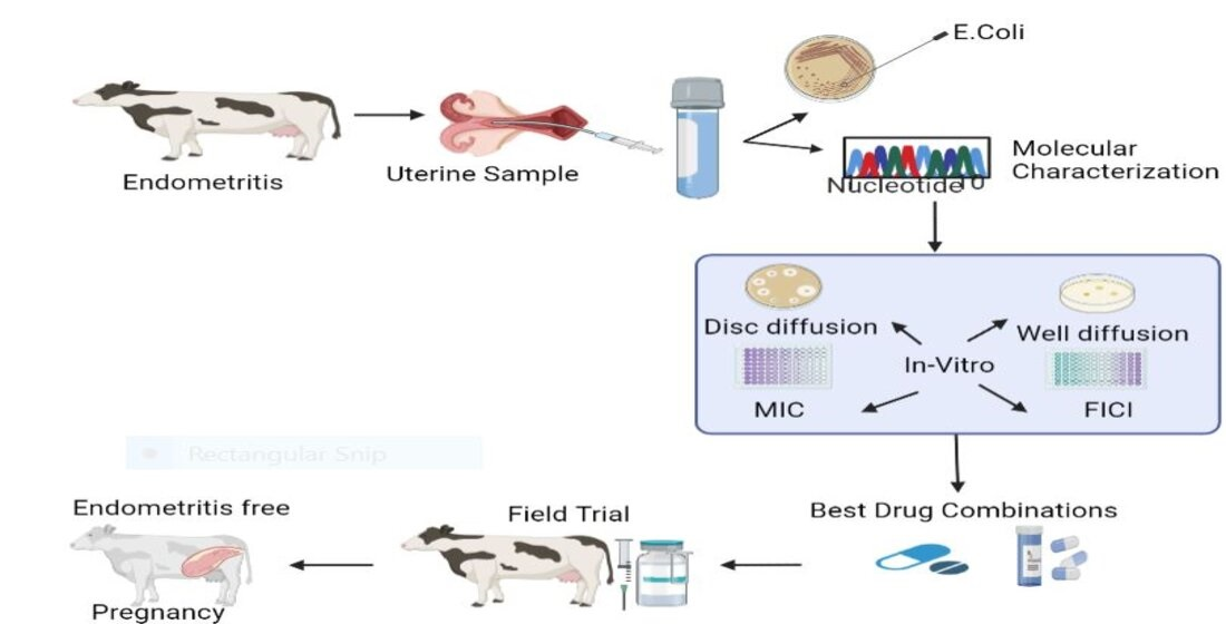
2. Materials and Methods

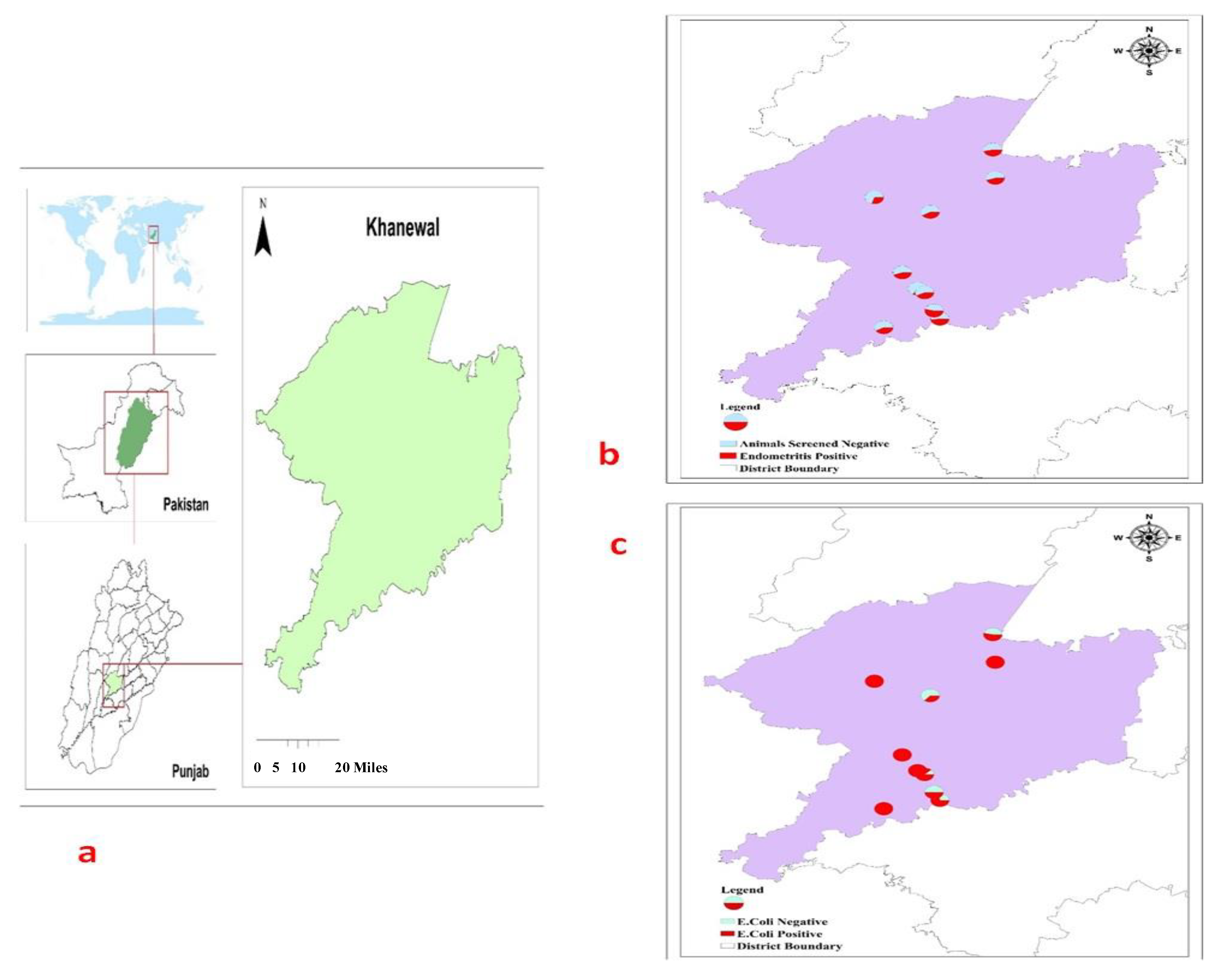
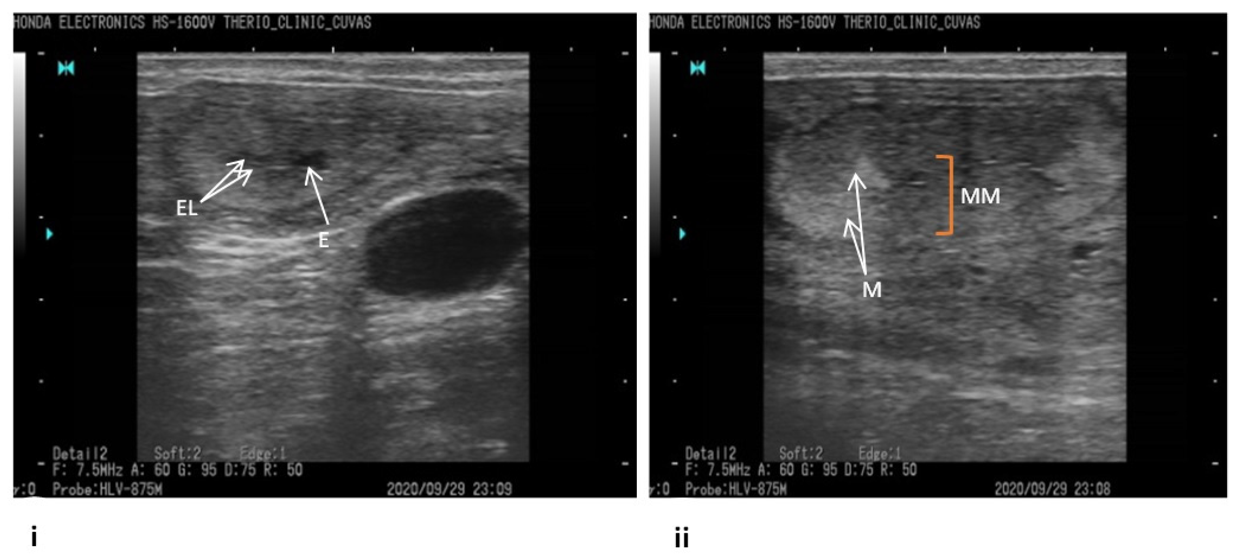
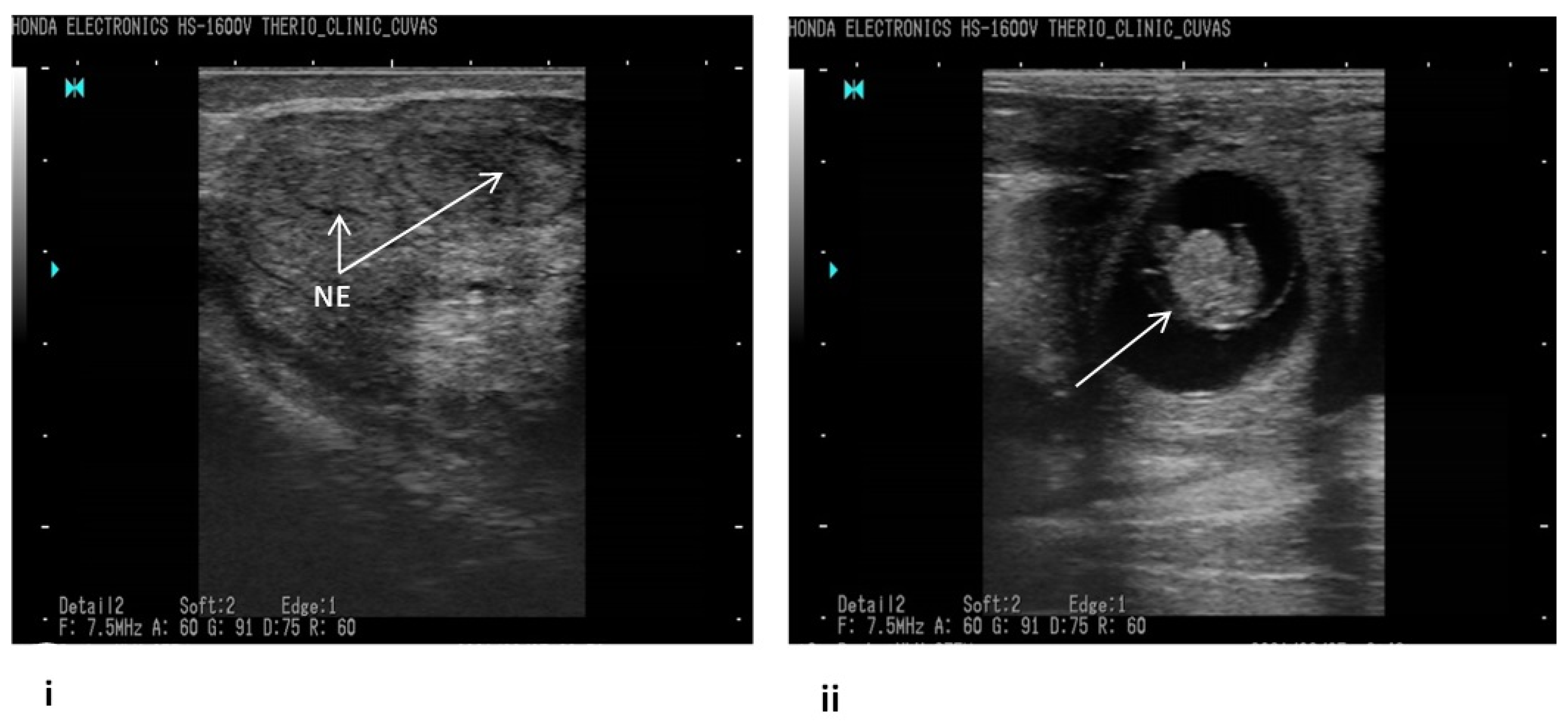
2.1. Tracking Bovine Endometritis

The accessible commercial dairy farms big enough to accommodate at least 50 active lactating dairy cows, endometritis identified through ultrasonography, and availability of professional veterinary supervision were the inclusion criteria for this study [17,18]. Keeping in view the set guidelines, commercial dairy farms (n = 10) from Khanewal district, Punjab, Pakistan, were selected for this study (Figure 1). Using a convenient sampling technique, n = 304 exotic cows (Holstein Frisien) from these farms were included in this study [19]. An ultrasound machine (B mode, 7.5 MHz linear array trans-rectal probe) was used to visualize the reproductive tract (Figure 2), and endometritis was characterized by distended lumen filled with partially echogenic snowy patches as recommended by Fissore et al. The fluid in the uterine environment is indicated black (non-echogenic) while the inflamed uterus, having pus cells, appears with distended lumen filled with whitish-grey areas (echogenic) as referred by Pearson et al. and Fissore et al. [20,21]. When some rays are passing through the structure, and many are coming back that appear hypoechoic (grey scale). If all rays are reflecting back from any structure, it appears hyperechoic (whitish), as referred by B Ihnatsenka et al. [22]. In the case of endometritis, pus is present inside the uterine lumen, due to which some ultrasound rays pass through it and some reflect back, and it appears as a mixture of hypo and hyperechoic images. Aseptically uterine flushing was done using an artificial insemination gun, and fluid was aspirated through a syringe and collected in a sterile sheath. The collected samples were shipped to Central Diagnostic Lab (CDL) of the Cholistan University of Veterinary Sciences, Bahawalpur, Pakistan, maintaining a cold chain at 4 °C.

2.2. Isolation and Confirmation of E. coli

The uterine samples were first incubated in nutrient broth for 24 h at 37 °C, while later, these samples were centrifuged at 6000× g for 10 min. The sediments were swabbed on blood agar and incubated at 37 °C for 24 h [23]. Harvested microbial colonies were streaked on MacConkey agar, and the resulting growth was subjected to a series of biochemical tests (gram staining, coagulase, and IMViC test), according to the guidelines of the Bergey’s Manual of Determinative Bacteriology [24].

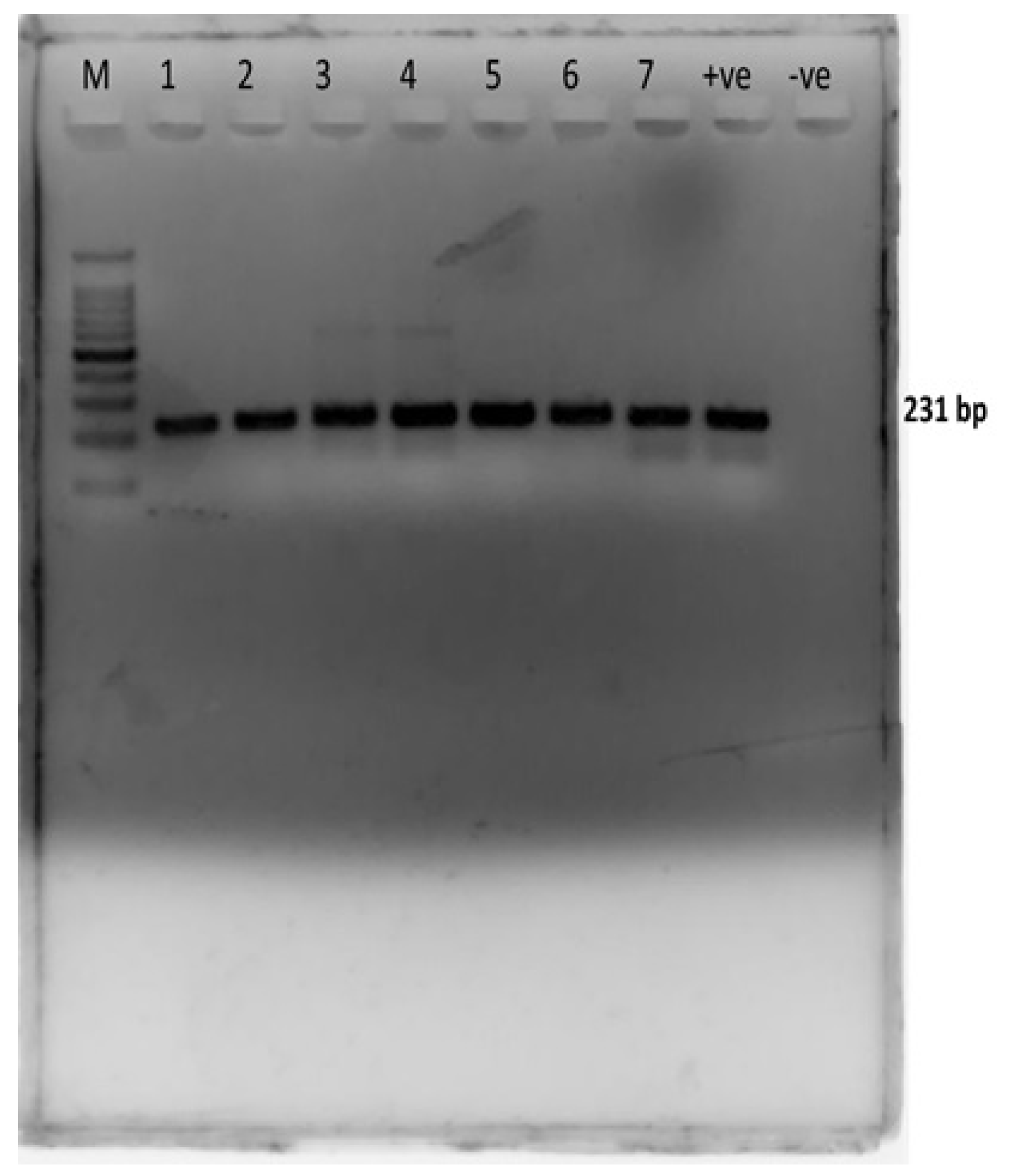
2.3. Molecular Confirmation of E. coli

For molecular confirmation of E. coli, PCR amplification was done using the E. coli 23S rRNA gene-specific primers (E23S-F: ATCAACCGAGATTCCCCCAGT; E23S-R: TCACTATCGGTCAGTCAGGAG), with amplification of 231 bp product (Figure 3). PCR conditions were as follows: denaturation at 95 °C for 3 min, followed by 35 cycles of 95 °C for 1 min, 60 °C for 1 min, and 72 °C for 1 min, and a final extension step at 72 °C for 7 min. The obtained PCR products were subjected to 2% agarose gel electrophoresis [25].

2.3.1. Sequencing of the Local E. coli Isolate

The sequence was submitted to NCBI (MZ344556). The sequencing results of the local E. coli isolate were analyzed using different bioinformatics tools. Nucleotide analysis of the nucleotide sequences was performed using BLAST to check the similarity with reference NCBI strain. The phylogenetic tree of nucleotide sequences was constructed using the MEGAX software by the neighbor-joining method [26]. Different (21) sequences were also involved in phylogenetic analysis of the nucleotide sequences of E. coli 23S ribosomal RNA gene. The tree was drawn to scale, with branch lengths in the same units as those of the evolutionary distances used to infer the phylogenetic tree. The evolutionary distances were computed using the maximum composite likelihood method and were in the units of the number of base substitutions per site.

2.3.2. Gene Structure and Motifs Elicitation

Additionally, conserved motifs of E. coli were explored using the MEME Suite (Multiple EM for Motif Elicitation) [27]. The 23S E. coli ribosomal RNA gene sequence isolated from bovine uterine samples from Pakistan was submitted in the MEME Suit for motif evaluation. Gene structure display server 2.0 was used for the structural analysis of the desired gene to visualize the exonic regions in the submitted data.

2.4. Assessment of the Antibiotic Resistance Profile of E. coli

Disc diffusion assay was used to access the antibiogram of previously confirmed isolates (n = 50) against commonly used antibiotics as per the Clinical and Laboratory Standards Institute (CLSI) guidelines [28]. The representative sample size of isolates was enrolled in such a way that n = 5 E. coli samples from each dairy farm (endometritis positive cows) were randomly selected, making a total of n = 50 for in vitro trials. The antibiotics tested included fusidic acid (10 µg), enrofloxacin (10 µg), ciprofloxacin (5 µg), trimethoprim-sulfamethoxazole (25 µg), Amoxicillin, chloramphenicol (30 µg), vancomycin (30 µg), gentamycin (10 µg), linezolid (30 µg), and cefoxitin (30 µg). Briefly, antibiotic discs were aseptically placed on pre-seeded cation-adjusted Mueller-Hinton agar plates. Following incubation for 24 h, the zone of inhibition around each antibiotic disc was compared with the standards provided by CLSI [28].

2.5. In Vitro Therapeutic Testing of Antibiotics

Commonly used antibiotics (alone/in combination) were tested against resistant E. coli isolates. In vitro testing was performed, initially using the well diffusion test and later using the broth microdilution method to determine synergistic combinations among a range of drugs, including co-amoxiclav, enrofloxacin, metronidazole, oxytetracycline, gentamicin, streptomycin, and amoxicillin. Well diffusion assay identified comparative activity of different drugs while minimum inhibitory concentration (MIC) was performed to authenticate their activity. MIC of individual drugs and drugs in combination was dealt with in the synergy testing section to find the best-suited combination for the field trial.

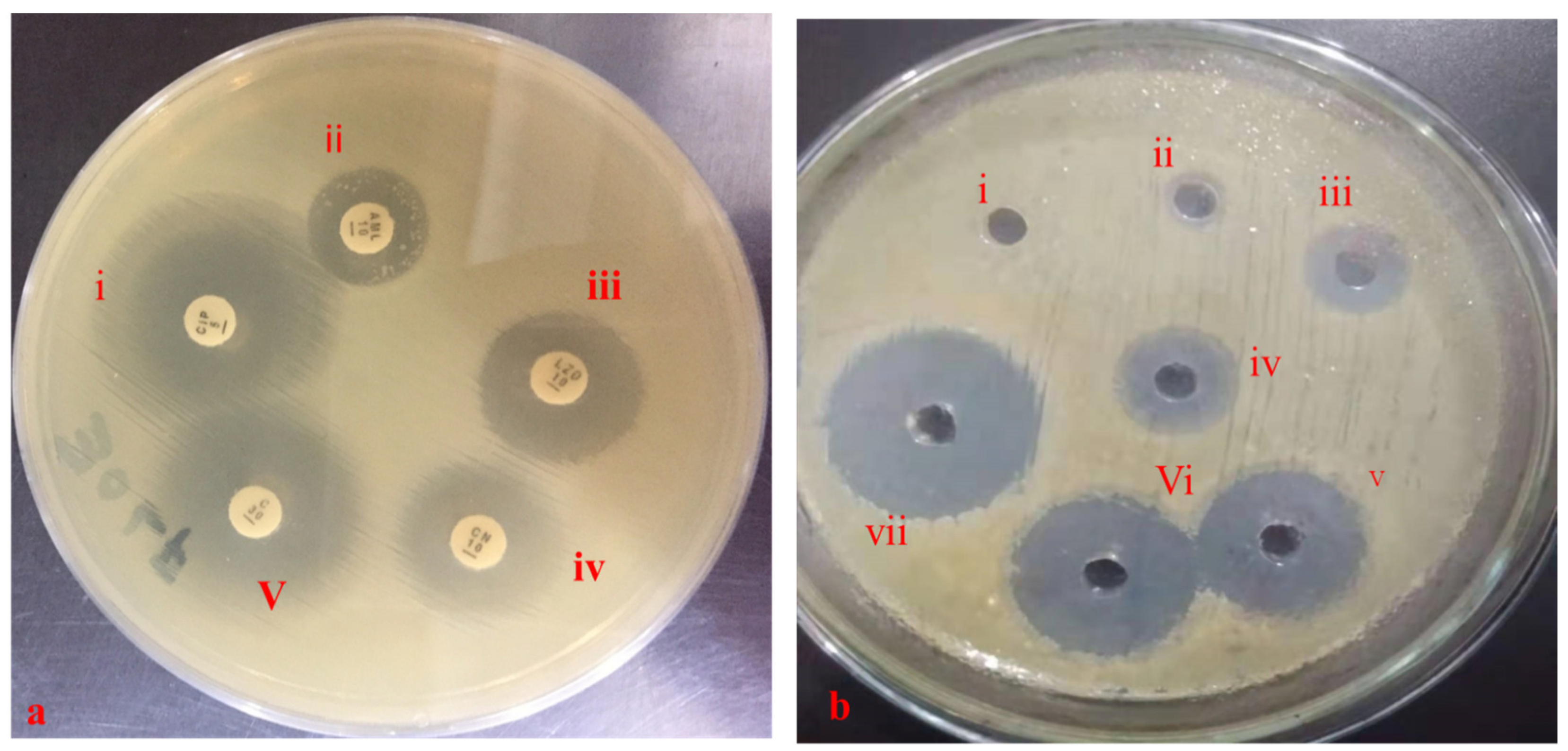
2.5.1. Well Diffusion Assay

Wells of 6–8 mm in diameter were dug on pre-seeded Mueller-Hinton agar plates using a well borer. Antibiotics to be tested (alone or in combination) were poured into the wells to assess their antibacterial potential. Overnight incubation at 37 °C was followed by measuring of zones of inhibition to determine synergistic drug combinations [29].

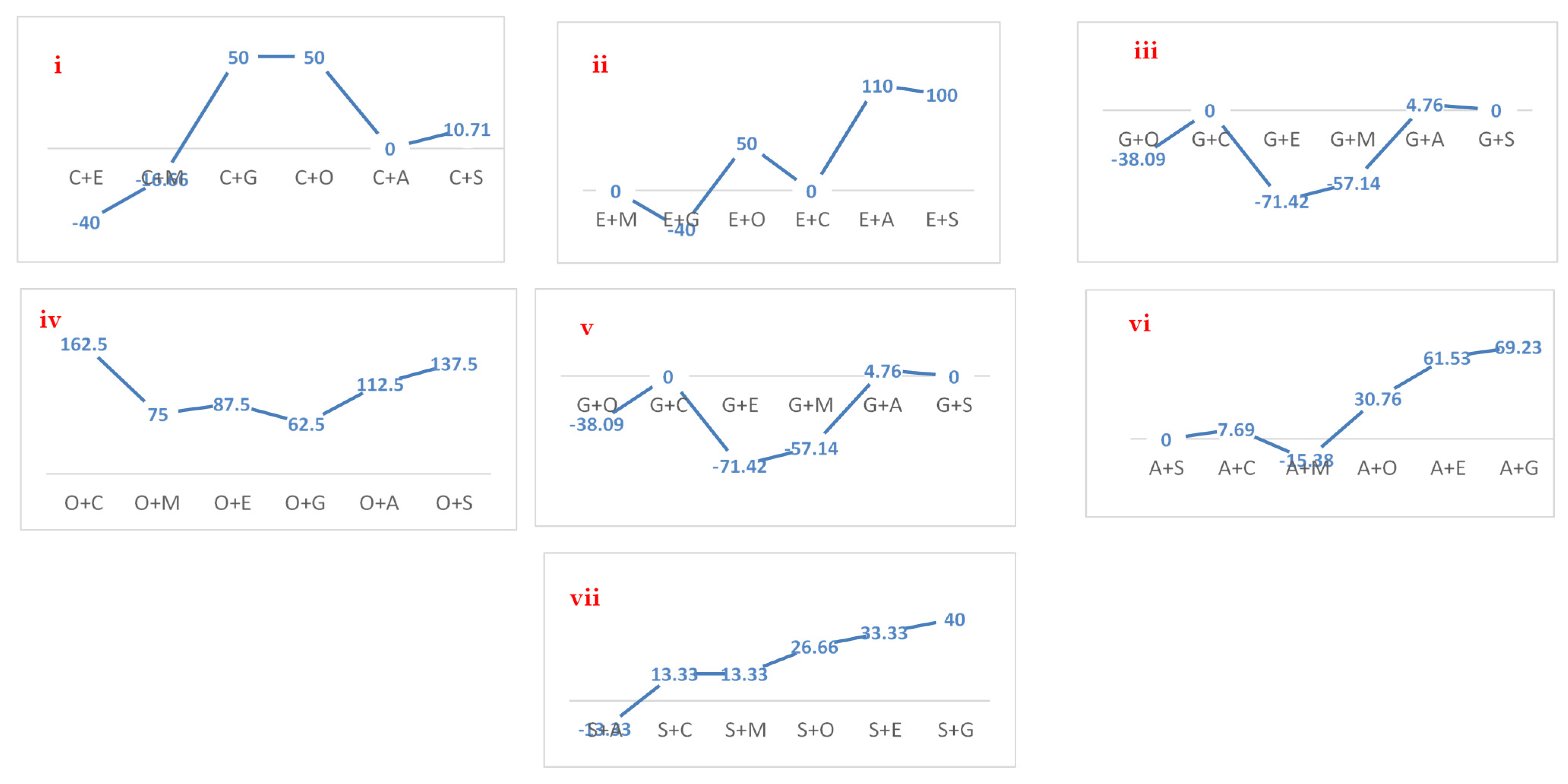
2.5.2. Synergy Testing Using Broth Dilution Method

Drugs combinations against resistant E. coli isolates (1–1.5 × 10⁵ CFU/mL) were assessed for their synergy using the checkerboard method. The optical density of 96 well plates was measured at 570 nm before and after 24 h incubation at 37 °C. Inhibitory concentration indices were measured as per the formula given below. To avoid errors, the experiment was performed in triplicate [29,30].
FICI = FIC of product A + FIC of product B
FIC of product A = MIC of product A in combination with product B/MIC of product A alone
FIC of product B = MIC of product B in combination with product A/MIC of product B alone
An FICI of ≤0.5 was considered as synergistic, >0.5 but ≤1.0 as an additive, >1.0 but <4.0 as indifferent, and >4.0 as antagonistic.

2.6. Field Evaluation of In Vitro Outcomes

Animals positive for E. coli-based endometritis, irrespective of the involvement of other bacteria, were selected for the evaluation of in vitro results of the study. The drugs that showed promising results in the in vitro investigations were tested in the field trial. The drug combinations and dosage regimens are shown in Supplementary Materials Table S1. The success rate was determined on the basis of the reproductive indices and physical observations such as disappearance of ailment signs in ultrasonography, negative culture results of uterine flushing, and establishment of pregnancy [18,31].

2.7. Statistical Analysis

Descriptive statistics such as univariate analysis were used for the analysis of prevalence and antibiotic susceptibility [32]. On the other hand, parametric tests such as t-test (to compare means of two groups) and ANOVA (to compare means of more than two groups) were applied to data obtained in quantitative form. Tukey test as a post hoc test following ANOVA was applied to find significant differences among different groups. Percentage increase in zone sizes and fractional inhibitory indices were calculated using the prescribed formulas [29,30]. The statistical analysis was carried out using the computer-based statistical program SPSS (version 20) at a 5% probability level.

3. Results

3.1. Prevalence of Endometritis and E. coli

The current study showed an overall prevalence of endometritis at 43.75%, while E. coli were 72.18% among these from different farms (Table 1).

3.2. Sequencing Results of the Local E. coli Isolate

3.2.1. Nucleotide Analysis

Nucleotide analysis revealed that the 23S rRNA nucleotides sequence of the local E. coli isolate showed 98% similarity to the nucleotide sequence of the reference E. coli strain (accession number CP067311.1) (Supplmentary Materials Figure S1).

3.2.2. Phylogenetic Analysis

The evolutionary history was inferred by using a phylogenetic tool. Two clades were observed in the phylogenetic tree (Figure S2), which was obtained after comparing the 23S ribosomal RNA gene (nucleotide sequences) of the local E. coli isolate with the other sequences available in the NCBI database. The nucleotide sequence of the 23S rRNA gene of the local E. coli isolate was found to be closely related to that of the E. coli isolate from human rectal swab (Laos) and was clustered together with the E. coli isolated from human rectal swab (Laos), human urine (USA), swine liver (China), dairy manure (Canada), human feces (Singapore), chicken meat inner strip (Romania), human urine (China), human (Spain), and pig feces (Canada). The percentage of replicate trees wherein the associated taxa clustered together in the bootstrap test (1000 replicates) was indicated next to the branches. In total, there were 211 positions in the final dataset after the removal of ambiguous positions from each sequence pair.

3.2.3. Gene Structure and Motif Analysis

To gain further insights into the 23S gene of E. coli, gene structure and motif analysis were predicted. Five motifs were identified on different positions in 23S rRNA gene sequence. E. value, sites and width of motifs is given in Figure S3. p-value (2.49 × 10−100) is the same for all five sequences (Figure S3). The position of motif 1 is from nucleotide 1 to 29, as shown in Figure S3 (i). The position of motif 2 is from nucleotide 40 to 89 Figure S3 (ii). The position of motif 3 is from nucleotide 106 to 155, as demonstrated in Figure S3 (iii). The position of motif 4 is from nucleotide 163 to 212, as shown in Figure S3 (iv). The position of motif 5 is from nucleotide 213 to 233, as shown in Figure S3 (v). Frequencies of nucleotides of 23S RNA gene are given in Table 2. Figure S4 shows the structural analysis of the 23S E. coli ribosomal gene coding region of Pakistan revealed that this gene possesses the same pattern of the coding region of E. coli 23S ribosomal gene isolated from different samples of E. coli from different countries.

3.3. Antibiogram of Endometritis-Originated E. coli

The resistive response of E. coli isolates against different antibiotics was in the following order: fusidic acid > vancomycin > amoxicillin > linezolid > cefoxitin > gentamicin ≥ enrofloxacin > ciprofloxacin ≥ trimethoprim-sulfamethoxazole ≥ chloramphenicol (Table 3). The resistance of E. coli against fusidic acid, vancomycin, and amoxicillin was 80%, 70%, and 50%, respectively, whereas none of the isolates was resistant against ciprofloxacin, trimethoprim-sulfamethoxazole, and chloramphenicol. The antibacterial activity of different antibiotics is presented in Figure 4a.

3.4. In Vitro Therapeutics of Commonly Used Anti-Microbials

3.4.1. Wells Zones of Microbial Growth Inhibition

The drugs, alone and in combinations, showed varying responses against E. coli. Inhibition zones of co-amoxiclav, enrofloxacin, metronidazole, amoxicillin, and streptomycin did not differ significantly when compared to their combinations (p > 0.05), while gentamicin and oxytetracycline showed significant differences (p < 0.05) when compared to their respective combinations (Table 4, Figure 4b).
Drug combination analysis via well diffusion assay revealed a maximum increase in the zone of inhibition (162.5%) for the oxytetracycline + co-amoxiclav combination in comparison to that of oxytetracycline (Figure 5). Similarly, increase in the zone of inhibition for metronidazole + oxytetracycline, metronidazole + co-amoxiclav, oxytetracycline + enrofloxacin, oxytetracycline + metronidazole, and oxytetracycline + gentamicin was 133.33%, 100%, 87.5%, 75%, and 62.5%, respectively, in comparison to that of the first antibiotic in each combination. Drug activities against E. coli using the well diffusion method are shown in Figure 4b.

3.4.2. Synergy Testing of Anti-Microbials

The MIC of the tested drugs varied significantly in the following order: enrofloxacin < gentamicin < co-amoxiclav < streptomycin < amoxicillin < metronidazole < oxytetracycline. All isolates were susceptible to enrofloxacin (2.20 + 1.37) and gentamicin (3.02 + 2.55) (Supplementary Materials Table S2).
Synergy testing of drug combinations against E. coli isolates showed no synergistic effect, while antagonism was noted for the combination of enrofloxacin with oxytetracycline. An additive effect was observed among the following combinations: amoxicillin + metronidazole, amoxicillin + gentamicin, amoxicillin + streptomycin, co-amoxiclav + gentamicin, co-amoxiclav + enrofloxacin, and co-amoxiclav + metronidazole. However, no additive effect was noted in the following combinations co-amoxiclav + metronidazole, co-amoxiclav + streptomycin, co-amoxiclav + enrofloxacin, metronidazole + gentamicin, and metronidazole + oxytetracycline (Table 5).

3.5. Field Trial Outcome

The highest recovery rates were noted for gentamicin + enrofloxacin (100%), gentamicin alone (100%), co-amoxiclav + gentamicin (84.61%), oxytetracycline alone (78.57%), and metronidazole + enrofloxacin (33.33%), with the success rate represented in terms of normalization of the uterine wall and pregnancy establishment (Figure 6). On the other hand, amoxicillin+ streptomycin, amoxicillin + metronidazole, and amoxicillin + gentamicin showed 15.33%, 10.50%, and 9.50%, respectively, success rate in the field trial indicating a difference of response to that of exhibited in vitro response among E. coli.

4. Discussion

Escherichia coli is an opportunistic bacterial pathogen, well known for its intrinsic and acquired resistance and ability to cause serious infections in animals [33]. Recently, 21 resistance pathways in E. coli have been identified against antibiotics [34], but a particular focus on E. coli from uterine samples is found in fewer than needed studies [35], despite its frequent isolation from endometritis [36]. Although wider ranges of Gram-positive and Gram-negative bacterial species are expected from endometritis [37], E. coli has been found as a common etiology [38]. The higher prevalence of endometritis and isolation of E. coli as the most prominent pathogen in the current study is in line with findings from a previous study [38]. The study found 83.33% endometritis, and from these samples, E. coli stood at 36.66%, appearing as the most prevalent pathogen. In contradiction to the current study, 26% of Japanese Holstein cattle were positive for endometritis, whereas the prevalence of E. coli stood as the most prominent microbe [2]. In another study, in agreement with the current study, the bacteriological investigations of uterine flushing of Holstein cattle showed E. coli as the most common isolate [39].
Pattern motifs in the current study were found similar to those reported on different species in literature by using MEME [27,40,41]. The present study revealed that the 23S rRNA nucleotides sequence of the local E. coli isolate showed 98% similarity to the nucleotide sequence of the reference E. coli strain (accession number CP067311.1). Gene sequencing has been proven to be a reliable conformational genetic marker as it is present in all bacteria, and its function has not changed over time [41,42,43]. Sanger sequencing of the variable gene (23S rRNA) was used for bacterial identification [42,44]. As defined in the CLSI guidelines, species identification can be assigned when the maximum score is 99% or higher and if the sequence similarity between the best and second-best species is greater than 0.5% using DNA target sequencing. Similar type of studies on different pathogens (Staphylococcus aureus, Pseudomonas aeruginosa, and E. coli) have been reported by [45,46,47] as a suitable tool for identification.
The current study was in line with findings of other studies in estimating the resistance of this E. coli to ciprofloxacin, i.e., 80%. The high resistance of E. coli against these antibiotics indicates the misuse and overuse of these drugs in hospitals and clinics without any cultural investigations [48]. Antibiogram of E. coli isolates from cattle depicted high resistance to amoxicillin and penicillin G, whereas good susceptibility was seen for amoxicillin and ciprofloxacin. Emerging resistance of the pathogen to chloramphenicol, gentamicin, and oxytetracycline is alarmingly evident [49]. Gentamicin resistance in E. coli has also been reported by Sabat et al. [40]. Additionally, high chloramphenicol resistance in the E. coli isolates that originated from livestock was reported previously [50]. It has also been reported that E. coli isolated from buffaloes showed high resistance to amoxicillin [51]. Resistance to these antibiotics is thought to be because of the O158:NM strain of E. coli [52]. The E. coli isolates of dairy origin showing high resistance to penicillin, cephalosporin, and enrofloxacin are in line with previous studies [41,43,44,53]. Resistance in E. coli is attributed to eae-A, F-41, stx-I, and stx-II genes, as reported in some studies [54,55,56,57]. In addition to this, production of extended-spectrum beta-lactamase and bla gene groups, most common of which are blaCTX-M15, blaCTX-M55, blaCTX-M14, ST-410, ST-23 complex, ST-10, and ST-167 genes [58]. MICs of enrofloxacin and gentamicin of the current study is in line with [59] and is supposed to be because of plasmid-based evolutionary pathways of microbial resistance [60]. The MIC of gentamicin was reported as 1 µg/mL (MIC 50), while that of chloramphenicol was 8 µg/mL against Gram-negative bacteria [61].
Antimicrobial resistance modulation by combination of antibiotics with non-steroidal anti-inflammatory drugs [29,30,62], plant extracts [63,64], and nano-particles [65,66] is well-documented with promising results. The combination of penicillin and gentamicin resulted in a 61% reduction in the MIC value of penicillin with a 100% cure rate against drug-resistant pathogens [67]. The current study showed an enhancement of zones of inhibition when certain antibiotics were used in combination were in agreement with recent studies [68,69]. Carfora et al. [70] reported very lower percentages (1.3%) of resistant isolates to combination therapy. Similar variability trends in the outcomes of in vitro and in vivo investigations, noted in the current study, were also reported in the literature [68,69,71,72]. Temperature variations, cellular binding pathways, the influence of cell matrix on drug action [71], local immunogenic response, optimization and validation of in vivo model to minimize individual variations, enzymatic degradation, activity stabilization [68], pharmacokinetic and pharmacodynamics parameters of the living body [72] are the contributing factors which cause alterations in laboratory and in vivo results; thus, making it difficult to achieve the reproducibility and repeatability of outcomes [73]. The synergy of antibiotics depends on active sites for drugs and how the bacteria respond to different antibiotics. Penicillin groups act on protein binding site 3, while cephalosporins in protein binding site 1. Amino glycosides bind to 30S ribosome subunits, inhibiting the migration of peptide-tRNA from site A to P, resulting in the misreading of mRNA. Antagonism between bacteriostatic and bactericidal antibiotics is also an obvious factor [74] that might be the reason for the failure of some of the drug combinations to inhibit E. coli. AS, antimicrobial resistance is the evolutionary mechanism under the effect of a constantly changing environment [12]. The poor response of enrofloxacin in this study might be because of the repeated use of this antibiotic in the study area. It was found on investigation that enrofloxacin had long been used in various health issues in the study region. Gentamicin is suggested to be used as an effective drug without enrofloxacin to avoid further resistance. Moreover, the current study focused on E. coli-based endometritis while there are significant chances of multiple bacterial etiologies that complicate cases. In that context, the outcome of this study may or may not be effective because the responses from multiple bacteria might be different. Studies are needed to address epidemiological and genetic discrepancies associated with the spread of such resistant microbes in routine dairy analysis. Similarly, antibiotics must be evaluated against other pathogenic bacterial isolates of dairy origin [18].

5. Conclusions

The study results showed a high prevalence of endometritis with context to E. coli in dairy cattle. The pathogen not only proved to be multidrug-resistant in an antibiogram trial but also showed substitution and addition of amino acids upon nucleotide sequence analysis. Additive drug interaction was noted when amoxicillin was combined with metronidazole, streptomycin, and gentamicin. The same response was observed in case of co-amoxiclav + gentamicin, enrofloxacin + gentamicin, and enrofloxacin + metronidazole. There was only one antagonism found when enrofloxacin was used in combination with oxytetracycline. The highest success rate in the field trial was observed in the case of application of gentamicin alone and in combination with enrofloxacin, followed by were co-amoxiclav + gentamicin, oxytetracycline alone, and metronidazole + enrofloxacin. This study thus standardizes the endometritis treatment regime by addressing in vitro as well as in vivo parameters of commonly used antibiotics and, thus, paves the way for further investigation on the behavior of E. coli at the molecular level. The study also recommends rotational use of antibiotics and additional focus on multiple bacterial etiologies to find the solution for complicated cases.

Supplementary Materials

The following are available online at https://www.mdpi.com/article/10.3390/antibiotics10080997/s1, Figure S1: Multiple sequence alignment of E. coli 23S ribosomal protein (reference and local isolates PK nucleotide sequences), Figure S2: Phylogenetic tree of E. coli 23S ribosomal gene (nucleotide sequences), Figure S3: Motifs locations in gene sequences (1 to 5), sequence of individual motif and p-value of motifs in sequence 1 to 5, Figure S4: Structural analysis of 23S E. coli ribosomal gene from Pakistan, Table S1: Drug combinations and dosage regimens, Table S2: MIC comparison of tested antibiotics against E. coli.

Author Contributions

L.S. performed the research work and prepared initial draft; S.W., H.P., and Q.L. conceived the idea and analyzed reproductive performance; A.I.A. conceived the idea, supervised research work, and finalized manuscript; M.A.N. analyzed data and prepared initial draft; Z.S. collected initial data about reproduction and prepared the initial draft of reproductive traits; M.M.A. carried out molecular characterization and analysis of outcomes; R.A. worked on microbiological and antibiotic sensitivity assays; A.S. prepared geographic maps; Q. and A.S.A. worked on therapeutic aspects and revised the manuscript; M.I. revised the final manuscript. All authors have read and agreed to the published version of the manuscript.

Funding

The present study was granted and supported by the National Natural Science Fund (U20A2051, 31760648 and 31860638), Guangxi Natural Science Foundation (AB18221120), and Guangxi Distinguished Scholars Program (201835).

Institutional Review Board Statement

All experimental study were approved and reviewed by the Experimental Animal Care and Use Committee of Guangxi University (NO. GXU-2021-127 and Dated. 30-6-2020).

Data Availability Statement

Data is contained within the article or supplementary material.

Acknowledgments

We would like to thank the State Key Laboratory for Conservation and Utilization of Subtropical Agro-Bioresources for technical support. We also thank the Department of Medicine and central diagnostic laboratory, Cholistan University of Veterinary and Animal Sciences, Bahawalpur, Pakistan who provided the experimental laboratory.

Conflicts of Interest

The authors declare that the research was conducted in the absence of any commercial or financial relationships that could be construed as a potential conflict of interest.

References

  1. Gilbert, R.O. Bovine endometritis: The burden of proof. Cornell Vet. 1992, 82, 11–14. [Google Scholar] [PubMed]
  2. Gautam, G.; Nakao, T.; Yusuf, M.; Koike, K. Prevalence of endometritis during the postpartum period and its impact on subsequent reproductive performance in two Japanese dairy herds. Anim. Reprod. Sci. 2009, 116, 175–187. [Google Scholar] [CrossRef]
  3. Blood, D.C. Veterinary Medicine: A Textbook of the Diseases of Cattle, Sheep, Pigs, Goats and Horses; James, A., Radostits, O.M., Eds.; Bailliere Tindall: London, UK, 1983; ISBN 0702009873. [Google Scholar]
  4. Turk, R.; Samardžija, M.; Bačić, G. Oxidative stress and reproductive disorders in dairy cows. In Dairy Cows: Nutrition, Fertility and Milk Production; Nova Science Publishers, Inc.: Hauppauge, NY, USA, 2013; pp. 57–98. ISBN 9781611229585. [Google Scholar]
  5. Abdel-Latif, M.A.; EL-Gohary, E.S.; Gabr, A.A.; El-Hawary, A.F.; Ahmed, S.A.; Ebrahim, S.A.; Fathala, M.M. Impact of Supplementing Propylene Glycol and Calcium Propionate to Primiparous Buffalo Cows During the Late Gestation and Early Lactation Period on Reproductive Performance and Metabolic Parameters. Alexandria J. Vet. Sci. 2016, 51, 114–121. [Google Scholar]
  6. Földi, J.; Kulcsar, M.; Pecsi, A.; Huyghe, B.; De Sa, C.; Lohuis, J.; Cox, P.; Huszenicza, G. Bacterial complications of postpartum uterine involution in cattle. Anim. Reprod. Sci. 2006, 96, 265–281. [Google Scholar] [CrossRef]
  7. Bicalho, R.C.; Santos, T.M.A.; Gilbert, R.O.; Caixeta, L.S.; Teixeira, L.M.; Bicalho, M.L.S.; Machado, V.S. Susceptibility of Escherichia coli isolated from uteri of postpartum dairy cows to antibiotic and environmental bacteriophages. Part I: Isolation and lytic activity estimation of bacteriophages. J. Dairy Sci. 2010, 93, 93–104. [Google Scholar] [CrossRef]
  8. Gautam, G.; Nakao, T.; Koike, K.; Long, S.T.; Yusuf, M.; Ranasinghe, R.; Hayashi, A. Spontaneous recovery or persistence of postpartum endometritis and risk factors for its persistence in Holstein cows. Theriogenology 2010, 73, 168–179. [Google Scholar] [CrossRef]
  9. Suojala, L.; Kaartinen, L.; Pyörälä, S. Treatment for bovine Escherichia coli mastitis—an evidence-based approach. J. Vet. Pharmacol. Ther. 2013, 36, 521–531. [Google Scholar] [CrossRef]
  10. Ericsson Unnerstad, H.; Lindberg, A.; Persson Waller, K.; Ekman, T.; Artursson, K.; Nilsson-Öst, M.; Bengtsson, B. Microbial aetiology of acute clinical mastitis and agent-specific risk factors. Vet. Microbiol. 2009, 137, 90–97. [Google Scholar] [CrossRef] [Green Version]
  11. Shoaib, M.; Rahman, S.U.; Aqib, A.I.; Ashfaq, K.; Naveed, A.; Kulyar, M.F.-A.; Bhutta, Z.A.; Younas, M.S.; Sarwar, I.; Naseer, M.A. Diversified Epidemiological Pattern and Antibiogram of mecA Gene in Staphylococcus aureus Isolates of Pets, Pet Owners and Environment. Pak. Vet. J. 2020, 40, 331–336. [Google Scholar]
  12. Lippolis, J.D.; Holman, D.B.; Brunelle, B.W.; Thacker, T.C.; Bearson, B.L.; Reinhardt, T.A.; Sacco, R.E.; Casey, T.A. Genomic and transcriptomic analysis of Escherichia coli strains associated with persistent and transient bovine mastitis and the role of colanic acid. Infect. Immun. 2018, 86, e00566-17. [Google Scholar] [CrossRef] [PubMed] [Green Version]
  13. Hasman, H.; Mevius, D.; Veldman, K.; Olesen, I.; Aarestrup, F.M. β-Lactamases among extended-spectrum β-lactamase (ESBL)-resistant Salmonella from poultry, poultry products and human patients in The Netherlands. J. Antimicrob. Chemother. 2005, 56, 115–121. [Google Scholar] [CrossRef] [Green Version]
  14. Mackeen, A.D.; Packard, R.E.; Ota, E.; Speer, L. Antibiotic regimens for postpartum endometritis. Cochrane Database Syst. Rev. 2015, 2015, CD001067. [Google Scholar] [CrossRef] [PubMed]
  15. Cheesman, M.J.; Ilanko, A.; Blonk, B.; Cock, I.E. Developing New Antimicrobial Therapies: Are Synergistic Combinations of Plant Extracts/Compounds with Conventional Antibiotics the Solution? Pharmacogn. Rev. 2017, 11, 57–72. [Google Scholar] [CrossRef] [Green Version]
  16. Aslam, B.; Wang, W.; Arshad, M.I.; Khurshid, M.; Muzammil, S.; Rasool, M.H.; Nisar, M.A.; Alvi, R.F.; Aslam, M.A.; Qamar, M.U.; et al. Antibiotic resistance: A rundown of a global crisis. Infect. Drug Resist. 2018, 11, 1645–1658. [Google Scholar] [CrossRef] [PubMed] [Green Version]
  17. Azawi, O.I.; Omran, S.N.; Hadad, J.J. Clinical, bacteriological, and histopathological study of toxic puerperal metritis in Iraqi buffalo. J. Dairy Sci. 2007, 90, 4654–4660. [Google Scholar] [CrossRef]
  18. Ahmadi, M.R.; Hosseini, A.; Gheisari, H.R.; Yavari, M. Preliminary trial in treatment of postpartum endometritis with intrauterine application of hyperimmune serum in dairy cows. Asian Pacific J. Trop. Dis. 2014, 4, 360–365. [Google Scholar] [CrossRef]
  19. Thrusfield, M. Sampling in Veterinary Epidemiology; Blackwell Science Ltd.: London, UK, 2007; pp. 214–256. [Google Scholar]
  20. Fissore, R.A.; Edmondson, A.J.; Pashen, R.L.; Bondurant, R.H. The use of ultrasonography for the study of the bovine reproductive tract. II. Non-pregnant, pregnant and pathological conditions of the uterus. Anim. Reprod. Sci. 1986, 12, 167–177. [Google Scholar] [CrossRef]
  21. Pierson, R.A.; Ginther, O.J. Ultrasonic imaging of the ovaries and uterus in cattle. Theriogenology 1988, 29, 21–37. [Google Scholar] [CrossRef]
  22. Ihnatsenka, B.; Boezaart, A.P. Ultrasound: Basic understanding and learning the language. Int. J. Shoulder Surg. 2010, 4, 55. [Google Scholar]
  23. Sarwar, I.; Ashar, A.; Mahfooz, A.; Aqib, A.I.; Saleem, M.I.; Butt, A.A.; Bhutta, Z.A.; Shoaib, M.; Kulyar, M.F.-A.; Ilyas, A. Evaluation of Antibacterial Potential of Raw Turmeric, Nano-Turmeric, and NSAIDs against Multiple Drug Resistant Staphylococcus aureus and E. coli Isolated from Animal Wounds. Pak. Vet. J. 2021, 41, 209–214. [Google Scholar]
  24. Holt, J.G.; Krieg, N.R.; Sneath, P.H.A.; Staley, J.T.; Williams, S.T. Bergey’s Manual Of Determinative Bacteriology, 9th ed.; William Wilkins: Baltimore, MD, USA, 1994. [Google Scholar]
  25. Sambrook, J.; Russell, D.W. Molecular Cloning-Sambrook & Russel-Vol. 1, 2, 3; Cold Springs Harbor Lab Press: Long Island, NY, USA, 2001. [Google Scholar]
  26. Kumar, S.; Stecher, G.; Li, M.; Knyaz, C.; Tamura, K. MEGA X: Molecular evolutionary genetics analysis across computing platforms. Mol. Biol. Evol. 2018, 35, 1547. [Google Scholar] [CrossRef] [PubMed]
  27. Bailey, T.L.; Williams, N.; Misleh, C.; Li, W.W. MEME: Discovering and analyzing DNA and protein sequence motifs. Nucleic Acids Res. 2006, 34, W369–W373. [Google Scholar] [CrossRef] [PubMed]
  28. Clinical and Laboratory Standards Institute. Performance Standards for Antimicrobial Susceptibility Testing of Anaerobic Bacteria: Informational Supplement; Clinical and Laboratory Standards Institute (CLSI): Annapolis, MD, USA, 2009. [Google Scholar]
  29. Anwar, M.A.; Aqib, A.I.; Ashfaq, K.; Deeba, F.; Khan, M.K.; Khan, S.R.; Muzammil, I.; Shoaib, M.; Naseer, M.A.; Riaz, T.; et al. Antimicrobial resistance modulation of MDR E. coli by antibiotic coated ZnO nanoparticles. Microb. Pathog. 2020, 148, 104450. [Google Scholar] [CrossRef] [PubMed]
  30. Aqib, A.; Saqib, M.; Khan, S.; Ahmad, T.; Raza, S.; Naseer, M.; Shoaib, M.; Sarwar, I.; Kulyar, M.F.; Bhutta, Z.; et al. Non-steroidal anti-inflammatory drugs, plant extracts, and characterized microparticles to modulate antimicrobial resistance of epidemic mecA positive S. aureus of dairy origin. Appl. Nanosci. 2021, 11, 553–563. [Google Scholar] [CrossRef]
  31. Shin, E.-K.; Jeong, J.-K.; Choi, I.-S.; Kang, H.-G.; Hur, T.-Y.; Jung, Y.-H.; Kim, I.-H. Relationships among ketosis, serum metabolites, body condition, and reproductive outcomes in dairy cows. Theriogenology 2015, 84, 252–260. [Google Scholar] [CrossRef]
  32. Thrusfield, M.V. Veterinary Epidemiology; Blackwell Science: Ames, IA, USA, 2007; ISBN 9781405156271. [Google Scholar]
  33. Wang, G.-Q.; Wu, C.-M.; Du, X.-D.; Shen, Z.-Q.; Song, L.-H.; Chen, X.; Shen, J.-Z. Characterization of integrons-mediated antimicrobial resistance among Escherichia coli strains isolated from bovine mastitis. Vet. Microbiol. 2008, 127, 73–78. [Google Scholar] [CrossRef]
  34. Hinthong, W.; Pumipuntu, N.; Santajit, S.; Kulpeanprasit, S.; Buranasinsup, S.; Sookrung, N.; Chaicumpa, W.; Aiumurai, P.; Indrawattana, N. Detection and drug resistance profile of Escherichia coli from subclinical mastitis cows and water supply in dairy farms in Saraburi Province, Thailand. PeerJ 2017, 5, e3431. [Google Scholar] [CrossRef] [Green Version]
  35. Sheldon, I.M.; Dobson, H. Postpartum uterine health in cattle. Anim. Reprod. Sci. 2004, 82, 295–306. [Google Scholar] [CrossRef]
  36. Deori, S.; Phookan, A. bovine postpartum metritis and its therapeutics: A Review. Indian J. Sci. Technol. 2015, 8, 1. [Google Scholar] [CrossRef]
  37. Azawi, O.I. Postpartum uterine infection in cattle. Anim. Reprod. Sci. 2008, 105, 187–208. [Google Scholar] [CrossRef]
  38. Palanisamy, K.; Udhayavel, S.; Malmarugan, S.; Rajeswar, J. Antibiogram pattern of bacteria causing endometritis in cows. Vet. World 2013, 6, 100–102. [Google Scholar] [CrossRef]
  39. Koba, I.S.; Lysenko, A.A.; Koshchaev, A.G.; Rodin, I.A.; Shantyz, A.U. Effective treatment of chronic endometritis in cows by Florinazol preparation. Indian Vet. J. 2017, 94, 15–18. [Google Scholar]
  40. Rehman, S.U.; Feng, T.; Wu, S.; Luo, X.; Lei, A.; Luobu, B.; Hassan, F.-U.; Liu, Q. Comparative Genomics, Evolutionary and Gene Regulatory Regions Analysis of Casein Gene Family in Bubalus bubalis. Front. Genet. 2021, 12, 662609. [Google Scholar] [CrossRef]
  41. Rehman, S.U.; Nadeem, A.; Javed, M.; Hassan, F.-U.; Luo, X.; Khalid, R.B.; Liu, Q. Genomic Identification, Evolution and Sequence Analysis of the Heat-Shock Protein Gene Family in Buffalo. Genes 2020, 11, 1388. [Google Scholar] [CrossRef]
  42. Sabat, A.J.; van Zanten, E.; Akkerboom, V.; Wisselink, G.; van Slochteren, K.; de Boer, R.F.; Hendrix, R.; Friedrich, A.W.; Rossen, J.W.A.; Kooistra-Smid, A.M.D.M. Targeted next-generation sequencing of the 16S-23S rRNA region for culture-independent bacterial identification—increased discrimination of closely related species. Sci. Rep. 2017, 7, 3434. [Google Scholar] [CrossRef]
  43. Patel, J.B. 16S rRNA gene sequencing for bacterial pathogen identification in the clinical laboratory. Mol. Diagn. J. Devoted Underst. Hum. Dis. Clin. Appl. Mol. Biol. 2001, 6, 313–321. [Google Scholar] [CrossRef]
  44. Liu, J.; Guan, G.; Li, Y.; Liu, A.; Luo, J.; Yin, H. A Molecular Survey of Babesia Species and Detection of a New Babesia Species by DNA Related to B. venatorum from White Yaks in Tianzhu, China. Front. Microbiol. 2017, 8, 419. [Google Scholar] [CrossRef] [Green Version]
  45. Bhattacharya, D.; Sarma, P.M.; Krishnan, S.; Mishra, S.; Lal, B. Evaluation of genetic diversity among Pseudomonas citronellolis strains isolated from oily sludge-contaminated sites. Appl. Environ. Microbiol. 2003, 69, 1435–1441. [Google Scholar] [CrossRef] [Green Version]
  46. Becker, K.; Harmsen, D.; Mellmann, A.; Meier, C.; Schumann, P.; Peters, G.; Von Eiff, C. Development and evaluation of a quality-controlled ribosomal sequence database for 16S ribosomal DNA-based identification of Staphylococcus species. J. Clin. Microbiol. 2004, 42, 4988–4995. [Google Scholar] [CrossRef] [Green Version]
  47. Li, Y.; Liu, D.; Cao, B.; Han, W.; Liu, Y.; Liu, F.; Guo, X.; Bastin, D.A.; Feng, L.; Wang, L. Development of a serotype-specific DNA microarray for identification of some Shigella and pathogenic Escherichia coli strains. J. Clin. Microbiol. 2006, 44, 4376–4383. [Google Scholar] [CrossRef] [Green Version]
  48. Oladeinde, B.H.; Omoregie, R.; Olley, M.; Anunibe, J.A. Urinary tract infection in a rural community of Nigeria. N. Am. J. Med. Sci. 2011, 3, 75. [Google Scholar] [CrossRef]
  49. Mohanty, N.; Das, P.; Pany, S.; Sarangi, L.N.; Ranabijuli, S.; Panda, H. Isolation and antibiogram of Staphylococcus, Streptococcus and Escherichia coli isolates from clinical and subclinical cases of bovine mastitis. Vet. World 2013, 6, 739–743. [Google Scholar] [CrossRef] [Green Version]
  50. Haftu, R.; Menghistu, H.T.; Gugsa, G.; Kalayou, S. Prevalence, bacterial causes, and antimicrobial susceptibility profile of mastitis isolates from cows in large-scale dairy farms of Northern Ethiopia. Trop. Anim. Health Prod. 2012, 44, 1765–1771. [Google Scholar] [CrossRef]
  51. Kocyigit, R.; Yilmaz, O.; Özenc, E.; Mehmet, U. Effect of some risk factors on subclinical mastitis in dairy cows. Kocatepe Vet. Derg. 2016, 9, 185–193. [Google Scholar] [CrossRef]
  52. Wenz, J.R.; Barrington, G.M.; Garry, F.B.; McSweeney, K.D.; Dinsmore, R.P.; Goodell, G.; Callan, R.J. Bacteremia associated with naturally occuring acute coliform mastitis in dairy cows. J. Am. Vet. Med. Assoc. 2001, 219, 976–981. [Google Scholar] [CrossRef] [PubMed]
  53. Najeeb, M.; Anjum, A.; Ahmad, M.-D.; Khan, H.; Ali, M.; Sattar, M.M.K. Bacterial etiology of subclinical mastitis in dairy goats and multiple drug resistance of the isolates. J. Anim. Plant. Sci. 2013, 23, 1541–1544. [Google Scholar]
  54. Hemmatinezhad, B.; Khamesipour, F.; Mohammadi, M.; Safarpoor Dehkordi, F.; Mashak, Z. Microbiological Investigation of O-Serogroups, Virulence Factors and Antimicrobial Resistance Properties of Shiga Toxin-Producing Escherichia Coli Isolated from Ostrich, Turkey and Quail Meats. J. Food Saf. 2015, 35, 491–500. [Google Scholar] [CrossRef]
  55. Dehkordi, F.S.; Yazdani, F.; Mozafari, J.; Valizadeh, Y. Virulence factors, serogroups and antimicrobial resistance properties of Escherichia coli strains in fermented dairy products. BMC Res. Notes 2014, 7, 217. [Google Scholar] [CrossRef]
  56. Shahrani, M.; Dehkordi, F.S.; Momtaz, H. Characterization of Escherichia coli virulence genes, pathotypes and antibiotic resistance properties in diarrheic calves in Iran. Biol. Res. 2014, 47, 28. [Google Scholar] [CrossRef] [Green Version]
  57. Bessalah, S.; Fairbrother, J.M.; Salhi, I.; Vanier, G.; Khorchani, T.; Seddik, M.M.; Hammadi, M. Antimicrobial resistance and molecular characterization of virulence genes, phylogenetic groups of Escherichia coli isolated from diarrheic and healthy camel-calves in Tunisia. Comp. Immunol. Microbiol. Infect. Dis. 2016, 49, 1–7. [Google Scholar] [CrossRef]
  58. Su, Y.; Yu, C.-Y.; Tsai, Y.; Wang, S.-H.; Lee, C.; Chu, C. Fluoroquinolone-resistant and extended-spectrum β-lactamase-producing Escherichia coli from the milk of cows with clinical mastitis in Southern Taiwan. J. Microbiol. Immunol. Infect. 2016, 49, 892–901. [Google Scholar] [CrossRef] [Green Version]
  59. Salmon, S.A.; Watts, J.L. Minimum inhibitory concentration determinations for various antimicrobial agents against 1570 bacterial isolates from turkey poults. Avian Dis. 2000, 1, 85–98. [Google Scholar] [CrossRef]
  60. San Millan, A.; Escudero, J.A.; Gifford, D.R.; Mazel, D.; MacLean, R.C. Multicopy plasmids potentiate the evolution of antibiotic resistance in bacteria. Nat. Ecol. Evol. 2016, 1, 10. [Google Scholar] [CrossRef]
  61. Ibrahem, E.J.; Yasin, Y.S.; Jasim, O.K. Antibacterial Activity of Zinc Oxide Nanoparticles Against Staphylococcus Aureus and Pseudomonas Aeruginosa Isolated from Burn Wound Infections. Cihan Univ. Sci. J. 2017, 10, 24086. [Google Scholar] [CrossRef] [Green Version]
  62. Chan, E.W.L.; Yee, Z.Y.; Raja, I.; Yap, J.K.Y. Synergistic effect of non-steroidal anti-inflammatory drugs (NSAIDs) on antibacterial activity of cefuroxime and chloramphenicol against methicillin-resistant Staphylococcus aureus. J. Glob. Antimicrob. Resist. 2017, 10, 70–74. [Google Scholar] [CrossRef]
  63. Diaz-Sanchez, S.; D’Souza, D.; Biswas, D.; Hanning, I. Botanical alternatives to antibiotics for use in organic poultry production1. Poult. Sci. 2015, 94, 1419–1430. [Google Scholar] [CrossRef]
  64. Palombo, E.A.; Semple, S.J. Antibacterial activity of Australian plant extracts against methicillin-resistant Staphylococcus aureus (MRSA) and vancomycin-resistant enterococci (VRE). J. Basic Microbiol. 2002, 42, 444–448. [Google Scholar] [CrossRef]
  65. Punjabi, K.; Mehta, S.; Chavan, R.; Chitalia, V.; Deogharkar, D.; Deshpande, S. Efficiency of biosynthesized silver and zinc nanoparticles against multi-drug resistant pathogens. Front. Microbiol. 2018, 9, 2207. [Google Scholar] [CrossRef] [PubMed] [Green Version]
  66. Banerjee, S.; Vishakha, K.; Das, S.; Dutta, M.; Mukherjee, D.; Mondal, J.; Mondal, S.; Ganguli, A. Antibacterial, anti-biofilm activity and mechanism of action of pancreatin doped zinc oxide nanoparticles against methicillin resistant Staphylococcus aureus. Colloids Surf. B Biointerfaces 2020, 190, 110921. [Google Scholar] [CrossRef] [PubMed]
  67. Tateda, K.; Matsumoto, T.; Miyazaki, S.; Yamaguchi, K. Efficacy of β-lactam antibiotics combined with gentamicin against penicillin-resistant pneumococcal pneumonia in CBA/J mice. J. Antimicrob. Chemother. 1999, 43, 367–371. [Google Scholar] [CrossRef] [Green Version]
  68. Halassy, B.; Kurtović, T.; Brgles, M.; Lang Balija, M.; Forčić, D. Factors influencing preclinical in vivo evaluation of mumps vaccine strain immunogenicity. Hum. Vaccin. Immunother. 2015, 11, 2446–2454. [Google Scholar] [CrossRef] [Green Version]
  69. Bohn, T.; Carriere, F.; Day, L.; Deglaire, A.; Egger, L.; Freitas, D.; Golding, M.; Le Feunteun, S.; Macierzanka, A.; Menard, O.; et al. Correlation between in vitro and in vivo data on food digestion. What can we predict with static in vitro digestion models? Crit. Rev. Food Sci. Nutr. 2018, 58, 2239–2261. [Google Scholar] [CrossRef] [Green Version]
  70. Carfora, V.; Caprioli, A.; Marri, N.; Sagrafoli, D.; Boselli, C.; Giacinti, G.; Giangolini, G.; Sorbara, L.; Dottarelli, S.; Battisti, A.; et al. Enterotoxin genes, enterotoxin production, and methicillin resistance in Staphylococcus aureus isolated from milk and dairy products in Central Italy. Int. Dairy J. 2015, 42, 12–15. [Google Scholar] [CrossRef] [Green Version]
  71. Davies, S.N.; Martin, D.; Millar, J.D.; Aram, J.A.; Church, J.; Lodge, D. Differences in results from in vivo and in vitro studies on the use-dependency of N-methylaspartate antagonism by MK-801 and other phencyclidine receptor ligands. Eur. J. Pharmacol. 1988, 145, 141–151. [Google Scholar] [CrossRef]
  72. Nagadurga, D.H. Bioavailability and Bioequivalence Studies. In Pharmaceutical Formulation Design-Recent Practices; IntechOpen: London, UK, 2019. [Google Scholar]
  73. Voehringer, P.; Nicholson, J.R. Minimum Information in In Vivo Research BT—Good Research Practice in Non-Clinical Pharmacology and Biomedicine; Bespalov, A., Michel, M.C., Steckler, T., Eds.; Springer International Publishing: Cham, Switzerland, 2020; pp. 197–222. ISBN 978-3-030-33656-1. [Google Scholar]
  74. Ocampo, P.S.; Lázár, V.; Papp, B.; Arnoldini, M.; Abel zur Wiesch, P.; Busa-Fekete, R.; Fekete, G.; Pál, C.; Ackermann, M.; Bonhoeffer, S. Antagonism between bacteriostatic and bactericidal antibiotics is prevalent. Antimicrob. Agents Chemother. 2014, 58, 4573–4582. [Google Scholar] [CrossRef] [PubMed] [Green Version]
Figure 1. Tracking maps of study area, endometritis, and E. coli. (a) Green map indicates area of study from where samples were collected. (b) Number of positive endometritis cases indicated in red in the circles. Each circle is a complete farm. (c) Prevalence of E. coli indicated in red (circle). Each circle indicates individual dairy farm. Red area of the circle is indicative of positive samples.
Antibiotics 10 00997 g001
Figure 2. Ultrasonographic pictures showing endometritis in cattle. (i) EL—endometrial lining; E—endometrium, white arrows indicate fluid; (ii) MM—myometrium; M—metrium, white arrows indicate pus.
Antibiotics 10 00997 g002
Figure 3. Genotypic confirmation of E. coli from endometritis samples. M = marker 1000 bp, wells 1–7 Sample at 231 bp, +ve = positive control, −ve = negative control.
Antibiotics 10 00997 g003
Figure 4. Antibacterial activity of different antibiotics/drugs against E. coli using disc diffusion and well diffusion test. (a) Antibiotic disc diffusion. i—Ciprofloxacin; ii—Amoxicillin; iii—Linezolid; iv—Gentamicin; v—Chloramphenicol. (b) Well diffusion test. i—no zones, negative control; iv—Amoxicillin alone; iii—Enrofloxacin alone; ii—metronidazole; v—Gentamicin + Amoxicillin; vi—Amoxicillin + Enrofloxacin; vii—positive control.
Antibiotics 10 00997 g004
Figure 5. Percentage variation (increase/decrease) in zone of inhibition (ZOI) of individual drugs when compared with their combinations with other drugs. (i)—% variation in ZOI of Co-amoxiclav (C) when it was compared with its combination with other drugs; (ii)—% variation in ZOI of Enrofloxacin (E) when it was compared with its combination with other drugs; (iii)—% variation in ZOI of Gentamicin (G) when it was compared with its combination with other drugs; (iv)—% variation in ZOI of Oxytertracylin (O) when it was compared with its combination with other drugs; (v)—% variation in ZOI of Metronidazole (M) when it was compared with its combination with other drugs; (vi)—% variation in ZOI of Amoxicillin (A) when it was compared with its combination with other drugs, (vii)—% variation in ZOI of Streptomycin (S) when it was compared with its combination with other drugs.
Antibiotics 10 00997 g005
Figure 6. Ultrasonographic pictures after treatment. (i) NE = normal endometrium. (ii) Arrow indicates the fetus as a sign of established pregnancy.
Antibiotics 10 00997 g006
Table 1. Prevalence of endometritis and E. coli.
Farm NamePrevalence of EndometritisPrevalence of E. coli
No. of Animals Screened (A)Endometritis Positive (B)Prevalence (B/A × 100)Confidence Interval (95%)E. coli Positive (C)E. coli % (C/B × 100)Confidence Interval (95%)
Usama Dairies27933.3318.64–52.17910070.09–100
Sahu Dairies27829.6315.85–48.48810067.56–100
United Dairies18844.4424.56–66.28810067.56–100
Rajpoot Dairies341441.1826.37–57.78535.7116.34–61.23
Horizon Dairies542648.1535.4–61.151973.0853.92–86.3
Abdullah Dairies12541.6719.33–68.05510056.55–100
Masab Dairies281346.4329.53–64.19753.8529.15–76.8
Sifari Dairies482654.16740.29–67.43135032.06–67.94
Sial Dairies381642.1027.86–57.811487.5063.98–96.5
Hiraj Dairies18844.4424.56–66.28810067.56–100
Total30413343.75 9672.18
Table 2. Frequencies of purines and pyrimidines in 23S gene sequence.
NucleotideFrequency
A0.227
C0.273
T0.227
G0.273
Table 3. Antibiogram of E. coli isolates.
AntibioticsPotency (µg)Resistant (%)Intermediate (%)Sensitive (%)
Fusidic acid1080200
Enrofloxacin10102070
Ciprofloxacin503070
Trimethoprim
Sulfamethoxazole
2501090
Amoxicillin5502030
Chloramphenicol3003070
Vancomycin3070300
Gentamicin10104050
Linezolid30403030
Cefoxitin30303040
Table 4. Comparison of zones of inhibitions (mm) of E. coli isolates against different combinations of antibiotics/drugs.
Drugs/Antibiotics UsedMean ± Std.p-Value
Drug’s NameCombination of Drugs
Co-amoxiclavAlone7 ± 1.4140.073
C + E5 ± 1.414
C + M6 ± 2.828
C + G10.5 ± 2.121
C + O10.5 ± 0.707
C + A7.0 ± 1.414
C + S8.5 ± 0.707
EnrofloxacinAlone5 ± 0.000.162
E + M5 ± 1.414
E + G3 ± 0.00
E + O7.5 ± 2.121
E + C5 ± 1.414
E + A10.5 ± 6.364
E + S10.0 ± 1.414
MetronidazoleAlone3 ± 00.246
M + G4.5 ± 0.707
M + O7 ± 1.414
M + C6 ± 2.828
M + E5 ± 1.414
M + A5.5 ± 0.707
M + S8.5 ± 3.535
OxytetracyclineAlone4 ± 1.4140.016
O + C10.5 ± 0.707
O + M7 ± 1.414
O + E7.5 ± 2.121
O + G6.5 ± 0.707
O + A8.5 ± 0.707
O + S9.5 ± 0.707
GentamicinAlone10.5 ± 0.707 0.001
G + O6.5 ± 0.707
G + C10.5 ± 2.121
G + E3 ± 0
G + M4.5 ± 0.707
G + A11.0 ± 1.414
G + S10.5 ± 0.707
AmoxicillinAlone6.5 ± 0.7070.344
A + S6.5 ± 0.707
A + C7.0 ± 1.414
A + M5.5 ± 0.707
A + O8.5 ± 0.707
A + E10.5 ± 6.364
A + G11.0 ± 1.414
StreptomycinAlone7.5 ± 0.7070.266
S + A6.5 ± 0.707
S + C8.5 ± 0.707
S + M8.5 ± 3.535
S + O9.5 ± 0.707
S + E10.0 ± 1.414
S + G10.5 ± 0.707
p < 0.05 indicate significant difference. C—co-amoxiclav; E—enrofloxacin; A—amoxicillin; S—streptomycin; M—metronidazole; O—oxytetracycline; G—gentamicin.
Table 5. Synergy testing of drugs against E. coli isolates.
CombinationsMIC ABMIC AFIC AMIC BAMIC BFIC BFICIResults
amoxi + co-amoxiclav23.437515.6251.55.8593757.81250.752.25Indifferent
amoxi + metro7.812515.6250.562.5187.50.3333330.833333Additive
amoxi + enro5.85937515.6250.3750.9765631.5136720.6451611.020161Indifferent
amoxi + strepto5.85937515.6250.3757.812518.229170.4285710.803571Additive
amoxi + genta4.55729215.6250.2916671.9531252.9296880.6666670.958333Additive
amoxi + oxy31.2515.625231.2523.43751.3333333.333333Indifferent
co-amoxiclav + metro5.8593757.81250.75125187.50.6666671.416667Indifferent
co-amoxiclav + enro3.906257.81250.50.9765631.5136720.6451611.145161Indifferent
co-amoxiclav + strepto2.9296887.81250.37515.62518.229170.8571431.232143Indifferent
co-amoxiclav + genta3.906257.81250.50.9765632.9296880.3333330.833333Additive
co-amoxiclav + oxy7.81257.8125131.2523.43751.3333332.333333Indifferent
metro + enro72.91667187.50.3888890.4882811.5136720.3225810.71147Additive
metro + strepto250187.51.33333315.62518.229170.8571432.190476Indifferent
metro + genta125187.50.6666671.9531252.9296880.6666671.333333Indifferent
metro + oxy250187.51.33333331.2523.43751.3333332.666667Indifferent
enro + strepto3.906251.5136722.58064520.5078118.229171.1253.705645Indifferent
enro + genta0.4882811.5136720.3225811.9531252.9296880.6666670.989247Additive
enro + oxy4.5572921.5136723.01075331.2523.43751.3333334.344086Antagonistic
strepto + genta15.62518.229170.8571433.906252.9296881.3333332.190476Indifferent
strepto + oxy20.5078118.229171.12515.62523.43750.6666671.791667Indifferent
genta + oxy4.5572922.9296881.55555631.2523.43751.3333332.888889Indifferent
MIC—minimum inhibitory concentration; FICI—fractional inhibitory concentration index; amoxi—amoxicillin; enro—enrofloxacin; A—amoxicillin; strpto—streptomycin; metro—metronidazole; oxy—oxytetracycline; genta—gentamicin.
Publisher’s Note: MDPI stays neutral with regard to jurisdictional claims in published maps and institutional affiliations.

