
Pharmaceutical Prospects of Bee Products: Special Focus on Anticancer, Antibacterial, Antiviral, and Antiparasitic Properties

 ,
,  , ,
, ,  and
and 
Abstract
:1. Introduction
2. Types of Bee Products: Description and Components
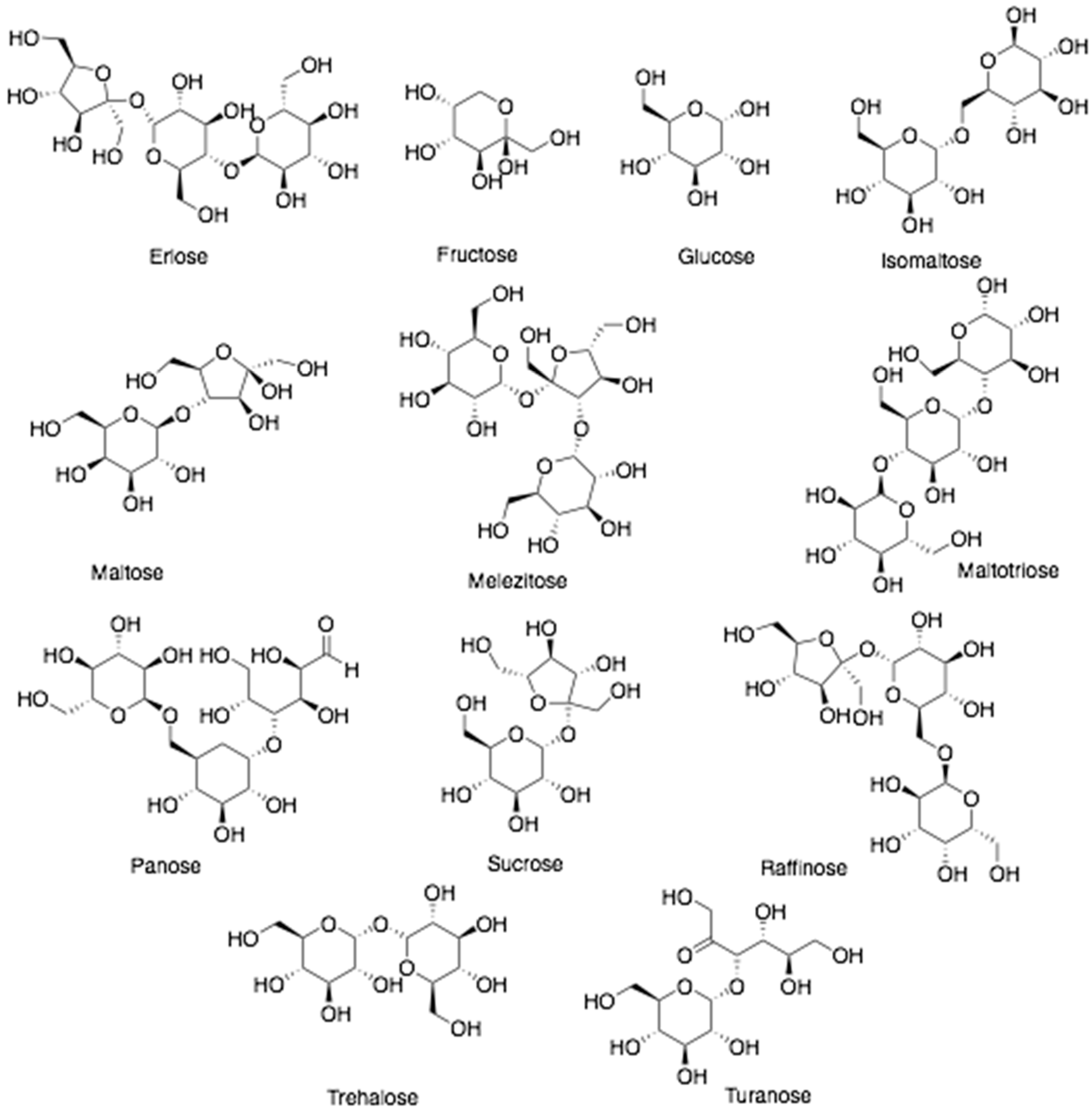
2.1. Honey
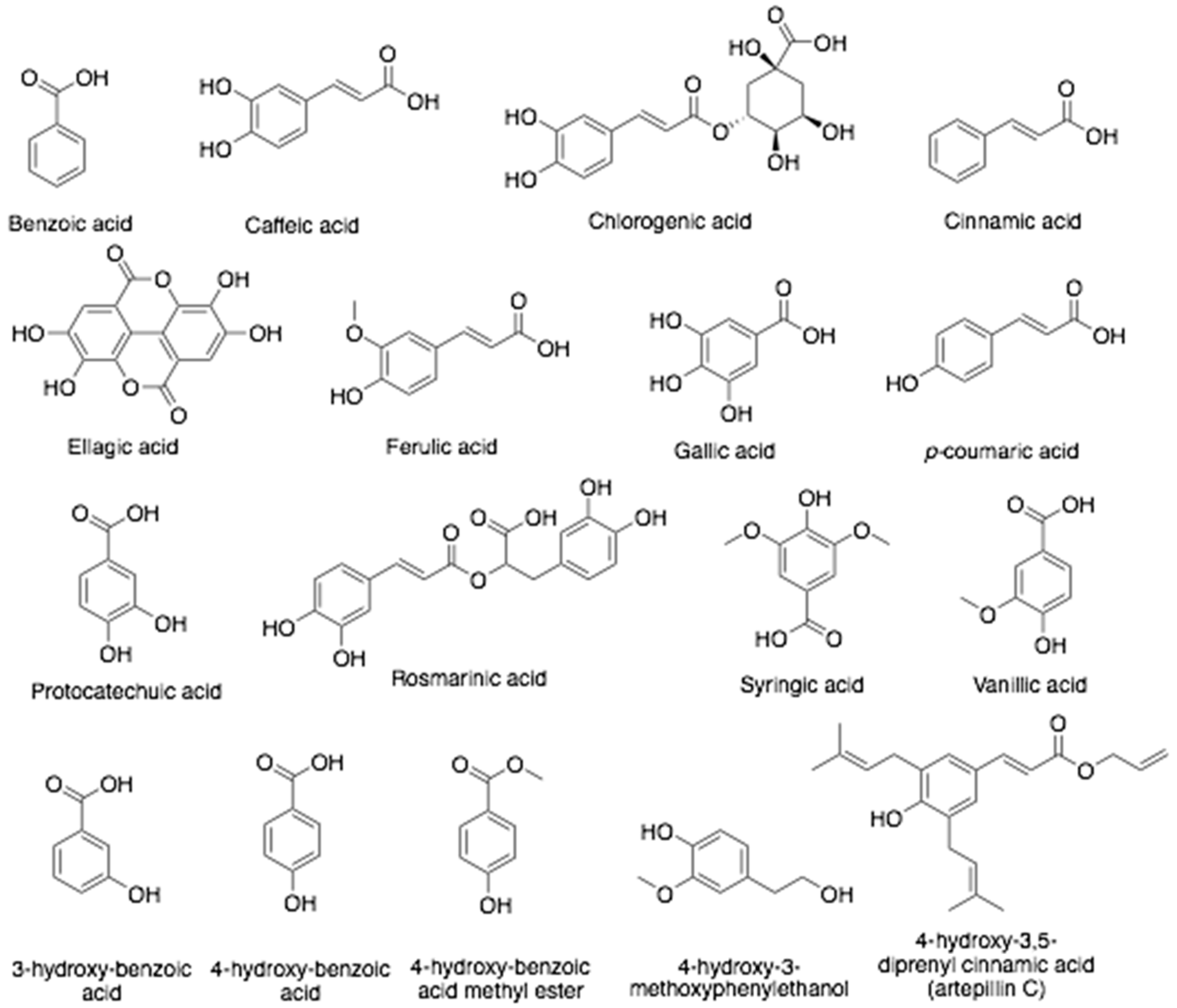
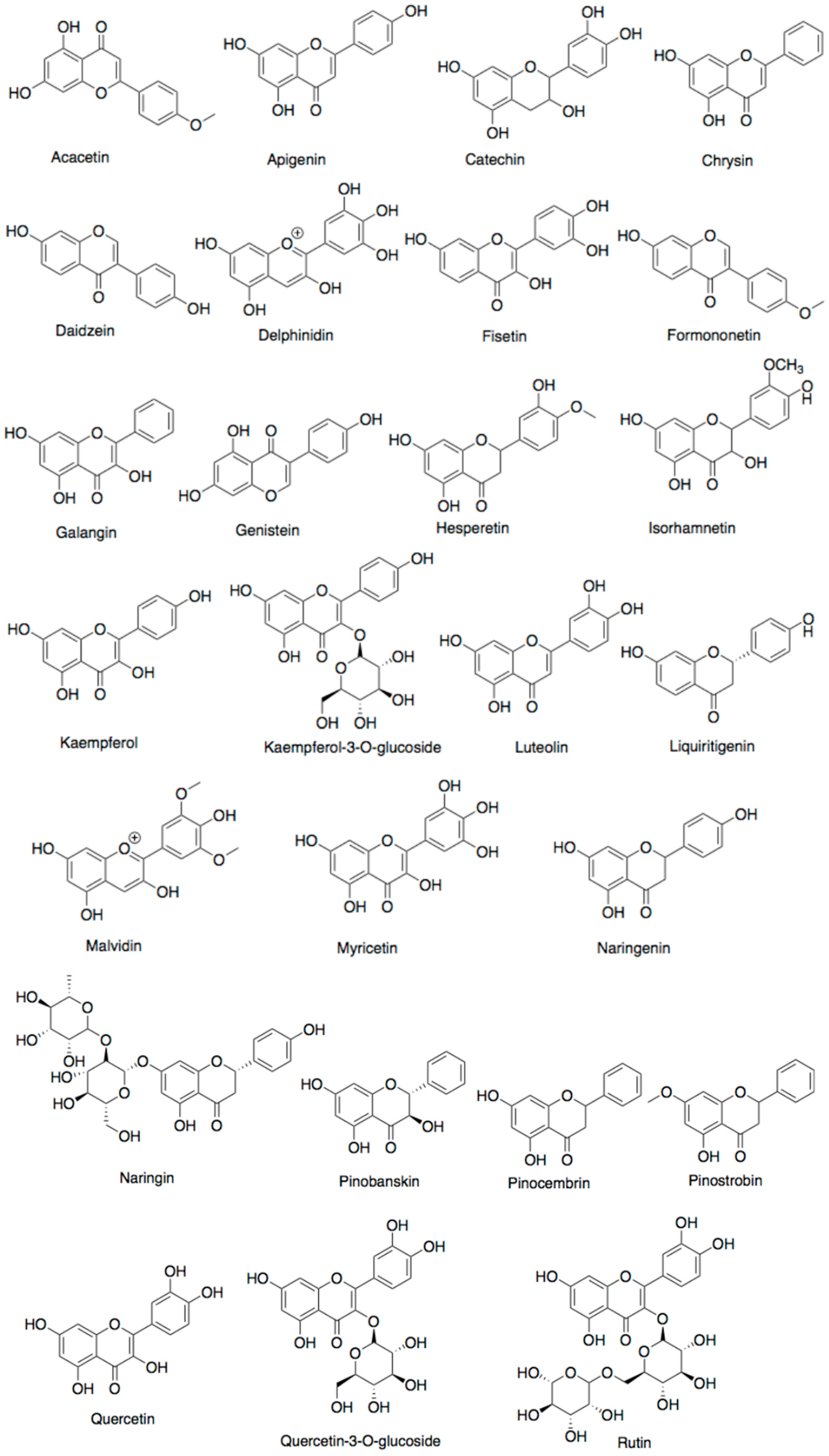
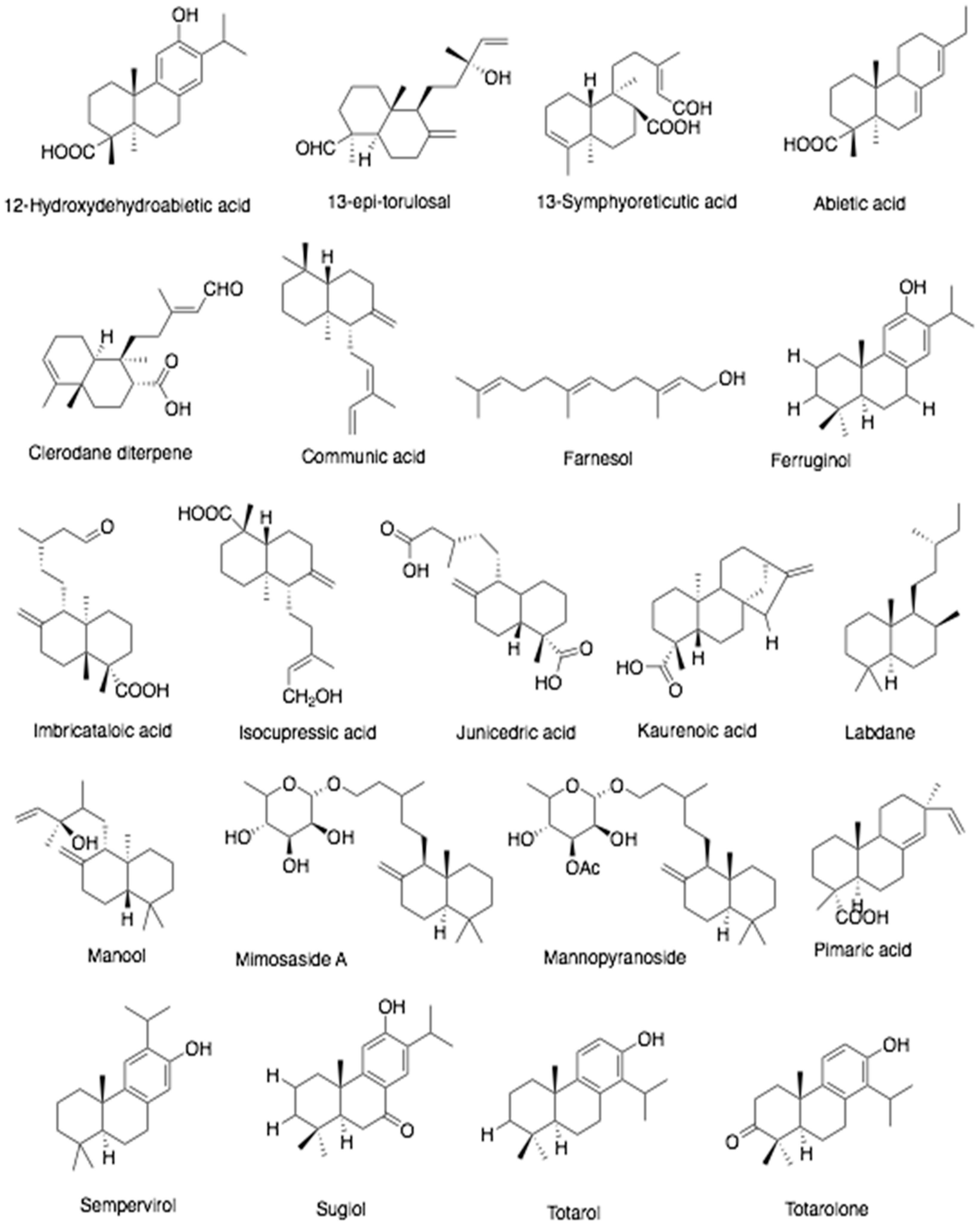
2.2. Propolis
2.3. Bee Pollen
2.4. Royal Jelly
2.5. Bee Bread
2.6. Beeswax
2.7. Bee Venom
2.8. Drone Brood
3. Anticancer Properties of Bee Products
5. Antiparasitic Potential of Bee Products
6. Bee Product-Derived Nanoparticles as Potential Therapeutic Agents
7. Concluding Remarks and Future Directions
Author Contributions
Funding
Institutional Review Board Statement
Informed Consent Statement
Data Availability Statement
Conflicts of Interest
References
- Samarghandian, S.; Farkhondeh, T.; Samini, F. Honey and Health: A Review of Recent Clinical Research. Pharmacogn. Res. 2017, 9, 121–127. [Google Scholar] [CrossRef]
- Al-Hatamleh, M.A.I.; Boer, J.C.; Wilson, K.L.; Plebanski, M.; Mohamud, R.; Mustafa, M.Z. Antioxidant-Based Medicinal Properties of Stingless Bee Products: Recent Progress and Future Directions. Biomolecules 2020, 10, 923. [Google Scholar] [CrossRef]
- Cornara, L.; Biagi, M.; Xiao, J.; Burlando, B. Therapeutic Properties of Bioactive Compounds from Different Honeybee Products. Front. Pharm. 2017, 8, 412. [Google Scholar] [CrossRef]
- Sung, H.; Ferlay, J.; Siegel, R.L.; Laversanne, M.; Soerjomataram, I.; Jemal, A.; Bray, F. Global Cancer Statistics 2020: GLOBOCAN Estimates of Incidence and Mortality Worldwide for 36 Cancers in 185 Countries. CA Cancer J. Clin. 2021, 71, 209–249. [Google Scholar] [CrossRef]
- Münstedt, K.; Männle, H. Bee products and their role in cancer prevention and treatment. Complement. Ther. Med. 2020, 51, 102390. [Google Scholar] [CrossRef]
- Afrin, S.; Haneefa, S.M.; Fernandez-Cabezudo, M.J.; Giampieri, F.; Al-Ramadi, B.K.; Battino, M. Therapeutic and preventive properties of honey and its bioactive compounds in cancer: An evidence-based review. Nutr. Res. Rev. 2020, 33, 50–76. [Google Scholar] [CrossRef]
- Rady, I.; Siddiqui, I.A.; Rady, M.; Mukhtar, H. Melittin, a major peptide component of bee venom, and its conjugates in cancer therapy. Cancer Lett. 2017, 402, 16–31. [Google Scholar] [CrossRef] [Green Version]
- Kurek-Górecka, A.; Górecki, M.; Rzepecka-Stojko, A.; Balwierz, R.; Stojko, J. Bee Products in Dermatology and Skin Care. Molecules 2020, 25, 556. [Google Scholar] [CrossRef] [Green Version]
- Meo, S.A.; Al-Asiri, S.A.; Mahesar, A.L.; Ansari, M.J. Role of honey in modern medicine. Saudi J. Biol. Sci. 2017, 24, 975–978. [Google Scholar] [CrossRef]
- Anjum, S.I.; Ullah, A.; Khan, K.A.; Attaullah, M.; Khan, H.; Ali, H.; Bashir, M.A.; Tahir, M.; Ansari, M.J.; Ghramh, H.A.; et al. Composition and functional properties of propolis (bee glue): A review. Saudi J. Biol. Sci. 2019, 26, 1695–1703. [Google Scholar] [CrossRef]
- Yildirim, A.; Duran, G.G.; Duran, N.; Jenedi, K.; Bolgul, B.S.; Miraloglu, M.; Muz, M. Antiviral Activity of Hatay Propolis Against Replication of Herpes Simplex Virus Type 1 and Type 2. Med. Sci. Monit. 2016, 22, 422–430. [Google Scholar] [CrossRef]
- Münstedt, K. Bee products and the treatment of blister-like lesions around the mouth, skin and genitalia caused by herpes viruses—A systematic review. Complement. Med. 2019, 43, 81–84. [Google Scholar] [CrossRef]
- Watanabe, K.; Rahmasari, R.; Matsunaga, A.; Haruyama, T.; Kobayashi, N. Anti-influenza Viral Effects of Honey In Vitro: Potent High Activity of Manuka Honey. Arch. Med. Res. 2014, 45, 359–365. [Google Scholar] [CrossRef]
- Zareie, P.P. Honey as an Antiviral Agent against Respiratory Syncytial Virus. Ph.D. Dissertation, University of Waikato, Waikato, New Zealand, 2011. [Google Scholar]
- Amoros, M.; Lurton, E.; Boustie, J.; Girre, L.; Sauvager, F.; Cormier, M. Comparison of the anti-herpes simplex virus activities of propolis and 3-methyl-but-2-enyl caffeate. J. Nat. Prod. 1994, 57, 644–647. [Google Scholar] [CrossRef] [PubMed]
- Serkedjieva, J.; Manolova, N.; Bankova, V. Anti-influenza virus effect of some propolis constituents and their analogues (esters of substituted cinnamic acids). J. Nat. Prod. 1992, 55, 294–302. [Google Scholar] [CrossRef]
- Shimizu, T.; Hino, A.; Tsutsumi, A.; Park, Y.K.; Watanabe, W.; Kurokawa, M. Anti-Influenza Virus Activity of Propolis in Vitro and its Efficacy against Influenza Infection in Mice. Antivir. Chem. Chemother. 2008, 19, 7–13. [Google Scholar] [CrossRef] [Green Version]
- Governa, P.; Cusi, M.G.; Borgonetti, V.; Sforcin, J.M.; Terrosi, C.; Baini, G.; Miraldi, E.; Biagi, M. Beyond the Biological Effect of a Chemically Characterized Poplar Propolis: Antibacterial and Antiviral Activity and Comparison with Flurbiprofen in Cytokines Release by LPS-Stimulated Human Mononuclear Cells. Biomedicines 2019, 7, 73. [Google Scholar] [CrossRef] [Green Version]
- Ito, J.; Chang, F.-R.; Wang, H.-K.; Park, Y.K.; Ikegaki, M.; Kilgore, N.; Lee, K.-H. Anti-AIDS Agents. 48. Anti-HIV Activity of Moronic Acid Derivatives and the New Melliferone-Related Triterpenoid Isolated from Brazilian Propolis. J. Nat. Prod. 2001, 64, 1278–1281. [Google Scholar] [CrossRef]
- Shvarzbeyn, J.; Huleihel, M. Effect of propolis and caffeic acid phenethyl ester (CAPE) on NFκB activation by HTLV-1 Tax. Antivir. Res. 2011, 90, 108–115. [Google Scholar] [CrossRef]
- El-Hadya, F.K.A.; Hegazi, A.G.; Wollenweber, E. Effect of Egyptian Propolis on the Susceptibility of LDL to Oxidative Modification and its Antiviral Activity with Special Emphasis on Chemical Composition. Z. Für. Nat. C 2007, 62, 645–655. [Google Scholar] [CrossRef]
- Takeshita, T.; Watanabe, W.; Toyama, S.; Hayashi, Y.; Honda, S.; Sakamoto, S.; Matsuoka, S.; Yoshida, H.; Takeda, S.; Hidaka, M.; et al. Effect of Brazilian Propolis on Exacerbation of Respiratory Syncytial Virus Infection in Mice Exposed to Tetrabromobisphenol A, a Brominated Flame Retardant. Evid. Based Complement. Altern. Med. 2013, 2013, 698206. [Google Scholar] [CrossRef]
- Búfalo, M.C.; Figueiredo, A.S.; de Sousa, J.P.; Candeias, J.M.; Bastos, J.K.; Sforcin, J.M. Anti-poliovirus activity of Baccharis dracunculifolia and propolis by cell viability determination and real-time PCR. J. Appl. Microbiol. 2009, 107, 1669–1680. [Google Scholar] [CrossRef] [PubMed]
- Soroy, L.; Bagus, S.; Yongkie, I.P.; Djoko, W. The effect of a unique propolis compound (Propoelix™) on clinical outcomes in patients with dengue hemorrhagic fever. Infect. Drug Resist. 2014, 7, 323–329. [Google Scholar] [CrossRef] [Green Version]
- Lyu, S.Y.; Rhim, J.Y.; Park, W.B. Antiherpetic activities of flavonoids against herpes simplex virus type 1 (HSV-1) and type 2 (HSV-2) in vitro. Arch. Pharm. Res. 2005, 28, 1293–1301. [Google Scholar] [CrossRef]
- Lee, I.K.; Hwang, B.S.; Kim, D.W.; Kim, J.Y.; Woo, E.E.; Lee, Y.J.; Choi, H.J.; Yun, B.S. Characterization of Neuraminidase Inhibitors in Korean Papaver rhoeas Bee Pollen Contributing to Anti-Influenza Activities In Vitro. Planta Med. 2016, 82, 524–529. [Google Scholar] [CrossRef] [Green Version]
- Lima, W.G.; Brito, J.C.M.; da Cruz Nizer, W.S. Bee products as a source of promising therapeutic and chemoprophylaxis strategies against COVID-19 (SARS-CoV-2). Phytother. Res. 2021, 35, 743–750. [Google Scholar] [CrossRef]
- Santos-Buelga, C.; González-Paramás, A.M. Phenolic Composition of Propolis. In Bee Products—Chemical and Biological Properties; Alvarez-Suarez, J.M., Ed.; Springer International Publishing: Cham, Switzerland, 2017; pp. 99–111. [Google Scholar] [CrossRef]
- Mohammed, S.E.A.; Kabashi, A.S.; Koko, W.S.; Azim, M.K. Antigiardial activity of glycoproteins and glycopeptides from Ziziphus honey. Nat. Prod. Res. 2015, 29, 2100–2102. [Google Scholar] [CrossRef]
- Svečnjak, L.; Chesson, L.A.; Gallina, A.; Maia, M.; Martinello, M.; Mutinelli, F.; Muz, M.N.; Nunes, F.M.; Saucy, F.; Tipple, B.J.; et al. Standard methods for Apis mellifera beeswax research. J. Apic. Res. 2019, 58, 1–108. [Google Scholar] [CrossRef] [Green Version]
- Fratini, F.; Cilia, G.; Turchi, B.; Felicioli, A. Beeswax: A minireview of its antimicrobial activity and its application in medicine. Asian Pac. J. Trop. Med. 2016, 9, 839–843. [Google Scholar] [CrossRef]
- Ratiu, I.A.; Al-Suod, H.; Bukowska, M.; Ligor, M.; Buszewski, B. Correlation Study of Honey Regarding their Physicochemical Properties and Sugars and Cyclitols Content. Molecules 2020, 25, 34. [Google Scholar] [CrossRef] [Green Version]
- De-Melo, A.A.M.; de Almeida-Muradian, L.B. Chemical Composition of Bee Pollen. In Bee Products—Chemical and Biological Properties; Alvarez-Suarez, J.M., Ed.; Springer International Publishing: Cham, Switzerland, 2017; pp. 221–259. [Google Scholar] [CrossRef]
- Viuda-Martos, M.; Pérez-Alvarez, J.A.; Fernández-López, J. Royal Jelly: Health Benefits and Uses in Medicine. In Bee Products—Chemical and Biological Properties; Alvarez-Suarez, J.M., Ed.; Springer International Publishing: Cham, Switzerland, 2017; pp. 199–218. [Google Scholar] [CrossRef]
- Siheri, W.; Alenezi, S.; Tusiimire, J.; Watson, D.G. The Chemical and Biological Properties of Propolis. In Bee Products—Chemical and Biological Properties; Alvarez-Suarez, J.M., Ed.; Springer International Publishing: Cham, Switzerland, 2017; pp. 137–178. [Google Scholar] [CrossRef]
- Abd El-Wahed, A.A.; Khalifa, S.A.M.; Sheikh, B.Y.; Farag, M.A.; Saeed, A.; Larik, F.A.; Koca-Caliskan, U.; AlAjmi, M.F.; Hassan, M.; Wahabi, H.A.; et al. Bee Venom Composition: From Chemistry to Biological Activity. In Studies in Natural Products Chemistry; Atta ur, R., Ed.; Elsevier: Amsterdam, The Netherlands, 2019; Volume 60, pp. 459–484. [Google Scholar]
- Hassan, M.I.; Mohamed, A.F.; Amer, M.A.; Hammad, K.M.; Riad, S.A. Monitoring of the antiviral potential of bee venom and wax extracts against Adeno-7 (DNA) and Rift Valley fever virus (RNA) viruses models. J. Egypt Soc. Parasitol. 2015, 45, 193–198. [Google Scholar] [CrossRef] [PubMed]
- Uddin, M.B.; Lee, B.H.; Nikapitiya, C.; Kim, J.H.; Kim, T.H.; Lee, H.C.; Kim, C.G.; Lee, J.S.; Kim, C.J. Inhibitory effects of bee venom and its components against viruses in vitro and in vivo. J. Microbiol. 2016, 54, 853–866. [Google Scholar] [CrossRef] [PubMed]
- Aw Yong, P.Y.; Islam, F.; Harith, H.H.; Israf, D.A.; Tan, J.W.; Tham, C.L. The Potential use of Honey as a Remedy for Allergic Diseases: A Mini Review. Front. Pharm. 2020, 11, 599080. [Google Scholar] [CrossRef] [PubMed]
- Santos-Buelga, C.; González-Paramás, A.M. Chemical Composition of Honey. In Bee Products—Chemical and Biological Properties; Alvarez-Suarez, J.M., Ed.; Springer International Publishing: Cham, Switzerland, 2017; pp. 43–82. [Google Scholar] [CrossRef]
- Gündoğdu, E.; Cakmakci, S.; Şat, İ. An Overview of Honey: Its Composition, Nutritional and Functional Properties. J. Food Sci. Eng. 2019, 9, 10–14. [Google Scholar] [CrossRef]
- Sousa, J.M.; de Souza, E.L.; Marques, G.; Meireles, B.; de Magalhães Cordeiro, Â.T.; Gullón, B.; Pintado, M.M.; Magnani, M. Polyphenolic profile and antioxidant and antibacterial activities of monofloral honeys produced by Meliponini in the Brazilian semiarid region. Food Res. Int. 2016, 84, 61–68. [Google Scholar] [CrossRef]
- Ahmed, S.; Sulaiman, S.A.; Baig, A.A.; Ibrahim, M.; Liaqat, S.; Fatima, S.; Jabeen, S.; Shamim, N.; Othman, N.H. Honey as a Potential Natural Antioxidant Medicine: An Insight into Its Molecular Mechanisms of Action. Oxid. Med. Cell Longev. 2018, 2018, 8367846. [Google Scholar] [CrossRef] [Green Version]
- Alqarni, A.S.; Owayss, A.A.; Mahmoud, A.A.; Hannan, M.A. Mineral content and physical properties of local and imported honeys in Saudi Arabia. J. Saudi Chem. Soc. 2014, 18, 618–625. [Google Scholar] [CrossRef] [Green Version]
- Rao, P.V.; Krishnan, K.T.; Salleh, N.; Gan, S.H. Biological and therapeutic effects of honey produced by honey bees and stingless bees: A comparative review. Rev. Bras. De Farmacogn. 2016, 26, 657–664. [Google Scholar] [CrossRef] [Green Version]
- Piszcz, P.; Głód, B.K. Antioxidative Properties of Selected Polish Honeys. J. Apic. Sci. 2019, 63, 81–91. [Google Scholar] [CrossRef] [Green Version]
- Mohammed, S.E.A.; Kabbashi, A.S.; Koko, W.S.; Ansari, M.J.; Adgaba, N.; Al-Ghamdi, A. In vitro activity of some natural honeys against Entamoeba histolytica and Giardia lamblia trophozoites. Saudi J. Biol. Sci. 2019, 26, 238–243. [Google Scholar] [CrossRef]
- Kocot, J.; Kiełczykowska, M.; Luchowska-Kocot, D.; Kurzepa, J.; Musik, I. Antioxidant Potential of Propolis, Bee Pollen, and Royal Jelly: Possible Medical Application. Oxid. Med. Cell. Longev. 2018, 2018, 7074209. [Google Scholar] [CrossRef]
- Pasupuleti, V.R.; Sammugam, L.; Ramesh, N.; Gan, S.H. Honey, Propolis, and Royal Jelly: A Comprehensive Review of Their Biological Actions and Health Benefits. Oxid. Med. Cell Longev. 2017, 2017, 1259510. [Google Scholar] [CrossRef]
- Bonamigo, T.; Campos, J.F.; Alfredo, T.M.; Balestieri, J.B.P.; Cardoso, C.A.L.; Paredes-Gamero, E.J.; de Picoli Souza, K.; dos Santos, E.L. Antioxidant, Cytotoxic, and Toxic Activities of Propolis from Two Native Bees in Brazil: Scaptotrigona depilis and Melipona quadrifasciata anthidioides. Oxid. Med. Cell. Longev. 2017, 2017, 1038153. [Google Scholar] [CrossRef] [PubMed] [Green Version]
- Huang, S.; Zhang, C.P.; Wang, K.; Li, G.Q.; Hu, F.L. Recent advances in the chemical composition of propolis. Molecules 2014, 19, 9610. [Google Scholar] [CrossRef] [Green Version]
- Hashem, N.M.; Hassanein, E.M.; Simal-Gandara, J. Improving Reproductive Performance and Health of Mammals Using Honeybee Products. Antioxidants 2021, 10, 336. [Google Scholar] [CrossRef] [PubMed]
- Wang, X.; Sankarapandian, K.; Cheng, Y.; Woo, S.O.; Kwon, H.W.; Perumalsamy, H.; Ahn, Y.-J. Relationship between total phenolic contents and biological properties of propolis from 20 different regions in South Korea. BMC Complement. Altern. Med. 2016, 16, 65. [Google Scholar] [CrossRef] [Green Version]
- El-Seedi, H.R.; Khalifa, S.A.M.; El-Wahed, A.A.; Gao, R.; Guo, Z.; Tahir, H.E.; Zhao, C.; Du, M.; Farag, M.A.; Musharraf, S.G.; et al. Honeybee products: An updated review of neurological actions. Trends Food Sci. Technol. 2020, 101, 17–27. [Google Scholar] [CrossRef]
- Takashima, M.; Ichihara, K.; Hirata, Y. Neuroprotective effects of Brazilian green propolis on oxytosis/ferroptosis in mouse hippocampal HT22 cells. Food Chem. Toxicol. 2019, 132, 110669. [Google Scholar] [CrossRef]
- Berretta, A.A.; Silveira, M.A.D.; Cóndor Capcha, J.M.; De Jong, D. Propolis and its potential against SARS-CoV-2 infection mechanisms and COVID-19 disease. Biomed. Pharmacother. 2020, 131, 110622. [Google Scholar] [CrossRef]
- Vasconcelos, M.R.D.S.; Duarte, A.W.F.; Gomes, E.P.; Silva, S.C.D.; López, A.M.Q. Physicochemical composition and antioxidant potential of bee pollen from different botanical sources in Alagoas, Brazil. Ciência E Agrotecnologia 2017, 41, 447–458. [Google Scholar] [CrossRef] [Green Version]
- Thakur, M.; Nanda, V. Composition and functionality of bee pollen: A review. Trends Food Sci. Technol. 2020, 98, 82–106. [Google Scholar] [CrossRef]
- Gardana, C.; Del Bo’, C.; Quicazán, M.C.; Corrrea, A.R.; Simonetti, P. Nutrients, phytochemicals and botanical origin of commercial bee pollen from different geographical areas. J. Food Compos. Anal. 2018, 73, 29–38. [Google Scholar] [CrossRef]
- Komosinska-Vassev, K.; Olczyk, P.; Kaźmierczak, J.; Mencner, L.; Olczyk, K. Bee pollen: Chemical composition and therapeutic application. Evid Based Complement. Altern. Med. 2015, 2015, 297425. [Google Scholar] [CrossRef] [PubMed] [Green Version]
- Abdelnour, S.A.; Abd El-Hack, M.E.; Alagawany, M.; Farag, M.R.; Elnesr, S.S. Beneficial impacts of bee pollen in animal production, reproduction and health. J. Anim. Physiol. Anim. Nutr. 2019, 103, 477–484. [Google Scholar] [CrossRef] [PubMed]
- Li, Q.-Q.; Wang, K.; Marcucci, M.C.; Sawaya, A.C.H.F.; Hu, L.; Xue, X.-F.; Wu, L.-M.; Hu, F.-L. Nutrient-rich bee pollen: A treasure trove of active natural metabolites. J. Funct. Foods 2018, 49, 472–484. [Google Scholar] [CrossRef]
- Dong, X.; Zhou, Z.; Wang, L.; Saremi, B.; Helmbrecht, A.; Wang, Z.; Loor, J.J. Increasing the availability of threonine, isoleucine, valine, and leucine relative to lysine while maintaining an ideal ratio of lysine:methionine alters mammary cellular metabolites, mammalian target of rapamycin signaling, and gene transcription. J. Dairy Sci 2018, 101, 5502–5514. [Google Scholar] [CrossRef] [PubMed] [Green Version]
- Dong, J.; Yang, Y.; Wang, X.; Zhang, H. Fatty acid profiles of 20 species of monofloral bee pollen from China. J. Apic. Res. 2015, 54, 503–511. [Google Scholar] [CrossRef]
- Kostić, A.Ž.; Milinčić, D.D.; Gašić, U.M.; Nedić, N.; Stanojević, S.P.; Tešić, Ž.L.; Pešić, M.B. Polyphenolic profile and antioxidant properties of bee-collected pollen from sunflower (Helianthus annuus L.) plant. LWT 2019, 112, 108244. [Google Scholar] [CrossRef]
- Sousa, C.; Andrade, P.B.; Valentão, P. Relationships of Echium plantagineum L. bee pollen, dietary flavonoids and their colonic metabolites with cytochrome P450 enzymes and oxidative stress. RSC Adv. 2016, 6, 6084–6092. [Google Scholar] [CrossRef]
- Sattler, J.A.G.; de Melo, I.L.P.; Granato, D.; Araújo, E.; da Silva de Freitas, A.; Barth, O.M.; Sattler, A.; de Almeida-Muradian, L.B. Impact of origin on bioactive compounds and nutritional composition of bee pollen from southern Brazil: A screening study. Food Res. Int. 2015, 77, 82–91. [Google Scholar] [CrossRef] [Green Version]
- Sattler, J.A.G.; De-Melo, A.A.M.; Nascimento, K.S.D.; Melo, I.L.P.D.; Mancini-Filho, J.; Sattler, A.; Almeida-Muradian, L.B.D. Essential minerals and inorganic contaminants (barium, cadmium, lithium, lead and vanadium) in dried bee pollen produced in Rio Grande do Sul State, Brazil. Food Sci. Technol. 2016, 36, 505–509. [Google Scholar] [CrossRef] [Green Version]
- Zhang, Y.; Yang, F.; Jamali, M.A.; Peng, Z. Antioxidant Enzyme Activities and Lipid Oxidation in Rape (Brassica campestris L.) Bee Pollen Added to Salami during Processing. Molecules 2016, 21, 1439. [Google Scholar] [CrossRef]
- Denisow, B.; Denisow-Pietrzyk, M. Biological and therapeutic properties of bee pollen: A review. J. Sci. Food Agric. 2016, 96, 4303–4309. [Google Scholar] [CrossRef]
- Nassar, A.M.K.; Salim, Y.M.M.; Eid, K.S.A.; Shaheen, H.M.; Saati, A.A.; Hetta, H.F.; Elmistekawy, A.; Batiha, G.E. Ameliorative Effects of Honey, Propolis, Pollen, and Royal Jelly Mixture against Chronic Toxicity of Sumithion Insecticide in White Albino Rats. Molecules 2020, 25, 2633. [Google Scholar] [CrossRef] [PubMed]
- Pavel, C.I.; Mărghitaş, L.A.; Dezmirean, D.S.; Tomoş, L.I.; Bonta, V.; Şapcaliu, A.; Buttstedt, A. Comparison between local and commercial royal jelly—Use of antioxidant activity and 10-hydroxy-2-decenoic acid as quality parameter. J. Apic. Res. 2014, 53, 116–123. [Google Scholar] [CrossRef]
- Ab Hamid, N.; Abu Bakar, A.B.; Mat Zain, A.A.; Nik Hussain, N.H.; Othman, Z.A.; Zakaria, Z.; Mohamed, M. Composition of Royal Jelly (RJ) and Its Anti-Androgenic Effect on Reproductive Parameters in a Polycystic Ovarian Syndrome (PCOS) Animal Model. Antioxidants 2020, 9, 499. [Google Scholar] [CrossRef]
- El-Guendouz, S.; Machado, A.M.; Aazza, S.; Lyoussi, B.; Miguel, M.G.; Mateus, M.C.; Figueiredo, A.C. Chemical Characterization and Biological Properties of Royal Jelly Samples From the Mediterranean Area. Nat. Prod. Commun. 2020, 15, 8080. [Google Scholar] [CrossRef] [Green Version]
- Wang, Y.; Ma, L.; Zhang, W.; Cui, X.; Wang, H.; Xu, B. Comparison of the nutrient composition of royal jelly and worker jelly of honey bees (Apis mellifera). Apidologie 2016, 47, 48–56. [Google Scholar] [CrossRef] [Green Version]
- Ramadan, M.F.; Al-Ghamdi, A. Bioactive compounds and health-promoting properties of royal jelly: A review. J. Funct. Foods 2012, 4, 39–52. [Google Scholar] [CrossRef]
- Kanelis, D.; Tananaki, C.; Liolios, V.; Dimou, M.; Goras, G.; Rodopoulou, M.A.; Karazafiris, E.; Thrasyvoulou, A. A suggestion for royal jelly specifications. Arh. Hig. Rada Toksikol. 2015, 66, 275–284. [Google Scholar] [CrossRef] [Green Version]
- Kolayli, S.; Sahin, H.; Can, Z.; Yildiz, O.; Malkoc, M.; Asadov, A. A Member of Complementary Medicinal Food: Anatolian Royal Jellies, Their Chemical Compositions, and Antioxidant Properties. J. Evid. Based Complement. Altern. Med. 2016, 21, Np43–NP48. [Google Scholar] [CrossRef] [Green Version]
- Maghsoudlou, A.; Mahoonak, A.S.; Mohebodini, H.; Toldra, F. Royal Jelly: Chemistry, Storage and Bioactivities. J. Apic. Sci. 2019, 63, 17–40. [Google Scholar] [CrossRef] [Green Version]
- Ahmad, S.; Campos, M.G.; Fratini, F.; Altaye, S.Z.; Li, J. New Insights into the Biological and Pharmaceutical Properties of Royal Jelly. Int. J. Mol. Sci. 2020, 21, 382. [Google Scholar] [CrossRef] [Green Version]
- Park, M.J.; Kim, B.Y.; Park, H.G.; Deng, Y.; Yoon, H.J.; Choi, Y.S.; Lee, K.S.; Jin, B.R. Major royal jelly protein 2 acts as an antimicrobial agent and antioxidant in royal jelly. J. Asia-Pac. Entomol. 2019, 22, 684–689. [Google Scholar] [CrossRef]
- Petelin, A.; Kenig, S.; Kopinč, R.; Deželak, M.; Černelič Bizjak, M.; Jenko Pražnikar, Z. Effects of Royal Jelly Administration on Lipid Profile, Satiety, Inflammation, and Antioxidant Capacity in Asymptomatic Overweight Adults. Evid. Based Complement. Altern. Med. 2019, 2019, 4969720. [Google Scholar] [CrossRef] [Green Version]
- Ghanbari, E.; Nejati, V.; Khazaei, M. Antioxidant and protective effects of Royal jelly on histopathological changes in testis of diabetic rats. Int. J. Reprod. Biomed. 2016, 14, 519–526. [Google Scholar] [CrossRef] [Green Version]
- Dranca, F.; Ursachi, F.; Oroian, M. Bee Bread: Physicochemical Characterization and Phenolic Content Extraction Optimization. Foods 2020, 9, 1358. [Google Scholar] [CrossRef] [PubMed]
- Kieliszek, M.; Piwowarek, K.; Kot, A.M.; Błażejak, S.; Chlebowska-Śmigiel, A.; Wolska, I. Pollen and bee bread as new health-oriented products: A review. Trends Food Sci. Technol. 2018, 71, 170–180. [Google Scholar] [CrossRef]
- Khalifa, S.A.M.; Elashal, M.; Kieliszek, M.; Ghazala, N.E.; Farag, M.A.; Saeed, A.; Xiao, J.; Zou, X.; Khatib, A.; Göransson, U.; et al. Recent insights into chemical and pharmacological studies of bee bread. Trends Food Sci. Technol. 2020, 97, 300–316. [Google Scholar] [CrossRef]
- Mayda, N.; Özkök, A.; Ecem Bayram, N.; Gerçek, Y.C.; Sorkun, K. Bee bread and bee pollen of different plant sources: Determination of phenolic content, antioxidant activity, fatty acid and element profiles. J. Food Meas. Charact. 2020, 14, 1795–1809. [Google Scholar] [CrossRef]
- Kaplan, M.; Karaoglu, Ö.; Eroglu, N.; Silici, S. Fatty Acid and Proximate Composition of Bee Bread. Food Technol. Biotechnol. 2016, 54, 497–504. [Google Scholar] [CrossRef] [PubMed]
- Sobral, F.; Calhelha, R.C.; Barros, L.; Dueñas, M.; Tomás, A.; Santos-Buelga, C.; Vilas-Boas, M.; Ferreira, I.C. Flavonoid Composition and Antitumor Activity of Bee Bread Collected in Northeast Portugal. Molecules 2017, 22, 248. [Google Scholar] [CrossRef] [Green Version]
- Bakour, M.; Al-Waili, N.S.; El Menyiy, N.; Imtara, H.; Figuira, A.C.; Al-Waili, T.; Lyoussi, B. Antioxidant activity and protective effect of bee bread (honey and pollen) in aluminum-induced anemia, elevation of inflammatory makers and hepato-renal toxicity. J. Food Sci. Technol. 2017, 54, 4205–4212. [Google Scholar] [CrossRef]
- Kaškonienė, V.; Adaškevičiūtė, V.; Kaškonas, P.; Mickienė, R.; Maruška, A. Antimicrobial and antioxidant activities of natural and fermented bee pollen. Food Biosci. 2020, 34, 100532. [Google Scholar] [CrossRef]
- Pucca, M.B.; Cerni, F.A.; Oliveira, I.S.; Jenkins, T.P.; Argemí, L.; Sørensen, C.V.; Ahmadi, S.; Barbosa, J.E.; Laustsen, A.H. Bee Updated: Current Knowledge on Bee Venom and Bee Envenoming Therapy. Front. Immunol. 2019, 10. [Google Scholar] [CrossRef] [PubMed] [Green Version]
- Elieh Ali Komi, D.; Shafaghat, F.; Zwiener, R.D. Immunology of Bee Venom. Clin. Rev. Allergy Immunol. 2018, 54, 386–396. [Google Scholar] [CrossRef] [PubMed]
- Carpena, M.; Nuñez-Estevez, B.; Soria-Lopez, A.; Simal-Gandara, J. Bee Venom: An Updating Review of Its Bioactive Molecules and Its Health Applications. Nutrients 2020, 12, 3360. [Google Scholar] [CrossRef]
- Lee, K.S.; Kim, B.Y.; Yoon, H.J.; Choi, Y.S.; Jin, B.R. Secapin, a bee venom peptide, exhibits anti-fibrinolytic, anti-elastolytic, and anti-microbial activities. Dev. Comp. Immunol. 2016, 63, 27–35. [Google Scholar] [CrossRef]
- Mohamed, W.A.; Abd-Elhakim, Y.M.; Ismail, S.A.A. Involvement of the anti-inflammatory, anti-apoptotic, and anti-secretory activity of bee venom in its therapeutic effects on acetylsalicylic acid-induced gastric ulceration in rats. Toxicology 2019, 419, 11–23. [Google Scholar] [CrossRef]
- Zhang, S.; Liu, Y.; Ye, Y.; Wang, X.R.; Lin, L.T.; Xiao, L.Y.; Zhou, P.; Shi, G.X.; Liu, C.Z. Bee venom therapy: Potential mechanisms and therapeutic applications. Toxicon 2018, 148, 64–73. [Google Scholar] [CrossRef]
- Sawczuk, R.; Karpinska, J.; Miltyk, W. What do we need to know about drone brood homogenate and what is known. J. Ethnopharmacol. 2019, 245, 111581. [Google Scholar] [CrossRef] [PubMed]
- Sidor, E.; Dżugan, M. Drone Brood Homogenate as Natural Remedy for Treating Health Care Problem: A Scientific and Practical Approach. Molecules 2020, 25, 5699. [Google Scholar] [CrossRef] [PubMed]
- Imtara, H.; Kmail, A.; Touzani, S.; Khader, M.; Hamarshi, H.; Saad, B.; Lyoussi, B. Chemical analysis and cytotoxic and cytostatic effects of twelve honey samples collected from different regions in morocco and palestine. Evid. Based Complement. Altern. Med. 2019, 2019, 1–11. [Google Scholar] [CrossRef]
- Portokalakis, I.; Yusof, H.I.M.; Ghanotakis, D.F.; Nigam, P.S.; Owusu-Apenten, R. Manuka honey-induced cytotoxicity against MCF7 breast cancer cells is correlated to total phenol content and antioxidant power. J. Adv. Biol. Biotechnol. 2016, 1–10. [Google Scholar] [CrossRef] [Green Version]
- Aryappalli, P.; Al-Qubaisi, S.S.; Attoub, S.; George, J.A.; Arafat, K.; Ramadi, K.B.; Mohamed, Y.A.; Al-Dhaheri, M.M.; Al-Sbiei, A.; Fernandez-Cabezudo, M.J. The IL-6/STAT3 signaling pathway is an early target of manuka honey-induced suppression of human breast cancer cells. Front. Oncol. 2017, 7, 167. [Google Scholar] [CrossRef] [PubMed] [Green Version]
- Henderson, K.; Aldhirgham, T.; Nigam, P.S.-N.; Owusu-Apenten, R.K. Evaluation of Manuka honey estrogen activity using the MCF-7 cell proliferation assay. J. Adv. Biol. Biotechnol. 2016, 10, 1–11. [Google Scholar] [CrossRef] [Green Version]
- Aumeeruddy, M.Z.; Aumeeruddy-Elalfi, Z.; Neetoo, H.; Zengin, G.; van Staden, A.B.; Fibrich, B.; Lambrechts, I.A.; Rademan, S.; Szuman, K.M.; Lall, N. Pharmacological activities, chemical profile, and physicochemical properties of raw and commercial honey. Biocatal. Agric. Biotechnol. 2019, 18, 101005. [Google Scholar] [CrossRef]
- Salleh, M.A.M.; Eshak, Z.; Ismail, W.I.W. Acacia honey induces apoptosis in human breast adenocarcinoma cell lines (MCF-7). J. Teknol. 2017, 79, 9–16. [Google Scholar] [CrossRef] [Green Version]
- Kaya, B.; Yıldırım, A. Determination of the antioxidant, antimicrobial and anticancer properties of the honey phenolic extract of five different regions of Bingöl province. J. Food Sci. Technol. 2021, 58, 2420–2430. [Google Scholar] [CrossRef]
- Waheed, M.; Hussain, M.B.; Javed, A.; Mushtaq, Z.; Hassan, S.; Shariati, M.A.; Khan, M.U.; Majeed, M.; Nigam, M.; Mishra, A.P. Honey and cancer: A mechanistic review. Clin. Nutr. 2019, 38, 2499–2503. [Google Scholar] [CrossRef]
- da Silva, P.M.; Gauche, C.; Gonzaga, L.V.; Costa, A.C.O.; Fett, R. Honey: Chemical composition, stability and authenticity. Food Chem. 2016, 196, 309–323. [Google Scholar] [CrossRef]
- Machado De-Melo, A.A.; Almeida-Muradian, L.B.d.; Sancho, M.T.; Pascual-Maté, A. Composition and properties of Apis mellifera honey: A review. J. Apic. Res. 2018, 57, 5–37. [Google Scholar] [CrossRef]
- Biluca, F.C.; da Silva, B.; Caon, T.; Mohr, E.T.B.; Vieira, G.N.; Gonzaga, L.V.; Vitali, L.; Micke, G.; Fett, R.; Dalmarco, E.M. Investigation of phenolic compounds, antioxidant and anti-inflammatory activities in stingless bee honey (Meliponinae). Food Res. Int. 2020, 129, 108756. [Google Scholar] [CrossRef] [PubMed]
- Beretta, G.; Moretti, R.M.; Nasti, R.; Cincinelli, R.; Dallavalle, S.; Marelli, M.M. Apoptosis-mediated anticancer activity in prostate cancer cells of a chestnut honey (Castanea sativa L.) quinoline–pyrrolidine gamma-lactam alkaloid. Amino Acids 2021, 53, 869–880. [Google Scholar] [CrossRef]
- Fauzi, A.N.; Yaacob, N.S. Cell cycle and apoptosis pathway modulation by Tualang honey in ER-dependent and -independent breast cancer cell lines. J. Apic. Res. 2016, 55, 366–374. [Google Scholar] [CrossRef]
- Demir, S.; Aliyazicioglu, Y.; Turan, I.; Misir, S.; Mentese, A.; Yaman, S.O.; Akbulut, K.; Kilinc, K.; Deger, O. Antiproliferative and proapoptotic activity of Turkish propolis on human lung cancer cell line. Nutr. Cancer 2016, 68, 165–172. [Google Scholar] [CrossRef]
- Elnakady, Y.A.; Rushdi, A.I.; Franke, R.; Abutaha, N.; Ebaid, H.; Baabbad, M.; Omar, M.O.M.; Al Ghamdi, A.A. Characteristics, chemical compositions and biological activities of propolis from Al-Bahah, Saudi Arabia. Sci. Rep. 2017, 7, 1–13. [Google Scholar] [CrossRef] [PubMed] [Green Version]
- Noureddine, H.; Hage-Sleiman, R.; Wehbi, B.; Fayyad-Kazan, H.; Hayar, S.; Traboulssi, M.; Alyamani, O.A.; Faour, W.H.; ElMakhour, Y. Chemical characterization and cytotoxic activity evaluation of Lebanese propolis. Biomed. Pharmacother. 2017, 95, 298–307. [Google Scholar] [CrossRef] [PubMed]
- Czyżewska, U.; Siemionow, K.; Zaręba, I.; Miltyk, W. Proapoptotic activity of propolis and their components on human tongue squamous cell carcinoma cell line (CAL-27). PLoS ONE 2016, 11, e0157091. [Google Scholar] [CrossRef]
- Touzani, S.; Embaslat, W.; Imtara, H.; Kmail, A.; Kadan, S.; Zaid, H.; ElArabi, I.; Badiaa, L.; Saad, B. In vitro evaluation of the potential use of propolis as a multitarget therapeutic product: Physicochemical properties, chemical composition, and immunomodulatory, antibacterial, and anticancer properties. BioMed Res. Int. 2019, 2019, 1–11. [Google Scholar] [CrossRef] [PubMed] [Green Version]
- Celińska-Janowicz, K.; Zaręba, I.; Lazarek, U.; Teul, J.; Tomczyk, M.; Pałka, J.; Miltyk, W. Constituents of propolis: Chrysin, caffeic acid, p-coumaric acid, and ferulic acid induce PRODH/POX-dependent apoptosis in human tongue squamous cell carcinoma cell (CAL-27). Front. Pharmacol. 2018, 9, 336. [Google Scholar] [CrossRef] [Green Version]
- Sun, L.; Guo, Y.; Zhang, Y.; Zhuang, Y. Antioxidant and anti-tyrosinase activities of phenolic extracts from rape bee pollen and inhibitory melanogenesis by cAMP/MITF/TYR pathway in B16 mouse melanoma cells. Front. Pharmacol. 2017, 8, 104. [Google Scholar] [CrossRef] [Green Version]
- Zou, Y.; Hu, J.; Huang, W.; Zhu, L.; Shao, M.; Dordoe, C.; Ahn, Y.-J.; Wang, D.; Zhao, Y.; Xiong, Y. The botanical origin and antioxidant, anti-BACE1 and antiproliferative properties of bee pollen from different regions of South Korea. BMC Complement. Med. Ther. 2020, 20, 236. [Google Scholar] [CrossRef] [PubMed]
- Saisavoey, T.; Sangtanoo, P.; Srimongkol, P.; Reamtong, O.; Karnchanatat, A. Hydrolysates from bee pollen could induced apoptosis in human bronchogenic carcinoma cells (ChaGo-K-1). J. Food Sci. Technol. 2021, 58, 752–763. [Google Scholar] [CrossRef]
- Liu, C.-C.; Hao, D.-J.; Zhang, Q.; An, J.; Zhao, J.-J.; Chen, B.; Zhang, L.-L.; Yang, H. Application of bee venom and its main constituent melittin for cancer treatment. Cancer Chemother. Pharmacol. 2016, 78, 1113–1130. [Google Scholar] [CrossRef] [PubMed]
- Moga, M.A.; Dimienescu, O.G.; Arvătescu, C.A.; Ifteni, P.; Pleş, L. Anticancer activity of toxins from bee and snake venom—An overview on ovarian cancer. Molecules 2018, 23, 692. [Google Scholar] [CrossRef] [Green Version]
- Sangboonruang, S.; Kitidee, K.; Chantawannakul, P.; Tragoolpua, K.; Tragoolpua, Y. Melittin from Apis florea Venom as a Promising Anticancer Agent. Antibiotics 2020, 9, 517. [Google Scholar] [CrossRef] [PubMed]
- Plasay, M.; Wahid, S.; Natzir, R.; Miskad, U.A. Selective cytotoxicity Assay in anticancer drug of Melittin Isolated from Bee Venom (Apis cerana indica) to several human cell lines: HeLa, WiDr and Vero. J. Chem. Pharm. Sci 2016, 9, 2674–2676. [Google Scholar]
- Zolfagharian, H.; Khalilifard Borojeni, S.; Babaie, M. Evaluation of anticancer effects induced by Apis mellifera venom on breast cancer cell line. Alborz Univ. Med. J. 2020, 9, 357–366. [Google Scholar]
- Mahmoodzadeh, A.; Zarrinnahad, H.; Bagheri, K.P.; Moradia, A.; Shahbazzadeh, D. First report on the isolation of melittin from Iranian honey bee venom and evaluation of its toxicity on gastric cancer AGS cells. J. Chin. Med. Assoc. 2015, 78, 574–583. [Google Scholar] [CrossRef] [Green Version]
- Hou, K.K.; Pan, H.; Lanza, G.M.; Wickline, S.A. Melittin derived peptides for nanoparticle based siRNA transfection. Biomaterials 2013, 34, 3110–3119. [Google Scholar] [CrossRef] [Green Version]
- Yu, X.; Dai, Y.; Zhao, Y.; Qi, S.; Liu, L.; Lu, L.; Luo, Q.; Zhang, Z. Melittin-lipid nanoparticles target to lymph nodes and elicit a systemic anti-tumor immune response. Nat. Commun. 2020, 11, 1–14. [Google Scholar] [CrossRef] [Green Version]
- Holle, L.; Song, W.; Holle, E.; Wei, Y.; Wagner, T.; Yu, X. A matrix metalloproteinase 2 cleavable melittin/avidin conjugate specifically targets tumor cells in vitro and in vivo. Int. J. Oncol. 2003, 22, 93–98. [Google Scholar] [CrossRef] [PubMed]
- Su, M.; Chang, W.; Zhang, K.; Cui, M.; Wu, S.; Xu, T. Expression and purification of recombinant ATF-mellitin, a new type fusion protein targeting ovarian cancer cells, in P. pastoris. Oncol. Rep. 2016, 35, 1179–1185. [Google Scholar] [CrossRef] [PubMed]
- Zhang, S.; Shao, Q.; Geng, H.; Su, S. The effect of royal jelly on the growth of breast cancer in mice. Oncol. Lett. 2017, 14, 7615–7621. [Google Scholar] [CrossRef]
- Miyata, Y.; Sakai, H. Anti-cancer and protective effects of royal jelly for therapy-induced toxicities in malignancies. Int. J. Mol. Sci. 2018, 19, 3270. [Google Scholar] [CrossRef] [PubMed] [Green Version]
- Filipič, B.; Gradišnik, L.; Rihar, K.; Šooš, E.; Pereyra, A.; Potokar, J. The influence of royal jelly and human interferon-alpha (HuIFN-αN3) on proliferation, glutathione level and lipid peroxidation in human colorectal adenocarcinoma cells in vitro. Arh. Za Hig. Rada I Toksikol. 2015, 66. [Google Scholar] [CrossRef] [Green Version]
- Miyata, Y.; Ohba, K.; Matsuo, T.; Mitsunari, K.; Sakai, H. A randomized, double-blinded clinical trial of royal jelly intake for anticancer effects and suppressing adverse events in renal cell carcinoma patients treated with tyrosine kinase inhibitors. J. Clin. Oncol. 2020, 38, 697. [Google Scholar] [CrossRef]
- Araki, K.; Miyata, Y.; Ohba, K.; Nakamura, Y.; Matsuo, T.; Mochizuki, Y.; Sakai, H. Oral intake of royal jelly has protective effects against tyrosine kinase inhibitor-induced toxicity in patients with renal cell carcinoma: A randomized, double-blinded, placebo-controlled trial. Medicines 2019, 6, 2. [Google Scholar] [CrossRef] [Green Version]
- Osama, H.; Abdullah, A.; Gamal, B.; Emad, D.; Sayed, D.; Hussein, E.; Mahfouz, E.; Tharwat, J.; Sayed, S.; Medhat, S. Effect of honey and royal jelly against cisplatin-induced nephrotoxicity in patients with cancer. J. Am. Coll. Nutr. 2017, 36, 342–346. [Google Scholar] [CrossRef]
- Wang, S.-Y.; Chen, C.-W. Effects of royal jelly extracts on growth inhibition, differentiation human leukemic U937 cells and its immunomodulatory activity. Biocell 2019, 43, 29–41. [Google Scholar]
- Elsayed, N.; El-Din, H.S.; Altemimi, A.B.; Ahmed, H.Y.; Pratap-Singh, A.; Abedelmaksoud, T.G. In Vitro Antimicrobial, Antioxidant and Anticancer Activities of Egyptian Citrus Beebread. Molecules 2021, 26, 2433. [Google Scholar] [CrossRef] [PubMed]
- Aru, B.; Güzelmeric, E.; Akgül, A.; Demirel, G.Y.; Kırmızıbekmez, H. Antiproliferative activity of chemically characterized propolis from Turkey and its mechanisms of action. Chem. Biodivers. 2019, 16, e1900189. [Google Scholar] [CrossRef]
- Emran, T.B.; Uddin, M.M.N.; Rahman, M.A.; Uddin, M.Z.; Islam, M.M. Phytochemical, antimicrobial, cytotoxic, analgesic and anti-inflammatory properties of Azadirachta indica: A therapeutic study. J. Bioanal. Biomed. 2015, S12, 1–7. [Google Scholar] [CrossRef]
- Udaondo, Z.; Matilla, M.A. Mining for novel antibiotics in the age of antimicrobial resistance. Microb. Biotechnol. 2020, 13, 1702–1704. [Google Scholar] [CrossRef] [PubMed]
- Peraman, R.; Sure, S.K.; Dusthackeer, V.N.A.; Chilamakuru, N.B.; Yiragamreddy, P.R.; Pokuri, C.; Kutagulla, V.K.; Chinni, S. Insights on recent approaches in drug discovery strategies and untapped drug targets against drug resistance. Future J. Pharm. Sci. 2021, 7, 56. [Google Scholar] [CrossRef]
- Sartini, S.; Permana, A.D.; Mitra, S.; Tareq, A.M.; Salim, E.; Ahmad, I.; Harapan, H.; Emran, T.B.; Nainu, F. Current State and Promising Opportunities on Pharmaceutical Approaches in the Treatment of Polymicrobial Diseases. Pathogens 2021, 10, 245. [Google Scholar] [CrossRef]
- Didaras, N.A.; Karatasou, K.; Dimitriou, T.G.; Amoutzias, G.D.; Mossialos, D. Antimicrobial Activity of Bee-Collected Pollen and Beebread: State of the Art and Future Perspectives. Antibiotics 2020, 9, 811. [Google Scholar] [CrossRef]
- Combarros-Fuertes, P.; Fresno, J.M.; Estevinho, M.M.; Sousa-Pimenta, M.; Tornadijo, M.E.; Estevinho, L.M. Honey: Another Alternative in the Fight against Antibiotic-Resistant Bacteria? Antibiotics 2020, 9, 774. [Google Scholar] [CrossRef]
- Nolan, V.C.; Harrison, J.; Wright, J.E.E.; Cox, J.A.G. Clinical Significance of Manuka and Medical-Grade Honey for Antibiotic-Resistant Infections: A Systematic Review. Antibiotics 2020, 9, 766. [Google Scholar] [CrossRef]
- Uddin, M.Z.; Rana, M.S.; Hossain, S.; Dutta, E.; Ferdous, S.; Dutta, M.; Emran, T.B. In vivo neuroprotective, antinociceptive, anti-inflammatory potential in Swiss albino mice and in vitro antioxidant and clot lysis activities of fractionated Holigarna longifolia Roxb. bark extract. J. Complement. Integr. Med. 2019, 17, 1–10. [Google Scholar] [CrossRef]
- Bucekova, M.; Jardekova, L.; Juricova, V.; Bugarova, V.; Di Marco, G.; Gismondi, A.; Leonardi, D.; Farkasovska, J.; Godocikova, J.; Laho, M.; et al. Antibacterial Activity of Different Blossom Honeys: New Findings. Molecules 2019, 24, 1573. [Google Scholar] [CrossRef] [PubMed] [Green Version]
- El Sohaimy, S.A.; Masry, S.H.D.; Shehata, M.G. Physicochemical characteristics of honey from different origins. Ann. Agric. Sci. 2015, 60, 279–287. [Google Scholar] [CrossRef] [Green Version]
- Silici, S.; Sagdic, O.; Ekici, L. Total phenolic content, antiradical, antioxidant and antimicrobial activities of Rhododendron honeys. Food Chem. 2010, 121, 238–243. [Google Scholar] [CrossRef]
- Almasaudi, S. The antibacterial activities of honey. Saudi J. Biol. Sci. 2021, 28, 2188–2196. [Google Scholar] [CrossRef] [PubMed]
- Al-Hatamleh, M.A.I.; Hatmal, M.M.M.; Sattar, K.; Ahmad, S.; Mustafa, M.Z.; Bittencourt, M.D.C.; Mohamud, R. Antiviral and Immunomodulatory Effects of Phytochemicals from Honey against COVID-19: Potential Mechanisms of Action and Future Directions. Molecules 2020, 25, 5017. [Google Scholar] [CrossRef]
- Hadagali, M.D.; Chua, L.S. The anti-inflammatory and wound healing properties of honey. Eur. Food Res. Technol. 2014, 239, 1003–1014. [Google Scholar] [CrossRef]
- Hazrati, M.; Mehrabani, D.; Japoni, A.; Montasery, H.; Azarpira, N.; Hamidian-shirazi, A.R.; Tanideh, N. Effect of Honey on Healing of Pseudomonas aeruginosa Infected Burn Wounds in Rat. J. Appl. Anim. Res. 2010, 37, 161–165. [Google Scholar] [CrossRef]
- Lusby, P.E.; Coombes, A.L.; Wilkinson, J.M. Bactericidal Activity of Different Honeys against Pathogenic Bacteria. Arch. Med. Res. 2005, 36, 464–467. [Google Scholar] [CrossRef]
- Hossain, K.S.; Hossain, M.G.; Moni, A.; Rahman, M.M.; Rahman, U.H.; Alam, M.; Kundu, S.; Rahman, M.M.; Hannan, M.A.; Uddin, M.J. Prospects of honey in fighting against COVID-19: Pharmacological insights and therapeutic promises. Heliyon 2020, 6, e05798. [Google Scholar] [CrossRef]
- Shahzad, A.; Cohrs, R.J. In vitro antiviral activity of honey against varicella zoster virus (VZV): A translational medicine study for potential remedy for shingles. Transl. Biomed. 2012, 3. [Google Scholar] [CrossRef] [Green Version]
- Zeina, B.; Othman, O.; al-Assad, S. Effect of honey versus thyme on Rubella virus survival in vitro. J. Altern Complement. Med. 1996, 2, 345–348. [Google Scholar] [CrossRef]
- Hashemipour, M.A.; Tavakolineghad, Z.; Arabzadeh, S.A.M.; Iranmanesh, Z.; Nassab, S.A.H.G. Antiviral Activities of Honey, Royal Jelly, and Acyclovir Against HSV-1. Wounds 2014, 26, 47–54. [Google Scholar]
- Al-Waili, N.S.; Al-Waili, T.N.; Al-Waili, A.N.; Saloom, K.S. Influence of Natural Honey on Biochemical and Hematological Variables in AIDS: A case study. Sci. World J. 2006, 6, 389430. [Google Scholar] [CrossRef] [PubMed]
- Socarras, K.M.; Theophilus, P.A.S.; Torres, J.P.; Gupta, K.; Sapi, E. Antimicrobial Activity of Bee Venom and Melittin against Borrelia burgdorferi. Antibiotics 2017, 6, 31. [Google Scholar] [CrossRef] [Green Version]
- Sforcin, J.M.; Bankova, V. Propolis: Is there a potential for the development of new drugs? J. Ethnopharmacol. 2011, 133, 253–260. [Google Scholar] [CrossRef] [PubMed]
- Veiga, R.S.; De Mendonça, S.; Mendes, P.B.; Paulino, N.; Mimica, M.J.; Lagareiro Netto, A.A.; Lira, I.S.; López, B.G.-C.; Negrão, V.; Marcucci, M.C. Artepillin C and phenolic compounds responsible for antimicrobial and antioxidant activity of green propolis and Baccharis dracunculifolia DC. J. Appl. Microbiol. 2017, 122, 911–920. [Google Scholar] [CrossRef]
- Yoshimasu, Y.; Ikeda, T.; Sakai, N.; Yagi, A.; Hirayama, S.; Morinaga, Y.; Furukawa, S.; Nakao, R. Rapid Bactericidal Action of Propolis against Porphyromonas gingivalis. J. Dent. Res. 2018, 97, 928–936. [Google Scholar] [CrossRef] [PubMed]
- Veloz, J.J.; Alvear, M.; Salazar, L.A. Antimicrobial and Antibiofilm Activity against Streptococcus mutans of Individual and Mixtures of the Main Polyphenolic Compounds Found in Chilean Propolis. BioMed Res. Int. 2019, 2019, 7602343. [Google Scholar] [CrossRef] [Green Version]
- Seibert, J.B.; Bautista-Silva, J.P.; Amparo, T.R.; Petit, A.; Pervier, P.; dos Santos Almeida, J.C.; Azevedo, M.C.; Silveira, B.M.; Brandão, G.C.; de Souza, G.H.B.; et al. Development of propolis nanoemulsion with antioxidant and antimicrobial activity for use as a potential natural preservative. Food Chem. 2019, 287, 61–67. [Google Scholar] [CrossRef] [PubMed]
- Ali, A.M.; Kunugi, H. Propolis, Bee Honey, and Their Components Protect against Coronavirus Disease 2019 (COVID-19): A Review of In Silico, In Vitro, and Clinical Studies. Molecules 2021, 26, 1232. [Google Scholar] [CrossRef]
- Bridi, R.; Atala, E.; Pizarro, P.N.; Montenegro, G. Honeybee Pollen Load: Phenolic Composition and Antimicrobial Activity and Antioxidant Capacity. J. Nat. Prod. 2019, 82, 559–565. [Google Scholar] [CrossRef] [PubMed]
- Kacániová, M.; Vuković, N.; Chlebo, R.; Haščík, P.; Rovna, K.; Cubon, J.; Džugan, M.; Pasternakiewicz, A. The antimicrobial activity of honey, bee pollen loads and beeswax from Slovakia. Arch. Biol. Sci. 2012, 64, 927–934. [Google Scholar] [CrossRef]
- Graikou, K.; Kapeta, S.; Aligiannis, N.; Sotiroudis, G.; Chondrogianni, N.; Gonos, E.; Chinou, I. Chemical analysis of Greek pollen—Antioxidant, antimicrobial and proteasome activation properties. Chem. Cent. J. 2011, 5, 33. [Google Scholar] [CrossRef] [Green Version]
- Morais, M.; Moreira, L.; Feás, X.; Estevinho, L.M. Honeybee-collected pollen from five Portuguese Natural Parks: Palynological origin, phenolic content, antioxidant properties and antimicrobial activity. Food Chem. Toxicol. 2011, 49, 1096–1101. [Google Scholar] [CrossRef] [PubMed] [Green Version]
- Pascoal, A.; Rodrigues, S.; Teixeira, A.; Feás, X.; Estevinho, L.M. Biological activities of commercial bee pollens: Antimicrobial, antimutagenic, antioxidant and anti-inflammatory. Food Chem. Toxicol. 2014, 63, 233–239. [Google Scholar] [CrossRef]
- Fatrcová-Šramková, K.; Nôžková, J.; Kačániová, M.; Máriássyová, M.; Rovná, K.; Stričík, M. Antioxidant and antimicrobial properties of monofloral bee pollen. J. Environ. Sci. Health Part B 2013, 48, 133–138. [Google Scholar] [CrossRef] [PubMed]
- AbdElsalam, E.; Foda, H.S.; Abdel-Aziz, M.S.; Abd, F.K. Antioxidant and Antimicrobial activities of Egyptian Bee Pollen. Middle East. J. Appl. Sci. 2018, 8, 1248–1255. [Google Scholar]
- Tareq, A.M.; Farhad, S.; Uddin, A.N.; Hoque, M.; Nasrin, M.S.; Uddin, M.M.R.; Hasan, M.; Sultana, A.; Munira, M.S.; Lyzu, C.; et al. Chemical profiles, pharmacological properties, and in silico studies provide new insights on Cycas pectinata. Heliyon 2020, 6, e04061. [Google Scholar] [CrossRef] [PubMed]
- Talukder, M.E.U.; Aklima, J.; Emran, T.B.; Islam, S.; Rahman, A.; Bhuiyan, R.H. In vitro antioxidant potential of Momordica charantia fruits extracts. J. Pharm. Res. 2013, 3, 963–971. [Google Scholar] [CrossRef]
- McCloskey, M.C.; Shaheen, S.; Rabago, L.; Hulverson, M.A.; Choi, R.; Barrett, L.K.; Arnold, S.L.M. Evaluation of in vitro and in vivo antibiotic efficacy against a novel bioluminescent Shigella flexneri. Sci. Rep. 2019, 9, 13567. [Google Scholar] [CrossRef] [PubMed]
- Dash, R.; Ahsan, M.T.; Hosen, S.M.Z.; Rahman, M.G.; Emran, T.B.; Uddin, M.M.N. Evolution of selective COX-2 inhibitor from Alangium salvifolium: An in silico approach. J. Appl. Pharm. Sci. 2015, 5, 89–93. [Google Scholar] [CrossRef] [Green Version]
- Sukarsih, Y.; Arfiansyah, R.; Roska, T.P.; Murdifin, M.; Kasim, S.; Nainu, F. Protective Effect of Ethanol Extract of Legundi (Vitex trifolia L.) Leaves against Staphylococcus aureus in Drosophila Infection Model. Biointerface Res. Appl. Chem. 2021, 11, 13989–13996. [Google Scholar]
- Anisa, A.N.; Rahmasari, M.; Roska, T.P.; Arfiansyah, R.; Sartini, S.; Djide, M.N.; Harapan, H.; Nainu, F. Antimicrobial Effect of Roselle (Hibiscus sabdariffa L.) Water Fraction against Pseudomonas aeruginosa using Drosophila Infection Model. Biointerface Res. Appl. Chem. 2021, 11, 12877–12885. [Google Scholar]
- Nainu, F.; Djide, M.N.; Subehan, S.; Sartini, S.; Roska, T.P.; Salim, E.; Kuraishi, T. Protective Signatures of Roselle (Hibiscus sabdariffa L.) Calyx Fractions against Staphylococcus aureus in Drosophila Infection Model. HAYATI J. Biosci. 2020, 27, 306–313. [Google Scholar]
- Ahsan, M.; Gonsales, A.V.; Sartini, S.; Wahyudin, E.; Nainu, F. In vivo anti-staphylococcal activity of roselle (Hibiscus sabdariffa L.) calyx extract in Drosophila model of infection. J. Herbmed. Pharmacol. 2019, 8, 41–46. [Google Scholar] [CrossRef]
- Nainu, F.; Asri, R.M.; Djide, M.N.; Ahsan, M.; Arfiansyah, R.; Sartini, S.; Alam, G. Protective effect of green algae Ulva reticulata against Pseudomonas aeruginosa in Drosophila infection model. HAYATI J. Biosci. 2019, 26, 163–171. [Google Scholar]
- Nainu, F.; Asri, R.M.; Arsyad, A.; Manggau, M.A.; Amir, M.N. In vivo antibacterial activity of green algae Ulva reticulata against Staphylococcus aureus in Drosophila model of infection. Pharmacogn. J. 2018, 10, 993–997. [Google Scholar] [CrossRef] [Green Version]
- Wu, W.; Li, R.; Li, X.; He, J.; Jiang, S.; Liu, S.; Yang, J. Quercetin as an Antiviral Agent Inhibits Influenza A Virus (IAV) Entry. Viruses 2016, 8, 6. [Google Scholar] [CrossRef]
- Al Naggar, Y.; Giesy, J.P.; Abdel-Daim, M.M.; Javed Ansari, M.; Al-Kahtani, S.N.; Yahya, G. Fighting against the second wave of COVID-19: Can honeybee products help protect against the pandemic? Saudi J. Biol. Sci. 2021, 28, 1519–1527. [Google Scholar] [CrossRef]
- Cokcetin, N.N.; Pappalardo, M.; Campbell, L.T.; Brooks, P.; Carter, D.A.; Blair, S.E.; Harry, E.J. The Antibacterial Activity of Australian Leptospermum Honey Correlates with Methylglyoxal Levels. PLoS ONE 2016, 11, e0167780. [Google Scholar] [CrossRef] [PubMed]
- Stagos, D.; Soulitsiotis, N.; Tsadila, C.; Papaeconomou, S.; Arvanitis, C.; Ntontos, A.; Karkanta, F.; Adamou-Androulaki, S.; Petrotos, K.; Spandidos, D.A.; et al. Antibacterial and antioxidant activity of different types of honey derived from Mount Olympus in Greece. Int. J. Mol. Med. 2018, 42, 726–734. [Google Scholar] [CrossRef] [PubMed] [Green Version]
- Das, A.; Datta, S.; Mukherjee, S.; Bose, S.; Ghosh, S.; Dhar, P. Evaluation of antioxidative, antibacterial and probiotic growth stimulatory activities of Sesamum indicum honey containing phenolic compounds and lignans. LWT Food Sci. Technol. 2015, 61, 244–250. [Google Scholar] [CrossRef]
- Almasaudi, S.B.; Al-Nahari, A.A.M.; Abd El-Ghany, E.S.M.; Barbour, E.; Al Muhayawi, S.M.; Al-Jaouni, S.; Azhar, E.; Qari, M.; Qari, Y.A.; Harakeh, S. Antimicrobial effect of different types of honey on Staphylococcus aureus. Saudi J. Biol. Sci. 2017, 24, 1255–1261. [Google Scholar] [CrossRef] [PubMed] [Green Version]
- Morroni, G.; Alvarez-Suarez, J.M.; Brenciani, A.; Simoni, S.; Fioriti, S.; Pugnaloni, A.; Giampieri, F.; Mazzoni, L.; Gasparrini, M.; Marini, E.; et al. Comparison of the Antimicrobial Activities of Four Honeys From Three Countries (New Zealand, Cuba, and Kenya). Front. Microbiol. 2018, 9. [Google Scholar] [CrossRef] [Green Version]
- Junie, L.; Vica, M.; Mirel, G.; Matei, H. Physico-chemical characterization and antibacterial activity of different types of honey tested on strains isolated from hospitalized patients. J. Apic. Sci. 2016, 60, 5–18. [Google Scholar] [CrossRef] [Green Version]
- Massaro, C.F.; Simpson, J.B.; Powell, D.; Brooks, P. Chemical composition and antimicrobial activity of honeybee (Apis mellifera ligustica) propolis from subtropical eastern Australia. Sci. Nat. 2015, 102, 68. [Google Scholar] [CrossRef]
- Bittencourt, M.L.F.; Ribeiro, P.R.; Franco, R.L.P.; Hilhorst, H.W.M.; de Castro, R.D.; Fernandez, L.G. Metabolite profiling, antioxidant and antibacterial activities of Brazilian propolis: Use of correlation and multivariate analyses to identify potential bioactive compounds. Food Res. Int. 2015, 76, 449–457. [Google Scholar] [CrossRef] [Green Version]
- Campos, J.F.; Santos, U.P.D.; Rocha, P.D.S.D.; Damião, M.J.; Balestieri, J.B.P.; Cardoso, C.A.L.; Paredes-Gamero, E.J.; Estevinho, L.M.; de Picoli Souza, K.; Santos, E.L.D. Antimicrobial, Antioxidant, Anti-Inflammatory, and Cytotoxic Activities of Propolis from the Stingless Bee Tetragonisca fiebrigi (Jataí). Evid. Based Complement. Altern. Med. 2015, 2015, 296186. [Google Scholar] [CrossRef] [Green Version]
- Regueira, M.S.; Tintino, S.R.; da Silva, A.R.P.; Costa, M.d.S.; Boligon, A.A.; Matias, E.F.F.; de Queiroz Balbino, V.; Menezes, I.R.A.; Melo Coutinho, H.D. Seasonal variation of Brazilian red propolis: Antibacterial activity, synergistic effect and phytochemical screening. Food Chem. Toxicol. 2017, 107, 572–580. [Google Scholar] [CrossRef]
- Barua, N.; Aziz, M.A.I.; Tareq, A.M.; Sayeed, M.A.; Alam, N.; ul Alam, N.; Uddin, M.A.; Lyzu, C.; Emran, T.B. In vivo and in vitro evaluation of pharmacological activities of Adenia trilobata (Roxb.). Biochem. Biophys. Rep. 2020, 23, 100772. [Google Scholar] [CrossRef] [PubMed]
- Nina, N.; Quispe, C.; Jiménez-Aspee, F.; Theoduloz, C.; Feresín, G.E.; Lima, B.; Leiva, E.; Schmeda-Hirschmann, G. Antibacterial Activity, Antioxidant Effect and Chemical Composition of Propolis from the Región del Maule, Central Chile. Molecules 2015, 20, 18144–18167. [Google Scholar] [CrossRef] [Green Version]
- AL-Ani, I.; Zimmermann, S.; Reichling, J.; Wink, M. Antimicrobial Activities of European Propolis Collected from Various Geographic Origins Alone and in Combination with Antibiotics. Medicines 2018, 5, 2. [Google Scholar] [CrossRef] [PubMed] [Green Version]
- De Marco, S.; Piccioni, M.; Pagiotti, R.; Pietrella, D. Antibiofilm and Antioxidant Activity of Propolis and Bud Poplar Resins versus Pseudomonas aeruginosa. Evid. Based Complement. Altern. Med. 2017, 2017, 5163575. [Google Scholar] [CrossRef] [Green Version]
- Rakib, A.; Ahmed, S.; Islam, M.A.; Uddin, M.M.N.; Paul, A.; Chy, M.N.U.; Emran, T.B.; Seidel, V. Pharmacological studies on the antinociceptive, anxiolytic and antidepressant activity of Tinospora crispa. Phytother. Res. 2020, 34, 2978–2984. [Google Scholar] [CrossRef] [PubMed]
- Popova, M.; Giannopoulou, E.; Skalicka-Woźniak, K.; Graikou, K.; Widelski, J.; Bankova, V.; Kalofonos, H.; Sivolapenko, G.; Gaweł-Bęben, K.; Antosiewicz, B.; et al. Characterization and Biological Evaluation of Propolis from Poland. Molecules 2017, 22, 1159. [Google Scholar] [CrossRef] [PubMed]
- Chen, Y.-W.; Ye, S.-R.; Ting, C.; Yu, Y.-H. Antibacterial activity of propolins from Taiwanese green propolis. J. Food Drug Anal. 2018, 26, 761–768. [Google Scholar] [CrossRef] [PubMed] [Green Version]
- Atsalakis, E.; Chinou, I.; Makropoulou, M.; Karabournioti, S.; Graikou, K. Evaluation of Phenolic Compounds in Cistus creticus Bee Pollen from Greece. Antioxidant and Antimicrobial Properties. Nat. Prod. Commun. 2017, 12, 1141. [Google Scholar] [CrossRef] [Green Version]
- Akhir, R.A.M.; Bakar, M.F.A.; Sanusi, S.B. Antioxidant and antimicrobial activity of stingless bee bread and propolis extracts. AIP Conf. Proc. 2017, 1891, 020090. [Google Scholar] [CrossRef] [Green Version]
- Rahman, J.; Tareq, A.M.; Hossain, M.M.; Sakib, S.A.; Islam, M.N.; Uddin, A.B.M.N.; Hoque, M.; Nasrin, M.S.; Ali, M.H.; Caiazzo, E.; et al. Biological evaluation, DFT calculations and molecular docking studies on the antidepressant and cytotoxicity activities of Cycas pectinata Buch.-Ham. Compounds. Pharmaceuticals 2020, 13, 232. [Google Scholar] [CrossRef]
- Coutinho, D.; Karibasappa, S.; Mehta, D. Royal Jelly Antimicrobial Activity against Periodontopathic Bacteria. J. Interdiscip. Dent. 2018, 8, 18–22. [Google Scholar] [CrossRef]
- Susilowati, H.; Murakami, K.; Yumoto, H.; Amoh, T.; Hirao, K.; Hirota, K.; Matsuo, T.; Miyake, Y. Royal Jelly Inhibits Pseudomonas aeruginosa Adherence and Reduces Excessive Inflammatory Responses in Human Epithelial Cells. BioMed Res. Int. 2017, 2017, 3191752. [Google Scholar] [CrossRef] [PubMed] [Green Version]
- Bakour, M.; Fernandes, Â.; Barros, L.; Sokovic, M.; Ferreira, I.C.F.R.; Badiaa, l. Bee bread as a functional product: Chemical composition and bioactive properties. LWT 2019, 109, 276–282. [Google Scholar] [CrossRef] [Green Version]
- Ivanišová, E.; Kačániová, M.; Frančáková, H.; Petrová, J.; Hutková, J.; Brovarskyi, V.; Velychko, S.; Adamchuk, L.; Schubertová, Z.; Musilová, J. Bee bread—Perspective source of bioactive compounds for future. Potravin. Slovak J. Food Sci. 2015, 9, 592–598. [Google Scholar] [CrossRef] [Green Version]
- Marques Pereira, A.F.; Albano, M.; Bérgamo Alves, F.C.; Murbach Teles Andrade, B.F.; Furlanetto, A.; Mores Rall, V.L.; Delazari dos Santos, L.; de Oliveira Orsi, R.; Fernandes Júnior, A. Influence of apitoxin and melittin from Apis mellifera bee on Staphylococcus aureus strains. Microb. Pathog. 2020, 141, 104011. [Google Scholar] [CrossRef]
- Haktanir, I.; Masoura, M.; Mantzouridou, F.T.; Gkatzionis, K. Mechanism of antimicrobial activity of honeybee (Apis mellifera) venom on Gram-negative bacteria: Escherichia coli and Pseudomonas spp. AMB Express 2021, 11, 54. [Google Scholar] [CrossRef] [PubMed]
- Lamas, A.; Arteaga, V.; Regal, P.; Vázquez, B.; Miranda, J.M.; Cepeda, A.; Franco, C.M. Antimicrobial Activity of Five Apitoxins from Apis mellifera on Two Common Foodborne Pathogens. Antibiotics 2020, 9, 367. [Google Scholar] [CrossRef]
- Sunyoto, T.; Verdonck, K.; El Safi, S.; Potet, J.; Picado, A.; Boelaert, M. Uncharted territory of the epidemiological burden of cutaneous leishmaniasis in sub-Saharan Africa—A systematic review. PLoS Negl. Trop. Dis. 2018, 12, e0006914. [Google Scholar] [CrossRef] [Green Version]
- Bristy, T.A.; Barua, N.; Tareq, A.M.; Sakib, S.A.; Etu, S.T.; Chowdhury, K.H.; Jyoti, M.A.; Aziz, M.A.I.; Reza, A.S.M.A.; Caiazzo, E.; et al. Deciphering the pharmacological properties of methanol extract of Psychotria calocarpa leaves by in vivo, in vitro and in silico approaches. Pharmaceuticals 2020, 13, 183. [Google Scholar] [CrossRef]
- Tzani, M.; Barrasa, A.; Vakali, A.; Georgakopoulou, T.; Mellou, K.; Pervanidou, D. Surveillance data for human leishmaniasis indicate the need for a sustainable action plan for its management and control, Greece, 2004 to 2018. Eurosurveillance 2021, 26, 2000159. [Google Scholar] [CrossRef]
- Sundar, S.; Chakravarty, J. Leishmaniasis: An update of current pharmacotherapy. Expert Opin. Pharmacother. 2013, 14, 53–63. [Google Scholar] [CrossRef] [PubMed]
- Sangshetti, J.N.; Khan, F.A.K.; Kulkarni, A.A.; Arote, R.; Patil, R.H. Antileishmanial drug discovery: Comprehensive review of the last 10 years. RSC Adv. 2015, 5, 32376–32415. [Google Scholar] [CrossRef]
- Alvar, J.; Arana, B. Leishmaniasis, Impact and Therapeutic Needs. In Drug Discovery for Leishmaniasis; Rivas, L., Gil, C., Eds.; Royal Society of Chemistry: London, UK, 2017. [Google Scholar]
- Cauich-Kumul, R.; Campos, M.R.S. Bee Propolis: Properties, Chemical Composition, Applications, and Potential Health Effects. In Bioactive Compounds; Elsevier: Amsterdam, The Netherlands, 2019; pp. 227–243. [Google Scholar]
- El-Guendouz, S.; Lyoussi, B.; Miguel, M.G. Insight on propolis from mediterranean countries: Chemical composition, biological activities and application fields. Chem. Biodivers. 2019, 16, e1900094. [Google Scholar] [CrossRef]
- Siheri, W.; Zhang, T.; Ebiloma, G.U.; Biddau, M.; Woods, N.; Hussain, M.Y.; Clements, C.J.; Fearnley, J.; Ebel, R.E.; Paget, T. Chemical and antimicrobial profiling of propolis from different regions within Libya. PLoS ONE 2016, 11, e0155355. [Google Scholar] [CrossRef] [Green Version]
- Omar, R.; Igoli, J.O.; Zhang, T.; Gray, A.I.; Ebiloma, G.U.; Clements, C.J.; Fearnley, J.; Ebel, R.E.; Paget, T.; De Koning, H.P. The chemical characterization of Nigerian propolis samples and their activity against Trypanosoma brucei. Sci. Rep. 2017, 7, 1–10. [Google Scholar] [CrossRef] [Green Version]
- Alotaibi, A.; Ebiloma, G.U.; Williams, R.; Alenezi, S.; Donachie, A.-M.; Guillaume, S.; Igoli, J.O.; Fearnley, J.; De Koning, H.P.; Watson, D.G. European propolis is highly active against trypanosomatids including Crithidia fasciculata. Sci. Rep. 2019, 9, 1–10. [Google Scholar] [CrossRef] [PubMed] [Green Version]
- Tareq, A.M.; Sohel, M.; Uddin, M.; Mahmud, M.H.; Hoque, M.; Reza, A.A.; Nasrin, M.S.; Kader, F.B.; Emran, T.B. Possible neuropharmacological effects of Apis cerana indica beehive in the Swiss Albino mice. J. Adv. Biotechnol. Exp. Ther. 2020, 3, 128–134. [Google Scholar] [CrossRef]
- da Silveira Regueira-Neto, M.; Tintino, S.R.; Rolón, M.; Coronal, C.; Vega, M.C.; de Queiroz Balbino, V.; de Melo Coutinho, H.D. Antitrypanosomal, antileishmanial and cytotoxic activities of Brazilian red propolis and plant resin of Dalbergia ecastaphyllum (L) Taub. Food Chem. Toxicol. 2018, 119, 215–221. [Google Scholar] [CrossRef]
- Silici, S.; Kutluca, S. Chemical composition and antibacterial activity of propolis collected by three different races of honeybees in the same region. J. Ethnopharmacol. 2005, 99, 69–73. [Google Scholar] [CrossRef]
- Silva, M.P.; Silva, T.M.; Mengarda, A.C.; Salvadori, M.C.; Teixeira, F.S.; Alencar, S.M.; Luz Filho, G.C.; Bueno-Silva, B.; de Moraes, J. Brazilian red propolis exhibits antiparasitic properties in vitro and reduces worm burden and egg production in an mouse model harboring either early or chronic Schistosoma mansoni infection. J. Ethnopharmacol. 2021, 264, 113387. [Google Scholar] [CrossRef]
- Rebouças-Silva, J.; Celes, F.S.; Lima, J.B.; Barud, H.S.; de Oliveira, C.I.; Berretta, A.A.; Borges, V.M. Parasite killing of Leishmania (V) braziliensis by standardized propolis extracts. Evid. Based Complement. Altern. Med. 2017, 2017, 1–14. [Google Scholar] [CrossRef] [Green Version]
- Devequi-Nunes, D.; Machado, B.A.S.; Barreto, G.d.A.; Rebouças Silva, J.; da Silva, D.F.; da Rocha, J.L.C.; Brandão, H.N.; Borges, V.M.; Umsza-Guez, M.A. Chemical characterization and biological activity of six different extracts of propolis through conventional methods and supercritical extraction. PLoS ONE 2018, 13, e0207676. [Google Scholar] [CrossRef]
- Santiago de Sousa, A.R.; Cruz de Moraes, S.Z.; Barbosa Viana-Júnior, A.; Divino de Araujo, E. Toward a Novel Pharmacology and Therapeutic Understanding of Brazilian Propolis: A Meta-Analytical Approach. Pharmacogn. Rev. 2020, 14, 1–7. [Google Scholar] [CrossRef]
- Dutra, R.P.; Bezerra, J.L.; da Silva, M.C.P.; Batista, M.C.A.; Patrício, F.J.B.; Nascimento, F.R.F.; Ribeiro, M.N.S.; Guerra, R.N.M. Antileishmanial activity and chemical composition from Brazilian geopropolis produced by stingless bee Melipona fasciculata. Rev. Bras. De Farmacogn. 2019, 29, 287–293. [Google Scholar] [CrossRef]
- Gomes, A.N.P.; Camara, C.A.; dos Santos Sousa, A.; dos Santos, F.d.A.R.; de Santana Filho, P.C.; Dorneles, G.P.; Romão, P.R.T.; Silva, T.M.S. Chemical Composition of Bee Pollen and Leishmanicidal Activity of Rhusflavone. Rev. Bras. De Farmacogn. 2021, 31, 176–183. [Google Scholar] [CrossRef]
- Cuesta-Rubio, O.; Fernández, M.C.; Hernández, I.M.; Jaramillo, C.G.J.; González, V.H.; Porto, R.M.D.O.; Delange, D.M.; Fidalgo, L.M.; Piccinelli, A.L.; Campone, L. Chemical profile and anti-leishmanial activity of three Ecuadorian propolis samples from Quito, Guayaquil and Cotacachi regions. Fitoterapia 2017, 120, 177–183. [Google Scholar] [CrossRef]
- Cunha, B.C.; De Miranda, M.B.; Afonso, L.C.C.; Abreu, S.R.L.; Da Silva, G.R.; Testasicca, M.C.S.; Moura, S.A.L. Brazilian green propolis hydroalcoholic extract as a therapeutic adjuvant to treat cutaneous leishmaniasis. J. Appl. Pharm. Sci. 2020, 10, 124–132. [Google Scholar]
- Emran, T.B.; Mowla, T.-E.; Ahmed, S.; Zahan, S.; Rakib, A.; Hasan, M.S.; Amin, M.N.; Mow, T.R.; Uddin, M.M.N. Sedative, anxiolytic, antinociceptive, anti-inflammatory and antipyretic effects of a chloroform extract from the leaves of Urena sinuata (Borss) L. in rodents. J. Appl. Life Sci. Int. 2018, 16, 1–19. [Google Scholar] [CrossRef]
- de Miranda, M.B.; Lanna, M.F.; Nascimento, A.L.B.; de Paula, C.A.; de Souza, M.E.; Felipetto, M.; da Silva Barcelos, L.; de Moura, S.A.L. Hydroalcoholic extract of Brazilian green propolis modulates inflammatory process in mice submitted to a low protein diet. Biomed. Pharmacother. 2019, 109, 610–620. [Google Scholar] [CrossRef] [PubMed]
- dos Santos Thomazelli, A.P.F.; Tomiotto-Pellissier, F.; da Silva, S.S.; Panis, C.; Orsini, T.M.; Cataneo, A.H.D.; Miranda-Sapla, M.M.; Custódio, L.A.; Tatakihara, V.L.H.; Bordignon, J. Brazilian propolis promotes immunomodulation on human cells from American Tegumentar Leishmaniasis patients and healthy donors infected with L. braziliensis. Cell. Immunol. 2017, 311, 22–27. [Google Scholar] [CrossRef] [Green Version]
- Yesmin, S.; Paul, A.; Naz, T.; Rahman, A.B.M.A.; Akhter, S.F.; Wahed, M.I.I.; Emran, T.B.; Siddiqui, S.A. Membrane stabilization as a mechanism of the anti-inflammatory activity of ethanolic root extract of Choi (Piper chaba). Clin. Phytosci. 2020, 6, 59. [Google Scholar] [CrossRef]
- Hegazi, A.G.; Al Guthami, F.M.; Al Gethami, A.F.; El Fadaly, H.A. Beneficial Effects of Capparis spinosa Honey on the Immune Response of Rats Infected with Toxoplasma gondii. J. Pharmacopunct. 2017, 20, 112–118. [Google Scholar] [CrossRef]
- Menna-Barreto, R.F.S.; Salomão, K.; Dantas, A.P.; Santa-Rita, R.M.; Soares, M.J.; Barbosa, H.S.; de Castro, S.L. Different cell death pathways induced by drugs in Trypanosoma cruzi: An ultrastructural study. Micron 2009, 40, 157–168. [Google Scholar] [CrossRef]
- Araujo, J.M.E.; Mendonça-Melo, L.S.; Araujo, E.D.; Fernandes, R.P.M.; Scher, R. Phenolic composition and leishmanicidal activity of red propolis and Dalbergia ecastaphyllum (L.) Taub (Fabaceae) Extracts from Sergipe, Brazil. Braz. Arch. Biol. Technol. 2018, 61. [Google Scholar] [CrossRef]
- Arruda, C.; Ribeiro, V.P.; Mejía, J.A.A.; Almeida, M.O.; Goulart, M.O.; Candido, A.C.B.B.; dos Santos, R.A.; Magalhães, L.G.; Martins, C.H.G.; Bastos, J.K. Green propolis: Cytotoxic and leishmanicidal activities of artepillin C, p-coumaric acid, and their degradation products. Rev. Bras. De Farmacogn. 2020, 30, 169. [Google Scholar] [CrossRef]
- Mohamed, A.H.; El-Nabi, S.E.H.; Bayomi, A.E.; Abdelaal, A.A. Effect of bee venom or proplis on molecular and parasitological aspects of Schistosoma mansoni infected mice. J. Parasit. Dis. 2016, 40, 390–400. [Google Scholar] [CrossRef] [Green Version]
- Hassan, S.E.; Abou-El-Dobal, S.K.; Hegazi, A.G. Bioassay of Egyptian propolis on Toxocara vitulorum adult worms. World Appl.Sci.J. 2016, 34, 283–289. [Google Scholar]
- Mokhtar, A.B.; El-Gayar, E.K.; Habib, E.S. In vitro anti-protozoal activity of propolis extract and cysteine proteases inhibitor (phenyl vinyl sulfone) on Blastocystis species. J. Egypt. Soc. Parasitol. 2016, 46, 261–272. [Google Scholar] [CrossRef] [PubMed]
- Soufy, H.; Nadia, M.; Nasr, S.M.; Abd El-Aziz, T.H.; Khalil, F.A.M.; Ahmed, Y.F.; Abou Zeina, H.A.A. Effect of Egyptian propolis on cryptosporidiosis in immunosuppressed rats with special emphasis on oocysts shedding, leukogram, protein profile and ileum histopathology. Asian Pac. J. Trop. Med. 2017, 10, 253–262. [Google Scholar] [CrossRef] [PubMed]
- Hegazi, A.G.; Al Guthami, F.M.; Al Gethami, A.F.; Barakat, A.M. Egyptian propolis 12: Influence of Propolis on Cytokines of Toxoplasma gondii Infected Rats. Int. J. Curr. Microbiol. Appl. Sci. 2017, 6, 202–211. [Google Scholar] [CrossRef] [Green Version]
- Al-Ghandour, A.M.F.; Ahmed, H.K.; Salem, A.E.; Tealeb, A.-S.M.; Mohamed, R.M.S.M.; Yousef, A.M. Efficacy of olibanum and propolis medicinal extracts versus metronidazole in Giardia lamblia experimentally infected mice. Microbes Infect. Dis. 2020, 1, 209–220. [Google Scholar] [CrossRef]
- Alanazi, S.; Alenzi, N.; Alenazi, F.; Tabassum, H.; Watson, D. Chemical characterization of Saudi propolis and its antiparasitic and anticancer properties. Sci. Rep. 2021, 11, 1–9. [Google Scholar]
- Afrouzan, H.; Zakeri, S.; Mehrizi, A.A.; Molasalehi, S.; Tahghighi, A.; Shokrgozar, M.A.; Es-Haghi, A.; Djadid, N.D. Anti-plasmodial assessment of four different Iranian propolis extracts. Arch. Iran. Med. 2017, 20, 270–281. [Google Scholar]
- Tavakoli, P.; Shaddel, M.; Yakhchali, M.; Raoufi, N.; Shamsi, H.; Dastgheib, M. Antileishmanial effects of propolis against Leishmania major in vitro and in vivo. Ann. Mil. Health Sci. Res. 2020, 18, e100630. [Google Scholar] [CrossRef] [Green Version]
- Deghbar, N.; Mezioug, D.; Kahina, T.; Medjdoub, Y.-M.; Touil-Boukoffa, C. Antihydatic and immunomodulatory effects of Algerian propolis ethanolic extract: In vitro and in vivo study. Asian Pac. J. Trop. Med. 2019, 12, 106. [Google Scholar]
- Nina, N.; Lima, B.; Feresin, G.E.; Giménez, A.; Salamanca Capusiri, E.; Schmeda-Hirschmann, G. Antibacterial and leishmanicidal activity of Bolivian propolis. Lett. Appl. Microbiol. 2016, 62, 290–296. [Google Scholar] [CrossRef]
- Omar, R.M.K.; Igoli, J.; Gray, A.I.; Ebiloma, G.U.; Clements, C.; Fearnley, J.; Edrada Ebel, R.A.; Zhang, T.; De Koning, H.P.; Watson, D.G. Chemical characterisation of Nigerian red propolis and its biological activity against Trypanosoma brucei. Phytochem. Anal. 2016, 27, 107–115. [Google Scholar] [CrossRef] [PubMed] [Green Version]
- Alenezi, S.S.; Natto, M.J.; Igoli, J.O.; Gray, A.I.; Fearnley, J.; Fearnley, H.; de Koning, H.P.; Watson, D.G. Novel flavanones with anti-trypanosomal activity isolated from Zambian and Tanzanian propolis samples. Int. J. Parasitol. Drugs Drug Resist. 2020, 14, 201–207. [Google Scholar] [CrossRef] [PubMed]
- Marin, S.; Vlasceanu, G.M.; Tiplea, R.E.; Bucur, I.R.; Lemnaru, M.; Marin, M.M.; Grumezescu, A.M. Applications and toxicity of silver nanoparticles: A recent review. Curr Top. Med. Chem 2015, 15, 1596–1604. [Google Scholar] [CrossRef]
- Al-Yousef, H.M.; Amina, M.; Alqahtani, A.S.; Alqahtani, M.S.; Malik, A.; Hatshan, M.R.; Siddiqui, M.R.H.; Khan, M.; Shaik, M.R.; Ola, M.S.; et al. Pollen Bee Aqueous Extract-Based Synthesis of Silver Nanoparticles and Evaluation of Their Anti-Cancer and Anti-Bacterial Activities. Processes 2020, 8, 524. [Google Scholar] [CrossRef]
- El-Guendouz, S.; Lyoussi, B.; Lourenço, J.P.; Rosa da Costa, A.M.; Miguel, M.G.; Barrocas Dias, C.; Manhita, A.; Jordao, L.; Nogueira, I.; Faleiro, M.L. Magnetite nanoparticles functionalized with propolis against methicillin resistant strains of Staphylococcus aureus. J. Taiwan Inst. Chem. Eng. 2019, 102, 25–33. [Google Scholar] [CrossRef]
- Chen, X.; Liu, B.; Li, X.; An, T.T.; Zhou, Y.; Li, G.; Wu-Smart, J.; Alvarez, S.; Naldrett, M.J.; Eudy, J.; et al. Identification of anti-inflammatory vesicle-like nanoparticles in honey. J. Extracell Vesicles 2021, 10, e12069. [Google Scholar] [CrossRef] [PubMed]
- Iadnut, A.; Mamoon, K.; Thammasit, P.; Pawichai, S.; Tima, S.; Preechasuth, K.; Kaewkod, T.; Tragoolpua, Y.; Tragoolpua, K. In Vitro Antifungal and Antivirulence Activities of Biologically Synthesized Ethanolic Extract of Propolis-Loaded PLGA Nanoparticles against Candida albicans. Evid. Based Complement. Altern. Med 2019, 2019, 3715481. [Google Scholar] [CrossRef] [PubMed] [Green Version]
- do Nascimento, T.G.; da Silva, P.F.; Azevedo, L.F.; da Rocha, L.G.; de Moraes Porto, I.C.; Lima, E.M.T.F.; Basílio-Júnior, I.D.; Grillo, L.A.; Dornelas, C.B.; Fonseca, E.J.; et al. Polymeric Nanoparticles of Brazilian Red Propolis Extract: Preparation, Characterization, Antioxidant and Leishmanicidal Activity. Nanoscale Res. Lett 2016, 11, 301. [Google Scholar] [CrossRef] [Green Version]
- Saber, A.E.S.; Abdelwahab, A.K.; Amir, A.M.E.; Nassar, M.I.; Zohdi, H.F. Bee venom loaded chitosan nanoparticles as treatment for amoebiasis in mice. J. Egypt. Soc. Parasitol. 2017, 47, 443–458. [Google Scholar] [CrossRef]


| Bee Products | Identified Substance(s) | Tested Cell Lines | Type(s) of Cancer | Possible Mechanism(s) | Ref. |
|---|---|---|---|---|---|
| Honey | Gallic acid, caffeic acid, syringe acid, chlorogenic acid, p-coumaric acid, ferulic acid, catechin, quercetin, chrysin | PC-3 | Human prostate adenocarcinoma | Not examined | [106] |
| Honey (Tualang) | Not examined | MCF-7, MDA-MB-231 | Breast cancer | Modulation of apoptotic signalling pathway by enhancing the expression of p53, p21 and FADD protein | [112] |
| Honey (Manuka) | Not examined | MCF-7, MDA-MB-231 | Breast cancer | Reduction of interleukin (IL)-6 and inhibition of pY-STAT3 signalling. Inhibition of cell invasion and migration | [102] |
| Honey (Chesnut honey) | 3–2′-pyrrilonidinyl-kynurenic acid | CRPC | Castration-resistant prostate cancer | Induction of apoptosis via caspase-3 | [111] |
| Bee pollen | <65 kDa peptides | ChaGo-K-1 | Human bronchogenic carcinoma | Induction of apoptosis (biomolecular pathway not known yet) | [121] |
| Propolis | Pinocembrin as the major constituent | MCF-7, HCT and THP-1 | Breast cancer, human colon cancer, and human leukemia model | Enhanced production of interleukin-10 (IL-10) and decreased production of TNF-a and IL-6 | [117] |
| Propolis | Triterpenes, steroids derivatives, and diterpenes | Jurkat HEP-62 SW-756 | T-lymphocyte leukemia, human liver carcinoma, Squamose carcinoma. | Interaction with microtubules; induction of tubulin depolymerisation | [114] |
| Propolis | Chrysin, galangin and p-coumaric acid | CAL-27 | Human tongue squamosa cancer | Activation of apoptotic cascades via caspase-3, -8 and -9 | [116] |
| Propolis | Chrysin, caffeic acid, p-coumaric acid and ferulic acid | CAL-27 | Human tongue squamosa cancer | Decreased level of proline in cancer cells via proline dehydrogenase/proline oxidase activity | [118] |
| Propolis | 3-O-methylquercetin, chrysin, caffeic acid, CAPE, galangin and pinocembrin | MCF-7, HGC-27, A549 | Breast cancer, human gastric carcinoma, human lung adenocarcinoma | Induction of apoptosis, promotion of cell cycle arrest via activation of p21 | [140] |
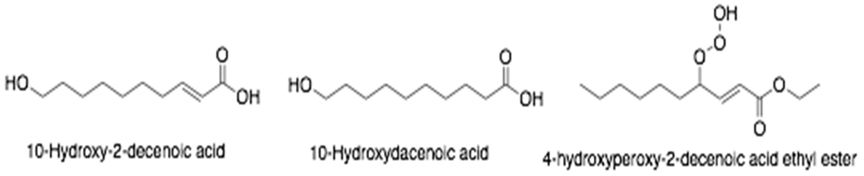
| Royal jelly | 10-hydroxy-2-decenoic acid | MCF-7 | Breast cancer | Not examined | [133] |
| Royal jelly | Not examined | Mouse 4T1 | Mouse mammary carcinoma | Increased concentration of IL-2 and interferon (INF)-α; decreased level of IL-10 | [132] |
| Royal jelly | 10-hydroxy-2-decenoic acid | U-937 | Leukemia | Induce secretion of cytokines by mononuclear cells | [138] |
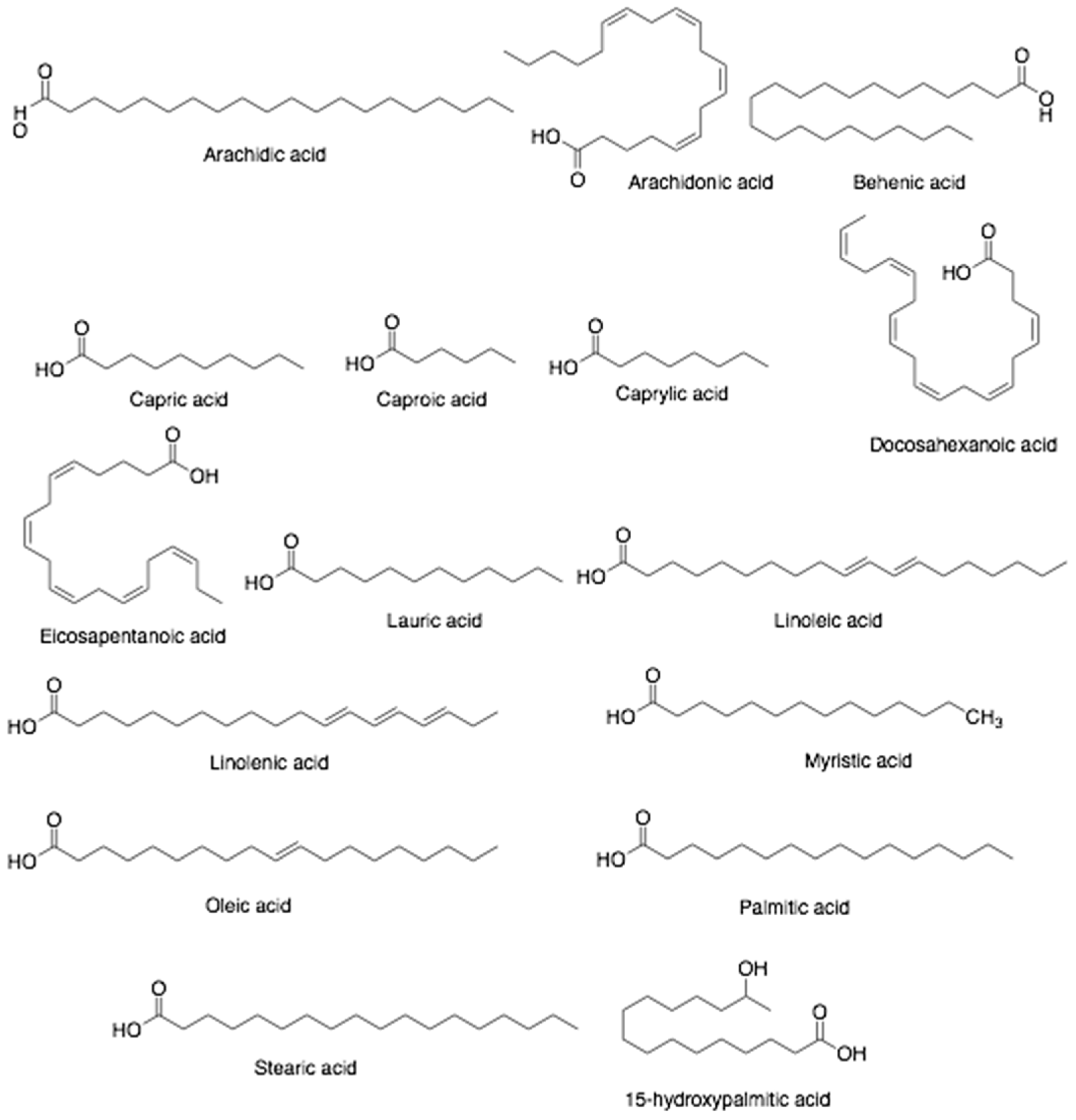
| Bee bread | Polyunsaturated fatty acids (51%) and monounsaturated fatty acids (9.9%) | Caco-2 PC-3 | Human colorectal adenocarcinoma, Human prostate adenocarcinoma | Not examined | [139] |
| Bee bread | Flavonoids and polyphenols | MCF-7 HeLa | Breast cancer Cervical cancer | Not examined | [89] |
| Bee venom | Mellitin | AGS | Gastric cancer | Disruption of cell membrane causing necrosis to the affected cells | [127] |
| Bee venom | Mellitin | A375 | Human lung cancer | Induction of apoptosis via activation of caspase-9 and caspase-3, inhibition of invasion and migration of melanoma cells through interference of f-actin reorganisation and epidermal growth factor receptor (EGFR) activity | [124] |
| Bee Products | Country | Bacteria | Assay Method | Results | Ref. |
|---|---|---|---|---|---|
| Leptospermum honey (80 different honeys derived from Leptospermum species) | Australia | Staphylococcus aureus (ATCC 25923) | In vitro (phenol equivalence assay) | Majority of Australian Lectospermum honey tested in the experiments demonstrated non-peroxide antibacterial activity (NPA) and to a greater extent correlates to their high content of methylglyoxal (MGO) and dihydroxyacetone (DHA) | [188] |
| 21 types of honey of different botanical source, derived from the Olympus mountain | Greece | Clinical isolates of methicillin-resistant S. aureus (strain 1552) and carbapenem-resistant P. aeruginosa (strain 1773) | In vitro (agar well diffusion assay and microtiter plate assay) | All honey samples yielded antibacterial activity against both tested bacteria. Some honey samples were active in a manner dependent on the presence of hydrogen peroxide and proteinaceous compounds | [189] |
| Sesamum indicum honey (seven types of sesame honey obtained from different location in West Bengal) | India | Salmonella enterica serovar Typhi, S. enterica serovar Typhimurium, Escherichia coli and Vibrio cholerae | In vitro (disc diffusion assay and microbroth dilution assay) | The antibacterial activity of sesame honey against the tested enteropathogens was good, with the best recorded activity was against both Salmonella species | [190] |
| New Zealand’s Manuka Honey (UMF +20, UMF +16, +10), Sidr honey, and Nigella sativa honey | New Zealand, Saudi Arabia | Methicillin sensitive- (ATCC 29213 and 10 strains of MSSA clinical isolates) and methicillin resistant S. aureus (ATCC 26112 and 10 strains of MRSA clinical isolates) | In vitro (disc diffusion assay and microbroth dilution assay) | Manuka honey demonstrated bactericidal activity against both MSSA and MRSA while Sidr and Nigella sativa honey yielded only bacteriostatic effect at tested concentrations | [191] |
| Melipona beecheii honey (Cuba); Manuka honey (New Zealand), Apis mellifera honey (Cuba), and African A. mellifera honey (Kenya) | New Zealand, Cuba, and Kenya | 51 clinical isolates (34 Gram-positive, 17 Gram-negative) | In vitro (microtiter plate assay for antibiofilm activity) | All honey tested in the study demonstrated good antibacterial and antibiofilm activity with Cuban M. beecheii honey had the highest activity in both | [192] |
| Ten samples of honey of different origins (polyfloral, linden, acacia, manna, and sunflower) | Romania | S. aureus, Staphylococcus epidermidis, S. enterica serovar Typhimurium, Bacillus cereus, B. subtilis, Pseudomonas aeruginosa, E. coli, and Listeria monocytogenes | In vitro (disc diffusion assay) | All honey samples demonstrated good antibacterial activity against all tested pathogens with S. aureus and P. aeruginosa were the most sensitive ones. It seems that the origins and the color of honey, but not acidity, play a role in the antibacterial activity of honey | [193] |
| Apis mellifera ligustica propolis (extracted using methanol) | Australia | S. aureus (ATCC 25923) and Klebsiella pneumoniae (ATCC 13883) | In vitro (agar diffusion and broth dilution assays) | The examined Australian propolis demonstrated antibacterial effect against S. aureus (bactericidal) but did not yield any effect on the K. pneumoniae | [194] |
| Brazilian brown and green propolis (extracted either using ethanol, hexane, or dicholometane) | Brazil | S. aureus (ATCC 6538), B. subtilis (ATCC 6633) and Micrococcus luteus (ATCC 10240) | In vitro (micro-dilution assay) | Of all samples examined in the study, the dichloromethane extract of both brown propolis and green propolis yielded the highest antibacterial effect against the tested pathogens | [195] |
| Propolis of T. fiebrigi bees (extracted using ethanol) | Brazil | S. aureus (ATCC 43300), S. aureus (ESA 654), S. epidermidis (ATCC 12228), S. epidermidis (ESA 675), Enterococcus faecalis (ATCC 43300), E. faecalis (ESA 553), K. pneumonia (ATCC 4352), K. pneumoniae (ESA 154), P. aeruginosa (ATCC 15442), P. aeruginosa (ESA 22), Proteus mirabilis (ATCC 43300) and P. mirabilis (ESA 37) | In vitro (micro-dilution assay) | The Brazilian propolis demonstrated antibacterial effect against all tested microorganisms but mainly active against Gram-positive bacteria | [196] |
| Red propolis of Africanized Apis mellifera | Brazil | Standard strains of E. coli (ATCC 25922) and S. aureus ATCC 6538 and the clinical isolates of E. coli 06 (EC06), P. aeruginosa 03 (PA03), P. aeruginosa 24 (PA24) and S. aureus 10 (SA10) | In vitro (micro-dilution assay) | The Brazilian red propolis demonstrated antibacterial activity against all tested pathogens, including the clinical isolate ones. The range of MIC values was dependent on the pathogen species (128–512 μg/mL for E. coli strains), (64 μg/mL to ≥1024 μg/mL for S. aureus strains), and (512 μg/mL for P. aeruginosa strains) | [197] |
| Red propolis, green propolis, and brown propolis (traditionally extracted using ethanol or supercritical extraction method) | Brazil | S. aureus (ATCC 25923) and Enterococcus sp. (ATCC 29712), Klebsiella sp. (ATCC 1706/700603), and E. coli (ATCC 25922) | In vitro (micro-dilution assay) | Of all samples, Brazilian red propolis extract yielded the highest antibacterial activity. Green propolis extract demonstrated weak to moderate antibacterial activity for most samples and brown propolis extract did not yield any antibacterial effect against the tested strains. None of the examined samples was active against E. coli | [198] |
| 19 propolis samples (collected from six different regions and extracted using methanol) | Chile | Methicillin-sensitive S. aureus (ATCC 25923), methicillin-resistant S. aureus (ATCC 43300), E. coli (ATCC 25922), and the clinical isolates of Pseudomonas sp., E. coli, P. mirabilis, Salmonella enteritidis, Salmonella sp. and Yersinia enterocolítica | In vitro (micro-dilution assay) | The antibacterial effect of propolis obtained from central valley was better than the ones collected from the Andreas slopes or the coastal areas. The samples’ MICs were ranging from 31.5 to >1000 µg/mL and the ones with MIC ≤ 62.5 µg/mL demonstrated good antibacterial effect against Pseudomonas sp., E. coli, S. enteritidis, and Y. enterocolitica | [199] |
| Propolis collected from different geographical regions (extracted using either ethanol or water) | Germany, Irlandia, Czech Republic | S. aureus, Staphylococcus saprophyticus, S. epidermidis, S. pneumoniae, Streptococcus pyogenes, Streptococcus oralis, Streptococcus agalactia, Streptococcus thermophiles, B. subtilis, Enterococcus casseliflavus, K. pneumonia, Klebsiella oxytoca, E. coli, E. coli O157.H7, P. aeruginosa, Salmonella choleraesuis, Shigella flexneri, Haemophilue influenza, Acinetobacter baumanii, Burkholderia cepacia, Y. enterocolitis, Enterobacter cloacae, one strain of MRSA, and one strain of Vancomycin-resistant enterococci (VRE) | In vitro (micro-dilution assay) | Both ethanol and water propolis extracts demonstrated good antimicrobial activity against most of Gram-positive bacteria (range of MICs: 0.08–5 mg/mL), with the Irish propolis yielded the highest bactericidal effect followed by Czech and German. All propolis extracts demonstrated moderate antibacterial against MRSA and VRE and also against β-lactamase positive H. influenzae, and S. pneumoniae. Propolis ethanol extract, but not water extract, yielded moderate antibacterial activity against Gtam-negative pathogens tested in the study (MICs: 0.6–5 mg/mL) | [200] |
| Propolis and bud poplar resins (extracted using ethanol) | Italy | P. aeruginosa PAO1 (ATCC 15692) and transgenic P. aeruginosa (P1242) with the luciferase gene and luciferin substrate (under the control of a constitutive P1 integron promoter) | In vitro (micro-dilution assay) | Both ethanol extracts (propolis and bud poplar resins) demonstrated good antibacterial activity against P. aeruginosa biofilm and negatively affected the swimming and swarming motility properties of P. aeruginosa | [201] |
| 24 propolis samples (collected from different geographical location in Morocco; hydro-alcoholic extracts) | Morocco | S. aureus (ATCC 6538) and three clinical isolates of MRSA (MRSA2, MRSA 15, and MRSA 16) | In vitro (disk diffusion method) | Propolis extract (MIC 0.36 mg/mL) was able to attenuate the virulence of S. aureus ATCC 6538 and the MRSA strains. The impairment of biofilm formation was also observed | [202] |
| Propolis of Populus alba, P. nigra, P. tremula, Acer pseudoplatanus, Betula verucosa, Pinus silvestris, and Aesculus hippocastanum (extracted using methanol or dichlorometane) | Poland | S. aureus (ATCC 25923), S. epidermidis (ATCC 12228), P. aeruginosa (ATCC 227853), E. coli (ATCC 25922), E. cloacae (ATCC 13047), and K. pneumoniae (ATCC 13883) | In vitro (disk diffusion method) | The dichloromethane extract of propolis demonstrated good antibacterial activity against all tested pathogens (MICs: 0.90–1.34 mg/mL) | [203] |
| Green propolis (extracted using ethanol, methanol, diethyl ether or water) | Taiwan | S. aureus (BCRC 10780, BCRC 10781 and BCRC 10451), MRSA (ATCC 43300), B. subtilis (BCRC 10255), L. monocytogenes (BCRC 14845), P. aeruginosa (BCRC 10944), and E. coli (BCRC 10675) | In vitro (micro-dilution assay) | Taiwanese green propolis extracts demonstrated good antibacterial activity against MRSA and all propolins, particularly propolin C, yielded good efficacy against S. aureus, B. subtilis, and L. monocytogenes | [204] |
| 29 bee pollen samples (collected during the dry seasons of 2016, 2017, and 2018; extracted using ethanol) | Chile | S. aureus (ATCC 25923), P. aeruginosa (ATCC 27853), E. coli (ATCC 25922), and S. pyogenes (I.S.P. 364-00) | In vitro (disk diffusion method and broth dilution method) | All bee pollen extracts (collected in three different years) demonstrated good antibacterial activity against S. pyogenes but did not yield antibacterial effect on S. aureus, P. aeruginosa, and E. coli | [169] |
| Three Greek bee pollen (collected from from Cistus creticus L. (rock rose) at three different locations; extracted using ethanol, butanol, dichloromethane, or cyclohexane) | Greece | S. aureus (ATCC 25923), S. epidermidis (ATCC 12228), P. aeruginosa (ATCC 227853), E. coli (ATCC 25922), E. cloacae (ATCC 13047), and K. pneumoniae (ATCC 13883) | In vitro (micro-dilution assay) | The butanol extract demonstrated good antibacterial activity against all pathogens tested in the study, probably due to the high content of flavonoids, such as quercetin and kaempferol glucosides. No antimicrobial activity was seen in both cyclohexane and dichlorometane extracts | [205] |
| Bee bread and propolis of Stingless bee (Heterotrigona itama species; extracted using ethanol or hexane) | Malaysia | S. aureus, B. subtilis, E. coli, and Salmonella | In vitro (disk diffusion method and broth dilution method) | All bee bread and propolis extracts demonstrated good antimicrobial activity against all tested bacteria (MIC: <6.67–33.33 µL/mL), with higher preference to Gram-positive (S. aureus and B. cereus) than Gram-negative bacteria (E. coli and Salmonella). Ethanol extracts yielded stronger antibacterial activity than their hexanic counterparts | [206] |
| Castanea sativa Mill. pollen grains (collected at Erfelek (4 sites) and Ayancık (5 sites) district of Sinop; extracted using methanol) | Turkey | S. aureus (ATCC 6538), MRSA, E. faecalis (ATCC 51299), M. luteus, Bacillus cereus (7064), Vancomycin-resistant Enterococcus (VRE), E. coli (ATCC 11293), and K. pneumonia | In vitro (disk diffusion method) | Bee pollen extracts yielded higher antibacterial activity against Gram-positive bacteria than their Gram-negative counterparts | [207] |
| Royal jelly sample | Singapore | Fusobacterium mucleatum, Aggregatibacter actinomycetemcomitans, Porphyromonas gingivalis, and Prevotella intermedia | In vitro (micro-dilution assay) | Royal jelly demonstrated good antibacterial activity against periodontopathic bacteria tested in the study | [208] |
| Royal jelly sample (Yamada Bee Farm, Okayama, Japan) | Japan | P. aeruginosa (PAO1) wild-type strain and four clinical isolates (TUH-54, TUH-124, TUH-188, and TUH-213) | In vitro (micro-dilution assay and microtiter plate biofilm assay) | Royal jelly did not yield antipseudomonal activity at concentration of 50% w/v. However, at concentration of less than 25%, antiadherent activity was observed on both the abiotic surface and the epithelial cell model | [209] |
| Bee bread sample (hydromethanolic extract) | Morocco | S. aureus (ATCC 6538), B. cereus (food isolate), L. monocytogenes (NCTC 7973), E. coli (ATCC 35210), E. cloacae (human isolate), S. Typhimurium (ATCC 13311) | In vitro (micro-dilution assay) | Hydromethanolic extract of bee bread demonstrated antibacterial activity against all tested pathogens (MIC: 0.04–0.175 mg/mL; MBC: 0.08–0.35 mg/mL) | [210] |
| Bee bread (five different regions of Ukraine; extracted using ethanol) | Ukraine | S. aureus (CCM 4223), E. coli (CCM 3988), S. enterica subs. enterica (CCM 3807), and Bacillus thuringiensis (CCM 19) | In vitro (disk diffusion method) | All bee bread samples yielded good antibacterial activity against antibiotic-resistant Gram-positive and Gram-negative bacteria tested in the study. The antibacterial strength of bee bread samples on certain bacteria was varied, mainly depends on the geographical location of sample collection | [211] |
| Apis mellifera venom (compared to mellitin) | n/a | Borrelia burgdorferi strain B31 (ATCC 35210) | SYBR Green I/Propidium Iodide assay and biofilm assay | Both bee venom and mellitin demonstrated good antimicrobial activity against the free form of B. burgdorferi and the biofilm-associated form | [162] |
| Apis mellifera venom (compared to mellitin) | Brazil | S. aureus (ATCC 13565, ATCC 14558, ATCC 19095, and ATCC 23235, all Enterotoxigenic), and five clinical isolates of MRSA (recovered from human specimens) | The resazurin microtiter assay (REMA) | Apitoxin and melittin yielded antibacterial activity against MRSA (MIC: 7.2 μg/mL, and 6.7 μg/mL, respectively). Apitoxin and melittin had no effect on the production of enterotoxin and/or its release. | [212] |
| Apis mellifera venom (commercial, freeze-dried) | n/a | Pseudomonas putida (ATCC 700008), Pseudomonas fuorescens (NCIMB 9046) and E. coli K-12, MG1655 (ATCC 47076), | In vitro viability assay and biochemical analysis | Bee venom exerted its antibacterial activities against the tested pathogens via alteration in the membrane permeability, destruction of bacterial cell wall, cell contents leakage, and inactivation of bacterial metabolic activity leading to cell death | [213] |
| Apis mellifera venom (apitoxin) | Ecuador | Salmonella (CECT 4395) and L. monocytogenes (CECT 934) and 49 S. enterica and 7 L. monocytogenes strains isolated from poultry products | In vitro (micro-dilution assay) | Apitoxin is effective against foodborne pathogens tested in the study (MIC range: 16–32 µg/mL for L. monocytogenes and 256–1024 µg/mL for S. enterica) | [214] |
| Bee Products | Country | Parasites | Assay Method | Results | Refs. |
|---|---|---|---|---|---|
| Propolis extracts (12 samples from different location in Libya) | Libya | Trypanosoma brucei (s427); Leishmania donovani; Plasmodium falciparum | In vitro | All the extracts are to some extent effective against all of the tested protozoa. There are regional variations in the antiparasitic properties | [223] |
| Ethanolic extracts of European propolis (35 different samples) | Europe | Trypanosoma brucei 427WT; Trypanosoma brucei B48 (resistant strain); Trypanosoma congolense; Leishmania mexicana WT; Leishmania mexicana C12Rx (resistant strain) | In vitro | All extracts display high level activities against the parasites. Yet, there are regional variations in the antiparasitic properties | [225] |
| Hydroethanolic red propolis extracts | Brazil | Trypanosoma cruzi; Leishmania braziliensis vianna; Leishmania infantum promastigotes | In vitro | The extracts exhibit strong cytotoxic properties against the protozoan parasites | [227] |
| Ethanolic extracts of red propolis | Brazil | Schistosoma mansoni | In vitro, in vivo | Propolis displays antischistosomal properties by decreasing motility and producing: mortality of adult parasites; morphological disruptions in the schistosomes’ tegument; and substantial impairment in egg generation. Propolis also significantly lower the worm and egg burden in both early and chronic S. mansoni murine infection model | [229] |
| Ethanolic extracts of red propolis | Brazil | Trypanosoma cruzi | In vitro | Ethanolic propolis extracts have high inhibitory activity against T. cruzi | [198] |
| Dry, alcoholic, and glycolic green propolis extracts | Brazil | Leishmania (Viannia) braziliensis promastigotes and amastigotes | In vitro | The extracts exhibits antileishmanial activity against promastigotes and amastigotes stages of the parasite | [237] |
| Ethanolic extracts of red and green propolis | Brazil | Leishmania (Vianna) braziliensis | n.a | Both propolis extracts exhibits leishmanicidal effect in a dose-dependent manner. Yet, the red propolis extract displays a higher efficacy than the green extract | [231] |
| Hydroethanolic extract of red propolis | Brazil | Leishmania chagasi promastigote; Leishmania amazonensis promastigote | In vitro | The extract exhibits leishmanicidal potency againts the parasites | [243] |
| Hydroalcoholic extract of Brazilian propolis | Brazil | Leishmania (Viannia) braziliensis | In vitro | Propolis extract shows immunomodulatory effects, by enhancing IL-4 and IL-17 and lowering IL-10, and therefore, may protect against Leishmania infection and clear the parasite | [239] |
| Hydroalcoholic extract of Brazilian green propolis | Brazil | Leishmania amazonensis promastigotes and amastigotes | In vitro In vivo | Propolis extract decreases the viability of L. amazonensis amastigotes and promastigotes. The extract also decreases the parasites and stimulates the macrophage recruitment into the lesion caused by the L. amazonensis | [236] |
| Degradation products of major compounds of green propolis: Z-artepillin C and Z-p-coumaric Acid | Brazil | Leishmania amazonensis promastigotes and amastigotes | In vitro | Both Z-artepillin C and Z-p-coumaric acid display a promising and significant activity against L. amazonensis | [244] |
| Hydroalcoholic extract of Melipona fasciculata geopropolis | Brazil | Leishmania amazonensis promastigotes and amastigotes | In vitro | Geopropolis has an antileishmanial effect and effective in reducing the number of the L. amazonensis promastigotes and amastigotes | [233] |
| Isolated compound of Bee pollen monofloral | Brazil | Leishmania amazonensis | In vitro | The isolated compound identified as the biflavonoid rhusflavone shows high antileishmanial effect against L. amazonensis promastigotes and amastigotes | [234] |
| Bee venom and propolis | Egypt | Schistosoma mansoni | In vivo | Bee venom and propolis exerts anti-schistosomal activities by substantially lowering the mean total numbers of worm, mean values of immature and mature egg as well as the ova count in hepatic tissue | [245] |
| Egyptian propolis ethanolic extract | Egypt | Toxocara vitulorum | In vitro | The extract exhibits anthelmintic efficacy and the nematicidal effect is dose-dependent | [246] |
| Egyptian propolis ethanolic extract | Egypt | Blastocystis spp. | In vitro | The extract presents a notable obstructive effect on the growth of Blastocystis parasites | [247] |
| Egyptian propolis ethanolic and water extract | Egypt | Cryptosporidium spp. | In vivo | The prophylactic and therapeutic administration of the extracts moderately effective in reducing the oocysts shedding on cryptosporidiosis infected rats | [248] |
| Egyptian propolis ethanolic extract | Egypt | Toxoplasma gondii | In vivo | Propolis markedly decreases the amount of IL-1β, IL-6, and TNFα in T. gondii infected models | [249] |
| Egyptian propolis ethanolic extract | Egypt | Giardia lamblia | In vivo | Propolis markedly reduce the G. lamblia trophozoites count | [250] |
| Ziziphus spina-christi honey; Acacia nilotca honey; Acacia seyal honey; Cucurbita maxima honey | Saudi Arabia | Entamoeba histolytica; Giardia lamblia | In vitro | All honeys are potentially effective to be used as antiamoebic and antigiardial agents since they can halt the growth of the trophozoites | [47] |
| Ethanolic extraction of Saudi propolis | Saudi Arabia | Trypanosoma brucei | In vitro | The extract indicates a significant anti-trypanosomal activity | [251] |
| Capparis spinosa honey | Saudi Arabia | Toxoplasma gondii | In vivo | Honey elevates the amount of antibody titer and the cytokines (IFN-γ, IL-1, and IL-6) in T. gondii infected rats | [241] |
| Ethanol and dichloromethane Propolis extracts | Iran | Plasmodium falciparum | In vitro | All extracts show concentration-dependent anti-plasmodial activity. Dichloromethane extract has the most potent inhibitory effect | [252] |
| Ethanolic extract of propolis | Iran | Leishmania major | In vitro, In vivo | Both tests indicate that the extract has an effective antileishmanial activity against L. major. The extract reduces the number of promastigotes and decreases the size of ulcers significantly | [253] |
| Ethanolic extract of algerian propolis | Algeria | Echinococcus granulosus | In vitro, In vivo | Both tests indicate that the extract is an effective antihydatic scolicidal effect since it has a major scolicidal activity against E. granulosus at all tested concentration and reduces cystic echinococcosis development in in vivo model | [254] |
| Methanolic extracts of propolis (ten different propolis from different geographical area in Bolivia) | Bolivia | Leishmania amazonensis; Leishmania braziliensis | In vitro | All propolis extracts show growth inhibition against both protozoa. Propolis with rich phenolic contents shows the best antiprotozoal effect | [255] |
| Methanolic extracts of propolis (three different propolis from different geographical area in Ecuador) | Ecuador | Leishmania amazonensis | In vitro | All propolis extracts show growth inhibition against the protozoa. Propolis with the richest flavonoids contents shows the best antiprotozoal effect | [235] |
| Ethanolic extracts of propolis (twelve different propolis from eight different geographical area in Nigeria) | Nigeria | Trypanosoma brucei (s427, wild-type); Trypanosoma brucei (B48, resistant strain); Trypanosoma brucei (aqp2/aqp3 null, resistant strain) | In vitro | The extracts are active against all the tested parasites | [224] |
| Isolated phenolic compounds of Nigerian red propolis | Nigeria | Trypanosoma brucei (s427, wild-type); Trypanosoma brucei (B48, resistant strain); Trypanosoma brucei (aqp2/aqp3 null, resistant strain) | In vitro | The extract displays moderate to high antitrypanosomal effectivity against all the tested parasites | [256] |
| Tanzanian propolis ethanolic extract | Tanzania | Trypanosoma brucei (s427, wild-type); Trypanosoma brucei (B48, resistant strain) | In vitro | The extract displays antitrypanosomal potency against both parasites | [257] |
| Zambian propolis ethanolic extract | Zambia | Trypanosoma brucei (s427, wild-type); Trypanosoma brucei (B48, resistant strain) | In vitro | The extract displays antitrypanosomal activity against the wild type of T. brucei and the multi-drug resistant clone | [257] |
Publisher’s Note: MDPI stays neutral with regard to jurisdictional claims in published maps and institutional affiliations. |
© 2021 by the authors. Licensee MDPI, Basel, Switzerland. This article is an open access article distributed under the terms and conditions of the Creative Commons Attribution (CC BY) license (https://creativecommons.org/licenses/by/4.0/).
Share and Cite
Nainu, F.; Masyita, A.; Bahar, M.A.; Raihan, M.; Prova, S.R.; Mitra, S.; Emran, T.B.; Simal-Gandara, J. Pharmaceutical Prospects of Bee Products: Special Focus on Anticancer, Antibacterial, Antiviral, and Antiparasitic Properties. Antibiotics 2021, 10, 822. https://doi.org/10.3390/antibiotics10070822
Nainu F, Masyita A, Bahar MA, Raihan M, Prova SR, Mitra S, Emran TB, Simal-Gandara J. Pharmaceutical Prospects of Bee Products: Special Focus on Anticancer, Antibacterial, Antiviral, and Antiparasitic Properties. Antibiotics. 2021; 10(7):822. https://doi.org/10.3390/antibiotics10070822
Chicago/Turabian StyleNainu, Firzan, Ayu Masyita, Muh. Akbar Bahar, Muhammad Raihan, Shajuthi Rahman Prova, Saikat Mitra, Talha Bin Emran, and Jesus Simal-Gandara. 2021. "Pharmaceutical Prospects of Bee Products: Special Focus on Anticancer, Antibacterial, Antiviral, and Antiparasitic Properties" Antibiotics 10, no. 7: 822. https://doi.org/10.3390/antibiotics10070822