Review

Recent Progress of Electrochemical Aptasensors toward AFB1 Detection (2018–2023)

1
Department of Analytical Chemistry, Faculty of Pharmacy, “Iuliu Haţieganu” University of Medicine and Pharmacy, 4 Pasteur Street, 400349 Cluj-Napoca, Romania
2
Department of Surgery, Faculty of Medicine, “Iuliu Haţieganu” University of Medicine and Pharmacy, 400012 Cluj-Napoca, Romania
*
Authors to whom correspondence should be addressed.
These authors contributed equally in this work.
Biosensors 2024, 14(1), 7; https://doi.org/10.3390/bios14010007
Submission received: 13 November 2023 / Revised: 12 December 2023 / Accepted: 20 December 2023 / Published: 22 December 2023
(This article belongs to the Special Issue Women in Biosensors (Volume II))

Abstract

:
Food contaminants represent possible threats to humans and animals as severe food safety hazards. Prolonged exposure to contaminated food often leads to chronic diseases such as cancer, kidney or liver failure, immunosuppression, or genotoxicity. Aflatoxins are naturally produced by strains of the fungi species Aspergillus, which is one of the most critical and poisonous food contaminants worldwide. Given the high percentage of contaminated food products, traditional detection methods often prove inadequate. Thus, it becomes imperative to develop fast, accurate, and easy-to-use analytical methods to enable safe food products and good practices policies. Focusing on the recent progress (2018–2023) of electrochemical aptasensors for aflatoxin B1 (AFB1) detection in food and beverage samples, without pretending to be exhaustive, we present an overview of the most important label-free and labeled sensing strategies. Simultaneous and competitive aptamer-based strategies are also discussed. The aptasensors are summarized in tabular format according to the detection mode. Sample treatments performed prior analysis are discussed. Emphasis was placed on the nanomaterials used in the aptasensors’ design for aptamer-tailored immobilization and/or signal amplification. The advantages and limitations of AFB1 electrochemical aptasensors for field detection are presented.

1. Introduction

Mycotoxins, a group of secondary metabolites produced by filamentous fungi, contaminate around 25% of the world’s agricultural products during different stages of their economic life: growth, harvest, storage, and processing [1]. They represent a large group of mycotoxins, but the most predominant are nivalenol, 3-acetyl-deoxynivalenol, 15-actyl-deoxinivalenol, ochratoxin (OTA), aflatoxin B1 (AFB1), and fumonisin B1 (FB1) [2,3]. AFB1 is probably the most widely known and definitely the most toxic of its group. It is known to have the ability to bind cell DNA and increase the risk of liver cancer, even if it might be found just in traces. Considering this, together with its mutagenic, teratogenic, immunosuppressive, and carcinogenic mechanism of action, AFB1 has been included by the International Agency for Research on Cancer in the number one group of carcinogens [2,3,4]. Without a doubt, there is a need to keep AFB1 concentrations under control and the contamination at a minimum level. Therefore, many local food and health agencies set a limit for AFB1 in different food products. Usually, the limits are between 0.05 and 20 ng/mL for cereals and cereal-derived products, where the risk of contamination is the highest [5]. But in some countries, like the USA, the limits can be set by the Food and Drug Administration (FDA) even for corn or peanuts [6].
In order to keep control of AFB1 levels, a continuous and rigorous analysis of its concentration in food products is needed. Traditional methods like high-performance liquid chromatography, mass spectrometry, or a combination of both [7,8,9,10] are reliable with very good sensitivity and selectivity, but they are very expensive, time-consuming, and lack the ability to analyze a high number of samples in different environments [11].
A good alternative to traditional methods could be electrochemical sensors. An electrochemical sensor stands for the sensor in which the transducer is an electrode surface. Some essential advantages derive from this, like (i) the use of the electron for signal acquisition, which is considered a green model for analytical applications, with minimal or no waste generation, (ii) the ability of miniaturization in order to develop portable devices, (iii) the short response time, and (iv) the low production costs [12]. The use of a recognition element in the development of an electrochemical sensor increases its selectivity. When the recognition element is represented by a biological-derived compound like an aptamer, antibody, bacteria, enzyme, or whole cell, it is called a biosensor. The use of a bioelement as a recognition system leads to high selectivity for the target analyte mainly because of the specific interaction between the analyte and the receptor. More importantly, the specific interaction prevents signal interference from other matrix components, which could modify the sensor’s response to a more specific one [13].
Aptamers are short single-stranded RNA or DNA sequences, selected by the SELEX (Systematic Evolution of Ligands by Exponential enrichment) technique from synthetic oligonucleotide libraries containing up to 1015 different sequences. Since the first SELEX selection described in 1990 [14,15], a significant number of aptamer sequences with high affinity and selectivity toward small molecules have been reported. The current SELEX technologies rely on the core process of the iterative in vitro selection of sequences based on successive key steps such as the binding, partitioning, and amplification of the bound sequences, but new features were introduced to reduce the selection time and costs and improve the aptamers’ stability and efficiency [16,17].
Compared with other biological recognition elements, they come with some advantages like good stability, wide target range, easy synthesis, low development cost, and most importantly, a good capacity for structural modification according to the scientific needs. As already stated, their ability to form different structures like hairpins, pseudoknots, convex rings, and G-quadruplexes leads to an increased conformational recognition of the targets similar to an antigen–antibody reaction [18]. In electrochemical sensing, their role is to bind different specific targeted molecules, from large to small organic molecules, such as proteins, biomarkers, xenobiotics, and toxins, to name a few [19]. When used in biosensors as bio-recognition elements, aptamers can be used directly linked to the transducer, both as a single-target or multi-target probe (labeled or label-free). Another possible approach is given by the possibility to use aptamer–target interaction to indirectly activate on–off devices in which the interaction with the analyte or the aptamer itself inhibits certain reactivity. Moreover, aptamers are subjected to significant conformational change caused by the interaction with the analyte that can be used as a recognition parameter when combined with appropriate transducers [20].
Ever since the development of the first AFB1-specific aptamer in 2012 (Patent > PCT/CA2010/001292), more and more AFB1 electrochemical aptasensors have been published, showing at the same time the need and the continuous interest toward fast and reliable AFB1 detection.
According to the Scopus database, 400 papers (original papers and reviews) were found using the terms “aptamer” and “AFB1” and 206 papers when using the terms “aptasensor” and “AFB1”. The publication trend over time saw a considerable decrease in 2021 due to the impact of the COVID-19 pandemic, but rapidly recovered by the end of 2022, outgrowing any values reached by then (Figure 1A). Among these, the electrochemical aptasensing of AFB1 accounts for 38% of original papers and for 78% of review papers (Figure 1B). In addition, regardless of the combination of keywords used in the literature search, the number of publications in 2022 almost doubled compared to the pre-pandemic years. Reviews reporting the detection of toxins [21,22], mycotoxins [18,23,24], or aflatoxins [25,26,27,28] based on analytical methods in general [21,27,29], or specifically by optical [22,23,26,28] and/or electrochemical [18,22,23,25] transduction methods, have been published relatively recently (2022–2023). However, only a few of these have devoted the discussion over the use of aptamers for aflatoxin detection [21,24,26], but none specifically on AFB1 via electrochemical detection strategies. For this reason, an overview of the latest progress in AFB1 electrochemical detection based on aptamers (2018–2023) is of high interest. This review will also provide insights into the challenges and prospects of electrochemical aptasensors for the on-site detection of AFB1.
The following sections present the most relevant approaches reporting AFB1 aptamer-based electrochemical detection, classified according to the detection mode: label-free and labeled assays (Figure 2), out of which competitive and simultaneous assays were treated as separate sections due to their possible future perspectives. Exceptional examples of aptamer DNA nanoarchitectures are presented. This review also examines the possible applications, challenges, and prospects of electrochemical aptasensors for the on-site detection of AFB1.

2. Food Contamination with AFB1 (Incidence, Toxicity, and Legal Limits)

Aflatoxins, among the numerous mycotoxins discovered so far, have emerged as a subject of profound scientific interest. This is due to their remarkable toxicity and carcinogenic potential, with some aflatoxin subtypes being officially included in Group 1 carcinogens by the International Agency for Research on Cancer [30]. These molecules are metabolites of the fungal species Aspergillus flavus and Aspergillus parasiticus, which are often found in food and feed, primarily in peanuts, corn, and rice. The tropical and subtropical climate favors the development of both fungi and toxins, as it provides the optimal temperature and humidity. However, their presence in food is not solely determined by geographical location but may also arise in response to inadequate conditions of storage, transportation, or crop processing [31,32].
When consumed in high quantities, aflatoxins can precipitate acute toxic responses, leading to a condition known as aflatoxicosis. Aflatoxicosis carries the potential for fatal consequences, primarily through the induction of hepatic failure. Additionally, aflatoxins can also have chronic toxic effects, manifesting as carcinogenicity affecting various organs, with a predilection for the liver and kidneys [33].
Under the action of cytochrome P450 enzymes, especially CYP3A4, the epoxidation of AFB1 occurs, resulting in the formation of 8,9-epoxy-AFB1. Only the 8,9-exo-epoxy-AFB1 isomer exhibits mutagenic activity by forming adducts with DNA or proteins, while the endo isomer is non-toxic. Due to interindividual variability in the expression of cytochrome P450 enzymes, some individuals may be more susceptible to the formation of the toxic metabolite, thus having an increased risk of developing liver carcinoma [34].
Hence, the presence of AFB1 in food items has been acknowledged as a serious food safety concern, resulting in the implementation of certain maximum levels by authorities. Permissible thresholds for AFB1 levels vary across different regions and countries around the world, with the maximum permissible for most consumer items in the European Union being 2 ng/mL, 10 ng/mL in Japan and Korea, and 20 ng/mL in the United States [35,36,37].

3. Nanomaterials Used for Electrode Modification

One of the biggest challenges in biosensors’ development is to obtain a very selective method and a good signal amplification to enable a low limit of detection (LOD). Since traditional electrochemical cells lack selectivity and sensitivity, different nanomaterials must be grafted on the surface of the working electrodes [38]. Hence, due to their functional groups and ease of conjugation, one key role relies on anchoring the aptamer sequences at the electrode surface. Depending on the application envisaged, the size and functionalities may vary.
Nanomaterials are divided according to their size and structure into zero dimensional (0D) nanomaterials like nanoparticles [39], QDs [24] or nanoclusters [40], 1D nanomaterials, such as nanotubes [41], nanowires [42], and nanorods [43], and 2D nanomaterials where the most representative examples are solid crystalline 1D nanomaterials that can form strong interlayer interactions [44]. In addition, they can form interactions with other layers of different nanomaterials, obtaining better physicochemical properties and new optical and electrochemical features, extremely helpful in DNA sensing [22,45]. The last and most recent category, 3D nanomaterials, is a combination of 0D–2D materials. Most often, they are developed by combining different conventional nanomaterials with a nanocomposite, or by the self-assembly of different nanoparticle systems [46].
Analyzing their composition, different types of nanomaterials have been used in the last couple of years for toxin detection and specifically for AFB1 electrochemical detection. Conductive polymers are widely known for their good electric conductivity acquired through redox moieties. Moreover, controlled polymerization allows for the formation of polymer structures that are not only similar on the entire WE surface, but also make possible the incorporation of different nanomaterials, which improves the accuracy [47]. Polyheteroaromatic polymers, such as polypyrrole, polyaniline (PANI), and their derivatives, have attracted a lot of attention lately because of their possible application toward electrochemical detection [48,49,50]. Being one of the most versatile conductive polymers, PANI was used in order to increase the electroconductive properties of a platform developed for AFB1 aptasensing in wine with very low LODs, down to 0.002 fg/mL [51].
Metallic nanoparticles (MNPs) are nanosized metals with sizes ranging from 10 nm to 100 nm. The development of novel MNPs with different structures, shapes, and dimensions has increasingly attracted the attention of the scientific world in the last few years. They have unique properties, such as high electrical conductivity, large surface-to-volume ratio, increased biocompatibility, and the ability to increase the catalytic activity of the electrochemical platform. All of these provide a huge space for MNPs in improving the sensors’ overall performance [52].
Carbon-based nanomaterials have some advantages compared with metal-based nanomaterials, including a lower price and higher surface area, thus leading to the possibility of the immobilization of a high amount of biological compounds. They are also very versatile, and a high number of structures can be obtained with specific characteristics [53]. From the carbon-based category, the most used are graphene oxides, which maintain their oxidized (GO) form, and reduced graphene oxides (rGOs), which have slightly modified electrochemical properties. GOs distinguish themselves by having distinctive properties like a greater hydrophilicity due to the high number of reactive oxygen groups, easily controllable electronic properties, wider potential window, and negligible residual current [54]. Beheshti-Marnani et al. developed an easy-to-use, electrochemical aptasensor for AFB1 detection, using only rGO nanosheets immobilized on the surface of a glassy carbon electrode (GCE) and an amino-labeled aptamer [55].
Nanomaterials play multiple roles in aptasensors’ design, but sometimes the sensitivity obtained by using different combinations of nanomaterials fails to meet the detection of AFB1 in trace amounts. Aptasensors based on multiple amplification strategies could be the solution to achieving the sensitivity goals. These include the use of enzymes, nanozymes, and DNAzymes [56]. An alternative could be the utilization of an exonuclease-assisted signal amplification strategy, usually promoted by exonuclease I (EXO I) [57,58], EXO II, or EXO III [59,60]. Zheng et al. performed a two-round signal amplification using EXO III and telomerase to obtain an LOD as low as 60 ag/mL using a voltametric technique and an AFB1 aptasensor. On the first amplification round, the telomerase was amplified to elongate the ssDNA probes on the gold nanoparticles deposed on the working electrode surface, leading to a “signal-off” electrochemical response. On the second step, the EXO III amplification had the role of hydrolyzing the 3′-end of the dsDNA right after the interaction with the AFB1 target, leading to a release of bound AFB1 back into the solution, starting a new recognition–amplification cycle [60]. The review of Rhouati et al. presents an in-depth analysis about enzyme-assisted amplification strategies in electrochemical aptasensors [56].

4. Aptamers for AFB1

Table 1 outlines the main aptamers for AFB1 used in the design of the electrochemical aptasensors evaluated in this review (48 studies), detailing their nucleotide content, length, affinity constants, original selection paper (or papers evaluating affinity constants of truncated versions), and the secondary structures. Notably, regardless of the detection strategy, the leading aptamer sequence is still the first patented aptamer for AFB1, denoted as Apt1 here, which was selected by Linda C. Le, Jorge A. Cruz-Aguado, and Gregory A. Penner from Neoventures Biotechnology LTD (London, ON, Canada) in 2012 [61]. Apt1 shares 60% of studies, followed by Apt 1 truncated versions 1 and 3, with 11% and 9%, respectively. However, to the best of our knowledge, only a few papers report the affinity constants of the aptamers and due to the considerable amount of the publications on AFB1 aptasensing, papers fail to report the source of the sequences used. This leads to misinformation and possible error generation, such as undesired sequence truncation or mismatches. For example, Apt 5 was selected by Cruz-Aguado and Penner for Ochratoxin A in 2008 [62]. No data were found concerning the affinity of the sequence towardsAFB1; instead, other papers might have reported it. The secondary structures were predicted by the Mfold webserver [63] by setting up the concentrations of 0.1 M Na+ and 0.05M Mg2+ and the temperature to 25 °C. Most aptamers present a central stem–loop secondary structure with a number of base pairs ranging from 1 up to 7. Truncated versions of the primary Apt1 retained good affinities toward AFB1, suggesting that the stem–loop has a great contribution in the interaction with the target. As not all sequences were found in the literature to compare the dissociation constants, Gibbs free energy (ΔG) could be an indicator of the nature of the aptamer–AFB1 interaction and the affinity of the aptamer. Generally, the lower the ΔG value, the higher the affinity [64]. Still, among all, Apt1 and its truncated version 2 have exhibited the lowest values.

5. Food and Beverage Samples Treatment

Because of the good thermal and chemical stability of AFB1, different pretreatment steps have been reported, mostly among the traditional sample pretreatment methods, such as liquid–liquid extraction, centrifugation, and filtration. Generally, the samples are treated with organic mixtures to ensure the extraction of AFB1, such as methanol:water (v/v) (6:4) [51], (7:3) [42,69], (8:2) [70] or acetonitrile buffer (v/v) (7:3) [71]. A mixing step of about 30 min [69] up to 5 h and a further centrifugation and filtration through 0.22 μm or 0.45 μm filter membranes are usually performed. However, limitations such as a high consumption of organic solvents, time-consuming steps, poor selectivity, and low recovery and enrichment factors are encountered. Solid-phase microextraction, magnetic solid-phase extraction, dispersive solid-phase extraction, and matrix solid-phase dispersion extraction can overcome the disadvantages of traditional methods. Materials with efficient adsorption properties, high and easily tailored surface areas, such as metallic organic frameworks (MOFs), covalent organic frameworks (COFs), molecularly imprinted polymers (MIPs), carbon-based nanomaterials, and aerogels, become of interest in the pretreatment step of foodstuffs [72]. MOFs, as emerging advanced materials, may serve both as a pretreatment tool and electrode surface modifier [73]. To improve the enrichment efficiency, magnetic particles can be incorporated into the MOF structure [74]. Enhanced selectivity can be achieved using biological (antibody) or biomimetic molecules (aptamer or molecularly imprinted polymers). Purification steps with immunoaffinity columns containing antibodies specific to AFB1 ensure the pre-concentration of and reduction in the matrix effect [75]. Although aptamers are stable in different media, aqueous mixtures are of election for electrochemical measurements, especially at screen-printed electrodes. Phosphate or TRIS buffers are usually used for the dilution of extracts prior to measurements. Corn flour and oil [42], wheat flour, peanuts [51,75,76] and peanut oil [42], beer [71], and wine [71,77] are commonly tested for the presence of AFB1. The standard addition method is frequently reported with spiking AFB1 solutions in the pg-ng/mL concentration range.
Advanced and efficient sample pretreatment methods for electrochemical sensing are still a major requirement nowadays. Recent reviews [72,78] report advanced methods for food sample preparation, but, generally, the analyte determination is further performed via conventional chromatographic methods. Sufficient enrichment of and efficient reduction in the matrix interference must be achieved to improve the accuracy and sensitivity of the electrochemical assays to address the food analysis requirements.

6. Applications of Electrochemical Aptasensors for AFB1 Detection

The increased need for fast, accurate, and feasible detection methods of mycotoxins turned the scientific world’s attention toward biosensing. Novel detection methods had to be found and applied in real samples, with very low LODs and high selectivity.
Aptamers have been incorporated into detection devices, such as biosensors, with the role of a biological recognition element and used more and more often for the detection of mycotoxins and, more specifically, AFB1. When the electrode is the transducer of the aptasensor, the detection tool emerges as the foundation stone toward portability. The electrochemical signal is generated directly or indirectly upon the aptamer–target interaction and amplified by different strategies. The past few years have witnessed a particular growth in the development of novel aptasensing approaches, electrochemical materials, and methods of aptamer incorporation into the sensing design. This review presents the latest publications regarding AFB1 electrochemical aptasensing, highlighting the most efficient aptamer sequences and analytical figures of merit of the aptasensors, as summarized in Table 2. The main electrochemical designs are schematically presented in Figure 2, whilst the most particular designs are discussed and illustrated. Challenges and possible solutions are discussed.

6.1. Label-Free Assays

Label-free approaches enable an electrochemical signal upon the changes that occur after the aptamer–target binding event that can be monitored at the electrochemical interface with the aid of an external redox probe or the intrinsic redox activity of the aptamer–target complex [116]. As a result, the difficulty in obtaining an aptasensor with a very good sensitivity and selectivity is increased. In these cases, the electrochemical platform has a very important role in order to obtain a good signal amplification and to allow a high ratio of aptamer immobilization [117]. Nevertheless, label-free assays also have some important advantages compared with the labeled ones. They allow for real-time quantification thanks to the capacity of kinetic monitorization of the receptor–target interaction [116] and the lack of labeling leads to lower development costs [118]. Most importantly, the binding affinity of the sequences would be unaffected by an extra labeling process [119] as the aptamer–target interaction is mostly monitored by non-invasive electrochemical techniques, such as EIS [120].
Multiple label-free electrochemical aptasensors based on carbon nanotubes [82,87], graphene oxide [80], reduced graphene oxide [55,77,82,85,87], AuNPs [42,76,77,87] or Au-based materials [51,83,86,90], polymers [49,77,80], and MOFs [79] have been reported recently.
As described above, AFB1 has a very harmful effect even in extremely low concentrations; therefore, it is almost mandatory to be able to detect it even in traces. To achieve this need, Wang P. et al. developed a very sensitive label-free electrochemical aptasensor based on Apt1 truncated v3, with the lowest LOD from this category to the best of our knowledge. Starting from an SPCE and using a combination of different nanomaterials like GO, MWCNT, and AuNPs, the authors report the detection of AFB1 as low as 15.4 ag/mL, with a wide dynamic range of 0.1–105 fg/mL, allowing for the detection of the toxin in traces but also in higher concentrations (Figure 3A) [87]. The downside of such an elaborate aptasensor is that the immobilization of a high number of nanomaterials can be time-consuming and that the 1 h interaction between the aptasensor and the analyte increases the risk of unspecific adsorption in complex matrixes, like the milk products used to demonstrate its application in real samples. Hence, as the maximum levels of AFB1 established by the competent legislation are in the ng/mL range, there is no need to lower the detection limits by 9 orders of magnitude.
In a different approach that could help save time, Apt1 was firstly immobilized on Fe3O4@Au nanospheres using the Au-SH affinity reaction and the remaining free Au sites were blocked using the -SH group of 6-Mercaptohexanol (MCH) in order to avoid unspecific adsorption in complex matrixes. In the end, the newly developed Fe3O4@Au-Apt nanosphere was dropped and kept on the surface of an SPCE using the magnetic field. After the interaction with the AFB1 solution, the analyte was detected in concentrations as low as 15 pg/mL (Figure 3B). Since the number of products that can be contaminated with AFB1 is quite high, with different possible concentrations and most importantly different matrix and chemical environments, in AFB1 electrochemical detection, one solution cannot fit all. Therefore, aptasensors have been developed specifically for the detection in some food samples: alcoholic beverages like wine or beer [77], peanut and corn oil [42], and even soy sauce [89].
Since, in some cases, there is also a need to detect the toxin not before but after its ingestion, it is important to be able to detect the toxin in biological samples fast, accurately, and in low doses. Even though there are still many steps to be made in order to achieve all three characteristics, there have already been label-free biosensors published detecting AFB1 in biological samples. One example belongs to M. Roushani et al. who developed a simple Au nanorod on a GCE-based, label-free aptasensor. Importantly, the time of contact between the aptasensor and AFB1 solution was only 15 min; this leads to fast detection, a critical aspect for point-of-care testing, especially for identifying an acute toxic event. The aptasensor that has an LOD of just 0.09 pg/mL was able to detect AFB1 in human serum samples, showing promising results toward clinical applications [90].

6.2. Labeled Assays

The labeled-based aptasensing approaches consist of redox-active molecules that are tagged at one end of the aptamer (5′ or 3′). Labeled complementary sequences can be used in the design to undergo the analytical signal [121,122]. Labeled sandwich assays that contain multiple aptamers are not common for AFB1 detection as the target is relatively small.
A typical labeled electrochemical aptasensor contains a redox probe, such as ferrocene (Fc), methylene blue (MB), thionine (Thi), or anthraquinone (AQ), tethered at the aptamer sequences or complementary strands, respectively. Generally, the detection mechanism lies on the conformational changes that occur in the aptamer 3D structure upon aptamer–target interaction, which cause a change in the electrochemical signal of the redox probe by distance regulation or steric effect strategies [17,123].
The “signal-on” approach is encountered when the folding of the aptamer upon analyte addition determines the proximity of the redox probe to the electrode surface, consequently increasing the obtained electrochemical signal [101,110]. On the contrary, the “signal-off” approach entails a greater distance between the label and the electrode after the aptamer–target interaction, thus decreasing the electrochemical signal [111].
Fc is widely used as a signal reporter in electrochemical aptasensors due to its intrinsic redox activity. Although far from being the optimal label due to its low solubility and poor adsorption, nanomaterials with increased electronic transport properties and large surface area can improve the features of Fc in electrochemical aptasensing [124]. Multiple Fc-labeled electrochemical aptasensors based on carbon nanotubes [92], reduced graphene oxide [93,94,96,97], AuNPs [94,95,96,100], and MOFs [103] have been reported recently. A recent study reported the use of hollow porous carbon spheres (HPCSs) tagged with a Fc label and tetrahedral DNA nanostructures for the sensitive detection of AFB1 in the range from 1.0 × 10⁻² pg/mL to 100.0 μg/mL with an LOD of 0.033 pg/mL (Figure 4A). Although the LOD is considerably low, the preparation of the aptasensor is quite laborious and requires multiple hours for platform preparation (>38 h), aptasensor development (7.5 h), and the assay itself (50 min) [96]. Most of the studies report the use of Apt1 [92,93,94,95,96,97,98] with LODs in the pg/mL to ng/mL range, whereas Apt3 version 1 [103] and Apt1 v1 reversed [100] seem to enable more sensitive aptasensors with LODs of 4.81 fg/mL and 12.00 fg/mL, respectively.
MB is one of the commonly used redox reporters and it is frequently labeled at the 3′-end of the aptamer strand [67]. However, an interesting approach uses the intercalation of the MB label at a specific thymine internal site of 26-mer Apt1v4, derived from the Apt1 sequence. The aptasensor based on a “signal-on” distance regulation strategy allowed for the detection of AFB1 by square wave voltammetry (SWV) [101]. By contrast, when two nucleotides were cut at both ends, resulting in the 22-mer Apt1 truncated v5, a LOD of 1.87 pg/mL (6 pM) was obtained [67]. Nevertheless, to prove the selectivity, all aptasensors were subjected to interferents from the class of mycotoxins.
Currently, the use of ratiometric biosensors has received attention toward mycotoxin detection [18]. Ratiometric-labeled electrochemical aptasensors use at least two redox reporters simultaneously to reduce the matrix effect in real samples and increase the sensitivity, reliability, and reproducibility of the assays by built-in signal corrections. The ratio of the electrochemical signals generates the output signal that is further correlated to the analyte concentration, either by a signal on–off mode when both receptors are labeled at the aptamer strand [98] or by a single signal/reference mode when at least one receptor is incorporated in the aptamer immobilization nanomaterial and acts as an internal redox reference [93,94,97,98]. A remarkable ratiometric aptasensor using both Fc and MB aptamer labels and AQ as the internal reference embedded in rGO enabled the detection of AFB1 as low as 0.01 pg/mL (Figure 4B) [98]. Nevertheless, the affinity of the aptamers suffers changes upon modification with redox tags as anchoring sites may become limited or unavailable to interact with the target molecules [125].

6.3. Competitive Assays

It is worth highlighting that competitive and simultaneous approaches do not stand alone as detection modes and are often based on labeled strategies. However, competitive assays involve the use of a cDNA strand to compete with the target molecules toward the interaction with the aptamer. The aptamer–target complex must have a higher stability than the dsDNA formed by hybridization with the cDNA to have an efficient detection system. The detection mechanism is mostly based on a target binding-induced aptamer or complementary strand displacement [121]. The signal amplification strategies in competitive aptasensors lie more on enzymatic assays (DNAzyme [69], alkaline phosphatase [50], Exo I [57]) or hybridization chain reaction (HCR) [108,111], but nanomaterials such as AuNPs [104,109,112,113], polymers [50], and MOFs [104,105] have also been reported.
Zhang et al. developed a label-free competitive aptasensor using an interesting signal amplifier probe. Cerium oxide nanoparticles were anchored on a porphyrinic MOF, subsequently modified with streptavidin (SA/CeO2NPs/PorMOFs). The cDNA was immobilized onto GE through Au-S bonds, while the unreacted gold surface was blocked with MCH. Furthermore, the 3′-biotinylated Apt1 was incubated and bound to the complementary strand. As depicted in Figure 5A, the absence of AFB1 allowed biotin and streptavidin to interact, therefore bringing the CeO2/PorMOFs near the electrode surface and determining a substantial oxygen reduction current. In contrast, when the target was present, the aptamer exhibited a preference for binding to AFB1 and, consequently, the lack of the signal probe resulted in a decrease in the analytical signal. The CeO2NPs increased the electrochemical current by being a catalyst for oxygen reduction, thus lowering the LOD of the aptasensor down to 9.37 fg/mL. With a sample incubation time of one hour, this aptasensor was successfully tested in complex matrixes, such as peanuts and milk, showing great reproducibility and accuracy [105].
In the competitive assays selected for this review, the most employed aptamer sequences were Apt1 and Apt1 truncated v3. Interestingly, only a few studies have reported the use of disposable transducers, namely SPCE [50] and SPGE [106], to facilitate the analysis for on-site applications. A label-free semiconductor QDs-based ratiometric electrochemical aptasensor was developed in several steps including endonuclease cleavage, magnetic separation, and dissolution in acidic media. Finally, the electrocatalytic oxidation of Pb2+ and Cd2+ by SWV stripping measurements was correlated inversely with the AFB1 concentration, as low as 4.5 pg/mL [107].
In the work of Liu et al., the novelty consists of a double “signal on” approach, both from a Fc-tagged aptamer and MB-tagged cDNA and DNA nanowire to work as a labeled competitive ratiometric sensor (Figure 5B). GCE was first modified with AuNPs, serving as a specific binding surface for Fc-Apt and MB-cDNA, which formed a dsDNA. Subsequently, an HP functioning as a substrate for the HCR was introduced onto the surface and MCH was immobilized to block any possible uncovered active sites. In parallel, the AFB1 samples were prepared with a mixture of an MB-labeled auxiliary HP1 and HP2. In the absence of AFB1, the sensor retained its original electrochemical signals from both Fc and MB. When AFB1 was present, it bound to Fc-Apt, leading to the release of MB-cDNA and triggering the unfolding of HP, which initiated an HCR between auxiliary HP1 and HP2. This reaction involved the binding of the two hairpin strands to form a long MB-tagged DNA nanowire. As the nanowire was formed, it generated an increased electrochemical signal in correlation with the quantity of MB. Simultaneously, the conformational change in Fc-DNA brought it closer to the electrode surface, facilitating electron transfer, which enhanced the electrochemical signal of Fc. Using HCR as a signal amplification strategy was highly effective in this case, providing a low LOD of 37 fg/mL. Moreover, the aptasensor’s functionality was proven by rapidly detecting AFB1 in moldy samples of peanut and cereals within just 20 min, setting a promising precedent for future on-site AFB1 detection [109]. Another labeled assay reports the detection of AFB1 upon 5 min incubation of the sample and possible regeneration and reuse of the aptasensor [111].
A similar competitive format using a label-free HCR amplification method has enabled the detection of AFB1 as low as 2.84 fg/mL [108]. Nevertheless, the signal amplification tools considerably add value to the aptasensor’s sensitivity, but overall increase the level of complexity in the work of principle of the sensor.

6.4. Simultaneous Aptasensing of Toxins

As contaminated foods may contain multiple toxins, selective and sensitive multi-target analysis becomes mandatory to fit the requirements nowadays of biotechnological advances. Dual electrochemical aptasensors for simultaneous AFB1 and OTA detection have been reported [114,115]. Both examples have reported the use of the Apt1 sequence and enabled LODs in the pg/mL level. Zhu et al. have developed a dual-ratiometric aptasensor using a Fc-labeled Apt1 and MB-labeled OTA aptamer. The aptamer strands specific to each target were immobilized by hybridization with a cDNA capture probe, owing to a hairpin configuration, grafted at the gold electrode surface. The cDNA was labeled with AQ and was used as a reference signal. The aptasensors enabled a “signal off” detection mode by a competitive approach, as the redox signals of Fc and MB decrease upon target addition. The ratio between Fc/AQ and MB/AQ was used to determine LODs as low as 4.3 pg/mL for AFB1 and 13.3 pg/mL for OTA. Interference studies were performed in the presence of other toxins, such as AFB2, OTB, FB1, and ZEN. The schematic representation of the aptasensor’s design and importance of the hairpin configuration of the capture probe are depicted in Figure 6.

7. Conclusions and Future Perspectives

This review aimed to present a short overview of the latest advances in electrochemical aptasensing strategies toward AFB1. The literature was selected mainly focusing on label-free and labeled aptasensors, but also competitive and simultaneous assays published in the time frame of 2018–2023. Particular strategies and novel materials were also highlighted. In addition, the aptamer sequences used in the sensor designs and the original selection sources were summarized in Table 1. In recent years, papers have reported the use of several aptamer sequences, but failed to report the affinity constants or original sources. More attention must be devoted when selecting the aptamer strands from other aptasensor papers due to possible error generation.
Although electrochemical approaches enable fast, highly sensitive, and selective detection tools, efforts have been made to lower the LODs of the developed electrochemical aptasensors to meet the values under the maximum accepted levels for AFB1.
Interestingly, although aimed at simple, fast, and on-site analysis, only a few electrochemical aptasensors analyzed in this review could pass the portability and/or ease-of-preparation tests and/or facile sample pretreatment steps. Therefore, many more optimizations must be performed in this direction as well.
There is still a large gap from the basic research to implementation in real scenarios. To the best of our knowledge, there is no commercially available electrochemical aptasensor for AFB1 or any other type of sensor to overcome the prototype phase. To this end, some crucial issues must be solved prior to use in regulated routine analysis as complementary to laboratory-based methods. In addition to long-term stability, and simple and efficient food extraction protocols, simultaneous screening becomes strongly required for on-site applications. Regardless of the features of electrochemical aptasensors, limitations related to substrate effects, low stability, non-specific adsorption, and electrolyte and temperature dependency are to be mentioned.
Interestingly, although aimed at simple, fast, and on-site analysis, only a few electrochemical aptasensors analyzed in this review could pass the portability and/or ease-of-preparation tests and/or facile sample treatment steps. Therefore, many more optimizations must be performed in this direction as well.
Overall, electrochemical aptasensors hold great value for the implementation of decentralized screening tools, to aid in the lower volume of samples analyzed by laboratory-based methods and reduce costs. Moreover, portability is required and easily employed by screen-printed transducers in combination with cost-effective and portable potentiostats.
The combination of aptamers with advanced materials to synergistically reinforce the electrochemical sensing properties is aimed at the identification and detection of traces of AFB1 in food and beverage samples. Efficient pretreatment steps and materials that allow for fast sample processing and detection are mandatory nowadays in the framework of food analysis and legislation requirements. Materials with efficient adsorption properties such as MOFs, COFs, MIPs, carbon-based nanomaterials, and aerogels can be used for removing the main impurities and selectively enriching AFB1 in the sample pretreatment steps. Hence, these materials can act both as pretreatment tools and electrode substrates to synergistically contribute to the aptasensors’ selectivity. For signal amplification, electrode modifiers such as carbon-based nanomaterials or Au nanostructures are mainly applied in the design of the aptasensors due to properties including an increased specific and electroactive area, providing a higher amount of immobilization sites for the aptamer strands and enhanced catalytic activity and electron transfer rates. Nevertheless, the fouling effect remains a critical issue to be addressed and multiple strategies based on polymers, hydrogels, peptides, or self-assembled monolayers can be adopted to prevent the unspecific adsorption of (bio)molecules.
Engineering efforts can contribute to the development of more robust and miniaturized sensing devices with multiplexed capabilities. Sensing platforms less prone to failure upon dramatic temperature, pH, or ionic strength alterations could represent possible solutions for apta-assays entering the market area. Biosensor arrays integrated into printed circuit boards dedicated to multiplexed detection in different pretreatment conditions could aid the improvement in the accuracy and reliability of electrochemical aptasensors. Hence, the simultaneous detection of different contaminants could ease the analysis process and food production flow.
Specific algorithms for data collection, processing, mining, and interpretation would be needed, whilst protocols could be useful for non-skilled personnel to follow the steps required for analysis completion. In addition to the above-mentioned critical steps, the commercialization of integrated electrochemical aptasensors will require intensive efforts in refining the analytical parameters and in consolidating electronics.
Coupled with smart electronics and advances in technology, aptasensors have a remarkable potential to pave a key milestone in biosensing technology.

Author Contributions

Conceptualization, O.H.-S.; methodology, D.C., O.H.-S., G.M. and F.O.; investigation, D.C., O.H.-S., G.M. and F.O.; writing—original draft preparation, D.C., O.H.-S., G.M., I.S. and F.O.; writing—review and editing, O.H.-S., G.M., I.S. and C.C.; supervision, O.H.-S., I.S. and C.C.; funding acquisition, O.H.-S. All authors have read and agreed to the published version of the manuscript.

Funding

This work was supported by the Romanian Ministry of Education and Research, CNCS-UEFISCDI, project number PN-III-P1-1.1-PD-2019-0631, within PNCDI III and the “Iuliu Hatieganu” University of Medicine and Pharmacy Cluj-Napoca, Romania from the internal grant number 35183/17.12.2021.

Institutional Review Board Statement

Not applicable.

Informed Consent Statement

Not applicable.

Data Availability Statement

Data sharing not applicable.

Conflicts of Interest

The authors declare no conflict of interest.

References

  1. Al-Taher, F.; Banaszewski, K.; Jackson, L.; Zweigenbaum, J.; Ryu, D.; Cappozzo, J. Rapid Method for the Determination of Multiple Mycotoxins in Wines and Beers by LC-MS/MS Using a Stable Isotope Dilution Assay. J. Agric. Food Chem. 2013, 61, 2378–2384. [Google Scholar] [CrossRef] [PubMed]
  2. Pereira, V.L.; Fernandes, J.O.; Cunha, S.C. Mycotoxins in Cereals and Related Foodstuffs: A Review on Occurrence and Recent Methods of Analysis. Trends Food Sci. Technol. 2014, 36, 96–136. [Google Scholar] [CrossRef]
  3. Li, F.-Q.; Li, Y.-W.; Wang, Y.-R.; Luo, X.-Y. Natural Occurrence of Aflatoxins in Chinese Peanut Butter and Sesame Paste. J. Agric. Food Chem. 2009, 57, 3519–3524. [Google Scholar] [CrossRef] [PubMed]
  4. Shim, W.-B.; Kim, M.J.; Mun, H.; Kim, M.-G. An Aptamer-Based Dipstick Assay for the Rapid and Simple Detection of Aflatoxin B1. Biosens. Bioelectron. 2014, 62, 288–294. [Google Scholar] [CrossRef] [PubMed]
  5. Babu, D.; Muriana, P.M. Immunomagnetic Bead-Based Recovery and Real Time Quantitative PCR (RT Iq-PCR) for Sensitive Quantification of Aflatoxin B1. J. Microbiol. Methods 2011, 86, 188–194. [Google Scholar] [CrossRef] [PubMed]
  6. Hu, Z.; Lustig, W.P.; Zhang, J.; Zheng, C.; Wang, H.; Teat, S.J.; Gong, Q.; Rudd, N.D.; Li, J. Effective Detection of Mycotoxins by a Highly Luminescent Metal–Organic Framework. J. Am. Chem. Soc. 2015, 137, 16209–16215. [Google Scholar] [CrossRef] [PubMed]
  7. Abia, W.A.; Warth, B.; Sulyok, M.; Krska, R.; Tchana, A.N.; Njobeh, P.B.; Dutton, M.F.; Moundipa, P.F. Determination of Multi-Mycotoxin Occurrence in Cereals, Nuts and Their Products in Cameroon by Liquid Chromatography Tandem Mass Spectrometry (LC-MS/MS). Food Control 2013, 31, 438–453. [Google Scholar] [CrossRef]
  8. Herzallah, S.M. Determination of Aflatoxins in Eggs, Milk, Meat and Meat Products Using HPLC Fluorescent and UV Detectors. Food Chem. 2009, 114, 1141–1146. [Google Scholar] [CrossRef]
  9. Tokuşoǧlu, Ö.; Ünal, M.K.; Yemiş, F. Determination of the Phytoalexin Resveratrol (3,5,4′-Trihydroxystilbene) in Peanuts and Pistachios by High-Performance Liquid Chromatographic Diode Array (HPLC-DAD) and Gas Chromatography−Mass Spectrometry (GC-MS). J. Agric. Food Chem. 2005, 53, 5003–5009. [Google Scholar] [CrossRef]
  10. Yazdanpanah, H.; Zarghi, A.; Shafaati, A.R.; Foroutan, S.M.; Aboul-Fathi, F.; Khoddam, A.; Nazari, F.; Shaki, F. Analysis of Aflatoxin B1 in Iranian Foods Using HPLC and a Monolithic Column and Estimation of Its Dietary Intake. Iran. J. Pharm. Res. IJPR 2013, 12, 83–89. [Google Scholar]
  11. Shim, W.-B.; Yang, Z.-Y.; Kim, J.-S.; Kim, J.-Y.; Kang, S.-J.; Woo, G.-J.; Chung, Y.-C.; Eremin, S.A.; Chung, D.-H. Development of Immunochromatography Strip-Test Using Nanocolloidal Gold-Antibody Probe for the Rapid Detection of Aflatoxin B1 in Grain and Feed Samples. J. Microbiol. Biotechnol. 2007, 17, 1629–1637. [Google Scholar] [PubMed]
  12. Simões, F.R.; Xavier, M.G. Electrochemical Sensors. In Nanoscience and Its Applications; Elsevier: Amsterdam, The Netherlands, 2017; pp. 155–178. ISBN 9780323497800. [Google Scholar]
  13. Ensafi, A.A. An Introduction to Sensors and Biosensors. In Electrochemical Biosensors; Elsevier: Amsterdam, The Netherlands, 2019; pp. 1–10. [Google Scholar]
  14. Ellington, A.D.; Szostak, J.W. In Vitro Selection of RNA Molecules That Bind Specific Ligands. Nature 1990, 346, 818–822. [Google Scholar] [CrossRef] [PubMed]
  15. Tuerk, C.; Gold, L. Systematic Evolution of Ligands by Exponential Enrichment: RNA Ligands to Bacteriophage T4 DNA Polymerase. Science 1990, 249, 505–510. [Google Scholar] [CrossRef] [PubMed]
  16. Ilgu, M.; Nilsen-Hamilton, M. Aptamers in Analytics. Analyst 2016, 141, 1551–1568. [Google Scholar] [CrossRef] [PubMed]
  17. Ștefan, G.; Hosu, O.; De Wael, K.; Lobo-Castañón, M.J.; Cristea, C. Aptamers in Biomedicine: Selection Strategies and Recent Advances. Electrochim. Acta 2021, 376, 137994. [Google Scholar] [CrossRef]
  18. Zhu, C.; Wang, X.; Yang, Y.; Chen, L.; Yu, D. Research Progress on Ratiometric Electrochemical Sensing of Mycotoxins. J. Electroanal. Chem. 2023, 929, 117115. [Google Scholar] [CrossRef]
  19. Hianik, T. Aptamer-Based Biosensors. In Encyclopedia of Interfacial Chemistry; Elsevier: Amsterdam, The Netherlands, 2018; pp. 11–19. [Google Scholar]
  20. Hermann, T.; Patel, D.J. Adaptive Recognition by Nucleic Acid Aptamers. Science 2000, 287, 820–825. [Google Scholar] [CrossRef]
  21. Kadam, U.S.; Hong, J.C. Advances in Aptameric Biosensors Designed to Detect Toxic Contaminants from Food, Water, Human Fluids, and the Environment. Trends Environ. Anal. Chem. 2022, 36, e00184. [Google Scholar] [CrossRef]
  22. Melinte, G.; Hosu, O.; Cristea, C.; Marrazza, G. DNA Sensing Technology a Useful Food Scanning Tool. TrAC Trends Anal. Chem. 2022, 154, 116679. [Google Scholar] [CrossRef]
  23. Fan, Y.; Li, J.; Amin, K.; Yu, H.; Yang, H.; Guo, Z.; Liu, J. Advances in Aptamers, and Application of Mycotoxins Detection: A Review. Food Res. Int. 2023, 170, 113022. [Google Scholar] [CrossRef]
  24. Shkembi, X.; Svobodova, M.; Skouridou, V.; Bashammakh, A.S.; Alyoubi, A.O.; O’Sullivan, C.K. Aptasensors for Mycotoxin Detection: A Review. Anal. Biochem. 2022, 644, 114156. [Google Scholar] [CrossRef] [PubMed]
  25. Pérez-Fernández, B.; de la Escosura-Muñiz, A. Electrochemical Biosensors Based on Nanomaterials for Aflatoxins Detection: A Review (2015–2021). Anal. Chim. Acta 2022, 1212, 339658. [Google Scholar] [CrossRef] [PubMed]
  26. Zhang, M.; Guo, X. Emerging Strategies in Fluorescent Aptasensor toward Food Hazard Aflatoxins Detection. Trends Food Sci. Technol. 2022, 129, 621–633. [Google Scholar] [CrossRef]
  27. Magdalena Pisoschi, A.; Iordache, F.; Stanca, L.; Ionescu Petcu, A.; Purdoiu, L.; Ionut Geicu, O.; Bilteanu, L.; Iren Serban, A. Comprehensive Overview and Critical Perspective on the Analytical Techniques Applied to Aflatoxin Determination—A Review Paper. Microchem. J. 2023, 191, 108770. [Google Scholar] [CrossRef]
  28. Wang, L.; He, K.; Wang, X.; Wang, Q.; Quan, H.; Wang, P.; Xu, X. Recent Progress in Visual Methods for Aflatoxin Detection. Crit. Rev. Food Sci. Nutr. 2022, 62, 7849–7865. [Google Scholar] [CrossRef] [PubMed]
  29. Yin, S.; Niu, L.; Liu, Y. Recent Progress on Techniques in the Detection of Aflatoxin B1 in Edible Oil: A Mini Review. Molecules 2022, 27, 6141. [Google Scholar] [CrossRef] [PubMed]
  30. IARC International Agency for Research on Cancer. List of Classifications by Cancer Sites with Sufficient or Limited Evidence in Humans, IARC Monographs Volumes 1–129 Cancer; International Agency for Research on Cancer: Lyon, France, 2021; pp. 1–12. [Google Scholar]
  31. Bennett, J.W.; Klich, M. Mycotoxins. Clin. Microbiol. Rev. 2003, 16, 497–516. [Google Scholar] [CrossRef] [PubMed]
  32. Pitt, J. Chemical Characterization. In Encyclopedia of Food Safety; Elsevier: Amsterdam, The Netherlands, 2014; Volume 2, pp. 289–294. ISBN 9780123786128. [Google Scholar]
  33. Li, C.; Liu, X.; Wu, J.; Ji, X.; Xu, Q. Research Progress in Toxicological Effects and Mechanism of Aflatoxin B 1 Toxin. PeerJ 2022, 10, e13850. [Google Scholar] [CrossRef]
  34. Smith, J.W.; Groopman, J.D. Aflatoxins. In Reference Module in Biomedical Sciences; Elsevier: Amsterdam, The Netherlands, 2018; pp. 1–14. ISBN 9780128012383. [Google Scholar]
  35. European Commission. Commission Regulation (EC) No 1881/2006 of 19 December 2006 Setting Maximum Levels for Certain Contaminants in Foodstuffs; European Commission: Brussels, Belgium, 2006. [Google Scholar]
  36. Anisakis—Nematode|Tokyo Food Safety Information Center|Bureau of Public Health, Tokyo Metropolitan Government. Available online: https://www.hokeniryo.metro.tokyo.lg.jp/shokuhin/eng/kabi/kabidoqa.html (accessed on 8 December 2023).
  37. FDA. Guidance for Industry: Action Levels for Poisonous or Deleterious Substances in Human Food and Animal Feed; FDA: Washington, DC, USA, 2011; pp. 1–15.
  38. Lim, M.-C.; Kim, Y.-R. Analytical Applications of Nanomaterials in Monitoring Biological and Chemical Contaminants in Food. J. Microbiol. Biotechnol. 2016, 26, 1505–1516. [Google Scholar] [CrossRef]
  39. Feier, B.; Cernat, A.; Melinte, G.; Stefan, G.; Cristea, C.; Hosu, O. Magnetic Nanomaterials-Based Biosensors. In Nanosensors for Smart Agriculture; Elsevier: Amsterdam, The Netherlands, 2022; pp. 81–115. ISBN 9780128245545. [Google Scholar]
  40. Pan, Y.; Han, Z.; Chen, S.; Wei, K.; Wei, X. Metallic Nanoclusters: From Synthetic Challenges to Applications of Their Unique Properties in Food Contamination Detection. Coord. Chem. Rev. 2023, 478, 214964. [Google Scholar] [CrossRef]
  41. Barsan, M.M.; Ghica, M.E.; Brett, C.M.A. Electrochemical Sensors and Biosensors Based on Redox Polymer/Carbon Nanotube Modified Electrodes: A Review. Anal. Chim. Acta 2015, 881, 1–23. [Google Scholar] [CrossRef] [PubMed]
  42. Zhong, T.; Li, S.; Li, X.; JiYe, Y.; Mo, Y.; Chen, L.; Zhang, Z.; Wu, H.; Li, M.; Luo, Q. A Label-Free Electrochemical Aptasensor Based on AuNPs-Loaded Zeolitic Imidazolate Framework-8 for Sensitive Determination of Aflatoxin B1. Food Chem. 2022, 384, 132495. [Google Scholar] [CrossRef]
  43. He, D.; Wu, Z.; Cui, B.; Xu, E. Aptamer and Gold Nanorod–Based Fumonisin B1 Assay Using Both Fluorometry and SERS. Microchim. Acta 2020, 187, 215. [Google Scholar] [CrossRef] [PubMed]
  44. Kou, J.; Nguyen, E.P.; Merkoçi, A.; Guo, Z. 2-Dimensional Materials-Based Electrical/Optical Platforms for Smart on-off Diagnostics Applications. 2D Mater. 2020, 7, 032001. [Google Scholar] [CrossRef]
  45. Sinha, S.; Kim, H.; Robertson, A.W. Preparation and Application of 0D-2D Nanomaterial Hybrid Heterostructures for Energy Applications. Mater. Today Adv. 2021, 12, 100169. [Google Scholar] [CrossRef]
  46. Hosu, O.; Florea, A.; Cristea, C.; Sandulescu, R. Functionalized Advanced Hybrid Materials for Biosensing Applications. In Advanced Biosensors for Health Care Applications; Elsevier: Amsterdam, The Netherlands, 2019; pp. 171–207. ISBN 9780128157435. [Google Scholar]
  47. Gopalan, A.I.; Lee, K.-P.; Manesh, K.M.; Santhosh, P.; Kim, J.H.; Kang, J.S. Electrochemical Determination of Dopamine and Ascorbic Acid at a Novel Gold Nanoparticles Distributed Poly(4-Aminothiophenol) Modified Electrode. Talanta 2007, 71, 1774–1781. [Google Scholar] [CrossRef] [PubMed]
  48. Picciani, P.H.S.; Shimizu, F.M.; Olimpio, Q.G.; Michel, R.C. Sensing Materials: Organic Polymers. In Encyclopedia of Sensors and Biosensors; Elsevier: Amsterdam, The Netherlands, 2023; Volume 1–4, pp. 130–147. ISBN 9780128225486. [Google Scholar]
  49. Ong, J.Y.; Phang, S.W.; Goh, C.T.; Pike, A.; Tan, L.L. Impedimetric Polyaniline-Based Aptasensor for Aflatoxin B1 Determination in Agricultural Products. Foods 2023, 12, 1698. [Google Scholar] [CrossRef]
  50. Selvolini, G.; Lettieri, M.; Tassoni, L.; Gastaldello, S.; Grillo, M.; Maran, C.; Marrazza, G. Electrochemical Enzyme-Linked Oligonucleotide Array for Aflatoxin B1 Detection. Talanta 2019, 203, 49–57. [Google Scholar] [CrossRef]
  51. Wang, C.; Qian, J.; An, K.; Ren, C.; Lu, X.; Hao, N.; Liu, Q.; Li, H.; Huang, X.; Wang, K. Fabrication of Magnetically Assembled Aptasensing Device for Label-Free Determination of Aflatoxin B1 Based on EIS. Biosens. Bioelectron. 2018, 108, 69–75. [Google Scholar] [CrossRef]
  52. Daniel, M.-C.; Astruc, D. Gold Nanoparticles: Assembly, Supramolecular Chemistry, Quantum-Size-Related Properties, and Applications toward Biology, Catalysis, and Nanotechnology|Chemical Reviews. Chem. Rev. 2004, 104, 293–346. [Google Scholar] [CrossRef]
  53. Smart, A.; Crew, A.; Pemberton, R.; Hughes, G.; Doran, O.; Hart, J.P. Screen-Printed Carbon Based Biosensors and Their Applications in Agri-Food Safety. TrAC Trends Anal. Chem. 2020, 127, 115898. [Google Scholar] [CrossRef]
  54. Dreyer, D.R.; Todd, A.D.; Bielawski, C.W. Harnessing the Chemistry of Graphene Oxide. Chem. Soc. Rev. 2014, 43, 5288. [Google Scholar] [CrossRef] [PubMed]
  55. Beheshti-Marnani, A.; Hatefi-Mehrjardi, A.; Es’haghi, Z. A Sensitive Biosensing Method for Detecting of Ultra-Trace Amounts of AFB1 Based on “Aptamer/Reduced Graphene Oxide” Nano-Bio Interaction. Colloids Surfaces B Biointerfaces 2019, 175, 98–105. [Google Scholar] [CrossRef] [PubMed]
  56. Rhouati, A.; Marty, J.-L.; Vasilescu, A. Electrochemical Biosensors Combining Aptamers and Enzymatic Activity: Challenges and Analytical Opportunities. Electrochim. Acta 2021, 390, 138863. [Google Scholar] [CrossRef]
  57. Hui, Y.; Wang, B.; Ren, R.; Zhao, A.; Zhang, F.; Song, S.; He, Y. An Electrochemical Aptasensor Based on DNA-AuNPs-HRP Nanoprobes and Exonuclease-Assisted Signal Amplification for Detection of Aflatoxin B1. Food Control 2020, 109, 106902. [Google Scholar] [CrossRef]
  58. Cui, H.; An, K.; Wang, C.; Chen, Y.; Jia, S.; Qian, J.; Hao, N.; Wei, J.; Wang, K. A Disposable Ratiometric Electrochemical Aptasensor with Exonuclease I-Powered Target Recycling Amplification for Highly Sensitive Detection of Aflatoxin B1. Sens. Actuators B Chem. 2022, 355, 131238. [Google Scholar] [CrossRef]
  59. Tang, J.; Huang, Y.; Liu, H.; Zhang, C.; Tang, D. Homogeneous Electrochemical Immunoassay of Aflatoxin B1 in Foodstuff Using Proximity-Hybridization-Induced Omega-like DNA Junctions and Exonuclease III-Triggered Isothermal Cycling Signal Amplification. Anal. Bioanal. Chem. 2016, 408, 8593–8601. [Google Scholar] [CrossRef]
  60. Zheng, W.; Teng, J.; Cheng, L.; Ye, Y.; Pan, D.; Wu, J.; Xue, F.; Liu, G.; Chen, W. Hetero-Enzyme-Based Two-Round Signal Amplification Strategy for Trace Detection of Aflatoxin B1 Using an Electrochemical Aptasensor. Biosens. Bioelectron. 2016, 80, 574–581. [Google Scholar] [CrossRef]
  61. Le, L.C.; Cruz-Aguando, J.A.; Penner, A.G. DNA Ligands for Aflatoxin and Zearalenone. U.S. Patent Application No. 13/391,426, 6 September 2012. [Google Scholar]
  62. Cruz-Aguado, J.A.; Penner, G. Determination of Ochratoxin A with a DNA Aptamer. J. Agric. Food Chem. 2008, 56, 10456–10461. [Google Scholar] [CrossRef]
  63. Zuker, M. Mfold Web Server for Nucleic Acid Folding and Hybridization Prediction. Nucleic Acids Res. 2003, 31, 3406–3415. [Google Scholar] [CrossRef]
  64. Azri, F.A.; Selamat, J.; Sukor, R.; Yusof, N.A.; Raston, N.H.A.; Eissa, S.; Zourob, M.; Chinnappan, R. Determination of Minimal Sequence for Zearalenone Aptamer by Computational Docking and Application on an Indirect Competitive Electrochemical Aptasensor. Anal. Bioanal. Chem. 2021, 413, 3861–3872. [Google Scholar] [CrossRef] [PubMed]
  65. Sun, L.; Zhao, Q. Direct Fluorescence Anisotropy Approach for Aflatoxin B1 Detection and Affinity Binding Study by Using Single Tetramethylrhodamine Labeled Aptamer. Talanta 2018, 189, 442–450. [Google Scholar] [CrossRef] [PubMed]
  66. Xu, G.; Wang, C.; Yu, H.; Li, Y.; Zhao, Q.; Zhou, X.; Li, C.; Liu, M. Structural Basis for High-Affinity Recognition of Aflatoxin B1 by a DNA Aptamer. Nucleic Acids Res. 2023, 51, 7666–7674. [Google Scholar] [CrossRef] [PubMed]
  67. Wang, C.; Liu, L.; Zhao, Q. Low Temperature Greatly Enhancing Responses of Aptamer Electrochemical Sensor for Aflatoxin B1 Using Aptamer with Short Stem. ACS Sensors 2020, 5, 3246–3253. [Google Scholar] [CrossRef] [PubMed]
  68. Ma, X.; Wang, W.; Chen, X.; Xia, Y.; Wu, S.; Duan, N.; Wang, Z. Selection, Identification, and Application of Aflatoxin B1 Aptamer. Eur. Food Res. Technol. 2014, 238, 919–925. [Google Scholar] [CrossRef]
  69. Liu, J.; Suo, Z.; Liu, Y.; He, B.; Wei, M. An Electrochemical Apta-Assay Based on Hybridization Chain Reaction and Aflatoxin B1-Driven Ag-DNAzyme as Amplification Strategy. Bioelectrochemistry 2023, 149, 108322. [Google Scholar] [CrossRef] [PubMed]
  70. He, H.; Sun, D.-W.; Pu, H.; Huang, L. Bridging Fe3O4@Au Nanoflowers and Au@Ag Nanospheres with Aptamer for Ultrasensitive SERS Detection of Aflatoxin B1. Food Chem. 2020, 324, 126832. [Google Scholar] [CrossRef]
  71. Yugender Goud, K.; Catanante, G.; Hayat, A.; Satyanarayana, M.; Gobi, K.V.; Marty, J.L. Disposable and Portable Electrochemical Aptasensor for Label Free Detection of Aflatoxin B1 in Alcoholic Beverages. Sens. Actuators B Chem. 2016, 235, 466–473. [Google Scholar] [CrossRef]
  72. Ling, Z.; Yang, J.; Zhang, Y.; Zeng, D.; Wang, Y.; Tian, Y.; Wang, H.; Xu, Z.; Sun, Y.; Shen, Y. Applications of Advanced Materials in the Pretreatment and Rapid Detection of Small Molecules in Foods: A Review. Trends Food Sci. Technol. 2023, 141, 104175. [Google Scholar] [CrossRef]
  73. Mohan, B.; Priyanka; Singh, G.; Chauhan, A.; Pombeiro, A.J.L.; Ren, P. Metal-Organic Frameworks (MOFs) Based Luminescent and Electrochemical Sensors for Food Contaminant Detection. J. Hazard. Mater. 2023, 453, 131324. [Google Scholar] [CrossRef]
  74. Alilou, S.; Amirzehni, M.; Eslami, P.A. A Simple Fluorometric Method for Rapid Screening of Aflatoxins after Their Extraction by Magnetic MOF-808/Graphene Oxide Composite and Their Discrimination by HPLC. Talanta 2021, 235, 122709. [Google Scholar] [CrossRef] [PubMed]
  75. Castillo, G.; Spinella, K.; Poturnayová, A.; Šnejdárková, M.; Mosiello, L.; Hianik, T. Detection of Aflatoxin B1 by Aptamer-Based Biosensor Using PAMAM Dendrimers as Immobilization Platform. Food Control 2015, 52, 9–18. [Google Scholar] [CrossRef]
  76. Feng, Z.; Gao, N.; Liu, J.; Li, H. Boron-Doped Diamond Electrochemical Aptasensors for Trace Aflatoxin B1 Detection. Anal. Chim. Acta 2020, 1122, 70–75. [Google Scholar] [CrossRef] [PubMed]
  77. Geleta, G.S.; Zhao, Z.; Wang, Z. A Novel Reduced Graphene Oxide/Molybdenum Disulfide/Polyaniline Nanocomposite-Based Electrochemical Aptasensor for Detection of Aflatoxin B1. Analyst 2018, 143, 1644–1649. [Google Scholar] [CrossRef]
  78. Alahmad, W.; Kaya, S.I.; Cetinkaya, A.; Varanusupakul, P.; Ozkan, S.A.A. Green Chemistry Methods for Food Analysis: Overview of Sample Preparation and Determination. Adv. Sample Prep. 2023, 5, 100053. [Google Scholar] [CrossRef]
  79. Jahangiri–Dehaghani, F.; Zare, H.R.; Shekari, Z. A Non-Label Electrochemical Aptasensor Based on Cu Metal–Organic Framework to Measure Aflatoxin B1 in Wheat Flour. Food Anal. Methods 2022, 15, 192–202. [Google Scholar] [CrossRef]
  80. Mo, R.; He, L.; Yan, X.; Su, T.; Zhou, C.; Wang, Z.; Hong, P.; Sun, S.; Li, C. A Novel Aflatoxin B1 Biosensor Based on a Porous Anodized Alumina Membrane Modified with Graphene Oxide and an Aflatoxin B1 Aptamer. Electrochem. Commun. 2018, 95, 9–13. [Google Scholar] [CrossRef]
  81. Zhang, T.; Xu, S.; Lin, X.; Liu, J.; Wang, K. Label-Free Electrochemical Aptasensor Based on the Vertically-Aligned Mesoporous Silica Films for Determination of Aflatoxin B1. Biosensors 2023, 13, 661. [Google Scholar] [CrossRef]
  82. Li, Y.; Meng, S.; Dong, N.; Wei, Y.; Wang, Y.; Li, X.; Liu, D.; You, T. Space-Confined Electrochemical Aptasensing with Conductive Hydrogels for Enhanced Applicability to Aflatoxin B1 Detection. J. Agric. Food Chem. 2023, 71, 14806–14813. [Google Scholar] [CrossRef]
  83. Zhang, X.; Wang, F.; Li, Z.; Hu, B.; Zheng, Q.; Piao, Y.; Feng, L.; Cao, J. Dual-Mode Electrochemical/Colorimetric Microfluidic Sensor Integrated Tetrahedral DNA Nanostructures with Au/Ni-Co LDH NCs Nanozyme for Ultrasensitive Detection of Aflatoxin B1. Sens. Actuators B Chem. 2023, 393, 134322. [Google Scholar] [CrossRef]
  84. Zhu, C.; Liu, D.; Li, Y.; Chen, T.; You, T. Label-Free Ratiometric Homogeneous Electrochemical Aptasensor Based on Hybridization Chain Reaction for Facile and Rapid Detection of Aflatoxin B1 in Cereal Crops. Food Chem. 2022, 373, 131443. [Google Scholar] [CrossRef] [PubMed]
  85. Guo, W.; Umar, A.; Algadi, H.; Albargi, H.; Ibrahim, A.A.; Cui, K.; Wang, L.; Pei, M.; Wang, Y. Design of a Unique “ON/OFF” Switch Electrochemical Aptasensor Driven by the PH for the Detection of Aflatoxin B1 in Acid Solutions Based on Titanium Carbide/Carboxylated Graphene Oxide-Poly(4-Vinyl Pyridine)/Aptamer Composite. Microchem. J. 2021, 169, 106548. [Google Scholar] [CrossRef]
  86. Liu, J.; Zhou, Y.; Dong, H.; Li, Q.; Zhang, Y.; Xu, M. Disposable Electrochemical Aptasensor for Ultrasensitive Determination of Aflatoxin B1 Using Copper Nanoparticles as Probes Research Article. Electroanalysis 2022, 34, 352–361. [Google Scholar] [CrossRef]
  87. Wang, P.; Luo, B.; Liu, K.; Wang, C.; Dong, H.; Wang, X.; Hou, P.; Li, A. A Novel COOH-GO-COOH-MWNT/PDA/AuNPs Based Electrochemical Aptasensor for Detection of AFB1. RSC Adv. 2022, 12, 27940–27947. [Google Scholar] [CrossRef] [PubMed]
  88. Chang, K.-Y.; Hong, C.-Y.; Yang, K.-C.; Hsieh, B.-C. A Comparative Study for Evaluating the Binding Affinity of MARAS-Selected Aptamer and a Patented Aptamer towards Aflatoxin B1 by Electrochemical Impedimetric Aptasensing. Int. J. Electrochem. Sci. 2023, 18, 100307. [Google Scholar] [CrossRef]
  89. Lin, T.; Shen, Y. Fabricating Electrochemical Aptasensors for Detecting Aflatoxin B1 via Layer-by-Layer Self-Assembly. J. Electroanal. Chem. 2020, 870, 114247. [Google Scholar] [CrossRef]
  90. Roushani, M.; Zare Dizajdizi, B.; Rahmati, Z.; Azadbakht, A. Development of Electrochemical Aptasensor Based on Gold Nanorod and Its Application for Detection of Aflatoxin B1 in Rice and Blood Serum Sample. Nanochem. Res. 2019, 4, 35–42. [Google Scholar] [CrossRef]
  91. Roushani, M.; Farokhi, S.; Rahmati, Z. Development of a Dual-Recognition Strategy for the Aflatoxin B1 Detection Based on a Hybrid of Aptamer-MIP Using a Cu2O NCs/GCE. Microchem. J. 2022, 178, 107328. [Google Scholar] [CrossRef]
  92. Shen, J.; Liu, J.; Yang, S.; Yao, X.; Fa, H.; Hou, C.; Yang, M. Novel Electrochemical Sensor Based on PDA/MXene/MWCNTs/NiCo2O4 Nanocomposites for Rapid, Sensitive, and Selective Detection of Aflatoxin B1. Food Anal. Methods 2023, 16, 1055–1068. [Google Scholar] [CrossRef]
  93. Li, Y.; Liu, D.; Zhu, C.; Wang, M.; Liu, Y.; You, T. A Ratiometry-Induced Successive Reusable Electrochemical Aptasensing Platform: Efficient Monitoring of Aflatoxin B1 in Peanut. Sens. Actuators B Chem. 2021, 336, 129021. [Google Scholar] [CrossRef]
  94. Li, Y.; Liu, D.; Zhu, C.; Shen, X.; Liu, Y.; You, T. Sensitivity Programmable Ratiometric Electrochemical Aptasensor Based on Signal Engineering for the Detection of Aflatoxin B1 in Peanut. J. Hazard. Mater. 2020, 387, 122001. [Google Scholar] [CrossRef] [PubMed]
  95. Wu, S.S.; Wei, M.; Wei, W.; Liu, Y.; Liu, S. Electrochemical Aptasensor for Aflatoxin B1 Based on Smart Host-Guest Recognition of β-Cyclodextrin Polymer. Biosens. Bioelectron. 2019, 129, 58–63. [Google Scholar] [CrossRef]
  96. Ren, X.; Jiao, X.; Wang, Y.; Yao, C.; Xu, X. A Sensitive Aflatoxin B1 Electrochemical Aptasensor Based on Ferrocene-Functionalized Hollow Porous Carbon Spheres as Signal Amplifier. Microchem. J. 2022, 181, 107649. [Google Scholar] [CrossRef]
  97. Lv, M.; Li, F.; Du, Y.; Guo, X.; Zhang, P.; Liu, Y. Ratiometric Electrochemical Aptasensor for AFB1 Detection in Peanut and Peanut Products. Int. J. Electrochem. Sci. 2023, 18, 9–15. [Google Scholar] [CrossRef]
  98. Li, Y.; Liu, D.; Meng, S.; Chen, T.; Liu, C.; You, T. Dual-Ratiometric Electrochemical Aptasensor Enabled by Programmable Dynamic Range: Application for Threshold-Based Detection of Aflatoxin B1. Biosens. Bioelectron. 2022, 195, 113634. [Google Scholar] [CrossRef] [PubMed]
  99. Zejli, H.; Goud, K.Y.; Marty, J.L. An Electrochemical Aptasensor Based on Polythiophene-3-Carboxylic Acid Assisted Methylene Blue for Aflatoxin B1 Detection. Sens. Bio-Sens. Res. 2019, 25, 100290. [Google Scholar] [CrossRef]
  100. Chen, T.; Li, Y.; Meng, S.; Liu, C.; Liu, D.; Dong, D.; You, T. Temperature and PH Tolerance Ratiometric Aptasensor: Efficiently Self-Calibrating Electrochemical Detection of Aflatoxin B1. Talanta 2022, 242, 123280. [Google Scholar] [CrossRef] [PubMed]
  101. Wang, C.; Zhao, Q. A Reagentless Electrochemical Sensor for Aflatoxin B1 with Sensitive Signal-on Responses Using Aptamer with Methylene Blue Label at Specific Internal Thymine. Biosens. Bioelectron. 2020, 167, 112478. [Google Scholar] [CrossRef]
  102. Huang, Q.; Lin, X.; Chen, D.; Tong, Q.X. Carbon Dots/α-Fe2O3-Fe3O4 Nanocomposite: Efficient Synthesis and Application as a Novel Electrochemical Aptasensor for the Ultrasensitive Determination of Aflatoxin B1. Food Chem. 2022, 373, 131415. [Google Scholar] [CrossRef]
  103. Wei, G.; Fan, Q.; Hong, N.; Cui, H.; Zhang, W.; Rustam, M.; Alim, A.; Jiang, T.; Dong, H.; Fan, H. A Reagentless Aptamer Sensor Based on a Self-Powered DNA Machine for Electrochemical Detection of AFB1. Electrocatalysis 2023, 14, 593–601. [Google Scholar] [CrossRef]
  104. Jahangiri–Dehaghani, F.; Zare, H.R.; Shekari, Z.; Benvidi, A. Development of an Electrochemical Aptasensor Based on Au Nanoparticles Decorated on Metal–Organic Framework Nanosheets and p-Biphenol Electroactive Label for the Measurement of Aflatoxin B1 in a Rice Flour Sample. Anal. Bioanal. Chem. 2022, 414, 1973–1985. [Google Scholar] [CrossRef]
  105. Zhang, J.; Gao, L.; Chai, B.; Zhao, J.; Yang, Z.; Yang, K. Electrochemical Aptasensor for Aflatoxin B1 Detection Using Cerium Dioxide Nanoparticle Supported on Iron-Porphyrinic Metal–Organic Framework as Signal Probes. Microchem. J. 2022, 181, 107716. [Google Scholar] [CrossRef]
  106. Sameiyan, E.; Lavaee, P.; Ramezani, M.; Alibolandi, M.; Khoshbin, Z.; Abnous, K.; Taghdisi, S.M. A Novel Electrochemical Method for the Sensitive Determination of Aflatoxin B 1 Using a Bivalent Binding Aptamer-cDNA Structure. Electroanalysis 2023, 35, e202200243. [Google Scholar] [CrossRef]
  107. Wang, C.; Qian, J.; An, K.; Lu, X.; Huang, X. A Semiconductor Quantum Dot-Based Ratiometric Electrochemical Aptasensor for the Selective and Reliable Determination of Aflatoxin B1. Analyst 2019, 144, 4772–4780. [Google Scholar] [CrossRef] [PubMed]
  108. Zhang, H.; Ye, S.; Huang, L.; Fan, S.; Mao, W.; Hu, Y.; Yu, Y.; Fu, F. An Electrochemical Biosensor for the Detection of Aflatoxin B1 Based on the Specific Aptamer and HCR Biological Magnification. Anal. Methods 2023, 15, 99–108. [Google Scholar] [CrossRef]
  109. Liu, C.; Wu, T.; Zeng, W.; Liu, J.; Hu, B.; Wu, L. Dual-Signal Electrochemical Aptasensor Involving Hybridization Chain Reaction Amplification for Aflatoxin B1 Detection. Sens. Actuators B Chem. 2022, 371, 132494. [Google Scholar] [CrossRef]
  110. Wang, C.; Li, Y.; Zhao, Q. A Signal-on Electrochemical Aptasensor for Rapid Detection of Aflatoxin B1 Based on Competition with Complementary DNA. Biosens. Bioelectron. 2019, 144, 111641. [Google Scholar] [CrossRef]
  111. Wang, C.; Li, Y.; Zhao, Q. A Competitive Electrochemical Aptamer-Based Method for Aflatoxin B1 Detection with Signal-off Response. Anal. Methods 2020, 12, 646–650. [Google Scholar] [CrossRef]
  112. Wu, D.; Liu, P.; Teng, Y.; Peng, L.; Deng, W.; Jia, Y. Wash-Free Electrochemical Aptasensor for the Detection of Aflatoxins by the Signal Amplification of Ferrocene-Capped Gold Nanoparticles. Int. J. Electrochem. Sci. 2022, 17, 220948. [Google Scholar] [CrossRef]
  113. Wang, C.; Zhao, X.; Gu, C.; Xu, F.; Zhang, W.; Huang, X.; Qian, J. Fabrication of a Versatile Aptasensing Chip for Aflatoxin B1 in Photothermal and Electrochemical Dual Modes. Food Anal. Methods 2022, 15, 3390–3399. [Google Scholar] [CrossRef]
  114. Jahangiri–Dehaghani, F.; Zare, H.R.; Shekari, Z. Simultaneous Measurement of Ochratoxin A and Aflatoxin B1 Using a Duplexed-Electrochemical Aptasensor Based on Carbon Nanodots Decorated with Gold Nanoparticles and Two Redox Probes Hemin@HKUST-1 and Ferrocene@HKUST-1. Talanta 2024, 266, 124947. [Google Scholar] [CrossRef]
  115. Zhu, C.; Liu, D.; Li, Y.; Ma, S.; Wang, M.; You, T. Hairpin DNA Assisted Dual-Ratiometric Electrochemical Aptasensor with High Reliability and Anti-Interference Ability for Simultaneous Detection of Aflatoxin B1 and Ochratoxin A. Biosens. Bioelectron. 2021, 174, 112654. [Google Scholar] [CrossRef] [PubMed]
  116. Malvano, F.; Pilloton, R.; Albanese, D. Label-Free Impedimetric Biosensors for the Control of Food Safety—A Review. Int. J. Environ. Anal. Chem. 2020, 100, 468–491. [Google Scholar] [CrossRef]
  117. Zaccari, I.; Davies, A.G.; Walti, C.; Laurenson, S.X. Label-Free Electrochemical Biosensors for Clinical Diagnostic. In Proceedings of the 2014 Cairo International Biomedical Engineering Conference (CIBEC), Giza, Egypt, 11–13 December 2014; pp. 15–18. [Google Scholar]
  118. Lima, H.R.S.; da Silva, J.S.; de Oliveira Farias, E.A.; Teixeira, P.R.S.; Eiras, C.; Nunes, L.C.C. Electrochemical Sensors and Biosensors for the Analysis of Antineoplastic Drugs. Biosens. Bioelectron. 2018, 108, 27–37. [Google Scholar] [CrossRef] [PubMed]
  119. Thevendran, R.; Citartan, M. Assays to Estimate the Binding Affinity of Aptamers. Talanta 2022, 238, 122971. [Google Scholar] [CrossRef] [PubMed]
  120. Štukovnik, Z.; Fuchs-Godec, R.; Bren, U. Nanomaterials and Their Recent Applications in Impedimetric Biosensing. Biosensors 2023, 13, 899. [Google Scholar] [CrossRef]
  121. Onaş, A.M.; Dascălu, C.; Raicopol, M.D.; Pilan, L. Critical Design Factors for Electrochemical Aptasensors Based on Target-Induced Conformational Changes: The Case of Small-Molecule Targets. Biosensors 2022, 12, 816. [Google Scholar] [CrossRef]
  122. Labuda, J.; Brett, A.M.O.; Evtugyn, G.; Fojta, M.; Mascini, M.; Ozsoz, M.; Palchetti, I.; Paleček, E.; Wang, J. Electrochemical Nucleic Acid-Based Biosensors: Concepts, Terms, and Methodology (IUPAC Technical Report). Pure Appl. Chem. 2010, 82, 1161–1187. [Google Scholar] [CrossRef]
  123. Liu, D.; Li, W.; Zhu, C.; Li, Y.; Shen, X.; Li, L.; Yan, X.; You, T. Recent Progress on Electrochemical Biosensing of Aflatoxins: A Review. TrAC Trends Anal. Chem. 2020, 133, 115966. [Google Scholar] [CrossRef]
  124. Han, H.; Liu, C.; Sha, J.; Wang, Y.; Dong, C.; Li, M.; Jiao, T. Ferrocene-Reduced Graphene Oxide-Polyoxometalates Based Ternary Nanocomposites as Electrochemical Detection for Acetaminophen. Talanta 2021, 235, 122751. [Google Scholar] [CrossRef]
  125. Amaya-González, S.; López-López, L.; Miranda-Castro, R.; De-los-Santos-Álvarez, N.; Miranda-Ordieres, A.J.; Lobo-Castañón, M.J. Affinity of Aptamers Binding 33-Mer Gliadin Peptide and Gluten Proteins: Influence of Immobilization and Labeling Tags. Anal. Chim. Acta 2015, 873, 63–70. [Google Scholar] [CrossRef] [PubMed]
Figure 1. (A) Literature report of AFB1 detection based on aptamers (2013–2023) and (B) the distribution of electrochemical methods out of all reported methods (Scopus database).
Biosensors 14 00007 g001
Figure 2. Schematic representation of main electrochemical aptasensor designs (label-free, labeled, competitive, and simultaneous) and the detection methods (EIS—electrochemical impedance spectroscopy; DPV—differential pulse voltammetry; SWV—square wave voltammetry; amperometry, where “a” and “b” represent the signal change upon target interaction). Created with Biorender.com.
Biosensors 14 00007 g002
Figure 3. Label-free electrochemical aptasensor based on (A) COOH–GO–COOH–MWNT/pDA/AuNPs (reprinted from [87] with permission from the Royal Society of Chemistry) and (B) Fe3O4@Au-Apt nanospheres (reprinted with permission from [51]).
Biosensors 14 00007 g003
Figure 4. (A) Labeled aptasensor based on Fc signal tag incorporated in HPCS through a layer-by-layer assembly and tetrahedral DNA nanostructures (TDNs). Reprinted with permission from [96]. (B) Ratiometric-labeled aptasensor following (a) the signal reaction process of HP and linear-HP (I and II) on a single sensing interface of the aptasensor and (b) the reaction process of HP on the surface of the aptasensor. Reprinted with permission from [98].
Biosensors 14 00007 g004
Figure 5. (A) CeO2/Fe-porphyrinic MOF composite aptasensor based on aptamer–target displacement mechanism. Reprinted with permission from [105]. (B) Ratiometric DECA method based on HCR signal amplification. Reprinted with permission from [109].
Biosensors 14 00007 g005
Figure 6. Simultaneous electrochemical analysis of AFB1 and OTA by a competitive ratiometric approach using Fc and MB labels as target indicators and AQ as a reference signal. The schematic representation of the hDNA (A) and ssDNA (B) configurations of the capture probe. Reprinted with permission from [115].
Biosensors 14 00007 g006
Table 1. Aptamer sequences used in the design of the electrochemical aptasensors selected for this review (48 studies from 2018–2023).
NameAFB1 Aptamer Seq. (from 5′ to 3′)LengthNo. of Studies Kd (nM) from Original Seq./ΔG (kcal/mol)Secondary Structures Generated in Mfold Web Server [63]Selection/Affinity Evaluation Ref.
Apt1GTTGGGCACGTGTTGTCTCTCTGTGTCTCGTGCCCTTCGCTAGGCCCACA50-mer2710 nM */−10.26Biosensors 14 00007 i001[61]
Apt1 truncated v1GTTGGGCACGTGTTGTCTCTCTGTGTCTCGTGCCCTTCGCTAGGCCC47-mer5Not mentioned/−10.26Biosensors 14 00007 i002[61]
Apt1 truncated v1 reversedCCCGGATCGCTTCCCGTGCTCTGTGTCTCTCTGTTGTGCACGGGTTG47-mer1-/−9.96Biosensors 14 00007 i003-
Apt1 truncated v2CCCGTTGGGCACGTGTTGTCTCTCTGTGTCTCGTGCCCTTCGCTAGGGCCC51-mer2Not mentioned/−11.35Biosensors 14 00007 i004[61]
Apt1 truncated v3GCACGTGTTGTCTCTCTGTGTCTCGTGC28-mer470 ± 2 nM *
30.9 ± 2.3 nM **i
35 ± 4.2 nM **ii/−4.67
Biosensors 14 00007 i005[65] *
[66] **i
[67] **ii
Apt1 truncated v4CACGTGTTGTCTCTCTGTGTCTCGTG26-mer149 ± 2 nM *
27.7 ± 2.4 nM **i
94 ± 24 nM **ii
21.8 nM ***/−2.16
Biosensors 14 00007 i006[65] *
[66] **i/***
[67] **ii
Apt1 truncated v5CGTGTTGTCTCTCTGTGTCTCG22-mer1341 ± 20 nM ** i
498 ± 37.2 nM ** ii/+1.23
Biosensors 14 00007 i007[66] **i
[67] **ii
Apt2AGCAGCACAGAGGTCCAGTCGTATAAATTTACATGGCGTGCTACCGTGAA50-mer111.39 nM */−2.8Biosensors 14 00007 i008[68]
Apt3 TGGGGTTTTGGTGGCGGGTGGTGTACGGGCGAGGG35-mer2-/−1.42Biosensors 14 00007 i009-
Apt3 truncated v1TGGGGTTTTGGTGGCGGTGGTGTACGGGCGAGGG34-mer2-/−1.43Biosensors 14 00007 i010-
Apt3 truncated v2TGGGGTTTGGTGGGTGGTGTACGGGCAGG29-mer1-/+0.04Biosensors 14 00007 i011-
Apt4GATCGGGTGTGGGTGGCGTAAAGGGAGCATCGGACA36-mer1-/−0.61Biosensors 14 00007 i012[62]
Kd determination technique: * fluorescence; ** isothermal titration calorimetry (i first example, ii second example); *** surface plasmon resonance.
Table 2. Electrochemical aptasensors for detection of AFB1 (2018–2023).
Aptamer Name (Modifications Made in Direction 5′ à 3′)Transducer PlatformAnalysis MethodDynamic RangeLODInterferentsSamples and Minimum Detectable AFB1 ConcentrationRef.
Label-free
HO(CH2)6-S-S-(CH2)6-Apt1GCE/rGO/MoS2/PANI@AuNPs/Apt/MCHDPV0.01–1.0 fg/mL0.002 fg/mLOTA, FB1Wine0.125 fg/mL [77]
HS-Apt1GCE/ZIF-8/AuNPs/Apt/MCHEIS10 pg/mL–0.1 µg/mL1.820 pg/mLAFB2, AFG1, AFG2Corn oil, peanut oil1 ng/mL[42]
NH2-C6-Apt1GCE/aminocaproic acid/Apt/AFB1/rGODPV0.15–1.25 ng/mL0.022 pg/mL-Human blood plasma and pasteurized cow milk150 pg/mL[55]
NH2-C12-Apt1GCE/CuMOFs/GA/Apt/BSAEIS 1.0 pg/mL–200.0 ng/mL0.830 pg/mLOTA, AFM1Wheat flour420 ng/mL[79]
NH2-Apt1GFE/PAA/Apt/GOCA1–20 ng/mL0.130 ng/mLOTA, AFG1, AFB2--[80]
Apt1-SHSPCE/PANI Fe3O4@Au-AptEIS20 pg/mL–50 ng/mL0.015 ng/mLFB1, AFB2, OTAPeanut0.5 ng/mL[51]
NH2-Apt1SPCE/PANI/GA/AptEIS9.37–24.98 pg/mL3.120 pg/mLAFB2, OTA, OTB, ZENPistachio nuts, cinnamon, cloves, corn, soybeans18.720 pg/mL[49]
SH-(CH2)6-Apt1BDDE/AuNPs/Apt/MCHEIS31.22 pg/mL‒3.12 µg/mL0.017 fg/mLAFB2, AFG1, AFG2Peanut powder0.031 fg/mL[76]
Apt1-NH2ITOE/O-VMSF/Apt/BSADPV3 pg/mL–3 µg/mL0.002 ng/mLZEN, OTA, AFB2Peanuts and corn0.1 ng/mL[81]
Apt1ITOE/Au-hydrogel/MB-dsDNADPV0.001 ng/mL–1000 ng/mL0.800 pg/mL-Peanut, soil5 ng/mL[82]
SH-(CH2)6-Apt1µPAD/TDNs/Apt-Au@Ni-Co LDH NCsDPV0.2 pg/mL–100 ng/mL0.071 pg/mLOTA, ZEN, DON, T-2Corn0.1 ng/mL[83]
Apt1 v1GCE/hDNA-Apt/HP1+HP2ACV100 pg/mL–100 ng/mL0.039 ng/mLAFB2, ZEN, OTA, FB1Corn, wheat, peanut, rice1 ng/mL[84]
NH2-Apt1 v1GCE/Ti3C2Tx/GO-COOH-P4VP/AptDPV0.01 ng/mL–50 ng/mL0.003 ng/mLAFG1, OTA Grape juice, milk, soy milk0.5 ng/mL[85]
HS-(CH2)6-Apt1 v1SPCE/AuNFs/APT/cDNA/CuNPsDPV0.031 fg/mL–31.22 pg/mL2.107 ag/mLAFB2, AFG1, AFG2Peanut, rice, soy, millet, corn, chestnut, beer0.312 pg/mL[86]
SH-(CH2)6-Apt1 v3SPCE/COOH–GO–COOH–MWCNTs/pDA/AuNPs/SH-Apt/MCHDPV0.1 fg/mL–100 pg/mL15.140 ag/mLAFB2, OTB, FB1, FB2, ZON, DONMilk0.1 pg/mL[87]
SH-Apt2GE/Apt/MCHEIS 0.312–31.227 ng/mL0.131 ng/mLOTA, AFB2, AFG1, AFG2Peanut0.312 ng/mL[88]
NH2-Apt3GCE/PDDA-GNs/PS-COOH/BSA/AptEIS0.001–0.1 ng/mL0.002 ng/mLOTAOil and soy sauce0.1 ng/mL[89]
NH2-Apt3 v1GCE/AuNRs/AptDPV0.31–78.07 pg/mL0.090 pg/mLAFM1, OTA, OTBHuman serum and rice samples0.312 pg/mL[90]
NH2–Apt3 v1GCE/Cu2O NCs/Apt/MIPEIS50 fg/mL–40 pg/mL0.012 pg/mLAFG2, OTA, OTB, AFM1Milk2 pg/mL[91]
Labeled
NH2-(CH2)6-Apt1SGPGE/NiCo2O4/MWCNTs/MXene/pDA/cDNA/MCH/Apt/TEMPO-COOHDPV2.5–200 ng/mL1.890 ng/mLAFB2, AFG1, AFG2, OTA, Vit B1, Citric acid, Glu, Gly, Na+, K+Corn flour, corn residue25 ng/mL[92]
Apt1GCE/THI-rGO/CS/Fc-AptACV0.01–100 ng/mL0.010 ng/mLAFB2, ZEN, OTA, FB1Peanut0.05 ng/mL[93]
Apt1GCE/THI-rGO/AuNPs/cDNA/MCH/Apt-FcACV0.05–20 ng/mL0.016 ng/mLFB1, AFB2, ZEN, OTAPeanut0.1 ng/mL[94]
Apt1GCE/AuNPs/β-CD/BSA/Fc-DNAEIS0.1 pg/mL–10 ng/mL0.049 pg/mLAFB2, OTA, FB1, ZEN, DON, SEBPeanut oil0.2 pg/mL[95]
Apt1GCE/Au@rGO/TDNs/BSA/Apt/cDNA/HPCS-FcDPV0.01 pg/mL–100 µg/mL0.033 pg/mLAFB2, OTA, ATP, BSAWheat powder1 ng/mL[96]
Apt1GCE/THI-rGO/CS/Apt/Fc-cDNAACV0.001–100 ng/mL0.330 pg/mLAFB2, AFM1, OTA, ZENPeanut, peanut butter, peanut oil0.1 ng/mL[97]
Apt1GCE/AQ-rGO/AuNPs/MB-HP/Apt-FcACV0.01 pg/mL–1 µg/mL0.010 pg/mLFB1, AFB2, ZEN, OTAPeanut1 pg/mL[98]
COOH-Apt1-MBSPCE/PT3C/HMDA/Apt-MBDPV2.5–30 ng/mL1.600 pg/mLOTACoffee5 ng/mL[99]
Apt1 v1 reversedGCE/AuNPs/sDNA/MCH/Fc-Apt/Fc-aDNAACV0.1–10000 pg/mL0.012 pg/mLFB1, ZEN, OTACorn powder10 ng/mL[100]
Apt1 v2ITOE/AuNFs/cDNA-MB/MCH/Apt-Fc/AFB1/ExoIDPV0.1–1000 pg/mL0.032 pg/mLAFB2, FB1, ZEN, OTA, Glu, BSA, Na+, K+, Mg2+, Zn2+, Fe3+, Al3+Peanut0.1 ng/mL[58]
SH-Apt1 v4GE/Apt-MB/MCHSWV2.500 pg/mL–0.936 µg/mL1.870 pg/mLOTA, OTB, FB1, FB2, ZEN, AFG1, AFG2White wine, milk, corn flour2.500 pg/mL[101]
SH-Apt1 v5GE/Apt-MB/MCHSWV2.500 pg/mL–0.195 µg/mL 1.870 pg/mLOTA, OTB, FB1, FB2, ZENBeer, grape juice, corn flour2.500 pg/mL[67]
NH2-(CH2)6-Apt3GCE/α-Fe2O3-Fe3O4/CDs/Apt/MBDPV312.27 ng/mL–31.227 mg/mL0.156 µg/mLAFB2, AFM1, AFG1, AFG2, miscellaneous
aspergillin (ST), OTA, FB1
Beer, rice, and peanut1.560 ng/mL[102]
Apt3 v2GE/PTFE/sDNA-Fc/DNAzyme/Apt/Mn2+@MOFDPV0.1 pg/mL–1000 ng/mL4.810 fg/mLAFB2, AFG1, AFG2, AFM1, OTA, OTB, ZENPeanut oil10 pg/mL[103]
Competitive
NH2-(CH2)3-Apt1GCE/Ni-MOFs/AuNPs/MPA/Apt/BSA/cDNA/PBPDPV5 pg/mL–150 ng/mL0.001 ng/mLOTA, AFM1Rice flour0.750 ng/mL[104]
BIO-Apt1GE/cDNA/MCH/Apt-AFB1/SA/
CeO2/PorMOFs
DPV0.03 pg/mL–3.12 ng/mL9.360 fg/mLAFB2, OTA, OTBPeanut, cow milk3.12 pg/mL[105]
BIO-Apt1GE/sDNA/MCH/HP1/HP2/SA-MBs/Ag+-DNAzyme/MBDPV1 pg/mL–50 ng/mL0.416 pg/mLT-2, OTA, ZEN, FB1, DONCorn flour, buckwheat powder, walnut powder, white peony powder, wine10 pg/mL[69]
BIO-TEG-Apt1SPCE/PANI-PAA/AFB1-BSA/Lys-Apt-BIO/SAP/1NPDPV0.1–10 ng/mL0.086 ng/mLAFG1Corn flour1 ng/mL[50]
SH-Apt1GCE/AuNPs/cDNA/MCH/Apt/(Exo I) AFB1-ssDNA-AuNPs-HRPDPV1 pg/mL–200 ng/mL0.330 pg/mLAFB2, AFG1, AFG2, AFM1, OTA, FB1Peanut, corn1 pg/mL[57]
SH-Apt1SPGE/Apt/cDNA/AFB1/MBDPV0.7–80 ng/mL0.100 ng/mLZEN, OTA, AFM1, DONRat serum samples10 ng/mL[106]
Apt1SiO2@PbS-Apt/MBs-CdTe/cDNA1/HindIII/MBs-cDNA2SWV5–50 ng/mL4.500 pg/mLFB1, OTA, AFB2, Glu, BSA, Ascorbic acid, K+, Fe3+Peanut0.500 ng/mL[107]
Apt1 v1GE/cDNA/MCH/Apt/HP1/HP2DPV0.01–100 pg/mL2.840 fg/mLAFB2, FB1, OTA, ZEN, DONCorn, coix seed, polygala root0.500 pg/mL[108]
Apt1 v2-SHGCE/AuNPs/Fc-Apt/MB-cDNASWV0.3 pg/mL–3.12 ng/mL0.037 pg/mLAFG1, AFG2, AFM1, DON, ZENPeanut, corn, wheat3.12 pg/mL[109]
SH-Apt1 v3GE/Apt-MB/MCH/cDNASWV0.62 ng/mL–1.24 µg/mL0.620 ng/mLOTA, OTB, FB1, FB2, ZENBeer, white wine0.620 ng/mL[110]
SH-Apt1 v3GE/cDNA/MCH/Apt-MBSWV0.62–156 ng/mL0.620 ng/mLOTA, OTB, FB1, FB2, ZENWhite wine0.620 ng/mL[111]
SH-(CH2)6-Apt1 v3GE/Apt/Cys/cDNA/Fc-AuNPsDPV0.01–7.5 pg/mL0.010 pg/mLAFM1, AFB2, AFG1, OTA, ZENBeer0.100 pg/mL[112]
Apt4-SHITOE/AuNPs/Apt/MCH/cDNA-Au@Fe3O4EIS, photothermal10 pg/mL–300 ng/mL0.005 ng/mLOTA, OTB, FB1, AFM1Peanut0.100 ng/mL[113]
Simultaneous
AFB1: NH2-(CH2)3-Apt1OTA: NH2-C6H12-GATCGGGTGTGGGTGGCGTAAAGGGAGCATCGGACACGCCACCCACACAGCE/AuNPs-CNDs/MPA/Apt(s)/BSA/BioconjDPV0.01–100 ng/mLAFB1: 5.200 pg/mL
OTA: 4.300 pg/mL
AFM1Corn flourAFB1: 0.100 ng/mL
OTA: 0.500 ng/mL
[114]
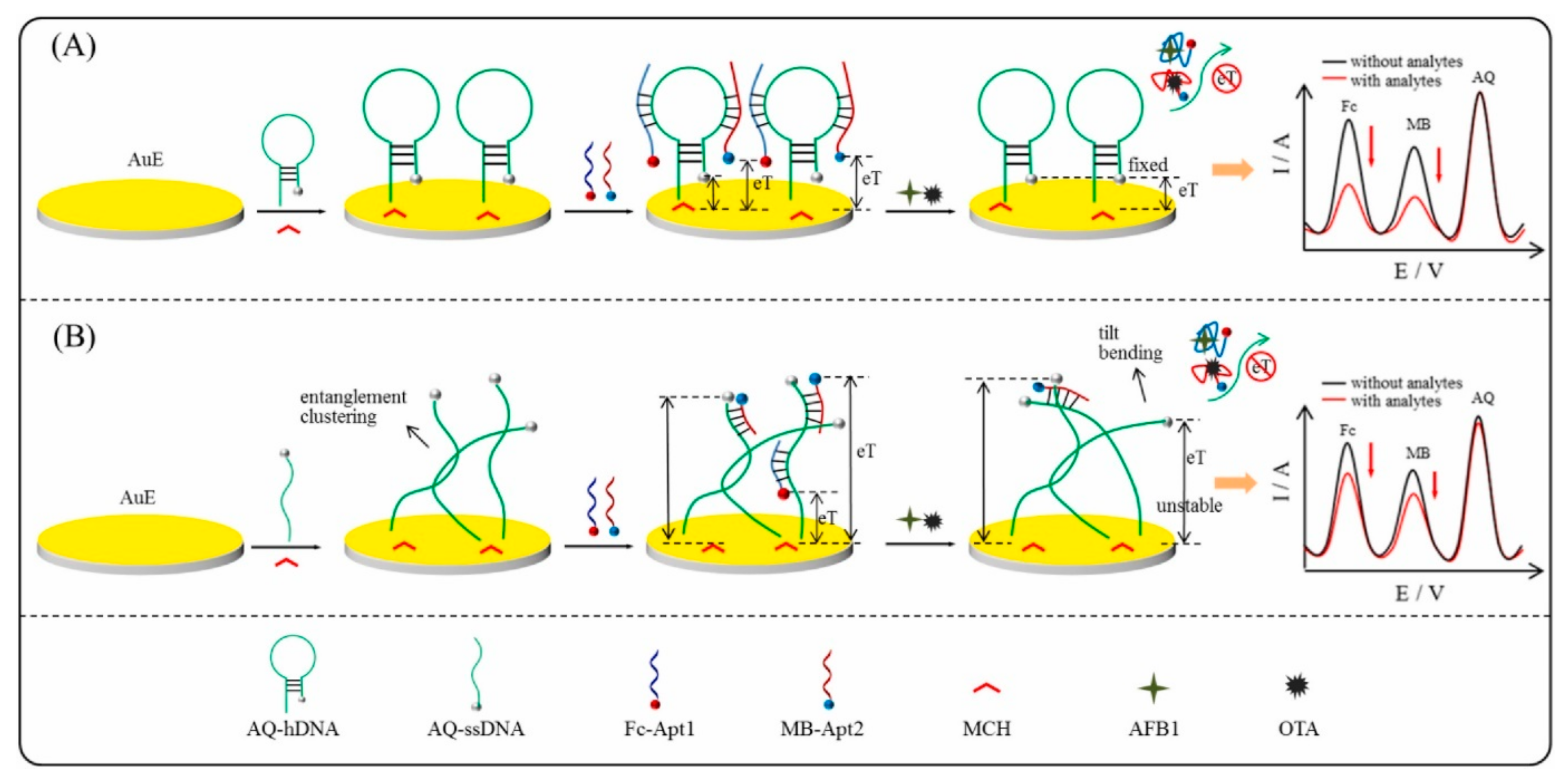
AFB1: Fc-Apt1 v1
OTA: GATCGGGTGTGGGTGGCGTAAAGGGAGCATCGGAC-MB
GE/AQ-hDNA/MCH/Fc-Apt1/MB-Apt2ACVAFB1: 10 pg/mL–3 ng/mL
OTA: 30 pg/mL–10 ng/mL
AFB1: 4.300 pg/mL
OTA: 13.300 pg/mL
AFB2, OTB, FB1, ZENCorn, wheatAFB1: 0.100 ng/mL
OTA: 0.100 ng/mL
[115]
Abbreviations: 1NP—1-naphtil phosphatase; ACV—alternating current voltammetry; aDNA—assistant DNA; AFB2—aflatoxin B2; AFG1—aflatoxin G1; AFG2—aflatoxin G2; AFM1—aflatoxin M1; Apt—aptamer; AQ—anthraquinone; AuNFs—gold nanoflowers; AuNPs—gold nanoparticles; ATP—adenosine triphosphate; AuNRs—gold nanorods; BDDE—boron-doped diamond electrode; BIO—biotin; Bioconj—bioconjugates; BSA—bovine serum albumin; CA—chronoamperometry; cDNA—complementary DNA; CDs—carbon dots; CNDs—carbon nanodots; CS—chitosan; Cu2O NCs—Cu2O nanocubes; CV—cyclic voltammetry; DON—deoxynivalenol; DPV—differential pulse voltammetry; dsDNA—double-stranded DNA; EIS—electrochemical impedance spectroscopy; Exo—exonuclease; FB1—fumonisin B1; FB2—fumonisin B2; Fc—ferrocene; GA—glutaraldehyde; GCE—glassy carbon electrode; GE—gold electrode; Glu—glucose; Gly—glycine; GNs—graphene nanosheets; GO—graphene oxide; HindIII—endonuclease; HMDA—hexamethylenediamine; hDNA—DNA hybridization chain reaction initiator; HP—hairpin DNA; HPCS—hollow porous carbon spheres; HRP—horseradish peroxidase; ITOE—indium tin oxide electrode; LDH NCs—layered double hydroxides nanocages; MB—methylene blue; MBs—magnetic beads; MCH—6-mercapto-1-hexanol; MOFs—metallic organic frameworks; MPA—3-mercaptopropionic acid; MIP—molecularly imprinted polymer; MWCNTs—multi-walled carbon nanotubes; NHS—N-hydroxysuccinimide; QDs—quantum dots; OTA—ochratoxin A; OTB—ochratoxin B; O-VMSF—3-glycidyloxypropyl trimethoxysilane-modified vertically ordered mesoporous silica film; P4VP—poly(4-vinyl pyridine); PAA—porous anodized aluminum; PANI—polyaniline; PANI-PAA—poly(aniline-anthranilic acid) copolymer; PBP—para-bisphenol; pDA—polydopamine; PDDA—poly(diallyl dimethylammonium chloride); PS—polystyrene; PTFE—polytetrafluoroethylene; PT3C—polythiophene-3 carboxylic; rGO—reduced graphene oxide; SA—streptavidin; SAP—streptavidin alkaline phosphatase; sDNA—substrate DNA; SEB—staphylococcal enterotoxin B; SGPGE—surface-graphenized pencil graphite electrode; SPCE—screen-printed carbon electrode; SPGE—screen-printed gold electrode; ssDNA—single-stranded DNA; SWV—square wave voltammetry; T-2—trichothecenes; TDNs—tetrahedral DNA nanostructures; TEG—triethylene glycol; TEMPO-COOH—4-carboxy-2,2,6,6-tetramethylpiperidine 1-oxyl free radical; THI—thionine; ZEN—zearalenone; ZIF-8—zeolitic imidazolate framework-8; β-CD—beta-cyclodextrin; μPAD—microfluidic paper-based analytical device.
Disclaimer/Publisher’s Note: The statements, opinions and data contained in all publications are solely those of the individual author(s) and contributor(s) and not of MDPI and/or the editor(s). MDPI and/or the editor(s) disclaim responsibility for any injury to people or property resulting from any ideas, methods, instructions or products referred to in the content.

