Recent Progress in Single-Nucleotide Polymorphism Biosensors

Abstract
:1. Introduction
2. Biosensors for SNP Detection
2.1. Fluorescent Biosensors
2.2. Electrochemical Biosensors
2.3. Other Biosensors
3. Conclusions
Author Contributions
Funding
Institutional Review Board Statement
Informed Consent Statement
Data Availability Statement
Conflicts of Interest
References
- Zhang, Y.; Chen, B.; Chen, D.; Wang, Y.; Lu, Q.; Tan, J.; Chen, L.; Zhou, L.; Tan, W.; Yang, Y.; et al. Electrical Detection Assay Based on Programmable Nucleic Acid Probe for Efficient Single-Nucleotide Polymorphism Identification. ACS Sens. 2023, 8, 2096–2104. [Google Scholar] [CrossRef] [PubMed]
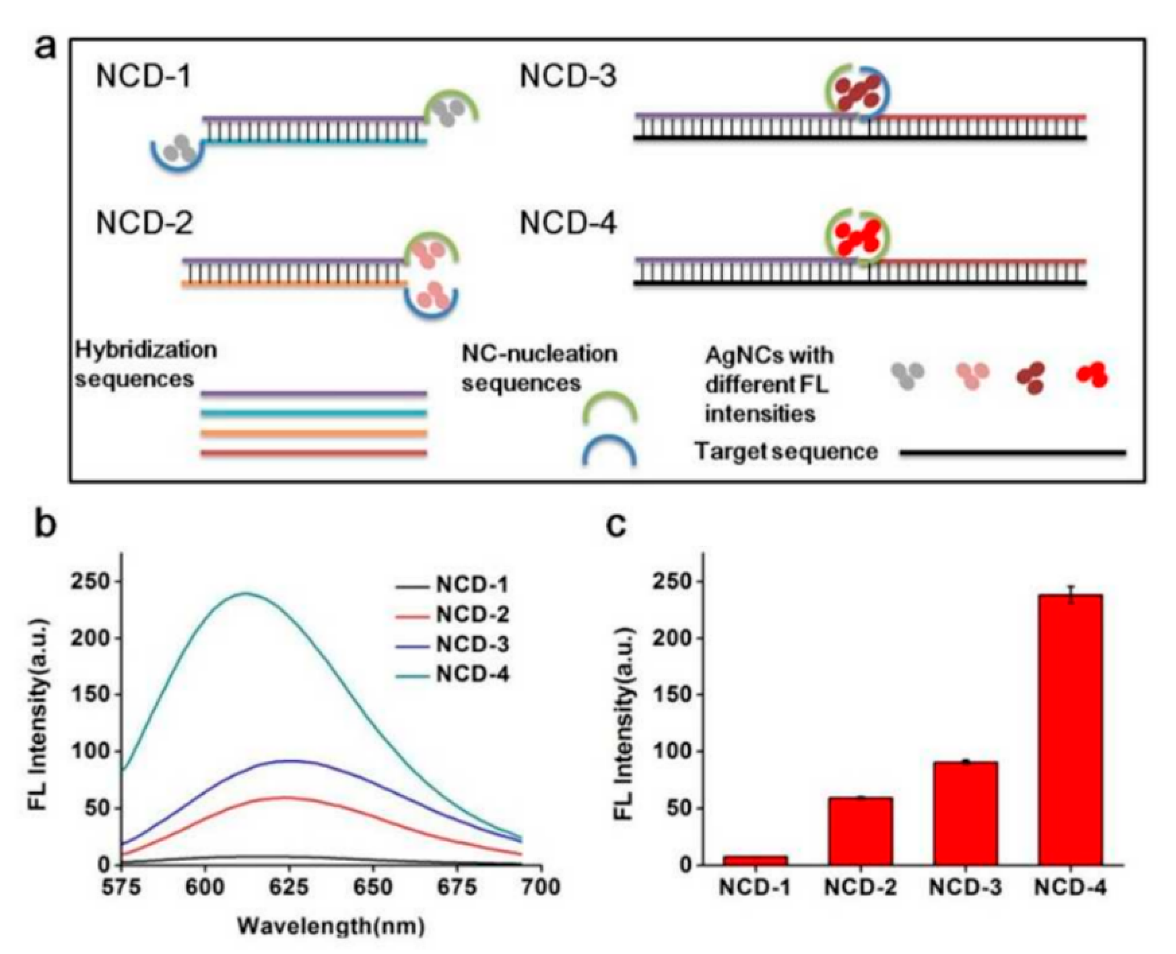
- Xu, M.; Wang, X.; Tian, J.; Chen, J.; Wei, X.; Li, W. A clamp-improved universal amplified system for ratiometric fluorescent detection of single-nucleotide polymorphisms coupled with a novel dual-emissive silver nanocluster. Sens. Actuators B Chem. 2022, 367, 132151. [Google Scholar] [CrossRef]
- Akekawatchai, C.; Phuegsilp, C.; Changsri, K.; Soimanee, T.; Sretapunya, W. Genotypic and allelic distribution of IFN-gamma +874T/A and TGF-beta1 -509C/T single-nucleotide polymorphisms in human immunodeficiency virus-infected Thais. J. Med. Virol. 2022, 94, 2882–2886. [Google Scholar] [CrossRef] [PubMed]
- Liu, B.; Jia, Y.; Ma, M.; Li, Z.; Liu, H.; Li, S.; Deng, Y.; Zhang, L.; Lu, Z.; Wang, W.; et al. High throughput SNP detection system based on magnetic nanoparticles separation. J. Biomed. Nanotechnol. 2013, 9, 247–256. [Google Scholar] [CrossRef] [PubMed]
- Gray, I.C.; Campbell, D.A.; Spurr, N.K. Single nucleotide polymorphisms as tools in human genetics. Hum. Mol. Genet. 2000, 9, 2403–2408. [Google Scholar] [CrossRef] [PubMed]
- Brookes, A.J. The essence of SNPs. Gene 1999, 234, 177–186. [Google Scholar] [CrossRef]
- Cui, M.; Xiao, X.; Zhao, M.; Zheng, B. Detection of single nucleotide polymorphism by measuring extension kinetics with T7 exonuclease mediated isothermal amplification. Analyst 2017, 143, 116–122. [Google Scholar] [CrossRef]
- Wang, H.B.; Ma, L.H.; Zhang, T.; Huang, K.C.; Zhao, Y.D.; Liu, T.C. Simple and accurate visual detection of single nucleotide polymorphism based on colloidal gold nucleic acid strip biosensor and primer-specific PCR. Anal. Chim. Acta 2020, 1093, 106–114. [Google Scholar] [CrossRef]
- Weng, Z.; You, Z.; Li, H.; Wu, G.; Song, Y.; Sun, H.; Fradlin, A.; Neal-Harris, C.; Lin, M.; Gao, X.; et al. CRISPR-Cas12a Biosensor Array for Ultrasensitive Detection of Unamplified DNA with Single-Nucleotide Polymorphic Discrimination. ACS Sens. 2023, 8, 1489–1499. [Google Scholar] [CrossRef]
- Yucel, M.; Koc, A.; Ulgenalp, A.; Akkoc, G.D.; Ceyhan, M.; Yildiz, U.H. PCR-Free Methodology for Detection of Single-Nucleotide Polymorphism with a Cationic Polythiophene Reporter. ACS Sens. 2021, 6, 950–957. [Google Scholar] [CrossRef]
- Van den Broeck, T.; Joniau, S.; Clinckemalie, L.; Helsen, C.; Prekovic, S.; Spans, L.; Tosco, L.; Van Poppel, H.; Claessens, F. The role of single nucleotide polymorphisms in predicting prostate cancer risk and therapeutic decision making. Biomed Res. Int. 2014, 2014, 627510. [Google Scholar] [CrossRef] [PubMed]
- Cheng, L.; Sun, B.; Sun, Y.; Xiao, P.; Ge, Q.; Zheng, Y.; Ke, X.; Zhou, Y.; Zhang, M.; Chen, P.; et al. Detection of multiple SNPs in numerous samples with polyacrylamide gel-based microarray. J. Nanosci. Nanotechnol. 2010, 10, 479–486. [Google Scholar] [CrossRef] [PubMed]
- Arancibia, T.; Morales-Pison, S.; Maldonado, E.; Jara, L. Association between single-nucleotide polymorphisms in miRNA and breast cancer risk: An updated review. Biol. Res. 2021, 54, 26. [Google Scholar] [CrossRef] [PubMed]
- Li, C.C.; Hu, J.; Luo, X.; Hu, J.; Zhang, C.Y. Development of a Single Quantum Dot-Mediated FRET Nanosensor for Sensitive Detection of Single-Nucleotide Polymorphism in Cancer Cells. Anal. Chem. 2021, 93, 14568–14576. [Google Scholar] [CrossRef] [PubMed]
- He, J.; Li, X.; Liao, L.; Zhou, W.; Jiang, B. Target-mediated assembly formation of multi-arm DNAzyme nanostructures for sensitive and accurate discrimination of single-nucleotide polymorphism in K-ras gene. Sens. Actuators B Chem. 2021, 346, 130535. [Google Scholar] [CrossRef]
- Xu, M.; Xing, S.; Zhao, Y.; Zhao, C. Peptide nucleic acid-assisted colorimetric detection of single-nucleotide polymorphisms based on the intrinsic peroxidase-like activity of hemin-carbon nanotube nanocomposites. Talanta 2021, 232, 122420. [Google Scholar] [CrossRef]
- Choi, W.; Jung, G.Y. Highly multiplex and sensitive SNP genotyping method using a three-color fluorescence-labeled ligase detection reaction coupled with conformation-sensitive CE. Electrophoresis 2017, 38, 513–520. [Google Scholar] [CrossRef]
- Yang, S.; Guo, H.; Wei, B.; Zhu, S.; Cai, Y.; Jiang, P.; Tang, J. Association of miR-502-binding site single nucleotide polymorphism in the 3′-untranslated region of SET8 and TP53 codon 72 polymorphism with non-small cell lung cancer in Chinese population. Acta Biochim. Biophys. Sin. 2014, 46, 149–154. [Google Scholar] [CrossRef]
- Li, S.; Liu, H.; Jia, Y.; Mou, X.; Deng, Y.; Lin, L.; Liu, B.; He, N. An automatic high-throughput single nucleotide polymorphism genotyping approach based on universal tagged arrays and magnetic nanoparticles. J. Biomed. Nanotechnol. 2013, 9, 689–698. [Google Scholar] [CrossRef]
- Tang, Y.; Ali, Z.; Dai, J.; Liu, X.; Wu, Y.; Chen, Z.; He, N.; Li, S.; Wang, L. Single-Nucleotide Polymorphism Genotyping of exoS in Pseudomonas aeruginosa Using Dual-Color Fluorescence Hybridization and Magnetic Separation. J. Biomed. Nanotechnol. 2018, 14, 206–214. [Google Scholar] [CrossRef]
- Mou, X.; Sheng, D.; Chen, Z.; Liu, M.; Liu, Y.; Deng, Y.; Xu, K.; Hou, R.; Zhao, J.; Zhu, Y. In-situ mutation detection by magnetic beads-probe based on single base extension and its application in genotyping of hepatitis B virus pre-C region 1896nt locus single nucleotide polymorphisms. J. Biomed. Nanotechnol. 2019, 15, 2393–2400. [Google Scholar] [CrossRef]
- Yang, H.; Yang, S.; Xia, X.; Deng, R.; Gao, H.; Dong, Y. Sensitive Detection of a Single-Nucleotide Polymorphism in Foodborne Pathogens Using CRISPR/Cas12a-Signaling ARMS-PCR. J. Agric. Food Chem. 2022, 70, 8451–8457. [Google Scholar] [CrossRef] [PubMed]
- Knez, K.; Spasic, D.; Janssen, K.P.; Lammertyn, J. Emerging technologies for hybridization based single nucleotide polymorphism detection. Analyst 2014, 139, 353–370. [Google Scholar] [CrossRef] [PubMed]
- Bidar, N.; Oroojalian, F.; Baradaran, B.; Eyvazi, S.; Amini, M.; Jebelli, A.; Hosseini, S.S.; Pashazadeh-Panahi, P.; Mokhtarzadeh, A.; de la Guardia, M. Monitoring of microRNA using molecular beacons approaches: Recent advances. TrAC Trends Anal. Chem. 2020, 131, 116021. [Google Scholar] [CrossRef]
- Zheng, M.; Chen, X.; Wang, S.; Wang, J.; Huang, M.; Xiao, S.; Cheng, X.; Chen, S.; Chen, X.; Lin, F.; et al. A TaqMan-MGB real-time RT-PCR assay with an internal amplification control for rapid detection of Muscovy duck reovirus. Mol. Cell Probes. 2020, 52, 101575. [Google Scholar] [CrossRef] [PubMed]
- Xue, S.S.; Li, Y.; Pan, W.; Li, N.; Tang, B. Multi-stimuli-responsive molecular fluorescent probes for bioapplications. Chem. Commun. 2023, 59, 3040–3049. [Google Scholar] [CrossRef]
- Bidar, N.; Amini, M.; Oroojalian, F.; Baradaran, B.; Hosseini, S.S.; Shahbazi, M.-A.; Hashemzaei, M.; Mokhtarzadeh, A.; Hamblin, M.R.; de la Guardia, M. Molecular beacon strategies for sensing purpose. TrAC Trends Anal. Chem. 2021, 134, 116143. [Google Scholar] [CrossRef]
- Samad Hosseini, S.; Jebelli, A.; Vandghanooni, S.; Jahanban-Esfahlan, A.; Baradaran, B.; Amini, M.; Bidar, N.; de la Guardia, M.; Mokhtarzadeh, A.; Eskandani, M. Perspectives and trends in advanced DNA biosensors for the recognition of single nucleotide polymorphisms. Chem. Eng. J. 2022, 441, 135988. [Google Scholar] [CrossRef]
- Mukhtar, M.; Sargazi, S.; Barani, M.; Madry, H.; Rahdar, A.; Cucchiarini, M. Application of Nanotechnology for Sensitive Detection of Low-Abundance Single-Nucleotide Variations in Genomic DNA: A Review. Nanomaterials 2021, 11, 1384. [Google Scholar] [CrossRef]
- Fujita, T.; Nagata, S.; Fujii, H. Oligoribonucleotide-Mediated Blockade of DNA Extension by Taq DNA Polymerases Increases Specificity and Sensitivity for Detecting Single-Nucleotide Differences. Anal. Chem. 2023, 95, 3442–3451. [Google Scholar] [CrossRef]
- Shen, W.; Tian, Y.; Ran, T.; Gao, Z. Genotyping and quantification techniques for single-nucleotide polymorphisms. TrAC Trends Anal. Chem. 2015, 69, 1–13. [Google Scholar] [CrossRef]
- Xu, J.; Li, L.; Chen, N.; She, Y.; Wang, S.; Liu, N.; Xiao, X. Endonuclease IV based competitive DNA probe assay for differentiation of low-abundance point mutations by discriminating stable single-base mismatches. Chem. Commun. 2017, 53, 9422–9425. [Google Scholar] [CrossRef] [PubMed]
- Krishan, N.V.N.K.P. APPLICATION OF MOLECULAR BEACON BASED BIOSENSOR AGAINST rs699 SNP IN HYPERTENSIVE AND NON- HYPERTENSIVE PUNJABI POPULATION. Int. J. Pharmacogn. 2018, 5, 37–50. [Google Scholar]
- Zhou, L.; He, X.; He, D.; Wang, K.; Qin, D. Biosensing technologies for Mycobacterium tuberculosis detection: Status and new developments. Clin. Dev. Immunol. 2011, 2011, 193963. [Google Scholar] [CrossRef] [PubMed]
- Duan, X.; Yue, W.; Liu, L.; Li, Z.; Li, Y.; He, F.; Zhu, D.; Zhou, G.; Wang, S. Single-nucleotide polymorphism (SNP) genotyping using cationic conjugated polymers in homogeneous solution. Nat. Protoc. 2009, 4, 984–991. [Google Scholar] [CrossRef]
- Ali, J.; Najeeb, J.; Asim Ali, M.; Farhan Aslam, M.; Raza, A. Biosensors: Their Fundamentals, Designs, Types and Most Recent Impactful Applications: A Review. J. Biosens. Bioelectron. 2017, 8, 1–9. [Google Scholar] [CrossRef]
- Girigoswami, K.; Akhtar, N. Nanobiosensors and fluorescence based biosensors: An overview. Int. J. Nano Dimens. 2019, 10, 1–17. [Google Scholar]
- Gong, L.; Zhao, L.; Tan, M.; Pan, T.; He, H.; Wang, Y.; He, X.; Li, W.; Tang, L.; Nie, L. Two-Photon Fluorescent Nanomaterials and Their Applications in Biomedicine. J. Biomed. Nanotechnol. 2021, 17, 509–528. [Google Scholar] [CrossRef]
- Wu, K.; He, X.; Wang, J.; Pan, T.; He, R.; Kong, F.; Cao, Z.; Ju, F.; Huang, Z.; Nie, L. Recent progress of microfluidic chips in immunoassay. Front. Bioeng. Biotechnol. 2022, 10, 1112327. [Google Scholar] [CrossRef]
- Zhou, L.; Yuan, G.; Hu, S. A 4-Hydroxy-1,8-Naphthalimide-Based Turn-on Two-Photon Fluorescent Probe for Hydrogen Polysulfide Sensing. J. Appl. Spectrosc. 2020, 86, 1071–1076. [Google Scholar] [CrossRef]
- Li, Q.; Guo, Y.M.; Li, G.L. Redox-regulated synthesis of fluorescent polydopamine nanoparticles for detection of butyrylcholinesterase activity. Spectrochim. Acta A Mol. Biomol. Spectrosc. 2022, 274, 121097. [Google Scholar] [CrossRef] [PubMed]
- Li, S.-X.; Wang, Y.-Q.; Chen, Z.-P.; Chen, Y. Probe technique-based generalized multivariate standard addition strategy for the analysis of fluorescence signals with matrix effects. Chemometr. Intell. Lab. 2019, 190, 41–47. [Google Scholar] [CrossRef]
- Chen, Y.; Tang, Y.; Wang, H. Quantification of ATP in cell by fluorescence spectroscopy based on generalized ratio quantitative analysis model. Spectrochim. Acta A Mol. Biomol. Spectrosc. 2021, 263, 120170. [Google Scholar] [CrossRef] [PubMed]
- Zuo, Q.; Chen, Y.; Chen, Z.P.; Yu, R.Q. A novel ratiometric fluorescent sensing method based on MnO(2) nanosheet for sensitive detection of alkaline phosphatase in serum. Talanta 2020, 209, 120528. [Google Scholar] [CrossRef] [PubMed]
- Xiao, X.; Wu, T.; Xu, L.; Chen, W.; Zhao, M. A branch-migration based fluorescent probe for straightforward, sensitive and specific discrimination of DNA mutations. Nucleic Acids Res. 2017, 45, e90. [Google Scholar] [CrossRef] [PubMed]
- Yu, Y.; Wu, T.; Johnson-Buck, A.; Li, L.; Su, X. A two-layer assay for single-nucleotide variants utilizing strand displacement and selective digestion. Biosens. Bioelectron. 2016, 82, 248–254. [Google Scholar] [CrossRef]
- Xu, H.; Yang, Q.; Li, F.; Tang, L.; Gao, S.; Jiang, B.; Zhao, X.; Wang, L.; Fan, C. A graphene-based platform for fluorescent detection of SNPs. Analyst 2013, 138, 2678–2682. [Google Scholar] [CrossRef]
- Hong, C.; Kim, D.M.; Baek, A.; Chung, H.; Jung, W.; Kim, D.E. Fluorescence-based detection of single-nucleotide changes in RNA using graphene oxide and DNAzyme. Chem. Commun. 2015, 51, 5641–5644. [Google Scholar] [CrossRef]
- Wu, W.; Hu, H.; Li, F.; Wang, L.; Gao, J.; Lu, J.; Fan, C. A graphene oxide-based nano-beacon for DNA phosphorylation analysis. Chem. Commun. 2011, 47, 1201–1203. [Google Scholar] [CrossRef]
- Huang, Y.; Yang, H.Y.; Ai, Y. DNA single-base mismatch study using graphene oxide nanosheets-based fluorometric biosensors. Anal. Chem. 2015, 87, 9132–9136. [Google Scholar] [CrossRef]
- Huang, J.; Wang, Z.; Kim, J.K.; Su, X.; Li, Z. Detecting Arbitrary DNA Mutations Using Graphene Oxide and Ethidium Bromide. Anal. Chem. 2015, 87, 12254–12261. [Google Scholar] [CrossRef] [PubMed]
- Wu, Z.K.; Zhou, D.M.; Wu, Z.; Chu, X.; Yu, R.Q.; Jiang, J.H. Single-base mismatch discrimination by T7 exonuclease with target cyclic amplification detection. Chem. Commun. 2015, 51, 2954–2956. [Google Scholar] [CrossRef] [PubMed]
- Khoothiam, K.; Treerattrakoon, K.; Iempridee, T.; Luksirikul, P.; Dharakul, T.; Japrung, D. Ultrasensitive detection of lung cancer-associated miRNAs by multiple primer-mediated rolling circle amplification coupled with a graphene oxide fluorescence-based (MPRCA-GO) sensor. Analyst 2019, 144, 4180–4187. [Google Scholar] [CrossRef]
- Devi, P.; Saini, S.; Kim, K.H. The advanced role of carbon quantum dots in nanomedical applications. Biosens. Bioelectron. 2019, 141, 111158. [Google Scholar] [CrossRef]
- Zhao, H.; Gao, L.; Luo, J.; Zhou, D.; Lu, Z. Massively parallel display of genomic DNA fragments by rolling-circle amplification and strand displacement amplification on chip. Talanta 2010, 82, 477–482. [Google Scholar] [CrossRef] [PubMed]
- Cao, G.; Chen, X.; Deng, Y.; Nie, F.; Liu, Y.; Wang, G.; Huo, D.; Hou, C. Single-nucleotide variant of PIK3CA (H1047R) gene assay by CRISPR/Cas12a combined with rolling circle amplification. Anal. Chim. Acta 2021, 1182, 338943. [Google Scholar] [CrossRef]
- Ma, Q.; Gao, Z. A simple and ultrasensitive fluorescence assay for single-nucleotide polymorphism. Anal. Bioanal. Chem. 2018, 410, 3093–3100. [Google Scholar] [CrossRef]
- Li, X.H.; Zhang, X.L.; Wu, J.; Lin, N.; Sun, W.M.; Chen, M.; Ou, Q.S.; Lin, Z.Y. Hyperbranched rolling circle amplification (HRCA)-based fluorescence biosensor for ultrasensitive and specific detection of single-nucleotide polymorphism genotyping associated with the therapy of chronic hepatitis B virus infection. Talanta 2019, 191, 277–282. [Google Scholar] [CrossRef]
- Sun, Y.; Han, B.; Sun, F. Ultra-specific genotyping of single nucleotide variants by ligase-based loop-mediated isothermal amplification coupled with a modified ligation probe. RSC Adv. 2021, 11, 17058–17063. [Google Scholar] [CrossRef]
- Liu, Y.; Liu, Y.; Qiao, L.; Liu, Y.; Liu, B. Advances in signal amplification strategies for electrochemical biosensing. Curr. Opin. Electrochem. 2018, 12, 5–12. [Google Scholar] [CrossRef]
- Yang, P.; Guo, X.; Zhang, J.; Chen, C.; Gan, Y.; Xie, W.; Du, Y.; Wu, Z. Picomolar thrombin detection by orchestration of triple signal amplification strategy with hierarchically porous Ti3C2Tx MXene electrode material-catalytic hairpin assembly reaction-metallic nanoprobes. Biosens. Bioelectron. 2022, 208, 114228. [Google Scholar] [CrossRef] [PubMed]
- Zhu, J.; Ding, Y.; Liu, X.; Wang, L.; Jiang, W. Toehold-mediated strand displacement reaction triggered isothermal DNA amplification for highly sensitive and selective fluorescent detection of single-base mutation. Biosens. Bioelectron. 2014, 59, 276–281. [Google Scholar] [CrossRef] [PubMed]
- Li, X.; Liao, L.; Jiang, B.; Yuan, R.; Xiang, Y. Invader assay-induced catalytic assembly of multi-DNAzyme junctions for sensitive detection of single nucleotide polymorphisms. Anal. Chim. Acta 2022, 1224, 340225. [Google Scholar] [CrossRef]
- Wu, F.; Chen, M.; Lan, J.; Xia, Y.; Liu, M.; He, W.; Li, C.; Chen, X.; Chen, J. A universal locked nucleic acid-integrated X-shaped DNA probe design for amplified fluorescence detection of single-nucleotide variant. Sens. Actuators B Chem. 2017, 241, 123–128. [Google Scholar] [CrossRef]
- Xiao, X.; Yang, H.; Jiang, P.; Chen, Z.; Ji, C.; Nie, L. Multi-Functional Fe3O4@mSiO(2)-AuNCs Composite Nanoparticles Used as Drug Delivery System. J. Biomed. Nanotechnol. 2017, 13, 1292–1299. [Google Scholar] [CrossRef]
- Lai, Y.; Wang, L.; Liu, Y.; Yang, G.; Tang, C.; Deng, Y.; Li, S. Immunosensors Based on Nanomaterials for Detection of Tumor Markers. J. Biomed. Nanotechnol. 2018, 14, 44–65. [Google Scholar] [CrossRef]
- Diez, I.; Ras, R.H. Fluorescent silver nanoclusters. Nanoscale 2011, 3, 1963–1970. [Google Scholar] [CrossRef]
- Li, T.; Hu, Z.; Yu, S.; Liu, Z.; Zhou, X.; Liu, R.; Liu, S.; Deng, Y.; Li, S.; Chen, H.; et al. DNA Templated Silver Nanoclusters for Bioanalytical Applications: A Review. J. Biomed. Nanotechnol. 2022, 18, 1237–1256. [Google Scholar] [CrossRef]
- Li, T.; Yi, H.; Liu, Y.; Wang, Z.; Liu, S.; He, N.; Liu, H.; Deng, Y. One-Step Synthesis of DNA Templated Water-Soluble Au-Ag Bimetallic Nanoclusters for Ratiometric Fluorescence Detection of DNA. J. Biomed. Nanotechnol. 2018, 14, 150–160. [Google Scholar] [CrossRef]
- Tang, J.; Wang, T.; Li, Q. Silver nanocluster-lightened catalytic hairpin assembly for enzyme-free and label-free mRNA detection. Microchem. J. 2021, 165, 106184. [Google Scholar] [CrossRef]
- Ding, S.; Xu, A.; Li, M.; Sun, A.; Zhang, Z.; Xia, Y.; Liu, Y. Theoretical study on the sensing mechanism of an ON(1)-OFF-ON(2) type fluoride fluorescent chemosensor. Spectrochim. Acta A Mol. Biomol. Spectrosc. 2020, 237, 118397. [Google Scholar] [CrossRef] [PubMed]
- Liu, J.; Lu, Y.; Feng, L.; Wang, S.; Zhang, S.; Zhu, X.; Sheng, L.; Zhang, S.; Zhang, X. Pinpoint the Positions of Single Nucleotide Polymorphisms by a Nanocluster Dimer. Anal. Chem. 2017, 89, 2622–2627. [Google Scholar] [CrossRef] [PubMed]
- Guo, W.; Yuan, J.; Dong, Q.; Wang, E. Highly Sequence-Dependent Formation of Fluorescent Silver Nanoclusters in Hybridized DNA Duplexes for Single Nucleotide Mutation Identification. J. Am. Chem. Soc. 2010, 132, 932–934. [Google Scholar] [CrossRef]
- Yeh, H.C.; Sharma, J.; Shih Ie, M.; Vu, D.M.; Martinez, J.S.; Werner, J.H. A fluorescence light-up Ag nanocluster probe that discriminates single-nucleotide variants by emission color. J. Am. Chem. Soc. 2012, 134, 11550–11558. [Google Scholar] [CrossRef] [PubMed]
- Liu, R.; Wang, C.; Hu, J.; Su, Y.; Lv, Y. DNA-templated copper nanoparticles: Versatile platform for label-free bioassays. TrAC Trends Anal. Chem. 2018, 105, 436–452. [Google Scholar] [CrossRef]
- Chen, C.A.; Wang, C.C.; Jong, Y.J.; Wu, S.M. Label-Free Fluorescent Copper Nanoclusters for Genotyping of Deletion and Duplication of Duchenne Muscular Dystrophy. Anal. Chem. 2015, 87, 6228–6232. [Google Scholar] [CrossRef] [PubMed]
- Tsai, T.T.; Chen, C.A.; Yi-Ju Ho, N.; Yang, S.; Chen, C.F. Fluorescent Double-Stranded DNA-Templated Copper Nanoprobes for Rapid Diagnosis of Tuberculosis. ACS Sens. 2019, 4, 2885–2892. [Google Scholar] [CrossRef]
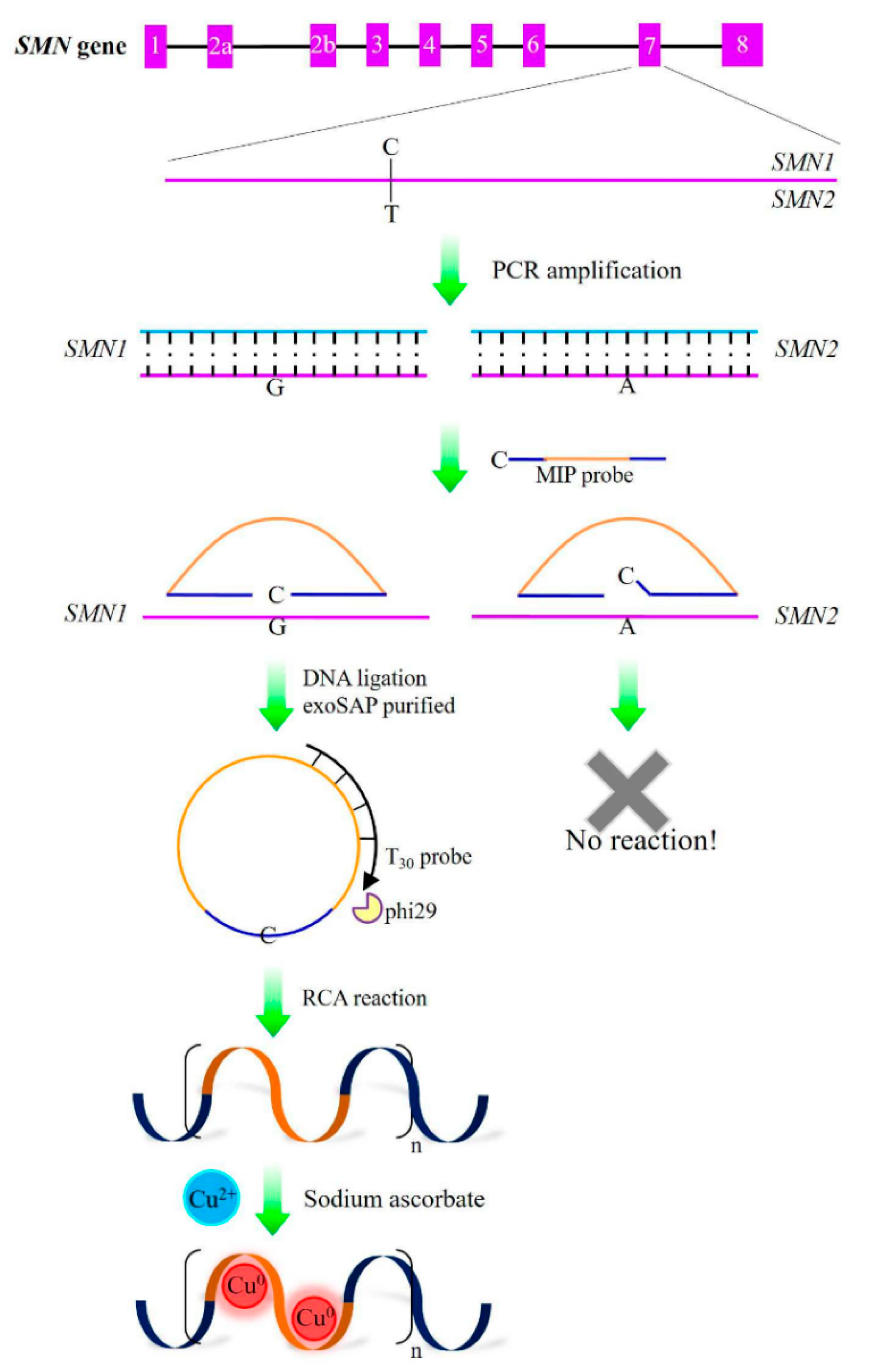
- Chen, C.A.; Wang, C.C.; Kou, H.S.; Wu, S.M. Molecular inversion probe-rolling circle amplification with single-strand poly-T luminescent copper nanoclusters for fluorescent detection of single-nucleotide variant of SMN gene in diagnosis of spinal muscular atrophy. Anal. Chim. Acta 2020, 1123, 56–63. [Google Scholar] [CrossRef]
- Jia, X.; Li, J.; Han, L.; Ren, J.; Yang, X.; Wang, E. DNA-Hosted Copper Nanoclusters for Fluorescent Identification of Single Nucleotide Polymorphisms. ACS Nano 2012, 6, 3311–3317. [Google Scholar] [CrossRef]
- Gerion, D.; Chen, F.; Kannan, B.; Fu, A.; Parak, W.J.; Chen, D.J.; Majumdar, A.; Alivisatos, A.P. Room-Temperature Single-Nucleotide Polymorphism and Multiallele DNA Detection Using Fluorescent Nanocrystals and Microarrays. Anal. Chem. 2003, 75, 4766–4772. [Google Scholar] [CrossRef]
- Guo, Q.; Bai, Z.; Liu, Y.; Sun, Q. A molecular beacon microarray based on a quantum dot label for detecting single nucleotide polymorphisms. Biosens. Bioelectron. 2016, 77, 107–110. [Google Scholar] [CrossRef] [PubMed]
- Suan Ng, S.; Ling Lee, H.; Bothi Raja, P.; Doong, R.A. Recent Advances in Nanomaterial-based Optical Biosensors as Potential Point-of-Care Testing (PoCT) Probes in Carcinoembryonic Antigen Detection. Chem. Asian J. 2022, 17, e202200287. [Google Scholar] [CrossRef] [PubMed]
- Zou, L.; Liu, X.; Zhou, Y.; Mei, W.; Wang, Q.; Yang, X.; Wang, K. Optical fiber amplifier and thermometer assisted point-of-care biosensor for detection of cancerous exosomes. Sens. Actuators B Chem. 2022, 351, 130893. [Google Scholar] [CrossRef]
- Watterson, J.H.; Raha, S.; Kotoris, C.C.; Wust, C.C.; Gharabaghi, F.; Jantzi, S.C.; Haynes, N.K.; Gendron, N.H.; Krull, U.J.; Mackenzie, A.E.; et al. Rapid detection of single nucleotide polymorphisms associated with spinal muscular atrophy by use of a reusable fibre-optic biosensor. Nucleic Acids Res. 2004, 32, e18. [Google Scholar] [CrossRef]
- Semeniak, D.; Cruz, D.F.; Chilkoti, A.; Mikkelsen, M.H. Plasmonic Fluorescence Enhancement in Diagnostics for Clinical Tests at Point-of-Care: A Review of Recent Technologies. Adv. Mater. 2022, 35, e2107986. [Google Scholar] [CrossRef]
- Cui, Y.; Niu, C.; Na, N.; Ouyang, J. Core–shell gold nanocubes for point mutation detection based on plasmon-enhanced fluorescence. J. Mater. Chem. B 2017, 5, 5329–5335. [Google Scholar] [CrossRef]
- Kim, D.-M.; Seo, J.; Kim, D.-W.; Jeong, W.; Hwang, S.-H.; Kim, D.-E. Fluorometric detection of single-nucleotide mutations using tandem gene amplification. Sens. Actuators B Chem. 2020, 314, 128071. [Google Scholar] [CrossRef]
- Kwon, W.Y.; Cha, B.S.; Kim, S.; Hwang, S.H.; Kim, J.M.; Kalimuthu, K.; Park, H.G.; Park, K.S. Fluorescence polarization-based detection of cancer-related mutations using target-initiated rolling circle amplification. Analyst 2019, 144, 4149–4152. [Google Scholar] [CrossRef]
- Freitas, M.; Nouws, H.P.A.; Delerue-Matos, C. Electrochemical Biosensing in Cancer Diagnostics and Follow-up. Electroanalysis 2018, 30, 1584–1603. [Google Scholar] [CrossRef]
- Deng, P.; Xiao, J.; Chen, J.; Feng, J.; Wei, Y.; Zuo, J.; Liu, J.; Li, J.; He, Q. Polyethylenimine-carbon nanotubes composite as an electrochemical sensing platform for sensitive and selective detection of toxic rhodamine B in soft drinks and chilli-containing products. J. Food Compos. Anal. 2022, 107, 104386. [Google Scholar] [CrossRef]
- Hassanpour, S.; Baradaran, B.; Hejazi, M.; Hasanzadeh, M.; Mokhtarzadeh, A.; de la Guardia, M. Recent trends in rapid detection of influenza infections by bio and nanobiosensor. TrAC Trends Anal. Chem. 2018, 98, 201–215. [Google Scholar] [CrossRef]
- Xiao, J.; Shi, S.; Yao, L.; Feng, J.; Zuo, J.; He, Q. Fast and Ultrasensitive Electrochemical Detection for Antiviral Drug Tenofovir Disoproxil Fumarate in Biological Matrices. Biosensors 2022, 12, 1123. [Google Scholar] [CrossRef] [PubMed]
- Wang, B.; He, Q.; Li, G.; Long, Y.; Zhang, G.; Liu, H.; Liu, J. Sensitive Determination of Trace 4-Nitrophenol in Ambient Environment Using a Glassy Carbon Electrode Modified with Formamide-Converted Nitrogen-Doped Carbon Materials. Int. J. Mol. Sci. 2022, 23, 12182. [Google Scholar] [CrossRef] [PubMed]
- Deng, P.; Zeng, Y.; Wei, Y.; Li, J.; Yao, L.; Liu, X.; Ding, J.; Li, K.; He, Q. An efficient electrochemical sensor based on multi-walled carbon nanotubes functionalized with polyethylenimine for simultaneous determination of o-nitrophenol and p-nitrophenol. Microchem. J. 2023, 186, 108340. [Google Scholar] [CrossRef]
- Labib, M.; Sargent, E.H.; Kelley, S.O. Electrochemical Methods for the Analysis of Clinically Relevant Biomolecules. Chem. Rev. 2016, 116, 9001–9090. [Google Scholar] [CrossRef]
- Li, W.; Xiao, J.; Yao, L.; Wei, Y.; Zuo, J.; Zeng, W.; Ding, J.; He, Q. Zirconium Molybdate Nanocomposites’ Sensing Platform for the Sensitive and Selective Electrochemical Detection of Adefovir. Molecules 2022, 27, 6022. [Google Scholar] [CrossRef]
- Guo, W.; Hu, C.; Li, S.; Wei, D.; Zhou, J.; Liu, X.; Chen, H.; Li, S.; Deng, Y. Selection and electrochemical-sensor application of an DNA-aptamer for methyl parathion detection. Anal. Chim. Acta 2023, 1241, 340780. [Google Scholar] [CrossRef]
- Mehmandoust, M.; Li, G.; Erk, N. Biomass-Derived Carbon Materials as an Emerging Platform for Advanced Electrochemical Sensors: Recent Advances and Future Perspectives. Ind. Eng. Chem. Res. 2022, 62, 4628–4635. [Google Scholar] [CrossRef]
- Zhang, J.; Feng, J.; Tian, Y.; Wu, Y.; Liu, X.; He, Q. Ultrasensitive electrochemical determination of tyrosine based on the α-Fe2O3@Co3O4-NRGO modified electrode. Microchem. J. 2021, 171, 106867. [Google Scholar] [CrossRef]
- Gao, Z.F.; Ling, Y.; Lu, L.; Chen, N.Y.; Luo, H.Q.; Li, N.B. Detection of single-nucleotide polymorphisms using an ON-OFF switching of regenerated biosensor based on a locked nucleic acid-integrated and toehold-mediated strand displacement reaction. Anal. Chem. 2014, 86, 2543–2548. [Google Scholar] [CrossRef]
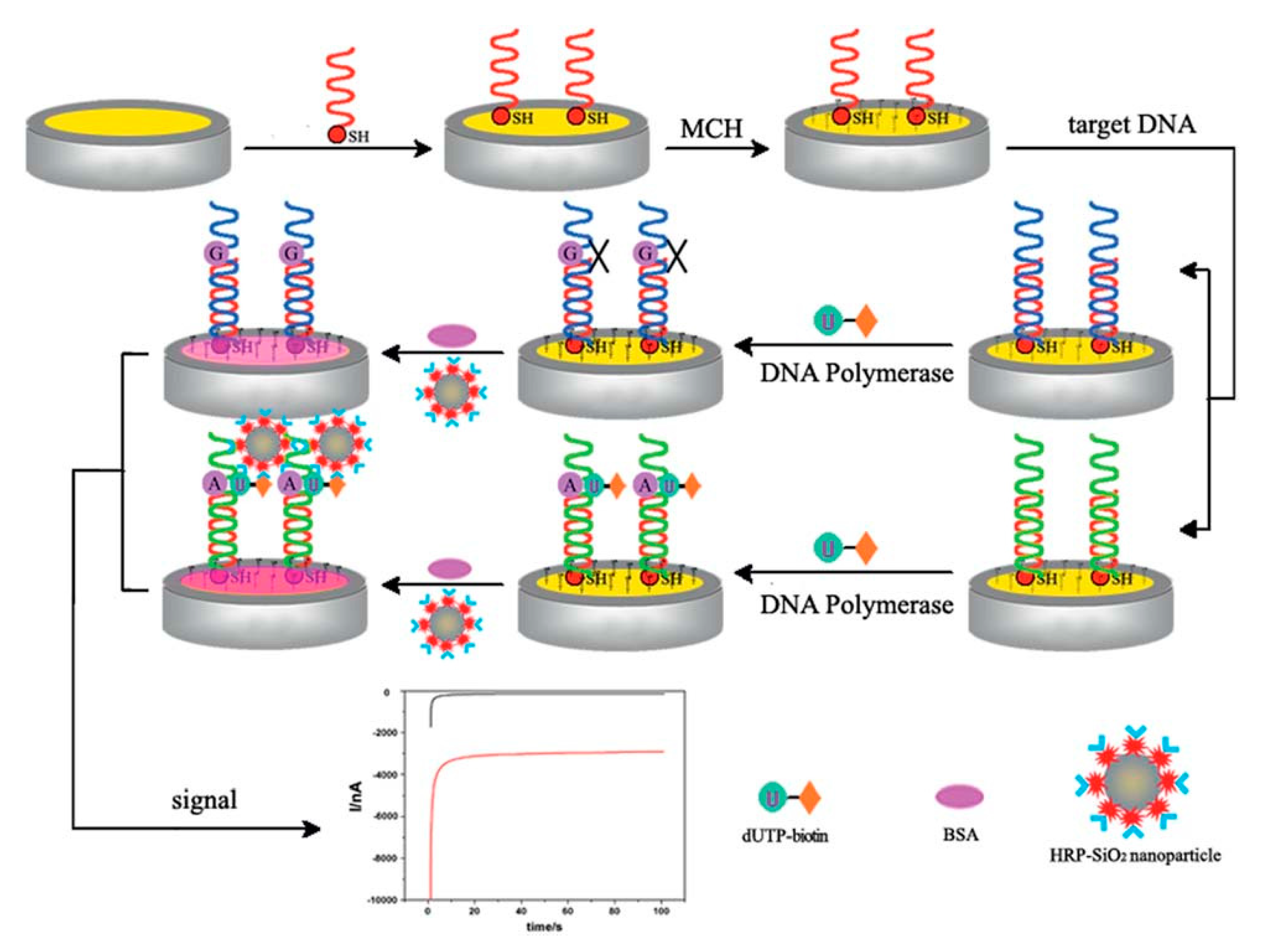
- Zhao, C.; Gao, F.; Weng, S.; Liu, Q.; Lin, L.; Lin, X. An electrochemical sensor based on DNA polymerase and HRP-SiO2 nanoparticles for the ultrasensitive detection of K-ras gene point mutation. RSC Adv. 2016, 6, 8669–8676. [Google Scholar] [CrossRef]
- Liu, Z.J.; Yang, L.Y.; Wei, Q.X.; Ye, C.L.; Xu, X.W.; Zhong, G.X.; Zheng, Y.J.; Chen, J.Y.; Lin, X.H.; Liu, A.L. A novel ligase chain reaction-based electrochemical biosensing strategy for highly sensitive point mutation detection from human whole blood. Talanta 2020, 216, 120966. [Google Scholar] [CrossRef] [PubMed]
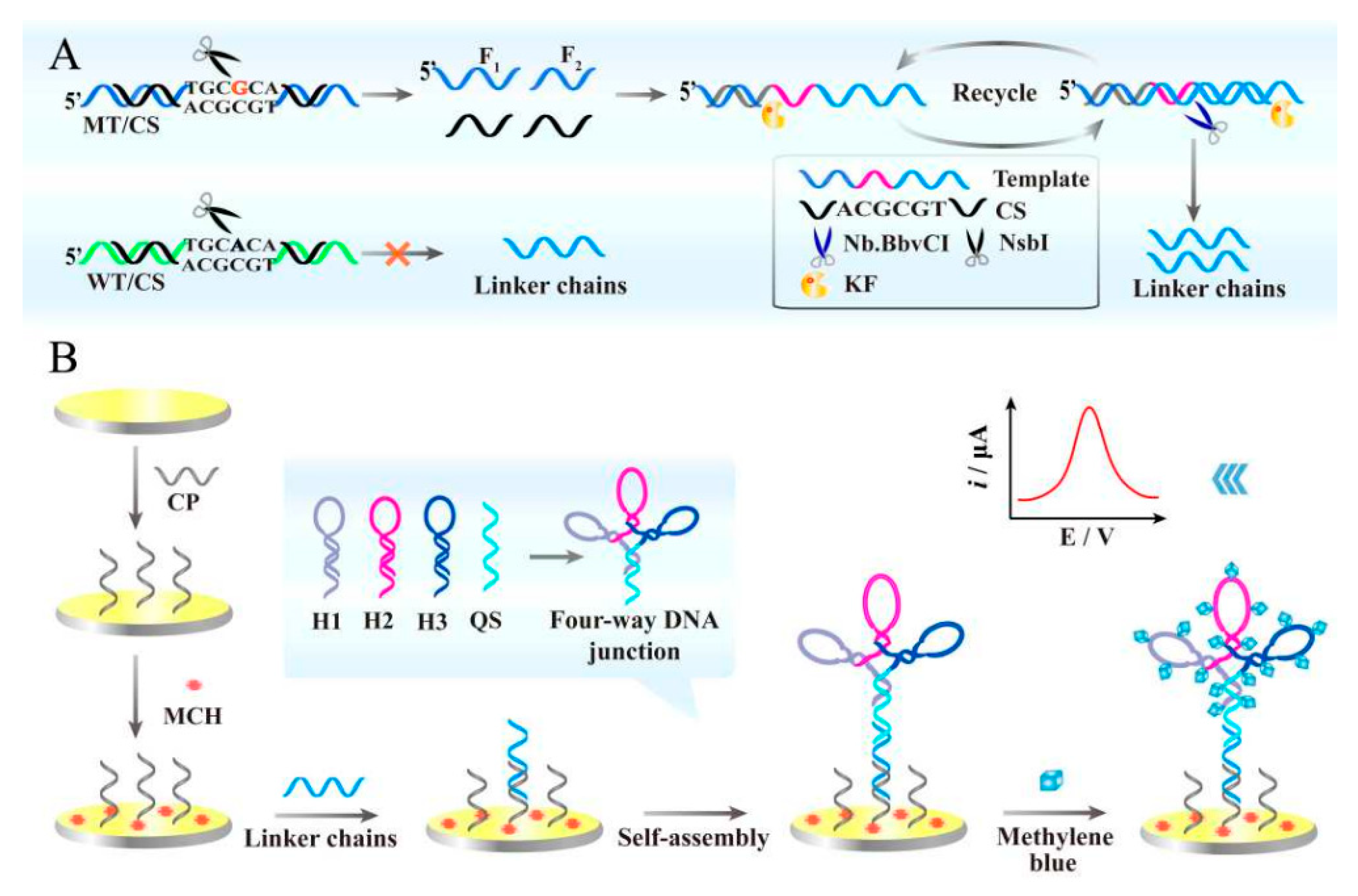
- Wang, T.; Peng, Q.; Guo, B.; Zhang, D.; Zhao, M.; Que, H.; Wu, H.; Yan, Y. An integrated electrochemical biosensor based on target-triggered strand displacement amplification and "four-way" DNA junction towards ultrasensitive detection of PIK3CA gene mutation. Biosens. Bioelectron. 2020, 150, 111954. [Google Scholar] [CrossRef] [PubMed]
- Liu, G.; Lao, R.; Xu, L.; Xu, Q.; Li, L.; Zhang, M.; Song, S.; Fan, C. Single-nucleotide polymorphism genotyping using a novel multiplexed electrochemical biosensor with nonfouling surface. Biosens. Bioelectron. 2013, 42, 516–521. [Google Scholar] [CrossRef] [PubMed]
- Wan, Y.; Lao, R.; Liu, G.; Song, S.; Wang, L.; Li, D.; Fan, C. Multiplexed Electrochemical DNA Sensor for Single-Nucleotide Polymorphism Typing by Using Oligonucleotide-Incorporated Nonfouling Surfaces. J. Phys. Chem. B 2010, 114, 6703–6706. [Google Scholar] [CrossRef]
- Lapitan, L.D., Jr.; Guo, Y.; Zhou, D. Nano-enabled bioanalytical approaches to ultrasensitive detection of low abundance single nucleotide polymorphisms. Analyst 2015, 140, 3872–3887. [Google Scholar] [CrossRef]
- Morais, S.L.; Magalhaes, J.; Domingues, V.F.; Delerue-Matos, C.; Ramos-Jesus, J.; Ferreira-Fernandes, H.; Pinto, G.R.; Santos, M.; Barroso, M.F. Development of an electrochemical DNA-based biosensor for the detection of the cardiovascular pharmacogenetic-altering SNP CYP2C9*3. Talanta 2023, 264, 124692. [Google Scholar] [CrossRef]
- Zhu, Q.; Liu, L.; Wang, R.; Zhou, X. A split aptamer (SPA)-based sandwich-type biosensor for facile and rapid detection of streptomycin. J. Hazard Mater. 2021, 403, 123941. [Google Scholar] [CrossRef]
- Nie, L.; Liu, F.; Ma, P.; Xiao, X. Applications of Gold Nanoparticles in Optical Biosensors. J. Biomed. Nanotechnol. 2014, 10, 2700–2721. [Google Scholar] [CrossRef]
- Lai, Y.; Deng, Y.; Yang, G.; Li, S.; Zhang, C.; Liu, X. Molecular Imprinting Polymers Electrochemical Sensor Based on AuNPs/PTh Modified GCE for Highly Sensitive Detection of Carcinomaembryonic Antigen. J. Biomed. Nanotechnol. 2018, 14, 1688–1694. [Google Scholar] [CrossRef]
- Liu, Y.; Deng, Y.; Li, T.; Chen, Z.; Chen, H.; Li, S.; Liu, H. Aptamer-Based Electrochemical Biosensor for Mercury Ions Detection Using AuNPs-Modified Glass Carbon Electrode. J. Biomed. Nanotechnol. 2018, 14, 2156–2161. [Google Scholar] [CrossRef] [PubMed]
- Liu, Y.; Lai, Y.; Yang, G.; Tang, C.; Deng, Y.; Li, S.; Wang, Z. Cd-Aptamer Electrochemical Biosensor Based on AuNPs/CS Modified Glass Carbon Electrode. J. Biomed. Nanotechnol. 2017, 13, 1253–1259. [Google Scholar] [CrossRef]
- Wang, Y.; Kong, S.L.; Su, X.D. A centrifugation-assisted visual detection of SNP in circulating tumor DNA using gold nanoparticles coupled with isothermal amplification. RSC Adv. 2020, 10, 1476–1483. [Google Scholar] [CrossRef] [PubMed]
- Han, S.; Liu, W.; Zheng, M.; Wang, R. Label-Free and Ultrasensitive Electrochemical DNA Biosensor Based on Urchinlike Carbon Nanotube-Gold Nanoparticle Nanoclusters. Anal. Chem. 2020, 92, 4780–4787. [Google Scholar] [CrossRef]
- Chakraborty, M.; Hashmi, M.S.J. Graphene as a Material—An Overview of Its Properties and Characteristics and Development Potential for Practical Applications. In Reference Module in Materials Science and Materials Engineering; Elsevier: Amsterdam, The Netherlands, 2018. [Google Scholar] [CrossRef]
- Khoshfetrat, S.M.; Mehrgardi, M.A. Amplified electrochemical genotyping of single-nucleotide polymorphisms using a graphene–gold nanoparticles modified glassy carbon platform. RSC Adv. 2015, 5, 29285–29293. [Google Scholar] [CrossRef]
- Hwang, M.T.; Wang, Z.; Ping, J.; Ban, D.K.; Shiah, Z.C.; Antonschmidt, L.; Lee, J.; Liu, Y.; Karkisaval, A.G.; Johnson, A.T.C.; et al. DNA Nanotweezers and Graphene Transistor Enable Label-Free Genotyping. Adv. Mater. 2018, 30, e1802440. [Google Scholar] [CrossRef]
- Bonanni, A.; Pumera, M. Graphene Platform for Hairpin-DNA-Based Impedimetric Genosensing. ACS Nano 2011, 5, 2356–2361. [Google Scholar] [CrossRef]
- Zeng, N.; Xiang, J. Detection of KRAS G12D point mutation level by anchor-like DNA electrochemical biosensor. Talanta 2019, 198, 111–117. [Google Scholar] [CrossRef]
- Hamidi-Asl, E.; Raoof, J.B.; Ojani, R.; Golabi, S.M.; Hejazi, M.S. A new peptide nucleotide acid biosensor for electrochemical detection of single nucleotide polymorphism in duplex DNA via triplex structure formation. J. Iran. Chem. Soc. 2013, 10, 1075–1083. [Google Scholar] [CrossRef]
- Aladag, N.; Ozkan-Ariksoysal, D.; Gezen-Ak, D.; Yilmazer, S.; Ozsoz, M. An Electrochemical DNA Biosensor for the Detection of the Apa I Polymorphism in the Vitamin D Receptor Gene Using Meldola’s Blue as a Hybridization Indicator. Electroanalysis 2010, 22, 590–598. [Google Scholar] [CrossRef]
- Gu, C.; Kong, X.; Liu, X.; Gai, P.; Li, F. Enzymatic Biofuel-Cell-Based Self-Powered Biosensor Integrated with DNA Amplification Strategy for Ultrasensitive Detection of Single-Nucleotide Polymorphism. Anal. Chem. 2019, 91, 8697–8704. [Google Scholar] [CrossRef]
- Uygun, Z.O.; Yeniay, L.; Gi Rgi, N.S.F. CRISPR-dCas9 powered impedimetric biosensor for label-free detection of circulating tumor DNAs. Anal. Chim. Acta 2020, 1121, 35–41. [Google Scholar] [CrossRef]
- Xu, W.; Jin, T.; Dai, Y.; Liu, C.C. Surpassing the detection limit and accuracy of the electrochemical DNA sensor through the application of CRISPR Cas systems. Biosens. Bioelectron. 2020, 155, 112100. [Google Scholar] [CrossRef] [PubMed]
- Sanromán-Iglesias, M.; Lawrie, C.H.; Liz-Marzán, L.M.; Grzelczak, M. The Role of Chemically Modified DNA in Discrimination of Single-Point Mutation through Plasmon-Based Colorimetric Assays. ACS Appl. Nano Mater. 2018, 1, 3741–3746. [Google Scholar] [CrossRef]
- Zhou, C.; Zou, H.; Sun, C.; Ren, D.; Chen, J.; Li, Y. Signal amplification strategies for DNA-based surface plasmon resonance biosensors. Biosens. Bioelectron. 2018, 117, 678–689. [Google Scholar] [CrossRef]
- Li, Y.; Gao, T.; Xu, G.; Xiang, X.; Zhao, B.; Han, X.X.; Guo, X. Direct Approach toward Label-Free DNA Detection by Surface-Enhanced Raman Spectroscopy: Discrimination of a Single-Base Mutation in 50 Base-Paired Double Helixes. Anal. Chem. 2019, 91, 7980–7984. [Google Scholar] [CrossRef] [PubMed]
- Chen, X.; Zhou, D.; Shen, H.; Chen, H.; Feng, W.; Xie, G. A universal probe design for colorimetric detection of single-nucleotide variation with visible readout and high specificity. Sci. Rep. 2016, 6, 20257. [Google Scholar] [CrossRef] [PubMed]
- Wu, S.; Liang, P.; Yu, H.; Xu, X.; Liu, Y.; Lou, X.; Xiao, Y. Amplified single base-pair mismatch detection via aggregation of exonuclease-sheared gold nanoparticles. Anal. Chem. 2014, 86, 3461–3467. [Google Scholar] [CrossRef]
- Deng, H.; Shen, W.; Gao, Z. Colorimetric detection of single nucleotide polymorphisms in the presence of 10(3)-fold excess of a wild-type gene. Biosens. Bioelectron. 2015, 68, 310–315. [Google Scholar] [CrossRef]
- Dai, B.; Xu, Y.; Wang, T.; Wang, S.; Tang, L.; Tang, J. Recent Advances in Agglomeration Detection and Dual-Function Application of Surface-Enhanced Raman Scattering (SERS). J. Biomed. Nanotechnol. 2022, 18, 1257–1275. [Google Scholar] [CrossRef]
- Gaidi, M.; Daoudi, K.; Tlili, A.; Columbus, S.; Leblanc-Lavoie, J.; Ramachandran, K.; Suleiman, B.; Alhazaa, A.N.; El Khakani, M.A. Fast, highly sensitive and label free detection of small genetic sequence difference of DNA using novel Surface-Enhanced Raman Spectroscopy nanostructured sensor. Sens. Bio-Sens. Res. 2021, 32, 100406. [Google Scholar] [CrossRef]
- Zhang, Y.; Zhan, D.S.; Xu, X.Y.; Zhang, Z.; Hafez, M.E.; He, Y.; Li, Y.; Li, D.W. Label-free detection of DNA methylation by surface-enhanced Raman spectroscopy using zirconium-modified silver nanoparticles. Talanta 2023, 253, 123941. [Google Scholar] [CrossRef] [PubMed]
- Lyu, N.; Rajendran, V.K.; Li, J.; Engel, A.; Molloy, M.P.; Wang, Y. Highly specific detection of KRAS single nucleotide polymorphism by asymmetric PCR/SERS assay. Analyst 2021, 146, 5714–5721. [Google Scholar] [CrossRef]
- Ngo, H.T.; Gandra, N.; Fales, A.M.; Taylor, S.M.; Vo-Dinh, T. Sensitive DNA detection and SNP discrimination using ultrabright SERS nanorattles and magnetic beads for malaria diagnostics. Biosens. Bioelectron. 2016, 81, 8–14. [Google Scholar] [CrossRef] [PubMed]
- Lowe, A.J.; Huh, Y.S.; Strickland, A.D.; Erickson, D.; Batt, C.A. Multiplex Single Nucleotide PolymorphismGenotyping Utilizing Ligase Detection ReactionCoupled Surface Enhanced Raman Spectroscopy. Anal. Chem. 2010, 82, 5810–5814. [Google Scholar] [CrossRef] [PubMed]
- Miura, S.; Nishizawa, S.; Suzuki, A.; Fujimoto, Y.; Ono, K.; Gao, Q.; Teramae, N. DNA-binding small-ligand-immobilized surface plasmon resonance biosensor for detecting thymine-related single-nucleotide polymorphisms. Chemistry 2011, 17, 14104–14110. [Google Scholar] [CrossRef] [PubMed]
- Rahman, M.S.; Anower, M.S.; Rahman, M.K.; Hasan, M.R.; Hossain, M.B.; Haque, M.I. Modeling of a highly sensitive MoS2-Graphene hybrid based fiber optic SPR biosensor for sensing DNA hybridization. Optik 2017, 140, 989–997. [Google Scholar] [CrossRef]
- Malmqvist, M. Biospecific interaction analysis using biosensor technology. Nature 1993, 361, 186–187. [Google Scholar] [CrossRef]
- Yi, X.; Xia, Y.; Ding, B.; Wu, L.; Hu, S.; Wang, Z.; Yang, M.; Wang, J. Dual-Channel Surface Plasmon Resonance for Quantification of ApoE Gene and Genotype Discrimination in Unamplified Genomic DNA Extracts. ACS Sens. 2018, 3, 2402–2407. [Google Scholar] [CrossRef]
- Huang, C.J.; Lin, Z.E.; Yang, Y.S.; Chan, H.W.; Chen, W.Y. Neutralized chimeric DNA probe for detection of single nucleotide polymorphism on surface plasmon resonance biosensor. Biosens. Bioelectron. 2018, 99, 170–175. [Google Scholar] [CrossRef]
- He, L.; Musick, M.D.; Nicewarner, S.R.; Salinas, F.G.; Benkovic, S.J.; Natan, M.J.; Keating, C.D. Colloidal Au-Enhanced Surface Plasmon Resonance for Ultrasensitive Detection of DNA Hybridization. Am. Chem. Soc. 2000, 122, 9071–9077. [Google Scholar] [CrossRef]
- Li, Y.; Wark, A.W.; Lee, H.J.; Corn, R.M. Single-Nucleotide Polymorphism Genotyping by Nanoparticle-Enhanced Surface Plasmon Resonance Imaging Measurements of Surface Ligation Reactions. Am. Chem. Soc. 2006, 78, 3158–3164. [Google Scholar] [CrossRef] [PubMed]
- Atay, S.; Pişkin, K.; Yılmaz, F.; Çakır, C.; Yavuz, H.; Denizli, A. Quartz crystal microbalance based biosensors for detecting highly metastatic breast cancer cells via their transferrin receptors. Anal. Methods 2016, 8, 153–161. [Google Scholar] [CrossRef]
- Hosseini, M.S.; Iraji zad, A.; Vossoughi, M.; Hosseini, M. L-lysine biodetector based on a TOCNFs-coated Quartz Crystal Microbalance (QCM). Eur. Polym. J. 2023, 186, 111831. [Google Scholar] [CrossRef]
- Pang, L.; Zhang, L.; Wang, Z.; Lu, G.; Sun, X.; Cheng, J.; Chen, S.; Qi, G.; Duan, X.; Xu, R.; et al. Identification of sweetpotato black spot disease caused by Ceratocystis fimbriata by quartz crystal microbalance array. Sens. Actuators B Chem. 2023, 386, 133761. [Google Scholar] [CrossRef]
- Lino, C.; Barrias, S.; Chaves, R.; Adega, F.; Fernandes, J.R.; Martins-Lopes, P. Development of a QCM-based biosensor for the detection of non-small cell lung cancer biomarkers in liquid biopsies. Talanta 2023, 260, 124624. [Google Scholar] [CrossRef]
- Lim, H.J.; Saha, T.; Tey, B.T.; Tan, W.S.; Hassan, S.S.; Ooi, C.W. Quartz crystal microbalance-based biosensing of hepatitis B antigen using a molecularly imprinted polydopamine film. Talanta 2022, 249, 123659. [Google Scholar] [CrossRef] [PubMed]
- Xi, X.; Niyonshuti, I.I.; Yu, N.; Yao, L.; Fu, Y.; Chen, J.; Li, Y. Label-Free Quartz Crystal Microbalance Biosensor Based on Aptamer-Capped Gold Nanocages Loaded with Polyamidoamine for Thrombin Detection. ACS Appl. Nano Mater. 2021, 4, 10047–10054. [Google Scholar] [CrossRef]
- Dronina, J.; Plausinaitis, D.; Samukaite-Bubniene, U.; Ramanavicius, A. Real-time label-free assessment of T7 DNA polymerase immobilization. Mater. Today Nano 2022, 19, 100232. [Google Scholar] [CrossRef]
- Bonyadi, F.; Kavruk, M.; Ucak, S.; Cetin, B.; Bayramoglu, G.; Dursun, A.D.; Arica, Y.; Ozalp, V.C. Real-Time Biosensing Bacteria and Virus with Quartz Crystal Microbalance: Recent Advances, Opportunities, and Challenges. Crit. Rev. Anal. Chem. 2023. [Google Scholar] [CrossRef] [PubMed]
- Wang, D.; Tang, W.; Wu, X.; Wang, X.; Chen, G.; Chen, Q.; Li, N.; Liu, F. Highly selective detection of single-nucleotide polymorphisms using a quartz crystal microbalance biosensor based on the toehold-mediated strand displacement reaction. Anal. Chem. 2012, 84, 7008–7014. [Google Scholar] [CrossRef] [PubMed]
- Simmel, F.C.; Yurke, B.; Singh, H.R. Principles and Applications of Nucleic Acid Strand Displacement Reactions. Chem. Rev. 2019, 119, 6326–6369. [Google Scholar] [CrossRef]
- Li, H.; Xiao, S.; Yao, D.; Lam, M.H.; Liang, H. A smart DNA-gold nanoparticle probe for detecting single-base changes on the platform of a quartz crystal microbalance. Chem. Commun. 2015, 51, 4670–4673. [Google Scholar] [CrossRef] [PubMed]
- Zhou, L.; Kato, F.; Iijima, M.; Nonaka, T.; Kuroda, S.; Ogi, H. Mass-Fabrication Scheme of Highly Sensitive Wireless Electrodeless MEMS QCM Biosensor with Antennas on Inner Walls of Microchannel. Anal. Chem. 2023, 95, 5507–5513. [Google Scholar] [CrossRef] [PubMed]


| Signal Amplification Strategies | Target Mutation | LOD | Ref. |
|---|---|---|---|
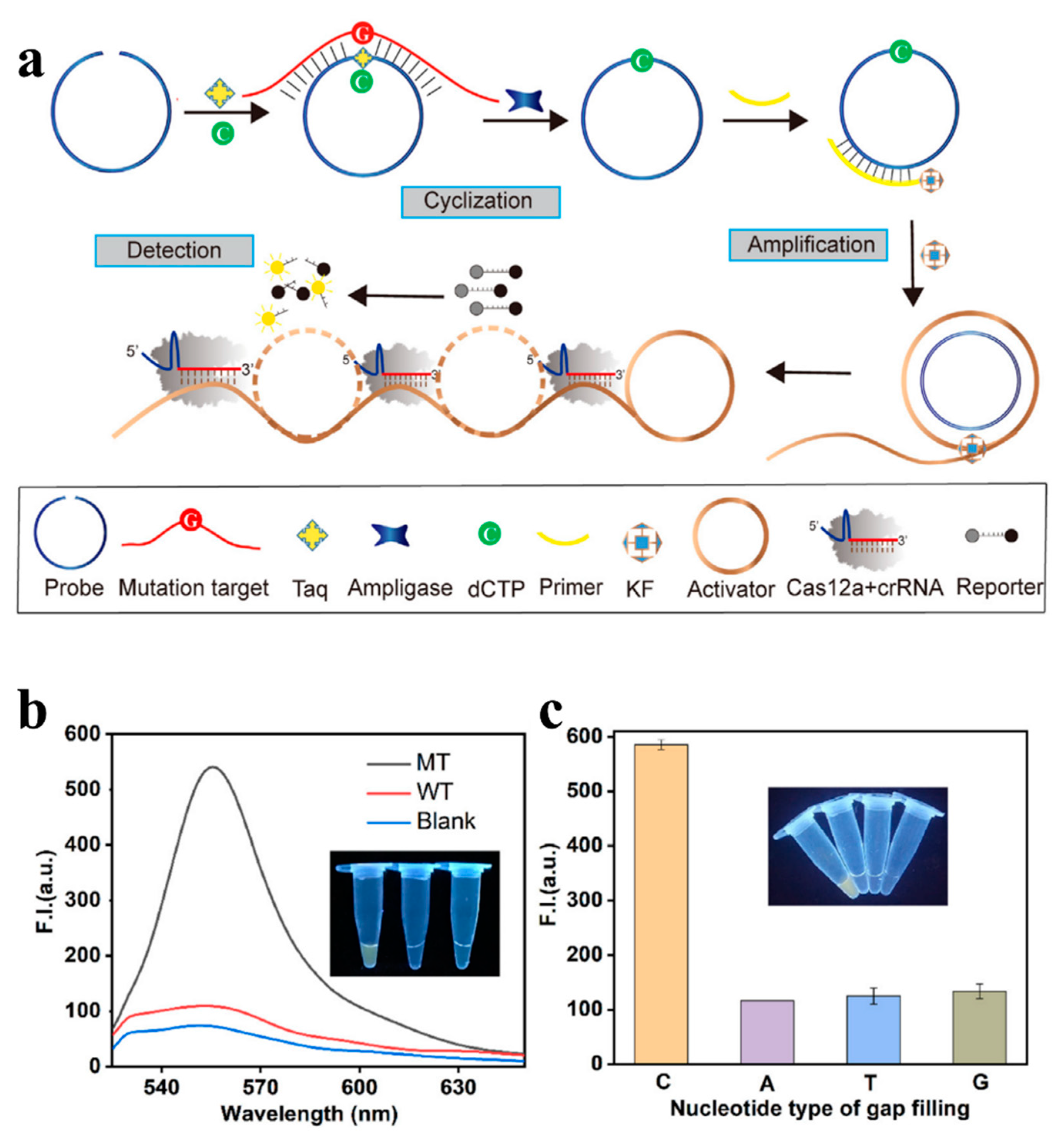
| CRISPR/Cas12a with RCA | SNV of the PIK3CA H1047R | 10 aM | [56] |
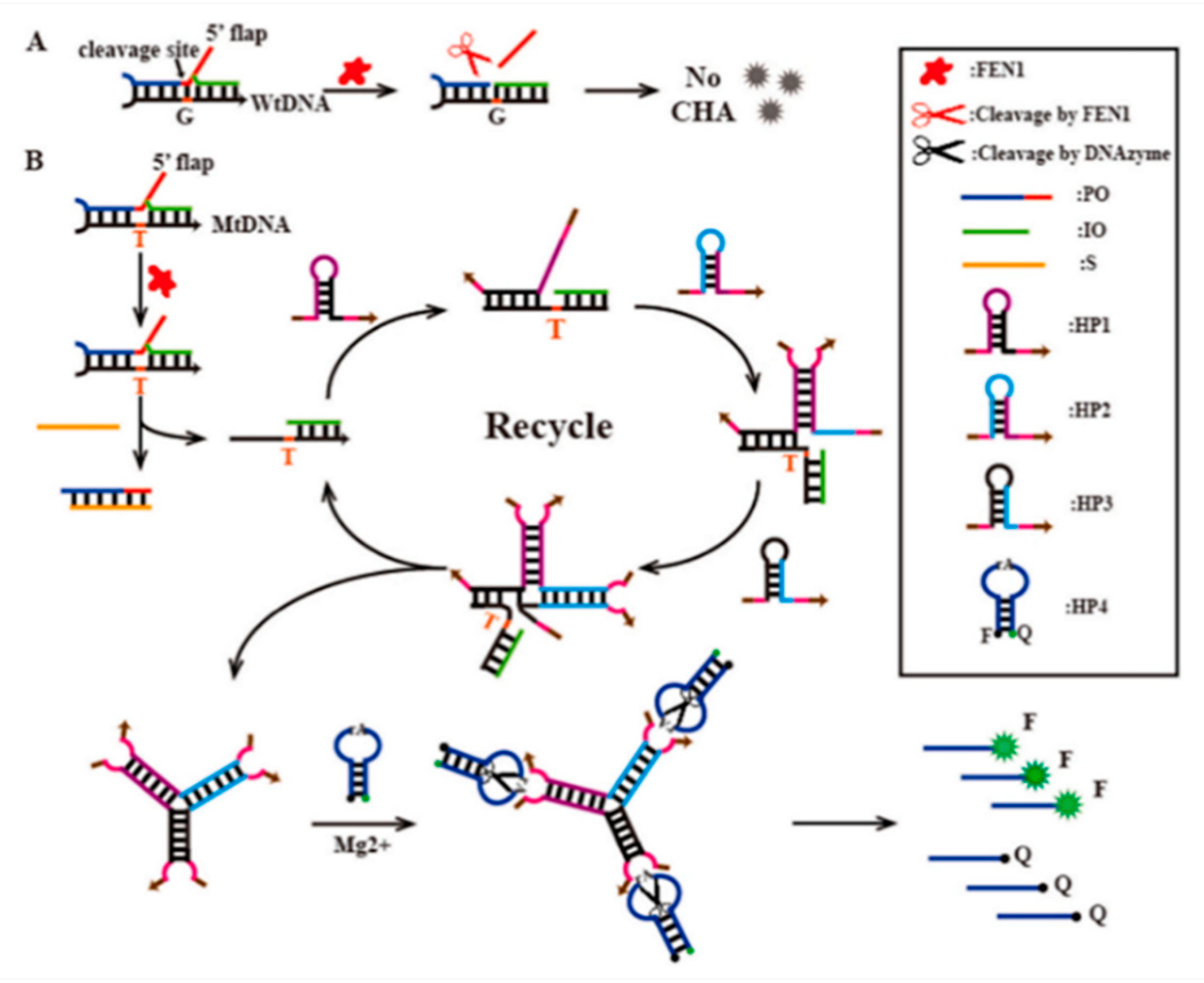
| Invader assay-induced multiDNAzyme junctions | SNP | 4.23 fM | [63] |
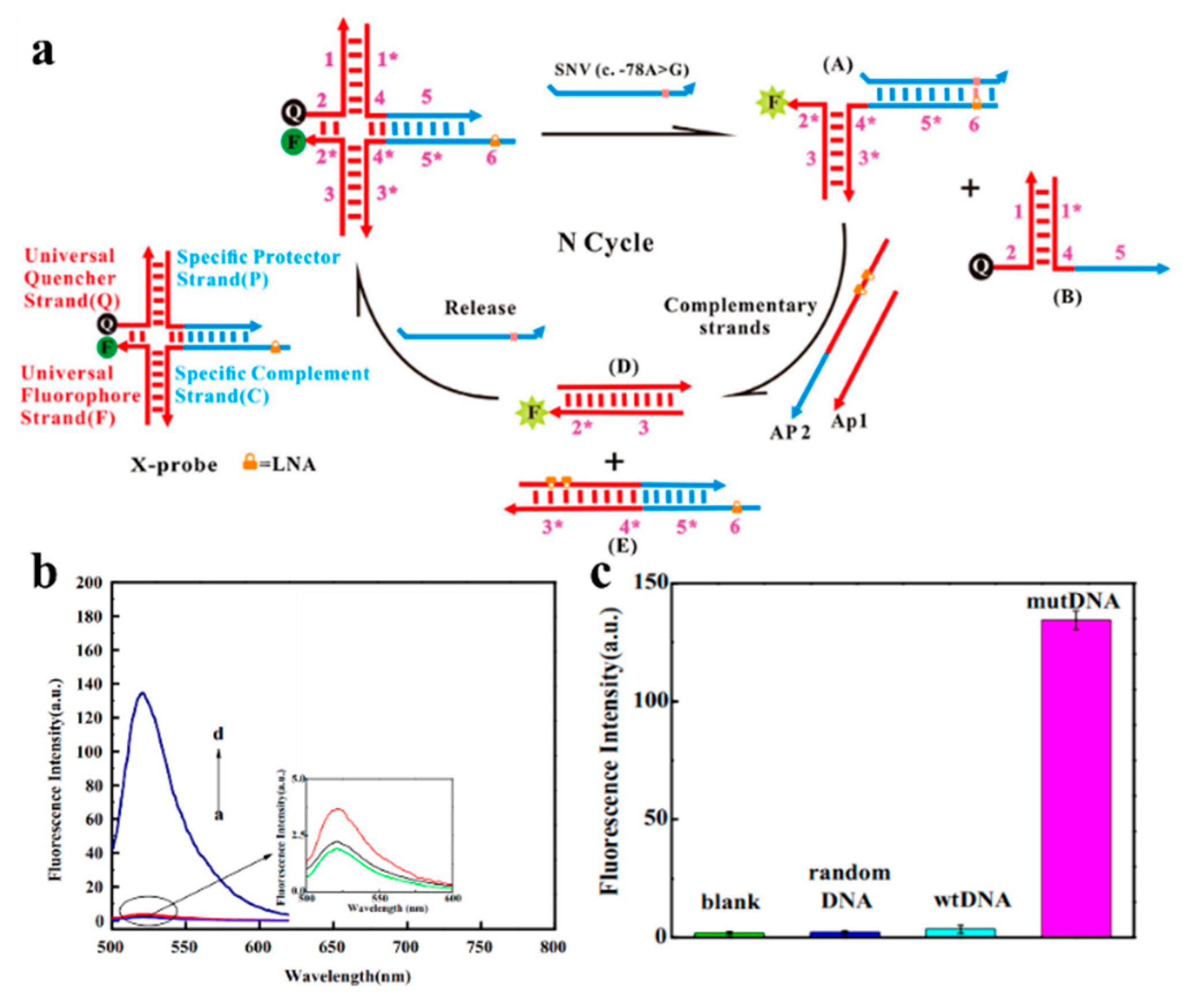
| Universal locked nucleic acid-integrated X-shaped probe | SNP | 6 fM | [64] |
| Core-shell gold nanocube (AuNC) and plasmon-enhanced fluorescence (PEF) | SNP | 1.3 pM | [86] |
| RT-PCR associated with G-quadruplex RCA | Multiple SNPs | 8.3 fg | [87] |
| Fluorescence polarization (FP) and target-initiated rolling circle amplification (RCA) | KRAS G13D and G12D mutations | 5.88 pM | [88] |
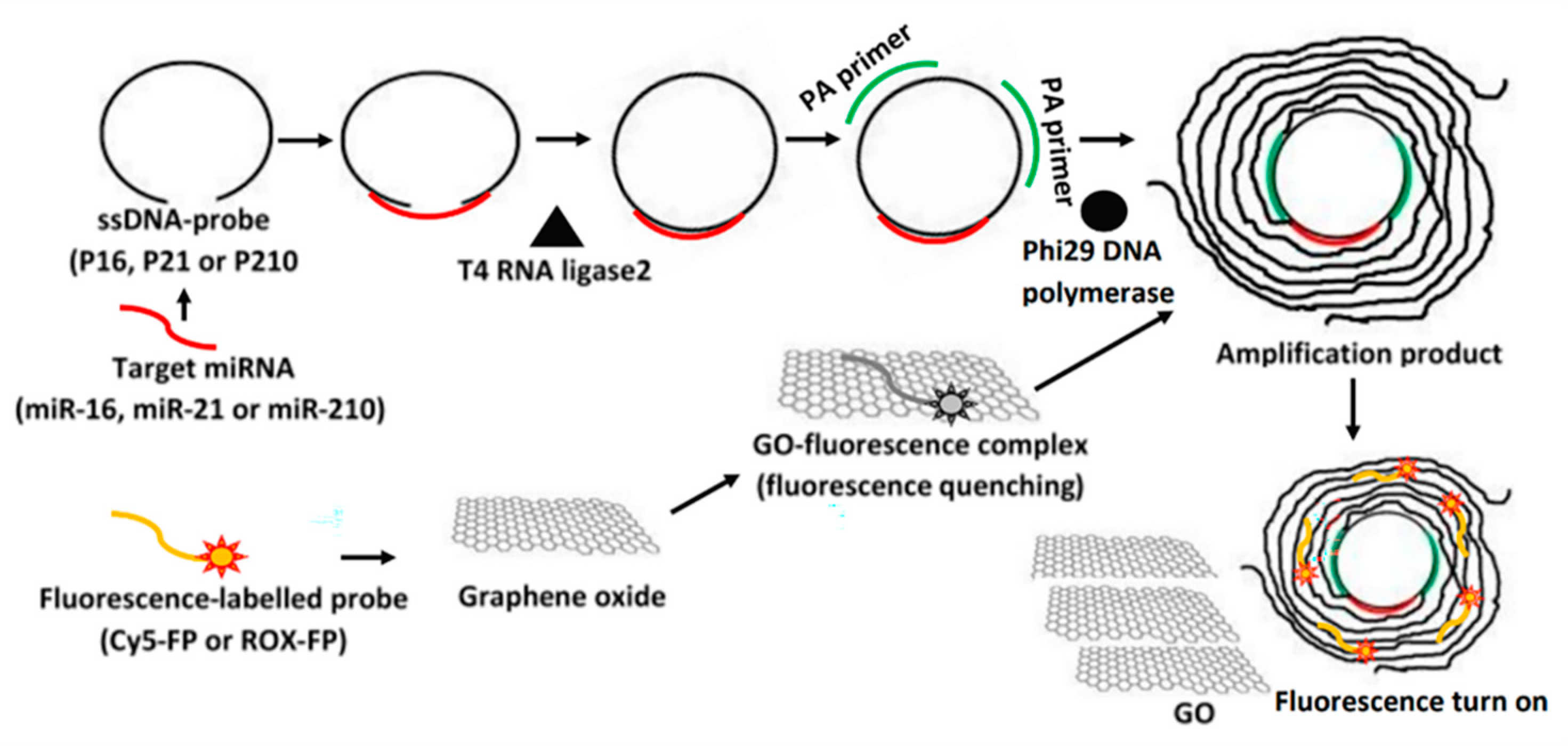
| Multiple primers-mediated RCA coupled with a graphene oxide-based fluorescence | Multiple SNPs | 0.87 fM | [53] |
| Signal Transduction | Biosensor Platform | Target Mutation | LOD | Ref. |
|---|---|---|---|---|
| Amperometric | Electrochemical ligase chain reaction (eLCR) | CYP2C19 (G681A) in human whole-blood samples | 0.5 fM | [102] |
| Impedimetric | Graphene | Apo E gene | G-SL: 50 nM G-FL: 6.6 pM | [118] |
| Impedimetric | Anchor-like DNA (alDNA) electrochemical biosensor | KRAS G12D mutation | 0.1 pM/100 pM (total/mutant DNA) | [119] |
| Amperometric | PNA/ds-DNA triplex formation | p53 gene | 10−6 M | [120] |
| Voltammetric | MDB as a hybridization indicator | VDR gene | 10.9 pmol/100 mL | [121] |
| Voltammetric | HCR and SDR | P53 gene | 20 aM | [122] |
| Impedimetric | CRISPR/dCas9-powered impedimetric | ctDNA, PIK3CA exon 9 mutation | 0.65 nM | [123] |
| Voltammetric | CRISPR/cas-enhanced electrochemical biosensor | SNPs | 10 fM | [124] |
Disclaimer/Publisher’s Note: The statements, opinions and data contained in all publications are solely those of the individual author(s) and contributor(s) and not of MDPI and/or the editor(s). MDPI and/or the editor(s) disclaim responsibility for any injury to people or property resulting from any ideas, methods, instructions or products referred to in the content. |
© 2023 by the authors. Licensee MDPI, Basel, Switzerland. This article is an open access article distributed under the terms and conditions of the Creative Commons Attribution (CC BY) license (https://creativecommons.org/licenses/by/4.0/).
Share and Cite
Wu, K.; Kong, F.; Zhang, J.; Tang, Y.; Chen, Y.; Chao, L.; Nie, L.; Huang, Z. Recent Progress in Single-Nucleotide Polymorphism Biosensors. Biosensors 2023, 13, 864. https://doi.org/10.3390/bios13090864
Wu K, Kong F, Zhang J, Tang Y, Chen Y, Chao L, Nie L, Huang Z. Recent Progress in Single-Nucleotide Polymorphism Biosensors. Biosensors. 2023; 13(9):864. https://doi.org/10.3390/bios13090864
Chicago/Turabian StyleWu, Kaimin, Feizhi Kong, Jingjing Zhang, Ying Tang, Yao Chen, Long Chao, Libo Nie, and Zhao Huang. 2023. "Recent Progress in Single-Nucleotide Polymorphism Biosensors" Biosensors 13, no. 9: 864. https://doi.org/10.3390/bios13090864