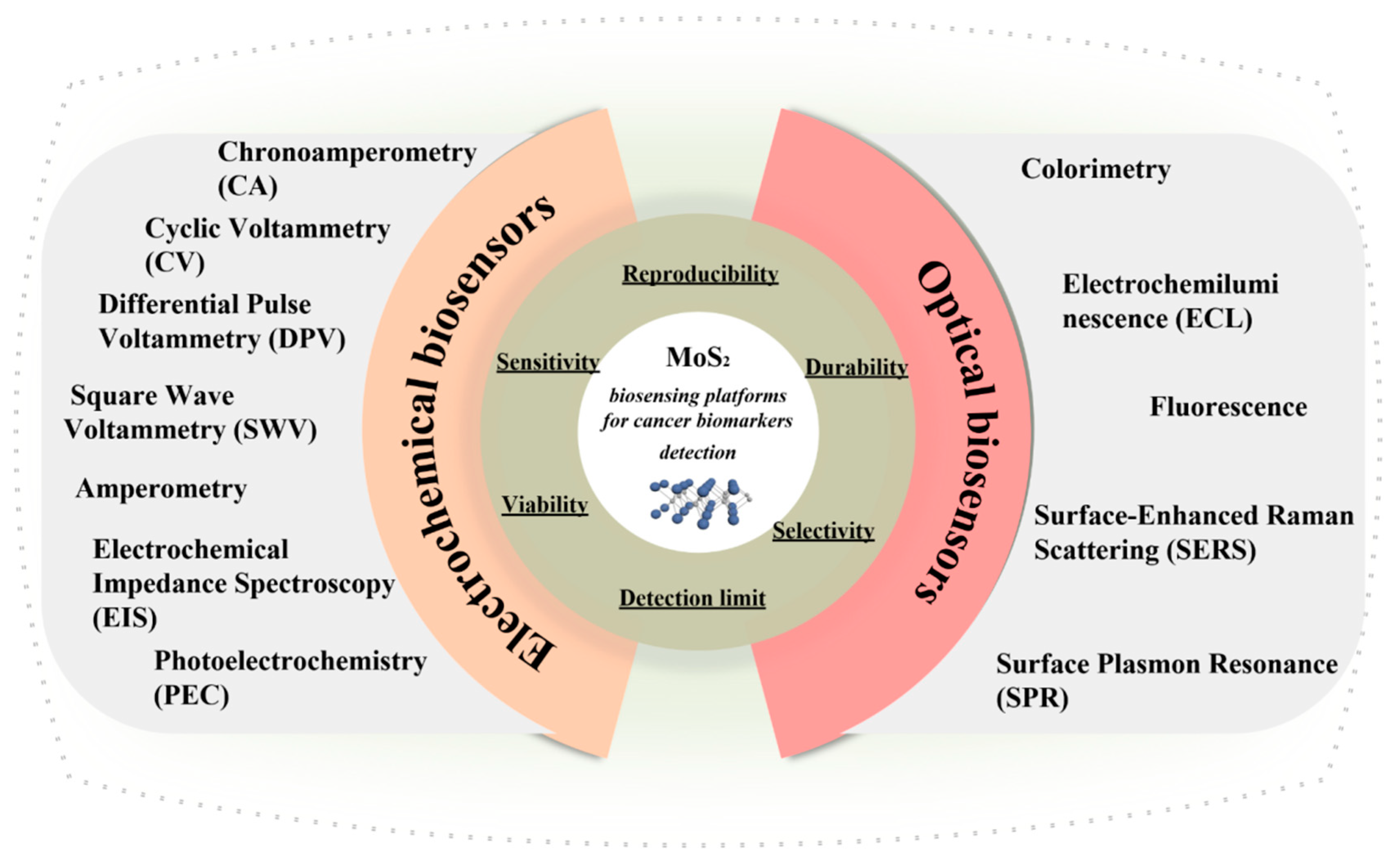
Molybdenum Disulfide as Tunable Electrochemical and Optical Biosensing Platforms for Cancer Biomarker Detection: A Review


Abstract
:1. Introduction
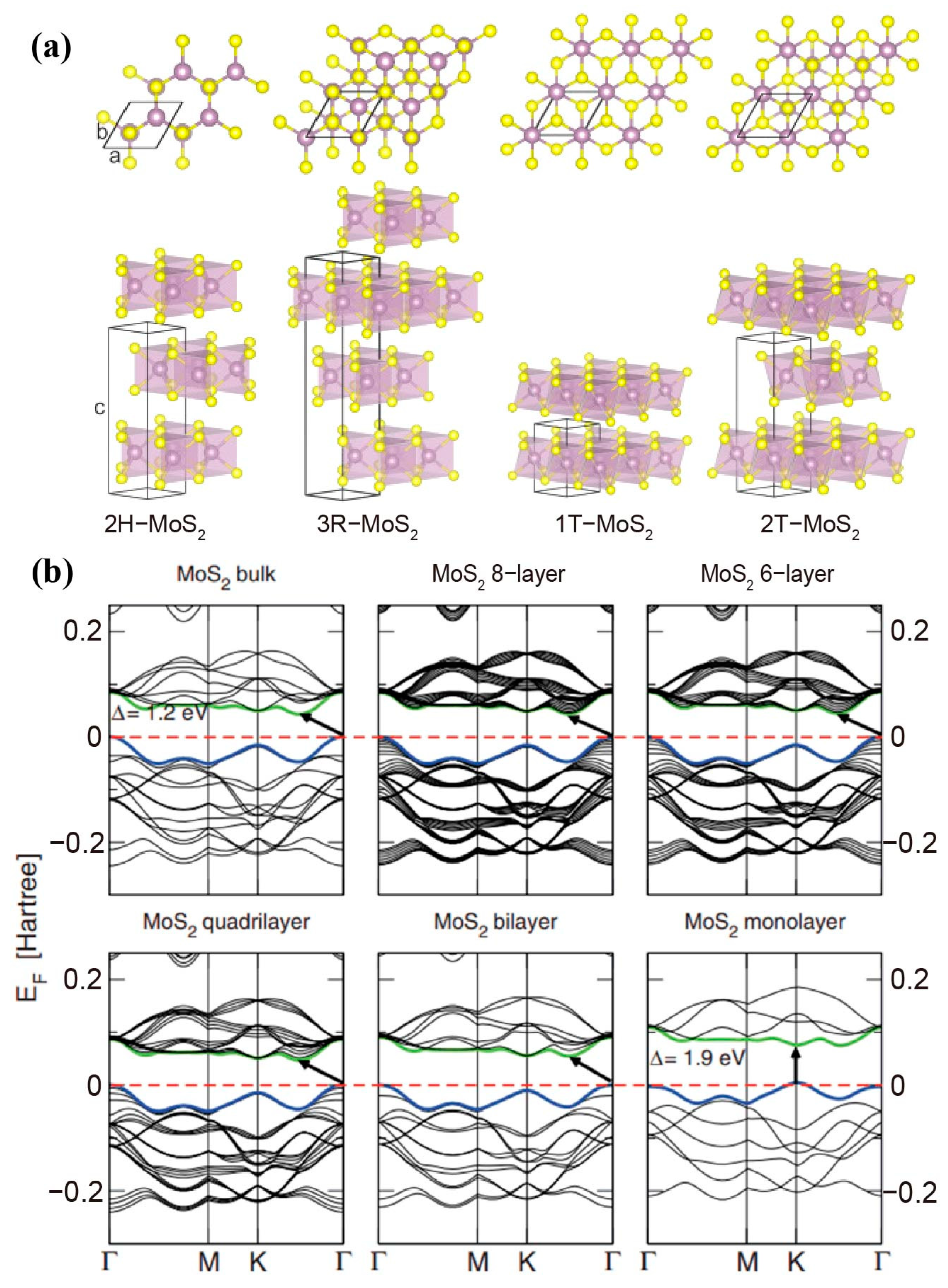
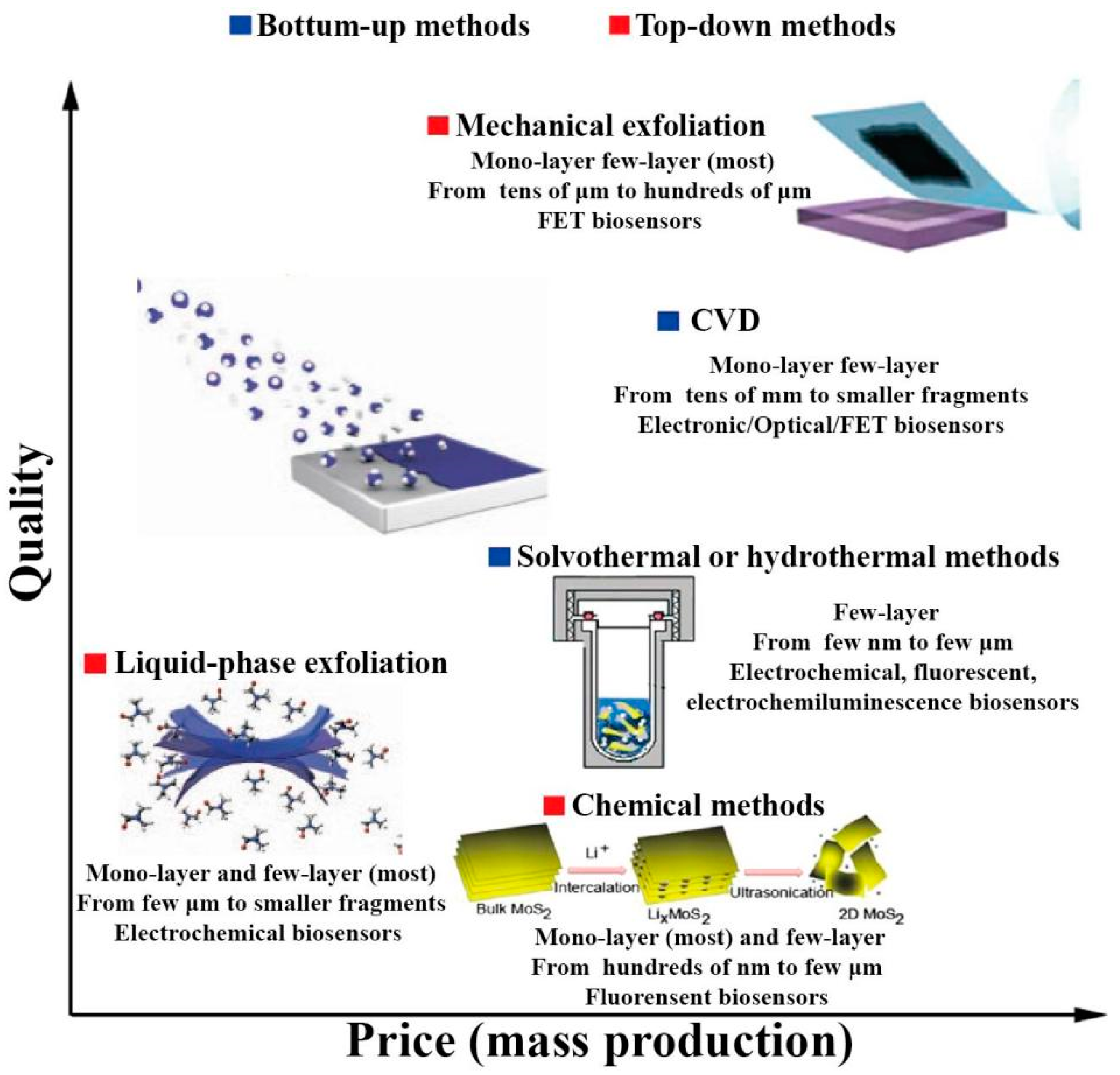
2. Synthesis and Characteristics of Molybdenum Disulfide
2.1. Top-Down Methods
2.2. Bottom-Up Methods
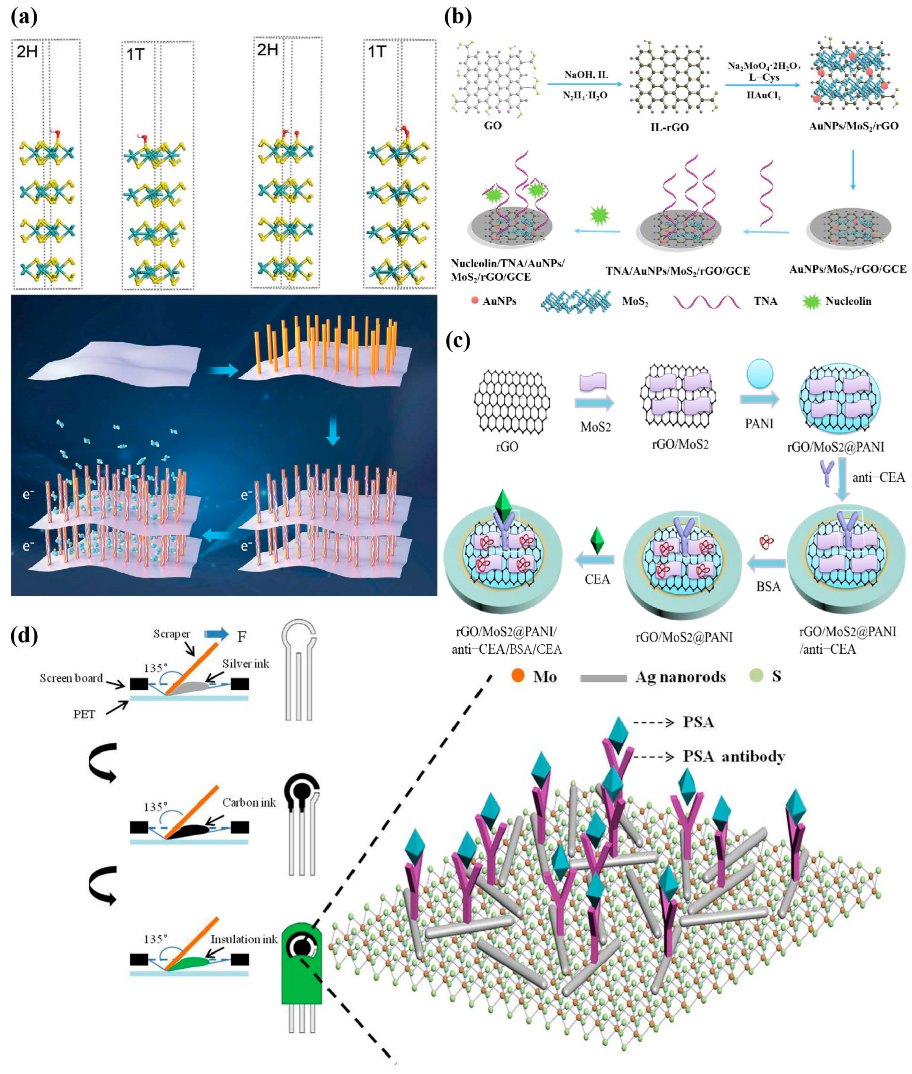
3. Electrochemical Biosensors for Cancer Biomarkers Detection Based on MoS2
3.1. Potentiometry
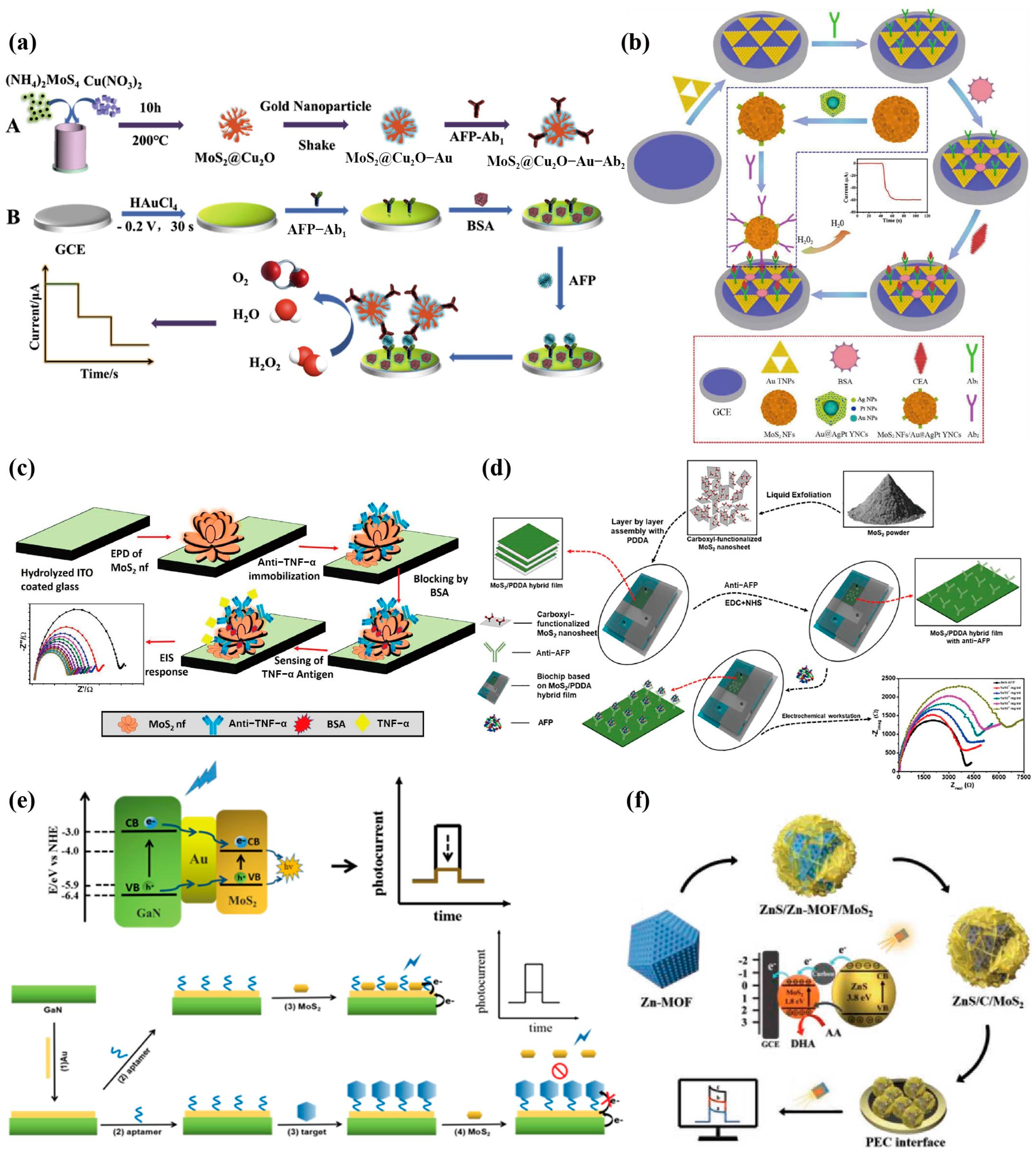
3.2. Amperometry

3.3. Impedimetry
3.4. Photoelectrochemistry (PEC)

4. Optical Biosensors for Cancer Biomarker Detection Based on MoS2
4.1. Colorimetry
4.2. Electrochemiluminescence (ECL)

4.3. Fluorescence
4.4. Surface Enhanced Raman Scattering (SERS)

4.5. Surface Plasmon Resonance (SPR)
5. Miscellaneous Biosensors for Cancer Biomarkers Detection Based on MoS2

6. Discussion and Outlook
7. Conclusions
Author Contributions
Funding
Institutional Review Board Statement
Informed Consent Statement
Data Availability Statement
Conflicts of Interest
Abbreviations
| AuNPs | gold nanoparticles; |
| rGO | reduced graphene oxide; |
| PDDA | poly dimethyl diallyl ammonium chloride; |
| Ag/AgCl | silver/silver chloride; |
| ITO | indium tin oxide; |
| AMACR | alpha-methylacyl-CoA racemase; |
| NaBH4 | sodium borohydride; |
| 4-NP | 4-nitrophenol; |
| ssDNA | single strand DNA; |
| AgNCs | gold nanocubes; |
| AuNS | gold nanostars; |
| CTC | circulating tumor cells; |
| PD-1 | programmed cell death protein 1; |
| CYFRA21-1 | cytokeratin 19 fragment (1); |
| T3 | triiodothyronine; |
| DSN | duplex-specific nuclease. |
| LSAW | Love-mode surface acoustic wave |
| OFA/iLRP | Oncofetal antigen/immature laminin receptor protein |
| BRCA1 | Breast cancer susceptibility gene type 1 |
References
- Jayanthi, V.S.; Das, A.B.; Saxena, U. Recent advances in biosensor development for the detection of cancer biomarkers. Biosens. Bioelectron. 2017, 91, 15–23. [Google Scholar] [CrossRef]
- Hasan, M.R.; Ahommed, M.S.; Daizy, M.; Bacchu, M.S.; Ali, M.R.; Al-Mamun, M.R.; Aly Saad Aly, M.; Khan, M.Z.H.; Hossain, S.I. Recent development in electrochemical biosensors for cancer biomarkers detection. Biosens. Bioelectron. X 2021, 8, 100075. [Google Scholar] [CrossRef]
- Rabie, A.M.I.; Ali, A.S.M.; Al-Zeer, M.A.; Barhoum, A.; El-Hallouty, S.; Shousha, W.G.; Berg, J.; Kurreck, J.; Khalil, A.S.G. Spontaneous Formation of 3D Breast Cancer Tissues on Electrospun Chitosan/Poly(ethylene oxide) Nanofibrous Scaffolds. ACS Omega 2022, 7, 2114–2126. [Google Scholar] [CrossRef] [PubMed]
- Gric, T.; Sokolovski, S.G.; Navolokin, N.; Glushkovskaya, O.S.; Rafailov, E.U. Metamaterial formalism approach for advancing the recognition of glioma areas in brain tissue biopsies. Opt. Mater. Express 2020, 10, 1607–1615. [Google Scholar] [CrossRef]
- Ranjan, R.; Esimbekova, E.N.; Kratasyuk, V.A. Rapid biosensing tools for cancer biomarkers. Biosens. Bioelectron. 2017, 87, 918–930. [Google Scholar] [CrossRef] [PubMed]
- Henry, N.L.; Hayes, D.F. Cancer biomarkers. Mol. Oncol. 2012, 6, 140–146. [Google Scholar] [CrossRef]
- Khan, H.; Shah, M.R.; Barek, J.; Malik, M.I. Cancer biomarkers and their biosensors: A comprehensive review. TrAC Trends Anal. Chem. 2023, 158, 116813. [Google Scholar] [CrossRef]
- Sharifi, M.; Avadi, M.R.; Attar, F.; Dashtestani, F.; Ghorchian, H.; Rezayat, S.M.; Saboury, A.A.; Falahati, M. Cancer diagnosis using nanomaterials based electrochemical nanobiosensors. Biosens. Bioelectron. 2019, 126, 773–784. [Google Scholar] [CrossRef]
- Agnolon, V.; Contato, A.; Meneghello, A.; Tagliabue, E.; Toffoli, G.; Gion, M.; Polo, F.; Fabricio, A.S.C. ELISA assay employing epitope-specific monoclonal antibodies to quantify circulating HER2 with potential application in monitoring cancer patients undergoing therapy with trastuzumab. Sci. Rep. 2020, 10, 3016. [Google Scholar] [CrossRef]
- Dueck, M.E.; Lin, R.; Zayac, A.; Gallagher, S.; Chao, A.K.; Jiang, L.X.; Datwani, S.S.; Hung, P.; Stieglitz, E. Precision cancer monitoring using a novel, fully integrated, microfluidic array partitioning digital PCR platform. Sci. Rep. 2019, 9, 19606. [Google Scholar] [CrossRef]
- Selvakumar, S.C.; Preethi, K.A.; Ross, K.; Tusubira, D.; Khan, M.W.A.; Mani, P.; Rao, T.N.; Sekar, D. CRISPR/Cas9 and next generation sequencing in the personalized treatment of Cancer. Mol. Cancer 2022, 24, 83. [Google Scholar] [CrossRef] [PubMed]
- Udo, R.; Katsumata, K.; Kuwabara, H.; Enomoto, M.; Ishizaki, T.; Sunamura, M.; Nagakawa, Y.; Soya, R.; Sugimoto, M.; Tsuchida, A. Urinary charged metabolite profiling of colorectal cancer using capillary electrophoresis-mass spectrometry. Sci. Rep. 2020, 10, 21057. [Google Scholar] [CrossRef] [PubMed]
- Barhoum, A.; Altintas, Z.; Shalini Devi, K.S.; Forster, R.J. Electrochemiluminescence biosensors for detection of cancer biomarkers in biofluids: Principles, opportunities, and challenges. Nano Today 2023, 50, 101874. [Google Scholar] [CrossRef]
- Ibrahim, J.; Peeters, M.; Camp, G.V.; Beeck, K.O. Methylation biomarkers for early cancer detection and diagnosis: Current and future perspectives. Eur. J. Cancer 2023, 178, 91–113. [Google Scholar] [CrossRef] [PubMed]
- Raghavendra, U.; Gudigar, A.; Paul, A.; Goutham, T.S.; Inamdar, M.A.; Hegde, A.; Devi, A.; Ooi, C.P.; Deo, R.C.; Barua, P.D.; et al. Brain tumor detection and screening using artificial intelligence techniques: Current trends and future perspectives. Comput. Biol. Med. 2023, 163, 107063. [Google Scholar] [CrossRef] [PubMed]
- Yang, G.; Xiao, Z.; Tang, C.; Deng, Y.; Huang, H.; He, Z. Recent advances in biosensor for detection of lung cancer biomarkers. Biosens. Bioelectron. 2019, 141, 111416. [Google Scholar] [CrossRef]
- Thévenot, D.R.; Toth, K.; Durst, R.A.; Wilson, G.S. Electrochemical biosensors: Recommended definitions and classification. Biosens. Bioelectron. 2001, 16, 121–131. [Google Scholar] [CrossRef]
- Choi, S.; Goryll, M.; Sin, L.Y.M.; Wong, P.K.; Chae, J. Microfluidic-based biosensors toward point-of-care detection of nucleic acids and proteins. Microfluid. Nanofluidics 2011, 10, 231–247. [Google Scholar] [CrossRef]
- Kukkar, M.; Tuteja, S.K.; Sharma, A.L.; Kumar, V.; Paul, A.K.; Kim, K.-H.; Sabherwal, P.; Deep, A. A New Electrolytic Synthesis Method for Few-Layered MoS2 Nanosheets and Their Robust Biointerfacing with Reduced Antibodies. ACS Appl. Mater. Interfaces 2016, 8, 16555–16563. [Google Scholar] [CrossRef]
- Turner, A.P. Biosensors: Sense and sensibility. Chem. Soc. Rev. 2013, 42, 3184–3196. [Google Scholar] [CrossRef]
- Vaddiraju, S.; Legassey, A.; Wang, Y.; Qiang, L.; Burgess, D.J.; Jain, F.; Papadimitrakopoulos, F. Design and fabrication of a high-performance electrochemical glucose sensor. J. Diabetes Sci. Technol. 2011, 5, 1044–1051. [Google Scholar] [CrossRef] [PubMed]
- Bozal-Palabiyik, B.; Uslu, B.; Marrazza, G. Chapter 11—Nanosensors in Biomarker Detection. In New Developments in Nanosensors for Pharmaceutical Analysis; Academic Press: Cambridge, MA, USA, 2019; pp. 327–380. [Google Scholar] [CrossRef]
- Kausar, M. Pesticidal activity of Pakistani Bacillus thuringiensis isolates against Helicoverpa armigera (Hubner) and Earias vittella (Lepidoptera: Noctuidae). IOSR J. Pharm. Biol. Sci. 2013, 4, 9–12. [Google Scholar] [CrossRef]
- Kaya, S.I.; Ozcelikay, G.; Mollarasouli, F.; Bakirhan, N.K.; Ozkan, S.A. Recent achievements and challenges on nanomaterial based electrochemical biosensors for the detection of colon and lung cancer biomarkers. Sens. Actuators B Chem. 2022, 351, 130856. [Google Scholar] [CrossRef]
- Nawz, T.; Safdar, A.; Hussain, M.; Sung Lee, D.; Siyar, M. Graphene to Advanced MoS2: A Review of Structure, Synthesis, and Optoelectronic Device Application. Crystals 2020, 10, 902. [Google Scholar] [CrossRef]
- Zhu, S.; Liu, Y.; Gu, Z.; Zhao, Y. Research trends in biomedical applications of two-dimensional nanomaterials over the last decade—A bibliometric analysis. Adv. Drug Deliv. Rev. 2022, 188, 114420. [Google Scholar] [CrossRef] [PubMed]
- Gan, X.; Zhao, H.; Quan, X. Two-dimensional MoS2: A promising building block for biosensors. Biosens. Bioelectron. 2017, 89, 56–71. [Google Scholar] [CrossRef] [PubMed]
- Samadi, M.; Sarikhani, N.; Zirak, M.; Zhang, H.; Zhang, H.; Moshfegh, A.Z. Group 6 transition metal dichalcogenide nanomaterials: Synthesis, applications and future perspectives. Nanoscale Horiz. 2018, 3, 90–204. [Google Scholar] [CrossRef]
- Joswig, J.O.; Lorenz, T.; Wendumu, T.B.; Gemming, S.; Seifert, G. Optics, mechanics, and energetics of two-dimensional MoS2 nanostructures from a theoretical perspective. Acc. Chem. Res. 2015, 48, 48–55. [Google Scholar] [CrossRef]
- Ataca, C.; Şahin, H.; Ciraci, S. Stable, Single-Layer MX2 Transition-Metal Oxides and Dichalcogenides in a Honeycomb-Like Structure. J. Phys. Chem. C 2012, 116, 8983–8999. [Google Scholar] [CrossRef]
- Lee, C.; Yan, H.; Brus, L.E.; Heinz, T.F.; Hone, J.; Ryu, S. Anomalous lattice vibrations of single- and few-layer MoS2. ACS Nano 2010, 4, 2695–2700. [Google Scholar] [CrossRef]
- Huang, Y.; Peng, C.; Chen, R.; Huang, Y.; Ho, C. Transport properties in semiconducting NbS2 nanoflakes. Appl. Phys. Lett. 2014, 105, 093106. [Google Scholar] [CrossRef]
- Chen, F.; Xia, J.; Ferry, D.K.; Tao, N. Dielectric screening enhanced performance in graphene FET. Nano Lett. 2009, 9, 2571–2574. [Google Scholar] [CrossRef] [PubMed]
- Kalantar-zadeh, K.; Ou, J.Z. Biosensors Based on Two-Dimensional MoS2. ACS Sens. 2016, 1, 5–16. [Google Scholar] [CrossRef]
- Zhang, W.; Zhang, P.; Su, Z.; Wei, G. Synthesis and sensor applications of MoS2-based nanocomposites. Nanoscale 2015, 7, 18364–18378. [Google Scholar] [CrossRef]
- Krishnan, U.; Kaur, M.; Singh, K.; Kumar, M.; Kumar, A. A synoptic review of MoS2: Synthesis to applications. Superlattices Microstruct. 2019, 128, 274–297. [Google Scholar] [CrossRef]
- Andrey, N.; Gotthard Seifert, E. Density-functional study of LixMoS2 intercalates (0 ≤ x ≤ 1). Comput. Theor. Chem. 2012, 999, 13–20. [Google Scholar] [CrossRef]
- Mortazavi, M.; Wang, C.; Deng, J.K.; Shenoy, V.B.; Medhekar, N.V. Ab initio characterization of layered MoS2 as anode for sodium-ion batteries. J. Power Sources 2014, 268, 279–286. [Google Scholar] [CrossRef]
- Zhao, W.; Pan, J.; Fang, Y.; Che, X.; Wang, D.; Bu, K.; Huang, F. Metastable MoS2: Crystal Structure, Electronic Band Structure, Synthetic Approach and Intriguing Physical Properties. Chemistry 2018, 24, 15942–15954. [Google Scholar] [CrossRef]
- Shi, S.; Sun, Z.; Hu, Y. Synthesis, stabilization and applications of 2-dimensional 1T metallic MoS2. J. Mater. Chem. A 2018, 6, 47. [Google Scholar] [CrossRef]
- Sha, R.; Bhattacharyya, T.K. MoS2-based nanosensors in biomedical and environmental monitoring applications. Electrochim. Acta 2020, 349, 136370. [Google Scholar] [CrossRef]
- Tang, H.; Morrison, S.R. Optimization of the anisotropy of composite MoS2 films. Thin Solid Films 1993, 227, 90–94. [Google Scholar] [CrossRef]
- Sundaram, R.S.; Engel, M.; Lombardo, A.; Krupke, R.; Ferrari, A.C.; Avouris, P.; Steiner, M. Electroluminescence in single layer MoS2. Nano Lett. 2013, 13, 1416–1421. [Google Scholar] [CrossRef]
- Kuc, A.; Zibouche, N.; Heine, T. Influence of quantum confinement on the electronic structure of the transition metal sulfide. Phys. Rev. B 2011, 83, 245213. [Google Scholar] [CrossRef]
- Zhang, X.; Biekert, N.; Choi, S.; Naylor, C.H.; De-Eknamkul, C.; Huang, W.; Zhang, X.; Zheng, X.; Wang, D.; Johnson, A.T.C.; et al. Dynamic Photochemical and Optoelectronic Control of Photonic Fano Resonances via Monolayer MoS2 Trions. Nano Lett. 2018, 18, 957–963. [Google Scholar] [CrossRef] [PubMed]
- Nalwa, H.S. A review of molybdenum disulfide (MoS2) based photodetectors: From ultra-broadband, self-powered to flexible devices. RSC Adv. 2020, 10, 30529–30602. [Google Scholar] [CrossRef] [PubMed]
- Lee, H.P.; Gaharwar, A.K. Light-Responsive Inorganic Biomaterials for Biomedical Applications. Adv. Sci. 2020, 7, 2000863. [Google Scholar] [CrossRef]
- Gopalakrishnan, D.; Damien, D.; Shaijumon, M.M. MoS2 quantum dot-interspersed exfoliated MoS2 nanosheets. ACS Nano 2014, 8, 5297–5303. [Google Scholar] [CrossRef]
- Nath, M.; Govindaraj, A.; Rao, C. Simple Synthesis of MoS2 and WS2 Nanotubes. Adv. Mater. 2001, 13, 283–286. [Google Scholar] [CrossRef]
- Lee, H.P.; Lokhande, G.; Singh, K.A.; Jaiswal, M.K.; Rajput, S.; Gaharwar, A.K. Light-Triggered In Situ Gelation of Hydrogels using 2D Molybdenum Disulfide (MoS2) Nanoassemblies as Crosslink Epicenter. Adv. Mater. 2021, 33, e2101238. [Google Scholar] [CrossRef]
- Shounak, R.; Kaivalya, A.D.; Singh, K.A.; Lee, H.P.; Jaiswal, A.; Gaharwar, A.K. Nano-bio interactions of 2D molybdenum disulfide. Adv. Drug Deliv. Rev. 2022, 187, 114361. [Google Scholar] [CrossRef]
- Najmaei, S.; Yuan, J.; Zhang, J.; Ajayan, P.; Lou, J. Synthesis and defect investigation of two-dimensional molybdenum disulfide atomic layers. Acc. Chem. Res. 2015, 48, 31–40. [Google Scholar] [CrossRef] [PubMed]
- Novoselov, K.S.; Fal’ko, V.I.; Colombo, L.; Gellert, P.R.; Schwab, M.G.; Kim, K. A roadmap for graphene. Nature 2012, 490, 192–200. [Google Scholar] [CrossRef]
- Matte, H.R.; Gomathi, A.; Manna, A.K.; Late, D.J.; Datta, R.; Pati, S.K.; Rao, C.N. MoS2 and WS2 analogues of graphene. Angew. Chem. 2010, 49, 4059–4062. [Google Scholar] [CrossRef] [PubMed]
- Yao, Y.; Lin, Z.; Lia, Z.; Song, X.; Moona, K.S.; Wong, C. Large-scale production of two-dimensional nanosheets. J. Mater. Chem. 2012, 22, 13494–13499. [Google Scholar] [CrossRef]
- Forsberg, V.; Zhang, R.; Bäckström, J.; Dahlström, C.; Andres, B.; Norgren, M.; Andersson, M.; Hummelgård, M.; Olin, H. Exfoliated MoS2 in Water without Additives. PLoS ONE 2016, 11, e0154522. [Google Scholar] [CrossRef] [PubMed]
- Varrla, E.; Backes, C.; Paton, K.R.; Harvey, A.; Gholamvand, Z.; McCauley, J.; Coleman, J.N. Large-Scale Production of Size-Controlled MoS2 Nanosheets by Shear Exfoliation. Chem. Mater. 2015, 27, 1129–1139. [Google Scholar] [CrossRef]
- Paton, K.R.; Varrla, E.; Backes, C.; Smith, R.J.; Khan, U.; O’Neill, A.; Boland, C.; Lotya, M.; Istrate, O.M.; King, P.; et al. Scalable production of large quantities of defect-free few-layer graphene by shear exfoliation in liquids. Nat. Mater. 2014, 13, 624–630. [Google Scholar] [CrossRef]
- Zhang, M.; Howe, R.C.T.; Woodward, R.I.; Kelleher, E.J.R.; Torrisi, F.; Hu, G.; Popov, S.V.; Taylor, J.R.; Hasan, T. Solution processed MoS2-PVA composite for sub-bandgap mode-locking of a wideband tunable ultrafast Er:fiber laser. Nano Res. 2015, 8, 1522–1534. [Google Scholar] [CrossRef]
- Liu, N.; Kim, P.; Kim, J.H.; Ye, J.H.; Kim, S.; Lee, C.J. Large-area atomically thin MoS2 nanosheets prepared using electrochemical exfoliation. ACS Nano 2014, 8, 6902–6910. [Google Scholar] [CrossRef]
- Coleman, J.N.; Lotya, M.; O’Neill, A.; Bergin, S.D.; King, P.J.; Khan, U.; Young, K.; Gaucher, A.; De, S.; Smith, R.J.; et al. Two-dimensional nanosheets produced by liquid exfoliation of layered materials. Science 2011, 331, 568–571. [Google Scholar] [CrossRef]
- Eda, G.; Yamaguchi, H.; Voiry, D.; Fujita, T.; Chen, M.; Chhowalla, M. Photoluminescence from chemically exfoliated MoS2. Nano Lett. 2011, 11, 5111–5116. [Google Scholar] [CrossRef] [PubMed]
- Fan, X.B.; Xu, P.T.; Zhou, D.; Sun, Y.F.; Li, Y.G.C.; Nguyen, M.A.T.; Terrones, M.; Mallouk, T.E. Fast and Efficient Preparation of Exfoliated 2H MoS2 Nanosheets by Sonication-Assisted Lithium Intercalation and Infrared Laser-Induced 1T to 2H Phase Reversion. Nano Lett. 2015, 15, 5956–5960. [Google Scholar] [CrossRef]
- Zeng, Z.; Yin, Z.; Huang, X.; Li, H.; He, Q.; Lu, G.; Boey, F.; Zhang, H. Single-Layer Semiconducting Nanosheets: High-Yield Preparation and Device Fabrication. Angew. Chem. Int. Ed. 2011, 50, 11093–11097. [Google Scholar] [CrossRef] [PubMed]
- Shi, Y.; Li, H.; Li, L. Recent advances in controlled synthesis of two-dimensional transition metal dichalcogenides via vapour deposition techniques. Chem. Soc. Rev. 2015, 44, 9. [Google Scholar] [CrossRef]
- Zhan, Y.; Liu, Z.; Najmaei, S.; Ajayan, P.M.; Lou, J. Large-area vapor-phase growth and characterization of MoS(2) atomic layers on a SiO(2) substrate. Small 2012, 8, 966–971. [Google Scholar] [CrossRef]
- Lin, Y.; Zhang, W.; Huang, J.; Liu, K.; Lee, Y.; Liang, C.; Chu, C.; Li, L. Wafer-scale MoS2 thin layers prepared by MoO3 sulfurization. Nanoscale 2012, 4, 20. [Google Scholar] [CrossRef]
- Endler, I.; Leonhardt, A.; König, U.; van den Berg, H.; Pitschke, W.; Sottke, V. Chemical vapour deposition of MoS2 coatings using the precursors MoCl5 and H2S. Surf. Coat. Technol. 1999, 120–121, 482–488. [Google Scholar] [CrossRef]
- Liu, H.; Wong, S.; Chi, D. CVD Growth of MoS2-based Two-dimensional Materials. Chem. Vap. Depos. 2015, 21, 241–259. [Google Scholar] [CrossRef]
- Vattikuti, S.V.P.; Byon, C.; Reddy, C.V.; Venkatesh, B.; Shim, J. Synthesis and structural characterization of MoS2 nanospheres and nanosheets using solvothermal method. J. Mater. Sci. 2015, 50, 5024–5038. [Google Scholar] [CrossRef]
- Liao, H.; Wang, Y.; Zhang, S.; Qian, Y. A Solution Low-Temperature Route to MoS2 Fiber. Chem. Mater. 2001, 6, 13. [Google Scholar] [CrossRef]
- Jeong, S.; Yoo, D.; Jang, J.; Kim, M.Y.; Cheon, J. Well-Defined Colloidal 2-D Layered Transition-Metal Chalcogenide Nanocrystals via Generalized Synthetic Protocols. J. Am. Chem. Soc. 2012, 134, 18233–18236. [Google Scholar] [CrossRef] [PubMed]
- Zhang, Z.; Li, Q.; Du, X.; Liu, M. Application of electrochemical biosensors in tumor cell detection. Thorac. Cancer 2020, 11, 840–850. [Google Scholar] [CrossRef] [PubMed]
- Chai, H.; Tang, Y.; Guo, Z.; Miao, P. Ratiometric Electrochemical Switch for Circulating Tumor DNA through Recycling Activation of Blocked DNAzymes. Anal. Chem. 2022, 94, 2779–2784. [Google Scholar] [CrossRef]
- Manzeli, S.; Dumcenco, D.; Migliato Marega, G.; Kis, A. Self-sensing, tunable monolayer MoS2 nanoelectromechanical resonators. Nat. Commun. 2019, 10, 4831. [Google Scholar] [CrossRef] [PubMed]
- Siao, M.; Shen, W.; Chen, R.; Chang, Z.; Shih, M.; Chiu, Y.; Cheng, C. Two-dimensional electronic transport and surface electron accumulation in MoS2. Nat. Commun. 2018, 9, 1442. [Google Scholar] [CrossRef]
- Tsai, Y.C.; Li, Y. Impact of Doping Concentration on Electronic Properties of Transition Metal-Doped Monolayer Molybdenum Disulfide. IEEE Trans. Electron. Devices 2018, 65, 733–738. [Google Scholar] [CrossRef]
- Sethulekshmi, A.S.; Saritha, A.; Joseph, K.; Aprem, A.S.; Sisupal, S.B. MoS2 based nanomaterials: Advanced antibacterial agents for future. J. Control. Release 2022, 348, 158–185. [Google Scholar] [CrossRef]
- Zhang, S.; Wang, J.; Torad, N.L.; Xia, W.; Aslam, M.A.; Kaneti, Y.V.; Hou, Z.; Ding, Z.; Da, B.; Fatehmulla, A.; et al. Rational Design of Nanoporous MoS2/VS2 Heteroarchitecture for Ultrahigh Performance Ammonia Sensors. Small 2020, 16, e1901718. [Google Scholar] [CrossRef]
- Ying, Z.; Feng, L.; Ji, D.; Zhang, Y.; Chen, W.; Dai, Y.; Janyasupab, M.; Li, X.; Wen, W.; Liu, C. Phase-Regulated Sensing Mechanism of MoS2 Based Nanohybrids toward Point-of-Care Prostate Cancer Diagnosis. Small 2020, 16, e2000307. [Google Scholar] [CrossRef]
- Li, F.; Zhang, L.; Li, J.; Lin, X.; Li, X.; Fang, Y.; Huang, J.; Li, W.; Tian, M.; Jin, J.; et al. Synthesis of Cu–MoS2/rGO hybrid as non-noble metal electrocatalysts for the hydrogen evolution reaction. J. Power Sources 2015, 292, 15–22. [Google Scholar] [CrossRef]
- Su, X.; Han, Y.; Liu, Z.; Fan, L.; Guo, Y. One-pot synthesized AuNPs/MoS2/rGO nanocomposite as sensitive electrochemical aptasensing platform for nucleolin detection. J. Electroanal. Chem. 2020, 859, 113868. [Google Scholar] [CrossRef]
- Jing, P.; Yi, H.; Xue, S.; Chai, Y.; Yuan, R.; Xu, W. A sensitive electrochemical aptasensor based on palladium nanoparticles decorated graphene-molybdenum disulfide flower-like nanocomposites and enzymatic signal amplification. Anal. Chim. Acta 2015, 853, 234–241. [Google Scholar] [CrossRef] [PubMed]
- Song, Y.; Cao, K.; Li, W.; Ma, C.; Qiao, X.; Li, H.; Hong, C. Optimal film thickness of rGO/MoS2@polyaniline nanosheets of 3D arrays for carcinoembryonic antigen high sensitivity detection. Microchem. J. 2020, 155, 104694. [Google Scholar] [CrossRef]
- Gui, J.; Han, L.; Du, C.; Yu, X.; Hu, K.; Li, L. An efficient label-free immunosensor based on ce-MoS2/AgNR composites and screen-printed electrodes for PSA detection. J. Solid State Electrochem. 2021, 25, 973–982. [Google Scholar] [CrossRef]
- Ma, N.; Zhang, T.; Fan, D.; Kuang, X.; Ali, A.; Wu, D.; Wei, Q. Triple amplified ultrasensitive electrochemical immunosensor for alpha fetoprotein detection based on MoS2@Cu2O-Au nanoparticles. Sens. Actuators B Chem. 2019, 297, 126821. [Google Scholar] [CrossRef]
- Ma, E.; Wang, P.; Yang, Q.; Yu, H.; Pei, F.; Li, Y.; Liu, Q.; Dong, Y. Electrochemical immunosensor based on MoS2 NFs/Au@AgPt YNCs as signal amplification label for sensitive detection of CEA. Biosens. Bioelectron. 2019, 142, 111580. [Google Scholar] [CrossRef]
- Jia, Q.; Huang, S.; Hu, M.; Song, Y.; Wang, M.; Zhang, Z.; He, L. Polyoxometalate-derived MoS2 nanosheets embedded around iron-hydroxide nanorods as the platform for sensitively determining miRNA-21. Sens. Actuators B Chem. 2020, 323, 128647. [Google Scholar] [CrossRef]
- Sri, S.; Chauhan, D.; Lakshmi, G.B.V.S.; Thakar, A.; Solanki, P.R. MoS2 nanoflower based electrochemical biosensor for TNF alpha detection in cancer patients. Electrochim. Acta 2022, 405, 139736. [Google Scholar] [CrossRef]
- Hu, T.; Zhang, M.; Wang, Z.; Chen, K.; Li, X.; Ni, Z. Layer-by-layer self-assembly of MoS2/PDDA hybrid film in microfluidic chips for ultrasensitive electrochemical immunosensing of alpha-fetoprotein. Microchem. J. 2020, 158, 105209. [Google Scholar] [CrossRef]
- Hu, D.; Cui, H.; Wang, X.; Luo, F.; Qiu, B.; Cai, W.; Huang, H.; Wang, J.; Lin, Z. Highly Sensitive and Selective Photoelectrochemical Aptasensors for Cancer Biomarkers Based on MoS2/Au/GaN Photoelectrodes. Anal. Chem. 2021, 93, 7341–7347. [Google Scholar] [CrossRef]
- Wei, Q.; Wang, C.; Li, P.; Wu, T.; Yang, N.; Wang, X.; Wang, Y.; Li, C. MOF Photochemistry: ZnS/C/MoS2 Nanocomposite Derived from Metal–Organic Framework for High-Performance Photo-Electrochemical Immunosensing of Carcinoembryonic Antigen. Small 2019, 15, 1970257. [Google Scholar] [CrossRef]
- Su, S.; Han, X.; Lu, Z.; Liu, W.; Zhu, D.; Chao, J.; Fan, C.; Wang, L.H.; Song, S.; Weng, L.; et al. Facile Synthesis of a MoS2-Prussian Blue Nanocube Nanohybrid-Based Electrochemical Sensing Platform for Hydrogen Peroxide and Carcinoembryonic Antigen Detection. ACS Appl. Mater. Interfaces 2017, 9, 12773–12781. [Google Scholar] [CrossRef] [PubMed]
- Wang, Y.; Zhao, G.; Zhang, Y.; Pang, X.; Cao, W.; Du, B.; Wei, Q. Sandwich-type electrochemical immunosensor for CEA detection based on Ag/MoS2@Fe3O4 and an analogous ELISA method with total internal reflection microscopy. Sens. Actuators B Chem. 2018, 266, 561–569. [Google Scholar] [CrossRef]
- Liu, L.; Wei, Y.; Jiao, S.; Zhu, S.; Liu, X. A novel label-free strategy for the ultrasensitive miRNA-182 detection based on MoS2/Ti3C2 nanohybrids. Biosens. Bioelectron. 2019, 137, 45–51. [Google Scholar] [CrossRef] [PubMed]
- Su, S.; Sun, Q.; Wan, L.; Gu, X.; Zhu, D.; Zhou, Y.; Chao, J.; Wang, L. Ultrasensitive analysis of carcinoembryonic antigen based on MoS2-based electrochemical immunosensor with triple signal amplification. Biosens. Bioelectron. 2019, 140, 111353. [Google Scholar] [CrossRef] [PubMed]
- Lin, Y.; Xiong, C.; Shi, J.; Zhang, J.; Wang, X. Electrochemical immunosensor based on Pd@Pt/MoS2-Gr for the sensitive detection of CEA. J. Solid. State Electrochem. 2021, 25, 2075–2085. [Google Scholar] [CrossRef]
- Li, S.; Hu, C.; Chen, C.; Zhang, J.; Bai, Y.; Tan, C.; Ni, G.; He, F.; Li, W.; Ming, D. Molybdenum Disulfide Supported on Metal-Organic Frameworks as an Ultrasensitive Layer for the Electrochemical Detection of the Ovarian Cancer Biomarker CA125. ACS Appl. Bio Mater. 2021, 4, 5494–5502. [Google Scholar] [CrossRef]
- Mehmandoust, M.; Karimi, F.; Erk, N. A zinc oxide nanorods/molybdenum disulfide nanosheets hybrid as a sensitive and reusable electrochemical sensor for determination of anti-retroviral agent indinavir. Chemosphere 2022, 300, 134430. [Google Scholar] [CrossRef]
- Li, W.; Qiao, X.; Hong, C.; Ma, C.; Song, Y. A sandwich-type electrochemical immunosensor for detecting CEA based on CeO2-MoS2 absorbed Pb2+. Anal. Biochem. 2020, 592, 113566. [Google Scholar] [CrossRef]
- Wang, Y.; Wang, Y.; Wu, D.; Ma, H.; Zhang, Y.; Fan, D.; Pang, X.; Du, B.; Wei, Q. Label-free electrochemical immunosensor based on flower-like Ag/MoS2/rGO nanocomposites for ultrasensitive detection of carcinoembryonic antigen. Sens. Actuators B Chem. 2018, 255, 125–132. [Google Scholar] [CrossRef]
- Gao, Z.; Li, Y.; Zhang, X.; Feng, J.; Kong, L.; Wang, P.; Chen, Z.; Dong, Y.; Wei, Q. Ultrasensitive electrochemical immunosensor for quantitative detection of HBeAg using Au@Pd/MoS2@MWCNTs nanocomposite as enzyme-mimetic labels. Biosens. Bioelectron. 2018, 102, 189–195. [Google Scholar] [CrossRef]
- Pei, F.; Wang, P.; Ma, E.; Yang, Q.; Yu, H.; Liu, J.; Yin, H.; Li, Y.; Liu, Q.; Dong, Y. A sensitive label-free immunosensor for alpha fetoprotein detection using platinum nanodendrites loaded on functional MoS2 hybridized polypyrrole nanotubes as signal amplifier. J. Electroanal. Chem. 2019, 835, 197–204. [Google Scholar] [CrossRef]
- Soni, A.; Pandey, C.M.; Pandey, M.K.; Sumana, G. Highly efficient Polyaniline-MoS2 hybrid nanostructures based biosensor for cancer biomarker detection. Anal. Chim. Acta 2019, 1055, 26–35. [Google Scholar] [CrossRef] [PubMed]
- Wang, C.; Wu, T.; Wang, X.; Wei, Q.; Wang, Y.; Li, C.; Sun, D. Ultrathin-layered carbon intercalated MoS2 hollow nanospheres integrated with gold nanoparticles for photoelectrochemical immunosensing of squamous cell carcinoma antigen. Sens. Actuators B Chem. 2019, 297, 126716. [Google Scholar] [CrossRef]
- Luo, J.R.; Zeng, Q.; Liu, S.P.; Wei, Q.Y.; Wang, Z.X.; Yang, M.H.; Zou, Y.P.; Lu, L.M. Highly sensitive photoelectrochemical sensing platform based on PM6:Y6 p-n heterojunction for detection of MCF-7 cells. Sens. Actuators B Chem. 2022, 363, 131814. [Google Scholar] [CrossRef]
- Singh, A.K.; Mittal, S.; Das, M.; Saharia, A.; Tiwari, M. Optical biosensors: A decade in review. Alex. Eng. J. 2023, 67, 673–691. [Google Scholar] [CrossRef]
- Lv, Q.; Chen, L.; Liu, H.; Zou, L. Peony-like 3D-MoS2/graphene nanostructures with enhanced mimic peroxidase performance for colorimetric determination of dopamine. Talanta 2022, 247, 123553. [Google Scholar] [CrossRef]
- Guo, X.; Wang, Y.; Wu, F.; Ni, Y.; Kokot, S. A colorimetric method of analysis for trace amounts of hydrogen peroxide with the use of the nano-properties of molybdenum disulfide. Analyst 2015, 140, 1119–1126. [Google Scholar] [CrossRef]
- Zhao, L.; Wang, J.; Su, D.; Zhang, Y.; Lu, H.; Yan, X.; Bai, J.; Gao, Y.; Lu, G. The DNA controllable peroxidase mimetic activity of MoS2 nanosheets for constructing a robust colorimetric biosensor. Nanoscale 2020, 12, 19420–19428. [Google Scholar] [CrossRef]
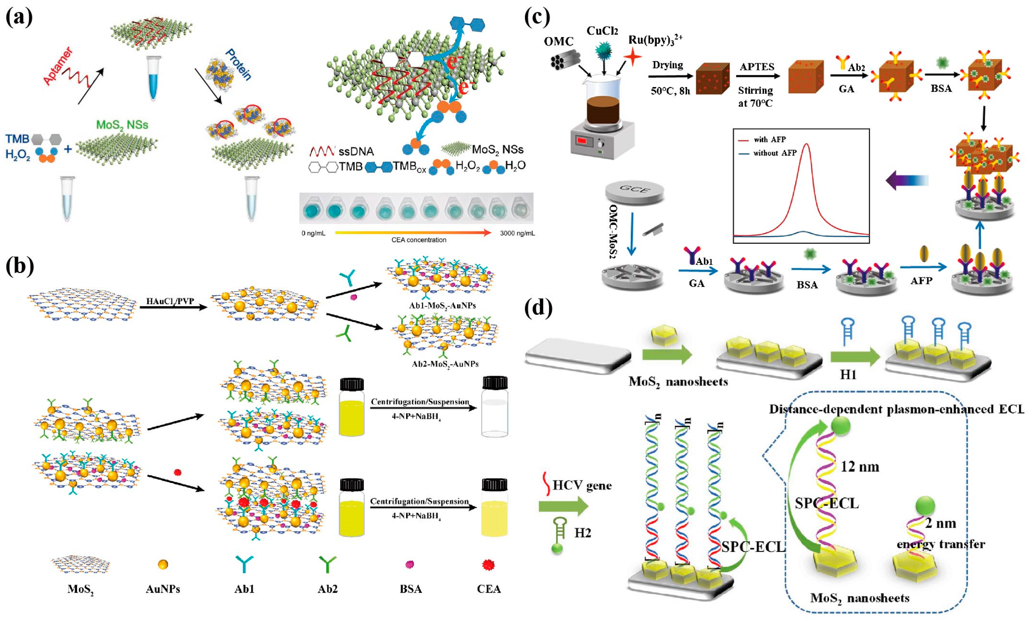
- Su, S.; Li, J.; Yao, Y.; Sun, Q.; Zhao, Q.; Wang, F.; Li, Q.; Liu, X.; Wang, L. Colorimetric Analysis of Carcinoembryonic Antigen Using Highly Catalytic Gold Nanoparticles. ACS Appl. Bio Mater. 2019, 2, 292–298. [Google Scholar] [CrossRef]
- Wang, X.; Cheng, S.; Wang, X.; Wei, L.; Kong, Q.; Ye, M.; Luo, X.; Xu, J.; Zhang, C.; Xian, Y. pH-Sensitive Dye-Based Nanobioplatform for Colorimetric Detection of Heterogeneous Circulating Tumor Cells. ACS Sens. 2021, 6, 1925–1932. [Google Scholar] [CrossRef] [PubMed]
- Ma, C.; Cao, Y.; Gou, X.; Zhu, J. Recent Progress in Electrochemiluminescence Sensing and Imaging. Anal. Chem. 2020, 92, 431–454. [Google Scholar] [CrossRef] [PubMed]
- Zhang, S.; Liu, Y. Recent Progress of Novel Electrochemiluminescence Nanoprobes and Their Analytical Applications. Front. Chem. 2021, 8, 626243. [Google Scholar] [CrossRef] [PubMed]
- Lv, X.; Li, Y.; Cui, B.; Fang, Y.; Wang, L. Electrochemiluminescent sensor based on an aggregation-induced emission probe for bioanalytical detection. Analyst 2022, 147, 2338–2354. [Google Scholar] [CrossRef] [PubMed]
- Du, L.; Zhang, H.; Wang, Z.; Zhuang, T.; Wang, Z. Boosting the electrochemiluminescence of luminol by high-intensity focused ultrasound pretreatment combined with 1T/2H MoS2 catalysis to construct a sensitive sensing platform. Ultrason. Sonochem. 2023, 92, 106264. [Google Scholar] [CrossRef] [PubMed]
- Ma, C.; Zhang, Z.; Tan, T.; Zhu, J. Recent Progress in Plasmonic based Electrochemiluminescence Biosensors: A Review. Biosensors 2023, 13, 200. [Google Scholar] [CrossRef]
- Zhang, Z.; Yu, H.; Zhang, Y.; Wang, Z.; Gao, H.; Rong, S.; Meng, L.; Dai, J.; Pan, H.; Chang, D. A sandwich-configuration electrochemiluminescence immunoassay based on Cu2O@OMC-Ru nanocrystals and OMC-MoS2 nanocomposites for determination of alpha-fetoprotein. Mikrochim. Acta 2021, 188, 213. [Google Scholar] [CrossRef]
- Srinidhi, G.; Sudalaimani, S.; Giribabu, K.; Basha, S.J.S.; Suresh, C. Amperometric determination of hydrazine using a CuS-ordered mesoporous carbon electrode. Mikrochim. Acta 2020, 187, 359. [Google Scholar] [CrossRef]
- Liu, Y.; Nie, Y.; Wang, M.; Zhang, Q.; Ma, Q. Distance-dependent plasmon-enhanced electrochemiluminescence biosensor based on MoS2 nanosheets. Biosens. Bioelectron. 2020, 148, 111823. [Google Scholar] [CrossRef]
- Muhammad, M.; Khan, S.; Shehzadi, S.A.; Gul, Z.; Al-Saidi, H.M.; Kamran, A.W.; Alhumaydhi, F.A. Recent advances in colorimetric and fluorescent chemosensors based on thiourea derivatives for metallic cations: A review. Dye. Pigment. 2022, 205, 110477. [Google Scholar] [CrossRef]
- Yao, W.; Ling, J.; Zhang, W.; Ding, Y. Highly sensitive fluorescent aptasensor based on MoS2 nanosheets for one-step determination of methamphetamine. Anal. Sci. 2022, 38, 99–104. [Google Scholar] [CrossRef] [PubMed]
- Liang, J.; Yan, R.; Chen, C.; Yao, X.; Guo, F.; Wu, R.; Zhou, Z.; Chen, J.; Li, G. A novel fluorescent strategy for Golgi protein 73 determination based on aptamer/nitrogen-doped graphene quantum dots/molybdenum disulfide @ reduced graphene oxide nanosheets. Spectrochim. Acta A Mol. Biomol. Spectrosc. 2023, 294, 122538. [Google Scholar] [CrossRef] [PubMed]
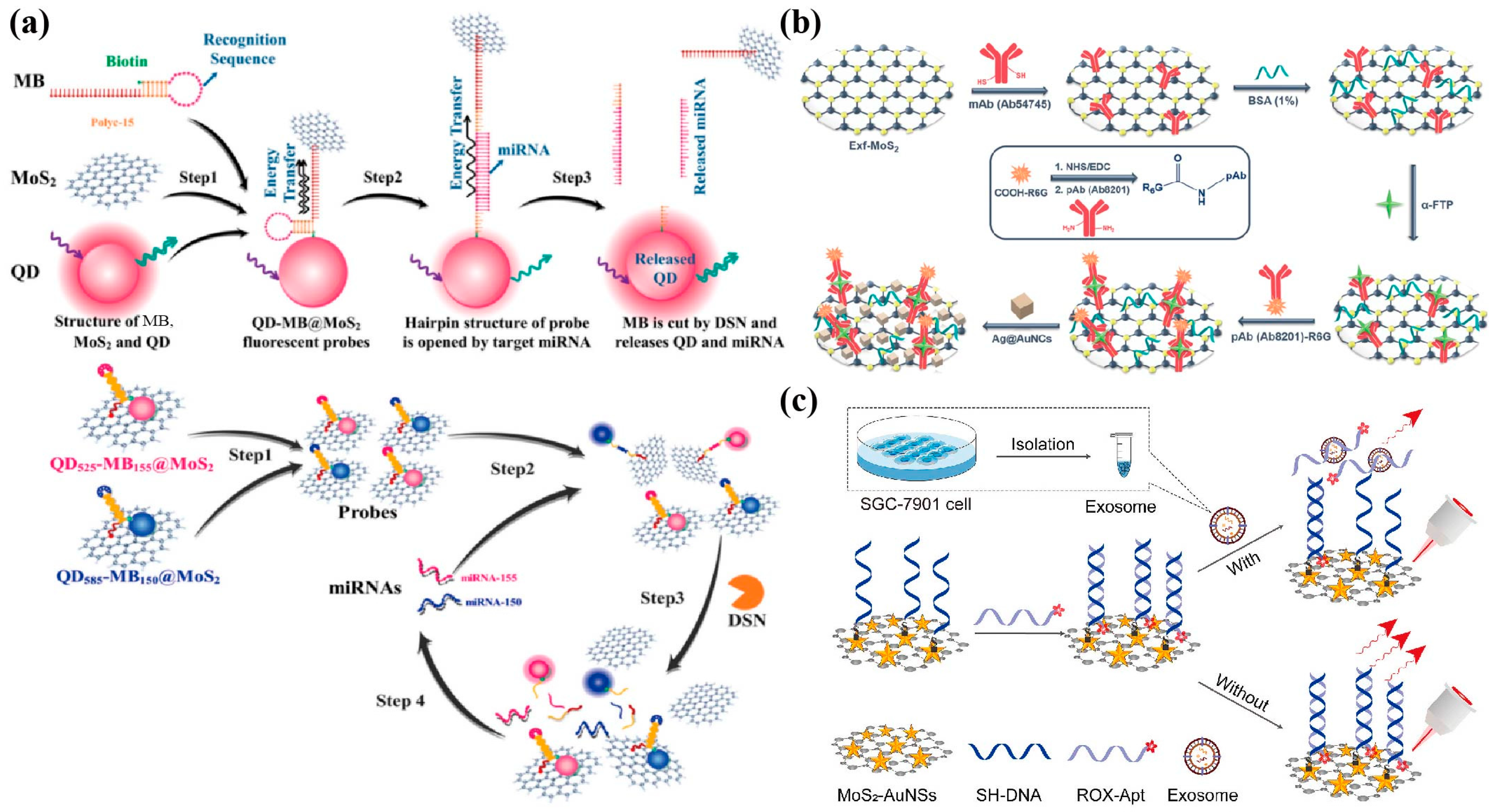
- Wang, J.; Liu, Y.; Ding, Z.; Zhang, L.; Han, C.; Yan, C.; Amador, E.; Yuan, L.; Wu, Y.; Song, C.; et al. The exploration of quantum dot-molecular beacon based MoS2 fluorescence probing for myeloma-related Mirnas detection. Bioact. Mater. 2022, 17, 360–368. [Google Scholar] [CrossRef] [PubMed]
- Chhowalla, M.; Shin, H.S.; Eda, G.; Li, L.J.; Loh, K.P.; Zhang, H. The chemistry of two-dimensional layered transition metal dichalcogenide nanosheets. Nat. Chem. 2013, 5, 263–275. [Google Scholar] [CrossRef] [PubMed]
- Ge, J.; Hu, Y.; Deng, R.; Li, Z.; Zhang, K.; Shi, M.; Yang, D.; Cai, R.; Tan, W. Highly Sensitive MicroRNA Detection by Coupling Nicking-Enhanced Rolling Circle Amplification with MoS2 Quantum Dots. Anal. Chem. 2020, 92, 13588–13594. [Google Scholar] [CrossRef]
- Strobbia, P.; Cupil-Garcia, V.; Crawford, B.M.; Fales, A.M.; Pfefer, T.J.; Liu, Y.; Maiwald, M.; Sumpf, B.; Vo-Dinh, T. Accurate in vivo tumor detection using plasmonic-enhanced shifted-excitation Raman difference spectroscopy (SERDS). Theranostics 2021, 11, 4090–4102. [Google Scholar] [CrossRef] [PubMed]
- Er, E.; Sánchez-Iglesias, A.; Silvestri, A.; Arnaiz, B.; Liz-Marzán, L.M.; Prato, M.; Criado, A. Metal Nanoparticles/MoS2 Surface-Enhanced Raman Scattering-Based Sandwich Immunoassay for α-Fetoprotein Detection. ACS Appl. Mater. Interfaces 2021, 13, 8823–8831. [Google Scholar] [CrossRef]
- Pan, H.; Dong, Y.; Gong, L.; Zhai, J.; Song, C.; Ge, Z.; Su, Y.; Zhu, D.; Chao, J.; Su, S.; et al. Sensing gastric cancer exosomes with MoS2-based SERS aptasensor. Biosens. Bioelectron. 2022, 215, 114553. [Google Scholar] [CrossRef]
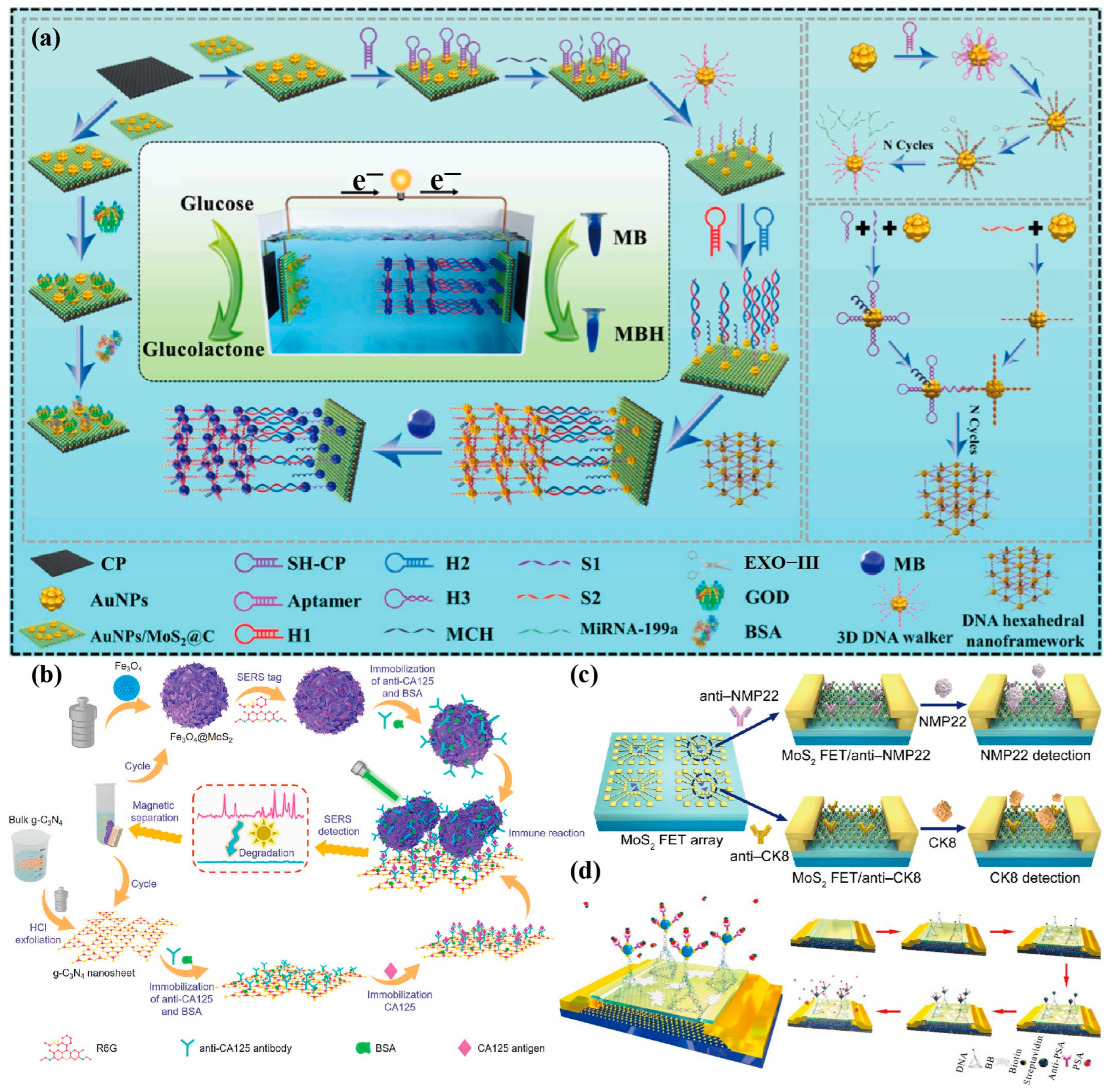
- Medetalibeyoglu, H.; Kotan, G.; Atar, N.; Yola, M.L. A novel sandwich-type SERS immunosensor for selective and sensitive carcinoembryonic antigen (CEA) detection. Anal. Chim. Acta 2020, 1139, 100–110. [Google Scholar] [CrossRef]
- Samy, O.; Zeng, S.; Birowosuto, M.D.; El Moutaouakil, A. A Review on MoS2 Properties, Synthesis, Sensing Applications and Challenges. Crystals 2021, 11, 355. [Google Scholar] [CrossRef]
- Chiu, N.F.; Yang, H.T. High-Sensitivity Detection of the Lung Cancer Biomarker CYFRA21-1 in Serum Samples Using a Carboxyl-MoS2 Functional Film for SPR-Based Immunosensors. Front. Bioeng. Biotechnol. 2020, 8, 234. [Google Scholar] [CrossRef] [PubMed]
- Xin, W.; Jiang, L.; Zong, L.; Zeng, H.; Shu, G.; Marks, R.; Zhang, X.; Shan, D. MoS2 quantum dots-combined zirconium-metalloporphyrin frameworks: Synergistic effect on electron transfer and application for bioassay. Sens. Actuators B Chem. 2018, 273, 566–573. [Google Scholar] [CrossRef]
- Hou, F.; Hu, X.; Ma, S.; Cao, J.; Liu, Y. Construction of electrochemiluminescence sensing platform with in situ generated coreactant strategy for sensitive detection of prostate specific antigen. J. Electroanal. Chem. 2020, 858, 113817. [Google Scholar] [CrossRef]
- Nie, Y.; Zhang, X.; Zhang, Q.; Liang, Z.; Ma, Q.; Su, X. A novel high efficient electrochemiluminescence sensor based on reductive Cu(I) particles catalyzed Zn-doped MoS2 QDs for HPV 16 DNA determination. Biosens. Bioelectron. 2020, 160, 112217. [Google Scholar] [CrossRef] [PubMed]
- Hou, S.; Wang, P.; Nie, Y.; Guo, Y.; Ma, Q. A novel work function tuning strategy-based ECL sensor with sulfur dots and Au NP@MoS2 nanosheet heterostructure for triple-negative breast cancer diagnosis. Chem. Eng. J. 2022, 446, 136906. [Google Scholar] [CrossRef]
- Liu, W.; Su, M.; Chen, A.; Peng, K.; Chai, Y.; Yuan, R. Highly Efficient Electrochemiluminescence Based on Luminol/MoS2 Quantum Dots@Zeolitic Imidazolate Framework-8 as an Emitter for Ultrasensitive Detection of MicroRNA. Anal. Chem. 2022, 94, 9106–9113. [Google Scholar] [CrossRef]
- Zhao, L.; Kong, D.; Wu, Z.; Liu, G.; Gao, Y.; Yan, X.; Liu, F.; Liu, X.; Wang, C.; Cui, J.; et al. Interface interaction of MoS2 nanosheets with DNA based aptameric biosensor for carbohydrate antigen 15–3 detection. Microchem. J. 2020, 155, 104675. [Google Scholar] [CrossRef]
- Peng, X.; Wang, Y.; Wen, W.; Chen, M.; Zhang, X.; Wang, S. Simple MoS2-Nanofiber Paper-Based Fluorescence Immunosensor for Point-of-Care Detection of Programmed Cell Death Protein 1. Anal. Chem. 2021, 93, 8791–8798. [Google Scholar] [CrossRef]
- Jiang, J.; Liu, H.; Li, X.; Chen, Y.; Gu, C.; Wei, G.; Zhou, J.; Jiang, T. Nonmetallic SERS-based immunosensor byintegrating MoS2 nanoflower and nanosheet towards the direct serum detection of carbohydrate antigen 19–9. Biosens. Bioelectron. 2021, 193, 113481. [Google Scholar] [CrossRef]
- Van der Zande, A.M.; Huang, P.S.Y.; Chenet, D.A.; Berkelbach, T.C.; You, Y.M.; Lee, G.H.; Heinz, T.F.; Reichman, D.R.; Muller, D.A.; Hone, J.C. Grains and grain boundaries in highly crystalline monolayer molybdenum disulphide. Nat. Mater. 2013, 12, 554–561. [Google Scholar] [CrossRef]
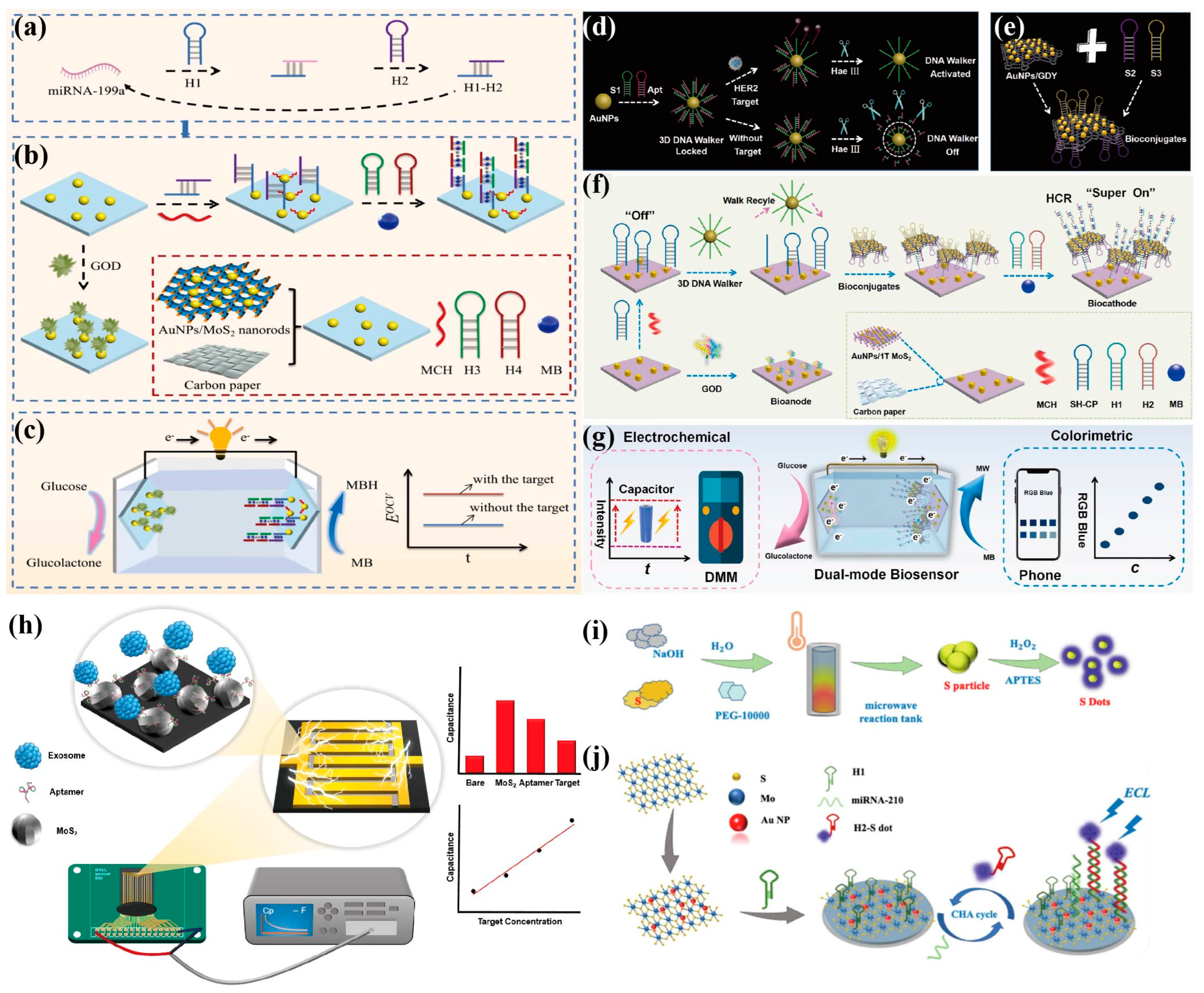
- Shi, J.; Qin, W.; Lin, Y.; Li, M.; Wu, Y.; Luo, H.; Yan, J.; Huang, K.; Tan, X. Enhancing biosensing with fourfold amplification and self-powering capabilities: MoS2@C hollow nanorods-mediated DNA hexahedral framework architecture for amol-level liver cancer tumor marker detection. Anal. Chim. Acta 2023, 1271, 341413. [Google Scholar] [CrossRef] [PubMed]
- Wang, R.; Liu, H.; Xu, T.; Zhang, Y.; Gu, C.; Jiang, T. SERS-based recyclable immunoassay mediated by 1T-2H mixed-phase magnetic molybdenum disulfide probe and 2D graphitic carbon nitride substrate. Biosens. Bioelectron. 2023, 227, 115160. [Google Scholar] [CrossRef] [PubMed]
- Yang, Y.; Zeng, B.; Li, Y.; Liang, H.; Yang, Y.; Yuan, Q. Construction of MoS2 field effect transistor sensor array for the detection of bladder cancer biomarkers. Sci. China Chem. 2020, 63, 997–1003. [Google Scholar] [CrossRef]
- Zhang, Y.; Feng, D.; Xu, Y.; Yin, Z.; Dou, W.; Habiba, U.M.E.; Pan, C.; Zhang, Z.; Mou, H.; Deng, H.; et al. DNA-based functionalization of two-dimensional MoS2 FET biosensor for ultrasensitive detection of PSA. Appl. Surf. Sci. 2021, 548, 149169. [Google Scholar] [CrossRef]
- Negahdary, M.; Angnes, L. Recent advances in electrochemical nanomaterial-based aptasensors for the detection of cancer biomarkers. Talanta 2023, 259, 124548. [Google Scholar] [CrossRef] [PubMed]
- Shi, J.; Song, Y.; Lin, Y.; Wu, Y.; Luo, H.; Yan, J.; Huang, K.; Tan, X. Sensitive detection of cancer biomarker with enzyme-free mediated cascade signal amplification empowered undisturbed dual-mode assay. Sens. Actuators B Chem. 2023, 394, 134430. [Google Scholar] [CrossRef]
- Hou, Y.; Xu, J.; Xie, W.; Huang, K.; Tan, X.; Zhao, B.; Zhang, S.; Gao, M. 3D DNA walker recognition-driven homogeneous dual-mode sensing strategy based on enzyme biofuel cell for ultrasensitive detection of HER2. Sens. Actuators B Chem. 2023, 376 (Pt B), 132998. [Google Scholar] [CrossRef]
- Lee, M.; Park, S.J.; Kim, G.; Park, C.; Lee, M.H.; Ahn, J.H.; Lee, T. A pretreatment-free electrical capacitance biosensor for exosome detection in undiluted serum. Biosens. Bioelectron. 2022, 199, 113872. [Google Scholar] [CrossRef]
- Kashefi-Kheyrabadi, L.; Kim, J.; Chakravarty, S.; Park, S.y.; Gwak, H.; Kim, S.; Mohammadniaei, M.; Lee, M.H.; Hyun, K.A.; Jung, H. Detachable microfluidic device implemented with electrochemical aptasensor (DeMEA) for sequential analysis of cancerous exosomes. Biosens. Bioelectron. 2020, 169, 112622. [Google Scholar] [CrossRef]
- Yagati, A.K.; Go, A.; Vu, N.H.; Lee, M.H. A MoS2–Au nanoparticle-modified immunosensor for T3 biomarker detection in clinical serum samples. Electrochim. Acta 2020, 342, 136065. [Google Scholar] [CrossRef]
- Jandas, P.J.; Luo, J.; Prabakaran, K.; Chen, F.; Fu, Y. Highly stable, love-mode surface acoustic wave biosensor using Au nanoparticle-MoS2-rGO nano-cluster doped polyimide nanocomposite for the selective detection of carcinoembryonic antigen. Mater. Chem. Phys. 2020, 246, 122800. [Google Scholar] [CrossRef]
- Torul, H.; Yarali, E.; Eksin, E.; Ganguly, A.; Benson, J.; Tamer, U.; Papakonstantinou, P.; Erdem, A. Paper-Based Electrochemical Biosensors for Voltammetric Detection of miRNA Biomarkers Using Reduced Graphene Oxide or MoS2 Nanosheets Decorated with Gold Nanoparticle Electrodes. Biosensors 2021, 11, 236. [Google Scholar] [CrossRef] [PubMed]
- Zhou, C.; Cui, K.; Liu, Y.; Li, L.; Zhang, L.; Hao, S.; Ge, S.; Yu, J. Bi2S3@MoS2 Nanoflowers on Cellulose Fibers Combined with Octahedral CeO2 for Dual-Mode Microfluidic Paper-Based MiRNA-141 Sensors. ACS Appl. Mater. Interfaces 2021, 13, 32780–32789. [Google Scholar] [CrossRef] [PubMed]
- Ma, J.; Xue, D.; Xu, T.; Wei, G.; Gu, C.; Zhang, Y.; Jiang, T. Nonmetallic SERS-based biosensor for ultrasensitive and reproducible immunoassay of ferritin mediated by magnetic molybdenum disulfide nanoflowers and black phosphorus nanosheets. Colloids Surf. B Biointerfaces 2023, 227, 113338. [Google Scholar] [CrossRef]
- Yan, R.; Lu, N.; Han, S.; Lu, Z.; Xiao, Y.; Zhao, Z.; Zhang, M. Simultaneous detection of dual biomarkers using hierarchical MoS2 nanostructuring and nano-signal amplification-based electrochemical aptasensor toward accurate diagnosis of prostate cancer. Biosens. Bioelectron. 2022, 197, 113797. [Google Scholar] [CrossRef]
- Shi, J.; Zhang, Y.; Wang, P.; Nie, Y.; Ma, Q. Luminous MoS2 nanosheet-based electrochemiluminescence biosensor with biomimetic vesicle for miRNA-210 detection. Talanta 2022, 237, 122969. [Google Scholar] [CrossRef]
- Wang, X.; Ji, J.; Yang, P.; Li, X.; Pang, Y.; Lu, P. A love-mode surface acoustic wave aptasensor with dummy fingers based on monolayer MoS2/Au NPs nanocomposites for alpha-fetoprotein detection. Talanta 2022, 243, 123328. [Google Scholar] [CrossRef]
- Sun, Z.W.; Tong, Y.; Zhao, L.; Li, J.; Gao, F.C.; Wang, C.X.; Li, H.; Du, L.T.; Jiang, Y.Y. MoS2@Ti3C2 nanohybrid-based photoelectrochemical biosensor: A platform for ultrasensitive detection of cancer biomarker exosomal miRNA. Talanta 2022, 238, 123077. [Google Scholar] [CrossRef]
- Rus, L.; Tertis, M.; Pop, A.; Fizeşan, L.; Bogdan, D.; Matei, E.; Oprea, D.; Diculescu, V.; Săndulescu, R.; Cristea, C. The use of a new selective AB3 aptamer for the hematologic tumor cells’ detection. Sens. Actuators B Chem. 2023, 394, 134389. [Google Scholar] [CrossRef]
- Majd, S.M.; Mirzapour, F.; Shamsipur, M.; Manouchehri, I.; Babaee, E.; Pashabadi, A.; Moradian, R. Design of a novel aptamer/molecularly imprinted polymer hybrid modified Ag–Au@Insulin nanoclusters/Au-gate-based MoS2 nanosheet field-effect transistor for attomolar detection of BRCA1 gene. Talanta 2023, 257, 124394. [Google Scholar] [CrossRef]
- Guo, Y.; Nie, Y.; Wang, P.; Li, Z.; Ma, Q. MoS2 QDs-MXene heterostructure-based ECL sensor for the detection of miRNA-135b in gastric cancer exosomes. Talanta 2023, 259, 124559. [Google Scholar] [CrossRef] [PubMed]
- Rahman, M.T.; Kumar, R.; Kumar, M.; Qiao, Q.Q. Two-dimensional transition metal dichalcogenides and their composites for lab-based sensing applications: Recent progress and future outlook. Sens. Actuators A Phys. 2021, 318, 112517. [Google Scholar] [CrossRef]
- Nakada, G.; Igarashi, Y.; Imai, H.; Oaki, Y. Materials-Informatics-Assisted High-Yield Synthesis of 2D Nanomaterials through Exfoliation. Adv. Theory Simul. 2019, 2, 1800180. [Google Scholar] [CrossRef]
- Hu, T.; Zhang, R.; Li, J.; Cao, J.; Qiu, F. Photodetectors based on two-dimensional MoS2 and its assembled heterostructures. Chip 2022, 1, 100017. [Google Scholar] [CrossRef]
- Kaur, J.; Singh, M.; Dell’Aversana, C.; Benedetti, R.; Giardina, P.; Rossi, M.; Valadan, M.; Vergara, A.; Cutarelli, A.; Montone, A.M.I.; et al. Biological interactions of biocompatible and water-dispersed MoS2 nanosheets with bacteria and human cells. Sci. Rep. 2018, 68, 16386. [Google Scholar] [CrossRef]
- Mohan, M.; Shetti, N.P.; Aminabhavi, T.M. Recent developments in MoS2-based flexible supercapacitors. Materials Today Chem. 2023, 27, 101333. [Google Scholar] [CrossRef]


| Method | Analytes | Electrode/Label | Linear Range | LOD | Ref. |
|---|---|---|---|---|---|
| Chronoamperometry | AMACR | Pt NWs array@2H-MoS2/SPE | 0.70–12.50 ng/µL | 0.5 pg/µL | [80] |
| CV | CEA | Ab/rGO/MoS2@PANI/GCE | 0.001–80 ng/mL | 0.3 pg/mL | [84] |
| CV | PSA | Ab/ce-MoS2/AgNR/SPE | 0.1–1000 ng/mL | 0.051 ng/mL | [85] |
| DPV | CEA | Ab/MoS2-PBNCs/GCE | 0.005–10 ng/mL | 0.54 pg/mL | [93] |
| DPV | CEA | Ag/MoS2@Fe3O4-Ab2/CEA/Ab1/Ag/MGCE | 0.0001–20 ng/mL | 0.03 pg/mL | [94] |
| DPV | miRNA-182 | ssRNA/MoS2/Ti3C2/GCE | 1 fM–0.1 nM | 0.43 fM | [95] |
| DPV | CEA | MoS2-AuNPs/HRP-Ab2/CEA/Ab1/MoS2-AuNPs/GCE | 10 fg/mL–1 ng/mL | 1.2 fg/mL | [96] |
| DPV | Nucleolin | TNA/AuNPs/MoS2/rGO/GCE | 0.5 nM–1.0 μM | 0.16 nM | [82] |
| DPV | CEA | Ab/Pd@Pt/MoS2-Gr/GCE | 0.00001–100 ng/mL | 0.005 pg/mL | [97] |
| DPV | CA125 | Ab/CuBTC@MoS2-AuNPs/SPE | 0.5 mU/mL–500 U/mL | 0.5 mU/mL | [98] |
| DPV | anti-retroviral agent indinavir | ZnO NRs/MoS2 NSs/SPE | 0.01–0.66 μM & 0.66–7.88 μM | 0.007 μM | [99] |
| SWV | CEA | CeO2-MoS2-Pb2+-Ab2/CEA/Ab1/AuNPs/GCE | 0.001–80 ng/mL | 0.3 pg/mL | [100] |
| Amperometry | CEA | Ab/Ag/MoS2/rGO/GCE | 0.01 pg/mL–100 ng/mL | 1.6 fg/mL | [101] |
| Amperometry | HBeAg | Au@Pd/MoS2@MWCNTs-Ab2/HBeAg/Ab1/p-GO@Au/GCE | 0.1–500 pg/mL | 26 fg/mL | [102] |
| Amperometry | AFP | MoS2@Cu2O-Au-Ab2/AFP/Ab1/AuNPs/GCE | 0.1 pg/mL–50 ng/mL | 0.037 pg/mL | [86] |
| Amperometry | AFP | Ab/Pt NDs/PDDA/MoS2@PPy NTs/GCE | 50 fg/mL–50 ng/mL | 17 fg/mL | [103] |
| Amperometry | CEA | MoS2 NFs/Au@AgPt YNCs -Ab2/CEA/Ab1/AuTNPs/GCE | 10 fg/mL–100 ng/mL | 3.09 fg/mL | [87] |
| EIS | CML | pDNA/PANI-MoS2/ITO | 10−17–10−6 M | 3 × 10⁻¹⁸ M | [104] |
| EIS | AFP | Ab/MoS2/PDDA/Ag/AgCl wire | 0.1–10 ng/mL | 0.033 ng/mL | [90] |
| EIS | miRNA-21 | cDNA/pd-MoS2@β-FeOOH/Au | 1 fM–5 nM | 0.11 fM | [88] |
| EIS | TNF-α | MoS2 NFs | 0.01–200 pg/ml | 0.202 pg/ml | [89] |
| PEC | CEA | ALP-Au-Ab2/CEA /Ab1/ZnS/C/MoS2/GCE | 2.0 pg/mL–10.0 ng/mL | 1.30 pg/mL | [92] |
| PEC | SCCA | Ab/AuNPs/C/MoS2/GCE | 0.005–8 ng/mL | 1.8 pg/mL | [105] |
| PEC | AFP | DNA/Au/GaN | 1.0–150 ng/mL | 0.3 ng/mL | [91] |
| PEC | MCF-7 cells | PM6:Y6/anti-EpCAM-MNs /Au NPs/Au-aptamer | 10–10,000 cell/mL | 9 cell/mL | [106] |
| Method | Analytes | Electrode/Label | Linear Range | LOD | Ref. |
|---|---|---|---|---|---|
| Colorimetry | CEA | Au NPs-MoS2-Ab2/ CEA/Ab1/MoS2-Au NPs | 5 pg/mL–10 ng/mL | 0.5 pg/mL | [111] |
| Colorimetry | CEA | DNA/MoS2 NSs | 50–1000 ng/mL | 50 ng/mL | [110] |
| Colorimetry | CTC | TP/SYL3C-MoS2 | 5–104 cells/mL | 2 cells/mL | [112] |
| ECL | CEA | Ab/MOF-545-Zn@MQDs/GCE | 0.18–1000 ng/mL | 0.45 pg/mL | [133] |
| ECL | PSA | GOD-SiO2-Ab2/PSA /Ab1/MoS2-AuNPs/GCE | 0.5 pg/mL–10.0 ng/mL | 0.20 pg/mL | [134] |
| ECL | HCV gene | S-BN QDs-hairpin DNA2 (H2)/MoS2 Ns | 0.5 pmoL/L–1 nmoL/L | 0.17 pmoL/L. | [121] |
| ECL | HPV 16 DNA | Zn-doped MoS2 QDs & QD-DNA/reductive Cu(I) particles | 0.1–200 nmolL | 0.03 nmol/L | [135] |
| ECL | miRNA-210 | S dots/Au NP@MoS2 NSs | 0.1 pM–10 nM | 0.03 pM | [136] |
| ECL | miRNA-21 | luminophore/MoS2 QDs@Zeolitic Imidazolate 2 Framework-8 | Buffer (0.1 mM PBS), co-reactant (2 mM H2O) | 14.6 aM | [137] |
| Fluorescence | CA15-3 | DNA/MoS2 NSs | 0.01–0.1 U/mL | 0.0039 U/mL | [138] |
| Fluorescence | PD-1 | MoS2-NFP | 125–8000 pg/mL | 85.5 pg/mL | [139] |
| Fluorescence | miRNA-155 & miRNA-150 | QD-MB @MoS2 | 10 fM–1 nM | 7.19 fM & 5.84 fM | [124] |
| SERS | CEA | MoS2 NFs@Au NPs/MBA-Ab2/CEA /Ab1/Fe3O4 NPs@Au NPs/ d-Ti3C2TX Mxene | 0.0001–100.0 ng/mL | 0.033 pg/mL | [132] |
| SERS | CA19-9 | R6G-tagged MoS2 NF | 5 × 10⁻³–100 IU/mL | 3.43 × 10⁻⁴ IU/mL | [140] |
| SERS | exosomes | MoS2-AuNSs/ROX-Apt | 55–5.5 × 10⁵ particles/μL | 17 particles/μL | [131] |
| SPR | CYFRA21-1 | Ab/COOH-MoS2/Au/Cr/BK7 | 0.05 pg/mL–100 ng/mL | 0.05 pg/mL | [132] |
| Method | Analytes | Electrode/Label | Linear Range | LOD | Ref. |
|---|---|---|---|---|---|
| FET array | NMP22 & CK8 | MoS2 NSs-FET | 10−6–10−1 pg/mL | 0.027–0.019 aM | [144] |
| DeMEA/ microfluidic | exosome | GASI microfluidic channel/anti-EpCAM | 1 × 10²–1 × 10⁹ exosomes/μL | 17 exosomes/μL | [150] |
| EIS/CV | T3 | Au-MoS2/anti-T3 electrodes | 0.01–100 ng/mL | 2.5 pg/mL | [151] |
| SAW | CEA | AuNP–MoS2-rGO/PI | 0.1–80 ng/mL. | 0.084 ng/mL | [152] |
| bio-FET | PSA | MoS2/B-SA system with DNA tetrahedron | 1 fg/mL–100 ng/mL (PBS) 1 fg/mL–100 ng/mL (serum) | 1 fg/mL | [145] |
| Paper-Based/DPV | miRNA-1 55miRNA-21 | AuNPs/RGO/PE AuNPs/MoS2/PE | 33.8–135.3 nM 135.6–406.8 nM | 12.0 nM 25.7 nM | [153] |
| Microfluidic/ electrochemical/visual | miRNA-141 | Bi2S3@MoS2 NFs/CeO2 | 10 fM–1 nM 0.5 fM–1 nM | 0.12 fM 2.65 fM | [154] |
| signal amplification/ 3D DNA walker | microRNA-199a | AuNPs/MoS2@C | 0.0001–100 pM | 4.94 amol/L | [142] |
| SERS/ELISA | ferritin | MoS2 @Fe3O4/BP | 10–10−4 μg/mL | 7.3 × 10⁻⁵ μg/mL | [155] |
| SERS/ELISA | CA125 | Fe3O4@MoS2/g-C3N4 NSs | 10−3–102 IU/mL | 4.96 × 10⁻⁴ IU/mL | [143] |
| colorimetric/ electrochemical dual mode | miRNA-199a | MoS2 NRs/CHA /HCR/MB | 0.1–105 fM & 0.1–107 fM | 0.0241 aM & 0.0345 aM | [147] |
| colorimetric/ electrochemical dual mode | HER2 | 3D DNA walker /1T-MoS2/graphdiyne | 0.0001–10 pg/mL | 0.03 pg/mL | [148] |
| capacitance electrobiosensor | exosome | MoS2 heterolayer/ IDMGE | 104–108 exosomes/mL | 2192.6 exosomes/mL | [149] |
| ECL/LSPR | miRNA-210 | S dots/AuNPs@MoS2 NSs heterostructure | 0.1 pM–10 nM | 0.03 pM | [136] |
| electrochemistry | PSA | MoS2/SiO2 NPs | 1 fg/mL–500 ng/mL | 2.5 fg/mL | [156] |
| ECL/CHA | miRNA-210 | MoS2 NSs@biomimetic magnetic vesicles | 1 fM–100 pM | 0.3 fM | [157] |
| LSAW aptasensor | AFP | monolayer MoS2/Au NPs/aaptamer | 0.01–100 ng/mL | 4.79 pg/mL | [158] |
| PEC/EIS | miR-92a-3p | MoS2@Ti3C2 | 1–108 fM | 0.27 fM | [159] |
| EIS/microscopic | OFA/iLRP | AB3/GO/SPE | 10–300 k cells/mL | 16 cells/mL | [160] |
| FET | BRCA1 | Ag-Au@Ins/Apta-MIP without template modified Au gate MoS2 FET | 10 aM–1.0 nM | 6.4 aM | [161] |
| Fluorescence/ ECL | miRNA-21 miRNA-135b | carbon dots MoS2 QDs | 0.1 pM–1 nM 30 fM–20 nM | 21.1 fM 10 fM | [162] |
Disclaimer/Publisher’s Note: The statements, opinions and data contained in all publications are solely those of the individual author(s) and contributor(s) and not of MDPI and/or the editor(s). MDPI and/or the editor(s) disclaim responsibility for any injury to people or property resulting from any ideas, methods, instructions or products referred to in the content. |
© 2023 by the authors. Licensee MDPI, Basel, Switzerland. This article is an open access article distributed under the terms and conditions of the Creative Commons Attribution (CC BY) license (https://creativecommons.org/licenses/by/4.0/).
Share and Cite
Qin, Z.; Zhang, J.; Li, S. Molybdenum Disulfide as Tunable Electrochemical and Optical Biosensing Platforms for Cancer Biomarker Detection: A Review. Biosensors 2023, 13, 848. https://doi.org/10.3390/bios13090848
Qin Z, Zhang J, Li S. Molybdenum Disulfide as Tunable Electrochemical and Optical Biosensing Platforms for Cancer Biomarker Detection: A Review. Biosensors. 2023; 13(9):848. https://doi.org/10.3390/bios13090848
Chicago/Turabian StyleQin, Ziyue, Jiawei Zhang, and Shuang Li. 2023. "Molybdenum Disulfide as Tunable Electrochemical and Optical Biosensing Platforms for Cancer Biomarker Detection: A Review" Biosensors 13, no. 9: 848. https://doi.org/10.3390/bios13090848