Lateral Flow Assay: A Summary of Recent Progress for Improving Assay Performance


Abstract
:1. Introduction
2. Methodology
3. Biorecognition Strategies
4. Assay Improvement
4.1. Assay Optimization
4.1.1. Controlling Capillary Flow Rate
4.1.2. Immobilization Efficiency of Biorecognition Elements
4.2. Signal Amplification
5. Extraction and Enrichment of the Target Molecule in the Sample
6. Conclusions and Future Outlook
Author Contributions
Funding
Acknowledgments
Conflicts of Interest
References
- Kuswandi, B.; Hidayat, M.A.; Noviana, E. Paper-based sensors for rapid important biomarkers detection. Biosens. Bioelectron. X 2022, 12, 100246. [Google Scholar] [CrossRef]
- Majdinasab, M.; Badea, M.; Marty, J.L. Aptamer-Based lateral flow assays: Current trends in clinical diagnostic rapid tests. Pharmaceuticals 2022, 15, 90. [Google Scholar] [CrossRef] [PubMed]
- Naghdi, T.; Ardalan, S.; Adib, Z.A.; Sharifi, A.R.; Golmohammadi, H. Moving toward smart biomedical sensing. Biosens. Bioelectron. 2022, 223, 115009. [Google Scholar] [CrossRef] [PubMed]
- Budd, J.; Miller, B.S.; Weckman, N.E.; Cherkaoui, D.; Huang, D.; Decruz, A.T.; Fongwen, N.; Han, G.-R.; Broto, M.; Estcourt, C.S. Lateral flow test engineering and lessons learned from COVID-19. Nat. Rev. Bioeng. 2023, 1, 13–31. [Google Scholar] [CrossRef]
- Jara, M.D.L.; Alvarez, L.A.C.; Guimarães, M.C.; Antunes, P.W.P.; de Oliveira, J.P. Lateral flow assay applied to pesticides detection: Recent trends and progress. Environ. Sci. Pollut. Res. 2022, 29, 46487–46508. [Google Scholar] [CrossRef]
- Khelifa, L.; Hu, Y.; Jiang, N.; Yetisen, A.K. Lateral flow assays for hormone detection. Lab A Chip 2022, 22, 2451–2475. [Google Scholar] [CrossRef]
- Preetam, S.; Nahak, B.K.; Patra, S.; Toncu, D.C.; Park, S.; Syväjärvi, M.; Orive, G.; Tiwari, A. Emergence of microfluidics for next generation biomedical devices. Biosens. Bioelectron. X 2022, 10, 100106. [Google Scholar] [CrossRef]
- Iles, A.H.; He, P.J.; Katis, I.N.; Galanis, P.P.; John, A.J.; Elkington, P.; Eason, R.W.; Sones, C.L. Semi-quantitative detection of inflammatory biomarkers using a laser-patterned multiplexed lateral flow device. Talanta 2022, 237, 122944. [Google Scholar] [CrossRef]
- Broccolo, F.; Fabris, S.; Ciccozzi, M.; Plebani, M. A rapid semi-quantitative test for determination of SARS-CoV-2 antibody levels. Clin. Chem. Lab. Med. 2022, 60, e101–e103. [Google Scholar] [CrossRef]
- Nuntawong, P.; Putalun, W.; Tanaka, H.; Morimoto, S.; Sakamoto, S. Lateral flow immunoassay for small-molecules detection in phytoproducts: A review. J. Nat. Med. 2022, 76, 521–545. [Google Scholar] [CrossRef]
- Mohammadinejad, A.; Nooranian, S.; Kazemi Oskuee, R.; Mirzaei, S.; Aleyaghoob, G.; Zarrabi, A.; Selda Gunduz, E.; Nuri Ertas, Y.; Sheikh Beig Goharrizi, M.A. Development of Lateral Flow Assays for Rapid Detection of Troponin I: A Review. Crit. Rev. Anal. Chem. 2022, 1–15. [Google Scholar] [CrossRef] [PubMed]
- Jiang, Q.; Han, T.; Ren, H.; Aziz, A.U.R.; Li, N.; Zhang, H.; Zhang, Z.; Liu, B. Bladder cancer hunting: A microfluidic paper-based analytical device. Electrophoresis 2020, 41, 1509–1516. [Google Scholar] [CrossRef] [PubMed]
- Ahmadi, A.; Mirzaeizadeh, Z.; Omidfar, K. Simultaneous detection of SARS-CoV-2 IgG/IgM antibodies, using gold nanoparticles-based lateral flow immunoassay. Monoclon. Antibodies Immunodiagn. Immunother. 2021, 40, 210–218. [Google Scholar] [CrossRef] [PubMed]
- Petrou, L.; Latvanen, E.; Seichepine, F.; Kim, S.H.; Bennett, P.R.; Sykes, L.; MacIntyre, D.A.; Terzidou, V.; Ladame, S. Lateral Flow Test (LFT) Detects Cell-Free MicroRNAs Predictive of Preterm Birth Directly from Human Plasma. Adv. NanoBiomed Res. 2022, 2, 2200026. [Google Scholar] [CrossRef]
- Wu, Y.; Hu, Y.; Jiang, N.; Anantharanjit, R.; Yetisen, A.K.; Cordeiro, M.F. Quantitative brain-derived neurotrophic factor lateral flow assay for point-of-care detection of glaucoma. Lab Chip 2022, 22, 3521–3532. [Google Scholar] [CrossRef]
- Sadeghi, P.; Sohrabi, H.; Hejazi, M.; Jahanban-Esfahlan, A.; Baradaran, B.; Tohidast, M.; Majidi, M.R.; Mokhtarzadeh, A.; Tavangar, S.M.; de la Guardia, M. Lateral flow assays (LFA) as an alternative medical diagnosis method for detection of virus species: The intertwine of nanotechnology with sensing strategies. TrAC Trends Anal. Chem. 2021, 145, 116460. [Google Scholar] [CrossRef]
- Park, J.S.; Kim, S.; Han, J.; Kim, J.H.; Park, K.S. Equipment-free, salt-mediated immobilization of nucleic acids for nucleic acid lateral flow assays. Sens. Actuators B Chem. 2022, 351, 130975. [Google Scholar] [CrossRef]
- Omidfar, K.; Moinfar, Z.; Sohi, A.N.; Tavangar, S.M.; Haghpanah, V.; Heshmat, R.; Kashanian, S.; Larijani, B. Expression of EGFRvIII in thyroid carcinoma: Immunohistochemical study by camel antibodies. Immunol. Investig. 2009, 38, 165–180. [Google Scholar] [CrossRef]
- Omidfar, K.; Darzianiazizi, M.; Ahmadi, A.; Daneshpour, M.; Shirazi, H. A high sensitive electrochemical nanoimmunosensor based on Fe3O4/TMC/Au nanocomposite and PT-modified electrode for the detection of cancer biomarker epidermal growth factor receptor. Sens. Actuators B Chem. 2015, 220, 1311–1319. [Google Scholar] [CrossRef]
- Omidfar, K.; Rasaee, M.; Modjtahedi, H.; Forouzandeh, M.; Taghikhani, M.; Bakhtiari, A.; Paknejad, M.; Kashanian, S. Production and characterization of a new antibody specific for the mutant EGF receptor, EGFRvIII, in Camelus bactrianus. Tumor Biol. 2004, 25, 179–187. [Google Scholar] [CrossRef]
- Omidfar, K.; Amjad Zanjani, F.S.; Hagh, A.G.; Azizi, M.D.; Rasouli, S.J.; Kashanian, S. Efficient growth inhibition of EGFR over-expressing tumor cells by an anti-EGFR nanobody. Mol. Biol. Rep. 2013, 40, 6737–6745. [Google Scholar] [CrossRef] [PubMed]
- Lee, A.S.; Kim, S.M.; Kim, K.R.; Park, C.; Lee, D.-G.; Heo, H.R.; Cha, H.J.; Kim, C.S. A colorimetric lateral flow immunoassay based on oriented antibody immobilization for sensitive detection of SARS-CoV-2. Sens. Actuators B Chem. 2023, 379, 133245. [Google Scholar] [CrossRef] [PubMed]
- Adesina, A.; Mashazi, P. Oriented antibody covalent immobilization for label-free impedimetric detection of C-reactive protein via direct and sandwich immunoassays. Front. Chem. 2021, 9, 587142. [Google Scholar] [CrossRef] [PubMed]
- Li, Z.; Wu, S.; Ji, J.; Bai, Y.; Jia, P.; Gong, Y.; Feng, S.; Li, F. Ball pen writing-without-ink: A truly simple and accessible method for sensitivity enhancement in lateral flow assays. RSC Adv. 2022, 12, 2068–2073. [Google Scholar] [CrossRef]
- Iles, A.H.; He, P.J.; Katis, I.N.; Horak, P.; Eason, R.W.; Sones, C.L. Optimization of flow path parameters for enhanced sensitivity lateral flow devices. Talanta 2022, 248, 123579. [Google Scholar] [CrossRef] [PubMed]
- Park, S.B.; Shin, J.H. Pressed lateral flow assay strips for flow delay-induced signal enhancement in lateral flow assay strips. BioChip J. 2022, 16, 480–489. [Google Scholar] [CrossRef] [PubMed]
- Srithong, P.; Chaiyo, S.; Pasomsub, E.; Rengpipat, S.; Chailapakul, O.; Praphairaksit, N. A novel delayed lateral flow immunoassay for enhanced detection of SARS-CoV-2 spike antigen. Microchim. Acta 2022, 189, 386. [Google Scholar] [CrossRef]
- Lee, D.; Ozkaya-Ahmadov, T.; Chu, C.-H.; Boya, M.; Liu, R.; Sarioglu, A.F. Capillary flow control in lateral flow assays via delaminating timers. Sci. Adv. 2021, 7, eabf9833. [Google Scholar] [CrossRef]
- Wang, X.; Xue, C.-H.; Yang, D.; Jia, S.-T.; Ding, Y.-R.; Lei, L.; Gao, K.-Y.; Jia, T.-T. Modification of a nitrocellulose membrane with nanofibers for sensitivity enhancement in lateral flow test strips. Rsc Adv. 2021, 11, 26493–26501. [Google Scholar] [CrossRef]
- Natarajan, S.; Joseph, J.; Prazeres, D.M.F. Graphene oxide coatings enhance fluorescence signals in a lateral flow immunoassay for the detection of UCH-L1, a marker for trauma brain injury. Sens. Actuators B Chem. 2023, 393, 134336. [Google Scholar] [CrossRef]
- Yang, J.M.; Kim, K.R.; Jeon, S.; Cha, H.J.; Kim, C.S. A sensitive paper-based lateral flow immunoassay platform using engineered cellulose-binding protein linker fused with antibody-binding domains. Sens. Actuators B Chem. 2021, 329, 129099. [Google Scholar] [CrossRef]
- Kim, H.-M.; Kim, J.; An, J.; Bock, S.; Pham, X.-H.; Huynh, K.-H.; Choi, Y.; Hahm, E.; Song, H.; Kim, J.-W. Au–Ag assembled on silica nanoprobes for visual semiquantitative detection of prostate-specific antigen. J. Nanobiotechnol. 2021, 19, 73. [Google Scholar] [CrossRef] [PubMed]
- Scarsi, A.; Pedone, D.; Pompa, P.P. A multi-line platinum nanozyme-based lateral flow device for the colorimetric evaluation of total antioxidant capacity in different matrices. Nanoscale Adv. 2023, 5, 2167–2174. [Google Scholar] [CrossRef] [PubMed]
- Song, E.; Kim, I.; Jeon, C.; Pyun, S. Development and optimization of surface-enhancement Raman scattering-based lateral flow immunoassays for the ultrasensitive detection of cardiac troponin I. Microchem. J. 2023, 193, 108962. [Google Scholar] [CrossRef]
- Li, G.; Niu, P.; Ge, S.; Cao, D.; Sun, A. SERS based lateral flow assay for rapid and ultrasensitive quantification of dual laryngeal squamous cell carcinoma-related miRNA biomarkers in human serum using Pd-Au core-shell nanorods and catalytic hairpin assembly. Front. Mol. Biosci. 2022, 8, 813007. [Google Scholar] [CrossRef] [PubMed]
- Gupta, R.; Gupta, P.; Wang, S.; Melnykov, A.; Jiang, Q.; Seth, A.; Wang, Z.; Morrissey, J.J.; George, I.; Gandra, S. Ultrasensitive lateral-flow assays via plasmonically active antibody-conjugated fluorescent nanoparticles. Nat. Biomed. Eng. 2023. [Google Scholar] [CrossRef]
- Wang, J.; Jiang, C.; Jin, J.; Huang, L.; Yu, W.; Su, B.; Hu, J. Ratiometric fluorescent lateral flow immunoassay for point-of-care testing of acute myocardial infarction. Angew. Chem. 2021, 133, 13152–13159. [Google Scholar] [CrossRef]
- Liu, H.; Chang, S.; Chen, S.; Du, Y.; Wang, H.; Wang, C.; Xiang, Y.; Wang, Q.; Li, Z.; Wang, S. Highly sensitive and rapid detection of SARS-CoV-2 via a portable CRISPR-Cas13a-based lateral flow assay. J. Med. Virol. 2022, 94, 5858–5866. [Google Scholar] [CrossRef]
- Joung, Y.; Kim, K.; Lee, S.; Chun, B.-S.; Lee, S.; Hwang, J.; Choi, S.; Kang, T.; Lee, M.-K.; Chen, L. Rapid and accurate on-site immunodiagnostics of highly contagious severe acute respiratory syndrome coronavirus 2 using portable surface-enhanced raman scattering-lateral flow assay reader. ACS Sens. 2022, 7, 3470–3480. [Google Scholar] [CrossRef]
- Wang, Y.; Qin, Z.; Boulware, D.R.; Pritt, B.S.; Sloan, L.M.; González, I.J.; Bell, D.; Rees-Channer, R.R.; Chiodini, P.; Chan, W.C. Thermal contrast amplification reader yielding 8-fold analytical improvement for disease detection with lateral flow assays. Anal. Chem. 2016, 88, 11774–11782. [Google Scholar] [CrossRef]
- Sharma, A.; Tok, A.I.Y.; Lee, C.; Ganapathy, R.; Alagappan, P.; Liedberg, B. Magnetic field assisted preconcentration of biomolecules for lateral flow assaying. Sens. Actuators B Chem. 2019, 285, 431–437. [Google Scholar] [CrossRef]
- Kim, C.; Yoo, Y.K.; Lee, N.E.; Lee, J.; Kim, K.H.; Lee, S.; Kim, J.; Park, S.J.; Lee, D.; Lee, S.W. Nanoelectrokinetic-assisted lateral flow assay for COVID-19 antibody test. Biosens. Bioelectron. 2022, 212, 114385. [Google Scholar] [CrossRef] [PubMed]
- Perju, A.; Wongkaew, N. Integrating high-performing electrochemical transducers in lateral flow assay. Anal. Bioanal. Chem. 2021, 413, 5535–5549. [Google Scholar] [CrossRef] [PubMed]
- Zhang, C.; Hu, J.; Wu, X.; Shi, J.; Hammock, B.D. Development of the Au@Pt-Labeled Nanobody Lateral-Flow Nanozyme Immunoassay for Visual Detection of 3-Phenoxybenzoic Acid in Milk and Lake Water. ACS Agric. Sci. Technol. 2022, 2, 573–579. [Google Scholar] [CrossRef]
- Zheng, T.; Li, X.; Si, Y.; Wang, M.; Zhou, Y.; Yang, Y.; Liang, N.; Ying, B.; Wu, P. Specific lateral flow detection of isothermal nucleic acid amplicons for accurate point-of-care testing. Biosens. Bioelectron. 2023, 222, 114989. [Google Scholar] [CrossRef]
- He, X.; Liu, Z.; Yang, Y.; Li, L.; Wang, L.; Li, A.; Qu, Z.; Xu, F. Sensitivity enhancement of nucleic acid lateral flow assays through a physical–chemical coupling method: Dissoluble saline barriers. ACS Sens. 2019, 4, 1691–1700. [Google Scholar] [CrossRef]
- Roy, L.; Buragohain, P.; Borse, V. Strategies for sensitivity enhancement of point-of-care devices. Biosens. Bioelectron. X 2022, 10, 100098. [Google Scholar] [CrossRef]
- Rivas, L.; Medina-Sánchez, M.; De La Escosura-Muñiz, A.; Merkoçi, A. Improving sensitivity of gold nanoparticle-based lateral flow assays by using wax-printed pillars as delay barriers of microfluidics. Lab Chip 2014, 14, 4406–4414. [Google Scholar] [CrossRef]
- Tang, Y.; Gao, H.; Kurth, F.; Burr, L.; Petropoulos, K.; Migliorelli, D.; Guenat, O.T.; Generelli, S. Nanocellulose aerogel inserts for quantitative lateral flow immunoassays. Biosens. Bioelectron. 2021, 192, 113491. [Google Scholar] [CrossRef]
- Alam, N.; Tong, L.; He, Z.; Tang, R.; Ahsan, L.; Ni, Y. Improving the sensitivity of cellulose fiber-based lateral flow assay by incorporating a water-dissolvable polyvinyl alcohol dam. Cellulose 2021, 28, 8641–8651. [Google Scholar] [CrossRef]
- Gürel-Gökmen, B.; Taslak, H.D.; Özcan, O.; Ipar, N.; Tunali-Akbay, T. Polycaprolactone/silk fibroin electrospun nanofibers-based lateral flow test strip for quick and facile determination of bisphenol A in breast milk. J. Biomed. Mater. Res. Part B Appl. Biomater. 2021, 109, 1455–1464. [Google Scholar] [CrossRef] [PubMed]
- Gan, S.Y.; Tye, G.J.; Chew, A.L.; Ng, W.K.; Lai, N.S. Linker-mediated oriented antibody immobilisation strategies for a more efficient immunosensor and diagnostic applications: A review. Biosens. Bioelectron. X 2023, 14, 100379. [Google Scholar] [CrossRef]
- Li, Y.; Zhu, Z.; Qu, W.; Yang, Q.; Liu, Y.; Wang, Q.; Duan, S.; Wu, J.; Gong, Z.; Xu, L. Universal probe with oriented antibody to improve the immunochromatographic assay of lead ions in Procambarus clarkii. Food Qual. Saf. 2023, 7, fyad015. [Google Scholar] [CrossRef]
- Bañuls, M.-J.; Jiménez-Meneses, P.; Meyer, A.; Vasseur, J.-J.; Morvan, F.; Escorihuela, J.; Puchades, R.; Maquieira, Á. Improved performance of DNA microarray multiplex hybridization using probes anchored at several points by thiol–ene or thiol–yne coupling chemistry. Bioconjug. Chem. 2017, 28, 496–506. [Google Scholar] [CrossRef] [PubMed]
- Naresh, V.; Lee, N. A review on biosensors and recent development of nanostructured materials-enabled biosensors. Sensors 2021, 21, 1109. [Google Scholar] [CrossRef]
- Mahmoudi, T.; Pourhassan-Moghaddam, M.; Shirdel, B.; Baradaran, B.; Morales-Narváez, E.; Golmohammadi, H. (Nano)tag–antibody conjugates in rapid tests. J. Mater. Chem. B 2021, 9, 5414–5438. [Google Scholar] [CrossRef]
- Ahmadi, A.; Khoshfetrat, S.M.; Kabiri, S.; Dorraji, P.S.; Larijani, B.; Omidfar, K. Electrochemiluminescence paper-based screen-printed electrode for HbA1c detection using two-dimensional zirconium metal-organic framework/Fe3O4 nanosheet composites decorated with Au nanoclusters. Microchim. Acta 2021, 188, 296. [Google Scholar] [CrossRef]
- Eshlaghi, S.N.; Syedmoradi, L.; Amini, A.; Omidfar, K. A Label-Free Electrochemical Aptasensor Based on Screen Printed Carbon Electrodes With Gold Nanoparticles-Polypyrrole Composite for Detection of Cardiac Troponin I. IEEE Sens. J. 2023, 23, 3439–3445. [Google Scholar] [CrossRef]
- Ahmadi, A.; Khoshfetrat, S.M.; Kabiri, S.; Fotouhi, L.; Dorraji, P.S.; Omidfar, K. Impedimetric paper-based enzymatic biosensor using electrospun cellulose acetate nanofiber and reduced graphene oxide for detection of glucose from whole blood. IEEE Sens. J. 2021, 21, 9210–9217. [Google Scholar] [CrossRef]
- Khodaei, R.; Ahmady, A.; Khoshfetrat, S.M.; Kashanian, S.; Tavangar, S.M.; Omidfar, K. Voltammetric immunosensor for E-cadherin promoter DNA methylation using a Fe3O4-citric acid nanocomposite and a screen-printed carbon electrode modified with poly(vinyl alcohol) and reduced graphene oxide. Microchim. Acta 2019, 186, 170. [Google Scholar] [CrossRef]
- Saeidi, M.; Amidian, M.A.; Sheybanikashani, S.; Mahdavi, H.; Alimohammadi, H.; Syedmoradi, L.; Mohandes, F.; Zarrabi, A.; Tamjid, E.; Omidfar, K. Multilayered mesoporous composite nanostructures for highly sensitive label-free quantification of cardiac troponin-I. Biosensors 2022, 12, 337. [Google Scholar] [CrossRef]
- Omidfar, K.; Khorsand, B.; Larijani, B. Development of a new sensitive immunostrip assay based on mesoporous silica and colloidal Au nanoparticles. Mol. Biol. Rep. 2012, 39, 1253–1259. [Google Scholar] [CrossRef] [PubMed]
- Omidfar, K.; Kia, S.; Larijani, B. Development of a colloidal gold-based immunochromatographic test strip for screening of microalbuminuria. Hybridoma 2011, 30, 117–124. [Google Scholar] [CrossRef]
- Majumder, S.; Sagor, M.M.H.; Arafat, M.T. Functional electrospun polymeric materials for bioelectronic devices: A review. Mater. Adv. 2022, 3, 6753–6772. [Google Scholar] [CrossRef]
- Xu, S.; Zhang, G.; Fang, B.; Xiong, Q.; Duan, H.; Lai, W. Lateral flow immunoassay based on polydopamine-coated gold nanoparticles for the sensitive detection of zearalenone in maize. ACS Appl. Mater. Interfaces 2019, 11, 31283–31290. [Google Scholar] [CrossRef] [PubMed]
- Tian, M.; Lei, L.; Xie, W.; Yang, Q.; Li, C.M.; Liu, Y. Copper deposition-induced efficient signal amplification for ultrasensitive lateral flow immunoassay. Sens. Actuators B Chem. 2019, 282, 96–103. [Google Scholar] [CrossRef]
- Wang, C.; Shi, D.; Wan, N.; Yang, X.; Liu, H.; Gao, H.; Zhang, M.; Bai, Z.; Li, D.; Dai, E. Development of spike protein-based fluorescence lateral flow assay for the simultaneous detection of SARS-CoV-2 specific IgM and IgG. Analyst 2021, 146, 3908–3917. [Google Scholar] [CrossRef]
- Huang, L.; Liao, T.; Wang, J.; Ao, L.; Su, W.; Hu, J. Brilliant pitaya-type silica colloids with central–radial and high-density quantum dots incorporation for ultrasensitive fluorescence immunoassays. Adv. Funct. Mater. 2018, 28, 1705380. [Google Scholar] [CrossRef]
- Liu, Y.; Zhan, L.; Qin, Z.; Sackrison, J.; Bischof, J.C. Ultrasensitive and highly specific lateral flow assays for point-of-care diagnosis. ACS Nano 2021, 15, 3593–3611. [Google Scholar] [CrossRef]
- Huttunen, A.; Aikio, S.; Kurkinen, M.; Mäkinen, J.-T.; Mitikka, R.; Kivimäki, L.; Harjumaa, M.; Takalo-Mattila, J.; Liedert, C.; Hiltunen, J. Portable low-cost fluorescence reader for LFA measurements. IEEE Sens. J. 2020, 20, 10275–10282. [Google Scholar] [CrossRef]
- Cao, X.E.; Ongagna-Yhombi, S.Y.; Wang, R.; Ren, Y.; Srinivasan, B.; Hayden, J.A.; Zhao, Z.; Erickson, D.; Mehta, S. A diagnostic platform for rapid, simultaneous quantification of procalcitonin and C-reactive protein in human serum. EBioMedicine 2022, 76, 103867. [Google Scholar] [CrossRef]
- Ganbaatar, U.; Liu, C. CRISPR-based COVID-19 testing: Toward next-generation point-of-care diagnostics. Front. Cell. Infect. Microbiol. 2021, 11, 663949. [Google Scholar] [CrossRef]
- Gootenberg, J.S.; Abudayyeh, O.O.; Kellner, M.J.; Joung, J.; Collins, J.J.; Zhang, F. Multiplexed and portable nucleic acid detection platform with Cas13, Cas12a, and Csm6. Science 2018, 360, 439–444. [Google Scholar] [CrossRef]
- Chen, J.S.; Ma, E.; Harrington, L.B.; Da Costa, M.; Tian, X.; Palefsky, J.M.; Doudna, J.A. CRISPR-Cas12a target binding unleashes indiscriminate single-stranded DNase activity. Science 2018, 360, 436–439. [Google Scholar] [CrossRef]
- Kumaran, A.; Jude Serpes, N.; Gupta, T.; James, A.; Sharma, A.; Kumar, D.; Nagraik, R.; Kumar, V.; Pandey, S. Advancements in CRISPR-Based Biosensing for Next-Gen Point of Care Diagnostic Application. Biosensors 2023, 13, 202. [Google Scholar] [CrossRef]
- Soh, J.H.; Balleza, E.; Rahim, M.N.A.; Chan, H.-M.; Ali, S.M.; Chuah, J.K.C.; Edris, S.; Atef, A.; Bahieldin, A.; Ying, J.Y. CRISPR-based systems for sensitive and rapid on-site COVID-19 diagnostics. Trends Biotechnol. 2022, 40, 1346–1360. [Google Scholar] [CrossRef]
- Fang, L.; Yang, L.; Han, M.; Xu, H.; Ding, W.; Dong, X. CRISPR-cas technology: A key approach for SARS-CoV-2 detection. Front. Bioeng. Biotechnol. 2023, 11, 1158672. [Google Scholar] [CrossRef]
- De Felice, M.; De Falco, M.; Zappi, D.; Antonacci, A.; Scognamiglio, V. Isothermal amplification-assisted diagnostics for COVID-19. Biosens. Bioelectron. 2022, 205, 114101. [Google Scholar] [CrossRef]
- Sena-Torralba, A.; Álvarez-Diduk, R.; Parolo, C.; Piper, A.; Merkoçi, A. Toward next generation lateral flow assays: Integration of nanomaterials. Chem. Rev. 2022, 122, 14881–14910. [Google Scholar] [CrossRef]
- Deng, Y.; Jiang, H.; Li, X.; Lv, X. Recent advances in sensitivity enhancement for lateral flow assay. Microchim. Acta 2021, 188, 379. [Google Scholar] [CrossRef]
- Kumar, S.; Ko, T.; Chae, Y.; Jang, Y.; Lee, I.; Lee, A.; Shin, S.; Nam, M.-H.; Kim, B.S.; Jun, H.S. Proof-of-Concept: Smartphone-and Cloud-Based Artificial Intelligence Quantitative Analysis System (SCAISY) for SARS-CoV-2-Specific IgG Antibody Lateral Flow Assays. Biosensors 2023, 13, 623. [Google Scholar] [CrossRef]
- Beduk, T.; Beduk, D.; Hasan, M.R.; Guler Celik, E.; Kosel, J.; Narang, J.; Salama, K.N.; Timur, S. Smartphone-based multiplexed biosensing tools for health monitoring. Biosensors 2022, 12, 583. [Google Scholar] [CrossRef] [PubMed]
- Lee, S.; Kim, S.; Yoon, D.S.; Park, J.S.; Woo, H.; Lee, D.; Cho, S.-Y.; Park, C.; Yoo, Y.K.; Lee, K.-B. Sample-to-answer platform for the clinical evaluation of COVID-19 using a deep learning-assisted smartphone-based assay. Nat. Commun. 2023, 14, 2361. [Google Scholar] [CrossRef] [PubMed]
- Madrid, R.E.; Ashur Ramallo, F.; Barraza, D.E.; Chaile, R.E. Smartphone-based biosensor devices for healthcare: Technologies, trends, and adoption by end-users. Bioengineering 2022, 9, 101. [Google Scholar] [CrossRef] [PubMed]
- Jung, Y.; Heo, Y.; Lee, J.J.; Deering, A.; Bae, E. Smartphone-based lateral flow imaging system for detection of food-borne bacteria E. coli O157:H7. J. Microbiol. Methods 2020, 168, 105800. [Google Scholar] [CrossRef]
- Fan, Y.; Li, J.; Guo, Y.; Xie, L.; Zhang, G. Digital image colorimetry on smartphone for chemical analysis: A review. Measurement 2021, 171, 108829. [Google Scholar] [CrossRef]
- Foysal, K.H.; Seo, S.E.; Kim, M.J.; Kwon, O.S.; Chong, J.W. Analyte quantity detection from lateral flow assay using a smartphone. Sensors 2019, 19, 4812. [Google Scholar] [CrossRef]
- Colombo, M.; Bezinge, L.; Tapia, A.R.; Shih, C.-J.; de Mello, A.J.; Richards, D.A. Real-time, smartphone-based processing of lateral flow assays for early failure detection and rapid testing workflows. Sens. Diagn. 2023, 2, 100–110. [Google Scholar] [CrossRef]
- Liu, X.; Du, K.; Lin, S.; Wang, Y. Deep learning on lateral flow immunoassay for the analysis of detection data. Front. Comput. Neurosci. 2023, 17, 1091180. [Google Scholar] [CrossRef]
- Lee, S.; Yoo, Y.K.; Wee, K.W.; Kim, C.; Lee, N.E.; Kim, K.H.; Kim, H.; Lee, D.; Han, S.I.; Lee, D.; et al. Machine-Learning-Assisted Lateral Flow Assay for COVID-19 and Influenza Detection. SSRN Electron. J. 2022. [Google Scholar] [CrossRef]
- Wong, N.C.; Meshkinfamfard, S.; Turbé, V.; Whitaker, M.; Moshe, M.; Bardanzellu, A.; Dai, T.; Pignatelli, E.; Barclay, W.; Darzi, A. Machine learning to support visual auditing of home-based lateral flow immunoassay self-test results for SARS-CoV-2 antibodies. Commun. Med. 2022, 2, 78. [Google Scholar] [CrossRef] [PubMed]
- Tania, M.H.; Lwin, K.T.; Shabut, A.M.; Najlah, M.; Chin, J.; Hossain, M.A. Intelligent image-based colourimetric tests using machine learning framework for lateral flow assays. Expert Syst. Appl. 2020, 139, 112843. [Google Scholar] [CrossRef]
- Beggs, A.D.; Caiado, C.C.; Branigan, M.; Lewis-Borman, P.; Patel, N.; Fowler, T.; Dijkstra, A.; Chudzik, P.; Yousefi, P.; Javer, A. Machine learning for determining lateral flow device results for testing of SARS-CoV-2 infection in asymptomatic populations. Cell Rep. Med. 2022, 3, 100784. [Google Scholar] [CrossRef] [PubMed]


| Approach | Method/Material | Detection of | Recognition Element | Improvement | Comp. to | Ref. |
|---|---|---|---|---|---|---|
| Assay Improvement | ||||||
| Assay optimization: flow rate decrease | ball pen writing-without-ink | HIV | DNA probe | 2-fold | conv. LFA | [24] |
| laser-patterned geometric control barriers | PCT | antibody | LOD of 1 ng/mL | conv. LFA | [25] | |
| apply pressure on the top of the membrane | CRP | antibody | 2-fold | conv. LFA | [26] | |
| trimethylsilyl cellulose barrier | SARS-CoV-2 spike antigen | antibody | 9.1- fold | conv. LFA | [27] | |
| imprinting barricades on the path of flow using water-insoluble ink | human chorionic gonadotropin | antibody | 8-fold | conv. LFA | [28] | |
| Assay optimization: increasing immobilization efficiency | nitrocellulose nanofibers | human chorionic gonadotropin | antibody | 50-fold | conv. LFA | [29] |
| graphene oxide | UCH-L1 | antibody | LOD of 11 pg/mL, 2–3-fold | no comparison | [30] | |
| CBP31-BC fusion | SARS-CoV-2 | antibody | 100% accuracy | RT-PCR | [22] | |
| CBP31-BC fusion | PSA | antibody | 10-fold | conv. LFA | [31] | |
| Signal amplification: chemical/physical modifications | Au-Ag alloy nanoparticles on silica surfaces | PSA | antibody | LOD of 0.30 ng/mL | no comparison | [32] |
| nanozyme (PtNPs) | TAC | no bioreceptor | no report | no comparison | [33] | |
| Signal amplification: new label design | SERS-LFA/GNPs | cardiac troponin I | antibody | 78-fold | gold nanoparticle-LFA | [34] |
| SERS-LFA/palladium—gold core–shell nanorods/catalytic hairpin assembly (CHA) | Squamous cell carcinoma/miRNA | streptavidin | LOD of fM | RT-PCR | [35] | |
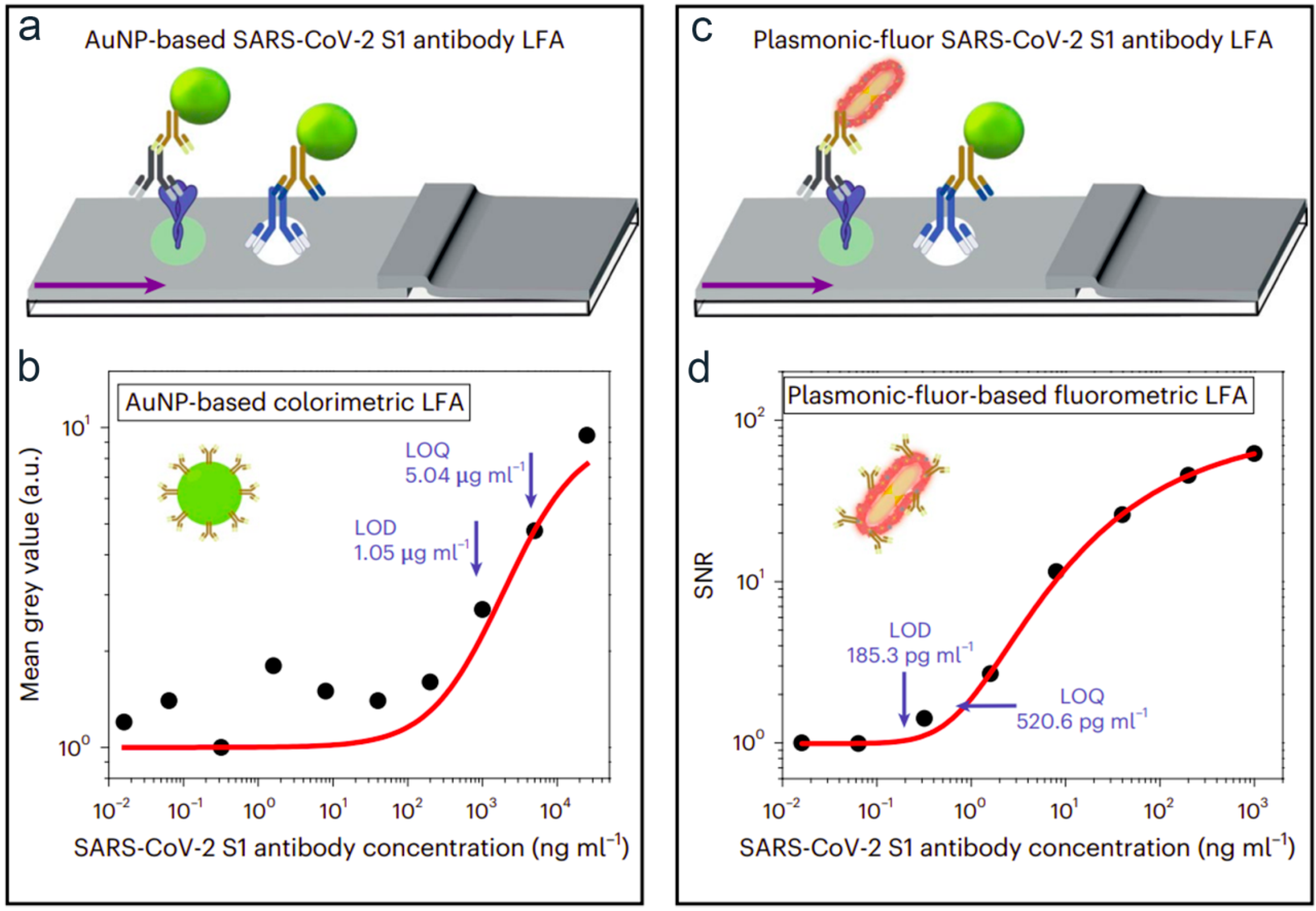
| plasmonic construct | SARS-CoV-2 S1 antibody | antibody | 5675-fold | gold nanoparticle-LFA | [36] | |
| Silica nanosphere/GNPs/rQDs | H-FABP | gQDs/antibody | LOD of 0.21 ng/mL | Conv. fluorescent LFA | [37] | |
| Signal amplification: reader use | CRISPR-Cas13a-LFA | SARS-CoV-2 | fluorescein isothiocyanate-secondary antibody | LOD of 0.25 copy/μL | RT-PCR | [38] |
| SERS-LFA/GNPs | SARS-CoV-2 | antibody | no enhancement | conv. LFA | [39] | |
| thermal contrast magnification | malaria | antibody | 8-fold | colorimetric reader | [40] | |
| Extraction and Enrichment of the Target Molecule in the Sample | magnetic field-assisted preconcentration | cardiac troponin | antibody | 10-fold | conv. LFA | [41] |
| nanoelectrokinetic (NEK)/preconcentration | SARS-CoV-2-IgG | antibody | 32-fold | conv. LFA | [42] |
Disclaimer/Publisher’s Note: The statements, opinions and data contained in all publications are solely those of the individual author(s) and contributor(s) and not of MDPI and/or the editor(s). MDPI and/or the editor(s) disclaim responsibility for any injury to people or property resulting from any ideas, methods, instructions or products referred to in the content. |
© 2023 by the authors. Licensee MDPI, Basel, Switzerland. This article is an open access article distributed under the terms and conditions of the Creative Commons Attribution (CC BY) license (https://creativecommons.org/licenses/by/4.0/).
Share and Cite
Omidfar, K.; Riahi, F.; Kashanian, S. Lateral Flow Assay: A Summary of Recent Progress for Improving Assay Performance. Biosensors 2023, 13, 837. https://doi.org/10.3390/bios13090837
Omidfar K, Riahi F, Kashanian S. Lateral Flow Assay: A Summary of Recent Progress for Improving Assay Performance. Biosensors. 2023; 13(9):837. https://doi.org/10.3390/bios13090837
Chicago/Turabian StyleOmidfar, Kobra, Fatemeh Riahi, and Soheila Kashanian. 2023. "Lateral Flow Assay: A Summary of Recent Progress for Improving Assay Performance" Biosensors 13, no. 9: 837. https://doi.org/10.3390/bios13090837