Application of Intelligent Medical Sensing Technology


Abstract
:1. Introduction
2. Calibration
2.1. Introduction to Calibration Methods
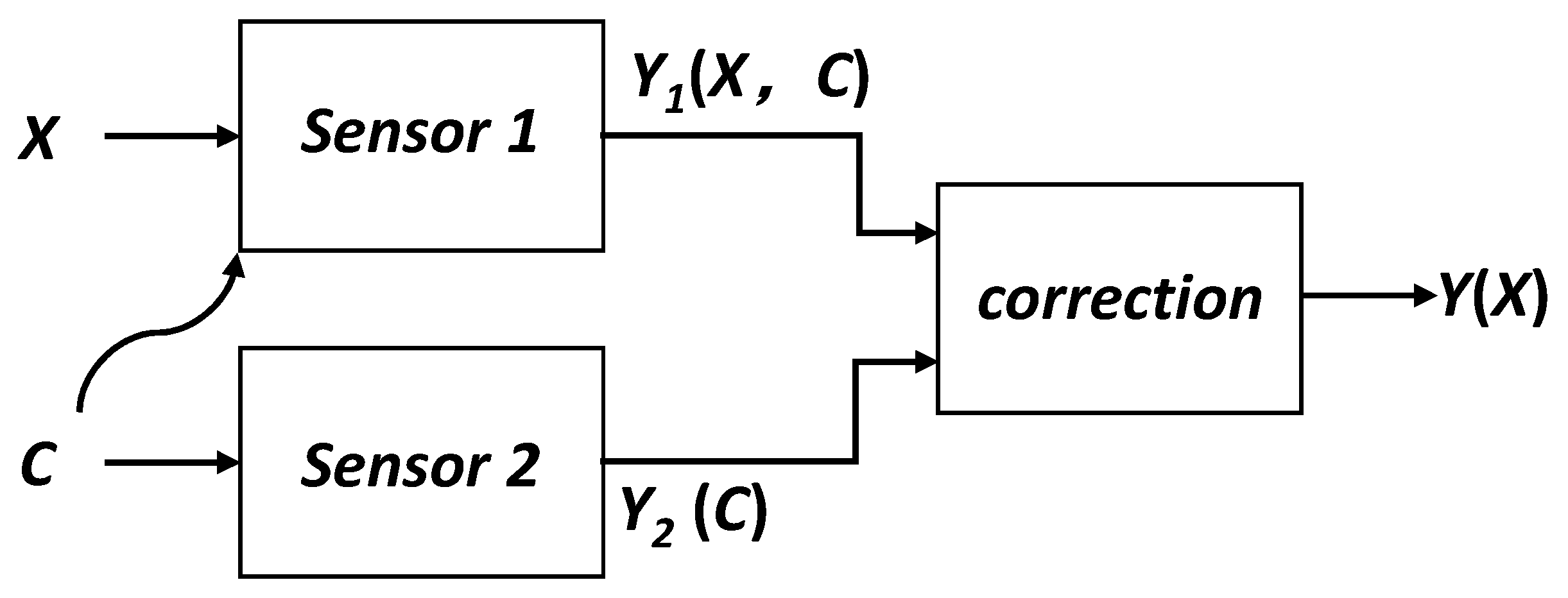
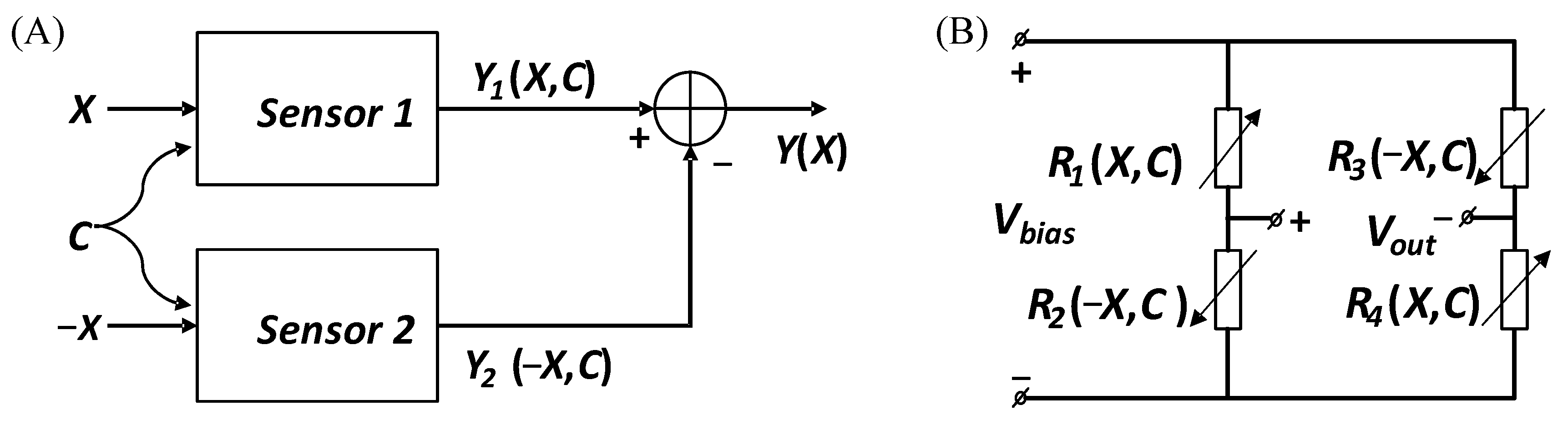
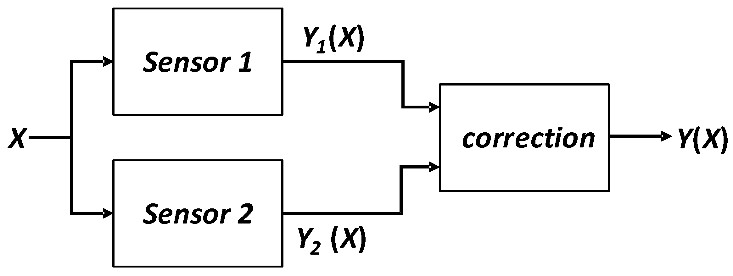
2.2. Self-Calibration by Combining Multiple Sensors
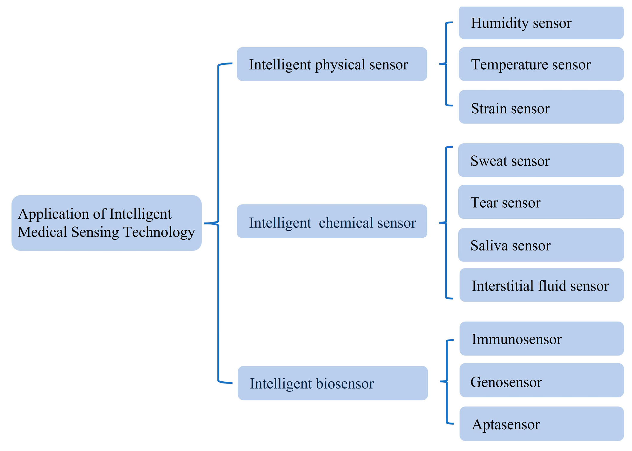
3. Intelligent Physical Sensor
3.1. Humidity Sensor
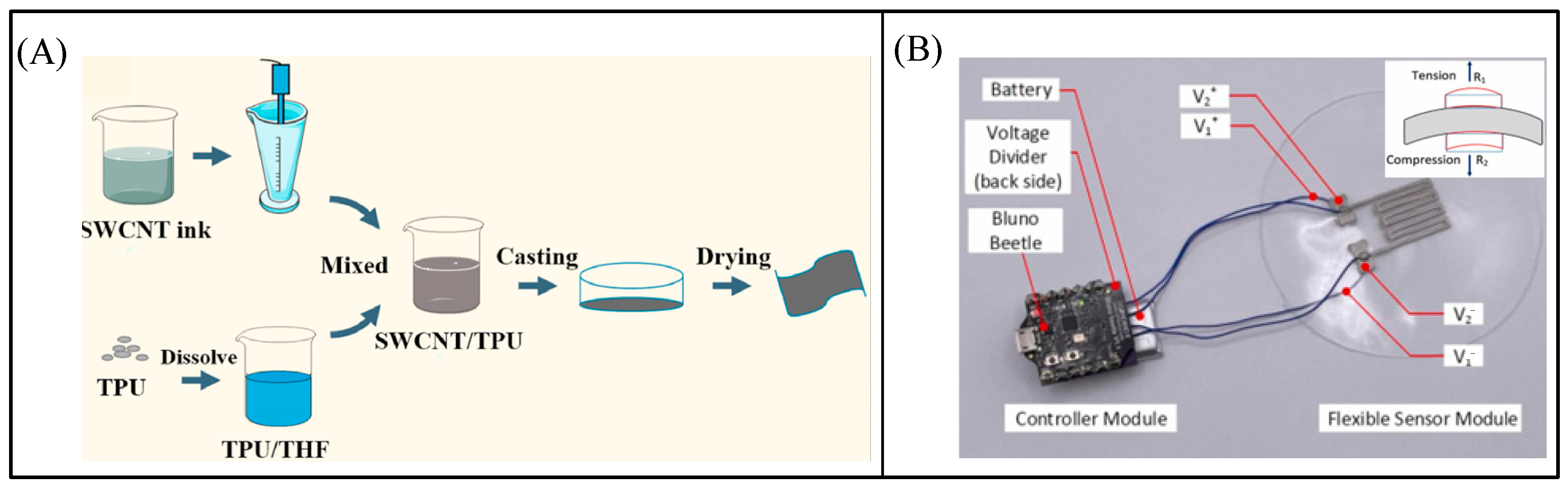
3.2. Temperature Sensor
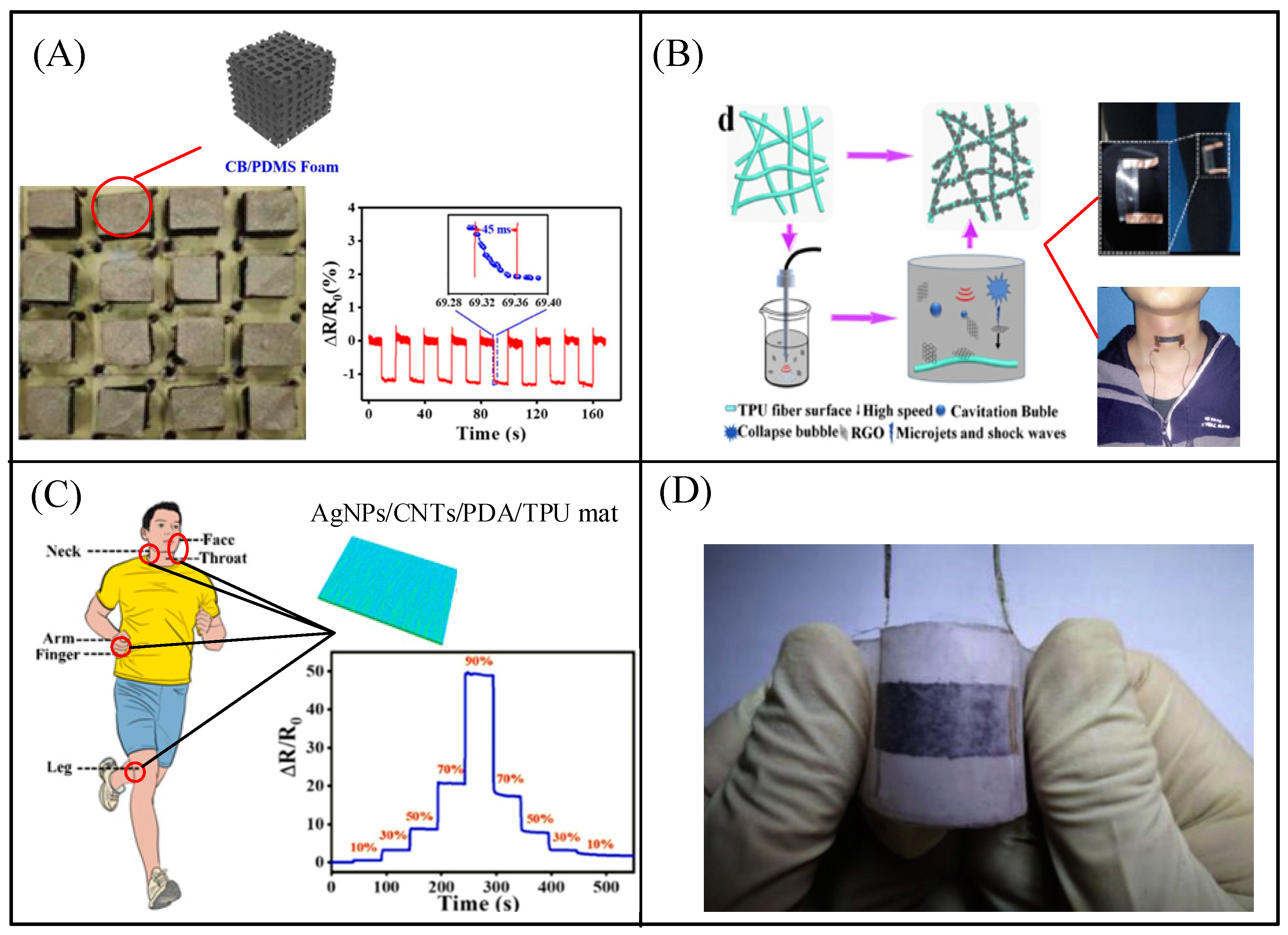
3.3. Strain Sensor
4. Intelligent Chemical Sensor
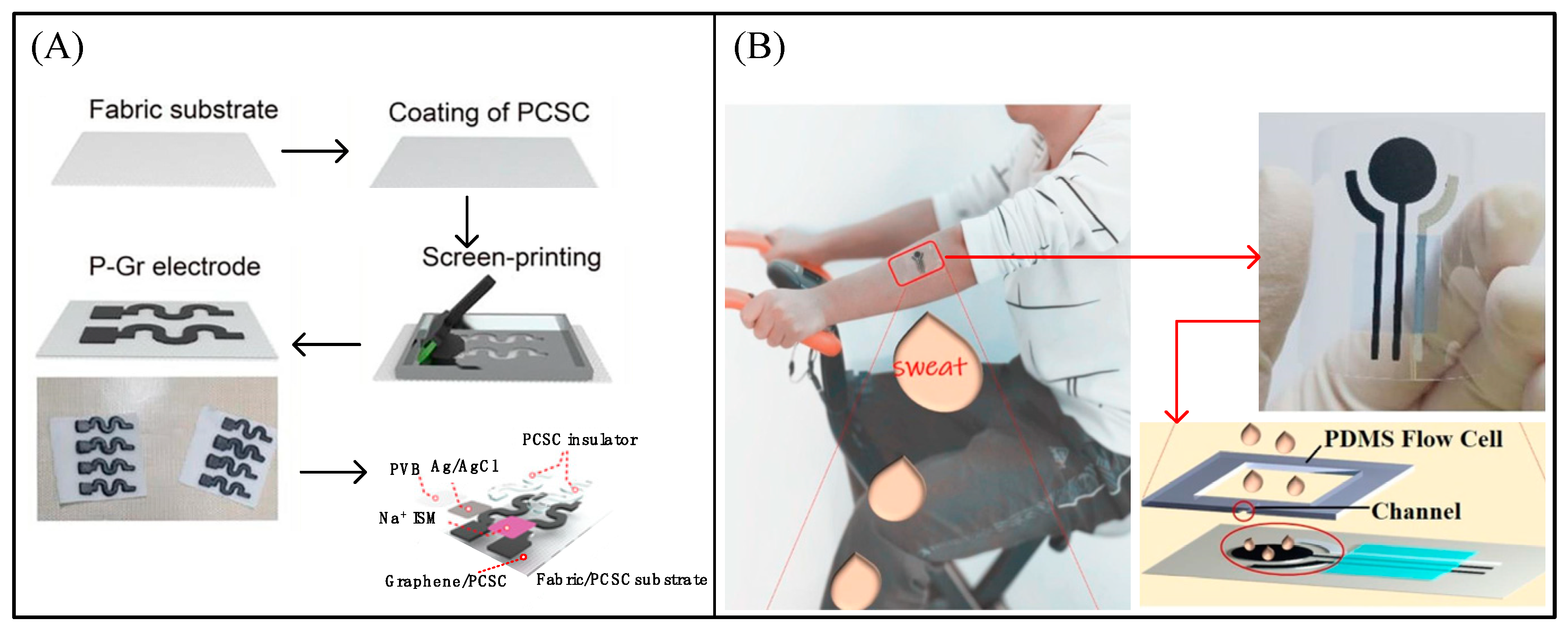
4.1. Sweat Sensor
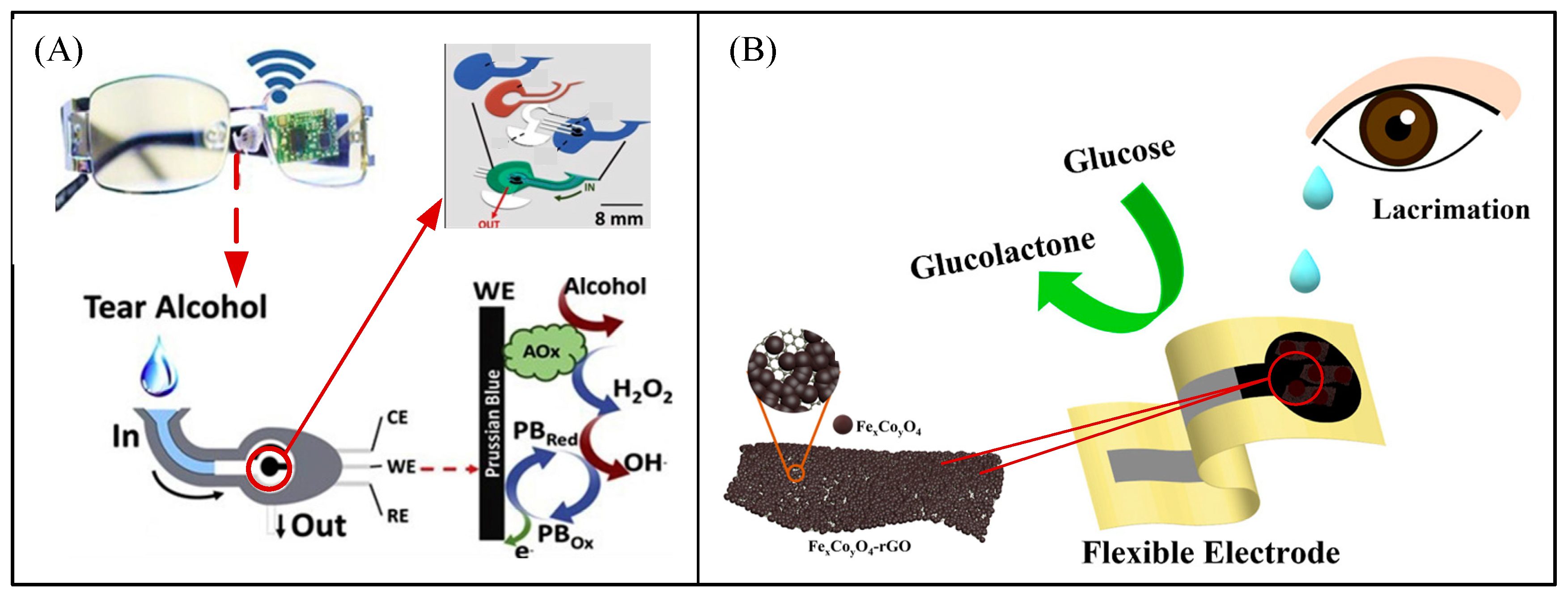
4.2. Tear Sensor
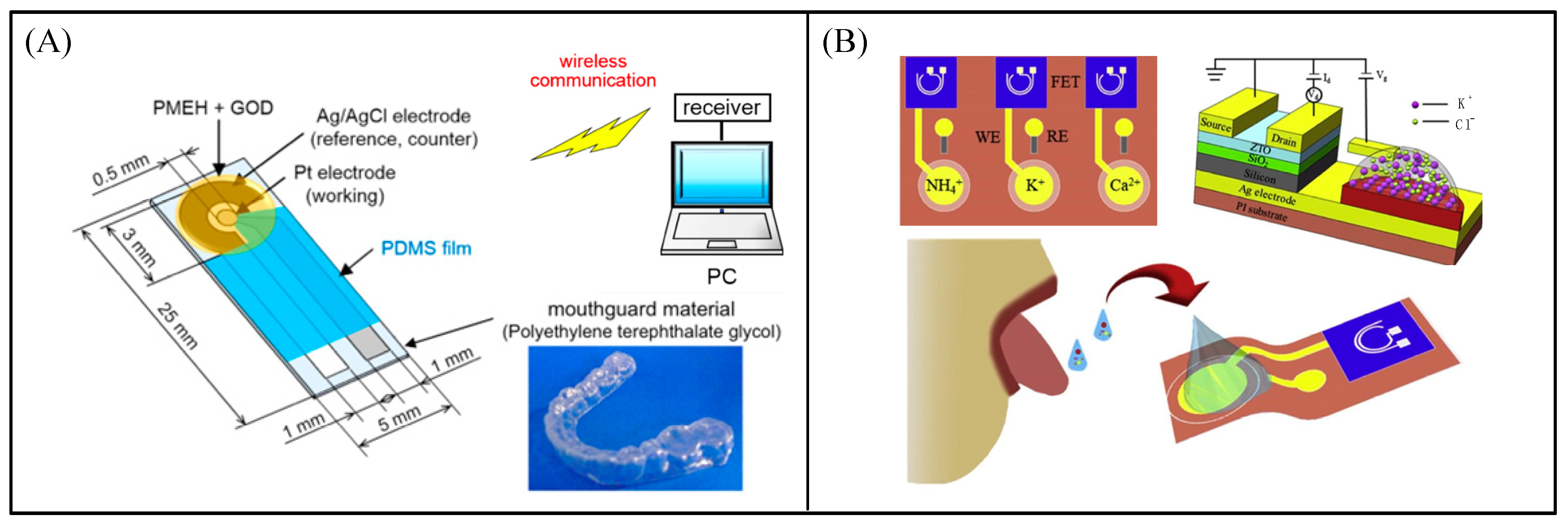
4.3. Saliva Sensor
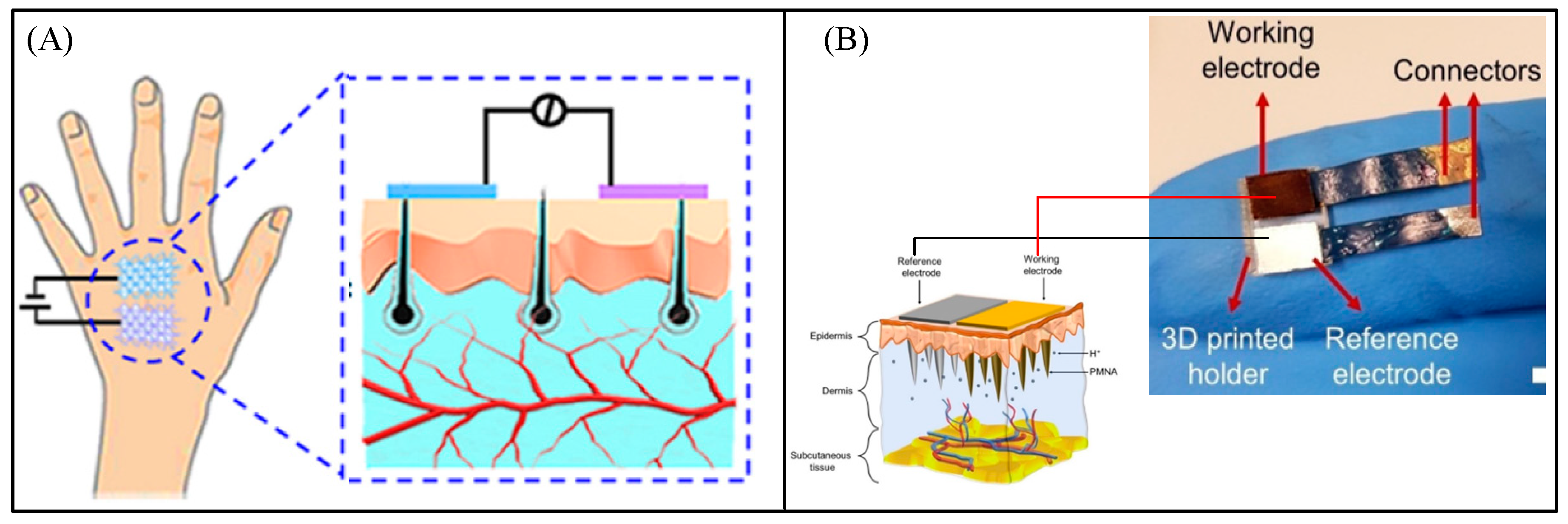
4.4. Interstitial Fluid Sensor
5. Intelligent Biosensor
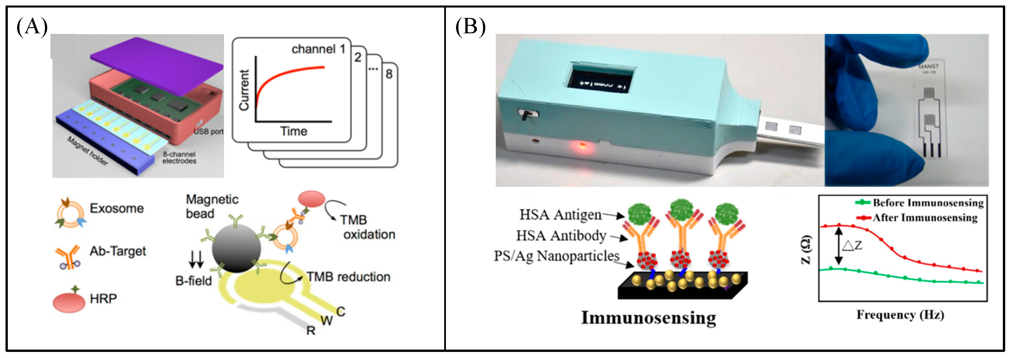
5.1. Immunosensor
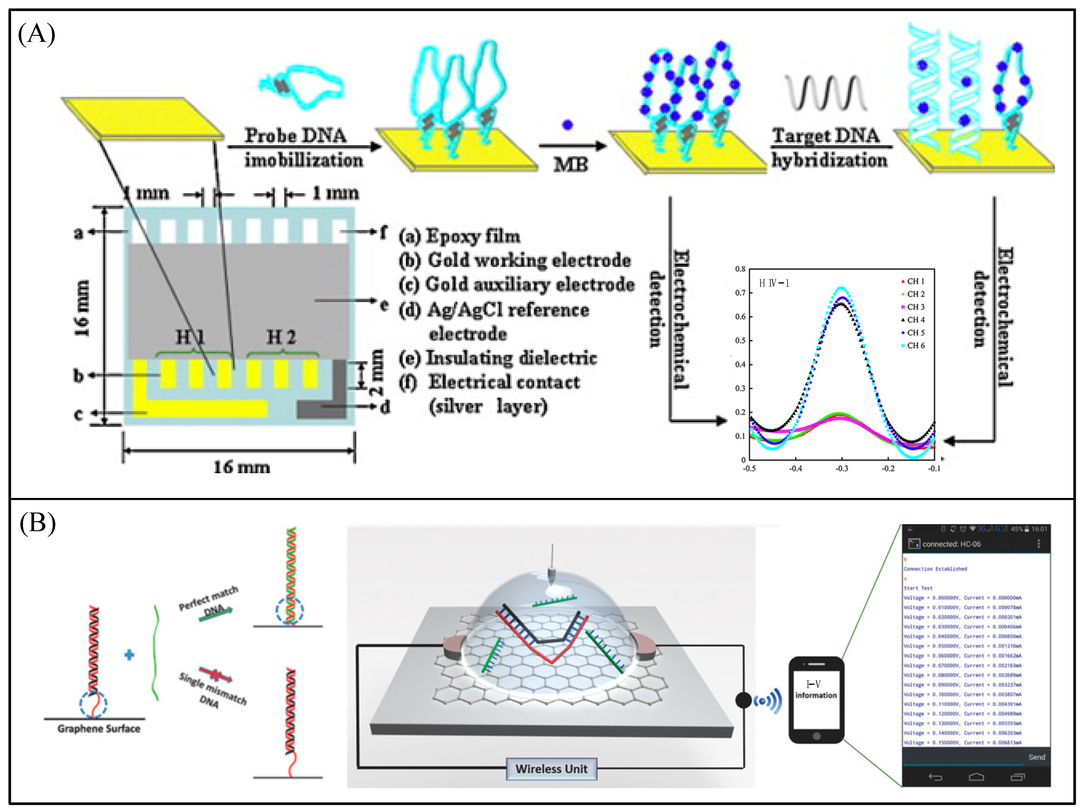
5.2. Genosensor
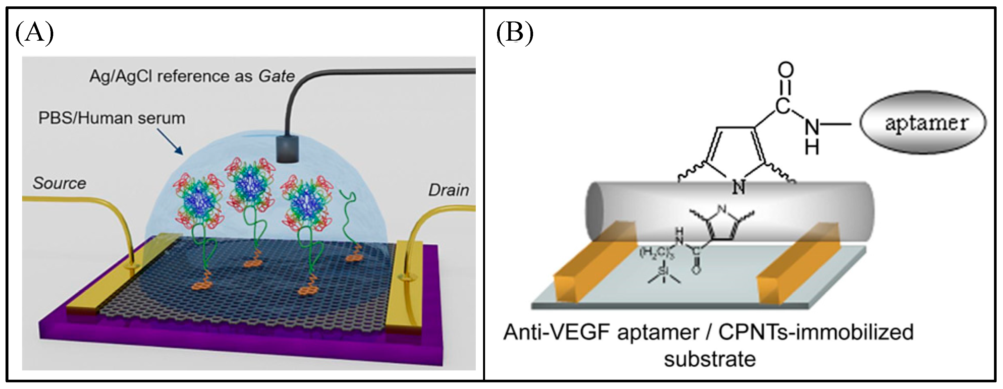
5.3. Aptasensor
6. Conclusions and Perspective
Author Contributions
Funding
Institutional Review Board Statement
Informed Consent Statement
Data Availability Statement
Conflicts of Interest
Abbreviations
References
- Tripathi, M.K.; Nickhil, C.; Kate, A.; Srivastva, R.M.; Mohapatra, D.; Jadam, R.S.; Yadav, A.; Modhera, B. Chapter 24–Biosensor: Fundamentals, biomolecular component, and applications. In Advances in Biomedical Polymers and Composites; Pal, K., Verma, S., Datta, P., Barui, A., Hashmi, S.A.R., Srivastava, A.K., Eds.; Elsevier: Amsterdam, The Netherlands, 2023; pp. 617–633. [Google Scholar]
- Li, Y.; Li, Y.; Zhang, R.; Li, S.; Liu, Z.; Zhang, J.; Fu, Y. Progress in wearable acoustical sensors for diagnostic applications. Biosens. Bioelectron. 2023, 237, 115509. [Google Scholar] [CrossRef]
- Zhang, Y.; Hu, Y.; Jiang, N.; Yetisen, A.K. Wearable artificial intelligence biosensor networks. Biosens. Bioelectron. 2023, 219, 114825. [Google Scholar] [CrossRef] [PubMed]
- Jain, U.; Chauhan, N.; Saxena, K. Chapter 3–Fundamentals of sensors and biosensors: An overview. In Multifaceted Bio-Sensing Technology; Singh, L., Mahapatra, D., Kumar, S., Eds.; Academic Press: Cambridge, MA, USA, 2023; Volume 4, pp. 31–44. [Google Scholar]
- Salehabadi, A.; Enhessari, M.; Ahmad, M.I.; Ismail, N.; Gupta, B.D. Chapter 2–Sensors and biosensors. In Metal Chalcogenide Biosensors; Salehabadi, A., Enhessari, M., Ahmad, M.I., Ismail, N., Gupta, B.D., Eds.; Woodhead Publishing: Sawston, UK, 2023; pp. 9–30. [Google Scholar]
- Das, T.R.; Patra, S.; Govender, P.P.; Shukla, S.K. Chapter 1–Biosensors: Principle, fundamentals history, recent trends and applications. In Biosensors for Emerging and Re-Emerging Infectious Diseases; Das, J., Dave, S., Radhakrishnan, S., Mohanty, P., Eds.; Academic Press: Cambridge, MA, USA, 2022; pp. 1–18. [Google Scholar]
- Zhang, C.; Liu, H.; Li, X.; Xu, F.; Li, Z. Modularized synthetic biology enabled intelligent biosensors. Trends Biotechnol. 2023, 41, 1055–1065. [Google Scholar] [CrossRef] [PubMed]
- Diao, H.; Yin, L.; Liang, B.; Chen, Y. An intelligent system control method based on visual sensor. Meas. Sens. 2023, 29, 100857. [Google Scholar] [CrossRef]
- Kuzubaşoğlu, B.A.; Tekçin, M.; Bahadır, S.K. Electronic Textiles (E-Textiles): Fabric Sensors and Material-Integrated Wearable Intelligent Systems. In Encyclopedia of Sensors and Biosensors, 1st ed.; Narayan, R., Ed.; Elsevier: Oxford, UK, 2023; pp. 80–100. [Google Scholar]
- Morris, A.S.; Langari, R. Chapter 11–Intelligent sensors. In Measurement and Instrumentation, 3rd ed.; Morris, A.S., Langari, R., Eds.; Academic Press: Cambridge, MA, USA, 2021; pp. 323–348. [Google Scholar]
- Desagani, D.; Ben-Yoav, H. Chemometrics meets electrochemical sensors for intelligent in vivo bioanalysis. TrAC Trends Anal. Chem. 2023, 164, 117089. [Google Scholar] [CrossRef]
- Perera, Y.S.; Ratnaweera, D.A.A.C.; Dasanayaka, C.H.; Abeykoon, C. The role of artificial intelligence-driven soft sensors in advanced sustainable process industries: A critical review. Eng. Appl. Artif. Intell. 2023, 121, 105988. [Google Scholar] [CrossRef]
- Zhou, Y.; Shen, M.; Cui, X.; Shao, Y.; Li, L.; Zhang, Y. Triboelectric nanogenerator based self-powered sensor for artificial intelligence. Nano Energy 2021, 84, 105887. [Google Scholar] [CrossRef]
- Lutz, É.; Coradi, P.C. Applications of new technologies for monitoring and predicting grains quality stored: Sensors, Internet of Things, and Artificial Intelligence. Measurement 2022, 188, 110609. [Google Scholar] [CrossRef]
- Heinssen, S.; Hillebrand, T.; Taddiken, M.; Tscherkaschin, K.; Paul, S.; Peters-Drolshagen, D. Design for reliability of generic sensor interface circuits. Microelectron. Reliab. 2018, 80, 184–197. [Google Scholar] [CrossRef]
- Czaja, Z. Time-domain measurement methods for R, L and C sensors based on a versatile direct sensor-to-microcontroller interface circuit. Sens. Actuators A Phys. 2018, 274, 199–210. [Google Scholar] [CrossRef]
- Xu, C.; Yang, Y.; Gao, W. Skin-Interfaced Sensors in Digital Medicine: From Materials to Applications. Matter 2020, 2, 1414–1445. [Google Scholar] [CrossRef]
- Nankali, M.; Nouri, N.M.; Navidbakhsh, M.; Malek, N.G.; Amindehghan, M.A.; Shahtoori, A.M.; Karimi, M.; Amjadi, M. Highly stretchable and sensitive strain sensors based on carbon nanotube-elastomer nanocomposites: The effect of environmental factors on strain sensing performance. J. Mater. Chem. C 2020, 8, 6185–6195. [Google Scholar] [CrossRef]
- Wu, D.; Zhang, G.; Liu, J.; Shen, S.; Yang, Z.; Pan, Y.; Zhao, X.; Yang, S.; Tian, Y.; Zhao, H.; et al. Influence of particle properties and environmental factors on the performance of typical particle monitors and low-cost particle sensors in the market of China. Atmos. Environ. 2022, 268, 118825. [Google Scholar] [CrossRef]
- Sun, T.; Yu, Z.-h. Moving target localization in distributed MIMO radar systems with sensor position errors in the presence of a calibration object. Digit. Signal Process. 2022, 131, 103751. [Google Scholar] [CrossRef]
- Ma, H.; Chen, J.; Tian, J.; Chen, X. Design of Personal Health Monitoring System Based on Body Sensor Network. Meas. Control Technol. 2015, 34, 24–27. [Google Scholar]
- Wang, S.; Shen, H.; Luo, C. Temperature Measurement with Bluetooth under Android Platform. Zhongguo Yi Liao Qi Xie Za Zhi = Chin. J. Med. Instrum. 2015, 39, 181–196. [Google Scholar]
- Becker, F.; Paul, O. Efficient cross-sensitivity compensation in multisensor systems by half-blind calibration. Sens. Actuators A Phys. 2017, 257, 154–164. [Google Scholar] [CrossRef]
- Sifuentes, E.; Casas, O.; Reverter, F.; Pallas-Areny, R. Improved direct interface circuit for resistive full- and half-bridge sensors. In Proceedings of the 4th International Conference on Electrical and Electronics Engineering, Mexico City, Mexico, 7 September 2007; pp. 100–103. [Google Scholar]
- de Graaf, G.; Wolffenbuttel, R.F. Circuit for readout and linearisation of sensor bridges. In Proceedings of the 30th European Solid-State Circuits Conference (ESSCIRC 2004), Leuven, Belgium, 21–23 September 2004; pp. 451–454. [Google Scholar]
- Haq, Y.-U.; Ullah, R.; Mazhar, S.; Khattak, R.; Qarni, A.A.; Haq, Z.-U.; Amin, S. Synthesis and characterization of 2D MXene: Device fabrication for humidity sensing. J. Sci. Adv. Mater. Devices 2022, 7, 100390. [Google Scholar] [CrossRef]
- Jia, G.; Zheng, A.; Wang, X.; Zhang, L.; Li, L.; Li, C.; Zhang, Y.; Cao, L. Flexible, biocompatible and highly conductive MXene-graphene oxide film for smart actuator and humidity sensor. Sens. Actuators B Chem. 2021, 346, 130507. [Google Scholar] [CrossRef]
- Xing, H.; Li, X.; Lu, Y.; Wu, Y.; He, Y.; Chen, Q.; Liu, Q.; Han, R.P.S. MXene/MWCNT electronic fabric with enhanced mechanical robustness on humidity sensing for real-time respiration monitoring. Sens. Actuators B Chem. 2022, 361, 131704. [Google Scholar] [CrossRef]
- Yang, M.-y.; Huang, M.-l.; Li, Y.-z.; Feng, Z.-s.; Huang, Y.; Chen, H.-j.; Xu, Z.-q.; Liu, H.-g.; Wang, Y. Printing assembly of flexible devices with oxidation stable MXene for high performance humidity sensing applications. Sens. Actuators B Chem. 2022, 364, 131867. [Google Scholar] [CrossRef]
- Jiang, Y.; Duan, Z.; Fan, Z.; Yao, P.; Yuan, Z.; Jiang, Y.; Cao, Y.; Tai, H. Power generation humidity sensor based on NaCl/halloysite nanotubes for respiratory patterns monitoring. Sens. Actuators B Chem. 2023, 380, 133396. [Google Scholar] [CrossRef]
- Zhu, G.; Wang, F.; Chen, L.; Wang, C.; Xu, Y.; Chen, J.; Chang, X.; Zhu, Y. Highly flexible TPU/SWCNTs composite-based temperature sensors with linear negative temperature coefficient effect and photo-thermal effect. Compos. Sci. Technol. 2022, 217, 109133. [Google Scholar] [CrossRef]
- Usman, M.; Jamhour, N.; Hettinger, J.; Xue, W. Smart Wearable Flexible Temperature Sensor with Compensation against Bending and Stretching Effects. Sens. Actuators A Phys. 2023, 353, 114224. [Google Scholar] [CrossRef]
- Zhai, W.; Xia, Q.; Zhou, K.; Yue, X.; Ren, M.; Zheng, G.; Dai, K.; Liu, C.; Shen, C. Multifunctional flexible carbon black/polydimethylsiloxane piezoresistive sensor with ultrahigh linear range, excellent durability and oil/water separation capability. Chem. Eng. J. 2019, 372, 373–382. [Google Scholar] [CrossRef]
- Chen, Z.; Ming, T.; Goulamaly, M.M.; Yao, H.; Nezich, D.; Hempel, M.; Hofmann, M.; Kong, J. Enhancing the Sensitivity of Percolative Graphene Films for Flexible and Transparent Pressure Sensor Arrays. Adv. Funct. Mater. 2016, 26, 5061–5067. [Google Scholar] [CrossRef]
- Kweon, O.Y.; Lee, S.J.; Oh, J.H. Wearable high-performance pressure sensors based on three-dimensional electrospun conductive nanofibers. NPG Asia Mater. 2018, 10, 540–551. [Google Scholar] [CrossRef] [Green Version]
- Wang, Y.; Hao, J.; Huang, Z.; Zheng, G.; Dai, K.; Liu, C.; Shen, C. Flexible electrically resistive-type strain sensors based on reduced graphene oxide-decorated electrospun polymer fibrous mats for human motion monitoring. Carbon 2018, 126, 360–371. [Google Scholar] [CrossRef]
- Zhan, P.; Jia, Y.; Zhai, W.; Zheng, G.; Dai, K.; Liu, C.; Shen, C. A fibrous flexible strain sensor with Ag nanoparticles and carbon nanotubes for synergetic high sensitivity and large response range. Compos. Part A Appl. Sci. Manuf. 2023, 167, 107431. [Google Scholar] [CrossRef]
- Lu, S.; Wang, S.; Wang, G.; Ma, J.; Wang, X.; Tang, H.; Yang, X. Wearable graphene film strain sensors encapsulated with nylon fabric for human motion monitoring. Sens. Actuators A Phys. 2019, 295, 200–209. [Google Scholar] [CrossRef]
- Maddocks, G.M.; Daniele, M.A. Chemical Sensors: Wearable Sensors. In Encyclopedia of Sensors and Biosensors, 1st ed.; Narayan, R., Ed.; Elsevier: Oxford, UK, 2023; pp. 260–280. [Google Scholar]
- Zhao, J.; Guo, H.; Li, J.; Bandodkar, A.J.; Rogers, J.A. Body-Interfaced Chemical Sensors for Noninvasive Monitoring and Analysis of Biofluids. Trends Chem. 2019, 1, 559–571. [Google Scholar] [CrossRef]
- Promphet, N.; Ummartyotin, S.; Ngeontae, W.; Puthongkham, P.; Rodthongkum, N. Non-invasive wearable chemical sensors in real-life applications. Anal. Chim. Acta 2021, 1179, 338643. [Google Scholar] [CrossRef] [PubMed]
- Legner, C.; Kalwa, U.; Patel, V.; Chesmore, A.; Pandey, S. Sweat sensing in the smart wearables era: Towards integrative, multifunctional and body-compliant perspiration analysis. Sens. Actuators A Phys. 2019, 296, 200–221. [Google Scholar] [CrossRef]
- Mohan, A.M.V.; Rajendran, V.; Mishra, R.K.; Jayaraman, M. Recent advances and perspectives in sweat based wearable electrochemical sensors. TrAC Trends Anal. Chem. 2020, 131, 116024. [Google Scholar] [CrossRef]
- Gyu Son, S.; Jun Park, H.; Kim, S.-M.; Jin Kim, S.; Sik Kil, M.; Jeong, J.-M.; Lee, Y.; Eom, Y.; Yeon Hwang, S.; Park, J.; et al. Ultra-fast self-healable stretchable bio-based elastomer/graphene ink using fluid dynamics process for printed wearable sweat-monitoring sensor. Chem. Eng. J. 2023, 454, 140443. [Google Scholar] [CrossRef]
- Jia, W.; Bandodkar, A.J.; Valdes-Ramirez, G.; Windmiller, J.R.; Yang, Z.; Ramirez, J.; Chan, G.; Wang, J. Electrochemical tattoo biosensors for real-time noninvasive lactate monitoring in human perspiration. Anal. Chem. 2013, 85, 6553–6560. [Google Scholar] [CrossRef]
- Zhang, Q.; Jiang, D.; Xu, C.; Ge, Y.; Liu, X.; Wei, Q.; Huang, L.; Ren, X.; Wang, C.; Wang, Y. Wearable electrochemical biosensor based on molecularly imprinted Ag nanowires for noninvasive monitoring lactate in human sweat. Sens. Actuators B Chem. 2020, 320, 128325. [Google Scholar] [CrossRef]
- Moreddu, R.; Wolffsohn, J.S.; Vigolo, D.; Yetisen, A.K. Laser-inscribed contact lens sensors for the detection of analytes in the tear fluid. Sens. Actuators B Chem. 2020, 317, 128183. [Google Scholar] [CrossRef]
- Pankratov, D.; González-Arribas, E.; Blum, Z.; Shleev, S. Tear Based Bioelectronics. Electroanalysis 2016, 28, 1250–1266. [Google Scholar] [CrossRef]
- Sempionatto, J.R.; Brazaca, L.C.; García-Carmona, L.; Bolat, G.; Campbell, A.S.; Martin, A.; Tang, G.; Shah, R.; Mishra, R.K.; Kim, J.; et al. Eyeglasses-based tear biosensing system: Non-invasive detection of alcohol, vitamins and glucose. Biosens. Bioelectron. 2019, 137, 161–170. [Google Scholar] [CrossRef]
- Zhou, F.; Zhao, H.; Chen, K.; Cao, S.; Shi, Z.; Lan, M. Flexible electrochemical sensor with Fe/Co bimetallic oxides for sensitive analysis of glucose in human tears. Anal. Chim. Acta 2023, 1243, 340781. [Google Scholar] [CrossRef] [PubMed]
- Swetha, P.; Balijapalli, U.; Feng, S.-P. Wireless accessing of salivary biomarkers based wearable electrochemical sensors: A mini-review. Electrochem. Commun. 2022, 140, 107314. [Google Scholar] [CrossRef]
- Arakawa, T.; Kuroki, Y.; Nitta, H.; Chouhan, P.; Toma, K.; Sawada, S.-i.; Takeuchi, S.; Sekita, T.; Akiyoshi, K.; Minakuchi, S.; et al. Mouthguard biosensor with telemetry system for monitoring of saliva glucose: A novel cavitas sensor. Biosens. Bioelectron. 2016, 84, 106–111. [Google Scholar] [CrossRef] [PubMed] [Green Version]
- Kim, J.; Valdes-Ramirez, G.; Bandodkar, A.J.; Jia, W.; Martinez, A.G.; Ramirez, J.; Mercier, P.; Wang, J. Non-invasive mouthguard biosensor for continuous salivary monitoring of metabolites. Analyst 2014, 139, 1632–1636. [Google Scholar] [CrossRef]
- Bao, C.; Kaur, M.; Kim, W.S. Toward a highly selective artificial saliva sensor using printed hybrid field effect transistors. Sens. Actuators B Chem. 2019, 285, 186–192. [Google Scholar] [CrossRef]
- Mannoor, M.S.; Tao, H.; Clayton, J.D.; Sengupta, A.; Kaplan, D.L.; Naik, R.R.; Verma, N.; Omenetto, F.G.; McAlpine, M.C. Graphene-based wireless bacteria detection on tooth enamel. Nat. Commun. 2012, 3, 763. [Google Scholar] [CrossRef]
- Tseng, P.; Napier, B.; Garbarini, L.; Kaplan, D.L.; Omenetto, F.G. Functional, RF-Trilayer Sensors for Tooth-Mounted, Wireless Monitoring of the Oral Cavity and Food Consumption. Adv. Mater. 2018, 30, e1703257. [Google Scholar] [CrossRef]
- Haslene-Hox, H.; Tenstad, O.; Wiig, H. Interstitial fluid—A reflection of the tumor cell microenvironment and secretome. Biochim. Biophys. Acta (BBA)-Proteins Proteom. 2013, 1834, 2336–2346. [Google Scholar] [CrossRef]
- Chang, H.; Zheng, M.; Yu, X.; Aung, T.; Seeni, R.Z.; Kang, R.; Tian, J.; Duong Phan, K.; Liu, L.; Chen, P.; et al. A Swellable Microneedle Patch to Rapidly Extract Skin Interstitial Fluid for Timely Metabolic Analysis. Adv. Mater. 2017, 29, 1702243. [Google Scholar] [CrossRef]
- Zhang, J.; Hao, N.; Liu, W.; Lu, M.; Sun, L.; Chen, N.; Wu, M.; Zhao, X.; Xing, B.; Sun, W.; et al. In-depth proteomic analysis of tissue interstitial fluid for hepatocellular carcinoma serum biomarker discovery. Br. J. Cancer 2017, 117, 1676–1684. [Google Scholar] [CrossRef] [Green Version]
- Hadrevi, J.; Ghafouri, B.; Sjors, A.; Antti, H.; Larsson, B.; Crenshaw, A.G.; Gerdle, B.; Hellstrom, F. Comparative metabolomics of muscle interstitium fluid in human trapezius myalgia: An in vivo microdialysis study. Eur. J. Appl. Physiol. 2013, 113, 2977–2989. [Google Scholar] [CrossRef] [Green Version]
- Kiang, T.K.L.; Schmitt, V.; Ensom, M.H.H.; Chua, B.; Haefeli, U.O. Therapeutic drug monitoring in interstitial fluid: A feasibility study using a comprehensive panel of drugs. J. Pharm. Sci. 2012, 101, 4642–4652. [Google Scholar] [CrossRef]
- Madden, J.; O’Mahony, C.; Thompson, M.; O’Riordan, A.; Galvin, P. Biosensing in dermal interstitial fluid using microneedle based electrochemical devices. Sens. Bio-Sens. Res. 2020, 29, 100348. [Google Scholar] [CrossRef]
- Zhao, R.; Wang, C.; Lu, F.; Du, L.; Fang, Z.; Guo, X.; Liu, J.-T.; Chen, C.-J.; Zhao, Z. A Flexible Interdigital Electrode Used in Skin Penetration Promotion and Evaluation with Electroporation and Reverse Iontophoresis Synergistically. Sensors 2018, 18, 1431. [Google Scholar] [CrossRef] [PubMed] [Green Version]
- Miller, P.R.; Taylor, R.M.; Bao Quoc, T.; Boyd, G.; Glaros, T.; Chavez, V.H.; Krishnakumar, R.; Sinha, A.; Poorey, K.; Williams, K.P.; et al. Extraction and biomolecular analysis of dermal interstitial fluid collected with hollow microneedlesis. Commun. Biol. 2018, 1, 173. [Google Scholar] [CrossRef] [PubMed] [Green Version]
- Vranic, E.; Tucak, A.; Sirbubalo, M.; Rahic, O.; Elezovic, A.; Hadziabdic, J. Microneedle-Based Sensor Systems for Real-Time Continuous Transdermal Monitoring of Analytes in Body Fluids. In Proceedings of the International Conference on Medical and Biological Engineering, Banja Luka, Bosnia and Herzegovina, 16–18 May 2019; Springer: Cham, Switzerland, 2020; pp. 167–172. [Google Scholar]
- Bollella, P.; Sharma, S.; Cass, A.E.G.; Antiochia, R. Minimally-invasive Microneedle-based Biosensor Array for Simultaneous Lactate and Glucose Monitoring in Artificial Interstitial Fluid. Electroanalysis 2019, 31, 374–382. [Google Scholar] [CrossRef] [Green Version]
- Yao, Y.; Chen, J.; Guo, Y.; Lv, T.; Chen, Z.; Li, N.; Cao, S.; Chen, B.; Chen, T. Integration of interstitial fluid extraction and glucose detection in one device for wearable non-invasive blood glucose sensors. Biosens. Bioelectron. 2021, 179, 113078. [Google Scholar] [CrossRef] [PubMed]
- Dervisevic, M.; Dervisevic, E.; Esser, L.; Easton, C.D.; Cadarso, V.J.; Voelcker, N.H. Wearable microneedle array-based sensor for transdermal monitoring of pH levels in interstitial fluid. Biosens. Bioelectron. 2023, 222, 114955. [Google Scholar] [CrossRef]
- Cajigas, S.; Soto, D.; Orozco, J. Biosensors: Biosensors With Signal Amplification. In Encyclopedia of Sensors and Biosensors, 1st ed.; Narayan, R., Ed.; Elsevier: Oxford, UK, 2023; pp. 429–457. [Google Scholar]
- Hamzah, A.A.; Nadzirah, S. Biosensor Development. In Encyclopedia of Sensors and Biosensors, 1st ed.; Narayan, R., Ed.; Elsevier: Oxford, UK, 2023; pp. 209–217. [Google Scholar]
- Bollella, P. Enzyme-based amperometric biosensors: 60 years later… Quo Vadis? Anal. Chim. Acta 2022, 1234, 340517. [Google Scholar] [CrossRef]
- Miura, D.; Asano, R. Biosensors: Immunosensors. In Encyclopedia of Sensors and Biosensors, 1st ed.; Narayan, R., Ed.; Elsevier: Oxford, UK, 2023; pp. 298–314. [Google Scholar]
- Jeong, S.; Park, J.; Pathania, D.; Castro, C.M.; Weissleder, R.; Lee, H. Integrated Magneto-Electrochemical Sensor for Exosome Analysis. ACS Nano 2016, 10, 1802–1809. [Google Scholar] [CrossRef] [Green Version]
- Shaikh, M.O.; Zhu, P.-Y.; Wang, C.-C.; Du, Y.-C.; Chuang, C.-H. Electrochemical immunosensor utilizing electrodeposited Au nanocrystals and dielectrophoretically trapped PS/Ag/ab-HSA nanoprobes for detection of microalbuminuria at point of care. Biosens. Bioelectron. 2019, 126, 572–580. [Google Scholar] [CrossRef] [PubMed]
- Ansah, F.; Krampa, F.; Donkor, J.K.; Owusu-Appiah, C.; Ashitei, S.; Kornu, V.E.; Danku, R.K.; Chirawurah, J.D.; Awandare, G.A.; Aniweh, Y.; et al. Ultrasensitive electrochemical genosensors for species-specific diagnosis of malaria. Electrochim. Acta 2022, 429, 140988. [Google Scholar] [CrossRef] [PubMed]
- Zhang, D.; Peng, Y.; Qi, H.; Gao, Q.; Zhang, C. Label-free electrochemical DNA biosensor array for simultaneous detection of the HIV-1 and HIV-2 oligonucleotides incorporating different hairpin-DNA probes and redox indicator. Biosens. Bioelectron. 2010, 25, 1088–1094. [Google Scholar] [CrossRef] [PubMed]
- Hwang, M.T.; Wang, Z.; Ping, J.; Ban, D.K.; Shiah, Z.C.; Antonschmidt, L.; Lee, J.; Liu, Y.; Karkisaval, A.G.; Johnson, A.T.C.; et al. DNA Nanotweezers and Graphene Transistor Enable Label-Free Genotyping. Adv. Mater. 2018, 30, e1802440. [Google Scholar] [CrossRef]
- Guedes, P.H.G.; Brussasco, J.G.; Moço, A.C.R.; Moraes, D.D.; Segatto, M.; Flauzino, J.M.R.; Mendes-Silva, A.P.; Vieira, C.U.; Madurro, J.M.; Brito-Madurro, A.G. A highly reusable genosensor for late-life depression diagnosis based on microRNA 184 attomolar detection in human plasma. Talanta 2023, 258, 124342. [Google Scholar] [CrossRef]
- Mujica, M.L.; Tamborelli, A.; Castellaro, A.; Barcudi, D.; Rubianes, M.D.; Rodríguez, M.C.; Saka, H.A.; Bocco, J.L.; Dalmasso, P.R.; Rivas, G.A. Impedimetric and amperometric genosensors for the highly sensitive quantification of SARS-CoV-2 nucleic acid using an avidin-functionalized multi-walled carbon nanotubes biocapture platform. Biosens. Bioelectron. X 2022, 12, 100222. [Google Scholar] [CrossRef]
- Bhardwaj, T.; Dalal, P.; Rathore, A.S.; Jha, S.K. An aptamer based microfluidic chip for impedimetric detection of Ranibizumab in a bioreactor. Sens. Actuators B Chem. 2020, 312, 127941. [Google Scholar] [CrossRef]
- Zhu, Q.; Liu, L.; Wang, R.; Zhou, X. A split aptamer (SPA)-based sandwich-type biosensor for facile and rapid detection of streptomycin. J. Hazard. Mater. 2021, 403, 123941. [Google Scholar] [CrossRef]
- Figueroa-Miranda, G.; Liang, Y.; Suranglikar, M.; Stadler, M.; Samane, N.; Tintelott, M.; Lo, Y.; Tanner, J.A.; Vu, X.T.; Knoch, J.; et al. Delineating charge and capacitance transduction in system-integrated graphene-based BioFETs used as aptasensors for malaria detection. Biosens. Bioelectron. 2022, 208, 114219. [Google Scholar] [CrossRef]
- Kwon, O.S.; Park, S.J.; Jang, J. A high-performance VEGF aptamer functionalized polypyrrole nanotube biosensor. Biomaterials 2010, 31, 4740–4747. [Google Scholar] [CrossRef]


| Circuit | Function |
|---|---|
| Impedance conversion circuit | When the output is high impedance, it is converted to low impedance |
| Amplifier circuit | Amplify weak output signal |
| Current-voltage conversion circuit | Current output converted to a voltage |
| Bridge circuit | Change of resistance, capacitance, and inductance into current or voltage |
| Frequency-voltage conversion circuit | Frequency output converted to current or voltage |
| Charge amplifier circuit | Charge generated by the output of the electric field sensor is converted into voltage |
| Filter circuit | Noise of the sensor is eliminated by low-pass and band-pass filter |
| Analyte | Sensing Strategies | LOD | Sensitivity | Detection Range | Ref |
|---|---|---|---|---|---|
| Humidity | MXene/MWCNT electronic fabric | \ | \ | 10–90% RH | [13] |
| Humidity | SA-MXene | \ | \ | 11–97% RH | [14] |
| Temperature | TPU/SWCNTs | \ | \ | 30–100 °C | [16] |
| Temperature | RTD | \ | 5.9 × 10⁻³/°C | 20–40 °C | [17] |
| Strain | CB/PDMS | \ | −1.12 | 0–91% | [18] |
| Strain | ACTM | 0.1% | 7030 & 4.8 × 10⁵ & 2 × 10⁶ | 0–230% & 230–520% & 520–640% | [22] |
| Na+ | PCSC/Gr/Na-selective membranes | 10−5.4 M | −62.35 mV/log[Na] | 10−4–10−1 M | [29] |
| Lactate | MIP-AgNWs | 0.22 μM | 4.5 μA/M | 10−6–10−1 M | [31] |
| Glucose | GOD/POD | 1.84 mM | 1.4 nm/mM | 0–20 mM | [32] |
| Glucose | FexCoyO4-RGO | 0.07 μM | 1510 μM cm−2 mA−1 | 0.1–906.4 μM | [35] |
| Glucose | G/CNTs/GOx | \ | 574.26 μA mM−1 cm−2 & 12.11 μA mM−1 cm−2 | 0–0.1 mM & 1.0–30 mM | [52] |
| miR-184 | Au/DEP1S | 10 automobiles L−1 | 0.15305 μA/log[miR-184] | 10−17–10−9 M | [64] |
| PfLDH | Aptamer/BSA | 0.78 fM | 1.3/decade | 0.78 fM–100 nM | [68] |
Disclaimer/Publisher’s Note: The statements, opinions and data contained in all publications are solely those of the individual author(s) and contributor(s) and not of MDPI and/or the editor(s). MDPI and/or the editor(s) disclaim responsibility for any injury to people or property resulting from any ideas, methods, instructions or products referred to in the content. |
© 2023 by the authors. Licensee MDPI, Basel, Switzerland. This article is an open access article distributed under the terms and conditions of the Creative Commons Attribution (CC BY) license (https://creativecommons.org/licenses/by/4.0/).
Share and Cite
Fu, J.; Gao, Q.; Li, S. Application of Intelligent Medical Sensing Technology. Biosensors 2023, 13, 812. https://doi.org/10.3390/bios13080812
Fu J, Gao Q, Li S. Application of Intelligent Medical Sensing Technology. Biosensors. 2023; 13(8):812. https://doi.org/10.3390/bios13080812
Chicago/Turabian StyleFu, Jie, Qiya Gao, and Shuang Li. 2023. "Application of Intelligent Medical Sensing Technology" Biosensors 13, no. 8: 812. https://doi.org/10.3390/bios13080812