Article

Ultrasensitive Nonenzymatic Real-Time Hydrogen Peroxide Monitoring Using Gold Nanoparticle-Decorated Titanium Dioxide Nanotube Electrodes

1
Faculty of Industrial Sciences and Technology, Universiti Malaysia Pahang, Kuantan 26300, Malaysia
2
Department of Chemistry and Biochemistry, Kent State University, Kent, OH 44242, USA
3
Centre for Sustainability of Ecosystem and Earth Resources (PUSAT ALAM), Universiti Malaysia Pahang, Kuantan 26300, Malaysia
4
Center for Advanced Intelligent Materials, Universiti Malaysia Pahang, Kuantan 26300, Malaysia
5
Faculty of Data Science and Information Technology, INTI International University, Nilai 71800, Malaysia
*
Authors to whom correspondence should be addressed.
Biosensors 2023, 13(7), 671; https://doi.org/10.3390/bios13070671
Submission received: 24 November 2022 / Revised: 13 December 2022 / Accepted: 14 December 2022 / Published: 22 June 2023
(This article belongs to the Special Issue New Biosensors and Nanosensors)

Abstract

:
An amperometric enzyme-free hydrogen peroxide (H2O2) sensor was developed by catalytically stabilizing active gold nanoparticles (Au NPs) of 4–5 nm on a porous titanium dioxide nanotube (TiO2 NTs) electrode. The Au NPs were homogeneously distributed on anatase TiO2 NTs with an outer diameter of ~102 nm, an inner diameter of ~60 nm, and a wall of thickness of ~40 nm. The cyclic voltammogram of the composite electrode showed a pair of redox peaks characterizing the electrocatalytic reduction of H2O2. The entrapping of Au NPs on TiO2 NTs prevented aggregation and facilitated good electrical conductivity and electron transfer rate, thus generating a wide linear range, a low detection limit of ~104 nM, and high sensitivity of ~519 µA/mM, as well as excellent selectivity, reproducibility, repeatability, and stability over 60 days. Furthermore, excellent recovery and relative standard deviation (RSD) were achieved in real samples, which were tap water, milk, and Lactobacillus plantarum bacteria, thereby verifying the accuracy and potentiality of the developed nonenzymatic sensor.

1. Introduction

Hydrogen peroxide (H2O2) is a multifunctional chemical that acts as an oxidizing agent in a variety of industrial environments [1] and as a signal messenger in mediating cellular processes [2]. H2O2 is utilized to disinfect food industry equipment used in the mixing, bottling, transport, and packing processes [3]. As an antibacterial agent, H2O2 is used to preserve milk and juice [4]. Generally, a large quantity of H2O2 at high concentrations (usually 35% and above) is used in industrial applications and might cause toxicity regardless of the exposure routes. Exposure to high concentrations of H2O2 preferentially induces necrosis, and moderate concentrations can cause apoptosis. Additionally, H2O2 is very stable and can rSeach diverse molecular targets far from the origin of generation [5].
H2O2 is produced by some bacteria, such as Lactobacillus plantarum, used in the food processing industry. L. plantarum is a microbial starter and probiotic that can produce H2O2 as well as other compounds, including organic acids and diacetyl [6]. Hence, the presence of H2O2 in food at an intolerable concentration for the human body could pose a threat to consumer health. Moreover, exogenously generated H2O2 can induce DNA damage, ATP depletion, apoptosis, necrosis, and severe cytotoxicity [7]. Therefore, precise and sensitive detection of H2O2, especially at the micro and nano levels, is required to ensure healthy lives and to promote well-being as stated in the sustainable development goal (SDG#3) adopted by the United Nations.
To date, chemiluminescence, spectrophotometry, chromatography, and electrochemical sensors have all been developed to detect H2O2 [8]. However, the electrochemical sensors have gained prominence due to their simplicity, sensitivity, selectivity, and low-detection capability [9]. More precisely, amperometry-based electrochemical sensors were developed using highly catalytically active horseradish peroxidase (HRP) and hemoglobin (Hb) [10]. However, complex immobilization, H2O2-induced inactivation of proteins, the high cost of enzymes, and their sensitivity to environmental conditions have limited the use of these molecules in sensor development [11,12].
Currently, nanomaterials are widely used in electrochemical H2O2 sensor fabrication, which has overcome the bottlenecks of HRP and Hb-modified sensors with high sensitivity, and are now leading the next generation of electrochemical sensors [13,14,15]. Plasmonic nanostructures such as gold (Au NPs), platinum (Pt NPs), and silver nanoparticles (Ag NPs) are frequently used for H2O2 sensing [16]. In particular, Au NPs are used for various chemical and biomolecule detections due to their desirable biocompatibility, large specific surface area, high extinction coefficients, and excellent conductivity [8]. Au NPs have excellent nanozyme activities resembling peroxidase, oxidase, catalase, and reductase [17]. This enzyme-mimicking property can promote electron transfer through the interface of Au NPs and expand the outer region of the modified electrode during sensing [18]. It has also been reported that nanomaterials with enzyme-like properties have the potential to overcome the intrinsic limitations of natural enzymes such as low stability and storage difficulties [19].
The high surface-to-volume ratio of Au NPs provides superior catalytic efficiency; however, it unfavorably reduces chemical stability and causes aggregation [20]. Moreover, the tiring and time-consuming re-dispersion cycle affects the performance of Au NPs [21]. Entrapment of Au NPs within a porous structure is a very sustainable approach, as the porous material can confine the metal nanoparticles and prevent aggregation. In addition, porous materials exhibit a size-selective property that ensures accurate interaction of the reactants with the metal surface [22].
Many metal oxides, such as copper(II) oxide (CuO), titanium dioxide(TiO2), manganese(IV) oxide (MnO2), zinc oxide (ZnO), tungsten trioxide (WO3), and tin(iv) oxide (SnO2) can be synthesized as porous structures with different shapes, such as nanotubes (NTs) [23,24]. TiO2 NTs have desired properties including a tubular structure, larger aspect ratio, corrosion resistance, biocompatibility, high chemical and thermal stability, non-toxicity, and chemical inertness, making them a suitable choice for developing Au NPs-TiO2 NTs composite sensors [25,26,27]. The TiO2 NTs synthesized via anodization offer a porous structure [28]. The tubular inner pores of TiO2 NTs help to effectively entrap platinum (Pt), palladium (Pd), and Au nanoparticles in their hollow structure and improve catalytic performance [29,30]. This entrapment also inhibits electron-hole pair recombination to achieve high charge transfer efficiency and catalytic activity [31]. In addition, TiO2 NTs-supported metals show better catalytic performance than carbon-based nanostructures [32]. To coat Au NPs on these porous TiO2 NTs, a weakly conductive chitosan (CS) polymer is reported to protect the electrode material without affecting the catalytic performance [33].
This study developed a simple, highly susceptible Au NPs-TiO2 NTs composite sensor by decorating Au NPs on TiO2 NTs for the real-time monitoring of H2O2. The nanostructure and morphology of the Au NPs-TiO2 NTs composite were characterized using field-emission scanning electron microscopy (FESEM) and X-ray powder diffraction (XRD). The electrochemical property and H2O2-sensing performances were evaluated using cyclic voltammetry and multi-step chronoamperometry.

2. Materials and Methods

2.1. Reagents and Materials

Titanium (Ti) foil, dimethyl sulfoxide (DMSO), chloroauric acid hydrate (HAuCl4·H2O), sodium citrate (Na3C6H5O7), sodium borohydride (NaBH4, 98%), chitosan (crab shells), acetic acid (CH3COOH, 99%), sodium phosphate monobasic (NaH2PO4, 99%) and sodium phosphate dibasic (Na2HPO4, 99%), sodium hydroxide (NaOH) pellet, hydrochloric acid (HCl), and hydrogen peroxide (H2O2, 30 wt%) were purchased from Sigma-Aldrich (St. Louis, MI, USA). Hydrofluoric acid (HF, 49%) was purchased from Fisher Chemical (Waltham, MA, USA). The Difco™ Lactobacilli MRS Agar and Lactobacilli MRS broth were purchased from Merck Millipore (Darmstadt, Germany). H2O2 was preserved at 4 °C. Ultrapure water (18 MΩ·cm) purified with a Nanopure® water system (Merck, Germany) was used to prepare all experiment solutions. All the reagents were of analytical grade.

2.2. Preparation of the Au NPs

The Au NPs used in this work were prepared following the citrate reduction method [34]. Briefly, first of all, 1 mL of 1% (w/w) sodium citrate solution was added to 100 mL of 0.01% (w/w) HAuCl4 aqueous solution at room temperature under continuous stirring. Then, after 1 min, 1.6 mL of 0.075% (w/w) NaBH4 that was prepared in the 1% (w/w) sodium citrate solution was added to the solution slowly and stirred continuously until its color turned red. The synthesized Au NPs were stored at 4 °C until further use.

2.3. Synthesis of TiO2 Nanotubes

The TiO2 nanotubes used in this study were synthesized using an anodic oxidation method [35,36]. Before the anodic oxidation, titanium (Ti) foil (0.8 × 1.0 × 0.05 cm) was cleaned with acetone and ethanol by ultrasonic treatment. The Ti foil was then washed with distilled water and etched in 18% HCl (v/v) solution for 10 min at 85 °C. After etching, the Ti foil was cleaned with ultrapure water. It was then used as the working electrode for anodic oxidation in a two-electrode electrochemical cell, where a platinum coil was used as the counter electrode. The anodic oxidation of Ti foil was performed by applying a voltage of 40 V for 8 h in an electrolyte containing DMSO and HF (2%). After anodization, the synthesized TiO2 NTs were rinsed with ultrapure water and subsequently ultrasonicated to remove surface residues. Finally, the TiO2 nanotubes were annealed at 450 °C for 1 h in an ambient atmospheric condition to enhance the crystalline properties and to remove remnants.

2.4. Fabrication of Au NPs-TiO2 NTs Composite Electrode

The Au NPs-TiO2 NTs composite electrode was prepared by direct casting of Au NPs onto the TiO2 nanotubes. Before casting the Au NPs, the prepared TiO2 NTs were cleaned using ethanol and ultrapure water for 5 min and dried in air. Then, 16 µL of Au NPs was immobilized on the TiO2 NTs surface with 9 µL of chitosan (2 mg/mL) and dried in air. These composites were used as a working electrode for further analysis.

2.5. Morphological Characterization and Electrochemical Measurement

The morphology of the Au NPs was studied using transmission electron microscopy (TEM) (Technai 20, FEI, Hillsboro, OR, USA). Field-emission scanning electron microscopy (FESEM) of TiO2 NTs and Au NPs-TiO2 NTs composite was performed using JSM-7800F (JEOL, Tokyo, Japan) at an acceleration voltage of 5 kV and an Energy Dispersive X-Ray (EDX) spectrometer. XRD patterns were acquired using an X-ray diffractometer (Miniflex II; Rigaku, Japan) by Cu-Kα radiation in the range of 2θ = 20°–70°. Electrochemical measurements were performed using a three-electrode configuration with the Gamry Potentiostat Instrument (INTERFACE1000E; 09218, UK). An Ag/AgCl (KCl saturated) electrode and a platinum wire electrode were used as the reference and counter electrode, respectively. For cyclic voltammetry (CV), the working electrode was cycled by applying a voltage of −0.1 V to 0.5 V at a scan rate of 10 mV/s. Multi-step chronoamperometry was carried out in a stirred cell by applying a potential of −0.35 V to the working electrode. All measurements were performed at room temperature with freshly prepared solutions.

2.6. Preparation and Analysis of Real Samples

The H2O2-sensing performance of the Au NPs-TiO2 NTs composite sensor in real samples was explored through multi-step chronoamperometry, by applying a potential of −0.35 V to the working electrode. To assess potency, tap water, full-cream milk, and two different sources (tapai and pickle) of L. plantarum bacteria were prepared. To evaluate the performance of H2O2 sensing in the tap water, 19 mL of tap water was added to 31 mL of PBS, and multi-step chronoamperometry was performed in this electrolyte. To perform the multi-step chronoamperometry with full-cream milk, 4 mL of commercial full-cream milk was added directly to 46 mL of PBS.
This study used L. plantarum to evaluate the effectiveness of the H2O2 biosensor. The isolated and pure L. plantarum broth culture was given by Glycobio International Sdn. Bhd., Malaysia. Then, the test sample was prepared from a two-day-old L. plantarum broth culture. The two-day-old L. plantarum broth culture (50 mL) was centrifuged at 4000× g for 20 min using an ultracentrifuge. The pellet was further centrifuged at 4000× g for 10 min, and then the collected pellet was added to 10 mL of PBS solution (pH 7.0), and this was our test sample. During the multi-step chronoamperometry, 1 mL bacteria test sample was added to 49 mL 0.1 M PBS (pH 7.0) and left for 15 min under continuous stirring for incubation. After incubation, multi-step chronoamperometry was carried out, and 10 µM H2O2 was added at 50 s intervals up to 300 s.
The changes in the reduction response were monitored, and the recovery of H2O2 from the solution was calculated by comparison with the standard (only 0.1 M PBS solution) amperometric curve of PBS.

3. Results and Discussion

3.1. Morphological Characterization of Au NPs and TiO2 Nanotubes

The particle size, shape, and distribution of Au NPs were examined using TEM. Figure 1a,b show the micrographs of spherical shape Au NPs with an average particle size of 4–5 nm. In the case of Au NPs, small-sized particles exhibited much higher catalytic activity than larger ones [37]. UV-Vis spectroscopy showed an absorption peak at 519 nm (data not shown here), indicating Au NPs formation.
The structural properties of porous TiO2 NTs grown on a Ti substrate were studied by FESEM. The top-view images presented in Figure 1c,d show that vertically oriented TiO2 NTs had open-mouth structures with an outer diameter of ~102 nm, an inner diameter (pore size of NTs) of ~60 nm, a wall of thickness of ~40 nm, and an average length of ~3 µm.

3.2. Morphological and Structural Studies of Au NPs-TiO2 NTs Composite

The Au NPs-TiO2 NTs composite electrode prepared by coating Au NPs on the TiO2 NTs surface was examined by FESEM and XRD. The presence of Au NPs on the top, inner, and outer surfaces of the nanotube walls are shown in Figure 1e,f. Nano spots (white color) indicated by the arrows in Figure 1e confirmed the presence of Au NPs on the surface of TiO2 NTs. Moreover, the area of Au NPs (black arrow) around TiO2 NTs (white arrow) is marked in Figure 1f. This composite property analysis revealed open-top characteristics where both materials were in their original structure.
The elemental mapping displayed in Figure 2a–c revealed the homogeneity of the deposited Au NPs along with Ti and O. The further EDX spectrum displayed in Figure 2d revealed the successful deposition of Au NPs. Au signal indicated Au NPs, and Ti and O signals represented TiO2 particles. Au NPs showed low intensity in EDX due to the small amount of casting suitable for nanocomposite [38].
The formation of Au NPs-TiO2 NTs composite and its elements was further studied using XRD, as displayed in Figure 3. Since the TiO2 NTs were synthesized on a Ti foil and TiO2 NTs were not detached from the Ti foil, the XRD pattern showed three diffraction peaks at 35.51°, 40.585°, and 53.404°, which corresponded to the (100), (101), and (102) crystallographic planes of Ti metal (Ti phase COD database (DB) card no. 9016190). The existence of the Ti metal phase in the XRD pattern was in good agreement with the previously published reports [39,40,41]. Some significant diffraction peaks were observed at 25.702°, 37.38°, 38.827°, 48.28°, 54.40°, 55.34°, 63.348°, and 69.24°, which corresponded to the (101), (103), (112), (200), (105), (211), (213), and (116) crystal planes of anatase TiO2 (anatase TiO2 phase COD database (DB) card no. 1010942). No diffraction peak corresponding to rutile TiO2 was observed. Anatase TiO2 was preferred as the catalyst support because its properties ensured proper distribution and homogeneity of the catalyst [42].
A small peak was observed at 2θ of 38.32°, which can be assigned to the (111) planes of gold (Au phase COD database (DB) card no. 9011612) [43]. The low peak intensity of Au NPs was in good agreement with the EDX intensity since a small amount of Au NPs were loaded. In addition, no peak of Au-Ti was found, suggesting that both Au NPs and TiO2 maintained their native structure and indicated the nanocomposite formation.

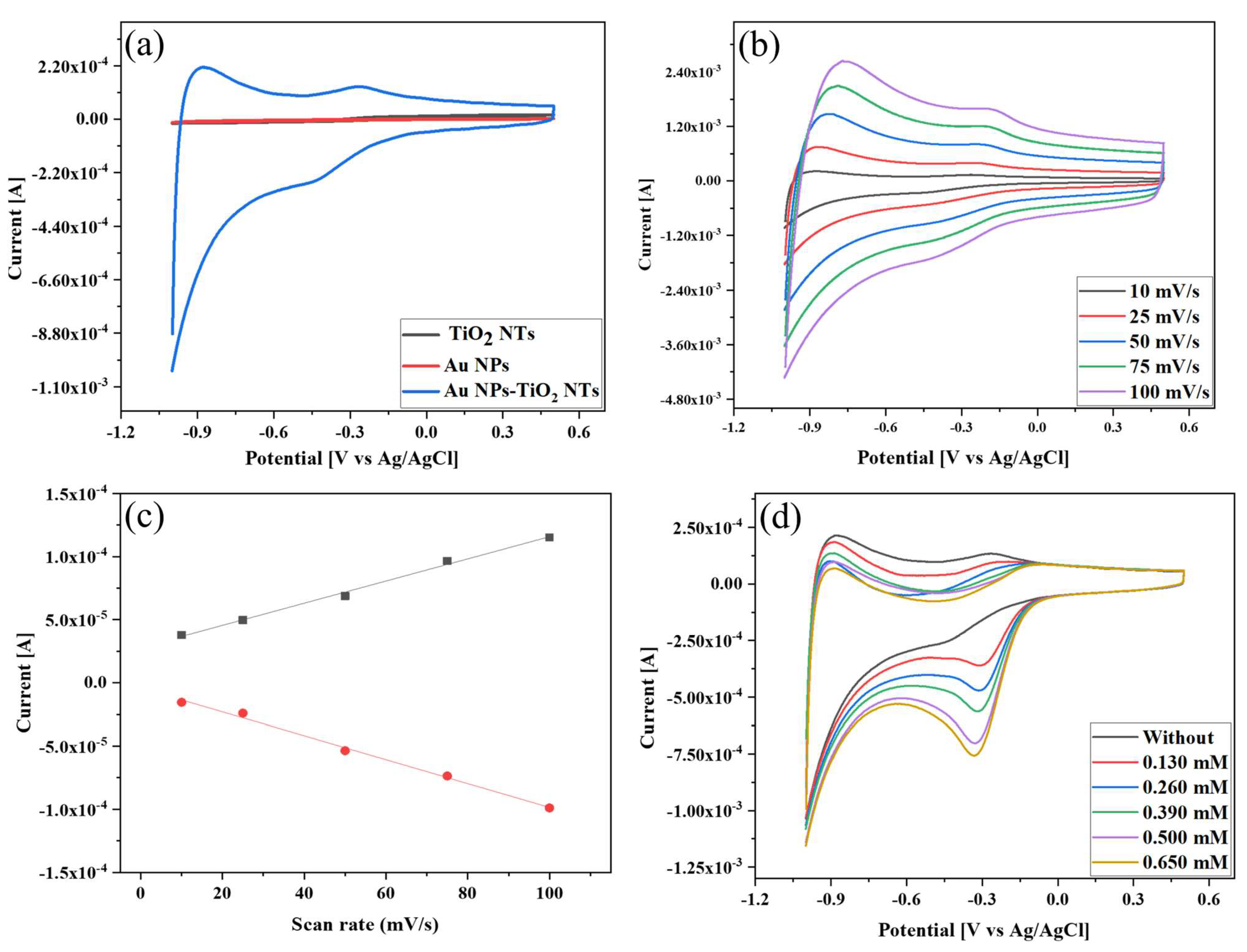
3.3. Electrochemical Properties of the Au NPs-TiO2 NTs Composite Electrode

Figure 4a illustrates the CV of TiO2 NTs, Au NPs, and Au NPs-TiO2 NTs electrodes in the potential range of −1.0 V to 0.5 V in 0.1 M PBS (pH 7.0) without H2O2 at a scan rate of 10 mV/s. The Au NPs-TiO2 NTs composite electrode exhibited a distinctly enhanced redox peak in comparison with the TiO2 NTs and Au NPs electrode. This redox peak indicated the increased electroactive active area and fast electron-transfer behavior of the Au NPs-TiO2 NTs composite. This redox peak was formed due to the formation of gold oxide during the forward scan and the subsequent reduction of gold oxide during the reverse scan [39,44]. Alongside this, the reaction kinetics of the composite electrode was investigated by recording the CV responses in 0.1 M PBS (pH 7.0) without H2O2 at different scan rates, as shown in Figure 4b. It was observed that the oxidation and reduction peak currents increased with increasing scan rate from 10 to 100 mV/s. Figure 4c shows that the anodic and cathodic peak current increase was linear with the scan rate, indicating that the electrochemical reaction was a surface-controlled process.

3.4. Electrochemical H2O2 Sensing of Au NPs-TiO2 NTs Composite Electrode

The electrocatalytic activity of Au NPs-TiO2 NTs composite electrode toward H2O2 was examined by adding 0 to 0.650 mM of H2O2 in 0.1 M PBS (pH 7.0) at a scan rate of 10 mV/s via CV, as shown in Figure 4d. With the continuous increase in the concentration, the reduction peak current gradually increased, and the reduction potential shifted toward negative. This negative shift and the broadening in peak potential with increasing H2O2 were consistent with a previous report [45]. This top-notch sensing behavior can be attributed to the porous structure of TiO2 NTs, which provided a large surface area effective for dispersing or stabilizing Au NPs. The detection mechanism can be expressed as follows [46]:
H 2 O 2 + e + Au Au OH ads + OH
Au OH   + e Au   +   OH
2 OH + 2 H +   2 H 2 O

3.5. Identification of Suitable Experimental Conditions

Selecting suitable working environments such as an optimal electrolyte solution, pH, buffer concentration, and reduction potential is a prerequisite for sensor development. Figure 5a presents the CV curves of Au NPs-TiO2 NTs composite electrode in the presence of 0.5 mM of H2O2 in different electrolyte media at 10 mV/s. All CV curves exhibited a single peak due to H2O2 reduction, where the maximum current was achieved in 0.1 M PBS. Hence, 0.1 M PBS was selected, and CV was subsequently run to identify the appropriate pH of 0.1 M PBS. Figure 5b demonstrates that the reduction peak current started to rise with increasing pH from 6.0 to 7.0, after which the reduction current did not increase with increasing pH. The highest reduction peak current was found at pH 7. Thus, pH 7 was selected for this study. Here, the H2O2 reduction was a pH-dependent reaction because the peak potential shifted to negative as the pH increased. Using this relationship, a potential vs. linear pH graph showed slope and R2 values of 71 mV and 0.98444, respectively (Figure 5c). Since the computed slope value was near the predicted Nernst value, the reduction of H2O2 was a two-electron two-proton reaction [47,48]. In addition, CV was also performed to identify the appropriate PBS concentration in the presence of 0.5 mM H2O2 at a scan rate of 10 mV/s. Figure 5d shows that 0.1 M PBS gave the maximum reduction current toward H2O2 compared with other concentrations. Hence, 0.1M PBS was considered.
Identifying the amperometric reduction potential is crucial because the appropriate potential affects sensor performances. Multi-step chronoamperometry was performed to determine the reduction potential by adding 60 μM H2O2 to 0.1 M PBS (pH 7.0). Figure 5e shows that all six potentials responded to the addition of H2O2 during the analysis. The rate of current change was low (below −0.35 V), and when it was increased further to −0.36 V and −0.38 V, it still responded less than −0.35 V. Therefore, −0.35 V was selected as the working potential.

3.6. Amperometric Detection of H2O2 on Au NPs-TiO2 NTs Composite Electrode

The detection sensitivity of the Au NPs-TiO2 NTs composite electrode was studied using multi-step chronoamperometry by adding H2O2 to 0.1 M PBS at −0.35 V under continuous stirring.
Figure 6a displays the amperometric current-time (I-t) curves from 1 µM to 5.5 mM of H2O2, while the inset displayed curves from 1 to 200 µM. The composite electrode achieved a steady current (95%) within 1.55 s after injection of H2O2. This speedy response was due to the active role played by the small-sized Au NPs on the electrode surface, which had tiny conduction centers [49].
The corresponding current vs. concentration calibration plots are displayed in Figure 6b,c, where all added H2O2 was linear with the current changes. Four linear ranges were obtained from the calibration plot due to different H2O2 adsorption and alteration in the electrocatalytic reduction kinetics of H2O2 on the electrode surface. At low concentrations, the rate-determining step of H2O2 reduction was dominated by H2O2 adsorption, while at high concentrations, H2O2 activation was the dominant determinant. In the middle area, the H2O2 reduction kinetics was simultaneously mediated by adsorption and activation [50,51]. The fitting curve in Figure 6b exhibits two linear ranges from 1 μM to 9.97806 μM (R2 = 0.99726) and from 19.93 μM to 198.47 μM (R2 = 0.99527). Another fitting curve is shown in Figure 6c, which exhibits two linear ranges from 297.29 μM to 987.89 μM (R2 = 0.93362) and from 1.48 mM to 5.413 mM (R2 = 0.97655). In addition, the sensitivity calculated from the linear curve was found to be 519.38 µA/mM. The limit of detection (LOD) of the sensor was estimated using the standard deviation of blank [52]:
L O D = 3 s b
where b is the calibration curve slope and s is the standard deviation of blank current. The detection limit was calculated to be 104.4 nM. The analytical performance of the developed H2O2 sensor was superior to or comparable with many previously advanced catalysts and even HRP and Hb-based sensors (Table 1).

3.7. Selectivity, Reproducibility, and Repeatability Study

The effects of intrusive compounds such as ascorbic acid, glucose, uric acid, NaNO3, KCl, ethanol, and acetic acid were studied through the amperometric I-t curve technique (Figure 7a). There was a sharp increase in the reduction current after the addition of 10 µM H2O2. However, after injecting 180 µM of the interfering compounds, no apparent change in the current response was observed that could affect the performance of the sensor. These results indicated the high selectivity of the Au NPs-TiO2 NTs composite electrode.
The reproducibility and repeatability of the Au NPs-TiO2 NTs composite electrode were explored by CV in the presence of 0.5 mM of H2O2 in 0.1 M PBS (pH 7.0) at 10 mV/s. The CV of four electrodes prepared under the same conditions (Figure 7b) exhibited almost the same reduction current response with a relative standard deviation (RSD) of 1.97%. The repeatability was explored in two different ways to assess the quality of the sensors. Figure 7c displays the CV curve of ten uninterrupted cycles with a slight fluctuation in the current response, where the RSD value was found to be 2.28%. Further, the repeatability of the electrode was performed in seven successive measurements at different times over 2 days. Figure 7d shows the RSD to be less than 1.51%.

3.8. Stability of the Electrode

To investigate the stability of the Au NPs-TiO2 NTs composite electrode, it was stored at room temperature, and CV was performed with 1 mM H2O2 in 0.1 M PBS (pH 7.0) at 10 mV/s. The developed electrode retained 96.4% of its initial current response for H2O2 up to 61 days with an RSD of 3.47%, as shown in Figure 8.

3.9. Real Sample Analysis

The H2O2-sensing performance of the Au NPs-TiO2 NTs composite electrode was evaluated using tap water, milk, and bacteria through multi-step chronoamperometry. During analysis, 10 µM H2O2 was injected every 50 s into the real samples containing 0.1 M PBS. The results are presented in Figure 9a, and Table 2 shows good recovery of H2O2, ranging from 109.72% to 100.62%.
The H2O2 sensing results in milk are shown in Figure 9b and Table 3. The sensor displayed perfect consistency during detection, where the recovery was from 98.33% to 111.15%. The concentration of H2O2 used in milk was lower than the H2O2 limit (14.7 μM) set by the US Food and Drug Administration (FDA) for food packaging [69].
The sensing performance on a real sample of Au NPs-TiO2 NTs composite electrode was also evaluated using L. plantarum bacteria from two different sources, namely tapai and pickle. Figure 9c displays the H2O2 sensing results in L. plantarum from tapai. The data tabulated in Table 4 demonstrate a satisfactory recovery in the range of 96.20% to 113.83%. Figure 9d displays the H2O2-sensing performance of the sensor on L. plantarum obtained from pickles, where the recovery range was from 95.10% to 111.63% (Table 5).
The current response did not fluctuate much after the addition of the real samples, indicating that the conductivity and resistance of the electrode did not change much after the addition of the real samples. The analysis of four different samples exhibited almost equivalent recovery percentages, thus suggesting the sensor’s versatility. Finally, it can be said that TiO2 NTs support efficiently facilitated the electron transfer of Au NPs and retained the catalytic activity for an extended period, resulting in good sensing performances. The comparison of experimental results (Table 1) and efficient practicality for detecting H2O2 in real samples suggested their potential use for food quality monitoring.

4. Conclusions

In summary, a porous TiO2 NTs-supported Au NPs-based nonenzymatic H2O2 sensor was developed via a simple drop-casting method. The aggregation of small-sized Au NPs was prevented by trapping them in porous TiO2 NTs, which played a key role in accelerating the detection sensitivity and stability of the sensor. Consequently, the developed sensor exhibited higher sensitivity, selectivity, stability, wide linearity, and nanomolar LOD over their enzymatic counterparts. Furthermore, the satisfactory recovery of H2O2 in tap water, milk, and L. plantarum bacteria by this Au NPs- TiO2 NTs composite sensor indicated its potential as a nonenzymatic sensor.

Author Contributions

Conceptualization, A.K.M.K. and N.S.A.; methodology, N.S.A. and M.A.K.; validation, N.S.A., M.S.H. and A.K.M.K.; formal analysis, M.A.K.; investigation, M.A.K.; resources, N.S.A.; data curation, M.A.K.; writing—original draft preparation, M.A.K.; writing—review and editing, M.S.H., N.S.A., A.K.M.K., K.W.G. and R.J.; visualization, M.A.K.; supervision, N.S.A.; project administration, N.S.A. and A.K.M.K.; funding acquisition, N.S.A., K.W.G. and M.S.H. All authors have read and agreed to the published version of the manuscript.

Funding

The authors would like to thank the Ministry of Higher Education for providing financial support under the Fundamental Research Grant Scheme (FRGS) No. FRGS/1/2019/STG05/UMP/02/8 (university reference RDU1901189) and Universiti Malaysia Pahang for laboratory facilities as well as additional financial support under the Postgraduate Research Grant Scheme PGRS 2003114.

Institutional Review Board Statement

Not applicable.

Informed Consent Statement

Not applicable.

Data Availability Statement

Not applicable.

Acknowledgments

Authors are highly thankful to Glycobio International Sdn. Bhd. for providing bacteria.

Conflicts of Interest

The authors declare no conflict of interest. The funders had no role in the design of the study; in the collection, analyses, or interpretation of data; in the writing of the manuscript; or in the decision to publish the results.

References

  1. Lima, L.S.; Rossini, E.L.; Pezza, L.; Pezza, H.R. Bioactive paper platform for detection of hydrogen peroxide in milk. J. Spec-Trochimica Acta Part A Mol. Biomol. Spectrosc. 2020, 227, 117774. [Google Scholar] [CrossRef] [PubMed]
  2. Lismont, C.; Revenco, I.; Fransen, M. Peroxisomal Hydrogen Peroxide Metabolism and Signaling in Health and Disease. Int. J. Mol. Sci. 2019, 20, 3673. [Google Scholar] [CrossRef] [PubMed] [Green Version]
  3. Mollarasouli, F.; Kurbanoglu, S.; Asadpour-Zeynali, K.; Ozkan, S.A. Non-enzymatic monitoring of hydrogen peroxide using novel nanosensor based on CoFe2O4@CdSeQD magnetic nanocomposite and rifampicin mediator. Anal. Bioanal. Chem. 2020, 412, 5053–5065. [Google Scholar] [CrossRef] [PubMed]
  4. Karimi, A.; Husain, S.W.; Hosseini, M.; Azar, P.A.; Ganjali, M.R. Rapid and sensitive detection of hydrogen peroxide in milk by Enzyme-free electrochemiluminescence sensor based on a polypyrrole-cerium oxide nanocomposite. Sens. Actuators B Chem. 2018, 271, 90–96. [Google Scholar] [CrossRef]
  5. Veal, E.A.; Day, A.M.; Morgan, B.A. Hydrogen Peroxide Sensing and Signaling. Mol. Cell 2007, 26, 1–14. [Google Scholar] [CrossRef] [PubMed]
  6. Arena, M.P.; Silvain, A.; Normanno, G.; Grieco, F.; Drider, D.; Spano, G.; Fiocco, D. Use of Lactobacillus plantarum Strains as a Bio-Control Strategy against Food-Borne Pathogenic Microorganisms. Front. Microbiol. 2016, 7, 464. [Google Scholar] [CrossRef] [Green Version]
  7. Panieri, E.; Gogvadze, V.; Norberg, E.; Venkatesh, R.; Orrenius, S.; Zhivotovsky, B. Reactive oxygen species generated in different compartments induce cell death, survival, or senescence. Free Radic. Biol. Med. 2013, 57, 176–187. [Google Scholar] [CrossRef]
  8. Chen, S.; Yuan, R.; Chai, Y.; Hu, F. Electrochemical sensing of hydrogen peroxide using metal nanoparticles: A review. Microchim. Acta 2012, 180, 15–32. [Google Scholar] [CrossRef]
  9. Zhong, Y.; Liu, M.-M.; Chen, Y.; Yang, Y.-J.; Wu, L.-N.; Bai, F.-Q.; Lei, Y.; Gao, F.; Liu, A.-L. A high-performance amperometric sensor based on a monodisperse Pt–Au bimetallic nanoporous electrode for determination of hydrogen peroxide released from living cells. Mikrochim. Acta 2020, 187, 1–9. [Google Scholar] [CrossRef]
  10. Ahammad, A.J.S. Hydrogen Peroxide Biosensors Based on Horseradish Peroxidase and Hemoglobin. J. Biosens. Bioelectron. 2012, s9, 1–11. [Google Scholar] [CrossRef] [Green Version]
  11. Achraf Ben Njima, M.; Legrand, L. Ag nanoparticles-oxidized green rust nanohybrids for novel and efficient non-enzymatic H2O2 electrochemical sensor. J. Electroanal. Chem. 2022, 906, 116015. [Google Scholar] [CrossRef]
  12. González-Sánchez, M.; Rubio-Retama, J.; López-Cabarcos, E.; Valero, E. Development of an acetaminophen amperometric biosensor based on peroxidase entrapped in polyacrylamide microgels. Biosens. Bioelectron. 2011, 26, 1883–1889. [Google Scholar] [CrossRef] [PubMed]
  13. Dhara, K.; Mahapatra, D.R. Recent advances in electrochemical nonenzymatic hydrogen peroxide sensors based on nanomaterials: A review. J. Mater. Sci. 2019, 54, 12319–12357. [Google Scholar] [CrossRef]
  14. Jackman, J.A.; Cho, N.; Nishikawa, M.; Yoshikawa, G.; Mori, T.; Shrestha, L.K.; Ariga, K. Materials Nanoarchitectonics for Mechanical Tools in Chemical and Biological Sensing. Chem. Asian J. 2018, 13, 3366–3377. [Google Scholar] [CrossRef] [PubMed]
  15. Pawar, D.; Kale, S.N. Correction to: A review on nanomaterial-modified optical fiber sensors for gases, vapors and ions. Mikrochim. Acta 2019, 186, 292. [Google Scholar] [CrossRef] [PubMed] [Green Version]
  16. Trujillo, R.; Barraza, D.; Zamora, M.; Cattani-Scholz, A.; Madrid, R. Nanostructures in Hydrogen Peroxide Sensing. Sensors 2021, 21, 2204. [Google Scholar] [CrossRef] [PubMed]
  17. Lou-Franco, J.; Das, B.; Elliott, C.; Cao, C. Gold Nanozymes: From Concept to Biomedical Applications. Nano-Micro Lett. 2020, 13, 1–36. [Google Scholar] [CrossRef]
  18. Gugoasa, L.A.D.; Pogacean, F.; Kurbanoglu, S.; Tudoran, L.; Serban, A.B.; Kacso, I.; Pruneanu, S. Graphene-gold nanoparticles nanozyme-based electrochemical sensor with en-hanced laccase-like activity for determination of phenolic substrates. J. Electrochem. Soc. 2021, 168, 067523. [Google Scholar] [CrossRef]
  19. Shu, Y.; Li, Z.; Yang, Y.; Tan, J.; Liu, Z.; Shi, Y.; Ye, C.; Gao, Q. Isolated Cobalt Atoms on N-Doped Carbon as Nanozymes for Hydrogen Peroxide and Dopamine Detection. ACS Appl. Nano Mater. 2021, 4, 7954–7962. [Google Scholar] [CrossRef]
  20. Zhang, G. Functional gold nanoparticles for sensing applications. Nanotechnol. Rev. 2013, 2, 269–288. [Google Scholar] [CrossRef]
  21. Zhou, Y.; Ping, T.; Maitlo, I.; Wang, B.; Akram, M.Y.; Nie1, J.; Zhu, X. Regional selective construction of nano-Au on Fe3O4@SiO2@PEI nanoparticles by photoreduction. Nanotechnology 2016, 27, 215301. [Google Scholar] [CrossRef] [PubMed]
  22. Wang, Q.; Zhang, X.; Chai, X.; Wang, T.; Cao, T.; Li, Y.; Zhang, L.; Fan, F.; Fu, Y.; Qi, W. An electrochemical sensor for H2O2 based on Au nanoparticles embedded in UiO-66 metal–organicframework films. ACS Appl. Nano Mater. 2021, 4, 6103–6110. [Google Scholar] [CrossRef]
  23. Huang, A.; He, Y.; Zhou, Y.; Zhou, Y.; Yang, Y.; Zhang, J.; Luo, L.; Mao, Q.; Hou, D.; Yang, J. A review of recent applications of porous metals and metal oxide in energy storage, sensing and catalysis. J. Mater. Sci. 2018, 54, 949–973. [Google Scholar] [CrossRef]
  24. Hassan, I.U.; Salim, H.; Naikoo, G.A.; Awan, T.; Dar, R.A.; Arshad, F.; Tabidi, M.A.; Das, R.; Ahmed, W.; Asiri, A.M.; et al. A review on recent advances in hierarchically porous metal and metal oxide nanostructures as electrode materials for supercapacitors and non-enzymatic glucose sensors. J. Saudi Chem. Soc. 2021, 25, 101228. [Google Scholar] [CrossRef]
  25. Li, Y.; Wang, S.; Dong, Y.; Mu, P.; Yang, Y.; Liu, X.; Lin, C.; Huang, Q. Effect of size and crystalline phase of TiO2 nanotubes on cell behaviors: A high throughput study using gradient TiO2 nanotubes. Bioact. Mater. 2020, 5, 1062–1070. [Google Scholar] [CrossRef]
  26. Durdu, S.; Cihan, G.; Yalcin, E.; Altinkok, A. Characterization and mechanical properties of TiO2 nanotubes formed on titanium by anodic oxidation. Ceram. Int. 2020, 47, 10972–10979. [Google Scholar] [CrossRef]
  27. Tremel, W. Inorganic nanotubes. Angew. Chem. Int. Ed. 1999, 38, 2175–2179. [Google Scholar] [CrossRef]
  28. Fraoucene, H.; Sugiawati, V.A.; Hatem, D.; Belkaid, M.S.; Vacandio, F.; Eyraud, M.; Pasquinelli, M.; Djenizian, T. Optical and Electrochemical Properties of Self-Organized TiO2 Nanotube Arrays From Anodized Ti−6Al−4V Alloy. Front. Chem. 2019, 7, 66. [Google Scholar] [CrossRef] [Green Version]
  29. Yang, X.; Wang, W.; Wu, L.; Li, X.; Wang, T.; Liao, S. Effect of confinement of TiO2 nanotubes over the Ru nanoparticles on Fischer-Tropsch syn-thesis. Appl. Catal. A: Gen. 2016, 526, 45–52. [Google Scholar] [CrossRef]
  30. Yang, X.; Wu, L.; Ma, L.; Li, X.; Wang, T.; Liao, S. Pd nano-particles (NPs) confined in titanate nanotubes (TNTs) for hydrogenation of cinnamal-dehyde. Catal. Commun. 2015, 59, 184–188. [Google Scholar] [CrossRef]
  31. Chen, X.; Wu, N.; Zhang, G.; Feng, S.; Xu, K.; Liu, W.; Pan, H. Functionalized TiO2 nanotubes as three-dimensional support for loading Au@ Pd nanopar-ticles: Facile preparation and enhanced materials for electrochemical sensor. Int. J. Electrochem. Sci. 2017, 12, 593–609. [Google Scholar] [CrossRef]
  32. Pisarek, M.; Kędzierzawski, P.; Andrzejczuk, M.; Hołdyński, M.; Mikołajczuk-Zychora, A.; Borodziński, A.; Janik-Czachor, M. TiO2 nanotubes with Pt and Pd nanoparticles as catalysts for electro-oxidation of formic acid. Materials 2020, 13, 1195. [Google Scholar] [CrossRef] [PubMed] [Green Version]
  33. Rizwan, M.; Elma, S.; Lim, S.A.; Ahmed, M.U. AuNPs/CNOs/SWCNTs/chitosan-nanocomposite modified electrochemical sensor for the label-free detection of carcinoembryonic antigen. Biosens. Bioelectron. 2018, 107, 211–217. [Google Scholar] [CrossRef] [PubMed]
  34. Wang, R.; Di, J.; Ma, J.; Ma, Z. Highly sensitive detection of cancer cells by electrochemical impedance spectroscopy. Electrochim. Acta 2012, 61, 179–184. [Google Scholar] [CrossRef]
  35. Kafi, A.; Wu, G.; Benvenuto, P.; Chen, A. Highly sensitive amperometric H2O2 biosensor based on hemoglobin modified TiO2 nanotubes. J. Electroanal. Chem. 2011, 662, 64–69. [Google Scholar] [CrossRef]
  36. Tian, M.; Adams, B.; Wen, J.; Asmussen, R.M.; Chen, A. Photoelectrochemical oxidation of salicylic acid and salicylaldehyde on titanium dioxide nanotube arrays. Electrochimica Acta 2009, 54, 3799–3805. [Google Scholar] [CrossRef]
  37. Suchomel, P.; Kvitek, L.; Prucek, R.; Panacek, A.; Halder, A.; Vajda, S.; Zboril, R. Simple size-controlled synthesis of Au nanoparticles and their size-dependent catalytic activity. Sci. Rep. 2018, 8, 4589. [Google Scholar] [CrossRef] [Green Version]
  38. Khatun, F.; Aziz, A.A.; Sim, L.C.; Monir, M.U. Plasmonic enhanced Au decorated TiO2 nanotube arrays as a visible light active catalyst towards photocatalytic CO2 conversion to CH4. J. Environ. Chem. Eng. 2019, 7, 103233. [Google Scholar] [CrossRef]
  39. Mers, S.S.; Kumar, E.T.; Ganesh, V. Gold nanoparticles-immobilized, hierarchically ordered, porous TiO2 nanotubes for biosensing of glutathione. Int. J. Nanomed. 2015, 10, 171–182. [Google Scholar]
  40. Hosseini, S.G.; Safshekan, S. Electrochemical detection of chlorate on a novel nano-Au/TiO2NT electrode. Mater. Res. Bull. 2017, 93, 290–295. [Google Scholar] [CrossRef]
  41. Jin, W.; Wu, G.; Chen, A. Sensitive and selective electrochemical detection of chromium(vi) based on gold nanoparticle-decorated titania nanotube arrays. Analyst 2014, 139, 235–241. [Google Scholar] [CrossRef] [PubMed]
  42. Bagheri, S.; Julkapli, N.M.; Hamid, S.B.A. Titanium Dioxide as a Catalyst Support in Heterogeneous Catalysis. Sci. World J. 2014, 2014, 1–21. [Google Scholar] [CrossRef] [PubMed] [Green Version]
  43. Wan, G.; Peng, X.; Zeng, M.; Yu, L.; Wang, K.; Li, X.; Wang, G. The preparation of Au@TiO2 yolk-shell nanostructure and its applications for degradation and detection of methylene blue. Nanoscale Res. Lett. 2017, 12, 535. [Google Scholar] [CrossRef] [PubMed] [Green Version]
  44. Chiu, W.-T.; Chang, T.-F.M.; Sone, M.; Tixier-Mita, A.; Toshiyoshi, H. Electrocatalytic activity enhancement of Au NPs-TiO2 electrode via a facile redis-tribution process towards the non-enzymatic glucose sensors. Sens. Actuators B Chem. 2020, 319, 128279. [Google Scholar] [CrossRef]
  45. Mathivanan, D.; Shalini Devi, K.S.; Sathiyan, G.; Tyagi, A.; da Silva, V.A.O.P.; Janegitz, B.C.; Prakash, J.; Gupta, R.K. Novel polypyrrole-graphene oxide-gold nanocomposite for high performance hydrogen peroxide sensing application. Sens. Actuators A Phys. 2021, 328, 112769. [Google Scholar] [CrossRef]
  46. Yuan, Y.; Zheng, Y.; Liu, J.; Wang, H.; Hou, S. Non-enzymatic amperometric hydrogen peroxide sensor using a glassy carbon electrode modified with gold nanoparticles deposited on CVD-grown graphene. Microchim. Acta 2017, 184, 4723–4729. [Google Scholar] [CrossRef]
  47. Qi, C.; Kan, Z.; Zhang, D.; Tang, J.; Ren, Z.; Jia, X.; Li, C.; Wang, F. Poly(1,3,5-tris(4-ethynylphenyl)-benzene) Conjugated Polymers as Electrochemical Sensors for Hydrogen Peroxide Detection. ACS Appl. Polym. Mater. 2020, 2, 685–690. [Google Scholar] [CrossRef]
  48. Simioni, N.B.; Silva, T.A.; Oliveira, G.G.; Fatibello-Filho, O. A nanodiamond-based electrochemical sensor for the determination of pyra-zinamide antibiotic. Sens. Actuators B Chem. 2017, 250, 315–323. [Google Scholar] [CrossRef]
  49. Bharathi, S.; Nogami, M.; Ikeda, S. Novel electrochemical interfaces with a tunable kinetic barrier by self-assembling organically modified silica gel and gold nanoparticles. Langmuir 2001, 17, 1–4. [Google Scholar] [CrossRef]
  50. Lu, J.; Hu, Y.; Wang, P.; Liu, P.; Chen, Z.; Sun, D. Electrochemical biosensor based on gold nanoflowers-encapsulated magnetic metal-organic framework nanozymes for drug evaluation with in-situ monitoring of H2O2 released from H9C2 cardiac cells. Sens. Actuators B Chem. 2020, 311, 127909. [Google Scholar] [CrossRef]
  51. Yin, D.; Tang, J.; Bai, R.; Yin, S.; Jiang, M.; Kan, Z.; Li, H.; Wang, F.; Li, C. Cobalt Phosphide (Co2P) with Notable Electrocatalytic Activity Designed for Sensitive and Selective Enzymeless Bioanalysis of Hydrogen Peroxide. Nanoscale Res. Lett. 2021, 16, 1–10. [Google Scholar] [CrossRef] [PubMed]
  52. Elewi, A.S.; Al-Shammaree, S.A.W.; Al Sammarraie, A.K.M.A. Hydrogen peroxide biosensor based on hemoglobin-modified gold nanoparticles–screen printed carbon electrode. Sens. Bio-Sens. Res. 2020, 28, 100340. [Google Scholar] [CrossRef]
  53. Huo, D.; Li, D.; Xu, S.; Tang, Y.; Xie, X.; Li, D.; Song, F.; Zhang, Y.; Li, A.; Sun, L. Disposable stainless-steel wire-based electrochemical microsensor for in vivo continuous monitoring of hydrogen peroxide in vein of tomato leaf. Biosensors 2022, 12, 35. [Google Scholar] [CrossRef] [PubMed]
  54. Garate, O.; Veiga, L.S.; Tancredi, P.; Medrano, A.V.; Monsalve, L.N.; Ybarra, G. High-performance non-enzymatic hydrogen peroxide electrochemical sensor prepared with a magnetite-loaded carbon nanotube waterborne ink. J. Electroanal. Chem. 2022, 915, 116372. [Google Scholar] [CrossRef]
  55. Rashed, M.; Ahmed, J.; Faisal, M.; Alsareii, S.; Jalalah, M.; Tirth, V.; Harraz, F.A. Surface modification of CuO nanoparticles with conducting polythiophene as a non-enzymatic amperometric sensor for sensitive and selective determination of hydrogen peroxide. Surf. Interfaces 2022, 31, 101998. [Google Scholar] [CrossRef]
  56. Subramanian, B.T.; Thomas, S.; Gumpu, M.B.; Biju, V.M.N. Aromatic carboxylic acid derived bimetallic nickel/cobalt electrocatalysts for oxygen evolution reaction and hydrogen peroxide sensing applications. J. Electroanal. Chem. 2022, 925, 116904. [Google Scholar] [CrossRef]
  57. Saeed, A.A.; Abbas, M.N.; El-Hawary, W.F.; Issa, Y.M.; Singh, B. A core-shell Au@TiO2 and multi-walled carbon nanotube-based sensor for the electroanalytical determination of H2O2 in human blood serum and saliva. Biosensors 2022, 12, 778. [Google Scholar] [CrossRef]
  58. Liu, D.; Cao, W.; Li, F.; Ding, Y.; Fan, B. Green facile synthesis of biowaste-converted Cu-Cu2O/BPC for non-enzymatic hydrogen peroxide sensing. Diam. Relat. Mater. 2022, 130, 109458. [Google Scholar] [CrossRef]
  59. Liu, T.; Zhang, X.; Fu, K.; Zhou, N.; Xiong, J.; Su, Z. Fabrication of Co3O4/NiCo2O4 nanocomposite for detection of H2O2 and dopamine. Biosensors 2021, 11, 452. [Google Scholar] [CrossRef]
  60. Mazzotta, E.; Di Giulio, T.; Mastronardi, V.; Pompa, P.P.; Moglianetti, M.; Malitesta, C. Bare platinum nanoparticles deposited on glassy carbon electrodes for elec-trocatalytic detection of hydrogen peroxide. ACS Appl. Nano Mater. 2021, 4, 7650–7662. [Google Scholar] [CrossRef]
  61. Tomassetti, M.; Leonardi, C.; Pezzilli, R.; Prestopino, G.; di Natale, C.; Medaglia, P.G. New voltammetric sensor based on LDH and H2O2 for L-proline determination in red and white wines. Crystals 2022, 12, 1474. [Google Scholar] [CrossRef]
  62. Pan, C.; Zheng, Y.; Yang, J.; Lou, D.; Li, J.; Sun, Y.; Liu, W. Pt–Pd Bimetallic Aerogel as High-Performance Electrocatalyst for Nonenzymatic Detection of Hydrogen Peroxide. Catalysts 2022, 12, 528. [Google Scholar] [CrossRef]
  63. Wu, B.; Yeasmin, S.; Liu, Y.; Cheng, L.-J. Ferrocene-grafted carbon nanotubes for sensitive non-enzymatic electrochemical detection of hydrogen peroxide. J. Electroanal. Chem. 2022, 908, 116101. [Google Scholar] [CrossRef] [PubMed]
  64. Haritha, V.; Kumar, S.S.; Rakhi, R. Non-enzymatic electrocatalytic detection of hydrogen peroxide using Tungsten disulphide nanosheets modified electrodes. Mater. Sci. Eng. B 2022, 285, 115932. [Google Scholar] [CrossRef]
  65. Thi, M.; Pham, V.; Bui, Q.; Ai-Le, P.; Nhac-Vu, H.-T. Novel nanohybrid of blackberry-like gold structures deposited graphene as a free-standing sensor for effective hydrogen peroxide detection. J. Solid State Chem. 2020, 286, 121299. [Google Scholar] [CrossRef]
  66. Ngamaroonchote, A.; Sanguansap, Y.; Wutikhun, T.; Karn-Orachai, K. Highly branched gold–copper nanostructures for non-enzymatic specific detection of glucose and hydrogen peroxide. Microchim. Acta 2020, 187, 559. [Google Scholar] [CrossRef] [PubMed]
  67. Dang, W.; Sun, Y.; Jiao, H.; Xu, L.; Lin, M. AuNPs-NH2/Cu-MOF modified glassy carbon electrode as enzyme-free electrochemical sensor detecting H2O2. J. Electroanal. Chem. 2020, 856, 113592. [Google Scholar] [CrossRef]
  68. Sardaremelli, S.; Hasanzadeh, M.; Razmi, H. Chemical binding of horseradish peroxidase enzyme with poly beta-cyclodextrin and its application as molecularly imprinted polymer for the monitoring of H2O2 in human plasma samples. J. Mol. Recognit. 2021, 34, 2884. [Google Scholar] [CrossRef]
  69. Riaz, M.A.; Zhai, S.; Wei, L.; Zhou, Z.; Yuan, Z.; Wang, Y.; Huang, Q.; Liao, X.; Chen, Y. Ultralow-platinum-loading nanocarbon hybrids for highly sensitive hydrogen peroxide detection. Sens. Actuators B Chem. 2019, 283, 304–311. [Google Scholar] [CrossRef]
Figure 1. Micrographs of different microscopes show the morphology and structure of the composites of Au NPs-TiO2 NTs. TEM images of Au NPs (a) low (×29,000) and (b) high magnifications (×240,000); FESEM images of TiO2 nanotubes. (c) top view (×1000) and (d) magnified top view (×10,000); FESEM Images of Au NPs-TiO2 NTs (e) ×15,000 (White arrows shows Au NPs), and (f) ×50,000 magnifications (White arrow = TiO2, Black arrow = Au NPs).
Biosensors 13 00671 g001
Figure 2. Elemental mapping images of (a) Ti, (b) O, (c) Au, and (d) EDX spectrum of Au NPs-TiO2 NTs.
Biosensors 13 00671 g002
Figure 3. X-ray diffraction (XRD) spectrum of Au NPs-TiO2 NTs (up) along with anatase TiO 2, gold and Ti (below). DB: database.
Biosensors 13 00671 g003
Figure 4. (a) Cyclic voltammetry (CV) of different modified electrodes without H2O2 in 0.1 M PBS (pH = 7.0) at a scan rate of 10 mV/s; (b) CV of Au NPs-TiO2 NTs composite electrode without H2O2 in 0.1 M PBS (pH = 7.0) at different scan rates; (c) corresponding anodic (black) and cathodic peak current (red) versus scan rate; (d) CV of Au NPs-TiO2 NTs composite electrode with different H2O2 in 0.1 M PBS (pH = 7.0) at a scan rate of 10 mV/s.
Biosensors 13 00671 g004
Figure 5. CV-based identification of (a) suitable electrolyte media; (b) suitable pH; (c) corresponding reduction potential vs. pH curve; (d) suitable PBS (pH 7) concentration in the presence of 0.5 mM of H2O2 at a scan rate of 10 mV/s; (e) appropriate amperometric reduction potential by adding 60 µM of H2O2 in 0.1 M PBS (pH 7.0).
Biosensors 13 00671 g005
Figure 6. (a) Multi-step chronoamperometry of Au NPs-TiO2 NTs composite electrode to the successive addition of H2O2 in 0.1 M PBS (pH 7.0) at −0.35 V. Inset: the magnified view of low concentrations of H2O2; respective calibration curve of H2O2 concentration vs. current (b) at lower concentrations and (c) at higher concentrations.
Biosensors 13 00671 g006
Figure 7. (a) Selectivity study of Au NPs-TiO2 NTs composite electrode exposed to H2O2 and ascorbic acid, glucose, uric acid, NaNO3, KCl, ethanol, and acetic acid in 0.1 M PBS (pH 7.0) at E = −0.35 V; (b) CV-based reproducibility study; repeatability study; (c) 10 continuous cycles and (d) different measurement times in 0.1 M PBS at 10 mV/s containing 0.5 mM H2O2.
Biosensors 13 00671 g007
Figure 8. Stability study of Au NPs-TiO2 NTs composite electrode with 1 mM of H2O2 in 0.1 M PBS at 10 mV/s.
Biosensors 13 00671 g008
Figure 9. Amperometric responses of the electrode upon stepwise addition of 10 μM H2O2 in PBS (pH 7) containing (a) tap water, (b) milk, (c) L. plantarum from tapai, and (d) L. plantarum from pickle.
Biosensors 13 00671 g009
Table 1. Comparison of sensor performance.
Electrode MaterialsLinear RangeDetection LimitStability
(Days)
Ref.
Au NPs-TiO2 NTs composite1–198.47 μM *
297.29–5413 μM *
104.4 nM61Current study
NF-Hb-Cys-Au NPs-SPCE3–240 μM0.6 µM30[52]
Au nanodots/SS electrode10 µM–1000 µM3.97 μM7[53]
Fe3O4–MWCNT ink0.001–2 mM0.5 µM21[54]
Pth-CuO/GCE20–3300 μM3.86 μM15[55]
GCE/Ni-Co ABDC0–7 mM0.18 mM16[56]
Au@TiO2/MWCNTs/GCE5–200 µM and
200 µM–6 mM
1.4 μM50 [57]
Cu-Cu2O/BPC-11–2830 μM
2830–8330 μM
0.35 μM30[58]
Co3O4/NiCo2O40.05–41.7 mM0.2578 µM9[59]
4 nm PtNPs/GCE0.025–0.75 mM10 µM10[60]
GCE-Ag(paste)-LDH125–3200 μM85 µM5[61]
Pt50Pd50 aerogel5.1–3190 μM2.21 μM6[62]
MWCNTs-FeC/SPCEs1–1000 μM0.49 μM10[63]
WS2/GCE10–90 µM0.88 µM14[64]
Au NPs-CNTs/3DF1–296 μM1.06 μM21[65]
Au-Cu/SPCE0.05–10 mM10.93 μM28[66]
Au NPs-NH2/Cu-MOF/GCE5–850 μM1.2 μM7[67]
HRP/ß-CD/GCE1–15 μM0.4 μM15[68]
* Different H2O2 adsorption and alteration in the electrocatalytic reduction kinetics of H2O2 on the electrode surface. ABDC: 2-aminobenzene-1,4-dicarboxylic acid, BPC: biomass porous carbon, CD: cyclodextrin, CNTs: carbon nanotubes, Co3O4: cobalt(II,III) oxide, Cu: copper, Cu2O: copper(I) oxide, Cys: cysteamine, Fe3O4: Iron(II,III) oxide, GCE: glassy carbon electrode, Hb: hemoglobin, HRP: horseradish peroxidase, LDH: layered double hydroxide, MOF: metal organic framework, MWCNTs: multi-walled carbon nanotubes, Ni: Nickle, NiCo2O4: Nickel cobaltite, Pth: parathormone, SPCE: screen printed carbon electrode, SS: stainless steel, WS2: Tungsten disulphide.
Table 2. Recovery calculation of H2O2 in tap water.
Addition No.H2O2 Added (μM)H2O2 Found (μM)Recovery (%)
19.99610.968109.72
219.98820.984104.98
329.97630.887103.03
439.9640.211100.62
Table 3. Recovery calculation of H2O2 in full-cream milk.
Addition No.H2O2 Added (μM)H2O2 Found (μM)Recovery (%)
19.9969.82998.33
219.98820.742103.77
329.97633.321111.15
439.9641.93104.93
Table 4. Recovery calculation of H2O2 in L. plantarum bacteria from tapai.
Addition No.H2O2 Added (μM)H2O2 Found (μM)Recovery (%)
110.019.6396.20
220.0222.65113.13
330.0033.25110.83
439.9941.66104.17
Table 5. Recovery calculation of H2O2 in L. plantarum bacteria from pickle.
Addition No.H2O2 Added (μM)H2O2 Found (μM)Recovery (%)
110.019.5295.10
220.0222.08110.28
330.0033.49111.63
439.9942.91107.30
Disclaimer/Publisher’s Note: The statements, opinions and data contained in all publications are solely those of the individual author(s) and contributor(s) and not of MDPI and/or the editor(s). MDPI and/or the editor(s) disclaim responsibility for any injury to people or property resulting from any ideas, methods, instructions or products referred to in the content.

