Article

A Ratiometric Fluorescent Probe for Hypochlorite and Lipid Droplets to Monitor Oxidative Stress

1
Molecular Sensors and Therapeutics Research Laboratory, Department of Chemistry, School of Natural Sciences, Shiv Nadar (Institute of Eminence Deemed to be) University, Delhi 201314, NCR, India
2
Department of Chemistry, University of California, Riverside, CA 92521, USA
*
Author to whom correspondence should be addressed.
These authors contributed equally to this work.
Biosensors 2023, 13(6), 662; https://doi.org/10.3390/bios13060662
Submission received: 3 May 2023 / Revised: 13 June 2023 / Accepted: 14 June 2023 / Published: 17 June 2023

Abstract

:
Mitochondria are valuable subcellular organelles and play crucial roles in redox signaling in living cells. Substantial evidence proved that mitochondria are one of the critical sources of reactive oxygen species (ROS), and overproduction of ROS accompanies redox imbalance and cell immunity. Among ROS, hydrogen peroxide (H2O2) is the foremost redox regulator, which reacts with chloride ions in the presence of myeloperoxidase (MPO) to generate another biogenic redox molecule, hypochlorous acid (HOCl). These highly reactive ROS are the primary cause of damage to DNA (deoxyribonucleic acid), RNA (ribonucleic acid), and proteins, leading to various neuronal diseases and cell death. Cellular damage, related cell death, and oxidative stress are also associated with lysosomes which act as recycling units in the cytoplasm. Hence, simultaneous monitoring of multiple organelles using simple molecular probes is an exciting area of research that is yet to be explored. Significant evidence also suggests that oxidative stress induces the accumulation of lipid droplets in cells. Hence, monitoring redox biomolecules in mitochondria and lipid droplets in cells may give a new insight into cell damage, leading to cell death and related disease progressions. Herein, we developed simple hemicyanine-based small molecular probes with a boronic acid trigger. A fluorescent probe AB that could efficiently detect mitochondrial ROS, especially HOCl, and viscosity simultaneously. When the AB probe released phenylboronic acid after reacting with ROS, the product AB–OH exhibited ratiometric emissions depending on excitation. This AB–OH nicely translocates to lysosomes and efficiently monitors the lysosomal lipid droplets. Photoluminescence and confocal fluorescence imaging analysis suggest that AB and corresponding AB–OH molecules are potential chemical probes for studying oxidative stress.

1. Introduction

The eukaryotic cells have multiple subcellular organelles such as mitochondria, lysosomes, nuclei, and Golgi bodies which play a crucial role in diverse biological functions in living cells. Among them, mitochondria are considered one of the essential membrane containing cell organelles as they produce energy for the entire body through cellular respiration [1,2]. A substantial amount of reactive oxygen species (ROS) superoxide anion radical (O2) converts into hydrogen peroxide (H2O2) via the catalytic activity of superoxide dismutase (SOD) [3]. This H2O2, a common intermediate of ROS, transforms into several potent cytotoxic oxidizing agents and functional biomolecules, such as hypochlorous acid (HOCl), which are generated via a reaction between H2O2 and chloride ion (Cl) in the presence of myeloperoxidase (MPO) in the cytoplasm [4]. As HOCl is in equilibrium with its conjugate base OCl (hypochlorite anion) at physiological pH (7.4), it acts as a potent oxidative agent [5]. It also plays a crucial role in the immune system by destroying many pathogens and effecting a few cellular processes, such as proliferation, differentiation, and anti-inflammatory response [6]. However, for a long time, whether HOCl is generated from mitochondria apart from cytoplasm was unclear. Later, Ma et al. proved that HOCl is also produced in mitochondria in macrophage cells [7]. Recently, we demonstrated OCl generation through NOX2 pathways in cancer cells [8]. Overproduction of OCl leads to oxidative stress and changes the microviscosity in the cellular organelles [9,10]. Furthermore, the oxidation of mitochondrial fatty acid (FA) upregulates and increases FA accumulation in lipid droplets during oxidative stress [11]. In general, an increased amount of mitochondrial ROS is associated with several diseases, such as chronic kidney disease progression, cancer, diabetes, metabolic disorders, atherosclerosis, and cardiovascular diseases [12,13,14].
Small molecular fluorescent probes and corresponding super-resolution imaging are widely considered one of the most powerful techniques for revealing facts at the molecular level due to their simplicity, high resolution, and low cost [15]. It has become one of the most emerging diagnostic modalities in biomedical research and clinical applications [16,17,18]. Although several small molecular probes are reported to detect an individual analyte, either HOCl or lipid droplets, there are no systematic studies for dual analytes, including OCl and lipid droplets, during oxidative stress [19]. Therefore, there is an urgent need to develop an efficient probe for monitoring both HOCl and lipid droplets (LDs) during drug-induced oxidative stress conditions.
A ratiometric fluorescent probe can overcome a few drawbacks compared to turn on/off by self-calibration, and can emerge as an attractive tool in the bio-imaging field [20,21,22,23,24,25,26,27,28,29,30,31,32,33]. However, designing a ratiometric probe and simultaneously targeting two analytes is always challenging [34]. Herein, we developed an efficient mitochondrial targeting hypochlorite-specific small molecular fluorescent probe, AB, which can also monitor mitochondrial viscosity. Confocal fluorescence imaging suggests that the hemicyanine based AB probe is nicely localized in mitochondria but translocated into lysosomal lipid droplets when the mitochondrial membrane is disrupted during oxidative stress. The phenylboronic triggering motif in AB rapidly converts it into AB–OH in the presence of hypochlorite. Due to substantial intramolecular charge transfer (ICT) in the basic environment, AB–OH exhibited ratiometric emission change and localized to lipid droplets. It showed a good sensitivity towards lipid droplets, the hallmark of cellular stress during oxidative stress.

2. Materials and Methods

2.1. General Information

Unless otherwise stated, all the compounds were used as such as they were purchased from commercial vendors without any additional purification. All the substances used in the reactions were bought from Sigma-Aldrich, Alfa Aesar, T.C.I. India, Spectrochem, and Rankem chemicals. Materials used for the spectroscopic and biological analysis are of highest quality available in the brand. Deuterated NMR solvents were purchased from Eurisotope. Dulbecco’s modified Eagle’s medium (DMEM), 10% fetal bovine serum (FBS), and 100× Antimycotic antibiotics were purchased from Thermo Fisher. Silica gel G-60 F254 aluminum TLC was used to monitor reaction completion, and short- and long-wavelength UV lamp chamber was used to visualize the TLC Silica gel with a mesh size of 100–200 was used for column chromatography. Distilled MilliQ water was used wherever water was needed.

2.2. General Instrumentation

NMR spectra of the synthesized molecules were recorded in the Bruker Avance 400 NMR spectrometer, Germany, using deuterated solvents. All the mass spectra were recorded in Agilent 6540, Q-TOF LC/MS system (Agilent Technologies, Santa Clara, CA, USA) connected with Agilent 1290 UPLC using DI water and acetonitrile along with 0.1% TFA as mobile phase with a gradient solvent system. The analytes 4% sodium hypochlorite solution was purchased from Qualigens (cat no. 7681-52-9) and 50% Hydrogen Peroxide solution from Fisher Scientific (cat no. 7722-84-1). Using an Agilent Cary 8454 UV-Vis diode array spectrophotometer, all the absorption spectra were measured, whereas the emission spectra were measured in HORIBA Fluorolog-3 spectrofluorometer (Model: FL3-2-IHR). The scan slits for excitation and emission were adjusted to 2 nm. A Waters Alliance System (Milford, MA, USA) equipped with an e2695 separation module, and a 2998 photodiode-array detector was used for the HPLC experiments. The cells were incubated in a Thermo Fisher CO2 incubator, and imaging was performed with Nikon confocal microscope.

2.3. Synthesis of AB

0.343 mmol of 2,3-dimethylbenzo[d]thiazol-3-ium (B) and 0.343 mol of (4-((4-formylphenoxy)methyl)phenyl)boronic acid (C) was mixed in dried ethanol in the presence of a catalytic amount of piperidine. The mixture was allowed to stir for 6 h, and the precipitate was filtered and washed with cold diethyl ether to render a 78% yield. The product formed was characterized using HRMS and NMR Spectroscopy. (Detailed schemes for the synthesis and the characterizations are provided in the supplementary material).

2.4. Cell Culture Experiments
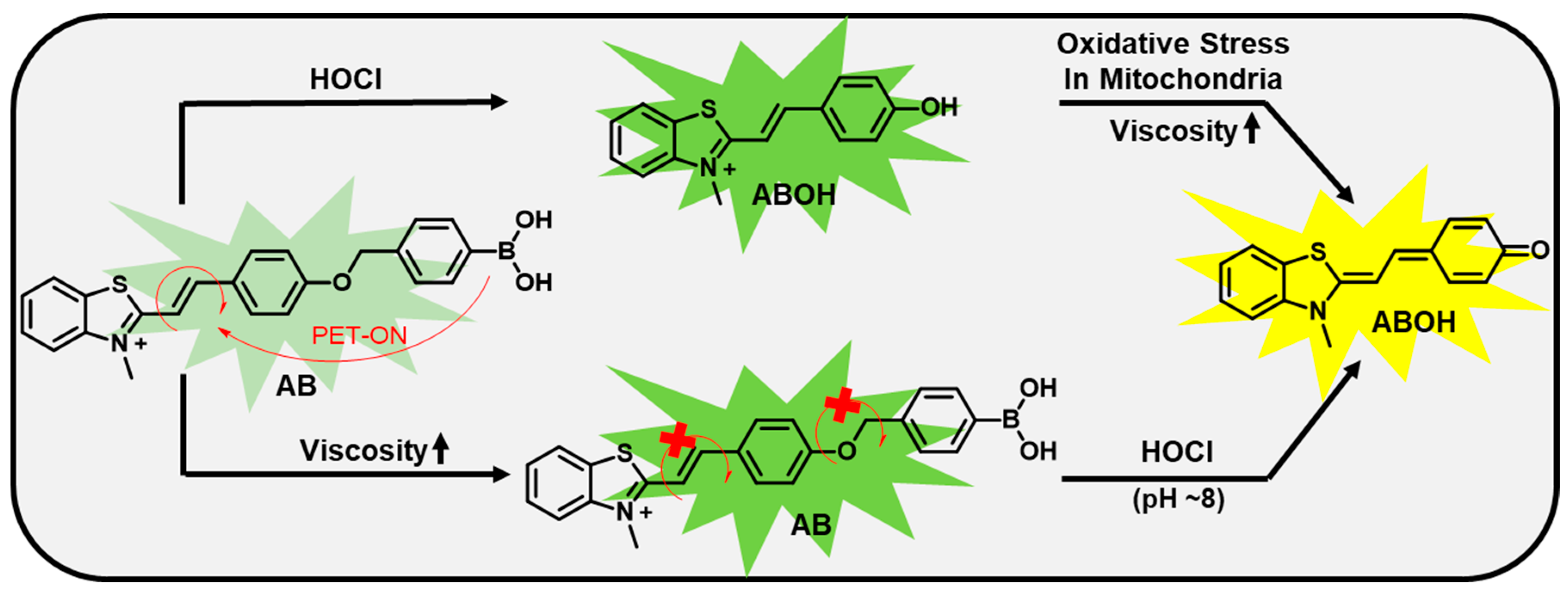
Adenocarcinoma human cell line A549 was purchased from NCCS Pune. All the cell culturing was performed in DMEM with 10% FBS and 1% Antimycotic under an atmosphere of 5% CO2 at 37 °C. The biocompatibility of the probe was determined using the standard MTT [(3-(4,5-Dimethylthiazol-2-yl)-2-5-Diphenyl tetrazolium Bromide] assay.
Cell imaging experiments were performed in 35mm confocal dishes with A549 cells. Cells were seeded one day before the experiment and treated with AB (10 μM, 30 min), in addition with and without the treatment, of different stress-inducing agents such as zymosan, PMA (Phorbol 12-myristate 13-acetate), MPO (Myeloperoxidase), and different drugs such as doxorubicin and paclitaxel. Images were taken in a confocal microscope.

3. Design Principle

To develop the targeted fluorescent probe, we conjugated a suitable trigger for reactive oxygen species (ROS) with a simple hemicyanine type of fluorophore. An aryl boronic acid has been used for developing several fluorescent probes for reactive oxygen species, especially hydrogen peroxide (H2O2) [35,36,37]. The basic concept was that the boronic acid ester will exhibit a photoinduced electron transfer (PET) mechanism and thus would be non-fluorescent after oxidation, and with the PET is switched off we get a fluorescent signal. A few reports also demonstrated that ONOO (peroxynitrite) [38,39] and HOCl [40,41] oxidized boronic acid derivatives to phenol, resulting in a turn-on fluorescence response. However, depending on fluorophores and triggers attached to boronic acid or boronic ester (boronic acid pinacol ester), the selectivity differs towards each specific reactive oxygen species. Herein, we also choose simple boronic acid, which has good water solubility and photostability and can be an active intermediate for targeting reactive oxygen species.
Furthermore, flexible hemicyanine fluorescent probes can respond to viscosity and associated lipid droplets during oxidative stress. Thus, a simple n-methylthiazolium acceptor was conjugated with phenylboronic acid via a Knoevenagel condensation reaction to yield our desired hemicyanine-based probe AB. The AB probe exhibited a ratiometric fluorescent response through an ICT process (Figure 1) while reacting with ROS, especially hypochlorite. Herein, we reported the development of probe AB and its application for dual analytes that include a hypochlorite in mitochondria and lipid droplets in lysosomes during oxidative stress with straightforward synthetic methodology.

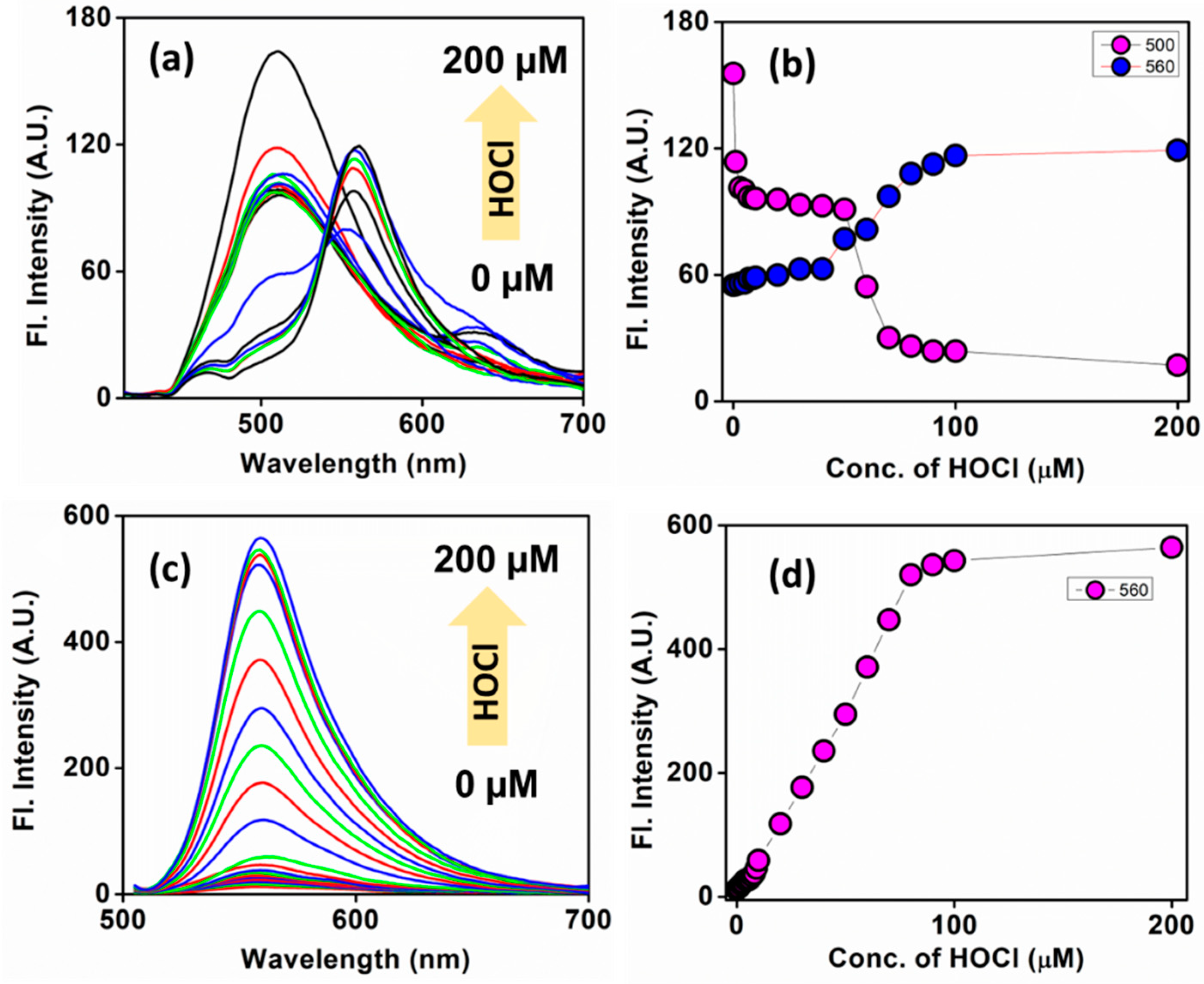
4. Photophysical Properties

Chang’s [42] and James’s groups [43] reported that boronic acid derivative is specific to H2O2. Shu et al. synthesized boronic acid derivatives, which showed specificity towards ONOO with low LOD (16 nM) [44]. However, this probe almost remained silent towards HOCl and reacted slowly with H2O2. Thus, we first screened our probe with probable reactive oxygen species, including H2O2, HOCl, OH·, and ONOO (Figure S1). Interestingly, we found the phenylboronic acid derivative of thiazolium was more specific towards hypochlorite than hydrogen peroxide (H2O2) and ONOO.
Firstly, we carefully checked the sensing ability of the AB probe for HOCl in alkaline pH (pH = 8), as the reaction is much more feasible in alkaline conditions (Figure 2). Since H2O2 is an intermediate for generating HOCl, we studied the photophysics concerning H2O2. We did a reaction kinetics with H2O2, as shown in Figure S2. All three oxygen reactive species (hypochlorous acid, hydrogen peroxide, and peroxynitrite) can cleave the arylboronic acid trigger, which acts as a receptor for photoinduced electron transfer in the buffer. The phenylboronic acid cleaved and formed a phenol derivative that exhibited intramolecular charge transfer (ICT) depending on pH and other microenvironments [45]. For a systematic study, a time-dependent kinetics reaction was recorded by monitoring at 400 nm the maximum absorbance in UV spectra for 80 min after adding 100 µM H2O2 and HOCl (Figures S2 and S3). The maximum absorbance at 400 nm gradually decreased and the new peak at 490 nm steadily increased with time (Figure S2). In the case of HOCl, the reaction kinetics were much faster than with H2O2, as the product peak reached a maximum within 15 min (Figure S3). A similar reaction kinetic experiment was conducted using maximum emission at 560 nm, and the reactivity towards HOCl was almost four times faster than H2O2 (Figure S6).
Next, the ratiometric emission change was analyzed towards variable concentrations of both the analytes (H2O2 and HOCl) ranging from 0−200 µM. The emission peak at ~500 nm was gradually decreased, and simultaneously a new peak at ~560 slowly appeared with increased concentration while exciting at 400 nm. When the same molecule was excited at 490 nm, a gradual increment at 560 nm with increased H2O2 and HOCl concentrations was observed. Hence, AB can be considered an efficient fluorescent probe for HOCl (Figure 2 and Figure S4) and remains partial towards H2O2.
Oxidative stress in living cells is associated with the overproduction of reactive oxygen species, including HOCl and H2O2. When AB reacts with these reactive species (HOCl or H2O2), it releases a quinone methide to form AB–OH. This phenomenon was confirmed using HPLC (Figure S7). AB–OH exhibited ratiometric emission properties due to the intramolecular charge transfer (ICT) from the donor phenoxide to the n-methylthiazolium acceptor in different pH. Thus, we first checked the absorbance and emission spectra of AB–OH in different pH. The absorbance at 400 nm was gradually reduced with increasing pH from 4 to 10. However, a new absorbance peak at 490 nm was gradually increased with pH from 4 to 10. The maximum emission at 510 nm was gradually decreased with increasing pH from 4 to10 upon excitation at 400 nm. However, the emission maxima at 560 nm increased when the pH was increased from 4 to 10 upon excitation at 490 nm. As the ratio of emission intensity at 500 and 560 nm changes, this probe can be considered a potential ratiometric probe toward pH alteration, which is associated with elevated ROS concentrations (Figure S5). We also performed a selectivity study after the addition of different analytes and found it to be reactive with all four ROS, but the best reactivity was observed in the case of HOCl, and thus we did all the further studies considering AB as an HOCl probe (Figure S8).
This AB/AB–OH fluorescent probe may respond to viscosity due to rotatable bonds. The fluorescence intensity at 500 nm significantly increased with an increase in viscosity when excited at around 400 nm (Figure S5). Hence, AB can be considered an efficient probe for hypochlorite and viscosity, and the corresponding AB–OH could be a viscosity sensor.

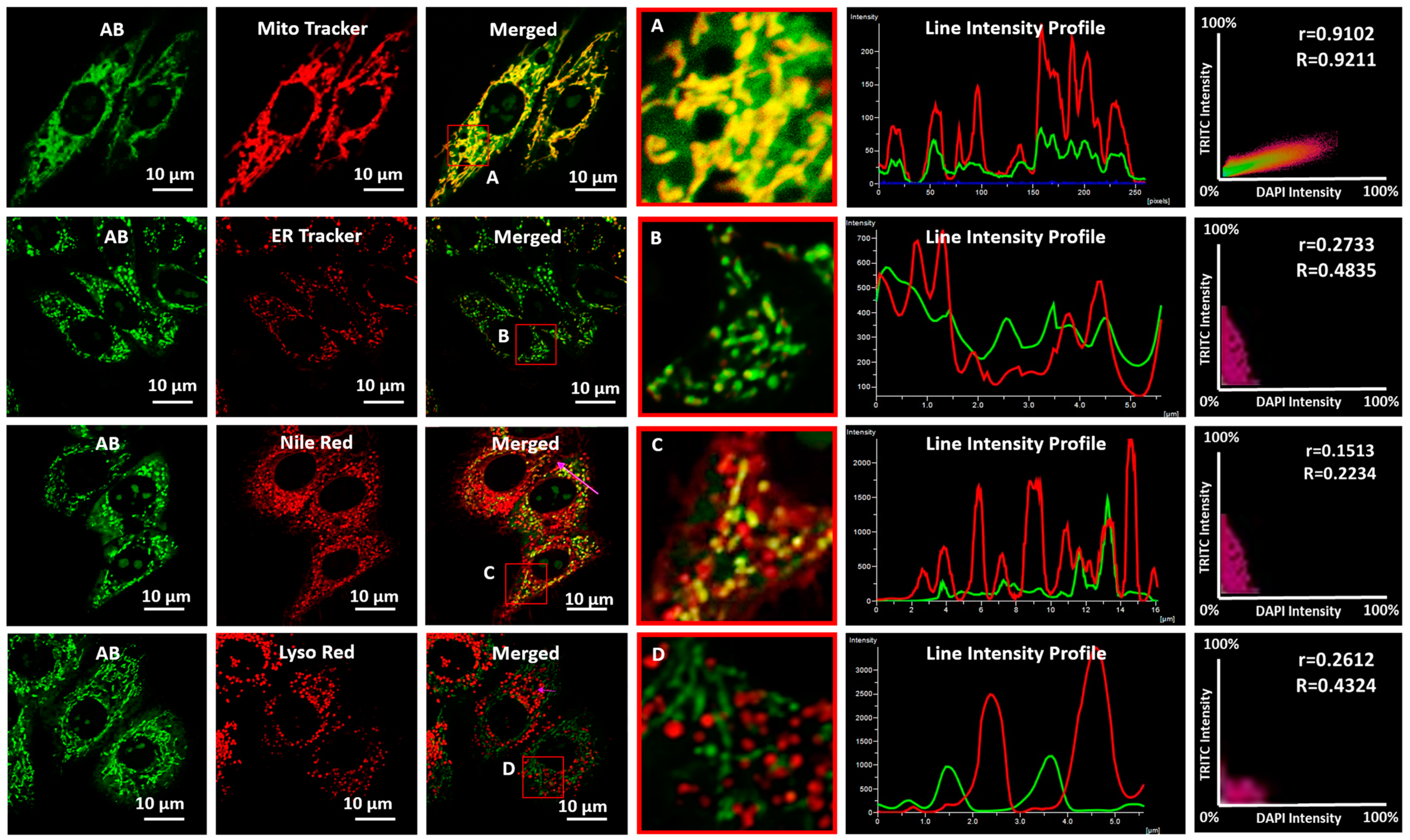
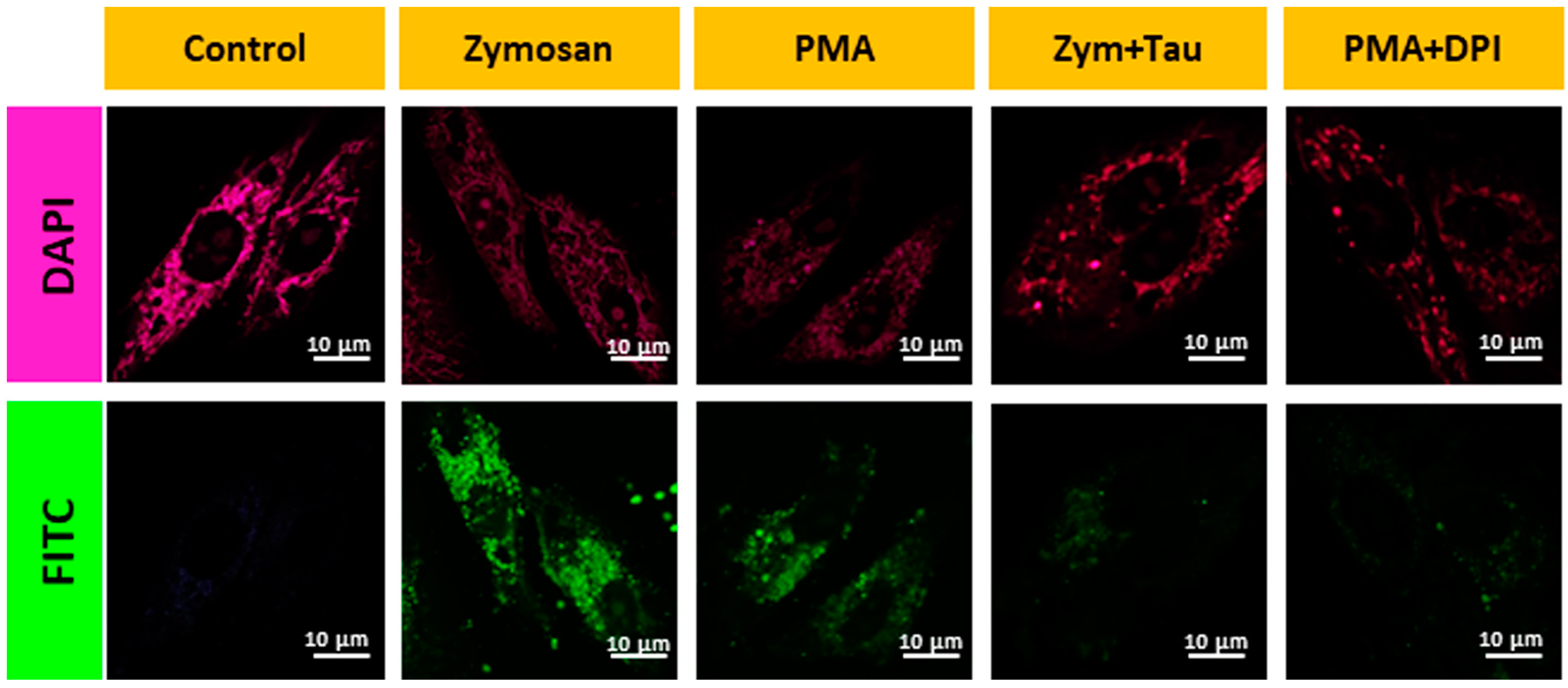
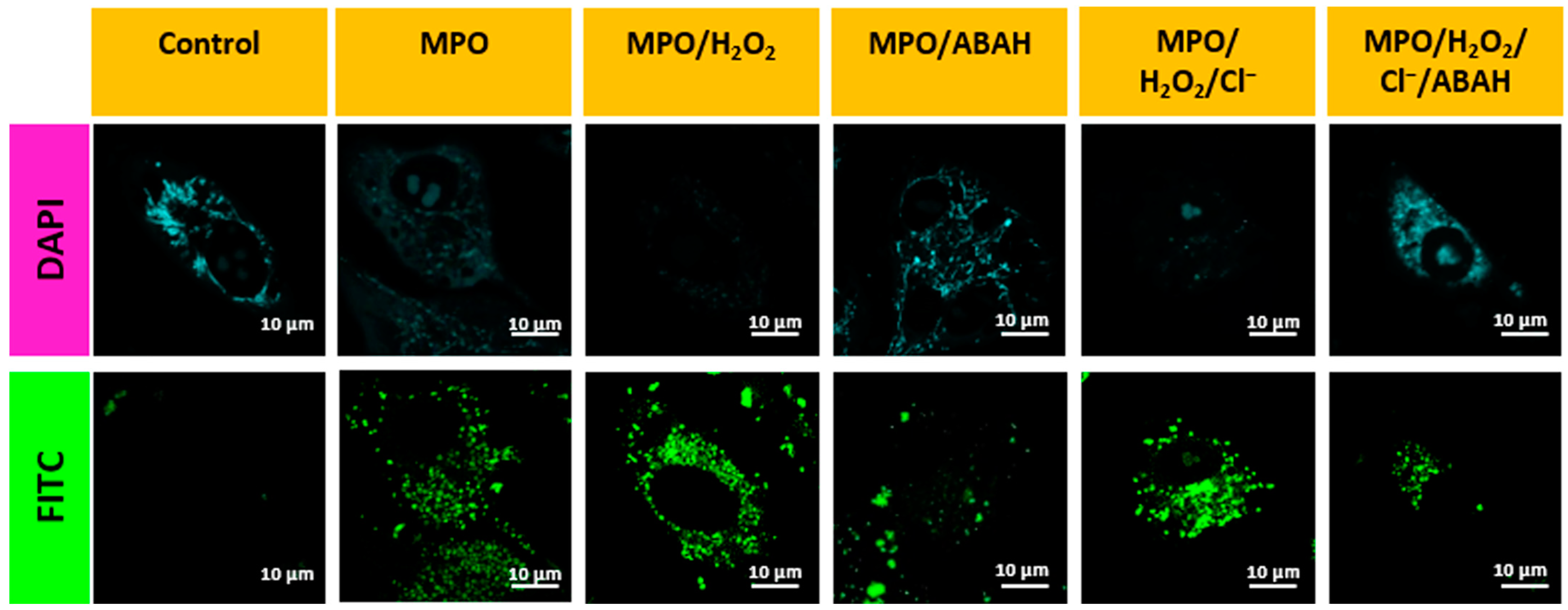
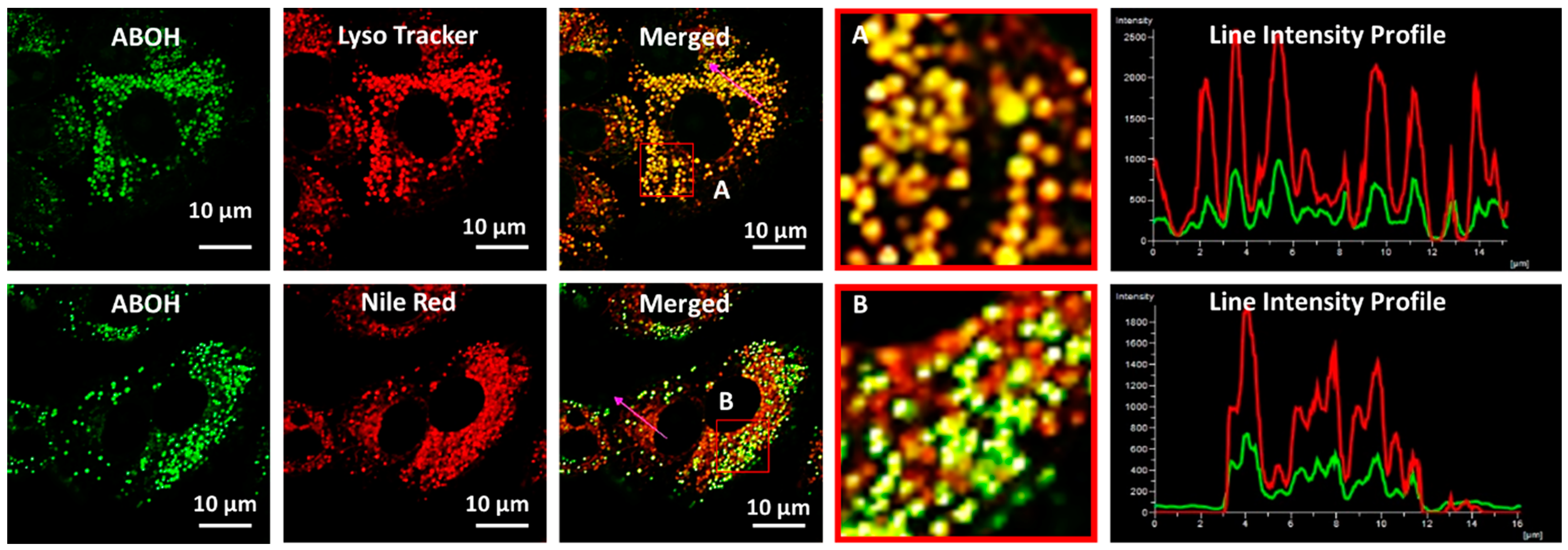
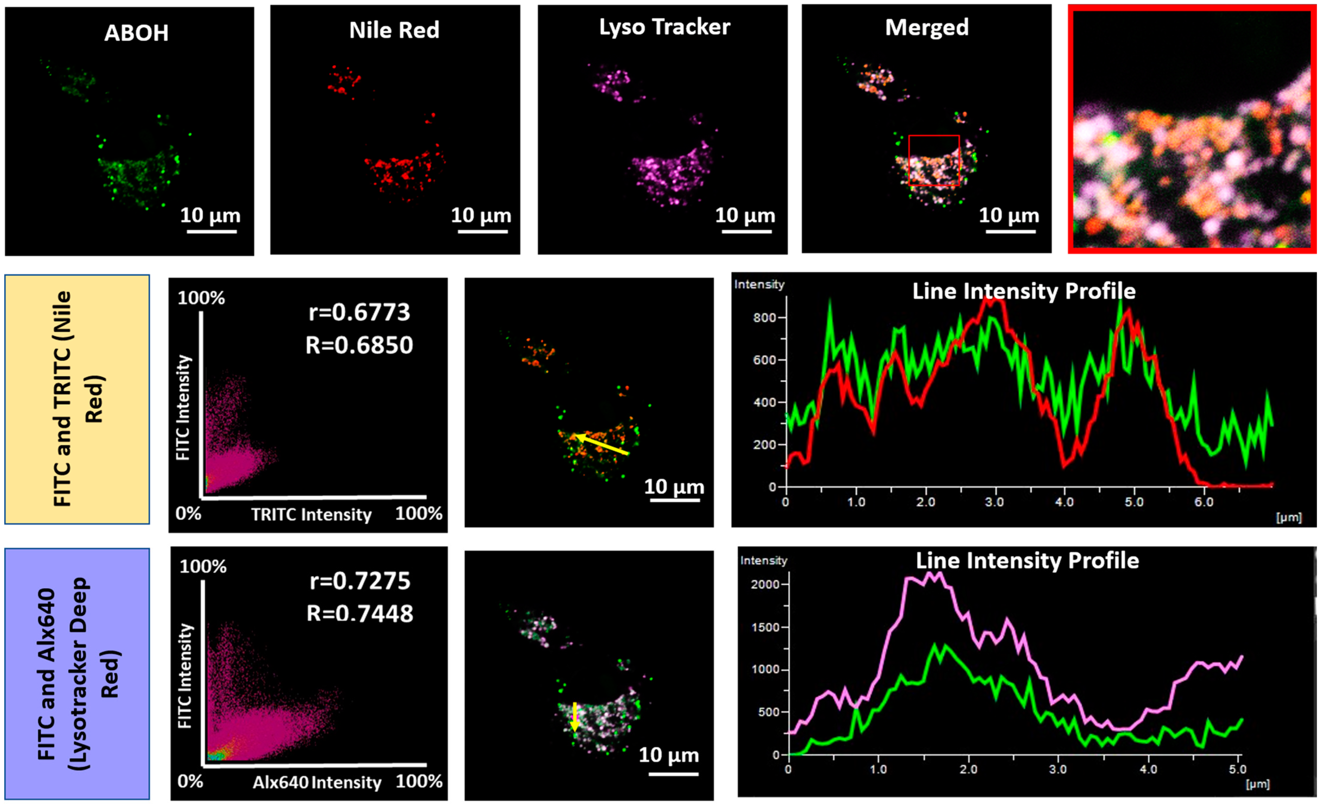
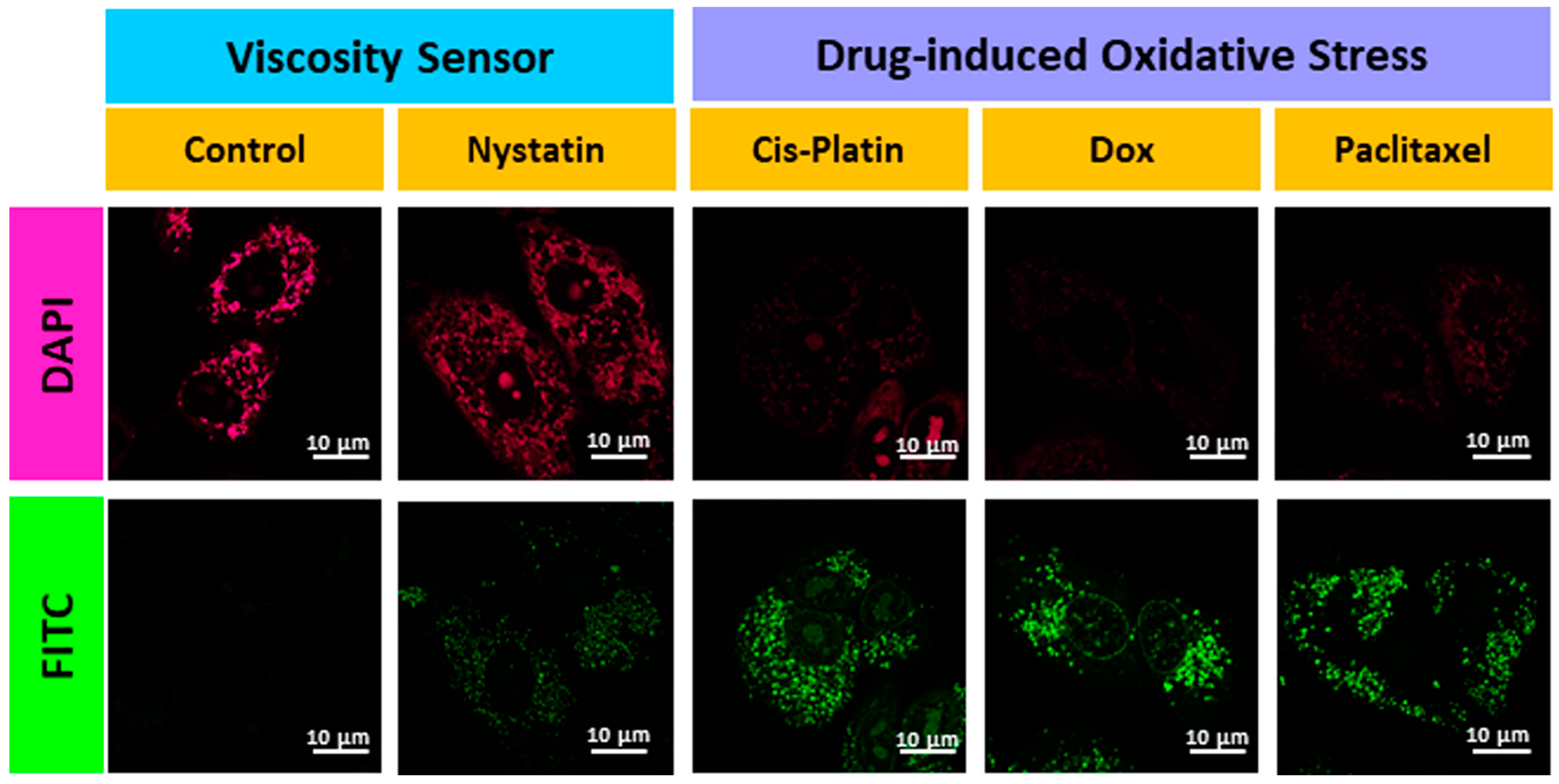
5. Biological Studies

Its biocompatibility is the first and foremost criterion for a fluorescent probe to be efficiently used for cellular imaging. To test this, we subjected our probe AB to A549 cells for 24 h and found it was almost nontoxic (Figure S9) by MTT assay compared to the 10 µM positive control doxorubicin. A co-localization study confirmed that the probe was nicely localized in mitochondria with a high Pearson’s coefficient (0.9102). There was a partial overlap with the endoplasmic reticulum (ER) but almost no overlap with lipid droplets or lysosomes (Figure 3). The high specificity to mitochondria suggested that the cationic AB probe has a great affinity to accumulate in the negative mitochondrial matrix.
Although AB responded to three reactive oxygen species, the most sensitivity towards hypochlorite encourages us to study intracellular ROS, especially hypochlorite, in living cells. We studied the efficiency of the AB probe in detecting endogenous mitochondrial HOCl in live cells. For this, we pretreated the cells with different known endogenous HOCl inducers such as PMA (500 nM, 1 h), Zymosan (10 µg/mL, 1 h), MPO, MPO/H2O2, MPO/H2O2/Cl, and then with AB for 30 min. Compared to the control, there was an apparent enhancement of fluorescence intensity for all the cases as the amount of HOCl was increased (Figure 4 and Figure 5). In contrast, cells treated with inhibitors such as 4-aminobenzoic acid hydrazine (4-ABAH), taurine, and DPI exhibited very low fluorescence in the FITC channel compared to the non-treated cells (Figure 4 and Figure 5). Therefore, all the above results confirm the AB probe’s proficiency in detecting endogenous HOCl generated via MPO and Zymosan.
Previously reported boronic acid-based fluorescent probes did not systematically study how the product, phenolic acid derivative (after reaction with ROS) behaves in cells during oxidative stress. Herein, we carefully studied the fate of the fluorescent probe after its response towards ROS using high-resolution confocal fluorescence microscopic images. The AB–OH, a product of AB during oxidative stress, was localized in the same or other intracellular organelles. As shown in Figure 6, AB–OH was nicely localized in lysosomes instead of mitochondria. These results indicate that the AB probe is initially localized in mitochondria but translocated to lysosomes when HOCl induced cleavage of phenylboronic acid. The mitochondria are depolarized during oxidative stress, resulting in a lowering of MMP (mitochondrial membrane potential). Thus, the probe leaks out from mitochondria and is localized to lysosomes.
As spectroscopic analysis of AB–OH exhibited an excellent response to viscosity, AB–OH can be a potential biomarker for lipid droplets that can monitor the oxidative process. A co-localization experiment suggested that AB–OH nicely localized in lysosomal lipid droplets with a Pearson coefficient (0.6773). However, it remains silent to non-lysosomal lipid droplets. As shown in Figure 7, AB–OH nicely colocalized with lysosomal LDs, confirmed by the merged images with commercial trackers Lyso Tracker Deep Red and Nile Red, along with the AB–OH probe in the FITC channel, respectively.
Further, to confirm the mitochondrial viscosity response of AB, cells were pretreated with nystatin, which is known to cause swelling of mitochondria, resulting in increased mitochondrial viscosity [46,47,48,49,50,51]. There was an enhancement in fluorescence intensity in the DAPI and FITC channels compared to the control (Figure 8). Finally, to test the drug-induced oxidative stress in cancerous cells, the cells were pretreated with well-known anticancer drugs such as doxorubicin, cisplatin, and paclitaxel for 1 h AB was incubated for 30 min before confocal microscopic imaging. A significant enhancement in fluorescence intensity was observed in the FITC channel compared to the untreated cells, and the change in morphology confirmed that the cells were under oxidative stress (Figure 8). This led to mitochondrial damage, consequently, changes in MMP and the formation of lipid droplets. Hence, a simple AB probe can efficiently sense mitochondrial ROS, especially HOCl and lysosomal viscosity in cells under oxidative stress conditions.

6. Conclusions

Reactive oxygen species (ROS) play crucial roles in redox signaling in living cells. However, overproduction of these reactive species may damage nucleic acids and oxidize proteins. These adverse effects may lead to cell death, neuronal diseases, cancer, and diabetes. Hence, we studied ROS, especially HOCl, in cancer cells during oxidative stress caused by viral infections and drug treatment. This may alter cellular microenvironments, including cellular viscosity and corresponding lipid droplets. Hence, we systematically studied the detection of hypochlorite in mitochondria during oxidative stress and the post-effect using a simple molecular probe that exhibited ratiometric properties. We successfully synthesized a derivative of hemicyanine dye with boronic acid as the trigger and receptor of photoinduced electron transfer. This AB showed good sensitivity towards hypochlorite compared to peroxynitrite (ONOO–) and the most common and highly abundant hydrogen peroxide (H2O2). It also responded towards viscosity with turn-on property. These excellent photophysical properties offer a new scope for studying confocal fluorescence imaging in living cells to monitor HOCl and viscosity during oxidative stress. AB showed a good co-localization in mitochondrial and responded towards HOCl ratiometrically during oxidative stress. We have experimentally verified and proved the significance of our designed probe during a change in mitochondrial viscosity. Confocal image analysis indicated that AB–OH, a product of AB, translocates to lysosomes and monitors the lysosomal lipid droplets during oxidative stress. Hence, this simple fluorescent probe can be a potential biomarker to study redox imbalance and lipid droplets during oxidative stress.

Supplementary Materials

The following supporting information can be downloaded at: https://www.mdpi.com/article/10.3390/bios13060662/s1.

Author Contributions

Conceptualization, M.B., A.J., N.P. and A.S.; Data curation, M.B., A.J., N.P. and S.S.; Formal analysis, M.B., A.J., N.P. and S.S.; Funding acquisition, A.S.; Investigation, A.S.; Methodology, M.B., A.J. and N.P.; Project administration, A.S.; Resources, A.S.; Supervision, A.S.; Validation, M.B. and A.J.; Visualization, M.B., A.J. and N.P.; Writing—original draft, M.B., A.J., N.P. and A.S.; Writing—review and editing, M.B., A.J., N.P. and A.S. All authors have read and agreed to the published version of the manuscript.

Funding

A.S. is grateful to the DST-SERB, India, for the DST-Core Research Grant (CRG/2021/002313) and the Ramanujan Grant (SB/S2/RJN148/2015) and to the Shiv Nadar Foundation for funding.

Institutional Review Board Statement

Not applicable.

Informed Consent Statement

Not applicable.

Data Availability Statement

Not applicable.

Acknowledgments

The authors thankfully acknowledge NCSS, Pune, for providing the A549 cell lines. Authors M.B., A.J., N.P. and S.S. thank the Shiv Nadar Institute of Eminence (SNIoE), Delhi NCR, and the Shiv Nadar Foundation for their research fellowships. The authors acknowledge the SNU DST-FIST grant [SR/FST/LS-1/2017/59(c)] for the confocal microscopy facility at Shiv Nadar University, Delhi NCR.

Conflicts of Interest

The authors declare no conflict of interest.

References

  1. Satori, C.P.; Henderson, M.M.; Krautkramer, E.A.; Kostal, V.; Distefano, M.M.; Arriaga, E.A. Bioanalysis of Eukaryotic Organelles. Chem. Rev. 2013, 113, 2733–2811. [Google Scholar] [CrossRef] [Green Version]
  2. Friedman, J.R.; Nunnari, J. Mitochondrial Form and Function. Nature 2014, 505, 335–343. [Google Scholar] [CrossRef] [Green Version]
  3. Osellame, L.D.; Blacker, T.S.; Duchen, M.R. Cellular and Molecular Mechanisms of Mitochondrial Function. Best Pract. Res. Clin. Endocrinol. Metab. 2012, 26, 711–723. [Google Scholar] [CrossRef] [PubMed] [Green Version]
  4. Khan, A.A.; Alsahli, M.A.; Rahmani, A.H. Myeloperoxidase as an Active Disease Biomarker: Recent Biochemical and Pathological Perspectives. Med. Sci. 2018, 6, 33. [Google Scholar] [CrossRef] [PubMed] [Green Version]
  5. Andrés, C.M.C.; de la Lastra, J.M.; Juan, C.A.; Plou, F.J.; Pérez-Lebeña, E. Hypochlorous Acid Chemistry in Mammalian Cells—Influence on Infection and Role in Various Pathologies. Int. J. Mol. Sci. 2022, 23, 10735. [Google Scholar] [CrossRef] [PubMed]
  6. Zorov, D.B.; Juhaszova, M.; Sollott, S.J. Mitochondrial Reactive Oxygen Species (ROS) and ROS-Induced ROS Release. Physiol. Rev. 2014, 94, 909–950. [Google Scholar] [CrossRef] [PubMed] [Green Version]
  7. Zhou, J.; Li, L.; Shi, W.; Gao, X.; Li, X.; Ma, H. HOCl Can Appear in the Mitochondria of Macrophages during Bacterial Infection as Revealed by a Sensitive Mitochondrial-Targeting Fluorescent Probe. Chem. Sci. 2015, 6, 4884–4888. [Google Scholar] [CrossRef] [Green Version]
  8. Baruah, M.; Jana, A.; Ali, M.; Mapa, K.; Samanta, A. An Efficient PeT Based Fluorescent Probe for Mapping Mitochondrial Oxidative Stress Produced via the Nox2 Pathway. J. Mater. Chem. B 2022, 10, 2230–2237. [Google Scholar] [CrossRef]
  9. Park, S.; Jung, W.-H.; Pittman, M.; Chen, J.; Chen, Y. The Effects of Stiffness, Fluid Viscosity, and Geometry of Microenvironment in Homeostasis, Aging, and Diseases: A Brief Review. J. Biomech. Eng. 2020, 142, 4048110. [Google Scholar] [CrossRef]
  10. Pizzino, G.; Irrera, N.; Cucinotta, M.; Pallio, G.; Mannino, F.; Arcoraci, V.; Squadrito, F.; Altavilla, D.; Bitto, A. Oxidative Stress: Harms and Benefits for Human Health. Oxid. Med. Cell. Longev. 2017, 2017, 8416763. [Google Scholar] [CrossRef] [Green Version]
  11. Jin, Y.; Tan, Y.; Chen, L.; Liu, Y.; Ren, Z. Reactive Oxygen Species Induces Lipid Droplet Accumulation in HepG2 Cells by Increasing Perilipin 2 Expression. Int. J. Mol. Sci. 2018, 19, 3445. [Google Scholar] [CrossRef] [PubMed] [Green Version]
  12. Tirichen, H.; Yaigoub, H.; Xu, W.; Wu, C.; Li, R.; Li, Y. Mitochondrial Reactive Oxygen Species and Their Contribution in Chronic Kidney Disease Progression Through Oxidative Stress. Front. Physiol. 2021, 12, 627837. [Google Scholar] [CrossRef] [PubMed]
  13. Demir, S.; Nawroth, P.P.; Herzig, S.; Ekim Üstünel, B. Emerging Targets in Type 2 Diabetes and Diabetic Complications. Adv. Sci. 2021, 8, 2100275. [Google Scholar] [CrossRef] [PubMed]
  14. Choi, T.G.; Kim, S.S. Physiological Functions of Mitochondrial Reactive Oxygen Species. In Free Radical Medicine and Biology; Das, K., Das, S., Biradar, M.S., Bobbarala, V., Tata, S.S., Eds.; IntechOpen: Rijeka, Croatia, 2019; p. Ch. 2; ISBN 978-1-78985-144-1. [Google Scholar]
  15. Tian, X.; Murfin, L.C.; Wu, L.; Lewis, S.E.; James, T.D. Fluorescent Small Organic Probes for Biosensing. Chem. Sci. 2021, 12, 3406–3426. [Google Scholar] [CrossRef]
  16. Arranz, A.; Ripoll, J. Advances in Optical Imaging for Pharmacological Studies. Front. Pharmacol. 2015, 6, 189. [Google Scholar] [CrossRef] [Green Version]
  17. Peters, T.M.; Linte, C.A. Image-Guided Interventions and Computer-Integrated Therapy: Quo Vadis? Med. Image Anal. 2016, 33, 56–63. [Google Scholar] [CrossRef]
  18. Mehrotra, P. Biosensors and Their Applications—A Review. J. Oral Biol. Craniofac. Res. 2016, 6, 153–159. [Google Scholar] [CrossRef] [Green Version]
  19. Ornatowski, W.; Lu, Q.; Yegambaram, M.; Garcia, A.E.; Zemskov, E.A.; Maltepe, E.; Fineman, J.R.; Wang, T.; Black, S.M. Complex Interplay between Autophagy and Oxidative Stress in the Development of Pulmonary Disease. Redox Biol. 2020, 36, 101679. [Google Scholar] [CrossRef]
  20. Bigdeli, A.; Ghasemi, F.; Abbasi-Moayed, S.; Shahrajabian, M.; Fahimi-Kashani, N.; Jafarinejad, S.; Farahmand Nejad, M.A.; Hormozi-Nezhad, M.R. Ratiometric Fluorescent Nanoprobes for Visual Detection: Design Principles and Recent Advances—A Review. Anal. Chim. Acta 2019, 1079, 30–58. [Google Scholar] [CrossRef] [PubMed]
  21. Lee, M.H.; Kim, J.S.; Sessler, J.L. Small Molecule-Based Ratiometric Fluorescence Probes for Cations, Anions, and Biomolecules. Chem. Soc. Rev. 2015, 44, 4185–4191. [Google Scholar] [CrossRef] [PubMed] [Green Version]
  22. Zhang, Q.; Zhu, Z.; Zheng, Y.; Cheng, J.; Zhang, N.; Long, Y.T.; Zheng, J.; Qian, X.; Yang, Y. A Three-Channel Fluorescent Probe That Distinguishes Peroxynitrite from Hypochlorite. J. Am. Chem. Soc. 2012, 134, 18479–18482. [Google Scholar] [CrossRef]
  23. Ren, M.; Li, Z.; Nie, J.; Wang, L.; Lin, W. A Photocaged Fluorescent Probe for Imaging Hypochlorous Acid in Lysosomes. Chem. Commun. 2018, 54, 9238–9241. [Google Scholar] [CrossRef]
  24. Pak, Y.L.; Park, S.J.; Xu, Q.; Kim, H.M.; Yoon, J. Ratiometric Two-Photon Fluorescent Probe for Detecting and Imaging Hypochlorite. Anal. Chem. 2018, 90, 9510–9514. [Google Scholar] [CrossRef] [PubMed]
  25. Hitomi, Y.; Takeyasu, T.; Funabiki, T.; Kodera, M. Detection of Enzymatically Generated Hydrogen Peroxide by Metal-Based Fluorescent Probe. Anal. Chem. 2011, 83, 9213–9216. [Google Scholar] [CrossRef]
  26. Liu, J.; Ren, J.; Bao, X.; Gao, W.; Wu, C.; Zhao, Y. PH-Switchable Fluorescent Probe for Spatially-Confined Visualization of Intracellular Hydrogen Peroxide. Anal. Chem. 2016, 88, 5865–5870. [Google Scholar] [CrossRef]
  27. Liu, J.; Liang, J.; Wu, C.; Zhao, Y. A Doubly-Quenched Fluorescent Probe for Low-Background Detection of Mitochondrial H2O2. Anal. Chem. 2019, 91, 6902–6909. [Google Scholar] [CrossRef]
  28. Peng, T.; Yang, D. HKGreen-3: A Rhodol-Based Fluorescent Probe for Peroxynitrite. Org. Lett. 2010, 12, 4932–4935. [Google Scholar] [CrossRef] [PubMed]
  29. Baruah, M.; Kwon, H.-Y.; Cho, H.; Chang, Y.-T.; Samanta, A. A Photoinduced Electron Transfer-Based Hypochlorite-Specific Fluorescent Probe for Selective Imaging of Proinflammatory M1 in a Rheumatoid Arthritis Model. Anal. Chem. 2023, 95, 4147–4154. [Google Scholar] [CrossRef] [PubMed]
  30. Yadav, R.; Munan, S.; Ali, M.; Mapa, K.; Samanta, A. A Systematic Design and Synthesis of PET-Based Fluorescent Probe for Monitoring PH during Mitophagy. Chem. Asian J. 2023, 2023, e202300308. [Google Scholar] [CrossRef]
  31. Yadav, R.; Munan, S.; Kardam, V.; Dutta Dubey, K.; Samanta, A. Esterase Specific Fluorescent Probe: Mechanistic Understanding Using QM/MM Calculation and Cell States Discrimination. Chem. A Eur. J. 2023, 2023, e202300244. [Google Scholar] [CrossRef]
  32. Munan, S.; Ali, M.; Yadav, R.; Mapa, K.; Samanta, A. PET- and ICT-Based Ratiometric Probe: An Unusual Phenomenon of Morpholine-Conjugated Fluorophore for Mitochondrial PH Mapping during Mitophagy. Anal. Chem. 2022, 94, 11633–11642. [Google Scholar] [CrossRef]
  33. Munan, S.; Kottarathil, S.; Joseph, M.M.; Jana, A.; Ali, M.; Mapa, K.; Maiti, K.K.; Samanta, A. IndiFluors: A New Full-Visible Color-Tunable Donor-Acceptor-Donor (D1-A-D2) Fluorophore Family for Ratiometric PH Imaging during Mitophagy. ACS Sens. 2021. [Google Scholar] [CrossRef]
  34. De Moliner, F.; Kielland, N.; Lavilla, R.; Vendrell, M. Modern Synthetic Avenues for the Preparation of Functional Fluorophores. Angew. Chem. Int. Ed. 2017, 56, 3758–3769. [Google Scholar] [CrossRef] [Green Version]
  35. Gatin-Fraudet, B.; Ottenwelter, R.; Le Saux, T.; Ephanie Norsikian, S.; Pucher, M.; Lomb, E.A.; Aur Elie Baron, T.; Durand, P.; Doisneau, G.; Bourdreux, Y.; et al. Evaluation of Borinic Acids as New, Fast Hydrogen Peroxide-Responsive Triggers. Proc. Natl. Acad. Sci. USA 2021, 118, e2107503118. [Google Scholar] [CrossRef] [PubMed]
  36. SK, M.; Banesh, S.; Trivedi, V.; Biswas, S. Selective and Sensitive Sensing of Hydrogen Peroxide by a Boronic Acid Functionalized Metal–Organic Framework and Its Application in Live-Cell Imaging. Inorg. Chem. 2018, 57, 14574–14581. [Google Scholar] [CrossRef] [PubMed]
  37. Kikuchi, H.; Nakamura, Y.; Inoue, C.; Nojiri, S.; Koita, M.; Kojima, M.; Koyama, H.; Miki, R.; Seki, T.; Egawa, Y. Hydrogen Peroxide-Triggered Conversion of Boronic Acid-Appended Insulin into Insulin and Its Application as a Glucose-Responsive Insulin Formulation. Mol. Pharm. 2021, 18, 4224–4230. [Google Scholar] [CrossRef] [PubMed]
  38. Sieracki, N.A.; Gantner, B.N.; Mao, M.; Horner, J.H.; Ye, R.D.; Malik, A.B.; Newcomb, M.E.; Bonini, M.G. Bioluminescent Detection of Peroxynitrite with a Boronic Acid-Caged Luciferin. Free. Radic. Biol. Med. 2013, 61, 40–50. [Google Scholar] [CrossRef] [Green Version]
  39. Sikora, A.; Zielonka, J.; Lopez, M.; Joseph, J.; Kalyanaraman, B. Direct Oxidation of Boronates by Peroxynitrite: Mechanism and Implications in Fluorescence Imaging of Peroxynitrite. Free. Radic. Biol. Med. 2009, 47, 1401–1407. [Google Scholar] [CrossRef] [Green Version]
  40. Xu, Q.; Lee, K.-A.; Lee, S.; Lee, K.M.; Lee, W.-J.; Yoon, J. A Highly Specific Fluorescent Probe for Hypochlorous Acid and Its Application in Imaging Microbe-Induced HOCl Production. J. Am. Chem. Soc. 2013, 135, 9944–9949. [Google Scholar] [CrossRef]
  41. Simões, E.F.C.; da Silva, L.P.; da Silva, J.C.G.E.; Leitão, J.M.M. Hypochlorite Fluorescence Sensing by Phenylboronic Acid-Alizarin Adduct Based Carbon Dots. Talanta 2020, 208, 120447. [Google Scholar] [CrossRef]
  42. Miller, E.W.; Albers, A.E.; Pralle, A.; Isacoff, E.Y.; Chang, C.J. Boronate-Based Fluorescent Probes for Imaging Cellular Hydrogen Peroxide. J. Am. Chem. Soc. 2005, 127, 16652–16659. [Google Scholar] [CrossRef] [Green Version]
  43. Sun, X.; Xu, S.-Y.; Flower, S.E.; Fossey, J.S.; Qian, X.; James, T.D. “Integrated” and “Insulated” Boronate-Based Fluorescent Probes for the Detection of Hydrogen Peroxide. Chem. Commun. 2013, 49, 8311–8313. [Google Scholar] [CrossRef] [Green Version]
  44. Shu, W.; Wu, Y.; Duan, Q.; Zang, S.; Su, S.; Jing, J.; Zhang, X. A Highly Selective Fluorescent Probe for Monitoring Exogenous and Endogenous ONOO− Fluctuations in HeLa Cells. Dye Pigment. 2020, 175, 108069. [Google Scholar] [CrossRef]
  45. Gatin-Fraudet, B.; Pucher, M.; Le Saux, T.; Doisneau, G.; Bourdreux, Y.; Jullien, L.; Vauzeilles, B.; Guianvarc’h, D.; Urban, D. Hydrogen Peroxide-Responsive Triggers Based on Borinic Acids: Molecular Insights into the Control of Oxidative Rearrangement. Chem. A Eur. J. 2022, 28, e202201543. [Google Scholar] [CrossRef]
  46. Gonzalez-Molina, J.; Zhang, X.; Borghesan, M.; Mendonça da Silva, J.; Awan, M.; Fuller, B.; Gavara, N.; Selden, C. Extracellular Fluid Viscosity Enhances Liver Cancer Cell Mechanosensing and Migration. Biomaterials 2018, 177, 113–124. [Google Scholar] [CrossRef] [PubMed]
  47. Soltoff, S.P.; Mandel, L.J. Potassium Transport in the Rabbit Renal Proximal Tubule: Effects of Barium, Ouabain, Valinomycin, and Other Ionophores. J. Membr. Biol. 1986, 94, 153–161. [Google Scholar] [CrossRef]
  48. Ma, C.; Sun, W.; Xu, L.; Qian, Y.; Dai, J.; Zhong, G.; Hou, Y.; Liu, J.; Shen, B. A Minireview of Viscosity-Sensitive Fluorescent Probes: Design and Biological Applications. J. Mater. Chem. B 2020, 8, 9642–9651. [Google Scholar] [CrossRef] [PubMed]
  49. Chen, B.; Li, C.; Zhang, J.; Kan, J.; Jiang, T.; Zhou, J.; Ma, H. Sensing and Imaging of Mitochondrial Viscosity in Living Cells Using a Red Fluorescent Probe with a Long Lifetime. Chem. Commun. 2019, 55, 7410–7413. [Google Scholar] [CrossRef] [PubMed]
  50. Peng, M.; Yin, J.; Lin, W. Tracking Mitochondrial Viscosity in Living Systems Based on a Two-Photon and near Red Probe. New J. Chem. 2019, 43, 16945–16949. [Google Scholar] [CrossRef]
  51. Yang, Z.; He, Y.; Lee, J.-H.; Park, N.; Suh, M.; Chae, W.-S.; Cao, J.; Peng, X.; Jung, H.; Kang, C.; et al. A Self-Calibrating Bipartite Viscosity Sensor for Mitochondria. J. Am. Chem. Soc. 2013, 135, 9181–9185. [Google Scholar] [CrossRef]
Figure 1. Design principle and sensing mechanism of AB. The probe AB is initially weak fluorescent (light green) in aqueous media due to the PET (photoinduced electron transfer) process but shows strong fluorescence in high viscous media (green color). ABOH exhibits stronger emission (represented as green) due to PET off mechanism. In mitochondrial pH (~8.0) and hence ABOH exhibits a longer wavelength (represented as yellow) due to ICT (intramolecular charge transfer) process.
Biosensors 13 00662 g001
Figure 2. Emission spectra of AB in the presence of different concentrations of HOCl (0–200 µM). (a) λex = 400 nm and (c) λex = 490 nm and their corresponding scatter plots (b,d). Probe concentration: 10 µM. The HOCl concentrations used were 100nm, 300 nm, 500 nm, 700 nm, 1–10 µM (10 points), 10–100 µM (10 points), and 200 µM.
Biosensors 13 00662 g002
Figure 3. Confocal fluorescence images for co-localization of AB (10 µM) in A549 cells. Images were taken after co-staining with Mito Tracker Deep red, ER Tracker Red, Nile red, and Lyso Tracker Deep red. The squares marked “A”, “B”, “C”, and “D” signify the zoomed-in picture. Corresponding line and scatter plots, wherein “r” and “R” represent Pearson’s correlation and Mander’s overlap coefficients, respectively. Images were captured using a 100× oil emersion lens with 2× zoom. Image scale bar = 10 µm.
Biosensors 13 00662 g003
Figure 4. Confocal images for A549 cells during the oxidative stress conditions using inducers Zymosan, PMA, and inhibitors such as taurine and DPI. Image scale = 10 µm.
Biosensors 13 00662 g004
Figure 5. Confocal images for A549 cells during the oxidative stress conditions using inducers MPO H2O2 and chloride ions, and inhibitor ABAH Image scale = 10 µm.
Biosensors 13 00662 g005
Figure 6. Confocal fluorescence images for co-localization of AB–OH (10 µM) in A549 cells. Images were taken after co-staining with Lyso Tracker Deep Red and Nile Red. The squares marked “A” and “B” signify the zoomed-in picture, followed by the corresponding line plots showing the overlap of the probe with commercial trackers. Images were captured using a 100× oil emersion lens with 2× zoom. Image scale bar = 10 µm.
Biosensors 13 00662 g006
Figure 7. Confocal fluorescence images for co-localization of AB–OH (10 µM) in A549 cells to confirm co-localization in lysosomal lipid droplets. After inducing oxidative-stress conditions, images were taken after co-staining with Lyso Tracker Deep Red and Nile Red together. The squares signify the zoomed-in picture. Below are the corresponding line and scatter plots, wherein “r” and “R” represent Pearson’s correlation coefficient and Mander’s overlap coefficient for two channels simultaneously. Images were captured using a 100× oil emersion lens with 2× zoom. Image scale bar = 10 µm.
Biosensors 13 00662 g007
Figure 8. Confocal images for A549 cells during increased mitochondrial viscosity after treatment with nystatin and in oxidative stress conditions using known drugs such as cis-platin, doxorubicin, and paclitaxel. Image scale = 10 µm.
Biosensors 13 00662 g008
Disclaimer/Publisher’s Note: The statements, opinions and data contained in all publications are solely those of the individual author(s) and contributor(s) and not of MDPI and/or the editor(s). MDPI and/or the editor(s) disclaim responsibility for any injury to people or property resulting from any ideas, methods, instructions or products referred to in the content.

