Advances in Tapered Optical Fiber Sensor Structures: From Conventional to Novel and Emerging

 , ,
, , 
Abstract
:1. Introduction
2. Theoretical Mechanism for Sensing
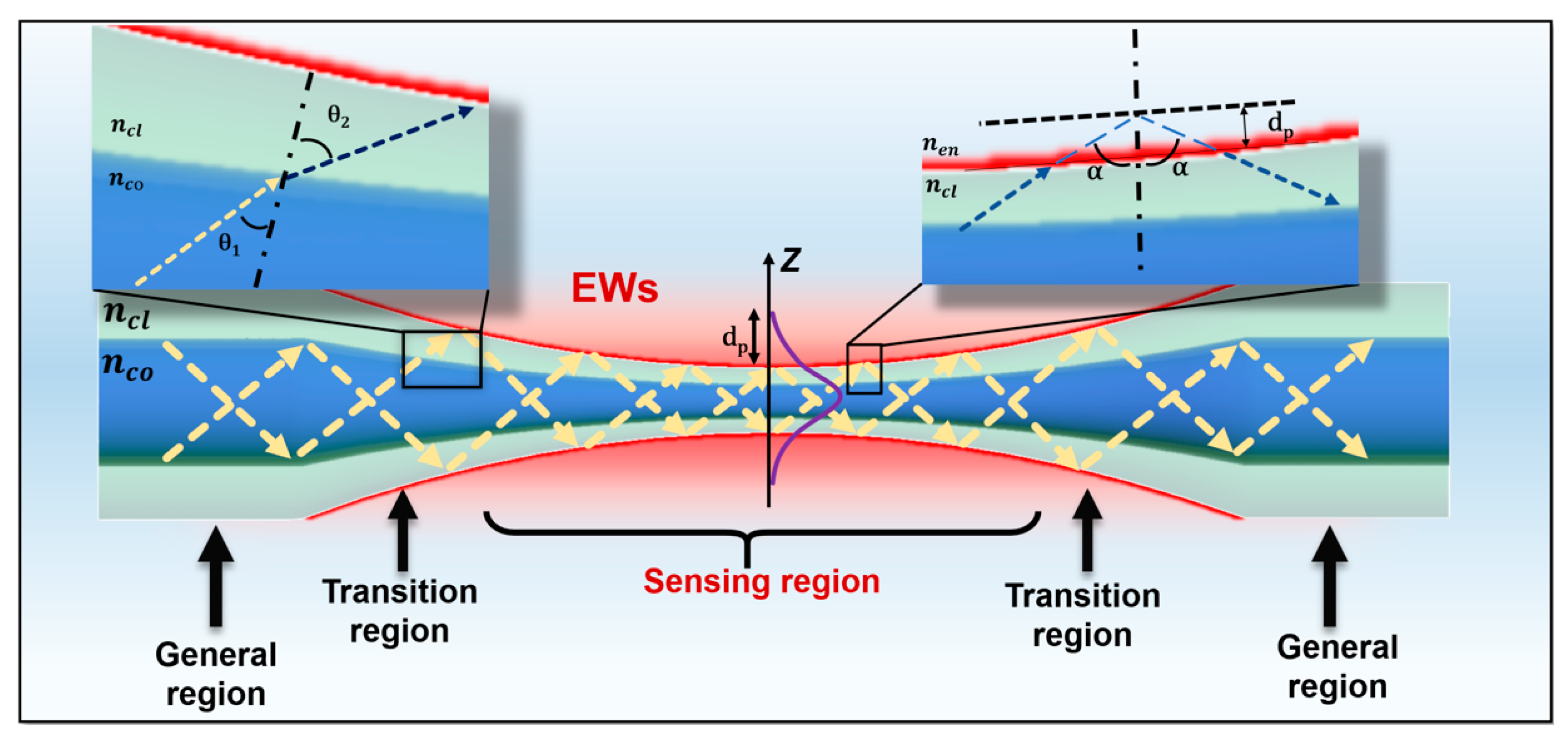
2.1. Basic Structure of TOF
2.2. Evanescent Waves Sensing Principle of TOF
2.3. SPR/LSPR Sensor Mechanism Based on TOF
2.4. In-Line MZI Theory Based on TOF
2.5. Grating Sensing Principle of TOF
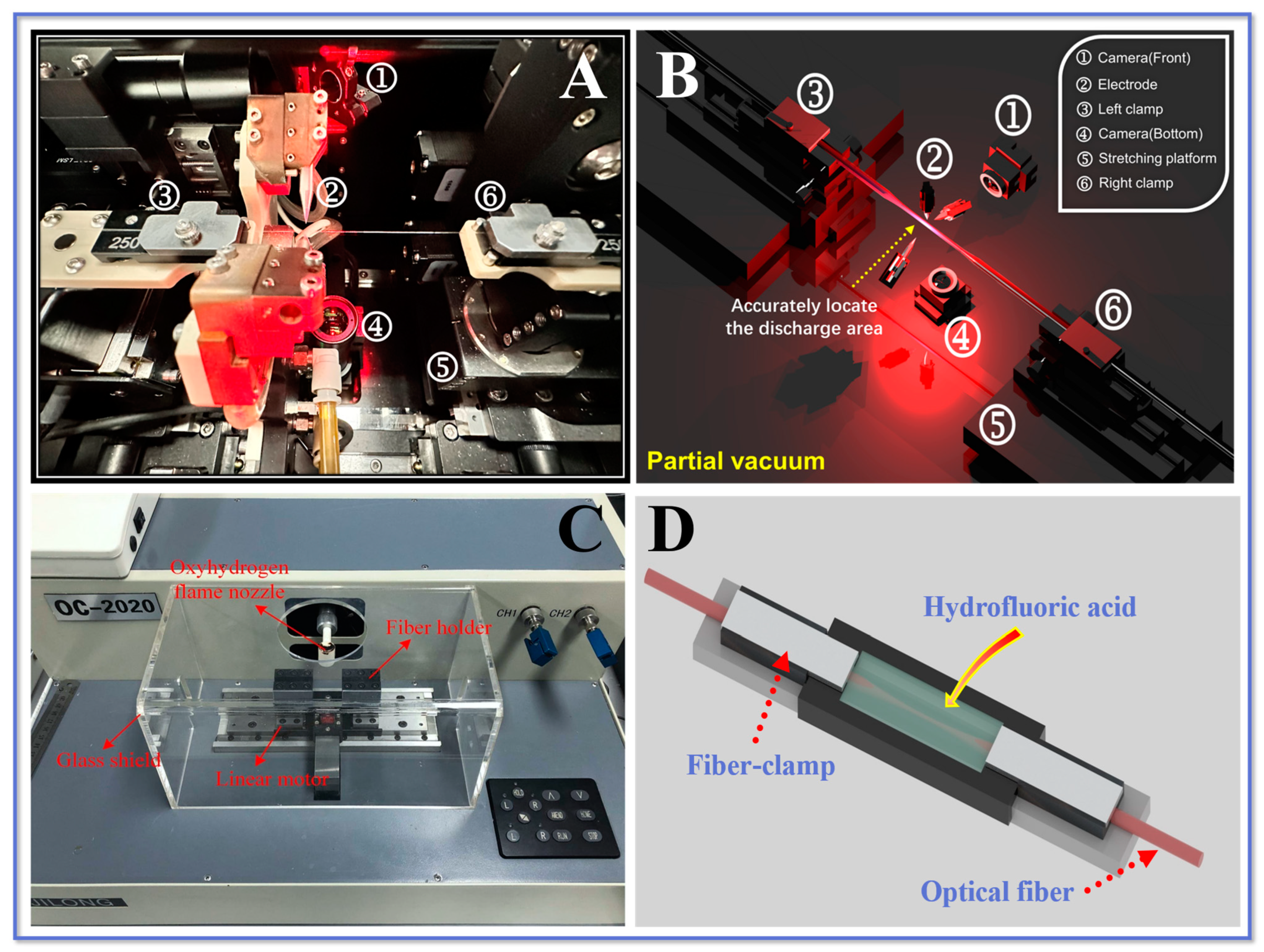
3. Fabrication Method of TOF
3.1. Arc Discharge Technology
3.2. Laser Processing Technology
3.3. Chemical Etching Technology
4. Signal Demodulation of TOF Sensors
4.1. Overview of Signal Demodulation Techniques
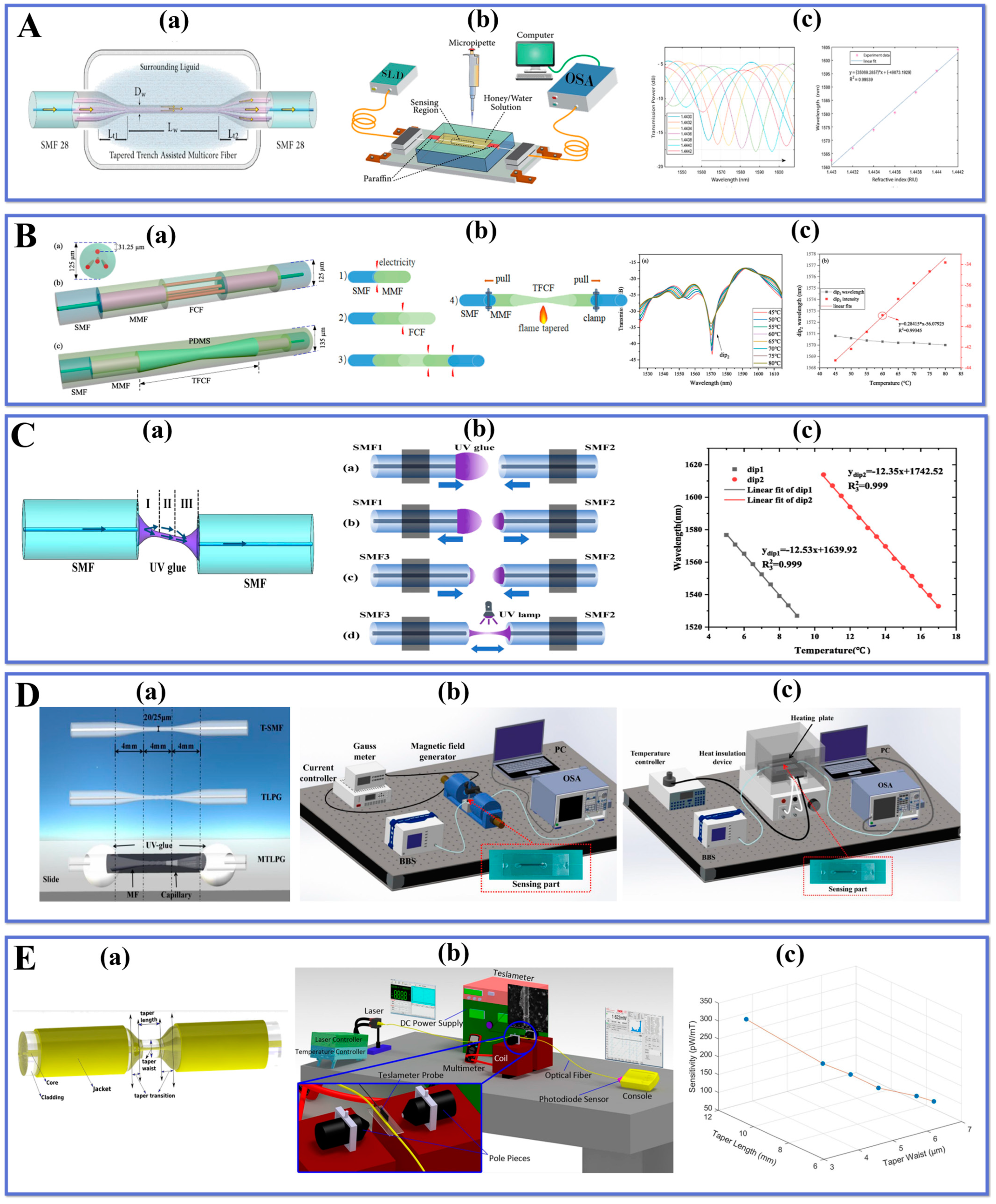
4.2. TOF Sensors Based on Signal Demodulation Techniques
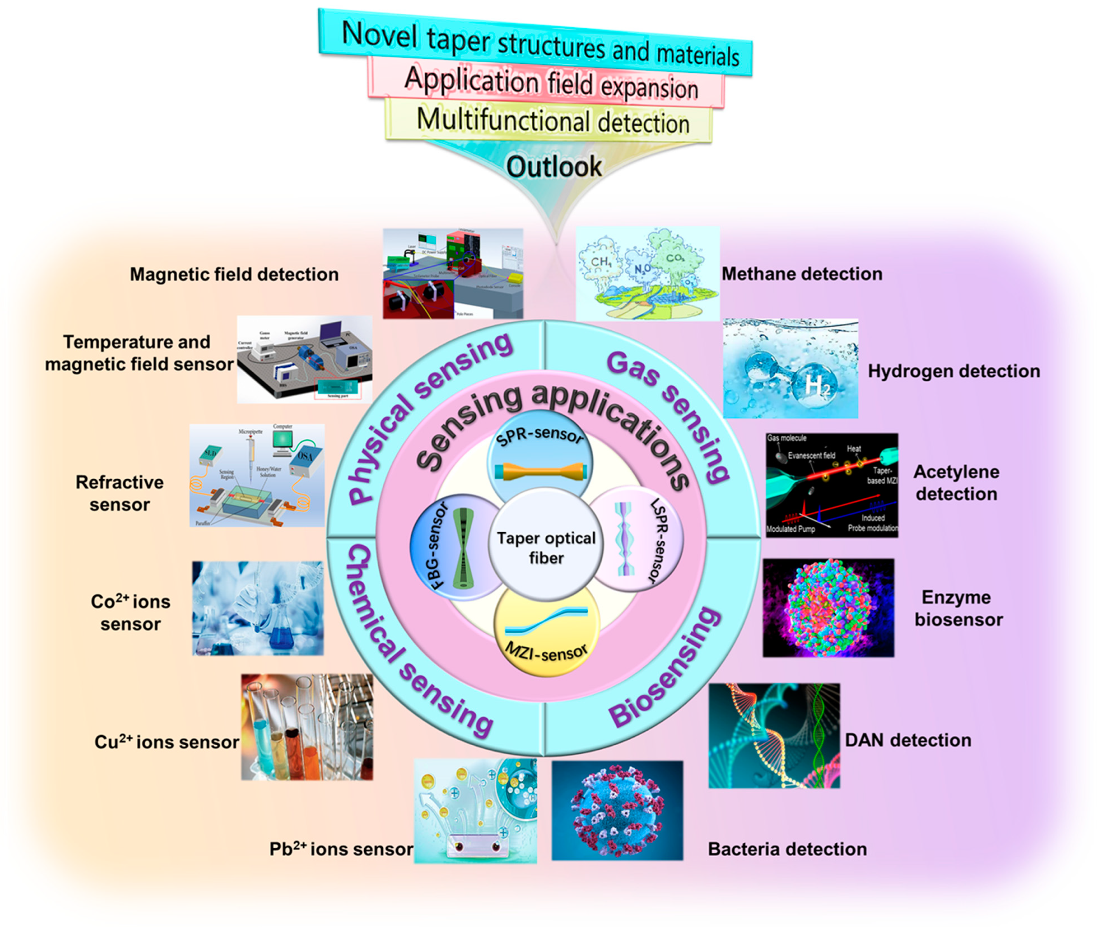
5. Applications of TOF Sensors
5.1. Physical Applications of TOF Sensors
5.2. Chemical Applications of TOF Sensors

5.3. TOF-Based Gas Sensors


5.4. TOF-Based Biosensors
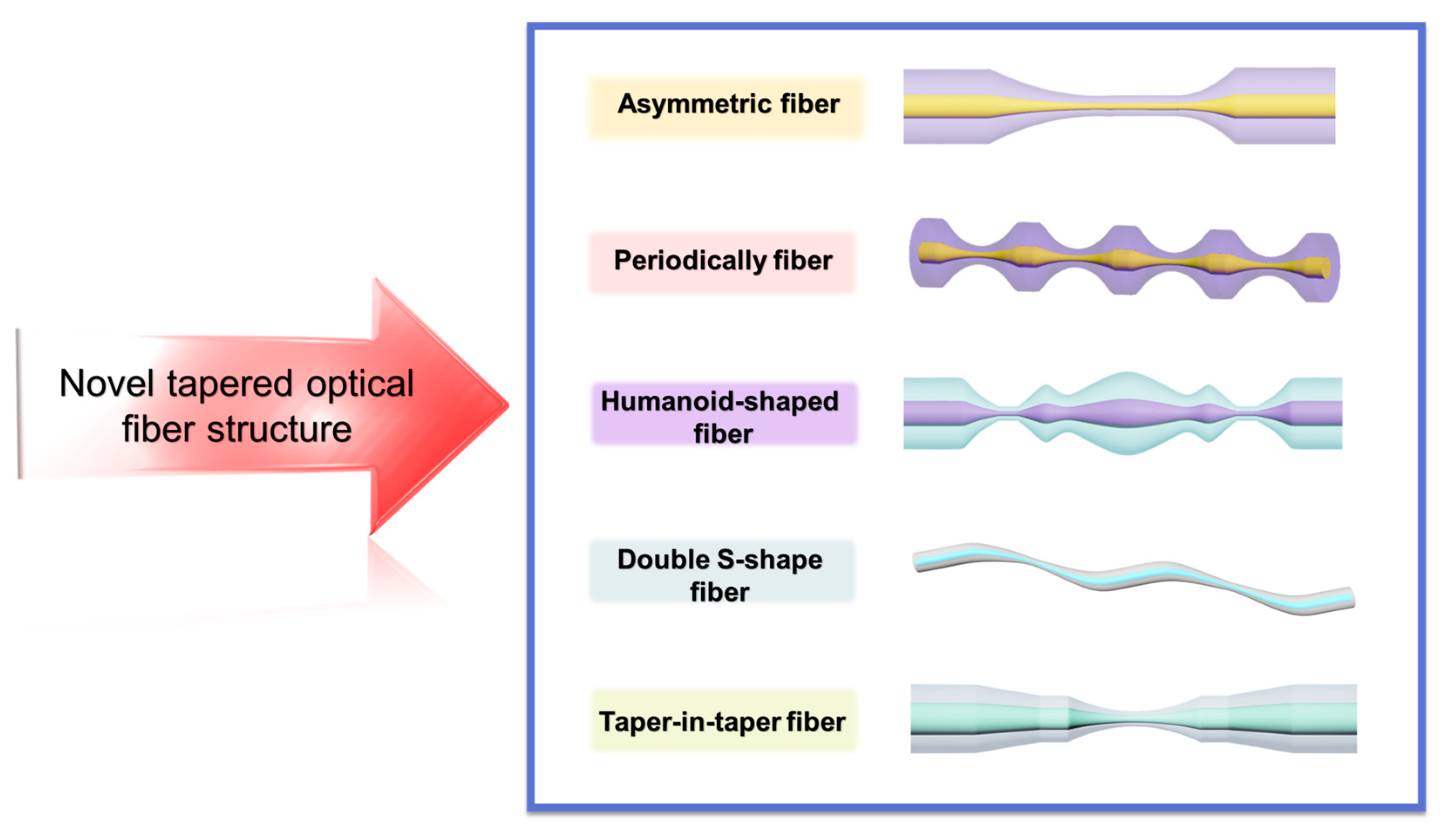
6. Novel TOF Structure
7. Future Prospects
8. Conclusions
Author Contributions
Funding
Institutional Review Board Statement
Informed Consent Statement
Data Availability Statement
Acknowledgments
Conflicts of Interest
References
- Hengoju, S.; Shvydkiv, O.; Tovar, M.; Roth, M.; Rosenbaum, M.A. Advantages of Optical Fibers for Facile and Enhanced Detection in Droplet Microfluidics. Biosens. Bioelectron. 2022, 200, 113910. [Google Scholar] [CrossRef] [PubMed]
- Temkina, V.; Medvedev, A.; Mayzel, A. Research on the Methods and Algorithms Improving the Measurements Precision and Market Competitive Advantages of Fiber Optic Current Sensors. Sensors 2020, 20, 5995. [Google Scholar] [CrossRef] [PubMed]
- Zhu, L.; Sun, G.; Bao, W.; You, Z.; Meng, F.; Dong, M. Structural Deformation Monitoring of Flight Vehicles Based on Optical Fiber Sensing Technology: A Review and Future Perspectives. Engineering 2022, 16, 39–55. [Google Scholar] [CrossRef]
- Komanec, M.; Dousek, D.; Suslov, D.; Zvanove, S. Hollow-Core Optical Fibers. Radioengineering 2020, 29, 417–430. [Google Scholar] [CrossRef]
- Pilipova, V.M.; Davydov, V.V.; Rud, V.Y. Development of a Fiber-Optic System for Testing Instruments for Monitoring Nuclear Power Plants. J. Phys. Conf. Ser. 2021, 2086, 012160. [Google Scholar] [CrossRef]
- Akram, S.; Sidén, J.; Duan, J.; Alam, M.F.; Bertilsson, K. Design and Development of a Battery Powered Electrofusion Welding System for Optical Fiber Microducts. IEEE Access 2020, 8, 173024–173043. [Google Scholar] [CrossRef]
- Raju, B.; Kumar, R.; Dhanalakshmi, S.; Dooly, G.; Duraibabu, D.B. Review of Fiber Optical Sensors and Its Importance in Sewer Corrosion Factor Analysis. Chemosensors 2021, 9, 118. [Google Scholar] [CrossRef]
- Zhang, S.; Peng, Y.; Wei, X.; Zhao, Y. High-Sensitivity Biconical Optical Fiber SPR Salinity Sensor with a Compact Size by Fiber Grinding Technique. Measurement 2022, 204, 112156. [Google Scholar] [CrossRef]
- Kuang, R.; Ye, Y.; Chen, Z.; He, R.; Savović, I.; Djordjevich, A.; Savović, S.; Ortega, B.; Marques, C.; Li, X.; et al. Low-Cost Plastic Optical Fiber Integrated with Smartphone for Human Physiological Monitoring. Opt. Fiber Technol. 2022, 71, 102947. [Google Scholar] [CrossRef]
- Yu, H.; Zheng, D.; Liu, Y.; Chen, S.; Wang, X.; Peng, W. Low-Cost Self-Calibration Data Glove Based on Space-Division Multiplexed Flexible Optical Fiber Sensor. Polymers 2022, 14, 3935. [Google Scholar] [CrossRef]
- Al-Qazwini, Y.; Noor, A.S.M.; Yaacob, M.H.; Harun, S.W.; Mahdi, M.A. Experimental Realization and Performance Evaluation of Refractive Index SPR Sensor Based on Unmasked Short Tapered Multimode-Fiber Operating in Aqueous Environments. Sens. Actuators A Phys. 2015, 236, 38–43. [Google Scholar] [CrossRef]
- Guo, Q.; Zhu, Y.; Shan, T.; Pan, X.; Liu, S.; Xue, Z.; Zheng, Z.; Chen, C.; Yu, Y. Intensity-Modulated Directional Torsion Sensor Based on a Helical Fiber Taper. Opt. Mater. Express OME 2021, 11, 80–88. [Google Scholar] [CrossRef]
- Liyanage, T.; Lai, M.; Slaughter, G. Label-Free Tapered Optical Fiber Plasmonic Biosensor. Anal. Chim. Acta 2021, 1169, 338629. [Google Scholar] [CrossRef]
- Li-hui, F.; Junfeng, D. Mixed Oil Detection Method Based on Tapered Fiber SPR Sensor. Opt. Fiber Technol. 2023, 78, 103322. [Google Scholar] [CrossRef]
- Taha, B.A.; Ali, N.; Sapiee, N.M.; Fadhel, M.M.; Mat Yeh, R.M.; Bachok, N.N.; Al Mashhadany, Y.; Arsad, N. Comprehensive Review Tapered Optical Fiber Configurations for Sensing Application: Trend and Challenges. Biosensors 2021, 11, 253. [Google Scholar] [CrossRef] [PubMed]
- Wang, P.; Zhao, H.; Wang, X.; Farrell, G.; Brambilla, G. A Review of Multimode Interference in Tapered Optical Fibers and Related Applications. Sensors 2018, 18, 858. [Google Scholar] [CrossRef] [Green Version]
- Chen, L.; Leng, Y.-K.; Liu, B.; Liu, J.; Wan, S.-P.; Wu, T.; Yuan, J.; Shao, L.; Gu, G.; Fu, Y.Q.; et al. Ultrahigh-Sensitivity Label-Free Optical Fiber Biosensor Based on a Tapered Singlemode- No Core-Singlemode Coupler for Staphylococcus Aureus Detection. Sens. Actuators B Chem. 2020, 320, 128283. [Google Scholar] [CrossRef]
- Zhao, Y.; Zhao, H.; Lv, R.; Zhao, J. Review of Optical Fiber Mach–Zehnder Interferometers with Micro-Cavity Fabricated by Femtosecond Laser and Sensing Applications. Opt. Lasers Eng. 2019, 117, 7–20. [Google Scholar] [CrossRef]
- Wang, F.; Pang, K.; Ma, T.; Wang, X.; Liu, Y. Folded-Tapered Multimode-No-Core Fiber Sensor for Simultaneous Measurement of Refractive Index and Temperature. Opt. Laser Technol. 2020, 130, 106333. [Google Scholar] [CrossRef]
- Wen, H.-Y.; Hsu, H.-C.; Tsai, Y.-T.; Feng, W.-K.; Lin, C.-L.; Chiang, C.-C. U-Shaped Optical Fiber Probes Coated with Electrically Doped GQDs for Humidity Measurements. Polymers 2021, 13, 2696. [Google Scholar] [CrossRef]
- Teng, C.; Shao, P.; Li, S.; Li, S.; Liu, H.; Deng, H.; Chen, M.; Yuan, L.; Deng, S. Double-Side Polished U-Shape Plastic Optical Fiber Based SPR Sensor for the Simultaneous Measurement of Refractive Index and Temperature. Opt. Commun. 2022, 525, 128844. [Google Scholar] [CrossRef]
- Bai, G.; Yin, Z.; Li, S.; Jing, X.; Chen, Q.; Zhang, M.; Shao, P. Enhancement of SPR Effect and Sensing Characteristics in D-Shaped Polished Grapefruit Microstructured Optical Fiber with Silver Film. Opt. Commun. 2023, 530, 129204. [Google Scholar] [CrossRef]
- Fu, X.; Li, D.; Zhang, Y.; Fu, G.; Jin, W.; Bi, W. High Sensitivity Refractive Index Sensor Based on Cascaded Core-Offset Splicing NCF-HCF-NCF Structure. Opt. Fiber Technol. 2022, 68, 102791. [Google Scholar] [CrossRef]
- Soares, M.S.; Silva, L.C.B.; Vidal, M.; Loyez, M.; Facão, M.; Caucheteur, C.; Segatto, M.E.V.; Costa, F.M.; Leitão, C.; Pereira, S.O.; et al. Label-Free Plasmonic Immunosensor for Cortisol Detection in a D-Shaped Optical Fiber. Biomed. Opt. Express 2022, 13, 3259–3274. [Google Scholar] [CrossRef]
- Wang, R.; Liu, C.; Wei, Y.; Jiang, T.; Liu, C.; Shi, C.; Zhao, X.; Li, L. Research and Application of Multi-Channel SPR Sensor Cascaded with Fiber U-Shaped Structure. Optik 2022, 266, 169603. [Google Scholar] [CrossRef]
- Wu, C.-W. Magnetic Field Sensor Based on Nickel-Coated S-Shaped Long Period Fiber Grating. Opt. Quant. Electron. 2018, 50, 357. [Google Scholar] [CrossRef]
- Sun, Y.; Guo, X.; Moreno, Y.; Sun, Q.; Yan, Z.; Zhang, L. Sensitivity Adjustable Biosensor Based on Graphene Oxide Coated Excessively Tilted Fiber Grating. Sens. Actuators B Chem. 2022, 351, 130832. [Google Scholar] [CrossRef]
- Liu, C.; Wu, P.; Shi, C.; Liu, C.; Wei, Y.; Hu, L.; Wang, R.; Jiang, T. Fiber SPR Micro Displacement Sensor Based on Heterocore Structure of Graded Index Multimode Fiber. Opt. Commun. 2023, 529, 129095. [Google Scholar] [CrossRef]
- Zhang, W.; Wu, M.; Jing, L.; Tong, Z.; Li, P.; Dong, M.; Tian, X.; Yan, G. Research on In-Line MZI Optical Fiber Salinity Sensor Based on Few-Mode Fiber with Core-Offset Structure. Measurement 2022, 202, 111857. [Google Scholar] [CrossRef]
- Liu, X.; Singh, R.; Li, M.; Li, G.; Min, R.; Marques, C.; Zhang, B.; Kumar, S. Plasmonic Sensor Based on Offset-Splicing and Waist-Expanded Taper Using Multicore Fiber for Detection of Aflatoxins B1 in Critical Sectors. Opt. Express 2023, 31, 4783. [Google Scholar] [CrossRef]
- Kumar, S.; Singh, R.; Zhu, G.; Yang, Q.; Zhang, X.; Cheng, S.; Zhang, B.; Kaushik, B.K.; Liu, F.-Z. Development of Uric Acid Biosensor Using Gold Nanoparticles and Graphene Oxide Functionalized Micro-Ball Fiber Sensor Probe. IEEE Trans. NanoBioscience 2020, 19, 173–182. [Google Scholar] [CrossRef] [PubMed]
- Liu, H.; Sun, Y.; Guo, J.; Liu, W.; Liu, L.; Meng, Y.; Yu, X. Temperature-Insensitive Label-Free Sensors for Human IgG Based on S-Tapered Optical Fiber Sensors. IEEE Access 2021, 9, 116286–116293. [Google Scholar] [CrossRef]
- Wu, Y.; Liu, B.; Nan, T.; Wu, J.; Mao, Y.; Ren, J.; Zhao, L.; Sun, T.; Wang, J.; Han, Y.; et al. Fiber Optic Hybrid Structure Based on an Air Bubble and Thin Taper for Measurement of Refractive Index, Temperature, and Transverse Load. Optik 2021, 241, 166962. [Google Scholar] [CrossRef]
- Gong, Z.; Lei, Y.; Wang, Z.; Zhang, J.; Sun, Z.; Li, Y.; Huang, J.; Chan, C.; Ouyang, X. A Taper-in-Taper Structured Interferometric Optical Fiber Sensor for Cu2+ Ion Detection. Sensors 2022, 22, 2709. [Google Scholar] [CrossRef] [PubMed]
- Li, Z.; Wang, Z.; Qi, Y.; Jin, W.; Ren, W. Improved Evanescent-Wave Quartz-Enhanced Photoacoustic CO Sensor Using an Optical Fiber Taper. Sens. Actuators B Chem. 2017, 248, 1023–1028. [Google Scholar] [CrossRef]
- Liu, Z.; Guo, C.; Yang, J.; Yuan, L. Tapered Fiber Optical Tweezers for Microscopic Particle Trapping: Fabrication and Application. Opt. Express OE 2006, 14, 12510–12516. [Google Scholar] [CrossRef]
- Anbalagan, T.; Haroon, H.; Zain, H.A.; Rafis, H.; Harun, S.W. Recent Progress of Tapered Optical Fiber for Biosensing Applications. JESTR 2022, 15, 204–209. [Google Scholar] [CrossRef]
- Bhardwaj, V.; Kishor, K.; Sharma, A.C. Tapered Optical Fiber Geometries and Sensing Applications Based on Mach-Zehnder Interferometer: A Review. Opt. Fiber Technol. 2020, 58, 102302. [Google Scholar] [CrossRef]
- Stasiewicz, K.A.; Jakubowska, I.; Dudek, M. Detection of Organosulfur and Organophosphorus Compounds Using a Hexafluorobutyl Acrylate-Coated Tapered Optical Fibers. Polymers 2022, 14, 612. [Google Scholar] [CrossRef]
- Liu, Y.; Liu, R.; Ai, C.; Wang, B.; Chu, R.; Wang, H.; Shui, L.; Zhou, F. Stick-Slip-Motion-Assisted Interfacial Self-Assembly of Noble Metal Nanoparticles on Tapered Optical Fiber Surface and Its Application in SERS Detection. Appl. Surf. Sci. 2022, 602, 154298. [Google Scholar] [CrossRef]
- Noor, A.S.M.; Talah, A.; Rosli, M.A.A.; Thirunavakkarasu, P.; Tamchek, N. Increased Sensitivity of Au-Pd Nanolayer on Tapered Optical Fiber Sensor for Detecting Aqueous Ethanol. J. Eur. Opt. Soc.-Rapid Publ. 2017, 13, 28. [Google Scholar] [CrossRef] [Green Version]
- Polokhin, A.A.; Shaman, Y.P.; Itrin, P.A.; Panyaev, I.S.; Sysa, A.A.; Selishchev, S.V.; Kitsyuk, E.P.; Pavlov, A.A.; Gerasimenko, A.Y. Tapered Optical Fiber Sensor Coated with Single-Walled Carbon Nanotubes for Dye Sensing Application. Micromachines 2023, 14, 579. [Google Scholar] [CrossRef] [PubMed]
- Karimi-Alavijeh, H.; Taslimi, A.; Maghsoudian, M.H.; Poorghadiri, M.H.; Kazemzadeh, M. Fabrication of Low-Loss Adiabatic Optical Microfibers Using an Attainable Arc-Discharge Fiber Tapering Setup. Opt. Commun. 2022, 522, 128669. [Google Scholar] [CrossRef]
- Hidayat, N.; Aziz, M.S.; Krishnan, G.; Johari, A.R.; Nur, H.; Taufiq, A.; Mufti, N.; Mukti, R.R.; Bakhtiar, H. Tapered Optical Fibers Using CO2 Laser and Their Sensing Performances. J. Phys. Conf. Ser. 2023, 2432, 012013. [Google Scholar] [CrossRef]
- Tang, Y.; Chen, X.; Zhang, J.; Lv, D.; Xiong, L.; Dong, X. Sensitivity-Enhanced Hot-Wire Anemometer by Using Cladding-Etched Fiber Bragg Grating. Photonic. Sens. 2023, 13, 230305. [Google Scholar] [CrossRef]
- Martan, T.; Kanka, J.; Kasik, I.; Matejec, V. Tapered Optical Fibres for Sensing. In Proceedings of the Photonics, Devices, and Systems IV, Prague, Czech Republic, 18 November 2008; SPIE: Bellingham, DC, USA, 2008; Volume 7138, pp. 256–261. [Google Scholar]
- Sun, M.; Liu, Y.; Chen, D.; Qian, Q. Multifunctional Cu-Se Alloy Core Fibers and Micro–Nano Tapers. Nanomaterials 2023, 13, 773. [Google Scholar] [CrossRef] [PubMed]
- Zhou, J.; He, X.; Yin, T.; Yang, J.; Guan, C.; Yuan, L. A Chirped Long Period Fiber Grating Sensor Based on Micro-Helix Taper. Opt. Fiber Technol. 2023, 78, 103289. [Google Scholar] [CrossRef]
- Fu, X.; Huang, Z.; Li, Q.; Cao, X.; Wang, Y.; Fu, G.; Jin, W.; Bi, W. A Cascaded Triple Waist-Enlarged Taper Few-Mode Fiber Temperature Sensor with Beaded Structure. Opt. Laser Technol. 2022, 156, 108621. [Google Scholar] [CrossRef]
- Aerts, J.T.; Andrén, P.E.; Jansson, E.T. Electrochemically Etched Tapered-Tip Stainless-Steel Electrospray-Ionization Emitters for Capillary Electrophoresis–Mass Spectrometry. J. Proteome Res. 2023, 22, 1377–1380. [Google Scholar] [CrossRef]
- Shaimerdenova, M.; Ayupova, T.; Ashikbayeva, Z.; Bekmurzayeva, A.; Blanc, W.; Tosi, D. Reflector-Less Shallow-Tapered Optical Fiber Biosensors for Rapid Detection of Cancer Biomarkers. J. Light. Technol. 2022, 1–10. [Google Scholar] [CrossRef]
- Ayupova, T.; Shaimerdenova, M.; Tosi, D. Shallow-Tapered Chirped Fiber Bragg Grating Sensors for Dual Refractive Index and Temperature Sensing. Sensors 2021, 21, 3635. [Google Scholar] [CrossRef] [PubMed]
- Zajíc, J.; Ripp, S.; Trögl, J.; Kuncová, G.; Pospíšilová, M. Repetitive Detection of Aromatic Hydrocarbon Contaminants with Bioluminescent Bioreporters Attached on Tapered Optical Fiber Elements. Sensors 2020, 20, 3237. [Google Scholar] [CrossRef]
- Chen, L.; Leng, Y.-K.; Qiu, S.; Liu, B.; Liu, J.; Wan, S.-P.; Wu, T.; Xu, H.; Xiong, Y.; Yuan, J.; et al. Ultrahigh-Sensitivity Label-Free Singlemode- Tapered No Core-Singlemode Fiber Immunosensor for Listeria Monocytogenes Detection. Sens. Actuators B Chem. 2023, 376, 132930. [Google Scholar] [CrossRef]
- Arjmand, M.; Saghafifar, H.; Alijanianzadeh, M.; Soltanolkotabi, M. A Sensitive Tapered-Fiber Optic Biosensor for the Label-Free Detection of Organophosphate Pesticides. Sens. Actuators B Chem. 2017, 249, 523–532. [Google Scholar] [CrossRef]
- Korposh, S.; James, S.W.; Lee, S.-W.; Tatam, R.P. Tapered Optical Fibre Sensors: Current Trends and Future Perspectives. Sensors 2019, 19, 2294. [Google Scholar] [CrossRef] [PubMed] [Green Version]
- Yang, Q.; Zhu, G.; Singh, L.; Wang, Y.; Singh, R.; Zhang, B.; Zhang, X.; Kumar, S. Highly Sensitive and Selective Sensor Probe Using Glucose Oxidase/Gold Nanoparticles/Graphene Oxide Functionalized Tapered Optical Fiber Structure for Detection of Glucose. Optik 2020, 208, 164536. [Google Scholar] [CrossRef]
- AL-Mashhadani, Z.A.A.; Navruz, I. Highly Sensitive Measurement of Surrounding Refractive Index Using Tapered Trench–Assisted Multicore Fiber. Opt. Fiber Technol. 2019, 48, 76–83. [Google Scholar] [CrossRef]
- Shao, F.; Li, S.; Lu, L.; Kuai, Y.; Cao, Z.; Xu, F.; Liu, Y.; Xie, F.; Xie, K.; Yu, B.; et al. High Sensitivity and Dual Parameters Micro-Tapered-LPG Sensor. Opt. Lasers Eng. 2023, 164, 107498. [Google Scholar] [CrossRef]
- Bakhshi, A.; Jalaly, M.; Vahedi, M. The Effect of GO–Fe3O4 Hybrid Coating on the Magnetic Field Detection by a Tapered Optical Fiber Sensor. Opt. Fiber Technol. 2022, 74, 103134. [Google Scholar] [CrossRef]
- Tan, Y.; Huang, T.; Sun, L.-P.; Jiang, S.; Liu, Y.; Guan, B.-O.; Jin, W. Dispersion Turning Point-Enhanced Photothermal Interferometry Gas Sensor with an Optical Microfiber Interferometer. Sens. Actuators B Chem. 2023, 385, 133690. [Google Scholar] [CrossRef]
- Danny, C.G.; Danny Raj, M.; Sai, V.V.R. Investigating the Refractive Index Sensitivity of U-Bent Fiber Optic Sensors Using Ray Optics. J. Light. Technol. 2020, 38, 1580–1588. [Google Scholar] [CrossRef] [Green Version]
- Tai, Y.-H.; Kamal, A.S.A.; Park, Y.-J.; Tsai, P.-C.; Ho, Y.-L.; Wei, P.-K.; Daiguji, H.; Delaunay, J.-J. Real-Time Monitoring of Frost/Defrost Processes Using a Tapered Optical Fiber. IEEE Sens. J. 2021, 21, 6188–6194. [Google Scholar] [CrossRef]
- Addanki, S.; Amiri, I.S.; Yupapin, P. Review of Optical Fibers-Introduction and Applications in Fiber Lasers. Results Phys. 2018, 10, 743–750. [Google Scholar] [CrossRef]
- Li, L.; Zhang, Y.; Zhou, Y.; Zheng, W.; Sun, Y.; Ma, G.; Zhao, Y. Optical Fiber Optofluidic Bio-Chemical Sensors: A Review. Laser Photonics Rev. 2021, 15, 2000526. [Google Scholar] [CrossRef]
- Chen, N.; Zhou, X.; Li, X. Highly Sensitive Humidity Sensor With Low-Temperature Cross-Sensitivity Based on a Polyvinyl Alcohol Coating Tapered Fiber. IEEE Trans. Instrum. Meas. 2021, 70, 9503308. [Google Scholar] [CrossRef]
- Cano Perez, J.L.; Gutiérrez-Gutiérrez, J.; Perezcampos Mayoral, C.; Pérez-Campos, E.L.; Pina Canseco, M.d.S.; Tepech Carrillo, L.; Mayoral, L.P.-C.; Vargas Treviño, M.; Apreza, E.L.; Rojas Laguna, R. Fiber Optic Sensors: A Review for Glucose Measurement. Biosensors 2021, 11, 61. [Google Scholar] [CrossRef]
- Zhao, Y.; Lei, M.; Liu, S.-X.; Zhao, Q. Smart Hydrogel-Based Optical Fiber SPR Sensor for PH Measurements. Sens. Actuators B Chem. 2018, 261, 226–232. [Google Scholar] [CrossRef]
- Yang, Y.; Ge, K.; Tao, P.; Dai, S.; Wang, X.; Zhang, W.; Xu, T.; Wang, Y.; Lin, T.-J.; Zhang, P. Mid-Infrared Evanescent Wave Sensor Based on Side-Polished Chalcogenide Fiber. Ceram. Int. 2023, 49, 1291–1297. [Google Scholar] [CrossRef]
- Jiang, J.; Wu, R.; Liu, J.; Wu, X.; Wu, Q.; Ranjan, P.; Zhang, X.; Liu, J. S-Taper Fiber Based Moisture Sensing in Power Transformer Oil. IEEE Trans. Instrum. Meas. 2023, 72, 9001208. [Google Scholar] [CrossRef]
- Patil, P.O.; Pandey, G.R.; Patil, A.G.; Borse, V.B.; Deshmukh, P.K.; Patil, D.R.; Tade, R.S.; Nangare, S.N.; Khan, Z.G.; Patil, A.M.; et al. Graphene-Based Nanocomposites for Sensitivity Enhancement of Surface Plasmon Resonance Sensor for Biological and Chemical Sensing: A Review. Biosens. Bioelectron. 2019, 139, 111324. [Google Scholar] [CrossRef]
- Shalabney, A.; Abdulhalim, I. Sensitivity-Enhancement Methods for Surface Plasmon Sensors: SPR Sensors Sensitivity Enhancement. Laser Photon. Rev. 2011, 5, 571–606. [Google Scholar] [CrossRef]
- Pandey, P.S.; Raghuwanshi, S.K.; Kumar, S. Recent Advances in Two-Dimensional Materials-Based Kretschmann Configuration for SPR Sensors: A Review. IEEE Sens. J. 2022, 22, 1069–1080. [Google Scholar] [CrossRef]
- Kumar, S.; Singh, R.; Wang, Z.; Li, M.; Liu, X.; Zhang, W.; Zhang, B.; Li, G. (Invited) Advances in 2D Nanomaterials-Assisted Plasmonics Optical Fiber Sensors for Biomolecules Detection. Results Opt. 2023, 10, 100342. [Google Scholar] [CrossRef]
- Wang, B.-T.; Wang, Q. Sensitivity-Enhanced Optical Fiber Biosensor Based on Coupling Effect Between SPR and LSPR. IEEE Sens. J. 2018, 18, 8303–8310. [Google Scholar] [CrossRef]
- Feng, J.; Gao, J.; Yang, W.; Liu, R.; Shafi, M.; Zha, Z.; Liu, C.; Xu, S.; Ning, T.; Jiang, S. LSPR Optical Fiber Sensor Based on 3D Gold Nanoparticles with Monolayer Graphene as a Spacer. Opt. Express 2022, 30, 10187. [Google Scholar] [CrossRef] [PubMed]
- Dos Santos, P.S.S.; Mendes, J.P.; Dias, B.; Pérez-Juste, J.; De Almeida, J.M.M.M.; Pastoriza-Santos, I.; Coelho, L.C.C. Spectral Analysis Methods for Improved Resolution and Sensitivity: Enhancing SPR and LSPR Optical Fiber Sensing. Sensors 2023, 23, 1666. [Google Scholar] [CrossRef] [PubMed]
- Mayer, K.M.; Hafner, J.H. Localized Surface Plasmon Resonance Sensors. Chem. Rev. 2011, 111, 3828–3857. [Google Scholar] [CrossRef] [PubMed]
- Zhang, J.; Kolhatkar, G.; Ruediger, A. Localized Surface Plasmon Resonance Shift and Its Application in Scanning Near-Field Optical Microscopy. J. Mater. Chem. C 2021, 9, 6960–6969. [Google Scholar] [CrossRef]
- Xia, F.; Zhao, Y.; Peng, Y. In-Line Microfiber MZI Operating at Two Sides of the Dispersion Turning Point for Ultrasensitive RI and Temperature Measurement. Sens. Actuators A Phys. 2020, 301, 111754. [Google Scholar] [CrossRef]
- Wang, Q.; Kong, L.; Dang, Y.; Xia, F.; Zhang, Y.; Zhao, Y.; Hu, H.; Li, J. High Sensitivity Refractive Index Sensor Based on Splicing Points Tapered SMF-PCF-SMF Structure Mach-Zehnder Mode Interferometer. Sens. Actuators B Chem. 2016, 225, 213–220. [Google Scholar] [CrossRef] [Green Version]
- Fu, X.; Huang, Z.; Zhou, J.; Zhang, W.; Jin, W.; Fu, G.; Liu, F.; Bi, W.; Luo, Y.; Peng, G.-D. A Dual-Parameter Sensing System for Temperature and Curvature Based on Nano-EYDF and in-Line Cascaded Mach-Zehnder Interferometer. Opt. Laser Technol. 2023, 163, 109353. [Google Scholar] [CrossRef]
- Xia, F.; Zhao, Y.; Zheng, H.; Li, L.; Tong, R. Ultra-Sensitive Seawater Temperature Sensor Using an FBG-Cascaded Microfiber MZI Operating at Dispersion Turning Point. Opt. Laser Technol. 2020, 132, 106458. [Google Scholar] [CrossRef]
- Riza, M.A.; Go, Y.I.; Harun, S.W.; Maier, R.R.J. FBG Sensors for Environmental and Biochemical Applications—A Review. IEEE Sens. J. 2020, 20, 7614–7627. [Google Scholar] [CrossRef]
- Jiao, L.; Zhong, N.; Zhao, X.; Ma, S.; Fu, X.; Dong, D. Recent Advances in Fiber-Optic Evanescent Wave Sensors for Monitoring Organic and Inorganic Pollutants in Water. TrAC Trends Anal. Chem. 2020, 127, 115892. [Google Scholar] [CrossRef]
- Gan, W.; Li, Y.; Liu, T.; Yang, Y.; Song, B.; Dai, S.; Xu, T.; Wang, Y.; Lin, T.-J.; Zhang, P. Rapid and Sensitive Detection of Ammonia in Water by a Long Period Fiber Grating Sensor Coated with Sol-Gel Silica. Opt. Express 2022, 30, 33817. [Google Scholar] [CrossRef]
- Zhang, W.; Zhuang, W.; Dong, M.; Zhu, L.; Meng, F. Dual-Parameter Optical Fiber Sensor for Temperature and Pressure Discrimination Featuring Cascaded Tapered-FBG and Ball-EFPI. IEEE Sens. J. 2019, 19, 5645–5652. [Google Scholar] [CrossRef]
- Son, G.; Jung, Y.; Yu, K. Tapered Optical Fiber Couplers Fabricated by Droplet-Based Chemical Etching. IEEE Photonics J. 2017, 9, 1–8. [Google Scholar] [CrossRef]
- Ascorbe, J.; Corres, J.M.; Matias, I.R.; Arregui, F.J. High Sensitivity Humidity Sensor Based on Cladding-Etched Optical Fiber and Lossy Mode Resonances. Sens. Actuators B Chem. 2016, 233, 7–16. [Google Scholar] [CrossRef] [Green Version]
- Li, Y.; Wang, Y.; Xiao, L.; Bai, Q.; Liu, X.; Gao, Y.; Zhang, H.; Jin, B. Phase Demodulation Methods for Optical Fiber Vibration Sensing System: A Review. IEEE Sens. J. 2022, 22, 1842–1866. [Google Scholar] [CrossRef]
- Zheng, Y.; Zhu, Z.-W.; Li, W.-J.; Gu, D.-M.; Xiao, W. Experimental Research on a Novel Optic Fiber Sensor Based on OTDR for Landslide Monitoring. Measurement 2019, 148, 106926. [Google Scholar] [CrossRef]
- Kwon, Y.; Naeem, K.; Jeon, M.Y.; Kwon, I.-B. Enhanced Sensitivity of Distributed-Temperature Sensor with Al-Coated Fiber Based on OFDR. Opt. Fiber Technol. 2019, 48, 229–234. [Google Scholar] [CrossRef]
- Liu, J.; Hou, Y.; Wang, J.; Zhong, G.; Zhang, L.; Zhuang, F.; Yu, L.; Wang, S. Multi-Parameter Demodulation for Temperature, Salinity and Pressure Sensor in Seawater Based on the Semi-Encapsulated Microfiber Mach-Zehnder Interferometer. Measurement 2022, 196, 111213. [Google Scholar] [CrossRef]
- Yu, Y.; Bian, Q.; Lu, Y.; Zhang, X.; Yang, J.; Liang, L. High Sensitivity All Optical Fiber Conductivity-Temperature-Depth (CTD) Sensing Based on an Optical Microfiber Coupler (OMC). J. Light. Technol. 2019, 37, 2739–2747. [Google Scholar] [CrossRef]
- Yi, X.; Li, Y.; Zhao, K.; Wu, Z.; Wang, Q.; Liu, B.; Buric, M.; Wright, R.; Chen, K.P. CO2 Laser Tapering of Intrinsic Fabry–Perot Interferometers for Sensing. IEEE Sens. J. 2023, 23, 5824–5830. [Google Scholar] [CrossRef]
- Niu, P.; Jiang, J.; Liu, K.; Wang, S.; Jing, J.; Xu, T.; Wang, T.; Liu, Y.; Liu, T. Fiber-Integrated WGM Optofluidic Chip Enhanced by Microwave Photonic Analyzer for Cardiac Biomarker Detection with Ultra-High Resolution. Biosens. Bioelectron. 2022, 208, 114238. [Google Scholar] [CrossRef] [PubMed]
- Chen, Q.; Yang, W.; Zhang, L.; Li, C.; Guan, C.; Xiong, Y.; Yang, Y.; Li, L. The Fiber Temperature Sensor with PDMS Sensitization Based on the T-MFM Fiber Structure. Opt. Fiber Technol. 2021, 67, 102701. [Google Scholar] [CrossRef]
- Liu, J.; Xu, M.; Abbas, L.G.; Jing, C.; Yao, W.; Zhou, Q.; Zhou, A. High-Sensitivity Temperature Sensor Based on Mach-Zehnder Interference of Asymmetric Taper-Shaped Ultraviolet Glue. Opt. Fiber Technol. 2022, 72, 102997. [Google Scholar] [CrossRef]
- Li, Y.; Song, Z.; Pan, J.; Lu, H.; Hu, J. In-Line Reflected Fiber Sensor for Simultaneous Measurement of Temperature and Liquid Level Based on Tapered Few-Mode Fiber. Opt. Express 2022, 30, 7870. [Google Scholar] [CrossRef]
- Lu, C.; Dashtabi, M.M.; Nikbakht, H.; Khoshmehr, M.T.; Akca, B.I. Sub-Nanometer Acoustic Vibration Sensing Using a Tapered-Tip Optical Fiber Microcantilever. Sensors 2023, 23, 924. [Google Scholar] [CrossRef]
- Fu, J.; Xu, Y.; Xu, M.; Abbas, L.G.; Zhou, A. Highly Sensitive Humidity Sensor Based on Tapered Dual Side-Hole Fiber. Optik 2022, 261, 169183. [Google Scholar] [CrossRef]
- Shao, M.; Han, L.; Liang, J.; Zhang, R.; Gao, H. A High-Sensitivity Liquid Level Sensor Based on Single-Mode Taper-Thin Core Taper Single-Mode Fiber Structure. Meas. Sci. Technol. 2020, 31, 105101. [Google Scholar] [CrossRef]
- Wu, Q.; Zhao, Y.; Zhang, Y.; Yang, Y. High Sensitive Applied Load Measurement Using Optical Fiber Tapered-Loop Probe with SPR Effect. Opt. Laser Technol. 2019, 114, 95–102. [Google Scholar] [CrossRef]
- Kumar, R. Magnetic Field Sensing Using Tapered Small-Core Optical Fibre Surrounded by Different Concentrations of Magnetic Fluid. Sensors 2022, 22, 8536. [Google Scholar] [CrossRef] [PubMed]
- Li, J.; Gan, W.; Li, H.; Xu, M.; Liu, J.; Zhou, A. Temperature Compensated Highly Sensitive Refractive Index Sensor Based on Mach-Zehnder Interferometer and FBG. Optik 2021, 241, 166838. [Google Scholar] [CrossRef]
- Ahsani, V.; Ahmed, F.; Jun, M.B.G.; Bradley, C. Tapered Fiber-Optic Mach-Zehnder Interferometer for Ultra-High Sensitivity Measurement of Refractive Index. Sensors 2019, 19, 1652. [Google Scholar] [CrossRef] [Green Version]
- Teng, C.; Li, M.; Cheng, Y.; Peng, H.; Deng, S.; Deng, H.; Yuan, L.; Chen, M. Investigation of U-Shape Tapered Plastic Optical Fibers Based Surface Plasmon Resonance Sensor for RI Sensing. Optik 2022, 251, 168461. [Google Scholar] [CrossRef]
- Mumtaz, F.; Cheng, P.; Li, C.; Cheng, S.; Du, C.; Yang, M.; Dai, Y.; Hu, W. A Design of Taper-Like Etched Multicore Fiber Refractive Index-Insensitive a Temperature Highly Sensitive Mach-Zehnder Interferometer. IEEE Sens. J. 2020, 20, 7074–7081. [Google Scholar] [CrossRef]
- Liao, Y.-C.; Liu, B.; Liu, J.; Wan, S.-P.; He, X.-D.; Yuan, J.; Fan, X.; Wu, Q. High Temperature (Up to 950 °C) Sensor Based on Micro Taper In-Line Fiber Mach–Zehnder Interferometer. Appl. Sci. 2019, 9, 2394. [Google Scholar] [CrossRef] [Green Version]
- Kang, X.; Wang, R.; Jiang, M.; Li, E.; Li, Y.; Yan, X.; Wang, T.; Ren, Z. Polydopamine Functionalized Graphene Oxide for High Sensitivity Micro-Tapered Long Period Fiber Grating Sensor and Its Application in Detection Co2+ Ions. Opt. Fiber Technol. 2022, 68, 102807. [Google Scholar] [CrossRef]
- Liu, T.; Wang, W.; Jian, D.; Li, J.; Ding, H.; Yi, D.; Liu, F.; Wang, S. Quantitative Remote and On-Site Hg2+ Detection Using the Handheld Smartphone Based Optical Fiber Fluorescence Sensor (SOFFS). Sens. Actuators B Chem. 2019, 301, 127168. [Google Scholar] [CrossRef]
- Wei, Y.; Ran, Z.; Liu, C.; Wang, R.; Jiang, T.; Liu, C.; Shi, C.; Ren, Z.; Wang, X.; Tan, W.; et al. Twisted Fiber SPR Sensor for Copper Ion Detection. Optik 2022, 271, 170208. [Google Scholar] [CrossRef]
- Wang, G.; Sun, D.; Liang, L.; Wang, G.; Ma, J. Highly Sensitive Detection of Trace Lead Ions Concentration Based on a Functional Film-Enhanced Optical Microfiber Sensor. Opt. Laser Technol. 2023, 161, 109171. [Google Scholar] [CrossRef]
- Yap, S.H.K.; Chan, K.K.; Zhang, G.; Tjin, S.C.; Yong, K.-T. Carbon Dot-Functionalized Interferometric Optical Fiber Sensor for Detection of Ferric Ions in Biological Samples. ACS Appl. Mater. Interfaces 2019, 11, 28546–28553. [Google Scholar] [CrossRef] [PubMed]
- Teng, P.; Jiang, Y.; Chang, X.; Shen, Y.; Liu, Z.; Copner, N.; Yang, J.; Li, K.; Bowkett, M.; Yuan, L.; et al. Highly Sensitive On-Line Detection of Trace Pb2+ Based on Tapered Fiber Integrated with Black Phosphorus. Opt. Fiber Technol. 2021, 66, 102668. [Google Scholar] [CrossRef]
- Chauhan, M.; Singh, V.K. TiO2 Coated Tapered Optical Fiber SPR Sensor for Alcohol Sensing Application. J. Opt. 2023, 1–11. [Google Scholar] [CrossRef]
- Chauhan, M.; Singh, V.K. Tapered MMF Sensor Fabrication Using SnO2-NPs for Alcohol Sensing Application. Opt. Fiber Technol. 2023, 75, 103167. [Google Scholar] [CrossRef]
- Yan, Y.; Fan, M.; Zhou, S.; Sun, X.; Ma, L.; Li, R.; Kost, A.R. Tapered Mach–Zehnder Interferometer Based on PbS Quantum Dots Modified by Polymers for Copper Ion Sensing. Appl. Opt. AO 2021, 60, 4807–4813. [Google Scholar] [CrossRef]
- Venkataraj, R.; Nampoori, V.P.N.; Radhakrishnan, P.; Kailasnath, M. Chemically Tapered Multimode Optical Fiber Probe for Fluoride Detection Based on Fluorescence Quenching of Curcumin. IEEE Sens. J. 2015, 15, 5584–5591. [Google Scholar] [CrossRef]
- Jiang, Y.; Feng, W.; Yang, X. Aluminium-Doped Zinc Oxide Sensing Membrane Integrated Fiber-Optic Michelson Interferometer for Trace Fluoride-Ion Detection. J. Phys. D Appl. Phys. 2021, 54, 475102. [Google Scholar] [CrossRef]
- Yusoff, A.; Azeman, N.H.; Mohamed Kassim, M.F.; Mobarak, N.N.; Haji Badri, K.; Mahdi, M.A.; Su’Ait, M.S.; Bakar, A.A.A. Bio-Based Polycationic Polyurethane as an Ion-Selective Membrane for Nitrate Tapered Optical Fiber Sensors. IEEE Access 2019, 7, 157103–157112. [Google Scholar] [CrossRef]
- Wang, R.; Kang, X.; Kong, D.; Jiang, M.; Ren, Z.; Hu, B.; He, Z. Highly Sensitive Metal Ion Sensing by Graphene Oxide Functionalized Micro-Tapered Long-Period Fiber Grating. Analyst 2022, 147, 3025–3034. [Google Scholar] [CrossRef]
- Alkhabet, M.M.; Girei, S.H.; Salih, H.K.; Thabit, R.; Issa, M.A.; Paiman, S.; Arsad, N.; Alresheedi, M.T.; Mahdi, M.A.; Yaacob, M.H. Room Temperature Operated Hydrogen Sensor Using Palladium Coated on Tapered Optical Fiber. Mater. Sci. Eng. B 2023, 287, 116092. [Google Scholar] [CrossRef]
- Bavili, N.; Balkan, T.; Morova, B.; Eryürek, M.; Uysallı, Y.; Kaya, S.; Kiraz, A. Highly Sensitive Optical Sensor for Hydrogen Gas Based on a Polymer Microcylinder Ring Resonator. Sens. Actuators B Chem. 2020, 310, 127806. [Google Scholar] [CrossRef]
- Zhang, Y.; Wang, M.; Zhu, N.; Han, B.; Liu, Y. Optical Fiber Hydrogen Sensor Based on Self-Assembled PDMS/Pd-WO3 Microbottle Resonator. Sens. Actuators B Chem. 2023, 375, 132866. [Google Scholar] [CrossRef]
- Wang, Q.; Fu, H.; Ding, J.; Yang, C.; Wang, S. Sensitivity Enhanced Microfiber Interferometer Ammonia Gas Sensor by Using WO3 Nanorods Coatings. Opt. Laser Technol. 2020, 125, 106036. [Google Scholar] [CrossRef]
- Mohammed, H.A.; Rashid, S.A.; Abu Bakar, M.H.; Ahmad Anas, S.B.; Mahdi, M.A.; Yaacob, M.H. Fabrication and Characterizations of a Novel Etched-Tapered Single Mode Optical Fiber Ammonia Sensors Integrating PANI/GNF Nanocomposite. Sens. Actuators B Chem. 2019, 287, 71–77. [Google Scholar] [CrossRef]
- Zhao, Y.; Zhang, S.-Y.; Wen, G.-F.; Han, Z.-X. Graphene-Based Optical Fiber Ammonia Gas Sensor. Instrum. Sci. Technol. 2018, 46, 12–27. [Google Scholar] [CrossRef]
- Fu, H.; Wang, Q.; Ding, J.; Zhu, Y.; Zhang, M.; Yang, C.; Wang, S. Fe2O3 Nanotube Coating Micro-Fiber Interferometer for Ammonia Detection. Sens. Actuators B Chem. 2020, 303, 127186. [Google Scholar] [CrossRef]
- Fu, H.; You, Y.; Wang, S.; Chang, H. SnO2 Nanomaterial Coating Micro-Fiber Interferometer for Ammonia Concentration Measurement. Opt. Fiber Technol. 2022, 68, 102819. [Google Scholar] [CrossRef]
- Girei, S.H.; Alkhabet, M.M.; Kamil, Y.M.; Lim, H.N.; Mahdi, M.A.; Yaacob, M.H. Wavelength Dependent Graphene Oxide-Based Optical Microfiber Sensor for Ammonia Gas. Sensors 2021, 21, 556. [Google Scholar] [CrossRef]
- Qi, Q.; Chu, L.; Zhou, W.; Zhang, P.; Wang, X.; Dai, S.; Xu, T. A Gas-Liquid Sensor Functionalized With Graphene-Oxide on Chalcogenide Tapered Fiber by Chemical Etching. J. Light. Technol. JLT 2021, 39, 6976–6984. [Google Scholar] [CrossRef]
- Johari, S.H.; Cheak, T.Z.; Rahim, H.R.A.; Jali, M.H.; Yusof, H.H.M.; Johari, M.A.M.; Harun, S.W. Formaldehyde Sensing Using Tapered U-Shape Plastic Optical Fiber Coated With Zinc Oxide Nanorods. IEEE Access 2022, 10, 91445–91451. [Google Scholar] [CrossRef]
- Alkhabet, M.M.; Yaseen, Z.M.; Eldirderi, M.M.A.; Khedher, K.M.; Jawad, A.H.; Girei, S.H.; Salih, H.K.; Paiman, S.; Arsad, N.; Mahdi, M.A.; et al. Palladium/Graphene Oxide Nanocomposite for Hydrogen Gas Sensing Applications Based on Tapered Optical Fiber. Materials 2022, 15, 8167. [Google Scholar] [CrossRef]
- Chua, W.H.; Yaacob, M.H.; Tan, C.Y.; Ong, B.H. Chemical Bath Deposition of H-MoO3 on Optical Fibre as Room-Temperature Ammonia Gas Sensor. Ceram. Int. 2021, 47, 32828–32836. [Google Scholar] [CrossRef]
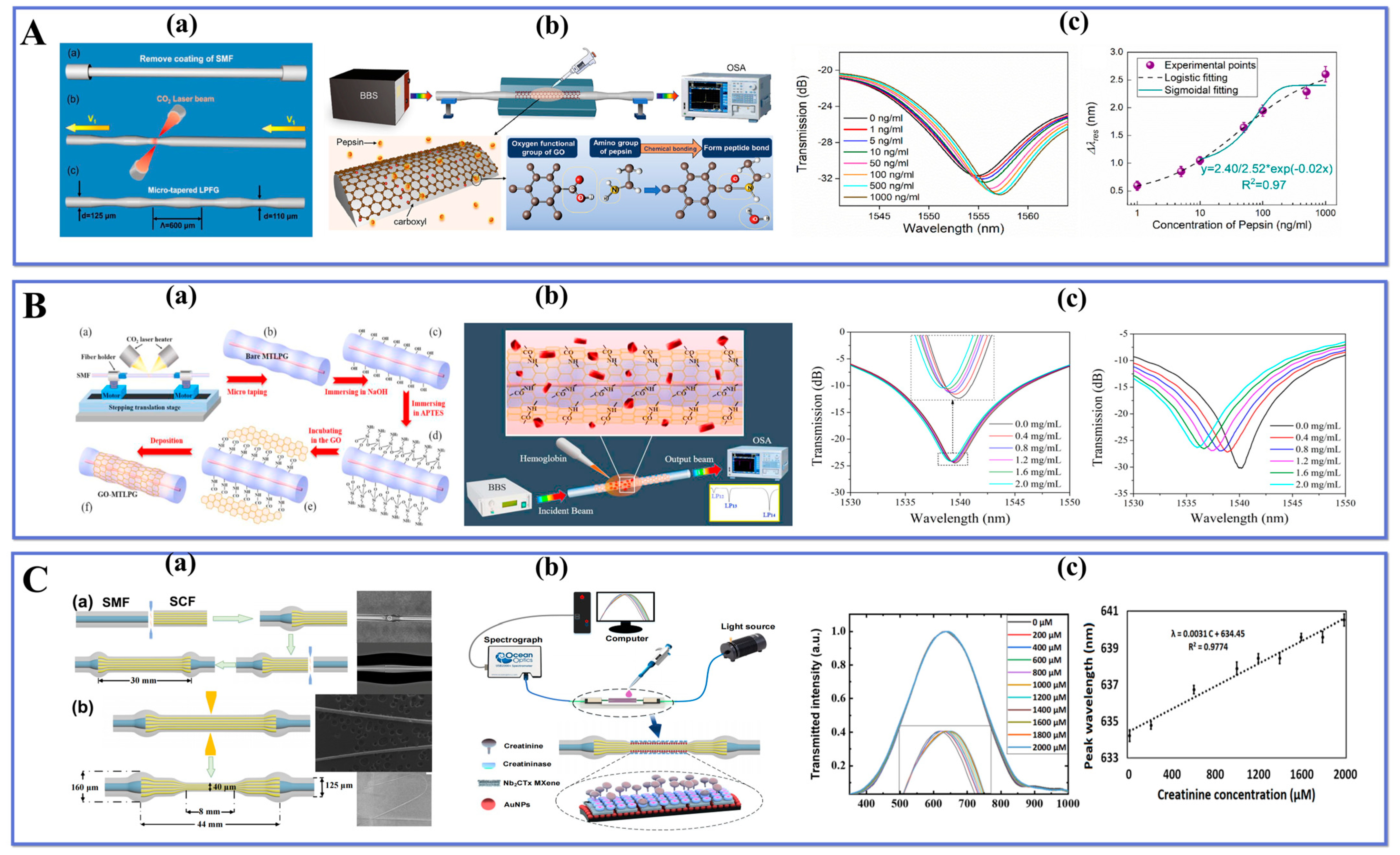
- Kang, X.; Wang, R.; Jiang, M.; Li, E.; Li, Y.; Wang, T.; Ren, Z. A Label-Free Biosensor for Pepsin Detection Based on Graphene Oxide Functionalized Micro-Tapered Long Period Fiber Grating. Sens. Actuators Rep. 2023, 5, 100139. [Google Scholar] [CrossRef]
- Wang, R.; Ren, Z.; Kong, D.; Hu, B.; He, Z. Highly Sensitive Label-Free Biosensor Based on Graphene-Oxide Functionalized Micro-Tapered Long Period Fiber Grating. Opt. Mater. 2020, 109, 110253. [Google Scholar] [CrossRef]
- Li, M.; Singh, R.; Soares, M.S.; Marques, C.; Zhang, B.; Kumar, S. Convex Fiber-Tapered Seven Core Fiber-Convex Fiber (CTC) Structure-Based Biosensor for Creatinine Detection in Aquaculture. Opt. Express 2022, 30, 13898–13914. [Google Scholar] [CrossRef]
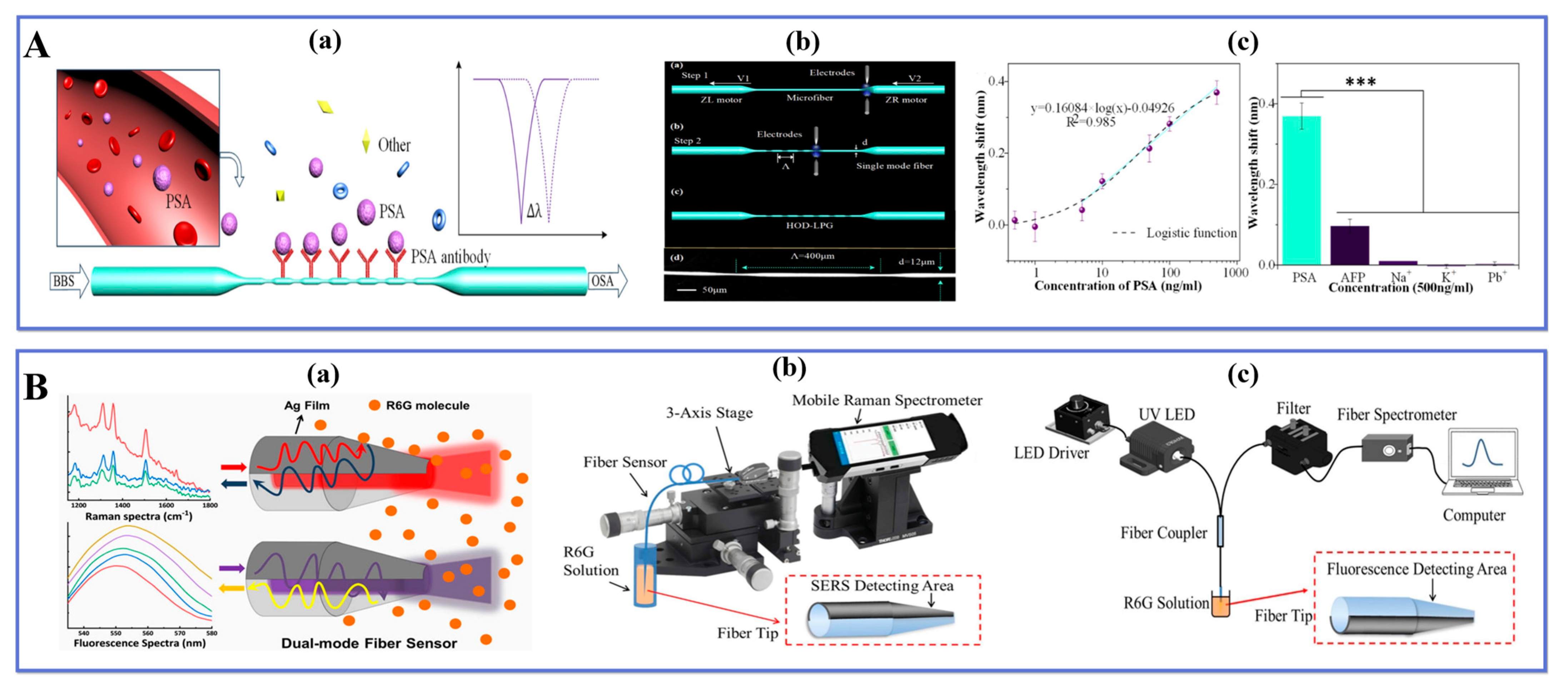
- Xiao, P.; Sun, Z.; Huang, Y.; Lin, W.; Ge, Y.; Xiao, R.; Li, K.; Li, Z.; Lu, H.; Yang, M.; et al. Development of an Optical Microfiber Immunosensor for Prostate Specific Antigen Analysis Using a High-Order-Diffraction Long Period Grating. Opt. Express OE 2020, 28, 15783–15793. [Google Scholar] [CrossRef]
- Li, M.; Yan, M.; Xu, B.; Zhao, C.; Wang, D.; Wang, Y.; Chen, H. A Dual-Mode Optical Fiber Sensor for SERS and Fluorescence Detection in Liquid. Spectrochim. Acta Part A Mol. Biomol. Spectrosc. 2023, 290, 122267. [Google Scholar] [CrossRef]
- Mustapha Kamil, Y.; Al-Rekabi, S.H.; Yaacob, M.H.; Syahir, A.; Chee, H.Y.; Mahdi, M.A.; Abu Bakar, M.H. Detection of Dengue Using PAMAM Dendrimer Integrated Tapered Optical Fiber Sensor. Sci. Rep. 2019, 9, 13483. [Google Scholar] [CrossRef] [Green Version]
- Zhu, G.; Wang, Y.; Wang, Z.; Singh, R.; Marques, C.; Wu, Q.; Kaushik, B.K.; Jha, R.; Zhang, B.; Kumar, S. Localized Plasmon-Based Multicore Fiber Biosensor for Acetylcholine Detection. IEEE Trans. Instrum. Meas. 2022, 71, 7000309. [Google Scholar] [CrossRef]
- Idris, S.; Azeman, N.H.; Noor Azmy, N.A.; Ratnam, C.T.; Mahdi, M.A.; Bakar, A.A.A. Gamma Irradiated Py/PVA for GOx Immobilization on Tapered Optical Fiber for Glucose Biosensing. Sens. Actuators B Chem. 2018, 273, 1404–1412. [Google Scholar] [CrossRef]
- Aziz, M.S.; Shamsudin, M.S.; Fahri, M.A.S.A.; Syuhada, A.; Raja Ibrahim, R.K.; Bakhtiar, H.; Harun, S.W. Glucose Oxidase-Based Enzyme Immobilised on Tapered Optical Fibre for Reliability Improvement in Selective Glucose Sensing. Optik 2022, 259, 168970. [Google Scholar] [CrossRef]
- Li, Y.; Du, M.; He, S.; Wang, R.; Zhang, Z.; Wang, Q. Sensitive Label-Free Hemoglobin Detection Based on Polydopamine Functionalized Graphene Oxide Coated Micro-Tapered Long-Period Fiber Grating. Optik 2023, 275, 170626. [Google Scholar] [CrossRef]
- Kumar, S.; Wang, Y.; Li, M.; Wang, Q.; Malathi, S.; Marques, C.; Singh, R.; Zhang, B. Plasmon-Based Tapered-in-Tapered Fiber Structure for p-Cresol Detection: From Human Healthcare to Aquaculture Application. IEEE Sens. J. 2022, 22, 18493–18500. [Google Scholar] [CrossRef]
- Lu, L.; Zhu, L.; Zhu, G.; Dong, M.; Zeng, Z. ZIF-8/Lipase Coated Tapered Optical Fiber Biosensor for the Detection of Triacylglycerides. IEEE Sens. J. 2020, 20, 14173–14180. [Google Scholar] [CrossRef]
- Mansor, M.; Abu Bakar, M.H.; Omar, M.F.; Mustapha Kamil, Y.; Zainol Abidin, N.H.; Mustafa, F.H.; Mahdi, M.A. Taper Biosensor in Fiber Ring Laser Cavity for Protein Detection. Opt. Laser Technol. 2020, 125, 106033. [Google Scholar] [CrossRef]
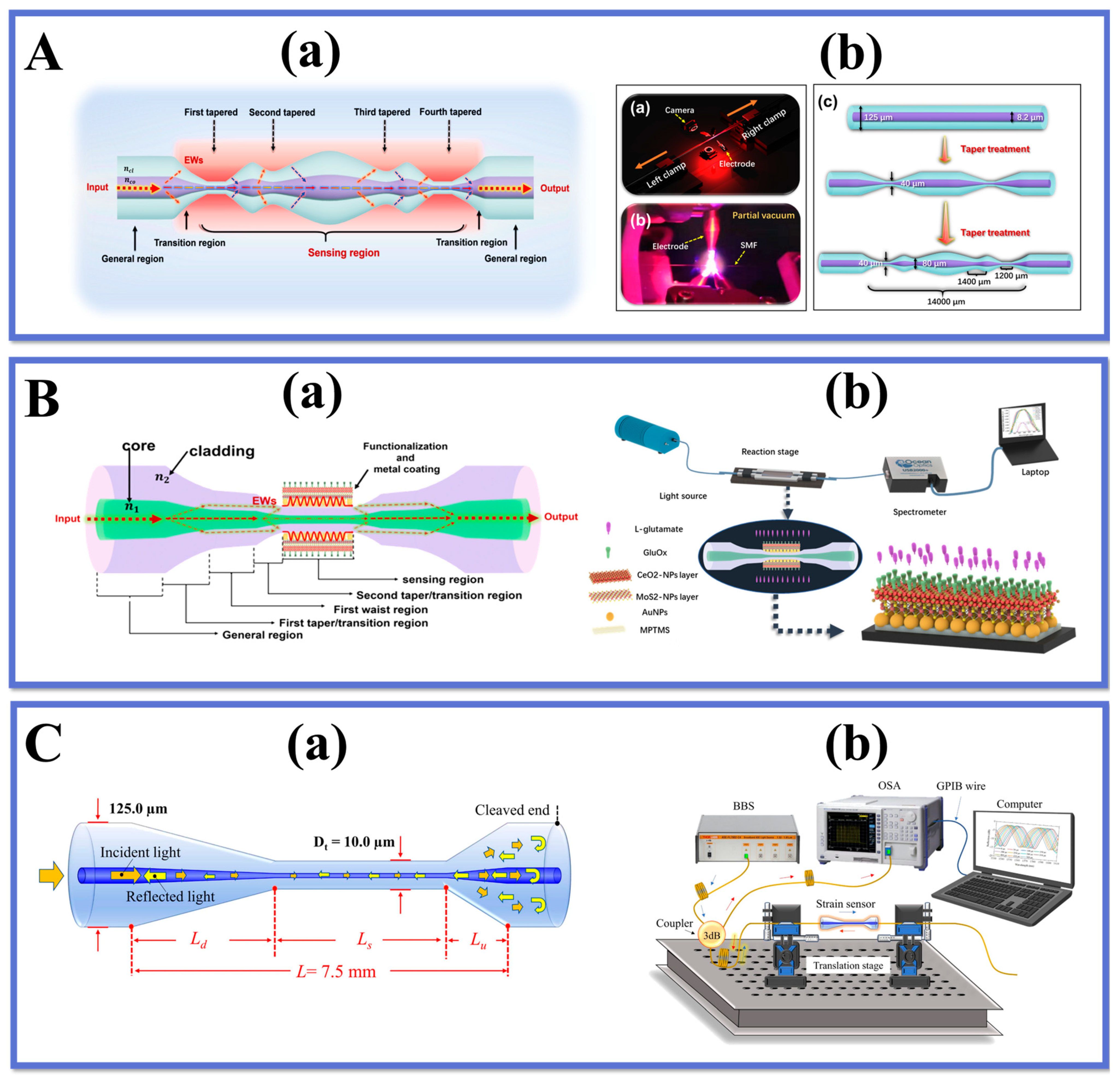
- Zhang, W.; Singh, R.; Wang, Z.; Li, G.; Xie, Y.; Jha, R.; Marques, C.; Zhang, B.; Kumar, S. Humanoid Shaped Optical Fiber Plasmon Biosensor Functionalized with Graphene Oxide/Multi-Walled Carbon Nanotubes for Histamine Detection. Opt. Express 2023, 31, 11788–11803. [Google Scholar] [CrossRef]
- Wang, Z.; Singh, R.; Marques, C.; Jha, R.; Zhang, B.; Kumar, S. Taper-in-Taper Fiber Structure-Based LSPR Sensor for Alanine Aminotransferase Detection. Opt. Express 2021, 29, 43793–43810. [Google Scholar] [CrossRef]
- Mumtaz, F.; Roman, M.; Zhang, B.; Huang, J. Assembly-Free Ultra-Sensitive Miniaturized Strain Sensor Based on an Asymmetric Optical Fiber Taper. Measurement 2023, 211, 112655. [Google Scholar] [CrossRef]
- Zhu, G.; Agrawal, N.; Singh, R.; Kumar, S.; Zhang, B.; Saha, C.; Kumar, C. A Novel Periodically Tapered Structure-Based Gold Nanoparticles and Graphene Oxide–Immobilized Optical Fiber Sensor to Detect Ascorbic Acid. Opt. Laser Technol. 2020, 127, 106156. [Google Scholar] [CrossRef]
- Li, G.; Singh, R.; Guo, J.; Zhang, B.; Kumar, S. Nb 2 CT x MXene-Assisted Double S-Tapered Fiber-Based LSPR Sensor with Improved Features for Tyramine Detection. Appl. Phys. Lett. 2023, 122, 083701. [Google Scholar] [CrossRef]


| Measured Parameter | Fiber Structure | Sensing Principle | Linear Detection Range | Sensitivity | LOD | Ref. |
|---|---|---|---|---|---|---|
| Acoustic Vibration | Tapered-Tip Fiber | Radiated modes interference | 1050 kHz | 15.7 V/nm | 0.1 nm | [100] |
| Humidity | Tapered dual side-hole fiber | Mach-Zehnder interferometer | 30.3%–60.1% RH | −0.142 nm/% RH | n.r. a | [101] |
| Liquid level | Single-mode taper-thin core taper single-mode fiber | Mach-Zehnder interferometer | 0–15 mm | 1.2416 nm/mm | n.r. a | [102] |
| Load | Fiber tapered-loop probe | SPR | 0–20 kPa | 1.473 nm/kPa | n.r. a | [103] |
| Magnetic Field | Tapered small core fiber | Modes interference | n.r.a | 0.46 nm/mT | n.r. a | [104] |
| RI/temperature | MMF-TSMF-MMF | Mach-Zehnder interferometer | 1–1.001849 25–80 °C | −3244.22 nm/RIU −35.18 pm/°C | n.r. a | [105] |
| RI | Tapered SMF | Mach-Zehnder interferometer | 1.332–1.384 1.384–1.4204 1.4204–1.4408 | 415 nm/RIU 1103 nm/RIU 4234 nm/RIU | n.r. a | [106] |
| RI | U-shape tapered plastic optical fiber | SPR | 1.335–1.41 | 1534.53 nm/RIU | n.r. a | [107] |
| Temperature | Taper-Like Etched Multicore Fiber | Mach-Zehnder interferometer | 24–130 °C | 89.19 pm/°C | n.r. a | [108] |
| Temperature | Micro Taper In-Line Fiber | Mach-Zehnder interferometer | 89–950 °C | 0.113 nm/°C | n.r. a | [109] |
| Measured Parameter | Fiber Structure | Sensing Principle | Linear Detection Range | Sensitivity | LOD | Ref. |
|---|---|---|---|---|---|---|
| Alcohol | Tapered fiber | SPR | Alcohol: 0–60% RI: 1.33–1.38 | 2350 nm/RIU | n.r. a | [116] |
| Alcohol | Tapered MMF | Transmission intensity modulation | 0–500 ppm | 22 counts/ppm | n.r. a | [117] |
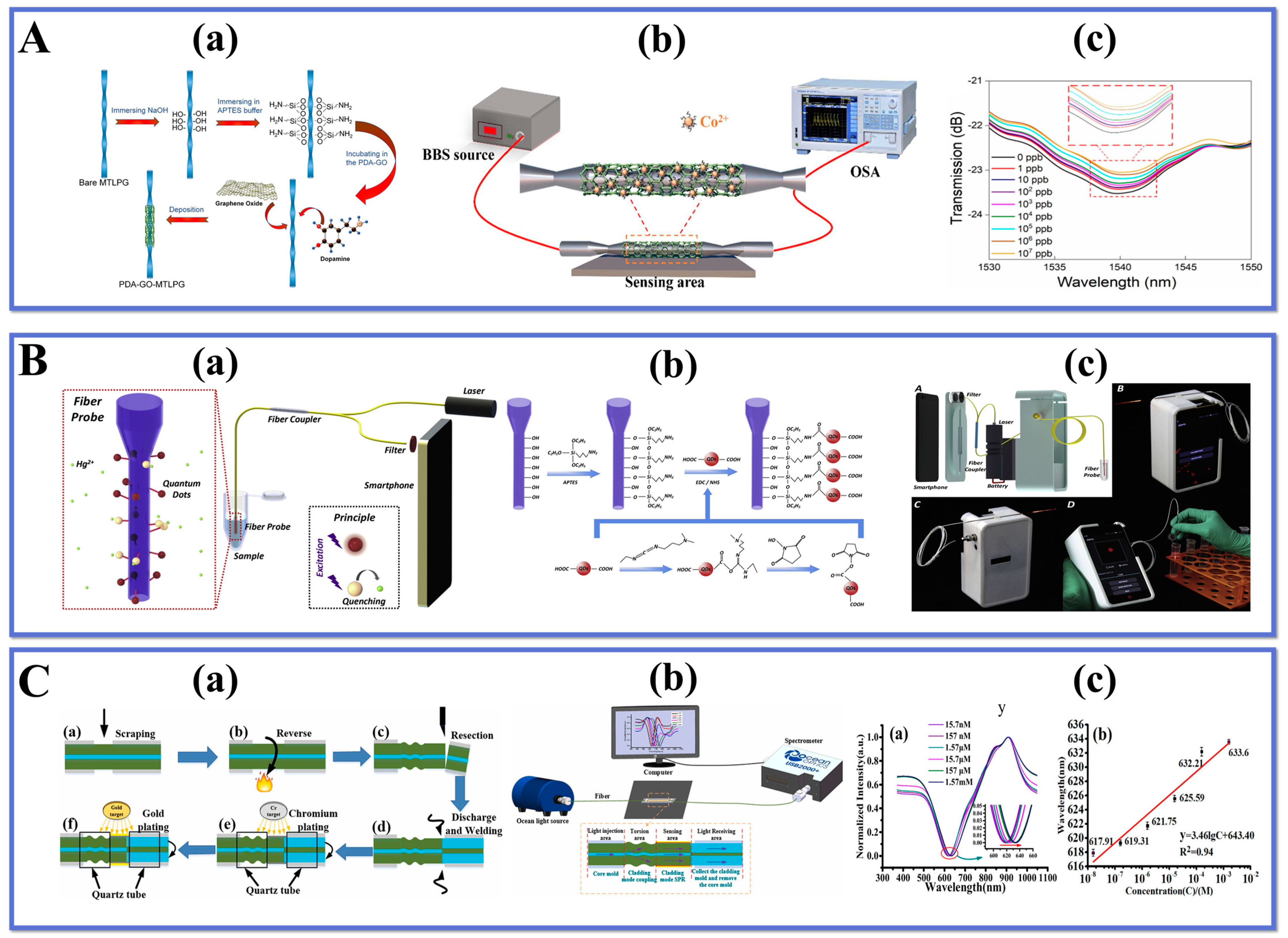
| Co2+ | Micro-tapered long-period fiber | Grating sensing | 1 ppb–107 ppb | 2.4 × 10⁻³ dB/ppb | n.r. a | [110] |
| Cu2+ | Tapered fiber | Mach-Zehnder interferometer | 0–1000 µM | 0.0091 nm/µM | 2.20 µM | [118] |
| Cu2+ | Taper in taper | Mode-mode interference | 0–0.1 mM | 78.03 nm/mM | n.r. a | [34] |
| Fluoride | Tapered Fiber Probe | EWs absorption | 2.08 × 10⁻⁶ 2.005 × 10⁻⁴ M | n.r. a | n.r. a | [119] |
| Fluoride-ion | Taper Michelson interferometric sensor | Interference wavelength shift | 0.01–0.10 ppb | 3341.23 pm/ppb | n.r. a | [120] |
| Nitrate | Tapered fiber | Polyurethane selective detection | n.r. a | 5.94 × 10⁻² μW/ppm | n.r. a | [121] |
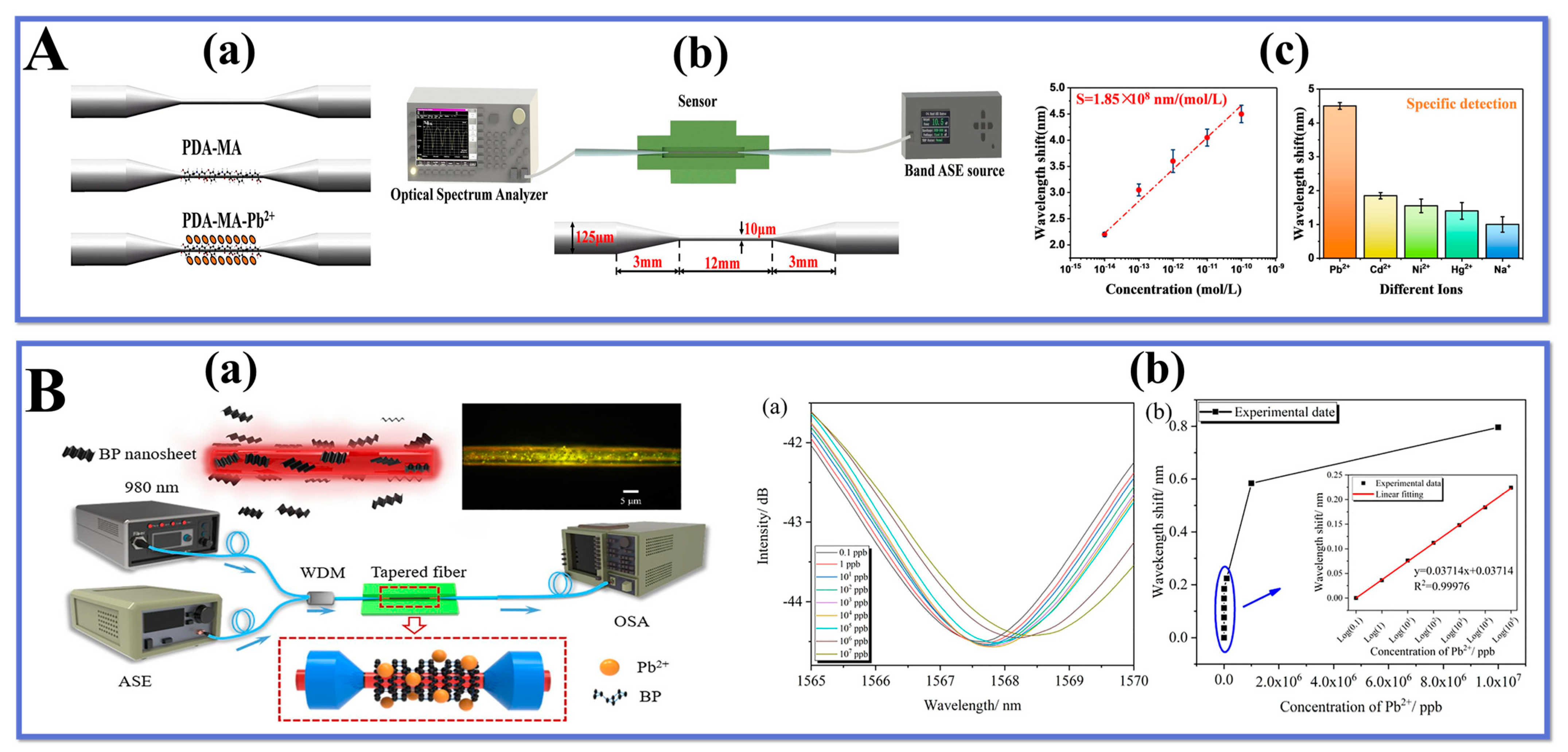
| Pb2+ | Tapered fiber | Interference wavelength shift | 0.1–105 ppb | 0.03714 nm/ppb | 0.0206 ppb | [115] |
| Sodium ions Manganese ions | Micro-tapered long period fiber | Grating sensing | 1–106 ppb | Sodium ions: 2.2 × 10⁻³ dB/ppb Manganese ions: 3.2 × 10⁻³ dB/ppb | 3.2 ppb | [122] |
| Measured Parameter | Fiber Structure | Sensing Principle | Linear Detection Range | Sensitivity | LOD | Ref. |
|---|---|---|---|---|---|---|
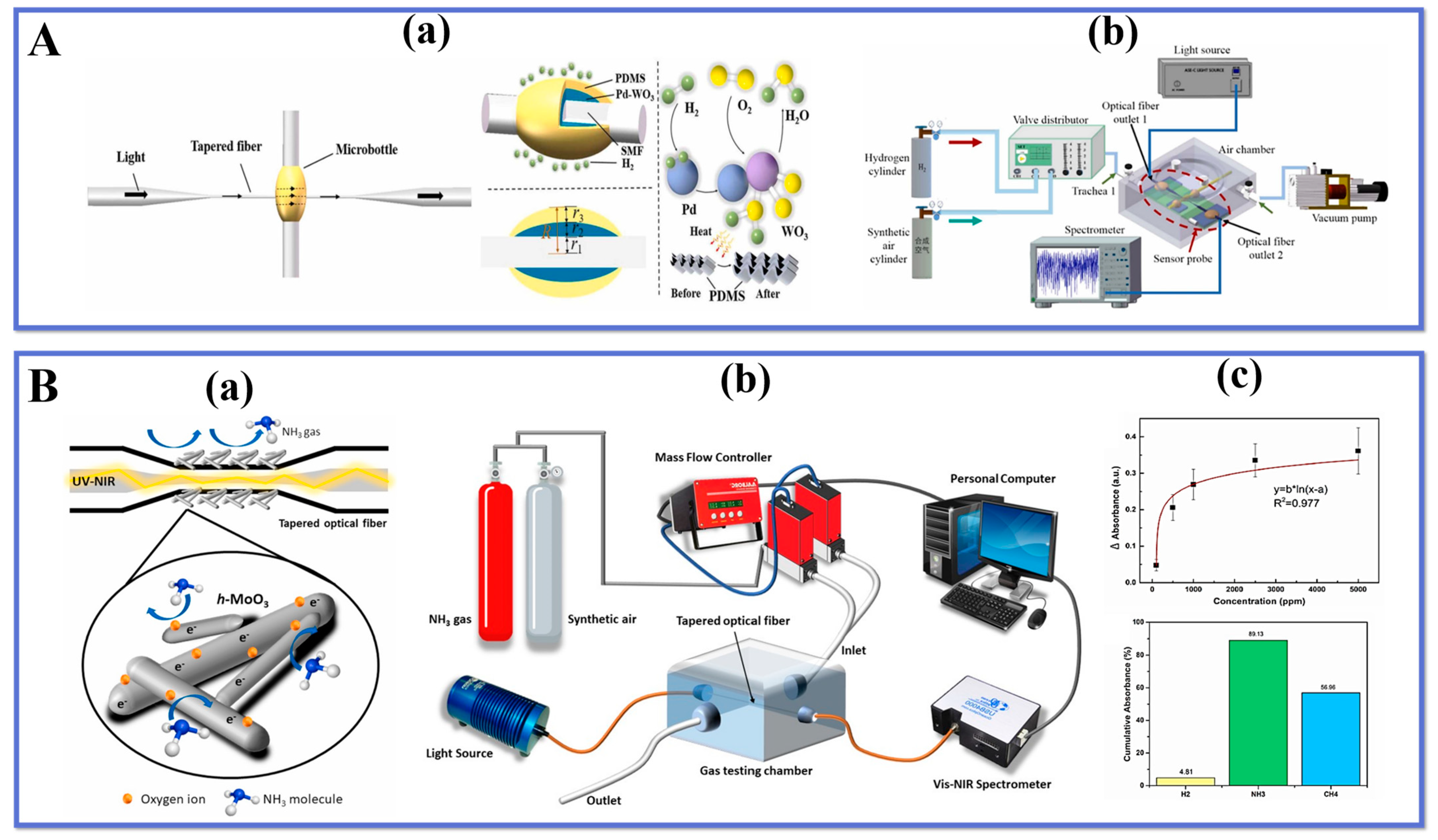
| Ammonia | Etched-tapered Single Mode Optical Fiber | EWs absorption | n.r. a | 300 au/% | 0.00142% | [127] |
| Ammonia | Taper cascade | EWs absorption | n.r. a | 0.015 nm/ppm | n.r. a | [128] |
| Ammonia | Tapered microfiber | EWs absorption | n.r. a | 1.30 pm/ppm | n.r. a | [129] |
| Ammonia | Fiber fusion and taper | Transmission spectrum shift | 0–10,476 ppm | 0.58 pm/ppm | n.r. a | [130] |
| Ammonia | Tapered fiber | Absorbance changes | n.r. a | 26.99 AU/% | 13 ppm | [131] |
| Butane | Tapered Fiber | EWs absorption | n.r. a | 0.4812 a.u./vol.% | n.r. a | [132] |
| Formaldehyde vapor | Tapered U-Shape Plastic Optical Fiber | EWs absorption | 5–20% | 0.00543 V/% | n.r. a | [133] |
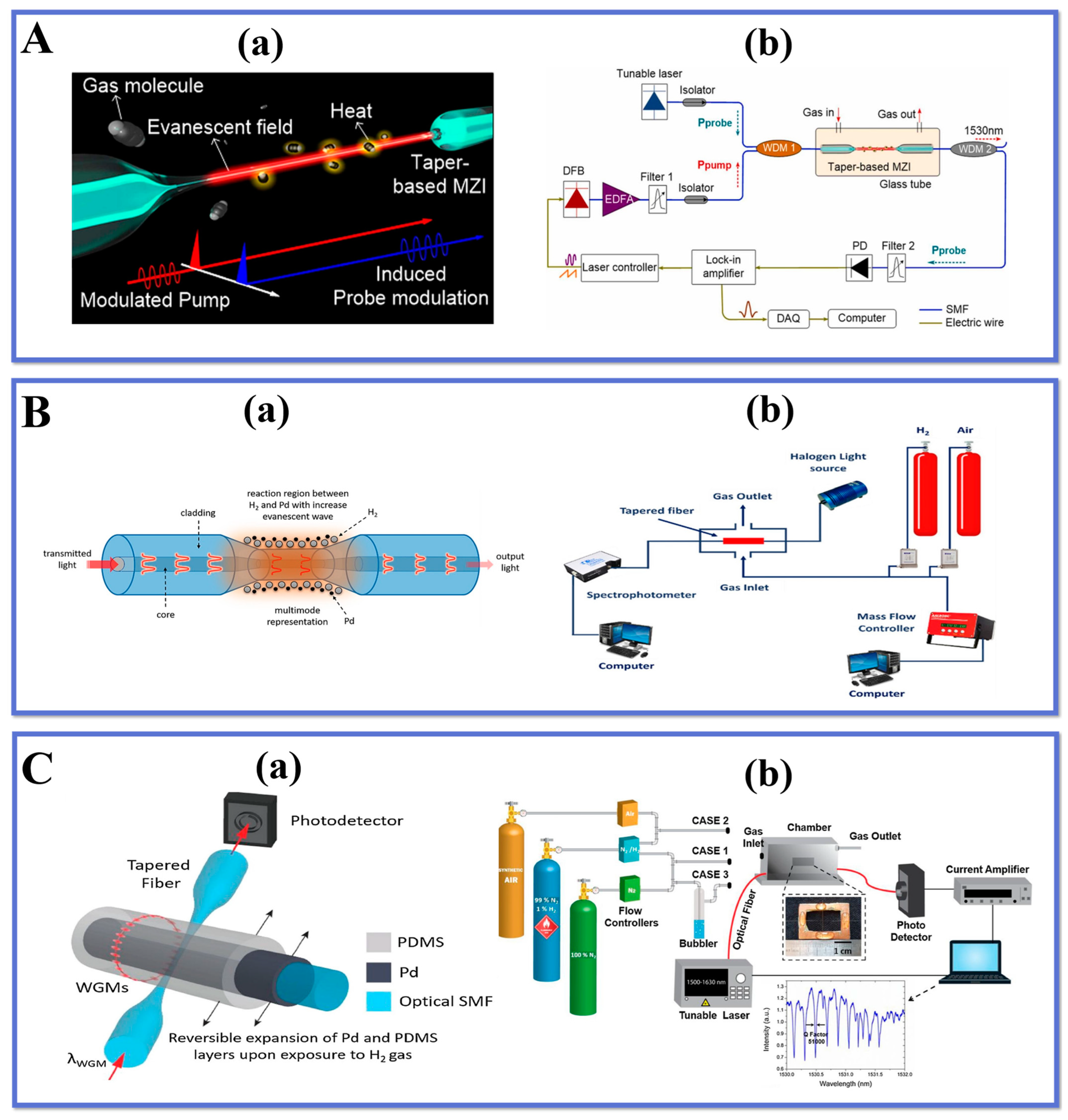
| Hydrogen gas | Tapered fiber | EWs absorption | 0.125–2.00% | 33.22/vol% | n.r. a | [134] |
| Hydrogen gas | Tapered fiber | EWs absorption | 0.125–2.00% | 18,645% | n.r. a | [123] |
| Measured Parameter | Fiber Structure | Sensing Principle | Linear Detection Range | Sensitivity | LOD | Ref. |
|---|---|---|---|---|---|---|
| Acetylcholine | Tapered/etched multicore fiber | LSPR | 0–1000 μM | 0.062 nm/μM | n.r. a | [142] |
| Creatinine | CTC structure | LSPR | 0–2000 µM | 3.1 pm/µM | 86.12 µM | [138] |
| Glucose | Tapered Optical Fiber | Evanescent field variations | n.r. a | 8.7 × 10⁻³ μW/mM | 0.31 μW | [143] |
| Glucose | Tapered fiber | LSPR | n.r. a | (5.01 ± 0.72) × 10−3 /a.u.(%) | n.r. a | [144] |
| Hemoglobin | Micro-tapered long-period fiber grating | Optical waves interference | n.r. a | 3.14 mg/mL | 0.057 mg/mL | [145] |
| p-Cresol | Tapered-in-Tapered fiber | LSPR | 0–1 mM | 3.8 pm/mM | 0.14 mM | [146] |
| Cancer Biomarkers | Reflector-Less Shallow-Tapered Optical Fiber | Optical fiber dispersion | 100 fM–10 nM | 1.33 nm/RIU | 16.4 pM | [51] |
| Triacylglycerides | Tapered Optical | EWs absorption | 0–50 nM | 0.9 nm/nM | 0.23 nM | [147] |
| Protein | Tapered fiber | Interferometric effect | 1–10 pM | 1.02 nm/pM | n.r. a | [148] |
Disclaimer/Publisher’s Note: The statements, opinions and data contained in all publications are solely those of the individual author(s) and contributor(s) and not of MDPI and/or the editor(s). MDPI and/or the editor(s) disclaim responsibility for any injury to people or property resulting from any ideas, methods, instructions or products referred to in the content. |
© 2023 by the authors. Licensee MDPI, Basel, Switzerland. This article is an open access article distributed under the terms and conditions of the Creative Commons Attribution (CC BY) license (https://creativecommons.org/licenses/by/4.0/).
Share and Cite
Zhang, W.; Lang, X.; Liu, X.; Li, G.; Singh, R.; Zhang, B.; Kumar, S. Advances in Tapered Optical Fiber Sensor Structures: From Conventional to Novel and Emerging. Biosensors 2023, 13, 644. https://doi.org/10.3390/bios13060644
Zhang W, Lang X, Liu X, Li G, Singh R, Zhang B, Kumar S. Advances in Tapered Optical Fiber Sensor Structures: From Conventional to Novel and Emerging. Biosensors. 2023; 13(6):644. https://doi.org/10.3390/bios13060644
Chicago/Turabian StyleZhang, Wen, Xianzheng Lang, Xuecheng Liu, Guoru Li, Ragini Singh, Bingyuan Zhang, and Santosh Kumar. 2023. "Advances in Tapered Optical Fiber Sensor Structures: From Conventional to Novel and Emerging" Biosensors 13, no. 6: 644. https://doi.org/10.3390/bios13060644