Novel Approaches to Enzyme-Based Electrochemical Nanobiosensors

 ,
,  ,
,  and
and
Abstract
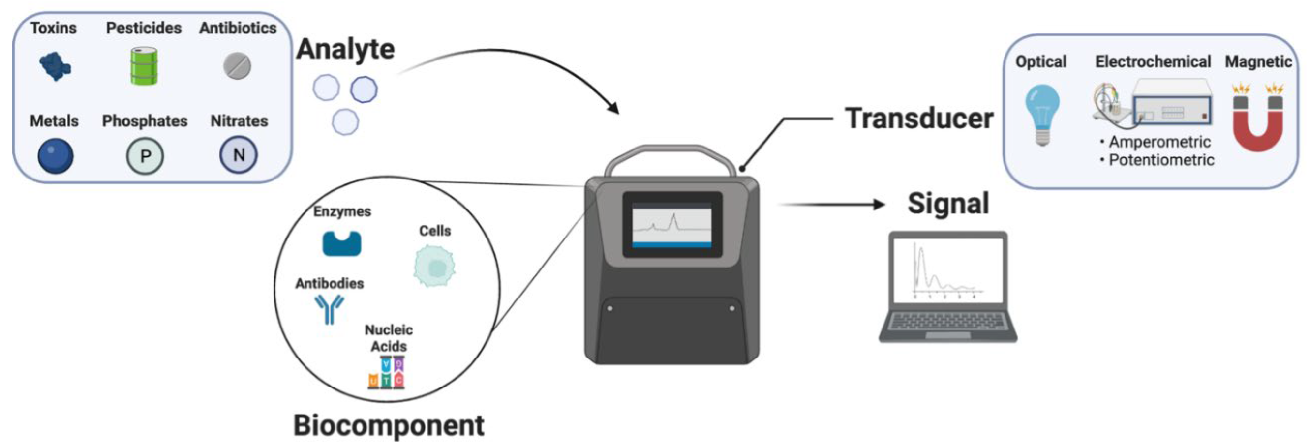
:1. Electrochemical Biosensors
2. Enzymatic Biosensors
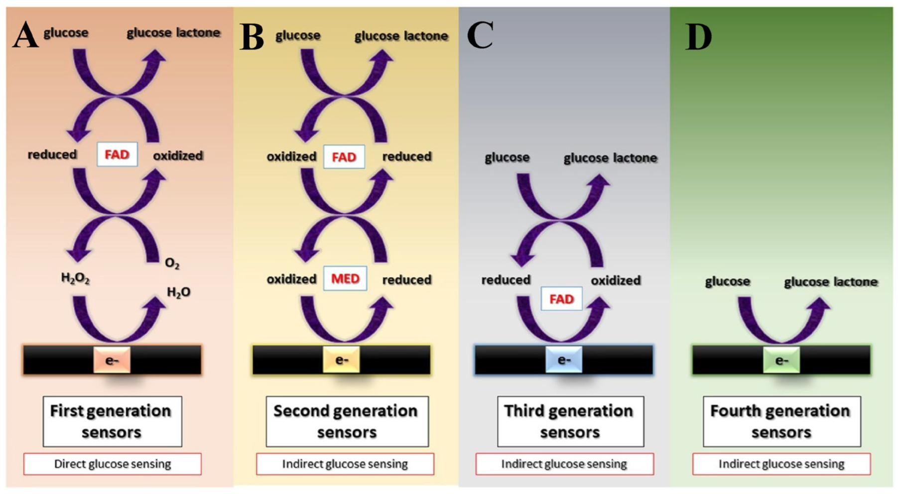
2.1. Types of Enzyme-Based Biosensors
2.2. Enzyme Immobilization and Its Techniques
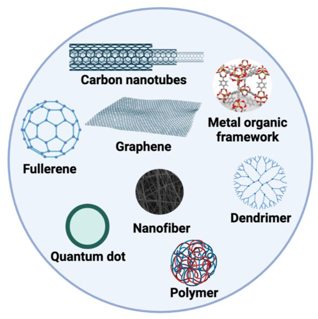
3. Nanomaterials That Are Generally Used in the Design of Enzyme-Based Electrochemical Nanobiosensors
4. Application of Enzyme-Based Electrochemical Nanobiosensors
5. Conclusions
Author Contributions
Funding
Institutional Review Board Statement
Informed Consent Statement
Data Availability Statement
Conflicts of Interest
References
- Bard, A.J.; Faulkner, L.R.; White, H.S. Electrochemical Methods: Fundamentals and Applications; John Wiley & Sons: New York, NY, USA, 2022; ISBN 978-1-119-33405-7. [Google Scholar]
- O’Mullane, A.P. Electrochemistry. In Reference Module in Chemistry, Molecular Sciences and Chemical Engineering; Elsevier: Amsterdam, The Netherlands, 2013; ISBN 978-0-12-409547-2. [Google Scholar]
- Yang, A.; Yan, F. Flexible Electrochemical Biosensors for Health Monitoring. ACS Appl. Electron. Mater. 2021, 3, 53–67. [Google Scholar] [CrossRef]
- Clark, L.C., Jr.; Lyons, C. Electrode Systems for Continuous Monitoring in Cardiovascular Surgery. Ann. N. Y. Acad. Sci. 1962, 102, 29–45. [Google Scholar] [CrossRef]
- Windmiller, J.R.; Wang, J. Wearable Electrochemical Sensors and Biosensors: A Review. Electroanalysis 2013, 25, 29–46. [Google Scholar] [CrossRef]
- Su, L.; Jia, W.; Hou, C.; Lei, Y. Microbial Biosensors: A Review. Biosens. Bioelectron. 2011, 26, 1788–1799. [Google Scholar] [CrossRef]
- Kurbanoglu, S.; Dogan-Topal, B.; Rodriguez, E.P.; Bozal-Palabiyik, B.; Ozkan, S.A.; Uslu, B. Advances in Electrochemical DNA Biosensors and Their Interaction Mechanism with Pharmaceuticals. J. Electroanal. Chem. 2016, 775, 8–26. [Google Scholar] [CrossRef]
- O’Sullivan, C.K. Aptasensors--the Future of Biosensing? Anal. Bioanal. Chem. 2002, 372, 44–48. [Google Scholar] [CrossRef]
- Hashemzadeh, H.; Khadivi-Khanghah, Z.; Allahverdi, A.; Hadipour, M.M.; Saievar-Iranizad, E.; Naderi-Manesh, H. A Novel Label-Free Graphene Oxide Nano-Wall Surface Decorated with Gold Nano-Flower Biosensor for Electrochemical Detection of Brucellosis Antibodies in Human Serum. Talanta Open 2023, 7, 100215. [Google Scholar] [CrossRef]
- Shekhar, S.; Yadav, A.K.; Khosla, A.; Solanki, P.R. Review—Interleukins Profiling for Biosensing Applications: Possibilities and the Future of Disease Detection. ECS Sens. Plus 2022, 1, 041601. [Google Scholar] [CrossRef]
- Bhalla, N.; Jolly, P.; Formisano, N.; Estrela, P. Introduction to Biosensors. Essays Biochem 2016, 60, 1–8. [Google Scholar] [CrossRef] [Green Version]
- Malhotra, B.D.; Ali, M.A. Nanomaterials in Biosensors. In Nanomaterials for Biosensors; Elsevier: Amsterdam, The Netherlands, 2018; pp. 1–74. [Google Scholar] [CrossRef]
- Sailapu, S.K.; Kraikaew, P.; Sabaté, N.; Bakker, E. Self-Powered Potentiometric Sensor Transduction to a Capacitive Electronic Component for Later Readout. ACS Sens. 2020, 5, 2909–2914. [Google Scholar] [CrossRef]
- Salvador, J.-P.; Kopper, K.; Miti, A.; Sanchis, A.; Marco, M.-P. Multiplexed Immunosensor Based on the Amperometric Transduction for Monitoring of Marine Pollutants in Sea Water. Sensors 2020, 20, 5532. [Google Scholar] [CrossRef]
- Bahadır, E.B.; Sezgintürk, M.K. A Review on Impedimetric Biosensors. Artif. Cells Nanomed. Biotechnol. 2016, 44, 248–262. [Google Scholar] [CrossRef] [PubMed]
- Ivanov, S.; Tsakova, V.; Mirsky, V.M. Conductometric Transducing in Electrocatalytical Sensors: Detection of Ascorbic Acid. Electrochem. Commun. 2006, 8, 643–646. [Google Scholar] [CrossRef]
- Yahya, R.; Shah, A.; Kokab, T.; Ullah, N.; Hakeem, M.K.; Hayat, M.; Haleem, A.; Shah, I. Electrochemical Sensor for Detection and Degradation Studies of Ethyl Violet Dye. ACS Omega 2022, 7, 34154–34165. [Google Scholar] [CrossRef]
- Ramanathan, K.; Danielsson, B. Principles and Applications of Thermal Biosensors. Biosens. Bioelectron. 2001, 16, 417–423. [Google Scholar] [CrossRef]
- Grieshaber, D.; MacKenzie, R.; Vörös, J.; Reimhult, E. Electrochemical Biosensors—Sensor Principles and Architectures. Sensors 2008, 8, 1400–1458. [Google Scholar] [CrossRef]
- Cho, I.-H.; Kim, D.H.; Park, S. Electrochemical Biosensors: Perspective on Functional Nanomaterials for on-Site Analysis. Biomater. Res. 2020, 24, 6. [Google Scholar] [CrossRef] [Green Version]
- Arakawa, T.; Dao, D.V.; Mitsubayashi, K. Biosensors and Chemical Sensors for Healthcare Monitoring: A Review. IEEJ Trans. Electr. Electron. Eng. 2022, 17, 626–636. [Google Scholar] [CrossRef]
- Mercante, L.A.; Pavinatto, A.; Pereira, T.S.; Migliorini, F.L.; dos Santos, D.M.; Correa, D.S. Nanofibers Interfaces for Biosensing: Design and Applications. Sens. Actuators Rep. 2021, 3, 100048. [Google Scholar] [CrossRef]
- Zhang, T.; Pan, W.; Zhang, Z.; Qi, N.; Chen, Z. Theoretical Study of Small Molecules Adsorption on Pristine and Transition Metal Doped GeSe Monolayer for Gas Sensing Application. Langmuir 2022, 38, 1287–1295. [Google Scholar] [CrossRef]
- Li, L.; Chen, S.; Jiang, S. Protein Adsorption on Alkanethiolate Self-Assembled Monolayers: Nanoscale Surface Structural and Chemical Effects. Langmuir 2003, 19, 2974–2982. [Google Scholar] [CrossRef]
- Ahmad, M.A.; Mustafa, F.; Panicker, N.; Rizvi, T.A. Optical Detection of SARS-CoV-2 Utilizing Antigen-Antibody Binding Interactions. Sensors 2021, 21, 6596. [Google Scholar] [CrossRef]
- Roth, S.; Ideses, D.; Juven-Gershon, T.; Danielli, A. Rapid Biosensing Method for Detecting Protein–DNA Interactions. ACS Sens. 2022, 7, 60–70. [Google Scholar] [CrossRef]
- Frew, J.E.; Hill, H.A.O. Electrochemical Biosensors. Anal. Chem. 1987, 59, 933A–944A. [Google Scholar] [CrossRef] [PubMed]
- Elgrishi, N.; Rountree, K.J.; McCarthy, B.D.; Rountree, E.S.; Eisenhart, T.T.; Dempsey, J.L. A Practical Beginner’s Guide to Cyclic Voltammetry. J. Chem. Educ. 2018, 95, 197–206. [Google Scholar] [CrossRef]
- Deffo, G.; Tene, T.F.N.; Dongmo, L.M.; Jiokeng, S.L.Z.; Temgoua, R.C.T. Differential Pulse and Square-Wave Voltammetry as Sensitive Methods for Electroanalysis Applications. In Reference Module in Chemistry, Molecular Sciences and Chemical Engineering; Elsevier: Amsterdam, The Netherlands, 2023; ISBN 978-0-12-409547-2. [Google Scholar]
- Magar, H.S.; Hassan, R.Y.A.; Mulchandani, A. Electrochemical Impedance Spectroscopy (EIS): Principles, Construction, and Biosensing Applications. Sensors 2021, 21, 6578. [Google Scholar] [CrossRef]
- Prodromidis, M.I. Impedimetric Immunosensors—A Review. Electrochim. Acta 2010, 55, 4227–4233. [Google Scholar] [CrossRef]
- Tsopela, A.; Laborde, A.; Salvagnac, L.; Ventalon, V.; Bedel-Pereira, E.; Séguy, I.; Temple-Boyer, P.; Juneau, P.; Izquierdo, R.; Launay, J. Development of a Lab-on-Chip Electrochemical Biosensor for Water Quality Analysis Based on Microalgal Photosynthesis. Biosens. Bioelectron. 2016, 79, 568–573. [Google Scholar] [CrossRef] [Green Version]
- Kumar, H.; Neelam, R. Enzyme-Based Electrochemical Biosensors for Food Safety: A Review. Nanobiosensors Dis. Diagn. 2016, 5, 29–39. [Google Scholar] [CrossRef] [Green Version]
- Escarpa, A.; González, M.C.; Lopez, M.Á. Agricultural and Food Electroanalysis; John Wiley & Sons: New York, NY, USA, 2015; ISBN 9781119961864. [Google Scholar]
- Karunakaran, C.; Madasamy, T.; Sethy, N.K. Enzymatic Biosensors; Elsevier Inc.: Amsterdam, The Netherlands, 2015; ISBN 9780128031018. [Google Scholar]
- Rahmawati, I.; Einaga, Y.; Ivandini, T.A.; Fiorani, A. Enzymatic Biosensors with Electrochemiluminescence Transduction. ChemElectroChem 2022, 9, e202200175. [Google Scholar] [CrossRef]
- Kucherenko, I.S.; Soldatkin, O.O.; Kucherenko, D.Y.; Soldatkina, O.V.; Dzyadevych, S.V. Advances in Nanomaterial Application in Enzyme-Based Electrochemical Biosensors: A Review. Nanoscale Adv. 2019, 1, 4560–4577. [Google Scholar] [CrossRef] [Green Version]
- Wang, J. Glucose Biosensors: 40 Years of Advances and Challenges. Electroanalysis 2001, 13, 983–988. [Google Scholar] [CrossRef]
- Yao, B.; Yao, J.; Fan, Z.; Zhao, J.; Zhang, K.; Huang, W. Rapid Advances of Versatile MXenes for Electrochemical Enzyme-Based Biosensors, Immunosensors, and Nucleic Acid-Based Biosensors. ChemElectroChem 2022, 9, e202200103. [Google Scholar] [CrossRef]
- Singh, S.; Wang, J.; Cinti, S. Review—An Overview on Recent Progress in Screen-Printed Electroanalytical (Bio)Sensors. ECS Sens. Plus 2022, 1, 023401. [Google Scholar] [CrossRef]
- David, S.; Walt, R.; Stubbs, J.; Andersson-Svahn, H.; Gilligan, M.; Wilson, E. Lab on a Chip Lab on a Chip. Lab A Chip 2014, 23, 1037–1043. [Google Scholar] [CrossRef]
- Cinti, S.; Talarico, D.; Palleschi, G.; Moscone, D.; Arduini, F. Novel Reagentless Paper-Based Screen-Printed Electrochemical Sensor to Detect Phosphate. Anal. Chim. Acta 2016, 919, 78–84. [Google Scholar] [CrossRef] [PubMed]
- Dzyadevych, S.V.; Arkhypova, V.N.; Soldatkin, A.P.; El’skaya, A.V.; Martelet, C.; Jaffrezic-Renault, N. Amperometric Enzyme Biosensors: Past, Present and Future. ITBM RBM 2008, 29, 171–180. [Google Scholar] [CrossRef]
- Tan, C.; Robbins, E.M.; Wu, B.; Cui, X.T. Recent Advances in in Vivo Neurochemical Monitoring. Micromachines 2021, 12, 208. [Google Scholar] [CrossRef]
- Rocchitta, G.; Spanu, A.; Babudieri, S.; Latte, G.; Madeddu, G.; Galleri, G.; Nuvoli, S.; Bagella, P.; Demartis, M.I.; Fiore, V.; et al. Enzyme Biosensors for Biomedical Applications: Strategies for Safeguarding Analytical Performances in Biological Fluids. Sensors 2016, 16, 780. [Google Scholar] [CrossRef] [Green Version]
- Das, P.; Das, M.; Chinnadayyala, S.R.; Singha, I.M.; Goswami, P. Recent Advances on Developing 3rd Generation Enzyme Electrode for Biosensor Applications. Biosens. Bioelectron. 2016, 79, 386–397. [Google Scholar] [CrossRef]
- Prodromidis, M.I.; Karayannis, M.I. Enzyme Based Amperometric Biosensors for Food Analysis. Electroanalysis 2002, 14, 241–261. [Google Scholar] [CrossRef]
- Pohanka, M. Glucose Electrochemical Biosensors: The Past and Current Trends. Int. J. Electrochem. Sci. 2021, 16, 47. [Google Scholar] [CrossRef]
- Bartlett, P.N.; Bradford, V.Q.; Whitaker, R.G. Enzyme Electrode Studies of Glucose Oxidase Modified with a Redox Mediator. Talanta 1991, 38, 57–63. [Google Scholar] [CrossRef]
- Gunasingham, H.; Tan, C.H.; Aw, T.C. Comparative Study of First-, Second- and Third-Generation Amperometric Glucose Enzyme Electrodes in Continuous-Flow Analysis of Undiluted Whole Blood. Anal. Chim. Acta 1990, 234, 321–330. [Google Scholar] [CrossRef]
- Scheller, F.W.; Schubert, F.; Neumann, B.; Pfeiffer, D.; Hintsche, R.; Dransfeld, I.; Wollenberger, U.; Renneberg, R.; Warsinke, A.; Johansson, G.; et al. Second Generation Biosensors. Biosens. Bioelectron. 1991, 6, 245–253. [Google Scholar] [CrossRef] [PubMed]
- Putzbach, W.; Ronkainen, N.J. Immobilization Techniques in the Fabrication of Nanomaterial-Based Electrochemical Biosensors: A Review. Sensors 2013, 13, 4811–4840. [Google Scholar] [CrossRef] [Green Version]
- Lee, I.; Loew, N.; Tsugawa, W.; Ikebukuro, K.; Sode, K. Development of a Third-Generation Glucose Sensor Based on the Open Circuit Potential for Continuous Glucose Monitoring. Biosens. Bioelectron. 2019, 124–125, 216–223. [Google Scholar] [CrossRef]
- Gorton, L.; Lindgren, A.; Larsson, T.; Munteanu, F.D.; Ruzgas, T.; Gazaryan, I. Direct Electron Transfer between Heme-Containing Enzymes and Electrodes as Basis for Third Generation Biosensors. Anal. Chim. Acta 1999, 400, 91–108. [Google Scholar] [CrossRef]
- Harper, A.; Anderson, M.R. Electrochemical Glucose Sensors-Developments Using Electrostatic Assembly and Carbon Nanotubes for Biosensor Construction. Sensors 2010, 10, 8248–8274. [Google Scholar] [CrossRef] [Green Version]
- Hassan, M.H.; Vyas, C.; Grieve, B.; Bartolo, P. Recent Advances in Enzymatic and Non-Enzymatic Electrochemical Glucose Sensing. Sensors 2021, 21, 4672. [Google Scholar] [CrossRef]
- Naikoo, G.A.; Awan, T.; Salim, H.; Arshad, F.; Hassan, I.U.; Pedram, M.Z.; Ahmed, W.; Faruck, H.L.; Aljabali, A.A.A.; Mishra, V.; et al. Fourth-Generation Glucose Sensors Composed of Copper Nanostructures for Diabetes Management: A Critical Review. Bioeng. Transl. Med. 2022, 7, e10248. [Google Scholar] [CrossRef]
- Fopase, R.; Paramasivam, S.; Kale, P.; Paramasivan, B. Strategies, Challenges and Opportunities of Enzyme Immobilization on Porous Silicon for Biosensing Applications. J. Environ. Chem. Eng. 2020, 8, 104266. [Google Scholar] [CrossRef]
- Swamy, N.K.; Sandeep, S.; Santhosh, A.S. Enzyme Immobilization Methods and Role of Conductive Polymers in Fabrication of Enzymatic Biosensors. Indian J. Adv. Chem. Sci. 2017, 2, 1–5. [Google Scholar] [CrossRef]
- Imam, H.T.; Marr, P.C.; Marr, A.C. Enzyme Entrapment, Biocatalyst Immobilization without Covalent Attachment. Green Chem. 2021, 23, 4980–5005. [Google Scholar] [CrossRef]
- Jesionowski, T.; Zdarta, J.; Krajewska, B. Enzyme Immobilization by Adsorption: A Review. Adsorption 2014, 20, 801–821. [Google Scholar] [CrossRef] [Green Version]
- Bernard, A.; Coordonnier, M.; LeBeault, J.M. Ddc Fermentation Process Control. Annu. Meet. Am. Inst. Chem. Eng. 1982, 103–125. [Google Scholar] [CrossRef]
- Luna, C.; Luna, D.; Calero, J.; Bautista, F.M.; Romero, A.A.; Posadillo, A.; Verdugo-Escamilla, C. Biochemical Catalytic Production of Biodiesel. In Handbook of Biofuels Production: Processes and Technologies, 2nd ed.; Elsevier: Amsterdam, The Netherlands, 2016. [Google Scholar]
- Nguyen, H.H.; Lee, S.H.; Lee, U.J.; Fermin, C.D.; Kim, M. Immobilized Enzymes in Biosensor Applications. Materials 2019, 12, 121. [Google Scholar] [CrossRef] [Green Version]
- Li, Y.; Zhao, Y.; Gui, F.; Teng, B.; Guan, C. Numerical Analysis of the Effects of Sinker Weight on the Hydrodynamics Behaviour of Gravity Cage Net in Uniform Flow*. J. Hydrodyn. 2006, 18, 77–83. [Google Scholar] [CrossRef]
- Apetrei, C.; Ghasemi-Varnamkhasti, M. Biosensors in Food PDO Authentication. In Comprehensive Analytical Chemistry; Elsevier: Amsterdam, The Netherlands, 2013; Volume 60. [Google Scholar]
- Kaur, J.; Choudhary, S.; Chaudhari, R.; Jayant, R.D.; Joshi, A. Enzyme-Based Biosensors; Elsevier Ltd.: Amsterdam, The Netherlands, 2019; ISBN 9780081024201. [Google Scholar]
- Kang, H.; Wang, L.; O’Donoghue, M.; Cao, Y.C.; Tan, W. Chapter 15—Nanoparticles for Biosensors. In Optical Biosensors, 2nd ed.; Ligler, F.S., Taitt, C.R., Eds.; Elsevier: Amsterdam, The Netherlands, 2008; pp. 583–621. ISBN 978-0-444-53125-4. [Google Scholar]
- Pirzada, M.; Altintas, Z. Nanomaterials for Healthcare Biosensing Applications. Sensors 2019, 19, 5311. [Google Scholar] [CrossRef] [PubMed] [Green Version]
- Idris, A.O.; Akanji, S.P.; Orimolade, B.O.; Olorundare, F.O.G.; Azizi, S.; Mamba, B.; Maaza, M. Using Nanomaterials as Excellent Immobilisation Layer for Biosensor Design. Biosensors 2023, 13, 192. [Google Scholar] [CrossRef]
- Meskher, H.; Ragdi, T.; Thakur, A.K.; Ha, S.; Khelfaoui, I.; Sathyamurthy, R.; Sharshir, S.W.; Pandey, A.K.; Saidur, R.; Singh, P.; et al. A Review on CNTs-Based Electrochemical Sensors and Biosensors: Unique Properties and Potential Applications. Crit. Rev. Anal. Chem. 2023, 1–24. [Google Scholar] [CrossRef]
- Ventura-Aguilar, R.I.; Bautista-Baños, S.; Mendoza-Acevedo, S.; Bosquez-Molina, E. Nanomaterials for Designing Biosensors to Detect Fungi and Bacteria Related to Food Safety of Agricultural Products. Postharvest Biol. Technol. 2023, 195, 112116. [Google Scholar] [CrossRef]
- Manusha, P.; Theyagarajan, K.; Elancheziyan, M.; Shankar, H.; Thenmozhi, K.; Senthilkumar, S. Anthraquinone Functionalized Ionic Liquid Based Disposable Electrochemical Sensor for the Enzyme-Free Detection of Hydrogen Peroxide. ECS Sens. Plus 2022, 1, 033601. [Google Scholar] [CrossRef]
- Khan, I.; Saeed, K.; Khan, I. Nanoparticles: Properties, Applications and Toxicities. Arab. J. Chem. 2019, 12, 908–931. [Google Scholar] [CrossRef]
- Maduraiveeran, G.; Jin, W. Nanomaterials Based Electrochemical Sensor and Biosensor Platforms for Environmental Applications. Trends Environ. Anal. Chem. 2017, 13, 10–23. [Google Scholar] [CrossRef]
- Chen, M.; Zeng, G.; Xu, P.; Lai, C.; Tang, L. How do Enzymes ‘Meet’ Nanoparticles and Nanomaterials? Trends Biochem. Sci. 2017, 42, 914–930. [Google Scholar] [CrossRef]
- Shin, W.-K.; Cho, J.; Kannan, A.G.; Lee, Y.-S.; Kim, D.-W. Cross-Linked Composite Gel Polymer Electrolyte Using Mesoporous Methacrylate-Functionalized SiO2 Nanoparticles for Lithium-Ion Polymer Batteries. Sci. Rep. 2016, 6, 26332. [Google Scholar] [CrossRef] [PubMed]
- Badea, M.; Curulli, A.; Palleschi, G. Oxidase Enzyme Immobilisation through Electropolymerised Films to Assemble Biosensors for Batch and Flow Injection Analysis. Biosens. Bioelectron. 2003, 18, 689–698. [Google Scholar] [CrossRef] [PubMed]
- Chavali, M.S.; Nikolova, M.P. Metal Oxide Nanoparticles and Their Applications in Nanotechnology. SN Appl. Sci. 2019, 1, 607. [Google Scholar] [CrossRef] [Green Version]
- Kumar, S.; Wang, Z.; Zhang, W.; Liu, X.; Li, M.; Li, G.; Zhang, B.; Singh, R. Optically Active Nanomaterials and Its Biosensing Applications—A Review. Biosensors 2023, 13, 85. [Google Scholar] [CrossRef]
- Zuliani, A.; Khiar, N.; Carrillo-Carrión, C. Recent Progress of Metal–Organic Frameworks as Sensors in (Bio)Analytical Fields: Towards Real-World Applications. Anal. Bioanal. Chem. 2023, 415, 2005–2023. [Google Scholar] [CrossRef]
- Li, B.; Suo, T.; Xie, S.; Xia, A.; Ma, Y.; Huang, H.; Zhang, X.; Hu, Q. Rational Design, Synthesis, and Applications of Carbon Dots@metal–Organic Frameworks (CD@MOF) Based Sensors. TrAC Trends Anal. Chem. 2021, 135, 116163. [Google Scholar] [CrossRef]
- Daniel, M.; Mathew, G.; Anpo, M.; Neppolian, B. MOF Based Electrochemical Sensors for the Detection of Physiologically Relevant Biomolecules: An Overview. Coord. Chem. Rev. 2022, 468, 214627. [Google Scholar] [CrossRef]
- Kajal, N.; Singh, V.; Gupta, R.; Gautam, S. Metal Organic Frameworks for Electrochemical Sensor Applications: A Review. Environ. Res. 2022, 204, 112320. [Google Scholar] [CrossRef]
- Mohan, B.; Kumar, S.; Xi, H.; Ma, S.; Tao, Z.; Xing, T.; You, H.; Zhang, Y.; Ren, P. Fabricated Metal-Organic Frameworks (MOFs) as Luminescent and Electrochemical Biosensors for Cancer Biomarkers Detection. Biosens. Bioelectron. 2022, 197, 113738. [Google Scholar] [CrossRef]
- Li, X.; Feng, Q.; Lu, K.; Huang, J.; Zhang, Y.; Hou, Y.; Qiao, H.; Li, D.; Wei, Q. Encapsulating Enzyme into Metal-Organic Framework during in-Situ Growth on Cellulose Acetate Nanofibers as Self-Powered Glucose Biosensor. Biosens. Bioelectron. 2021, 171, 112690. [Google Scholar] [CrossRef]
- Mayer, K.M.; Hafner, J.H. Localized Surface Plasmon Resonance Sensors. Chem. Rev. 2011, 111, 3828–3857. [Google Scholar] [CrossRef] [PubMed]
- Iftikhar, F.J.; Shah, A.; Akhter, M.S.; Kurbanoglu, S.; Ozkan, S.A. Chapter 1—Introduction to Nanosensors. In New Developments in Nanosensors for Pharmaceutical Analysis; Ozkan, S.A., Shah, A., Eds.; Academic Press: Cambridge, MA, USA, 2019; pp. 1–46. ISBN 978-0-12-816144-9. [Google Scholar]
- Husain, S.; Nandi, A.; Simnani, F.Z.; Saha, U.; Ghosh, A.; Sinha, A.; Sahay, A.; Samal, S.K.; Panda, P.K.; Verma, S.K. Emerging Trends in Advanced Translational Applications of Silver Nanoparticles: A Progressing Dawn of Nanotechnology. J. Funct. Biomater. 2023, 14, 47. [Google Scholar] [CrossRef] [PubMed]
- Zarlaida, F.; Adlim, M. Gold and Silver Nanoparticles and Indicator Dyes as Active Agents in Colorimetric Spot and Strip Tests for Mercury(II) Ions: A Review. Microchim. Acta 2017, 184, 45–58. [Google Scholar] [CrossRef]
- Loiseau, A.; Asila, V.; Boitel-Aullen, G.; Lam, M.; Salmain, M.; Boujday, S. Silver-Based Plasmonic Nanoparticles for and Their Use in Biosensing. Biosensors 2019, 9, 78. [Google Scholar] [CrossRef] [Green Version]
- Wei, W.; Yi, Y.; Song, J.; Chen, X.; Li, J.; Li, J. Tunable Graphene/Nitrocellulose Temperature Alarm Sensors. ACS Appl. Mater. Interfaces 2022, 14, 13790–13800. [Google Scholar] [CrossRef]
- Paukov, M.; Kramberger, C.; Begichev, I.; Kharlamova, M.; Burdanova, M. Functionalized Fullerenes and Their Applications in Electrochemistry, Solar Cells, and Nanoelectronics. Materials 2023, 16, 1276. [Google Scholar] [CrossRef] [PubMed]
- Er, S.; Demirkol, D.O. Graphene Oxide Incorporated Polystyrene Electrospun Nanofibers for Immunosensing of CD36 as a Marker of Diabetic Plasma. Bioelectrochemistry 2022, 145, 108083. [Google Scholar] [CrossRef]
- Oner, A.; Tufek, E.; Yezer, I.; Birol, A.; Demir, M.; Er, S.; Demirkol, D.O. High Generation Dendrimer Decorated Poly-Ɛ-Caprolactone/Polyacrylic Acid Electrospun Nanofibers for the Design of a Bioelectrochemical Sensing Surface. React. Funct. Polym. 2021, 161, 104853. [Google Scholar] [CrossRef]
- Unal, D.N.; Yıldırım, S.; Kurbanoglu, S.; Uslu, B. Current Trends and Roles of Surfactants for Chromatographic and Electrochemical Sensing. TrAC Trends Anal. Chem. 2021, 144, 116418. [Google Scholar] [CrossRef]
- Badmus, S.O.; Amusa, H.K.; Oyehan, T.A.; Saleh, T.A. Environmental Risks and Toxicity of Surfactants: Overview of Analysis, Assessment, and Remediation Techniques. Env. Sci. Pollut. Res. 2021, 28, 62085–62104. [Google Scholar] [CrossRef] [PubMed]
- Grueso, E.; Giráldez-Pérez, R.M.; Prado-Gotor, R.; Kuliszewska, E. Sodium Lauryl Sulfate-Conjugated Cationic Gemini-Surfactant-Capped Gold Nanoparticles as Model System for Biomolecule Recognition. Chemosensors 2023, 11, 207. [Google Scholar] [CrossRef]
- Cui, F.; Sun, H.; Yang, X.; Zhou, H.; Wu, Y.; Li, J.; Li, H.; Liu, J.; Zeng, C.; Qu, B.; et al. Laser-Induced Graphene (LIG)-Based Au@CuO/V2CTx MXene Non-Enzymatic Electrochemical Sensors for the Urine Glucose Test. Chem. Eng. J. 2023, 457, 141303. [Google Scholar] [CrossRef]
- Kurbanoglu, S.; Erkmen, C.; Uslu, B. Frontiers in Electrochemical Enzyme Based Biosensors for Food and Drug Analysis. TrAC Trends Anal. Chem. 2020, 124, 115809. [Google Scholar] [CrossRef]
- Abbasi, E.; Aval, S.F.; Akbarzadeh, A.; Milani, M.; Nasrabadi, H.T.; Joo, S.W.; Hanifehpour, Y.; Nejati-Koshki, K.; Pashaei-Asl, R. Dendrimers: Synthesis, Applications, and Properties. Nanoscale Res. Lett. 2014, 9, 1–10. [Google Scholar] [CrossRef] [PubMed] [Green Version]
- Klajnert, B.; Bryszewska, M. Dendrimers: Properties and Applications. Acta Biochim. Pol. 2001, 48, 199–208. [Google Scholar] [CrossRef] [PubMed] [Green Version]
- Dykes, G.M. Dendrimers: A Review of Their Appeal and Applications. J. Chem. Technol. Biotechnol. 2001, 76, 903–918. [Google Scholar] [CrossRef]
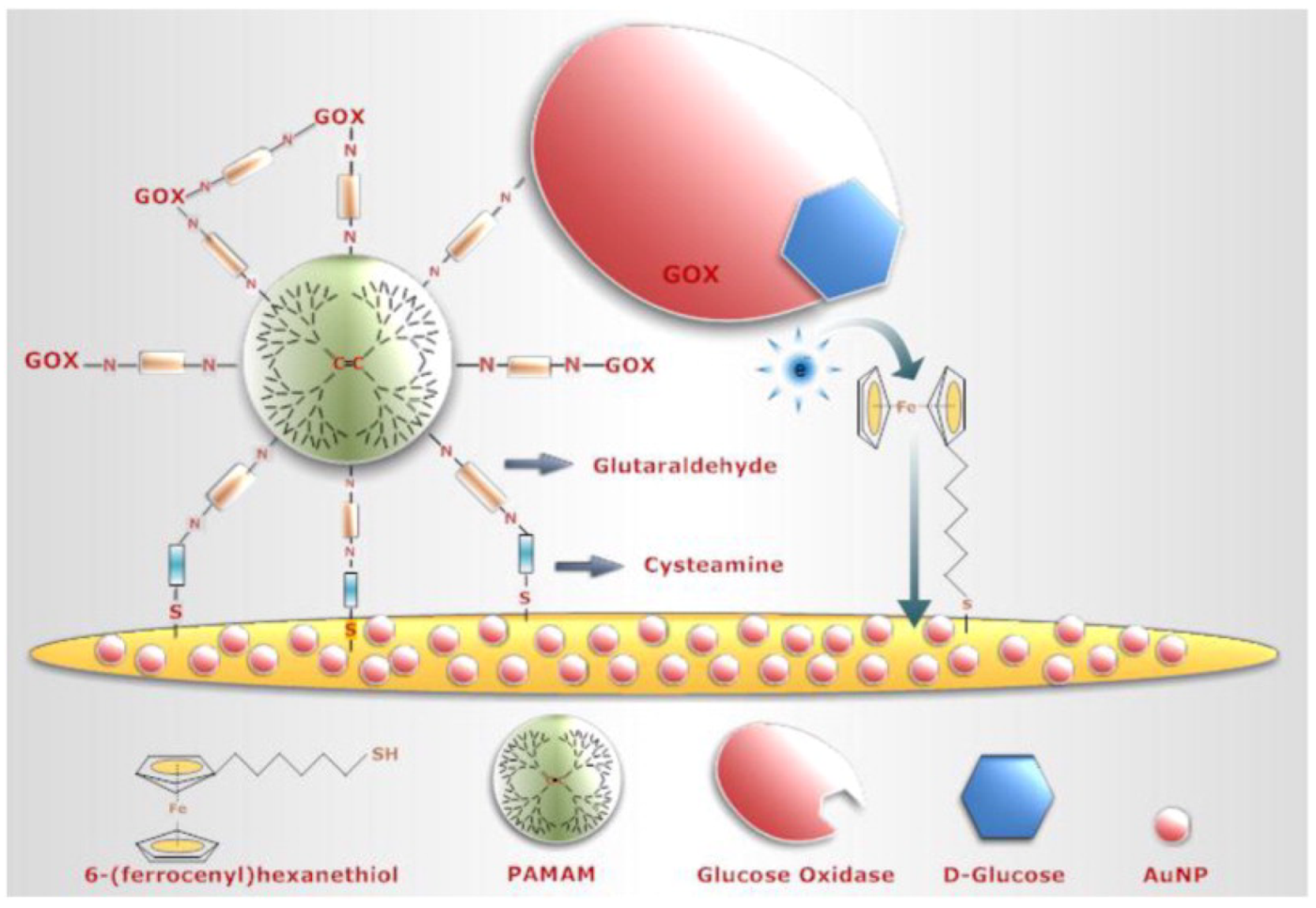
- Karadag, M.; Geyik, C.; Demirkol, D.O.; Ertas, F.N.; Timur, S. Modified Gold Surfaces by 6-(Ferrocenyl)Hexanethiol/Dendrimer/Gold Nanoparticles as a Platform for the Mediated Biosensing Applications. Mater. Sci. Eng. C 2013, 33, 634–640. [Google Scholar] [CrossRef]
- Ma, T.; Wang, Y.; Hou, Y.; Wang, E.; Yin, G.; Hasebe, Y.; Zhang, Z. An Amperometric Glucose Biosensor Based on Electrostatic Force Induced Layer-by-Layer GOD/Chitosan/Pyrite on a Glassy Carbon Electrode. Anal. Sci. 2022, 38, 553–562. [Google Scholar] [CrossRef] [PubMed]
- Brichkin, S.B.; Razumov, V.F. Colloidal Quantum Dots: Synthesis, Properties and Applications. Russ. Chem. Rev. 2016, 85, 1297–1312. [Google Scholar] [CrossRef]
- Jamieson, T.; Bakhshi, R.; Petrova, D.; Pocock, R.; Imani, M.; Seifalian, A.M.; Press, A.I.N. Biological Applications of Quantum Dots. Elsevier 2007, 28, 4717–4732. [Google Scholar] [CrossRef]
- Aftab, S.; Shah, A.; Erkmen, C.; Kurbanoglu, S.; Uslu, B. Chapter 1. Quantum dots: Synthesis and characterizations. In Electroanalytical Applications of Quantum Dot-Based Biosensors; Uslu, B., Ed.; Elsevier: Amsterdam, The Netherlands, 2021; pp. 1–35. [Google Scholar] [CrossRef]
- Shameem, M.M.; Sasikanth, S.M.; Annamalai, R.; Raman, R.G. Materials Today: Proceedings A Brief Review on Polymer Nanocomposites and Its Applications. Mater. Today Proc. 2021, 45, 2536–2539. [Google Scholar] [CrossRef]
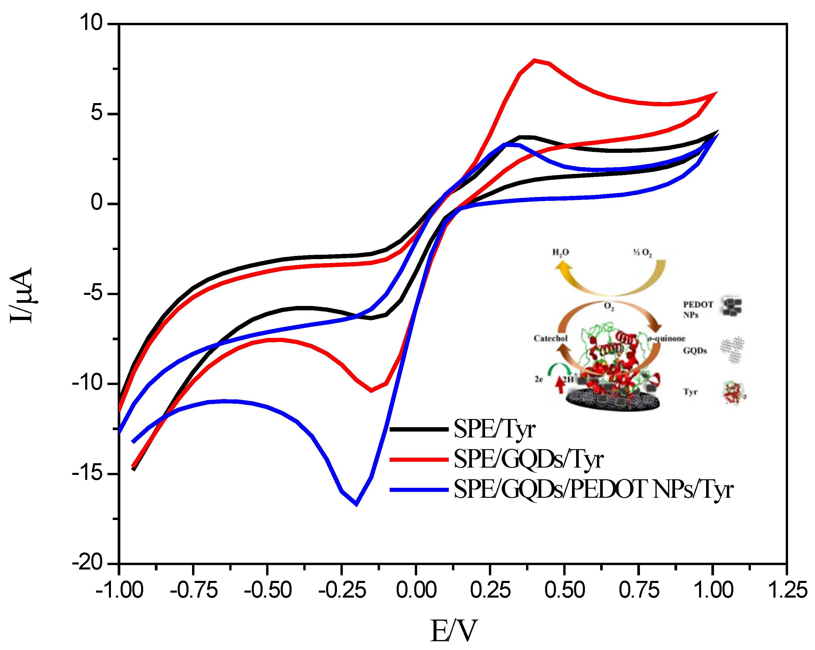
- Erkmen, C.; Demir, Y.; Kurbanoglu, S.; Uslu, B. Multi-Purpose Electrochemical Tyrosinase Nanobiosensor Based on Poly (3,4 Ethylenedioxythiophene) Nanoparticles Decorated Graphene Quantum Dots: Applications to Hormone Drugs Analyses and Inhibition Studies. Sens. Actuators B Chem. 2021, 343, 130164. [Google Scholar] [CrossRef]
- Jijana, A.N. Polyaniline Entrapped Water-Dispersible 3MPA-ZnSe Quantum Dots and Their Application for the Development of an Enzymatic Electrochemical Nanobiosensor for the Detection of 17β-Estradiol, an Endocrine-Disrupting Compound. Appl. Biochem. Biotechnol. 2023, 195, 3425–3455. [Google Scholar] [CrossRef]
- Wu, S.; Jiang, M.; Mao, H.; Zhao, N.; He, D.; Chen, Q.; Liu, D.; Zhang, W.; Song, X.M. A Sensitive Cholesterol Electrochemical Biosensor Based on Biomimetic Cerasome and Graphene Quantum Dots. Anal. Bioanal. Chem. 2022, 414, 3593–3603. [Google Scholar] [CrossRef]
- Kuzikov, A.V.; Filippova, T.A.; Masamrekh, R.A.; Shumyantseva, V.V. Biotransformation of Phenytoin in the Electrochemically-Driven CYP2C19 System. Biophys. Chem. 2022, 291, 106894. [Google Scholar] [CrossRef]
- Singh, A.K.; Jaiswal, N.; Tiwari, I.; Ahmad, M.; Silva, S.R.P. Electrochemical Biosensors Based on in Situ Grown Carbon Nanotubes on Gold Microelectrode Array Fabricated on Glass Substrate for Glucose Determination. Microchim. Acta 2023, 190, 55. [Google Scholar] [CrossRef] [PubMed]
- Sanz, C.G.; Onea, M.; Aldea, A.; Barsan, M.M. Disposable Superoxide Dismutase Biosensors Based on Gold Covered Polycaprolactone Fibers for the Detection of Superoxide in Cell Culture Media. Talanta 2022, 241, 123255. [Google Scholar] [CrossRef] [PubMed]
- Yan, C.; Jin, K.; Luo, X.; Piao, J.; Wang, F. Electrochemical Biosensor Based on Chitosan-and Thioctic-Acid-Modified Nanoporous Gold Co-Immobilization Enzyme for Glycerol Determination. Chemosensors 2022, 10, 258. [Google Scholar] [CrossRef]
- Ramanavičius, A.; Ramanavičienė, A.; Malinauskas, A. Electrochemical Sensors Based on Conducting Polymer—Polypyrrole. Electrochim. Acta 2006, 51, 6025–6037. [Google Scholar] [CrossRef]
- Ghorbani Zamani, F.; Moulahoum, H.; Ak, M.; Odaci Demirkol, D.; Timur, S. Current Trends in the Development of Conducting Polymers-Based Biosensors. TrAC Trends Anal. Chem. 2019, 118, 264–276. [Google Scholar] [CrossRef]
- Lou, C.; Jing, T.; Zhou, J.; Tian, J.; Zheng, Y.; Wang, C.; Zhao, Z.; Lin, J.; Liu, H.; Zhao, C.; et al. Laccase Immobilized Polyaniline/Magnetic Graphene Composite Electrode for Detecting Hydroquinone. Int. J. Biol. Macromol. 2020, 149, 1130–1138. [Google Scholar] [CrossRef]
- Kurbanoglu, S.; Cevher, S.C.; Toppare, L.; Cirpan, A.; Soylemez, S. Electrochemical Biosensor Based on Three Components Random Conjugated Polymer with Fullerene (C60). Bioelectrochemistry 2022, 147, 108219. [Google Scholar] [CrossRef]
- Ma, J.; Yuan, J.; Xu, Y.; Jiang, Y.; Bai, W.; Zheng, J. Ultrasensitive Electrochemical Determination of Bisphenol A in Food Samples Based on a Strategy for Activity Enhancement of Enzyme: Layer-by-Layer Self-Assembly of Tyrosinase between Two-Dimensional Porphyrin Metal–Organic Framework Nanofilms. Chem. Eng. J. 2022, 446, 137001. [Google Scholar] [CrossRef]
- Ivanov, A.; Davletshina, R.; Sharafieva, I.; Evtugyn, G. Electrochemical Biosensor Based on Polyelectrolyte Complexes for the Determination of Reversible Inhibitors of Acetylcholinesterase. Talanta 2019, 194, 723–730. [Google Scholar] [CrossRef]
- Liu, J.; Lang, Q.; Liang, B.; Zheng, Z.; Zhang, Y.; Liu, A. Sensitive Electrochemical Sequential Enzyme Biosensor for Glucose and Starch Based on Glucoamylase- and Glucose Oxidase-Controllably Co-Displayed Yeast Recombinant. Anal. Chim. Acta 2022, 1221, 340173. [Google Scholar] [CrossRef]
- Mohtar, L.G.; Aranda, P.; Messina, G.A.; Nazareno, M.A.; Pereira, S.V.; Raba, J.; Bertolino, F.A. Amperometric Biosensor Based on Laccase Immobilized onto a Nanostructured Screen-Printed Electrode for Determination of Polyphenols in Propolis. Microchem. J. 2019, 144, 13–18. [Google Scholar] [CrossRef]
- Li, J.; Li, X.; Zhao, Q.; Jiang, Z.; Tadé, M.; Wang, S.; Liu, S. Polydopamine-Assisted Decoration of TiO2 Nanotube Arrays with Enzyme to Construct a Novel Photoelectrochemical Sensing Platform. Sens. Actuators B Chem. 2018, 255, 133–139. [Google Scholar] [CrossRef]
- Vaghela, C.; Kulkarni, M.; Haram, S.; Aiyer, R.; Karve, M. A Novel Inhibition Based Biosensor Using Urease Nanoconjugate Entrapped Biocomposite Membrane for Potentiometric Glyphosate Detection. Int. J. Biol. Macromol. 2018, 108, 32–40. [Google Scholar] [CrossRef]
- Comba, F.N.; Romero, M.R.; Garay, F.S.; Baruzzi, A.M. Mucin and Carbon Nanotube-Based Biosensor for Detection of Glucose in Human Plasma. Anal. Biochem. 2018, 550, 34–40. [Google Scholar] [CrossRef] [Green Version]
- Wang, Y.; Liu, X.; Xu, X.; Yang, Y.; Huang, L.; He, Z.; Xu, Y.; Chen, J.; Feng, Z. Preparation and Characterization of Reduced Graphene Oxide/Fe3O4 Nanocomposite by a Facile in-Situ Deposition Method for Glucose Biosensor Applications. Mater. Res. Bull. 2018, 101, 340–346. [Google Scholar] [CrossRef]
- Sanko, V.; Şenocak, A.; Tümay, S.O.; Demirbas, E. A Novel Comparative Study for Electrochemical Urea Biosensor Design: Effect of Different Ferrite Nanoparticles (MFe2O4, M: Cu, Co, Ni, Zn) in Urease Immobilized Composite System. Bioelectrochemistry 2023, 149, 108324. [Google Scholar] [CrossRef]
- Maleki, N.; Kashanian, S.; Maleki, E.; Nazari, M. A Novel Enzyme Based Biosensor for Catechol Detection in Water Samples Using Artificial Neural Network. Biochem. Eng. J. 2017, 128, 1–11. [Google Scholar] [CrossRef]
- Sánchez-Paniagua López, M.; Redondo-Gómez, E.; López-Ruiz, B. Electrochemical Enzyme Biosensors Based on Calcium Phosphate Materials for Tyramine Detection in Food Samples. Talanta 2017, 175, 209–216. [Google Scholar] [CrossRef] [PubMed]
- Shoja, Y.; Rafati, A.A.; Ghodsi, J. Enzymatic Biosensor Based on Entrapment of D-Amino Acid Oxidase on Gold Nanofilm/MWCNTs Nanocomposite Modified Glassy Carbon Electrode by Sol-Gel Network: Analytical Applications for D-Alanine in Human Serum. Enzym. Microb. Technol. 2017, 100, 20–27. [Google Scholar] [CrossRef] [PubMed]
- Chen, W.; Yang, W.; Lu, Y.; Zhu, W.; Chen, X. Encapsulation of Enzyme into Mesoporous Cages of Metal-Organic Frameworks for the Development of Highly Stable Electrochemical Biosensors. Anal. Methods 2017, 9, 3213–3220. [Google Scholar] [CrossRef]
- Vlamidis, Y.; Gualandi, I.; Tonelli, D. Amperometric Biosensors Based on Reduced GO and MWCNTs Composite for Polyphenols Detection in Fruit Juices. J. Electroanal. Chem. 2017, 799, 285–292. [Google Scholar] [CrossRef]
- Ezhilan, M.; Gumpu, M.B.; Ramachandra, B.L.; Nesakumar, N.; Babu, K.J.; Krishnan, U.M.; Rayappan, J.B.B. Design and Development of Electrochemical Biosensor for the Simultaneous Detection of Melamine and Urea in Adulterated Milk Samples. Sens. Actuators B Chem. 2017, 238, 1283–1292. [Google Scholar] [CrossRef]
- Camargo, J.R.; Baccarin, M.; Raymundo-Pereira, P.A.; Campos, A.M.; Oliveira, G.G.; Fatibello-Filho, O.; Oliveira, O.N.; Janegitz, B.C. Electrochemical Biosensor Made with Tyrosinase Immobilized in a Matrix of Nanodiamonds and Potato Starch for Detecting Phenolic Compounds. Anal. Chim. Acta 2018, 1034, 137–143. [Google Scholar] [CrossRef] [PubMed]
- Cunha-Silva, H.; Arcos-Martinez, M.J. Dual Range Lactate Oxidase-Based Screen Printed Amperometric Biosensor for Analysis of Lactate in Diversified Samples. Talanta 2018, 188, 779–787. [Google Scholar] [CrossRef]
- Varmira, K.; Mohammadi, G.; Mahmoudi, M.; Khodarahmi, R.; Rashidi, K.; Hedayati, M.; Goicoechea, H.C.; Jalalvand, A.R. Fabrication of a Novel Enzymatic Electrochemical Biosensor for Determination of Tyrosine in Some Food Samples. Talanta 2018, 183, 1–10. [Google Scholar] [CrossRef]
- Rashidi, K.; Mahmoudi, M.; Mohammadi, G.; Zangeneh, M.M.; Korani, S.; Goicoechea, H.C.; Gu, H.W.; Jalalvand, A.R. Simultaneous Co-Immobilization of Three Enzymes onto a Modified Glassy Carbon Electrode to Fabricate a High-Performance Amperometric Biosensor for Determination of Total Cholesterol. Int. J. Biol. Macromol. 2018, 120, 587–595. [Google Scholar] [CrossRef]
- Sandeep, S.; Santhosh, A.S.; Swamy, N.K.; Suresh, G.S.; Melo, J.S.; Nithin, K.S. Electrochemical Detection of L-Dopa Using Crude Polyphenol Oxidase Enzyme Immobilized on Electrochemically Reduced RGO-Ag Nanocomposite Modified Graphite Electrode. Mater. Sci. Eng. B Solid-State Mater. Adv. Technol. 2018, 232–235, 15–21. [Google Scholar] [CrossRef]
- da Silva, M.K.L.; Vanzela, H.C.; Defavari, L.M.; Cesarino, I. Determination of Carbamate Pesticide in Food Using a Biosensor Based on Reduced Graphene Oxide and Acetylcholinesterase Enzyme. Sens. Actuators B Chem. 2018, 277, 555–561. [Google Scholar] [CrossRef] [Green Version]
- Samphao, A.; Butmee, P.; Saejueng, P.; Pukahuta, C.; Švorc, Ľ.; Kalcher, K. Monitoring of Glucose and Ethanol during Wine Fermentation by Bienzymatic Biosensor. J. Electroanal. Chem. 2018, 816, 179–188. [Google Scholar] [CrossRef]
- Hou, C.; Zhao, D.; Wang, Y.; Zhang, S.; Li, S. Preparation of Magnetic Fe3O4/PPy@ZIF-8 Nanocomposite for Glucose Oxidase Immobilization and Used as Glucose Electrochemical Biosensor. J. Electroanal. Chem. 2018, 822, 50–56. [Google Scholar] [CrossRef]
- Yazdanparast, S.; Benvidi, A.; Abbasi, S.; Rezaeinasab, M. Enzyme-Based Ultrasensitive Electrochemical Biosensor Using Poly(L-Aspartic Acid)/MWCNT Bio-Nanocomposite for Xanthine Detection: A Meat Freshness Marker. Microchem. J. 2019, 149, 104000. [Google Scholar] [CrossRef]
- Ma, L.; He, Y.; Wang, Y.; Wang, Y.; Li, R.; Huang, Z.; Jiang, Y.; Gao, J. Nanocomposites of Pt Nanoparticles Anchored on UiO66-NH2 as Carriers to Construct Acetylcholinesterase Biosensors for Organophosphorus Pesticide Detection. Electrochim. Acta 2019, 318, 525–533. [Google Scholar] [CrossRef]
- Wang, A.; Ding, Y.; Li, L.; Duan, D.; Mei, Q.; Zhuang, Q.; Cui, S.; He, X. A Novel Electrochemical Enzyme Biosensor for Detection of 17β-Estradiol by Mediated Electron-Transfer System. Talanta 2019, 192, 478–485. [Google Scholar] [CrossRef]
- Albishri, H.M.; Abd El-Hady, D. Hyphenation of Enzyme/Graphene Oxide-Ionic Liquid/Glassy Carbon Biosensors with Anodic Differential Pulse Stripping Voltammetry for Reliable Determination of Choline and Acetylcholine in Human Serum. Talanta 2019, 200, 107–114. [Google Scholar] [CrossRef]
- Malik, M.; Chaudhary, R.; Pundir, C.S. An Improved Enzyme Nanoparticles Based Amperometric Pyruvate Biosensor for Detection of Pyruvate in Serum. Enzym. Microb. Technol. 2019, 123, 30–38. [Google Scholar] [CrossRef]
- Narwal, V.; Pundir, C.S. Development of Glycerol Biosensor Based on Co-Immobilization of Enzyme Nanoparticles onto Graphene Oxide Nanoparticles Decorated Pencil Graphite Electrode. Int. J. Biol. Macromol. 2019, 127, 57–65. [Google Scholar] [CrossRef]
- Mirzaei, F.; Mirzaei, M.; Torkzadeh-Mahani, M. A Hydrophobin-Based-Biosensor Layered by an Immobilized Lactate Dehydrogenase Enzyme for Electrochemical Determination of Pyruvate. Bioelectrochemistry 2019, 130, 107323. [Google Scholar] [CrossRef]
- da Silva, W.; Ghica, M.E.; Ajayi, R.F.; Iwuoha, E.I.; Brett, C.M.A. Tyrosinase Based Amperometric Biosensor for Determination of Tyramine in Fermented Food and Beverages with Gold Nanoparticle Doped Poly(8-Anilino-1-Naphthalene Sulphonic Acid) Modified Electrode. Food Chem. 2019, 282, 18–26. [Google Scholar] [CrossRef]
- da Silva, W.; Ghica, M.; Brett, C.M.A. Choline Oxidase Inhibition Biosensor Based on Poly (Brilliant Cresyl Blue)–Deep Eutectic Solvent/Carbon Nanotube Modified Electrode for Dichlorvos. Sens. Actuators B Chem. 2019, 298, 126862. [Google Scholar] [CrossRef]
- Kurbanoglu, S.; Mayorga-Martinez, C.C.; Medina-Sánchez, M.; Rivas, L.; Ozkan, S.A.; Merkoçi, A. Antithyroid Drug Detection Using an Enzyme Cascade Blocking in a Nanoparticle-based Lab-on-a-chip System. Biosens. Bioelectron. 2015, 67, 670–676. [Google Scholar] [CrossRef]
- Turan, J.; Kesik, M.; Soylemez, S.; Goker, S.; Kolb, M.; Bahadir, M.; Toppare, L. Development of an Amperometric Biosensor Based on a Novel Conducting Copolymer for Detection of Anti-Dementia Drugs. J. Electroanal. Chem. 2014, 735, 43–50. [Google Scholar] [CrossRef]
- Kurbanoglu, S.; Rivas, L.; Ozkan, S.A.S.A.; Merkoçi, A. Electrochemically Reduced Graphene and Iridium Oxide Nanoparticles for Inhibition-Based Angiotensin-Converting Enzyme Inhibitor Detection. Biosens. Bioelectron. 2017, 88, 122–129. [Google Scholar] [CrossRef] [PubMed]
- Manan, F.A.A.; Hong, W.W.; Abdullah, J.; Yusof, N.A.; Ahmad, I. Nanocrystalline Cellulose Decorated Quantum Dots Based Tyrosinase Biosensor for Phenol Determination. Mater. Sci. Eng. C 2019, 99, 37–46. [Google Scholar] [CrossRef]
- Ali, M.; Shah, I.; Kim, S.W.; Sajid, M.; Lim, J.H.; Choi, K.H. Quantitative Detection of Uric Acid through ZnO Quantum Dots Based Highly Sensitive Electrochemical Biosensor. Sens. Actuators A Phys. 2018, 283, 282–290. [Google Scholar] [CrossRef]
- Wang, D.; Liang, Y.; Su, Y.; Shang, Q.; Zhang, C. Sensitivity Enhancement of Cloth-Based Closed Bipolar Electrochemiluminescence Glucose Sensor via Electrode Decoration with Chitosan/Multi-Walled Carbon Nanotubes/Graphene Quantum Dots-Gold Nanoparticles. Biosens. Bioelectron. 2019, 130, 55–64. [Google Scholar] [CrossRef]
- Çetin, M.Z.; Guven, N.; Apetrei, R.-M.; Camurlu, P. Highly Sensitive Detection of Glucose via Glucose Oxidase Immobilization onto Conducting Polymer-Coated Composite Polyacrylonitrile Nanofibers. Enzym. Microb. Technol. 2023, 164, 110178. [Google Scholar] [CrossRef] [PubMed]
- Wang, Q.; Jiao, C.; Wang, X.; Wang, Y.; Sun, K.; Li, L.; Fan, Y.; Hu, L. A Hydrogel-Based Biosensor for Stable Detection of Glucose. Biosens. Bioelectron. 2023, 221, 114908. [Google Scholar] [CrossRef]
- Wang, Y.; Guo, H.; Yuan, M.; Yu, J.; Wang, Z.; Chen, X. One-Step Laser Synthesis Platinum Nanostructured 3D Porous Graphene: A Flexible Dual-Functional Electrochemical Biosensor for Glucose and PH Detection in Human Perspiration. Talanta 2023, 257, 124362. [Google Scholar] [CrossRef]
- Trifan, A.G.; Apetrei, I.M. Development of Novel Electrochemical Biosensors Based on Horseradish Peroxidase for the Detection of Caffeic Acid. Appl. Sci. 2023, 13, 2526. [Google Scholar] [CrossRef]
- Baluta, S.; Romaniec, M.; Halicka-Stępień, K.; Alicka, M.; Pieła, A.; Pala, K.; Cabaj, J. A Novel Strategy for Selective Thyroid Hormone Determination Based on an Electrochemical Biosensor with Graphene Nanocomposite. Sensors 2023, 23, 602. [Google Scholar] [CrossRef]
- Tong, C.; Liu, H.; Mo, Y.; Li, J.; Liu, X.; Pang, L. In-Situ Growth of Enzyme/Copper Phosphate Hybrids on Carbon Cloth Surface as Self-Powered Electrochemical Glucose Biosensor. Biochem. Eng. J. 2023, 193, 108860. [Google Scholar] [CrossRef]
- Amin, M.; Abdullah, B.M.; Wylie, S.R.; Rowley-Neale, S.J.; Banks, C.E.; Whitehead, K.A. The Voltammetric Detection of Cadaverine Using a Diamine Oxidase and Multi-Walled Carbon Nanotube Functionalised Electrochemical Biosensor. Nanomaterials 2023, 13, 36. [Google Scholar] [CrossRef] [PubMed]
- Wei, Y.; Hui, Y.; Lu, X.; Liu, C.; Zhang, Y.; Fan, Y.; Chen, W. One-Pot Preparation of NiMn Layered Double Hydroxide-MOF Material for Highly Sensitive Electrochemical Sensing of Glucose. J. Electroanal. Chem. 2023, 933, 117276. [Google Scholar] [CrossRef]
- Yang, L.; Qu, L.; Zhang, X.; Li, M.; Liu, Z. Aptamer-Induced in-Situ Growth of Acetylcholinesterase-Cu3(PO4)2 Hybrid Nanoflowers for Electrochemical Detection of Organophosphorus Inhibitors. Nano Res. 2023. [Google Scholar] [CrossRef]
- Tvorynska, S.; Barek, J.; Josypcuk, B. High-Performance Amperometric Biosensor for Flow Injection Analysis Consisting of a Replaceable Lactate Oxidase-Based Mini-Reactor and a Silver Amalgam Screen-Printed Electrode. Electrochim. Acta 2023, 445, 142033. [Google Scholar] [CrossRef]
- Zhu, Y.; Qi, Y.; Xu, M.; Luo, J. Flexible Biosensor Based on Signal Amplification of Gold Nanoparticles-Composite Flower Clusters for Glucose Detection in Sweat. Colloids Surf. A Physicochem. Eng. Asp. 2023, 661, 130908. [Google Scholar] [CrossRef]
- Ozoglu, O.; Uzunoglu, A.; Unal, M.A.; Gumustas, M.; Ozkan, S.A.; Korukluoglu, M.; Gunes Altuntas, E. Electrochemical Detection of Lactate Produced by Foodborne Presumptive Lactic Acid Bacteria. J. Biosci. Bioeng. 2023, 135, 313–320. [Google Scholar] [CrossRef]
- Paneru, S.; Kumar, D. Ag-Doped-CuO Nanoparticles Supported Polyaniline (PANI) Based Novel Electrochemical Sensor for Sensitive Detection of Paraoxon-Ethyl in Three Real Samples. Sens. Actuators B Chem. 2023, 379, 133270. [Google Scholar] [CrossRef]
- Yang, L.; Bai, R.; Xie, B.; Zhuang, N.; Lv, Z.; Chen, M.; Dong, W.; Zhou, J.; Jiang, M. A Biosensor Based on Oriented Immobilization of an Engineered L-Glutamate Oxidase on a Screen-Printed Microchip for Detection of l-Glutamate in Fermentation Processes. Food Chem. 2023, 405, 134792. [Google Scholar] [CrossRef]
- Ji, X.; Zhao, X.; Zhang, Z.; Si, Y.; Qian, W.; Fu, H.; Chen, Z.; Wang, Z.; Jin, H.; Yang, Z.; et al. Scalable Fabrication of Graphene-Assembled Multifunctional Electrode with Efficient Electrochemical Detection of Dopamine and Glucose. Nano Res. 2023, 16, 6361–6368. [Google Scholar] [CrossRef]
- Lin, Y.; Yang, L.; Ma, Y.; Ye, J. Construction of Minitype Glutamate Sensor for in Vivo Monitoring of L-Glutamate in Plant. Microchem. J. 2023, 188, 108505. [Google Scholar] [CrossRef]
- Theansun, W.; Sriprachuabwong, C.; Chuenchom, L.; Prajongtat, P.; Techasakul, S.; Tuantranont, A.; Dechtrirat, D. Acetylcholinesterase Modified Inkjet-Printed Graphene/Gold Nanoparticle/Poly(3,4-Ethylenedioxythiophene): Poly(Styrenesulfonate) Hybrid Electrode for Ultrasensitive Chlorpyrifos Detection. Bioelectrochemistry 2023, 149, 108305. [Google Scholar] [CrossRef]
- Sun, X.; Xie, Y.; Chu, H.; Long, M.; Zhang, M.; Wang, Y.; Hu, X. A Highly Sensitive Electrochemical Biosensor for the Detection of Hydroquinone Based on a Magnetic Covalent Organic Framework and Enzyme for Signal Amplification. New J. Chem. 2022, 46, 11902–11909. [Google Scholar] [CrossRef]
- Cheraghi, S.; Taher, M.A.; Karimi-Maleh, H.; Karimi, F.; Shabani-Nooshabadi, M.; Alizadeh, M.; Al-Othman, A.; Erk, N.; Yegya Raman, P.K.; Karaman, C. Novel Enzymatic Graphene Oxide Based Biosensor for the Detection of Glutathione in Biological Body Fluids. Chemosphere 2022, 287, 132187. [Google Scholar] [CrossRef]
- Ketmen, S.; Er Zeybekler, S.; Gelen, S.S.; Odaci, D. Graphene Oxide-Magnetic Nanoparticles Loaded Polystyrene-Polydopamine Electrospun Nanofibers Based Nanocomposites for Immunosensing Application of C-Reactive Protein. Biosensors 2022, 12, 1175. [Google Scholar] [CrossRef]
- Kalkan, Z.; Yence, M.; Turk, F.; Bektas, T.U.; Ozturk, S.; Surdem, S.; Yildirim-Tirgil, N. Boronic Acid Substituted Polyaniline Based Enzymatic Biosensor System for Catechol Detection. Electroanalysis 2022, 34, 33–42. [Google Scholar] [CrossRef]
- Han, J.-H.; Hyun Park, S.; Kim, S.; Jungho Pak, J. A Performance Improvement of Enzyme-Based Electrochemical Lactate Sensor Fabricated by Electroplating Novel PdCu Mediator on a Laser Induced Graphene Electrode. Bioelectrochemistry 2022, 148, 108259. [Google Scholar] [CrossRef] [PubMed]
- Loguercio, L.F.; Thesing, A.; Demingos, P.; de Albuquerque, C.D.L.; Rodrigues, R.S.B.; Brolo, A.G.; Santos, J.F.L. Efficient Acetylcholinesterase Immobilization for Improved Electrochemical Performance in Polypyrrole Nanocomposite-Based Biosensors for Carbaryl Pesticide. Sens. Actuators B Chem. 2021, 339, 129875. [Google Scholar] [CrossRef]
- Nashruddin, S.N.A.; Abdullah, J.; Mohammad Haniff, M.A.S.; Mat Zaid, M.H.; Choon, O.P.; Mohd Razip Wee, M.F. Label Free Glucose Electrochemical Biosensor Based on Poly(3,4-Ethylenedioxy Thiophene):Polystyrene Sulfonate/Titanium Carbide/Graphene Quantum Dots. Biosensors 2021, 11, 267. [Google Scholar] [CrossRef] [PubMed]
- Kapan, B.; Kurbanoglu, S.; Esenturk, E.N.; Soylemez, S.; Toppare, L. Electrochemical Catechol Biosensor Based on β-Cyclodextrin Capped Gold Nanoparticles and Inhibition Effect of Ibuprofen. Process Biochem. 2021, 108, 80–89. [Google Scholar] [CrossRef]
- Akdag, A.; Işık, M.; Göktaş, H. Conducting Polymer-Based Electrochemical Biosensor for the Detection of Acetylthiocholine and Pesticide via Acetylcholinesterase. Biotechnol. Appl. Biochem. 2021, 68, 1113–1119. [Google Scholar] [CrossRef] [PubMed]
- Xia, T.; Liu, G.; Wang, J.; Hou, S.; Hou, S. MXene-Based Enzymatic Sensor for Highly Sensitive and Selective Detection of Cholesterol. Biosens. Bioelectron. 2021, 183, 113243. [Google Scholar] [CrossRef] [PubMed]
- Xu, W.; Sakran, M.; Fei, J.; Li, X.; Weng, C.; Yang, W.; Zhu, G.; Zhu, W.; Zhou, X. Electrochemical Biosensor Based on HRP/Ti3C2/Nafion Film for Determination of Hydrogen Peroxide in Serum Samples of Patients with Acute Myocardial Infarction. ACS Biomater. Sci. Eng. 2021, 7, 2767–2773. [Google Scholar] [CrossRef]
- Bathinapatla, A.; Kanchi, S.; Sabela, M.I.; Ling, Y.C.; Bisetty, K. Inamuddin Experimental and Computational Studies of a Laccase Immobilized ZnONPs/GO-Based Electrochemical Enzymatic Biosensor for the Detection of Sucralose in Food Samples. Food Anal. Methods 2020, 13, 2014–2027. [Google Scholar] [CrossRef]
- Yezer, I.; Demirkol, D.O. Cellulose Acetate–Chitosan Based Electrospun Nanofibers for Bio-Functionalized Surface Design in Biosensing. Cellulose 2020, 27, 10183–10197. [Google Scholar] [CrossRef]
- Sanli, S.; Celik, E.G.; Demir, B.; Gumus, Z.P.; Ilktac, R.; Aksuner, N.; Demirkol, D.O.; Timur, S. Magnetic Nanofiber Layers as a Functional Surface for Biomolecule Immobilization and One-Use ‘Sensing in-a-Drop’ Applications. ChemistrySelect 2018, 3, 13553–13560. [Google Scholar] [CrossRef]
- Esen, E.; Yazgan, I.; Demirkol, D.O.; Timur, S. Laccase Assay Based on Electrochemistry and Fluorescence Detection via Anthracene Sequestered Poly(Amic Acid) Films. React. Funct. Polym. 2018, 131, 36–43. [Google Scholar] [CrossRef]
- Kirbay, F.O.; Yalcinkaya, E.E.; Atik, G.; Evren, G.; Unal, B.; Demirkol, D.O.; Timur, S. Biofunctionalization of PAMAM-Montmorillonite Decorated Poly (Ɛ-Caprolactone)-Chitosan Electrospun Nanofibers for Cell Adhesion and Electrochemical Cytosensing. Biosens. Bioelectron. 2018, 109, 286–294. [Google Scholar] [CrossRef]
- Unal, B.; Yalcinkaya, E.E.; Demirkol, D.O.; Timur, S. An Electrospun Nanofiber Matrix Based on Organo-Clay for Biosensors: PVA/PAMAM-Montmorillonite. Appl. Surf. Sci. 2018, 444, 542–551. [Google Scholar] [CrossRef]
- Akin, M.; Prediger, A.; Yuksel, M.; Höpfner, T.; Demirkol, D.O.; Beutel, S.; Timur, S.; Scheper, T. A New Set up for Multi-Analyte Sensing: At-Line Bio-Process Monitoring. Biosens. Bioelectron. 2011, 26, 4532–4537. [Google Scholar] [CrossRef]
- Odaci, D.; Telefoncu, A.; Timur, S. Maltose Biosensing Based on Co-Immobilization of α-Glucosidase and Pyranose Oxidase. Bioelectrochemistry 2010, 79, 108–113. [Google Scholar] [CrossRef] [PubMed]
- Akin, M.; Yuksel, M.; Geyik, C.; Odaci, D.; Bluma, A.; Höpfner, T.; Beutel, S.; Scheper, T.; Timur, S. Alcohol Biosensing by Polyamidoamine (PAMAM)/Cysteamine/Alcohol Oxidase-Modified Gold Electrode. Biotechnol. Prog. 2010, 26, 896–906. [Google Scholar] [CrossRef] [PubMed]
- Tuncagil, S.; Ozdemir, C.; Demirkol, D.O.; Timur, S.; Toppare, L. Gold Nanoparticle Modified Conducting Polymer of 4-(2,5-Di(Thiophen-2-Yl)-1H-Pyrrole-1-l) Benzenamine for Potential Use as a Biosensing Material. Food Chem. 2011, 127, 1317–1322. [Google Scholar] [CrossRef]
- Odaci, D.; Telefoncu, A.; Timur, S. Pyranose Oxidase Biosensor Based on Carbon Nanotube (CNT)-Modified Carbon Paste Electrodes. Sens. Actuators B Chem. 2008, 132, 159–165. [Google Scholar] [CrossRef]
- Ozdemir, C.; Yeni, F.; Odaci, D.; Timur, S. Electrochemical Glucose Biosensing by Pyranose Oxidase Immobilized in Gold Nanoparticle-Polyaniline/AgCl/Gelatin Nanocomposite Matrix. Food Chem. 2010, 119, 380–385. [Google Scholar] [CrossRef]
- Yuksel, M.; Akin, M.; Geyik, C.; Demirkol, D.O.; Ozdemir, C.; Bluma, A.; Höpfner, T.; Beutel, S.; Timur, S.; Scheper, T. Offline Glucose Biomonitoring in Yeast Culture by Polyamidoamine/ Cysteamine-Modified Gold Electrodes. Biotechnol. Prog. 2011, 27, 530–538. [Google Scholar] [CrossRef]
- Odaci, D.; Kahveci, M.U.; Sahkulubey, E.L.; Ozdemir, C.; Uyar, T.; Timur, S.; Yagci, Y. In Situ Synthesis of Biomolecule Encapsulated Gold-Cross-Linked Poly(Ethylene Glycol) Nanocomposite as Biosensing Platform: A Model Study. Bioelectrochemistry 2010, 79, 211–217. [Google Scholar] [CrossRef] [PubMed]
- Damar, K.; Odaci Demirkol, D. Modified Gold Surfaces by Poly(Amidoamine) Dendrimers and Fructose Dehydrogenase for Mediated Fructose Sensing. Talanta 2011, 87, 67–73. [Google Scholar] [CrossRef]
- Çakar, İ.; Özdokur, K.V.; Demir, B.; Yavuz, E.; Demirkol, D.O.; Koçak, S.; Timur, S.; Ertaş, F.N. Molybdenum Oxide/Platinum Modified Glassy Carbon Electrode: A Novel Electrocatalytic Platform for the Monitoring of Electrochemical Reduction of Oxygen and Its Biosensing Applications. Sens. Actuators B Chem. 2013, 185, 331–336. [Google Scholar] [CrossRef]
- Ozdokur, K.V.; Demir, B.; Yavuz, E.; Ulus, F.; Erten, Ç.; Aydın, İ.; Demirkol, D.O.; Pelit, L.; Timur, S.; Ertaş, F.N. Pyranose Oxidase and Pt–MnOx Bionanocomposite Electrode Bridged by Ionic Liquid for Biosensing Applications. Sens. Actuators B Chem. 2014, 197, 123–128. [Google Scholar] [CrossRef]
- Unal, B.; Yalcinkaya, E.E.; Gumustas, S.; Sonmez, B.; Ozkan, M.; Balcan, M.; Demirkol, D.O.; Timur, S. Polyglycolide–Montmorillonite as a Novel Nanocomposite Platform for Biosensing Applications. New J. Chem. 2017, 41, 9371–9379. [Google Scholar] [CrossRef]
- Babadostu, A.; Guldu, O.K.; Demirkol, D.O.; Medine, E.I.; Unak, P.; Timur, S. Affinity Based Laccase Immobilization on Modified Magnetic Nanoparticles: Biosensing Platform for the Monitoring of Phenolic Compounds. Int. J. Polym. Mater. Polym. Biomater. 2015, 64, 260–266. [Google Scholar] [CrossRef]
- Ozdemir, C.; Akca, O.; Medine, E.I.; Demirkol, D.O.; Unak, P.; Timur, S. Biosensing Applications of Modified Core–Shell Magnetic Nanoparticles. Food Anal. Methods 2012, 5, 731–736. [Google Scholar] [CrossRef]


| Nanomaterial | Target | Enzyme | Support | Detection Principle | LOD | Application | Ref. |
|---|---|---|---|---|---|---|---|
| PCL:PAA/PAMAM/PyOx | Glucose | PyOx | GCE | CV, DPV and EIS | 0.0085 mM | Artificial serum, urine, salvia, sweat | [95] |
| Au/AuNP/(FcSH + Cyst)/PAMAM/GOx | Glucose | GOx | AuE | CV and AMP | 0.6 mM | Cherry juice Fizzy | [104] |
| AuE/PANI:3MPA-ZnSeQD/HRP | 17β-Estradiol | HRP | AuE | CV and DPV | 0.2 × 10⁻⁶ M | Wastewater | [111] |
| CNT | Glucose | GOx | Au MEA | CV and EIS | 0.2 ± 0.0014 µM | Blood serum samples | [114] |
| PANI/MG | hydroquinone | Lac | GCE | CV and CA | 2.94 μM | Actual water sample | [119] |
| Cu–TCPP | Bisphenol A | Tyr | GCE | DPV | 1.2 nM | Milk and plastic mineral water | [121] |
| AuNPs | polyphenols | lac | SPCE | CV | 0.83 μM | Polyphenols in propolis | [124] |
| TiO2 NT | H2O2 | HRP | PEC electrode | photoelectrochemical | 0.7 nM | NS | [125] |
| AuNPs | glyphosate | urease | Ion selective electrode (Elite 8051) | potentiometry | 0.5 ppm | Pesticides | [126] |
| Mucin and CNT | Glucose | Albumin | Pt | AMP | 3 μM | Human plasma | [127] |
| GO/Fe3O4 | Glucose | GOx | GCE | AMP | 106.5 μA mM−1 | Health | [128] |
| Ferrite NPs | Urea | Urease | GCE | CV and DPV | 0.17 µM | Soil and milk samples | [129] |
| PEDOT-GONs | Catechol | Lac | GCE | CV | 0.032 μM | Real water samples | [130] |
| Brushite cement-GA | Tyramine | PPO | GCE | CV | 4.85 × 10⁻⁸ M | Gouda and brie cheeses | [131] |
| Sol–gel/Au-NF/MWCNT | D-alanine | DAAO | GCE | LSV and CV | 20 nM | Human serum | [132] |
| MOF | Hydrogen peroxide | HRP | GCE | AMP and CV | 0.09 μM | NS | [133] |
| ERGO-MWCNTs | Catechol | Lac | GCE | CA | 0.3 μM | Fruit juice samples | [134] |
| Pyrogallol | |||||||
| Epicatechin | |||||||
| Gallic acid | |||||||
| 1,2-dihydroxybenzoic acid | |||||||
| Caffeic acid | |||||||
| Chlorogenic acid | |||||||
| Rutin | |||||||
| Catechin | |||||||
| Dopamine | |||||||
| Chitosan/ZnO | Melamine | AChE | PtE | CV | 3 pM for Melamine 1 pM for urea | Adulterated milk samples | [135] |
| Urea | |||||||
| ND-PS | Catechol | Tyr | GCE | DPV | 3.9 × 10⁻⁷ M | River and tap water | [136] |
| Cu-MOF/CS/Pt | Lactate | LOx | SPCE | CA | 0.75 μM | Sweat, saliva, red and white wines | [137] |
| PdPt NPs/Ch-IL/Gr-MWCNTs-IL | Tyrosine | TyrH | GCE | DPV | 0.009 × 10⁻⁹ M | Cheese, egg and yogurt | [138] |
| Au NPs/Chitin-IL/PEDOP/Gr-MWCNTs-Fr-IL | Cholesterol | ChO, ChE and HRP | GCE | CA | 0.07 μM | Rat plasma | [139] |
| rGO-AgNPs/Gr | L-dopa | PPO | GCE | CA | 1.85 μM | Urine | [140] |
| rGO | Carbamate | AChE | GCE | DPV | 1.9 nM | Tomato | [141] |
| Fe3O4@Au/(MnO2) | Glucose | GOx- ADH | CPE | CA | 0.1 mM for glucose and 60 mM for ethanol | Honey wine fermentation with wine yeast (Saccharomyces cerevisiae Type II) | [142] |
| Ethanol | |||||||
| Fe3O4/PPy@ZIF-8 | Glucose | GOx | GCE | CA | 0.333 μM | Serum | [143] |
| Poly(L-Asp)/MWCNT | Xanthine | XO | GCE | DPV | 3.5 × 10⁻⁴ μM | Fish meat | [144] |
| Pt@UiO66-NH2 | Organophosphorus pesticides | AChE | GCE | DPV | 4.9 × 10⁻¹⁵ M | Cabbage and apple | [145] |
| PLLY/CiA-GR | 17β-estradiol | Lac | GCE | DPV | 0.13 pM | Human urine | [146] |
| IL/GCE and GO-IL/GCE | Choline | AChE-ChO | GCE | ADPSV | 0.885 nM | Human serum | [147] |
| Acetylcholine | 1.352 nM | ||||||
| POxNPs/AuE | Pyruvate | POx | AuE | CA | 0.67 μM | Serum | [148] |
| GKNPs/GPONPs/GrONPs/ | Glycerol | GPO | PGE | CA | 0.002 μM | Blood serum | [149] |
| GrO/AuNPs/PVA/HFB1 | Pyruvate | Lactate dehydrogenase | GCE | DPV and CA | 8.69 nM | Serum | [150] |
| AuNP-PANSA | Tyramine | Tyr | GCE | CA | 0.71 μM | Fermented food and beverages | [151] |
| PBCBethaline-HNO3PTD/MWCNT | Dichlorvos | ChOx | GCE | CA | 1.6 nM | Orange juice | [152] |
| MNPs/IrOxNPs | Methimazole | Tyr | SPE | CA | 0.006 μM and 0.003 μM | Human serum and pharmaceutical dosage form | [153] |
| (poly(BODT-co-FMOC) | Donepezil | AChE-ChO | GE | CA | 0.027 μg/L for Donepezil | Tap water | [154] |
| Neostigmine | 0.559 μ/L for Neostigmine | ||||||
| ERGO/IrOxNPs | Captopril | Tyr | SPE | CA | 0.008 μM and 0.019 μM | Human serum and pharmaceutical dosage form | [155] |
| CTAB-NCC/QDs | Phenol | Tyr | SPCE | DPV | 0.082 μM | Lake water | [156] |
| Catechol | 0.125 μM | ||||||
| o-Cresol | 0.007 μM | ||||||
| 4-Chlorophenol | 0.021 μM | ||||||
| Nafion/ZnO QDs | Uric acid | Uricase | SPE | CA | 22.97 ± 10 μM | Urine samples | [157] |
| GQDs-AuNPs/PDDA-MWCNTs/CS/CBA | glucose | GOx | C-BPE | C-BP-ECL | 64 nM | human serum samples | [158] |
| PAN-MWCNTs/PEDOT | glucose | GOx | Pt disk electrode | AMP | 2.30 μM | Blood serum samples | [159] |
| PAN-MWCNTs/PPy | 2.38 μM | ||||||
| ZIF-8/CaCO3 NPs | glucose | GOx | Bare Au electrode | EIS | NS | Real honey samples | [160] |
| Pt-HEC/LSG | Glucose | GOx | Pt | CV and EIS | 0.23 μM | In human sweat | [161] |
| SPCE/HRP and SPCE/PB/HRP | Caffeic acid | HRP | SPCE and SPCE/PB | CV | 0.9 μM | In food supplements | [162] |
| GCE/Fe3O4@graphene/Ab/Lac | Free thyroid hormone | Lac | GCE | CV | 45.9 nM | In synthetic serum samples | [163] |
| enzyme-Cu3(PO4)2/CC | Glucose | GOx | GE | CV | 2.05 μM | NS | [164] |
| C-MWCNT/DAO/EDC-NHS/GA | Cadaverine | DAO | SPE | Voltammetry and DPV | 0.8 μg/mL | Stock solutions and artificial salvia | [165] |
| NiMn-LDH-MOF/GCE | Glucose | GOx | GCE | CV | 0.87 μM | In actual serum samples | [166] |
| AChE-Cu3(PO4)2 HNF/Apt/AuNP/CP | isocarbophos | AChE | CP | CV-EIS- SWV | 0.016 pM | Real agricultural samples (oilseed rape, cabbage, apple, and pear) | [167] |
| dichlorvos | 0.028 pM | ||||||
| methamidophos | 0.071 pM | ||||||
| parathion | 0.113 pM | ||||||
| SBA−15/APTES/GA/LOx mini-reactor connected in front of the AgA-SPE | L-lactic acid | LOx | SPE | AMP | 12.0 μmol L−1 | In wine and daily products | [168] |
| AuNPs/TA-APTES/aCC | Glucose | GOx | SPE | CV and EIS | 3.3 μM | In sweat sample | [169] |
| Ag/AgCl, Pt electrode and Pt wire | Lactate | LOx | Glassy electrode | CV and chronoamperometry | 31 μM | Lactate produced by foodborne lactic acid bacteria in real samples | [170] |
| AChE/Ag@CuO/PANI/ITO | Paraoxon-ethyl | AChE | ITO | CV | 11.35 pM | Banana, tomato and soil samples | [171] |
| ChBD-GluOx/PB/SPC | Glutamate | GluOx | Pt electrode | CV | 53.4 µA L mmol−1 cm−2 | Food ingredient | [172] |
| Nafion/GOx/GF | Dopamine | GOx | GCE | CV | 0.6 μM | Biological samples | [173] |
| Glucose | 0.41 μM | ||||||
| GluOx/PMPD/Pt/GRE | Glucose | GluOx | GRE | CV | 0.536 μM | Cucumber fruit and juice | [174] |
| AChE- CS/GP-AuNP-PEDOT:PSS/SPCE | Chlorpyrifos | AChE | SPCE | DPV | 0.07 nM | In real cabbage sample | [175] |
| Fe3O4@COF | Hydroquinone | HRP | GCE | DPV | 0.12 μM | Environmental water samples | [176] |
| GO/nafion/GCE | Glutathione | GSH-Px | GCE | DPV | 1.5 nM | Hemolyzed erythrocyte, dextrose saline, tablet | [177] |
| Polystyrene/rGO-MNP-PDA/Anti-CRP | CRP | GO | SPCE | CV, DPV and EIS | 0.33 ng/mL | Artificial saliva | [178] |
| 3-APBA | Catechol | Tyr | Au SPE GCE | CV | 0.25 μM | Green tea | [179] |
| PdCu | Lactate | LOx | LIG | AMP | 0.28 μM | NS | [180] |
| PPy-IC-DS1-AuNP | Carbaryl | AChE | ITO | AMP | 0.033 ng cm2 mL−1 | Tap water | [181] |
| PEDOT:PSS/Ti3C2/GQD | Glucose | GOx | SPE | DPV | 65 µM | NS | [182] |
| β-CD-AuNPs | Catechol | Tyr | GE | AMP | 0.42 μM | Drug Inhibition | [183] |
| Pt/PPy/Chi | Atch | AChE | Pt | DPV | 0.45 µM | NS | [184] |
| Paraoxon | 0.17 nM | ||||||
| Chi/Ti3C2Tx | Cholesterol | ChOx | GCE | DPV | 0.11 nM | Human serum | [185] |
| Ti3C2/Nafion film | Hydrogen peroxide | HRP | GCE | DPV | 1 µM | Human serum | [186] |
| ZnONPs-ATP-GO | Sucralose | Lac | GCE | DPV | 0.32 μM | Food samples | [187] |
| Cellulose acetate–CS/GOx | Glucose | GO | GCE | CV | 4.8 µM | Artificial tears, urine, sweat and serum | [188] |
| PVA-PEI/MNP/GO | Glucose | GO | SPE | CV and EIS | 11.5 μM | Synthetic spiked samples | [189] |
| Ant-PAA/Lac | Phenol | Lac | GCE | CV and EIS | 0.046 mM | Artificial wastewater | [190] |
| PCL-Chi/PAMAM-Mt | U87 | GCE | CV, DPV and EIS | NS | [191] | ||
| PVA/PAMAM-Mt | Glucose | PyOx | GCE | CV, DPV and EIS | 0.7 µM | Soft drink cola | [192] |
| Cysteamine/PAMAM | Glucose Ethanol | PyOx AOx | AuE | FIA-AMP | NS | Fermentation broth | [193] |
| AG/PyOx/CHIT–CNT AG/PyOx/CHIT | Maltose | PyOx α-glucosidase | GE | AMP | NS | Beer samples | [194] |
| PAMAM/Cyst/AOx | Ethanol | AOx | AuE | AMP | 0.016 mM | Alcoholic beverage and yeast cultivation samples | [195] |
| CP/AuNP/GOx | Glucose | GOx | GE | CV | 0.0021 and 0.0063 mM | Fizzy with orange Fizzy coke Lemonade Ice tea Peach juice Green tea Orange juice | [196] |
| CNTPE | Glucose | PyOx | CPE | CV | NS | In wine samples | [197] |
| AuNP/PANI/AgCl/Gelatin | Glucose | PyOx | GCE | CV and DPV | NS | Coke Lemonade Green tea Fruit juice Red Bull White wine Red wine | [198] |
| PDA/Cyst/AuNP | Glucose | GOx PyOx | AuE | FIA-AMP | 38.97 µM 1.27 µM | Yeast fermentation | [199] |
| PEG/AuNP/GOx | Glucose | GOx | PtE | CV | 0.06 mM | Ice tea Cherry juice | [200] |
| FDH/PAMAM | Fructose | FDH | AuE | CV | NS | Fruit juices Fizzy energy drink | [201] |
| Pt/MoO3/GCE/GOx | Glucose | GOx | GCE | CV | 0.025 mM | NS | [202] |
| PyOx–IL–Pt–MnOx/GCE | Glucose | PyOx | GCE | CV | 2.0 μM | Coke Orange juice | [203] |
| PGA–Mt/PyOx | Glucose | PyOx | GCE | AMP | 1.2 μM | Coke and other fizzy drinks | [204] |
| MNP-His/Cu/Lac | Phenol | Lac | CPE | CV | NS | In culture medium | [205] |
| MNP/GOx | Glucose | GOx | CPE | AMP | NS | Fruit juices | [206] |
Disclaimer/Publisher’s Note: The statements, opinions and data contained in all publications are solely those of the individual author(s) and contributor(s) and not of MDPI and/or the editor(s). MDPI and/or the editor(s) disclaim responsibility for any injury to people or property resulting from any ideas, methods, instructions or products referred to in the content. |
© 2023 by the authors. Licensee MDPI, Basel, Switzerland. This article is an open access article distributed under the terms and conditions of the Creative Commons Attribution (CC BY) license (https://creativecommons.org/licenses/by/4.0/).
Share and Cite
Kilic, N.M.; Singh, S.; Keles, G.; Cinti, S.; Kurbanoglu, S.; Odaci, D. Novel Approaches to Enzyme-Based Electrochemical Nanobiosensors. Biosensors 2023, 13, 622. https://doi.org/10.3390/bios13060622
Kilic NM, Singh S, Keles G, Cinti S, Kurbanoglu S, Odaci D. Novel Approaches to Enzyme-Based Electrochemical Nanobiosensors. Biosensors. 2023; 13(6):622. https://doi.org/10.3390/bios13060622
Chicago/Turabian StyleKilic, Nur Melis, Sima Singh, Gulsu Keles, Stefano Cinti, Sevinc Kurbanoglu, and Dilek Odaci. 2023. "Novel Approaches to Enzyme-Based Electrochemical Nanobiosensors" Biosensors 13, no. 6: 622. https://doi.org/10.3390/bios13060622