Article

PEGylation of Metal Oxide Nanoparticles Modulates Neutrophil Extracellular Trap Formation

by
Hunter T. Snoderly
,
Kasey A. Freshwater
,
Celia Martinez de la Torre
,
Dhruvi M. Panchal
,
Jenna N. Vito
and
Margaret F. Bennewitz
*
Department of Chemical and Biomedical Engineering, West Virginia University, Morgantown, WV 26506, USA
*
Author to whom correspondence should be addressed.
Biosensors 2022, 12(2), 123; https://doi.org/10.3390/bios12020123
Submission received: 18 December 2021 / Revised: 4 February 2022 / Accepted: 13 February 2022 / Published: 16 February 2022
(This article belongs to the Special Issue Bio–Nano-Interfaces for Engineering and Biomedical Applications)

Abstract

:
Novel metal oxide nanoparticle (NP) contrast agents may offer safety and functionality advantages over conventional gadolinium-based contrast agents (GBCAs) for cancer diagnosis by magnetic resonance imaging. However, little is known about the behavior of metal oxide NPs, or of their effect, upon coming into contact with the innate immune system. As neutrophils are the body’s first line of defense, we sought to understand how manganese oxide and iron oxide NPs impact leukocyte functionality. Specifically, we evaluated whether contrast agents caused neutrophils to release web-like fibers of DNA known as neutrophil extracellular traps (NETs), which are known to enhance metastasis and thrombosis in cancer patients. Murine neutrophils were treated with GBCA, bare manganese oxide or iron oxide NPs, or poly(lactic-co-glycolic acid) (PLGA)-coated metal oxide NPs with different incorporated levels of poly(ethylene glycol) (PEG). Manganese oxide NPs elicited the highest NETosis rates and had enhanced neutrophil uptake properties compared to iron oxide NPs. Interestingly, NPs with low levels of PEGylation produced more NETs than those with higher PEGylation. Despite generating a low rate of NETosis, GBCA altered neutrophil cytokine expression more than NP treatments. This study is the first to investigate whether manganese oxide NPs and GBCAs modulate NETosis and reveals that contrast agents may have unintended off-target effects which warrant further investigation.

1. Introduction

Cancers are diagnosed using a variety of screening methods, such as mammography, ultrasound, and magnetic resonance imaging (MRI). Although mammography is the current gold standard for breast imaging, it leads to inaccurate diagnoses, especially in younger patients where dense breasts can obscure cancer [1,2,3]. Therefore, ultrasound and MRI are used as secondary imaging tools in cases where mammography is suboptimal to visualize additional breast cancers [4,5,6]. Neither ultrasound or MRI have ionizing radiation, but ultrasound suffers from a limited depth of penetration which can miss some malignancies deeper in the breast [7,8]. In contrast, MRI acquires full 3D images of the breast with high spatial resolution and excellent soft tissue contrast that is capable of distinguishing between fat, muscle, organs, and tumors. However, MRI without additional contrast agents, suffers from exceedingly poor sensitivity [9]. To overcome this, contrast agents alter the relaxation rates of protons within water molecules in the body, thereby enhancing the magnitude of the signal in the tissues and organs where the contrast agent is incorporated [10].
Several types of contrast agents, including magnetic chelates and nanoparticles (NPs), have been created in recent decades to improve MRI sensitivity. Gadolinium (Gd) based contrast agents (GBCAs) and metal oxide NPs have both been used clinically to enhance the MRI-based tumor characterization [11]. While GBCAs are more commonly used than metal oxide contrast agents, GBCAs have recently fallen under increased regulatory scrutiny. The FDA has raised concerns around the safety of GBCAs due to reports of nephrogenic systemic fibrosis in renally challenged patients [12], gadolinium accumulation in the brain and other tissues following multiple contrast agent administrations [13,14,15], and in vitro neurotoxicity [16,17]. Additionally, one study suggested prenatal Gd exposure enhanced the risk of the child developing an inflammatory condition within 4 years after birth [18]. Due to these concerns and the favorable magnetic properties of metal oxides, metal oxide NPs have emerged as attractive possible alternatives to traditional GBCAs. Iron oxide (Fe2O3 or Fe3O4) and manganese oxide (MnO, MnO2, or Mn3O4) are two such NP formulations, with iron oxide providing T2 or T2 *-weighted contrast and manganese oxide providing T1-weighted contrast [19].
A key advantage of NPs lies in their customizability; different physicochemical properties of NPs can be altered to achieve the desired goal including size [20,21,22,23,24], shape [25,26,27], charge [24,28,29], type of contrast agent [19,30,31], polymer or lipid encapsulation [19,31,32,33,34], surface attachments (e.g., stealth polymers) [35], and the presence of targeting agents [36,37]. However, each modification alters how a given NP formulation will interact with blood cells as well as which tissues it accumulates in before clearance. Upon injection, NPs encounter a series of obstacles that influence their delivery to the target tissue depending on the particle formulation. As soon as NPs enter a patient’s body, they immediately interact with the circulation, resulting in the formation of a protein corona [38,39,40,41], the composition of which varies depending on the surface properties of the NPs [42,43,44]. In turn, this protein corona influences how well phagocytes recognize and internalize NPs. Tissue-resident phagocytes, such as macrophages, have been most studied in relation to NPs, as phagocytosis by these cells ultimately reduces the effective NP dose to the desired imaging areas [38,45]. However, neutrophils, which are circulating phagocytes and the most abundant immune cells in the body, also play a major role in NP clearance [46,47,48]. One NP surface modification technique used to reduce macrophage uptake is the attachment of poly(ethylene glycol) (PEG) chains onto NP surfaces. PEGylation is thought to decrease protein adsorption, thereby reducing phagocyte uptake and increasing the NP circulation time [49,50,51]. However, some studies have observed that PEGylation may have the opposite effect in neutrophils [52], suggesting that NP formulations designed solely with macrophages in mind are non-optimal upon initial delivery when neutrophils are the dominant phagocytes the NPs encounter. Additionally, as neutrophils are key mediators of inflammation and are the body’s first line of immunity, it is vital to consider whether NP uptake by neutrophils results in off-target effects that undermine the intended diagnostic or therapeutic utility of NPs.
When stimulated by inflammatory or pathogenic stimuli, neutrophil nuclei decondense and are exported from the cell in a fibrous structure composed of DNA, histones, and granular content [53,54]. This structure is known as a neutrophil extracellular trap (NET); NETosis refers to the process by which NETs are formed and released. The evolutionary purpose of NETs is predominantly antimicrobial defense, as NETs physically entrap bacteria, facilitating their phagocytosis or killing them directly through proteolytic enzymes decorating the fibers. However, inappropriate NETosis also occurs in the context of inflammation. Indeed, NETs have been observed to drive pathology in inflammatory diseases as diverse as type I diabetes [55], gout [56,57], cystic fibrosis [58], and ulcerative colitis [59]. As cancer is itself an inflammatory disease, it is unsurprising that NETosis also plays a role in tumor progression and spread. Mounting evidence suggests that NETs act as agents of metastasis by trapping circulating tumor cells [60,61,62,63,64]. As such, it is critical to ensure that current and emerging diagnostic tools do not enhance NETosis in a manner that could ultimately worsen cancer patients’ disease progression.
NETosis affects several cancers such as small bowel cancer, breast cancer, colorectal cancer, and lung cancer [53]. In recent years, researchers have found evidence that NETs enhance cancer progression in numerous ways. Higher levels of NET markers, such as cell-free double stranded DNA (dsDNA) [65], citrullinated histone 3 [66], neutrophil elastase [67], or myeloperoxidase [68], are associated with increased tumor cell proliferation and ultimately increased mortality in cancer patients [53,60,61,69]. NET formation also enhances thrombogenesis [70,71]. Since venous thromboembolism (VTE) is relatively common in many cancer patients [72,73], particularly those in later stages of disease, pro-thrombotic effects of diagnostic agents should be mitigated as much as possible. Therefore, it is critical that diagnostic tools such as MRI contrast agents do not enhance NETosis, even if this effect is transient. Higher rates of NETosis throughout the body represent a key opportunity for the establishment of metastatic lesions [61,74,75].
Little is known about how nanomaterials impact NETosis, much less whether specifically metal oxide NPs and GBCAs enhance NETosis. How the physiochemical attributes of NPs modulate neutrophil behavior has been studied in gold, silver, and iron oxide NPs [76,77,78,79,80]. Manganese oxide NPs have not been studied specifically in regards to NETosis, but inhaled Mn3O4 is known to cause respiratory inflammation; in fact, one study found that Mn3O4 induced increases of oxidative stress in human alveolar macrophages of up to 700%, as well as more modest increases in oxidative stress in human airway epithelial cells [81]. Therefore, it is reasonable to conclude that manganese oxide NPs may generate oxidative stress in neutrophils as well to contribute to inflammation and possibly NET formation.
Meanwhile, gold NPs (AuNPs) are known to promote NETosis by altering neutrophil surface charge density [79]. Bartneck et al. [76] treated human neutrophils with AuNPs of varying shapes and/or surface chemistries. NETosis was observed 15 min post-AuNP-exposure. Interestingly, the AuNP surface chemistry had a negligible impact on NET formation; however, released NETs tended to capture more positively charged AuNPs within their fibrous networks over time compared to negatively charged AuNPs. Elevated entrapment of positively charged particles was thought to be due to electrostatic attraction of the cationic AuNPs with the anionic NETs containing negatively charged DNA [76]. Hwang et al. have also shown that the NP surface charge modulates neutrophil uptake; liposomes treated with cationic surfactants enhanced neutrophil endocytosis, cytotoxicity, and subsequent NET release [82]. Conversely, Yang et al. [83] recently observed that negatively charged AuNPs synergistically enhanced NET formation in the presence of lipopolysaccharide, a bacterial membrane component and a classical NET stimulant. This effect was observed to be size-dependent, with smaller NPs being taken up more efficiently by neutrophils.
The capacity of NPs to cause NETosis has also been investigated in silver NPs (AgNPs), particularly in the context of whether AgNP size or concentration influences NET formation [77,79,80]. Human neutrophils were exposed to 5 nm AgNPs and 100 nm AgNPs at varying concentrations of 0 µg/mL to 2 µg/mL for 4 h. Starting at a concentration of 1.2 µg/mL, the 5 nm AgNPs induced significant NETosis which increased with higher NP concentrations; however, the 100 nm AgNPs did not induce NETosis regardless of concentration [77]. Another study reported that 15 nm AgNPs induced neutrophil necrosis, increased oxidative stress, and facilitated the release of interleukin-1β, which itself can stimulate further NETosis and enhance neutrophil recruitment [79,80,84].
Prior to this study, iron oxide was the only metal oxide MRI contrast agent that had been studied in the context of NETosis specifically. Bilyy et al. [78] examined iron oxide NPs with either lauric acid-treated or untreated surfaces. NPs were then exposed to serum albumin to form a corona. Interestingly, when placed within a magnetic field, NPs without an albumin coating formed aggregates which caused NETosis. In vivo, non-coated iron oxide NP aggregates further adhered to NETs and promoted vessel occlusion in rabbit veins which persisted for up to 3 days. While the stabilized NPs that remained dispersed in solution were not observed to cause NETosis, this raises the possibility that larger particles, or particles that form aggregates, could promote NETosis. Given the limited research into metal oxide NPs and NET formation, additional study is necessary to determine whether and to what extent the design of novel MRI contrast agents should account for an increased risk of NETosis.
In this study, we sought to determine: (1) whether NETosis is primarily dependent on NP metal oxide content or surface coating, and (2) how NETosis resulting from metal oxide NPs compares to NETosis caused by conventional GBCAs. BALB/c murine-derived neutrophils were treated with standardized concentrations of either chelated Gd or poly(lactic-co-glycolic acid) (PLGA)-coated metal oxide contrast agents (either Fe3O4 NPs or MnO NPs) with varying amounts of PEGylation. Chelated Gd was used as there is a dearth of information as to whether GBCAs induce NETosis; in fact, to the best of our knowledge, we are the first to investigate the impact of manganese oxide NPs and clinically relevant GBCAs on NETosis. We hypothesized that the surface treatment, particularly a higher PEGylation, which has previously been shown to enhance neutrophil phagocytosis [52], would increase NETosis by causing a greater neutrophil uptake, to produce a greater effective metal oxide dose. Additionally, we expected to observe increased NETosis in manganese oxide NP formulations, as Mn3O4 NPs have been shown to enhance the inflammatory response relative to Fe2O3 NPs in other cell populations, specifically epithelial cells [85]. While we did observe that neutrophils were more likely to undergo NETosis in the presence of MnO NPs compared to Fe3O4 NPs or a conventional GBCA, this effect did not linearly increase with PEGylation. Rather, NPs coated with an intermediate quantity of PEG produced the most NETosis for both MnO and Fe3O4-based NPs. Interestingly, MnO NPs were taken up more efficiently by neutrophils than Fe3O4 NPs; however, different levels of PEGylation did not appear to significantly impact overall particle uptake. Ultimately, this suggests that both the formulation and surface functionalization of metal oxide NPs can be tweaked in order to modulate the pro-NETotic response. The design of future MRI contrast agents utilizing metal oxide NPs should account for contrast agent-mediated NETosis in order to minimize the potential negative outcomes that NETs promote in the context of cancer.

2. Materials and Methods

2.1. Chemicals and Biologicals

Manganese (II) acetylacetonate (Mn(AcAc)2, technical grade, ≥97%), oleylamine (technical grade, 70%), and poly(vinyl alcohol) (PVA) were purchased from Sigma-Aldrich. Iron (III) acetylacetonate (Fe(AcAc)3, technical grade, ≥98%, TCI Chemicals), dibenzyl ether (≥99%, Acros Organics), dichloromethane (99.5% stabilized ACS, BDH Chemicals), Dulbecco’s phosphate-buffered saline (PBS), Hank’s Balanced Salt Solution (HBSS), serum-free RPMI-1640 media, and formaldehyde (16% Ultra-Pure EM Grade, Polysciences, Inc.) were purchased from VWR. Carboxylic acid-terminated, 50:50 poly(D,L-lactide-co-glycolide) (PLGA) (inherent viscosity: 0.15–0.25 dL/g) was obtained from LACTEL Absorbable Polymers and PLGA (7.5 kDa)–PEG (2 kDa)–alkene (ALK) was obtained from Nanosoft Polymers. Ethanol (Decon Labs, Inc.) was obtained internally from West Virginia University’s Environmental Health and Safety Services. Gado-DTPA™ (Gd-DTPA) was purchased from BioPAL. Hydrochloric acid (HCl) TraceMetal™ Grade, Hoescht 33342, CellTracker™ Deep Red, and a Quant-iT™ PicoGreen™ dsDNA Assay Kit were purchased from ThermoFisher Scientific. Osmium tetroxide (OsO4) and hexamethyldisilazane (HMDS) were obtained from Electron Microscopy Sciences. An OxiSelect™ In Vitro ROS/RNS Assay Kit and a Proteome Profiler Mouse Cytokine Array Kit, Panel A were purchased from Cell Biolabs and R&D Systems, respectively.

2.2. Animals

BALB/c mice (female, 8 weeks old) were purchased from The Jackson Laboratory (Bar Harbor, ME, USA). Mice were housed in standard cages prior to their use at approximately 12 weeks of age. Studies were conducted in accordance with the West Virginia University Institutional Animal Care and Use Committee (Protocol #1805014087_R1).

2.3. Metal Oxide Nanoparticle Fabrication

2.3.1. Synthesis of Manganese Oxide (MnO) and Iron Oxide (Fe3O4) Nanoparticles

NP synthesis and polymer encapsulation were performed under a chemical fume hood. According to established methods, MnO and Fe3O4 NPs were fabricated via thermal decomposition of Mn(AcAc)2, or Fe(AcAc)3, respectively [19,86,87]. Briefly, for thermal decomposition, 6 mmol of Mn(AcAc)2 for MnO and 3 mmol of Fe(AcAc)3 for Fe3O4 was dissolved in 10 mL of oleylamine and 50 mL of dibenzyl ether. To control the type of oxidation, the reaction was performed under inert conditions using nitrogen (N2) gas and was heated using a heating mantle connected to a programmable temperature controller. For MnO NPs, a one-step temperature ramp was used; the Mn(AcAc)2 solution was heated to 60 °C over 30 min and was held there for 1 min. Using a 10 °C/min ramp, the solution was then heated to 280 °C, and after 30 min at this temperature, the reaction was completed and allowed to cool to room temperature. For Fe3O4 NPs, the Fe(AcAc)3 solution was heated to 60 °C over 30 min and was held there for 1 min. Using a two-step temperature ramp of 22 °C/min, the solution was first heated to 110 °C and was held there for 1 h to allow for dehydration, which ensures the maximal formation of an Fe3O4 nucleus, which is required for nanocrystal formation [87]. Following dehydration, the solution was then heated to 300 °C to convert the Fe3O4 nucleus to Fe3O4 nanocrystals via the process of nucleation and it was then aged for 1 h to hold the oxidation state and ensure complete reaction. After aging, the reaction was completed and allowed to cool to room temperature. Following cooling, both MnO and Fe3O4 NPs were precipitated and washed using ethanol/hexane, collected by centrifugation, resuspended in hexane, and dried. The formation of Fe3O4 was confirmed based on attraction to a magnetic disc.

2.3.2. Polymer Encapsulation of Metal Oxide NPs

Using a previously established oil-in-water emulsion technique [19], the metal oxide NPs were encapsulated in PLGA with either 0%, 2.5%, or 5% (w/w) PLGA–PEG co-block polymer with dichloromethane (DCM) as the organic solvent and PVA as the stabilizing agent [19,88]. A 1:2 ratio (w/w) of metal oxide to polymer was added to the DCM. After the polymer was dissolved, the NP/polymer/solvent suspension was slowly added dropwise to an aqueous 10% (w/v) PVA solution under vortex. Once the addition was completed, the solution was vortexed for an additional 10 s and then ultrasonicated for 3, 15 s cycles using a Qsonica Sonicator 125 W to develop the single emulsified NPs. Immediately after sonication, the mixture was poured into a 0.3% (w/v) aqueous PVA solution under moderate stirring for 3 h to allow for the evaporation of the DCM. The resulting encapsulated NPs were collected by centrifugation, washed, and then resuspended in deionized water. The NPs were lyophilized without the use of cryoprotectants and stored at −80 °C until later use. The polymer encapsulated MnO will be referred to as Nano-, Encapsulated Manganese Oxide (NEMO) particles, and the polymer encapsulated Fe3O4 will be referred to as Nano-, Encapsulated Iron Oxide (NEIO) particles from here on in.

2.4. Nanoparticle Characterization

2.4.1. X-ray Diffraction (XRD)

A Panalytical X’Pert Pro X-ray diffractometer equipped with a Cu K-alpha X-ray source operating at 45 kV and 40 mA in the Bragg-Brentano geometry was used to acquire the XRD patterns of bare MnO and Fe3O4 NPs. A 1D silicon strip X-ray detector was used to capture spectra throughout a 2θ range of 20° to 85° with a step size of 0.033°. X’Pert HighScore Plus software was used to evaluate the collected XRD patterns. The software calculated the composition of our samples by comparing the XRD spectra of our produced bare MnO and Fe3O4 NPs with the known spectra for MnO/Mn3O4/Mn2O3 and Fe3O4/Fe2O3, respectively.

2.4.2. Electron Microscopy

Bare metal oxide NP samples were prepared for transmission electron microscopy (TEM), high-resolution TEM (HRTEM), and selected area electron diffraction (SAED) according to previous studies [19,86] and were imaged using a JEOL JEM-2100 transmission electron microscope at 200 kV. The dry-size diameters and d-parameters for the bare metal oxide NPs were obtained using ImageJ software.
Polymer encapsulated NEIO and NEMO particles were characterized with scanning electron microscopy (SEM) for morphology using a Hitachi scanning electron microscope S4700 operated at 5 kV. Metal oxide encapsulation by polymers was confirmed with SEM plus energy-dispersive X-ray spectroscopy (EDS) using an EDAX Team EDS System operated at 15 kV. During SEM-EDS, lower magnifications were used to decrease the risk of the NPs melting due to the strength of the electron beam.

2.4.3. Dynamic Light Scattering (DLS) and Zeta Potential (ζ-Potential)

Hydrodynamic size distributions and ζ-potential for NEMO and NEIO particles suspended in deionized water were measured with a Malvern Zetasizer Nano ZS (Malvern Instruments). Note that for NP populations that were polydisperse, the data processing tool “multiple narrow modes” was used.

2.4.4. Fourier Transform InfraRed Spectroscopy (FTIR)

A DIGILAB FTS 7000 FTIR spectrometer equipped with a GladiATR attenuated total reflectance module from PIKE Technologies was used to obtain FTIR spectra for bare metal oxide NPs, NEIO particles, and NEMO particles.

2.4.5. ThermoGravimetric Analysis (TGA)

TGA measurements of polymer encapsulated NEIO and NEMO particles were performed using an SDT 650 instrument (TA instruments) [89]. In brief, each sample was loaded at room temperature for 120 min while the chamber was flushed with N2 gas. To remove excess water, the samples were heated to 105 °C at 10 °C/min and aged for 1 h. After aging, the samples were heated to 500 °C with a 10 °C/min temperature ramp. Immediately after reaching 500 °C, the samples were heated to 800 °C with a 5 °C/min temperature ramp and were held isothermally for 1 h. Throughout the experiment, temperature, heat flow, and weight loss were recorded. By applying the known thermal decomposition temperatures for PLGA (205 °C–280 °C) [88,90] and PLGA-PEG (334.4 °C) [91], MATLAB® was used to calculate the grafting density and subsequently the % PLGA-PEG for each NP sample using the Equation (1) listed below [92,93]:
G r a f t i n g   D e n s i t y = W P L G A P E G 100 W P L G A P E G ρ P L G A V s h e l l N A M W P L G A P E G S A p a r t i c l e
where WPLGA-PEG is the weight loss due to the decomposition of PLGA-PEG (%), ρPLGA is the density of PLGA (1.2 g/mL [94]), Vshell is the volume of the polymer encapsulation determined as the difference between the bare metal oxide NP size (TEM) and the weighted average of the hydrodynamic size (DLS), NA is Avogadro’s number, MWPLGA-PEG is the molecular weight of PLGA-PEG (9.5 kDa), and SAparticle is the surface area of the polymer-encapsulated NEIO and NEMO particles.

2.4.6. Metal Content and Encapsulation Efficiency Assessment

As described in our previous research, the metal content and the encapsulation efficiency (EE) percentage of each type of NEMO or NEIO particles was measured by fully dissolving ~10 mg of NPs in 150 µL of HCl trace metal grade, followed by the analysis of metal content by inductively coupled plasma-optical emission spectrometry (ICP-OES) [19,86].

2.5. Neutrophil Extracellular Trap (NET) Assay

2.5.1. Neutrophil Isolation from Murine Bone Marrow

Neutrophils were isolated from BALB/c murine femur-derived bone marrow via the density gradient centrifugation method described by Swamydas and Lionakis [95]. Briefly, n = 3 mice were sacrificed via an isoflurane overdose and 6 femurs were collected. Serum-free RPMI 1640 cell culture medium was used to flush bone marrow out of the interior of each femur through a 100 µm cell strainer. Collected bone marrow was pooled, pelleted, and resuspended in ice-cold PBS, which was gently pipetted over a density column containing Histopaque 1119 overlayed with Histopaque 1077. After centrifugation at approximately 800× g for 30 min, without a break, neutrophils were collected at the Histopaque 1077–1119 interface, washed, and counted via a hemocytometer before plating.

2.5.2. NET Assay Dosing Calculation

The dosing of the NPs for the NET assay was calculated based on the clinically relevant dose of current GBCAs such as MultiHance® and Eovist®, where the dose of 0.05 mmol/kg of chelated construct [96,97] or 7.5 mg/kg Gd (Dosemetal), is 50% or 100% of their currently used dose, respectively. Equation (2) shows how each dosing was calculated based on the metal content of each type of NP:
D o s i n g   ( μ g   N P n e u t r o p h i l ) = D o s e m e t a l C n e u t r o p h i l V b l o o d N P m e t a l
where Cneutrophil is the average circulating neutrophil count from the female BALB/c mice at 8–16 weeks old (0.975 × 10³ neutrophil/µL [98]), Vblood is the average total blood volume in a mouse (78.5 mL/kg [99,100]), and the NPmetal is the metal content per mg of NP calculated from ICP-OES as described previously.

2.5.3. Ex Vivo NET Assay

Isolated neutrophils were stained with CellTracker™ Deep Red (2 µM) to visualize the cell membrane and Hoescht 33342 (8.1 µM) to visualize the nuclei and cell-free DNA, according to manufacturer instructions. Neutrophils were plated at 15,000 cells/well in 96 well plates and then incubated with Gd-DTPA, bare MnO NPs, 3 NEMO particle formulations, bare Fe3O4 NPs, or 3 NEIO particle formulations for 3 h at 37 °C in HBSS. Metal oxide NPs/GBCA dosing was standardized based on the overall quantity of metal loading determined from ICP-OES; thus, each well received a dose of contrast agent containing 1 µg metal (Fe, Mn, or Gd) per 10,000 neutrophils, which is approximately equivalent to a clinically relevant whole-body dose of 7.5 mg/kg Gd. NEMO and NEIO particle formulations were as follows: bare metal oxide NPs and PLGA encapsulated metal oxide NPs with either 0%, 2.5%, or 5% (w/w) PLGA-PEG. Each group was plated in triplicate. Unstimulated neutrophils were used as a negative control; these cells only received 10 µL of PBS (vehicle) and did not receive NPs or Gd-DTPA. Cells were fixed for 15 min at room temperature in 4% paraformaldehyde. Twenty fields of view (FOVs) were randomly collected per well on an upright Nikon A1R confocal microscope with a 40× objective.
Neutrophils and NETs were characterized automatically using a custom recipe in Nikon’s NIS-Elements general analysis software based on thresholding, size, and morphology. For preprocessing, functions including contrast enhancement, dark background removal, AI-assisted denoising, and a median filter were applied uniformly across all FOVs for neutrophils and NETs. Binary masks for neutrophils were then generated by filtering CellTracker™ Deep Red-positive objects between 8 µm and 20 µm in diameter and between 0.35 and 1 in circularity, where 1 is a perfect circle. Each NET mask was defined by filtering Hoescht 33342-positive objects greater than 10 µm in length on their longest axes with circularities between 0 and 0.7. We further required that the areas of the DNA objects include CellTracker™ Deep Red-positive objects to be characterized as NETs to distinguish them as having originated from neutrophils. The total NET area per neutrophil was calculated for each FOV to account for FOVs with more or fewer neutrophils.

2.6. Characterization of Neutrophil dsDNA Release, Reactive Oxygen Species Production, and Cytokine Release

Neutrophils were plated at approximately 250,000 cells/well in 6 well plates and were subsequently incubated with Gd-DTPA or a previously mentioned NP formulation for 3 h at 37 °C in HBSS. Metal dosing was standardized as discussed above. Unstimulated neutrophils were once more used as a negative control. The supernatant was removed from each well, aliquoted, and stored at −80 °C prior to downstream characterization studies.
Upon thawing the supernatant, a Quant-iT™ PicoGreen™ dsDNA Assay Kit was used according to manufacturer instructions to measure the quantity of cell-free double-stranded DNA released by neutrophils as a product of apoptosis or NETosis. Characterization of neutrophil inflammatory response was performed by measuring ROS presence within the supernatant via OxiSelect™ In Vitro ROS/RNS Assay Kit as per manufacturer instructions. Finally, neutrophil cytokine release in response to contrast agent stimulation was characterized with Proteome Profiler Mouse Cytokine Array Kit, Panel A according to the manufacturer’s instructions.

2.7. Scanning Electron Microscopy of NETs

Fifty thousand neutrophils/well were plated on coverslips within a 24 well plate and stimulated with contrast agents as previously described. Metal dosing was standardized as discussed above. Slides were gently rinsed with PBS 3 times and subsequently fixed with 4% paraformaldehyde after 3 h of incubation. Samples were then prepared for SEM characterization by first fixing the samples with 1% OsO4 followed by a series of washing steps with 0.1 M PBS. OsO4 has been found to crosslink cell components, particularly lipids, protecting the integrity of biological materials that would otherwise be damaged during preparation and imaging [101,102,103]. Then, the samples were dehydrated in a graded series of alcohol (30, 50, 70, 90, and 100% ethanol) followed by HMDS and allowed to air dry in a chemical fume hood overnight. HMDS is very volatile and quickly facilitates sample drying, ensuring the proper replacement of water and avoiding any shrinkage or loss of structure [102,104]. Images of NET structures were acquired with a Hitachi Scanning Electron Microscope S4700 in UHR-A mode at 5 kV.

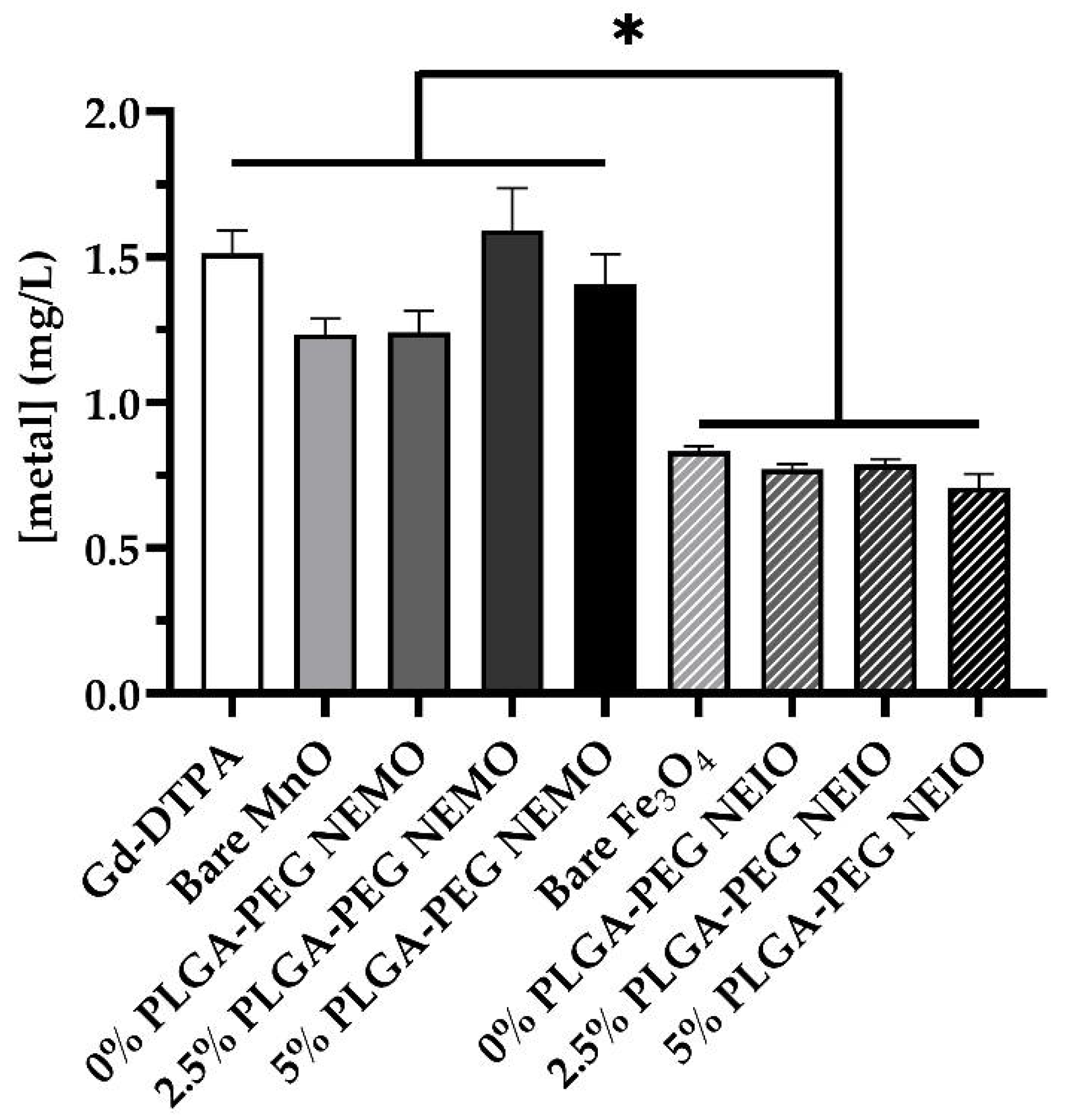
2.8. Neutrophil Metal Content

For metal content assessment, neutrophils were plated at approximately 250,000 cells/well in 6 well plates and were subsequently stimulated with contrast agents as described. Following incubation, wells were gently rinsed with PBS 3 times to remove free Gd-DTPA or NPs. After washing, the remaining neutrophils were digested with 0.5 mL of HCl trace metal grade. The resultant solution was run under EPA Method 200.8 Revision 5.4 on a PerkinElmer NexION 2000 ICP-mass spectrometer. All samples were preserved to pH ≤ 2 before analysis.

2.9. Statistical Analysis

Unless otherwise indicated, all data are expressed as mean ± standard error. Comparison between groups was conducted via ANOVA with a post hoc Holm correction. p-values less than 0.05 were considered significant.

3. Results

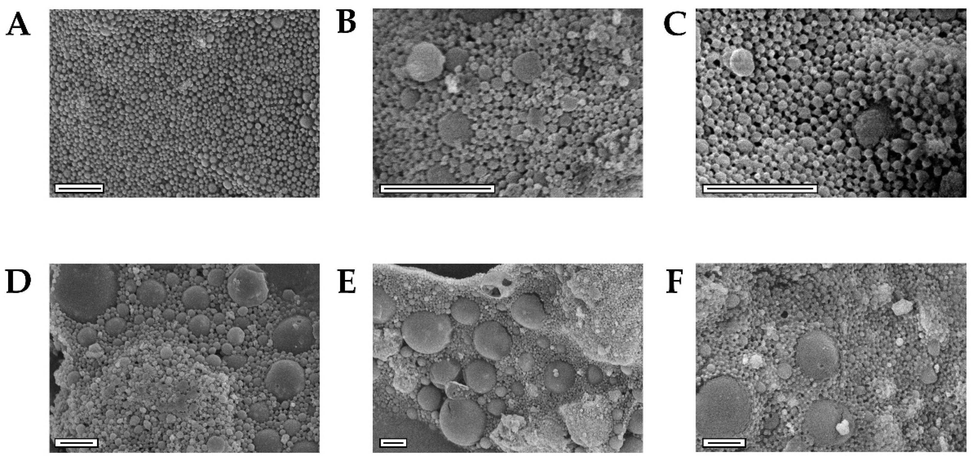
3.1. Synthesized MnO and Fe3O4 Bare NPs Displayed Small Sizes with Intended Oxidation State

Two optimized batches for both types of metal oxide NPs (MnO and Fe3O4) were successfully fabricated via a thermal decomposition reaction. The physicochemical properties of all NP samples were determined including morphology, crystallinity, size, surface chemistry, and metal loading.
TEM was used to assess bare MnO and Fe3O4 NP size (Figure S1) and morphology (Figure 1A,C). The MnO NPs displayed a rounded octagonal shape, similar to that found in our previous study [19] and in the study by Nolis et al. [105]. The morphology of the Fe3O4 NPs was irregular but possessed a dominant triangular shape similar to that described by Belaïd et al. and Laurent et al. [106,107]. The average size according to TEM for bare MnO and Fe3O4 NPs was 22 ± 7 nm and 13 ± 4 nm, respectively.
XRD was used to evaluate the bare MnO and Fe3O4 NP crystal structure and bulk composition (Figure 1B,D and Figure S2) for all batches. All of the synthesized bare metal oxide NPs clearly displayed the highest known characteristic peaks for MnO and Fe3O4 obtained from X’Pert HighScore. Table 1 and Table S1 show the percent composition of MnO, Mn3O4, and Mn2O3 for two different batches of NPs, and Fe3O4 and Fe2O3 for two different batches of NPs. The intended oxidation state of either MnO or Fe3O4 was present as the highest percent composition for all NP formulations. Additionally, complementary techniques, HRTEM and SAED, corroborated the crystal structure of both metal oxide NPs (Figures S3 and S4). FTIR spectra of the bare NPs (Figure S5) were taken to confirm the oleylamine capping of the NPs and metal oxide bonds [19,87,108,109,110].

3.2. NEIO Particles Displayed Larger Particle Size and Aggregation While NEMO Particles Displayed Smaller and More Consistent Particle Size

MnO and Fe3O4 NPs were encapsulated via a single emulsion technique using PLGA with either 0%, 2.5%, or 5% (w/w) PLGA-PEG co-block polymer. The physicochemical properties of the NEMO and NEIO particles were determined, including morphology, size, surface chemistry, surface charge, PEGylation percentage, and metal loading.
To confirm successful polymer encapsulation of the metal oxide NPs, FTIR (Figure S6) was used to ensure that the characteristic PLGA peaks were present [111,112,113]. Encapsulation was further confirmed with SEM-EDS where both metals, manganese and iron, were only present where the polymer NPs were observed (Figures S7 and S8). NEMO and NEIO particles were also evaluated by SEM to assess morphology (Figure 2) and general dry size after encapsulation.
All polymer encapsulated NPs exhibited spherical morphology; however, the NEMO particle size was more uniform compared to the NEIO particles, whose heterogeneity led to an increased overall dry size, as shown in Figure 2. To complement SEM, DLS was used to determine the hydrodynamic NP size distribution and the weighted average of the metal oxide NPs, as shown in Figure 3 and Table S2, respectively.
For the NEMO particles, the hydrodynamic size populations were comparable to those found using SEM. The majority of particles were uniformly distributed and similar in size, with a small population of larger-sized particles for all three samples (Figure 3A–C). The 0% PLGA-PEG NEIO particles were comparable in size and distribution to the NEMO particles; however, as shown by DLS in Figure 3, the 2.5% and 5% PLGA-PEG NEIO particles had larger-sized populations. We hypothesize that these large DLS populations are due to NP aggregation post-synthesis from dipole-dipole interaction as described in the literature [114,115,116,117]. Additionally, the presence of the larger-sized population shown using SEM (Figure 2) could contribute to the larger peak seen using DLS for PEGylated NEIO particles.
Aggregation is a common challenge to overcome in NP synthesis, as an ideal NP size for in vivo application is <200 nm to prevent vascular occlusion and increase cellular uptake [118,119,120,121,122]. There are several factors that can impact NP aggregation such as freeze-drying, the method of encapsulation, the surface charge, etc. [123,124,125]. Freeze-drying can cause mechanical stress and subsequent NP destabilization, and in this case, a cryoprotectant, such as trehalose, can be used to prevent aggregation [124,125]. As for the method of encapsulation, all variables were kept constant except for the type of metal oxide NP and the method of stirring. Due to the magnetic properties of Fe3O4 NPs, a non-ferrous, overhead stirrer was utilized in place of a magnetic stir plate during the solvent-evaporation step of the single emulsion technique. The overhead stirrer may have achieved different polymer and Fe3O4 NP mixing profiles than the conventional magnetic stir plate employed for the MnO polymer encapsulation process, resulting in large particles and aggregates as observed in all NEIO particle formulations using SEM and DLS (Figure 2 and Figure 3).
The surface charge of both NEMO and NEIO particles decreased with an increasing amount of PEGylation. Typically, more neutral charges lead to increased NP aggregation due to less interparticle electric repulsion forces; NEIO particles followed this trend and aggregated more as the ζ-potential came closer to zero, which may also be due to the dipole-dipole forces mentioned previously [114,115,116,117]. Interestingly, the NEMO particles did not tend to aggregate as the ζ-potential neutralized with PEGylation and instead maintained their stability. Additional studies will be required to uncover the mechanism behind these differences in aggregation between NEMO vs. NEIO particles. According to TGA (Figure S9), the actual PLGA-PEG percentage incorporated on the NP surface for 0%, 2.5%, and 5% PLGA-PEG for NEMO particles was 0%, 0.70%, and 1.1%, respectively, and for NEIO particles was 0%, 0.38%, and 0.77%, respectively. During the single emulsion technique, it is expected to see some loss of the PLGA-PEG, as any micelles formed with PLGA-PEG without metal oxide loading and any free-form polymer will be washed away during centrifugation.
Table 2 shows the metal element loading and encapsulation efficiency (EE) of the different NPs used for the set of cell experiments shown below. Comparable results were observed during SEM-EDS analysis (Figures S7 and S8). The metal loading was applied to calculate the corresponding dosage of NPs that would be equivalent to a clinically relevant dose of GBCA (0.05 mmol/kg [96,97], or 7.5 mg/kg Gd).

3.3. Ex Vivo Fluorescent Imaging Reveals That NEMO Particles Elicit the Highest NETosis

Neutrophils isolated from BALB/c mice were treated with standardized concentrations of Gd-DTPA, bare metal oxide NPs, or polymer encapsulated metal oxide NPs with varying levels of PEGylation. The NET formation was characterized using fluorescent staining; neutrophil supernatants were quantitatively evaluated for cell-free double-stranded DNA, ROS, and cytokine release. To confirm NET formation for each neutrophil, stimulated neutrophils were also imaged with SEM (Figure S10).
The fluorescent NET assay illustrated significant differences in the level of NETs produced in response to the various NP treatments (Figure 4). Gd-DTPA, bare MnO, bare Fe3O4, 0% PLGA-PEG NEIO particles (PLGA only), and 5% PLGA-PEG NEIO particles all increased the level of NETosis relative to unstimulated neutrophils, albeit non-significantly. However, the 0–5% PLGA-PEG NEMO groups and the 2.5% PLGA-PEG NEIO groups all elicited a robust pro-NETotic response that was significantly greater than the control group. For both NEMO and NEIO particles, the 2.5% PLGA-PEG coating caused the greatest NET release, showing that NP surface chemistry modulates NET formation. Representative FOVs for all groups are available in Figure S11. We also observed that the 0–5% PLGA-PEG blank NPs (without metal oxide) did not appear to significantly enhance NETosis on their own (Figure S12).

3.4. MRI Contrast Agents Provoke Non-NETotic Cell-Free dsDNA Release, Extracellular ROS, and Altered Cytokine Expression

We sought to confirm the trends seen in our microscopy-based ex vivo NET assay by measuring the total amount of dsDNA released by neutrophils for each stimulation group (Figure 5a). Although dsDNA is not a perfect marker of NETosis, as it can also be present due to cell death, the results were similar to the trends observed for NETosis, with several interesting caveats. For instance, NEMO and NEIO formulations appeared to elicit similar amounts of cell-free dsDNA release. If NEIO-stimulated neutrophils provoke similar overall amounts of cell-free dsDNA release, but fewer NETs, this suggests that dsDNA may be released by a different mechanism such as apoptosis or necrosis. Additionally, NEIO formulations appeared to cause dsDNA release that decreased slightly, yet significantly with greater PEGylation; this was not the case for NEMO formulations. This suggests that higher PEGylation may reduce cytotoxicity specifically for NEIO formulations, resulting in lower non-NETotic dsDNA release. Interestingly, Gd-DTPA appeared to release more dsDNA than bare MnO and Fe3O4 NPs, whereas they produced fewer NETs. In fact, bare NPs appeared to repress dsDNA release compared to the other groups, though this was only significant for MnO NPs.
While NETs both mediate and indicate an inflammatory state, NETosis alone is not the sole aspect of neutrophil involvement in inflammation. In vivo, NETs both promote inflammation and occur as a direct result of it [126]. We hypothesized that increased NETosis would correlate to increased oxidative stress. Thus, we measured the presence of ROS in the neutrophil supernatant post-stimulation (Figure 5b). Again, it appeared that the MnO groups elicited the greatest oxidative response, more so than all Fe3O4 groups. However, only the 0% and 5% PLGA-PEG NEMO groups reached significance relative to the control. This suggests that NP surface chemistry and composition both modulate NETosis, oxidative stress, and dsDNA release. Different NP formulations may favor NETosis, ROS production, or possibly neutrophil cell death, which can also result in dsDNA release. Overall, this data supported the hypothesis that increased ROS production supported increased NETosis, as NEMO particles generally provoked more NETosis and ROS production than NEIO particles. However, the 2.5% PLGA-PEG NEMO and NEIO particles did not significantly enhance extracellular ROS production despite provoking the most robust NETosis. As the ROS assay was applied only to the supernatant, it is possible that the 2.5% PLGA-PEG NEMO and NEIO particles could have increased intracellular ROS compared to the other formulations. Gd-DTPA did not elicit increased NETosis or ROS production compared to the control; this further supports the assertion that Gd-DTPA does not directly impact neutrophil-mediated oxidative stress.
To determine whether the neutrophil response to MRI contrast agents had the capacity to influence other immune cells, we sought to examine whether the neutrophil cytokine response varied with different NP formulations (Figure 6). We decided to study the 2.5% PLGA-PEG NEMO and NEIO particle stimulated neutrophils, as these provoked the most NETosis relative to each formulation. We also assessed the neutrophil cytokine response to Gd-DTPA, a clinically relevant MRI contrast agent. Unstimulated neutrophils were utilized as a negative control. Although the relative presence of 40 different cytokines was measured via western dot blot membranes, only two showed significant differences in expression compared to the control in at least one group (Figure S13): Stromal cell-derived factor-1 (SDF-1; alternatively, CXCL12) and the tissue inhibitor of matrix metalloproteinase-1 (TIMP-1). SDF-1 is known to enhance neutrophil recruitment, and it is primarily expressed in the bone marrow, where it acts to retain neutrophils until they mature. Although not conventionally thought to be released by neutrophils themselves, some studies have illustrated that neutrophils do in fact produce SDF-1 at least at the mRNA level [127]. TIMP-1 is also known to elicit NETosis, particularly in the context of pancreatic cancer [128]. The production of each cytokine, but lack of increased NETosis, suggests that GBCAs have the ability to influence neutrophil chemoattraction. In vivo, this may result in NETosis due to influences from other cells such as platelets and the endothelium on recruited neutrophils.

3.5. MRI Contrast Agents Yield Differential Leukocyte Phagocytosis Dependent on Formulation

Because we had observed significant increases in NETosis upon stimulation with NEMO particles specifically, we questioned whether this could be attributed to enhanced neutrophil uptake. As such, ICP-mass spectrometry was utilized to determine whether the differences in NETosis observed between various MRI contrast agent formulations corresponded with altered neutrophil phagocytosis (Figure 7). Indeed, internalization of all Fe3O4 formulations was significantly reduced compared to MnO and Gd-DTPA, suggesting that Fe3O4 may cause a smaller NETotic response simply because it is taken up less by neutrophils. Uptake, however, does not account for the differences seen between the groups within the metal oxide types, as these were not significantly different. Conversely, Gd-DTPA uptake appeared to be comparable to that of the MnO groups, but without pro-NETotic effects. Collectively, these results suggest that polymer coating, particularly PEG, may facilitate NETosis on its own in some capacity. Given the wide use of PEG in manufacturing NPs with biological applications, this merits further investigation.

4. Discussion

Because MRI contrast agents, particularly emerging NP-based contrast agents, may have the capacity to promote the formation of tumor-supporting NETs, we incubated murine neutrophils with Fe3O4 or MnO NPs encapsulated with or without PLGA or PLGA-PEG. We also stimulated neutrophils with Gd-DTPA to examine whether clinically utilized contrast agents altered neutrophil behavior. Our results suggest that MnO-based MRI contrast agents are more likely to elicit a pro-NETotic response than their Fe3O4 based counterparts. Our study suggests that the PEGylation percentage modulates NETosis, as both PEG-free particles and particles with 5% PLGA-PEG coating produced lower levels of NETosis than particles with 2.5% PLGA-PEG coating; in fact, there was no significant difference between the 0% PLGA-PEG and 5% PLGA-PEG NEIO particle-invoked NETosis compared to the control. PLGA encapsulation may also contribute to NETosis, as evidenced by bare MnO NPs causing substantially less NET formation despite similar uptake levels to the 0% PLGA-PEG NEMO particles. However, it is important to note that blank polymer NPs do not appear to significantly enhance NETosis on their own (Figure S12), at least at the percentage PEGylation levels studied. Instead, it appears that both polymer encapsulation and a metal oxide core must be present for NETosis to be enhanced. While polymer encapsulation does not appear to be necessary for metal oxide NP uptake in neutrophils, our data suggest that it is necessary for NETosis. Therefore, it is possible that NPs must reach a specific location within the cell, likely either the nucleus or mitochondria, which supply the DNA for NET fibers, in order to trigger NETosis. Collectively, these findings raise some concern about the safety of metal oxide NP MRI contrast agents in patients for which an increased inflammatory burden would be detrimental, as would be the case in cancer. It is notable that Gd-DTPA caused the smallest overall increase in NETosis relative to all other groups. Although the safety of GBCAs has been questioned due to heavy metal accumulation in the body, these concerns are not known to be directly linked to inflammation [129,130]. Our results support this assertion, as we did not observe elevated NETosis, DNA release, or ROS production in neutrophils stimulated with Gd-DTPA. As a chelate, rather than a polymer-encapsulated NP, Gd-DTPA likely benefits from enhanced stability compared to NEMO and NEIO particles, resulting in less direct interaction between metal ions and the intracellular environment; this may explain why Gd-DTPA had a lesser effect on neutrophil behavior in the majority of the studies conducted.
Although NEMO particles as a group elicited significantly more NET formation than NEIO particles, their overall capacity to cause neutrophils to release cell-free dsDNA was similar. Interestingly, the 2.5% PLGA-PEG NEMO and NEIO particles provoked similar overall dsDNA release relative to other surface chemistries despite provoking the most NETosis. One possibility is that groups exhibiting low NETosis, but high dsDNA release may actually be promoting necrosis more than NETosis. As discussed by Yousefi et al. [131], necrosis and NETosis both result in DNA release, but only NETs retain a distinctive, fibrous morphology. Necrotic DNA release forms a cloud-like structure [132]. Our morphology-based analysis of fluorescently labeled neutrophils and NETs does not characterize such structures as NETs (Figure S11). Therefore, dsDNA analysis alone would not accurately capture the nuances of how the degree of PEGylation differentially affects neutrophil NETosis and cell death. Additional studies characterizing neutrophil apoptosis and necrosis, as they relate to NETosis and the pathways that provoke it, are needed to further elucidate the underlying mechanisms promoting neutrophil response to contrast agents.
The upregulation of TIMP-1 and SDF-1 in Gd-DTPA treated neutrophils, despite the apparently low potential to trigger NET formation, merits further investigation. SDF-1 is a chemoattractant for both leukocytes and lymphocytes; it plays a role in keeping neutrophils within the bone marrow and in recruiting neutrophils to other tissues. Interestingly, elevated levels of TIMP-1 have been shown to promote liver metastasis of colorectal cancer by increasing hepatic SDF-1 expression, thereby enhancing metastasis by attracting neutrophils to the liver pre-metastatic niche where they take on tumor-supporting phenotypes, presumably including NETosis [133]. Similar TIMP-1 mediated phenomena have been observed in pancreatic and breast cancer [128,134]. Neutrophils possess a matrix metalloproteinase (MMP) known as MMP9, which can form a non-inhibitory complex with TIMP-1, despite the latter being an MMP inhibitor [135]. While the functional significance of this complex is unclear, it is conceivable that it is functionally analogous to the interaction between TIMP-2 and MMP2, which ultimately results in the activation of MMP2 via other MMPs that subsequently interact with this complex [136]. If this is the case for the non-inhibitory TIMP-1/MMP9 complex, MMP9 activity would be enhanced. MMP9 specifically contributes to the pro-inflammatory, proteolytic functions of NETs [62]. Outside of MMP9 inhibition, TIMP-1 may contribute to other tumor-supporting phenomena including enhanced proliferation [137] and epithelial to mesenchymal transition [134]. Therefore, the fact that Gd-DTPA appears to upregulate these factors may be cause for concern. Although the increases in expression that we observed were modest, it is notable that they are occurring in neutrophils, which produce a relatively minor quantity of these cytokines compared to populations such as endothelial cells. If Gd-DTPA, or GBCAs in general, were to provoke similar effects systemically, there may be concerning subclinical consequences for cancer patients receiving MRI with contrast.
This is both the first study to specifically examine MnO NPs in relation to NETosis and the first study directly comparing leukocyte response between a clinically utilized GBCA and metal oxide NPs being developed as MRI contrast agents, as well as one of the few existing studies examining how modulating NP properties impacts NET release (Table 3). However, there are some limitations to this study that need to be further explored to better characterize both how metal oxide NPs and traditional MRI contrast agents affect leukocyte behavior, and whether this may promote poor outcomes, particularly for cancer patients. For instance, this study solely utilized isolated bone marrow-derived neutrophils divorced from a physiologic context; thus, it cannot account for the effects of corona formation from plasma proteins on NPs. The protein corona has the potential to alter NP uptake and subsequent neutrophil behavior [138]. Varying surface chemistries have the potential to alter this corona and therefore the overall consequences of the NP–neutrophil interaction [139]. Additionally, the use of healthy bone marrow-derived leukocytes precludes the ability to draw conclusions about peripheral neutrophils in tumor-bearing environments. As the presence of either a primary tumor or local metastatic lesions is known to prime neutrophils towards a pro-NETotic state in order to support metastasis, the observed effects in our study may be more pronounced in isolated neutrophils from tumor-bearing mice, and therefore of greater clinical concern [140]. Utilizing peripheral or tissue-resident neutrophils, specifically from common metastatic sites, isolated from tumor-bearing animals would address this. Examining a broader range of surface chemistries such as other polymers and functional groups in addition to NPs without metal oxide encapsulation would better enable conclusions regarding the effects of surface functionalization versus encapsulated metal content on leukocyte behavior. Finally, given that we did not attain similar size distributions for PEGylated NEMO and NEIO formulations, this study cannot fully account for size-mediated effects. The apparent difference in size seems to be partially mediated by the aggregation of Fe3O4 NPs; however, our results stand in contrast to Bilyy et al.’s [78] study which showed that aggregated Fe3O4 NPs contributed to enhanced NET formation in rabbit vasculature. Despite this, it is notable that the observed trend of NEMO particles enhancing NETosis to a greater extent than NEIO particles still held for the 0% PLGA-PEG formulations, as both the NEMOs and the NEIOs had comparable size distributions for this encapsulation. For instance, if size alone had prevented the 2.5% and 5% PLGA-PEG NEIO particles from enhancing NETosis to the levels produced by NEMO particles, then we would expect that the 0% PLGA-PEG NEIO treatment would have provoked comparable NETosis to NEMO groups; however, this was not the case. This supports the assertion that the presence of MnO-based NPs specifically caused greater NETosis than Fe3O4 based NPs.

5. Conclusions

Although further work is needed to better characterize the effects of MRI contrast agents on neutrophil phenotype in vivo, our work constitutes the first examination of the impact of MnO NPs and GBCAs on NETosis. Consideration of off-target cellular level effects is critical in NP contrast agent design to ensure that diagnostic imaging procedures do not negatively impact patient health. In this study, MnO NPs substantially increased neutrophil NETosis and the oxidative burden for specific surface chemistries, both of which could enhance the metastasis-supporting phenotype of neutrophils in the context of cancer. Meanwhile, our data support the hypothesis that Fe3O4 NPs may be safer than MnO NPs, as they produced less overall NETosis, extracellular ROS, cytokine release, and neutrophil uptake. However, the elevated free dsDNA release associated with Fe3O4 NP exposure could indicate an increase in neutrophil necrosis, which merits further study. Even though Gd-DTPA did not provoke NETosis, free dsDNA release, or ROS production, it was found to upregulate cytokines that are known to play a role in metastasis. Because all of our studies relate solely to the neutrophil response, much remains to be explored regarding the interactions between MRI contrast agents and other blood cells such as the endothelium, monocytes, circulating tumor cells, erythrocytes, and platelets. Our studies illustrate the critical importance of examining a broader range of cellular population responses to NPs throughout the design process to produce truly optimized NPs free of off-target effects.

Supplementary Materials

The following supporting information can be downloaded at: https://www.mdpi.com/article/10.3390/bios12020123/s1, Figure S1. Size distributions of bare metal oxide NPs, Figure S2. XRD spectra confirm crystal structure of the second synthesized batches of bare metal oxide NPs, Table S1. Percent composition of the second synthesized batches of bare metal oxide NPs from X-Pert HighScore, Figure S3. High-Resolution TEM (HRTEM) and Selected Area Electron Diffraction (SAED) confirm the crystal structure of MnO NPs, Figure S4. HRTEM and SAED confirm the crystal structure of Fe3O4 NPs, Figure S5. FTIR spectra for bare metal oxide NPs confirm oleylamine surface coating, Figure S6. FTIR spectra for NEMO and NEIO particles confirm PLGA encapsulation, Figure S7. SEM-Energy Dispersive X-ray Spectroscopy (EDS) of NEMO particles verify the PLGA encapsulation, Figure S8. SEM-EDS of NEIO particles verify the PLGA encapsulation, Table S2. Average hydrodynamic size for NEMO and NEIO particles, Figure S9. ThermoGravimetric Analysis (TGA) for NP PEGylation quantification, Figure S10. SEM images reveal NETs produced by neutrophils exposed to contrast agents, Figure S11. Fluorescence ex vivo NET assay further confirms neutrophil NETotic response after incubation with contrast agents, Figure S12. Fluorescence ex vivo NET assay reveals that metal oxide-free (blank) PLGA-PEG NPs do not significantly enhance NETosis, Figure S13. Western dot blots show altered neutrophil cytokine release following contrast agent exposure. Reference [141] is cited in the supplementary materials.

Author Contributions

Conceptualization, H.T.S., K.A.F., C.M.d.l.T., D.M.P. and M.F.B.; methodology, H.T.S., K.A.F., C.M.d.l.T. and D.M.P.; validation, H.T.S., K.A.F., C.M.d.l.T., D.M.P. and J.N.V.; formal analysis, H.T.S., K.A.F., C.M.d.l.T., D.M.P. and J.N.V.; investigation, H.T.S., K.A.F., C.M.d.l.T., D.M.P. and J.N.V.; writing—original draft preparation, H.T.S., K.A.F., C.M.d.l.T., D.M.P., J.N.V. and M.F.B.; writing—review and editing, H.T.S., K.A.F., C.M.d.l.T., D.M.P., J.N.V. and M.F.B.; visualization, C.M.d.l.T., H.T.S., K.A.F., D.M.P. and M.F.B.; supervision, M.F.B.; project administration, K.A.F. and M.F.B.; funding acquisition, H.T.S., C.M.d.l.T. and M.F.B. All authors have read and agreed to the published version of the manuscript.

Funding

This research was funded by the National Institutes of Health—National Institute of General Medical Sciences (NIH-NIGMS) Tumor Microenvironment Centers of Biomedical Research Excellence (TME CoBRE) grant awarded to MFB (P20 GM121322), West Virginia University Start-up funds awarded to MFB, West Virginia University’s Provost Fellowship awarded to HTS, the NIH-NIGMS Cell and Molecular Biology T32 Training Program at West Virginia University awarded to HTS (5 T32 GM133369), and West Virginia University’s Health Sciences Center International Fellowship awarded to CMT. Fluorescent imaging experiments and subsequent image analysis were performed in the West Virginia University Microscope Imaging Facility, which has been supported by the WVU Cancer Institute and NIH grants P20RR016440 and P30RR032138/P30GM103488.

Institutional Review Board Statement

The study was conducted according to the protocol approved by the Institutional Animal Care and Use Committee at West Virginia University (Protocol #1805014087_R1, approved in June 2021).

Informed Consent Statement

Not applicable.

Data Availability Statement

All relevant data are within the manuscript and its Supporting Material.

Acknowledgments

The authors would like to thank Marcela Redigolo for protocol development for SEM of NETs in addition to advice on TEM and SEM sample preparation and imaging, Amanda Gatesman-Ammer for protocol development of the fluorescent ex vivo NET assay and image collection, Qiang Wang for interpretation and analysis of the XRD and FTIR spectra, Robert Vincent from the NRCCE Analytical Lab for performing ICP-OES/ICP-MS measurements of metal content, Xinwei Bai from John Hu’s research laboratory for performing TGA experiments, Leland E. Earp from Sonikpreet Aulakh’s research laboratory for supplying enhanced chemiluminescence substrate and providing guidance for the Cytokine Array Kit, Mara A. Looney-Sanders for helping conduct NP size analysis with ImageJ, Esra Al Abazaid for generating early pilot data by exposing neutrophils to NPs, the WVU Shared Research Facility for use of their equipment including the XRD, TEM, SEM, and FTIR, and the West Virginia University Microscope Imaging Facility for use of their equipment including the Nikon A1R and NIS Elements General Analysis software.

Conflicts of Interest

The authors declare no conflict of interest. The funders had no role in the design of the study; in the collection, analyses, or interpretation of data; in the writing of the manuscript, or in the decision to publish the results.

References

  1. Limitations of Mammograms|How Often Are Mammograms Wrong? Available online: https://www.cancer.org/cancer/breast-cancer/screening-tests-and-early-detection/mammograms/limitations-of-mammograms.html (accessed on 1 December 2021).
  2. Durand, M.A.; Friedewald, S.M.; Plecha, D.M.; Copit, D.S.; Barke, L.D.; Rose, S.L.; Hayes, M.K.; Greer, L.N.; Dabbous, F.M.; Conant, E.F. False-Negative Rates of Breast Cancer Screening with and without Digital Breast Tomosynthesis. Radiology 2021, 298, 296–305. [Google Scholar] [CrossRef] [PubMed]
  3. Clinicopathological Features of Breast Cancer without Mammographic Findings Suggesting Malignancy—PubMed. Available online: https://pubmed.ncbi.nlm.nih.gov/33285381/ (accessed on 1 December 2021).
  4. Supplemental MRI Screening for Women with Extremely Dense Breast Tissue|NEJM. Available online: https://www.nejm.org/doi/full/10.1056/NEJMoa1903986 (accessed on 1 December 2021).
  5. Comstock, C.E.; Gatsonis, C.; Newstead, G.M.; Snyder, B.S.; Gareen, I.F.; Bergin, J.T.; Rahbar, H.; Sung, J.S.; Jacobs, C.; Harvey, J.A.; et al. Comparison of Abbreviated Breast MRI vs Digital Breast Tomosynthesis for Breast Cancer Detection Among Women With Dense Breasts Undergoing Screening. JAMA 2020, 323, 746–756. [Google Scholar] [CrossRef] [PubMed]
  6. Green, L.A.; Karow, J.A.; Toman, J.E.; Lostumbo, A.; Xie, K. Review of Breast Augmentation and Reconstruction for the Radiologist with Emphasis on MRI. Clin. Imaging 2018, 47, 101–117. [Google Scholar] [CrossRef]
  7. Iranmakani, S.; Mortezazadeh, T.; Sajadian, F.; Ghaziani, M.F.; Ghafari, A.; Khezerloo, D.; Musa, A.E. A Review of Various Modalities in Breast Imaging: Technical Aspects and Clinical Outcomes. Egypt. J. Radiol. Nucl. Med. 2020, 51, 57. [Google Scholar] [CrossRef] [Green Version]
  8. Larson, E.D.; Lee, W.-M.; Roubidoux, M.A.; Goodsitt, M.M.; Lashbrook, C.; Davis, C.E.; Kripfgans, O.D.; Carson, P.L. Preliminary Clinical Experience with a Combined Automated Breast Ultrasound and Digital Breast Tomosynthesis System. Ultrasound Med. Biol. 2018, 44, 734–742. [Google Scholar] [CrossRef] [PubMed]
  9. Pierre, V.C.; Allen, M.J.; Caravan, P. Contrast Agents for MRI: 30+ Years and Where Are We Going? J. Biol. Inorg. Chem. 2014, 19, 127–131. [Google Scholar] [CrossRef] [PubMed] [Green Version]
  10. Xiao, Y.-D.; Paudel, R.; Liu, J.; Ma, C.; Zhang, Z.-S.; Zhou, S.-K. MRI Contrast Agents: Classification and Application (Review). Int. J. Mol. Med. 2016, 38, 1319–1326. [Google Scholar] [CrossRef] [PubMed] [Green Version]
  11. Caspani, S.; Magalhães, R.; Araújo, J.P.; Sousa, C.T. Magnetic Nanomaterials as Contrast Agents for MRI. Materials 2020, 13, 2586. [Google Scholar] [CrossRef]
  12. Kitajima, K.; Maeda, T.; Watanabe, S.; Ueno, Y.; Sugimura, K. Recent Topics Related to Nephrogenic Systemic Fibrosis Associated with Gadolinium-based Contrast Agents. Int. J. Urol. 2012, 19, 806–811. [Google Scholar] [CrossRef] [Green Version]
  13. Kanda, T.; Fukusato, T.; Matsuda, M.; Toyoda, K.; Oba, H.; Kotoku, J.; Haruyama, T.; Kitajima, K.; Furui, S. Gadolinium-Based Contrast Agent Accumulates in the Brain Even in Subjects without Severe Renal Dysfunction: Evaluation of Autopsy Brain Specimens with Inductively Coupled Plasma Mass Spectroscopy. Radiology 2015, 276, 228–232. [Google Scholar] [CrossRef]
  14. Choi, J.W.; Moon, W.-J. Gadolinium Deposition in the Brain: Current Updates. Korean J. Radiol. 2019, 20, 134–147. [Google Scholar] [CrossRef] [PubMed]
  15. Rogosnitzky, M.; Branch, S. Gadolinium-Based Contrast Agent Toxicity: A Review of Known and Proposed Mechanisms. Biometals 2016, 29, 365–376. [Google Scholar] [CrossRef] [Green Version]
  16. Ariyani, W.; Iwasaki, T.; Miyazaki, W.; Khongorzul, E.; Nakajima, T.; Kameo, S.; Koyama, H.; Tsushima, Y.; Koibuchi, N. Effects of Gadolinium-Based Contrast Agents on Thyroid Hormone Receptor Action and Thyroid Hormone-Induced Cerebellar Purkinje Cell Morphogenesis. Front. Endocrinol. 2016, 7, 115. [Google Scholar] [CrossRef] [PubMed] [Green Version]
  17. Khairinisa, M.A.; Ariyani, W.; Tsushima, Y.; Koibuchi, N. Effects of Gadolinium Deposits in the Cerebellum: Reviewing the Literature from In Vitro Laboratory Studies to In Vivo Human Investigations. Int. J. Environ. Res. Public Health 2021, 18, 7214. [Google Scholar] [CrossRef] [PubMed]
  18. Ray, J.G.; Vermeulen, M.J.; Bharatha, A.; Montanera, W.J.; Park, A.L. Association between MRI Exposure during Pregnancy and Fetal and Childhood Outcomes. JAMA 2016, 316, 952–961. [Google Scholar] [CrossRef]
  19. Martinez de la Torre, C.; Grossman, J.H.; Bobko, A.A.; Bennewitz, M.F. Tuning the Size and Composition of Manganese Oxide Nanoparticles through Varying Temperature Ramp and Aging Time. PLoS ONE 2020, 15, e0239034. [Google Scholar] [CrossRef] [PubMed]
  20. Cabral, H.; Matsumoto, Y.; Mizuno, K.; Chen, Q.; Murakami, M.; Kimura, M.; Terada, Y.; Kano, M.R.; Miyazono, K.; Uesaka, M.; et al. Accumulation of Sub-100 Nm Polymeric Micelles in Poorly Permeable Tumours Depends on Size. Nat. Nanotechnol. 2011, 6, 815–823. [Google Scholar] [CrossRef] [PubMed]
  21. Liu, D.; Mori, A.; Huang, L. Role of Liposome Size and RES Blockade in Controlling Biodistribution and Tumor Uptake of GM1-Containing Liposomes. Biochim. Biophys. Acta 1992, 1104, 95–101. [Google Scholar] [CrossRef]
  22. Tang, L.; Yang, X.; Yin, Q.; Cai, K.; Wang, H.; Chaudhury, I.; Yao, C.; Zhou, Q.; Kwon, M.; Hartman, J.A.; et al. Investigating the Optimal Size of Anticancer Nanomedicine. Proc. Natl. Acad. Sci. USA 2014, 111, 15344–15349. [Google Scholar] [CrossRef] [Green Version]
  23. Chen, R.; Ling, D.; Zhao, L.; Wang, S.; Liu, Y.; Bai, R.; Baik, S.; Zhao, Y.; Chen, C.; Hyeon, T. Parallel Comparative Studies on Mouse Toxicity of Oxide Nanoparticle- and Gadolinium-Based T1 MRI Contrast Agents. ACS Nano 2015, 9, 12425–12435. [Google Scholar] [CrossRef]
  24. He, C.; Hu, Y.; Yin, L.; Tang, C.; Yin, C. Effects of Particle Size and Surface Charge on Cellular Uptake and Biodistribution of Polymeric Nanoparticles. Biomaterials 2010, 31, 3657–3666. [Google Scholar] [CrossRef] [PubMed]
  25. Black, K.C.L.; Wang, Y.; Luehmann, H.P.; Cai, X.; Xing, W.; Pang, B.; Zhao, Y.; Cutler, C.S.; Wang, L.V.; Liu, Y.; et al. Radioactive 198Au-Doped Nanostructures with Different Shapes for in Vivo Analyses of Their Biodistribution, Tumor Uptake, and Intratumoral Distribution. ACS Nano 2014, 8, 4385–4394. [Google Scholar] [CrossRef] [PubMed]
  26. Geng, Y.; Dalhaimer, P.; Cai, S.; Tsai, R.; Tewari, M.; Minko, T.; Discher, D.E. Shape Effects of Filaments versus Spherical Particles in Flow and Drug Delivery. Nat. Nanotechnol. 2007, 2, 249–255. [Google Scholar] [CrossRef] [PubMed]
  27. Nguyen, T.-D.; Do, T.-O. Size- and Shape-Controlled Synthesis of Monodisperse Metal Oxide and Mixed Oxide Nanocrystals. Nanocrystal 2011, 66, 55–84. [Google Scholar] [CrossRef] [Green Version]
  28. Arvizo, R.R.; Miranda, O.R.; Moyano, D.F.; Walden, C.A.; Giri, K.; Bhattacharya, R.; Robertson, J.D.; Rotello, V.M.; Reid, J.M.; Mukherjee, P. Modulating Pharmacokinetics, Tumor Uptake and Biodistribution by Engineered Nanoparticles. PLoS ONE 2011, 6, e24374. [Google Scholar] [CrossRef] [PubMed] [Green Version]
  29. Lunov, O.; Syrovets, T.; Loos, C.; Beil, J.; Delacher, M.; Tron, K.; Nienhaus, G.U.; Musyanovych, A.; Mailänder, V.; Landfester, K.; et al. Differential Uptake of Functionalized Polystyrene Nanoparticles by Human Macrophages and a Monocytic Cell Line. ACS Nano 2011, 5, 1657–1669. [Google Scholar] [CrossRef] [PubMed]
  30. Iancu, S.D.; Albu, C.; Chiriac, L.; Moldovan, R.; Stefancu, A.; Moisoiu, V.; Coman, V.; Szabo, L.; Leopold, N.; Bálint, Z. Assessment of Gold-Coated Iron Oxide Nanoparticles as Negative T2 Contrast Agent in Small Animal MRI Studies. Int. J. Nanomed. 2020, 15, 4811–4824. [Google Scholar] [CrossRef]
  31. Chhour, P.; Gallo, N.; Cheheltani, R.; Williams, D.; Al-Zaki, A.; Paik, T.; Nichol, J.L.; Tian, Z.; Naha, P.C.; Witschey, W.R.; et al. Nanodisco Balls: Control over Surface versus Core Loading of Diagnostically Active Nanocrystals into Polymer Nanoparticles. ACS Nano 2014, 8, 9143–9153. [Google Scholar] [CrossRef] [PubMed] [Green Version]
  32. Chen, Y.; Yin, Q.; Ji, X.; Zhang, S.; Chen, H.; Zheng, Y.; Sun, Y.; Qu, H.; Wang, Z.; Li, Y.; et al. Manganese Oxide-Based Multifunctionalized Mesoporous Silica Nanoparticles for PH-Responsive MRI, Ultrasonography and Circumvention of MDR in Cancer Cells. Biomaterials 2012, 33, 7126–7137. [Google Scholar] [CrossRef]
  33. Van Schooneveld, M.M.; Cormode, D.P.; Koole, R.; van Wijngaarden, J.T.; Calcagno, C.; Skajaa, T.; Hilhorst, J.; Hart, D.C.; Fayad, Z.A.; Mulder, W.J.M.; et al. A Fluorescent, Paramagnetic and PEGylated Gold/Silica Nanoparticle for MRI, CT and Fluorescence Imaging. Contrast Media Mol. Imaging 2010, 5, 231–236. [Google Scholar] [CrossRef] [Green Version]
  34. Israel, L.L.; Galstyan, A.; Holler, E.; Ljubimova, J.Y. Magnetic Iron Oxide Nanoparticles for Imaging, Targeting and Treatment of Primary and Metastatic Tumors of the Brain. J. Control. Release 2020, 320, 45–62. [Google Scholar] [CrossRef] [PubMed]
  35. Cui, J.; Alt, K.; Ju, Y.; Gunawan, S.T.; Braunger, J.A.; Wang, T.-Y.; Dai, Y.; Dai, Q.; Richardson, J.J.; Guo, J.; et al. Ligand-Functionalized Poly(Ethylene Glycol) Particles for Tumor Targeting and Intracellular Uptake. Biomacromolecules 2019, 20, 3592–3600. [Google Scholar] [CrossRef] [PubMed]
  36. Capolla, S.; Garrovo, C.; Zorzet, S.; Lorenzon, A.; Rampazzo, E.; Spretz, R.; Pozzato, G.; Núñez, L.; Tripodo, C.; Macor, P.; et al. Targeted Tumor Imaging of Anti-CD20-Polymeric Nanoparticles Developed for the Diagnosis of B-Cell Malignancies. Int. J. Nanomed. 2015, 10, 4099–4109. [Google Scholar] [CrossRef] [Green Version]
  37. Huang, X.; Schwind, S.; Yu, B.; Santhanam, R.; Wang, H.; Hoellerbauer, P.; Mims, A.; Klisovic, R.; Walker, A.R.; Chan, K.K.; et al. Targeted Delivery of MicroRNA-29b by Transferrin-Conjugated Anionic Lipopolyplex Nanoparticles: A Novel Therapeutic Strategy in Acute Myeloid Leukemia. Clin. Cancer Res. 2013, 19, 2355–2367. [Google Scholar] [CrossRef] [Green Version]
  38. Blanco, E.; Shen, H.; Ferrari, M. Principles of Nanoparticle Design for Overcoming Biological Barriers to Drug Delivery. Nat. Biotechnol. 2015, 33, 941–951. [Google Scholar] [CrossRef]
  39. Ray, P.; Haideri, N.; Haque, I.; Mohammed, O.; Chakraborty, S.; Banerjee, S.; Quadir, M.; Brinker, A.; Banerjee, S. The Impact of Nanoparticles on the Immune System: A Gray Zone of Nanomedicine. J. Immunol. Sci. 2021, 5, 19–33. [Google Scholar] [CrossRef]
  40. Uz, M.; Bulmus, V.; Alsoy Altinkaya, S. Effect of PEG Grafting Density and Hydrodynamic Volume on Gold Nanoparticle–Cell Interactions: An Investigation on Cell Cycle, Apoptosis, and DNA Damage. Langmuir 2016, 32, 5997–6009. [Google Scholar] [CrossRef] [Green Version]
  41. Dobrovolskaia, M.A.; Shurin, M.; Shvedova, A.A. Current Understanding of Interactions between Nanoparticles and the Immune System. Toxicol. Appl. Pharmacol. 2016, 299, 78–89. [Google Scholar] [CrossRef] [Green Version]
  42. Lima, T.; Bernfur, K.; Vilanova, M.; Cedervall, T. Understanding the Lipid and Protein Corona Formation on Different Sized Polymeric Nanoparticles. Sci. Rep. 2020, 10, 1129. [Google Scholar] [CrossRef] [PubMed]
  43. Xiao, W.; Gao, H. The Impact of Protein Corona on the Behavior and Targeting Capability of Nanoparticle-Based Delivery System. Int. J. Pharm. 2018, 552, 328–339. [Google Scholar] [CrossRef] [PubMed]
  44. Lundqvist, M.; Stigler, J.; Cedervall, T.; Berggård, T.; Flanagan, M.B.; Lynch, I.; Elia, G.; Dawson, K. The Evolution of the Protein Corona around Nanoparticles: A Test Study. ACS Nano 2011, 5, 7503–7509. [Google Scholar] [CrossRef] [PubMed]
  45. La-Beck, N.M.; Gabizon, A.A. Nanoparticle Interactions with the Immune System: Clinical Implications for Liposome-Based Cancer Chemotherapy. Front. Immunol. 2017, 8, 416. [Google Scholar] [CrossRef] [PubMed] [Green Version]
  46. Fromen, C.A.; Kelley, W.J.; Fish, M.B.; Adili, R.; Noble, J.; Hoenerhoff, M.J.; Holinstat, M.; Eniola-Adefeso, O. Neutrophil–Particle Interactions in Blood Circulation Drive Particle Clearance and Alter Neutrophil Responses in Acute Inflammation. ACS Nano 2017, 11, 10797–10807. [Google Scholar] [CrossRef]
  47. Jones, S.W.; Roberts, R.A.; Robbins, G.R.; Perry, J.L.; Kai, M.P.; Chen, K.; Bo, T.; Napier, M.E.; Ting, J.P.; DeSimone, J.M. Nanoparticle Clearance Is Governed by Th1/Th2 Immunity and Strain Background. J. Clin. Investig. 2013, 123, 3061–3073. [Google Scholar] [CrossRef] [PubMed] [Green Version]
  48. Naumenko, V.; Nikitin, A.; Garanina, A.; Melnikov, P.; Vodopyanov, S.; Kapitanova, K.; Potashnikova, D.; Vishnevskiy, D.; Alieva, I.; Ilyasov, A. Neutrophil-Mediated Transport Is Crucial for Delivery of Short-Circulating Magnetic Nanoparticles to Tumors. Acta Biomater. 2020, 104, 176–187. [Google Scholar] [CrossRef]
  49. Fang, Y.; Xue, J.; Gao, S.; Lu, A.; Yang, D.; Jiang, H.; He, Y.; Shi, K. Cleavable PEGylation: A Strategy for Overcoming the “PEG Dilemma” in Efficient Drug Delivery. Drug Deliv. 2017, 24, 22–32. [Google Scholar] [CrossRef] [PubMed] [Green Version]
  50. Walkey, C.D.; Olsen, J.B.; Song, F.; Liu, R.; Guo, H.; Olsen, D.W.H.; Cohen, Y.; Emili, A.; Chan, W.C.W. Protein Corona Fingerprinting Predicts the Cellular Interaction of Gold and Silver Nanoparticles. ACS Nano 2014, 8, 2439–2455. [Google Scholar] [CrossRef]
  51. Paciotti, G.F.; Myer, L.; Weinreich, D.; Goia, D.; Pavel, N.; McLaughlin, R.E.; Tamarkin, L. Colloidal Gold: A Novel Nanoparticle Vector for Tumor Directed Drug Delivery. Drug Deliv. 2004, 11, 169–183. [Google Scholar] [CrossRef]
  52. Kelley, W.J.; Fromen, C.A.; Lopez-Cazares, G.; Eniola-Adefeso, O. PEGylation of Model Drug Carriers Enhances Phagocytosis by Primary Human Neutrophils. Acta Biomater. 2018, 79, 283–293. [Google Scholar] [CrossRef]
  53. Jorch, S.K.; Kubes, P. An Emerging Role for Neutrophil Extracellular Traps in Noninfectious Disease. Nat Med 2017, 23, 279–287. [Google Scholar] [CrossRef]
  54. Brinkmann, V.; Reichard, U.; Goosmann, C.; Fauler, B.; Uhlemann, Y.; Weiss, D.S.; Weinrauch, Y.; Zychlinsky, A. Neutrophil Extracellular Traps Kill Bacteria. Science 2004, 303, 1532–1535. [Google Scholar] [CrossRef] [PubMed]
  55. Lee, M.K.; Sreejit, G.; Nagareddy, P.R.; Murphy, A.J. Attack of the NETs! NETosis Primes IL-1β-Mediated Inflammation in Diabetic Foot Ulcers. Clin. Sci. 2020, 134, 1399–1401. [Google Scholar] [CrossRef] [PubMed]
  56. Schett, G.; Schauer, C.; Hoffmann, M.; Herrmann, M. Why Does the Gout Attack Stop? A Roadmap for the Immune Pathogenesis of Gout. RMD Open 2015, 1, e000046. [Google Scholar] [CrossRef] [Green Version]
  57. Chatfield, S.M.; Grebe, K.; Whitehead, L.W.; Rogers, K.L.; Nebl, T.; Murphy, J.M.; Wicks, I.P. Monosodium Urate Crystals Generate Nuclease-Resistant Neutrophil Extracellular Traps via a Distinct Molecular Pathway. J. Immunol. 2018, 200, 1802–1816. [Google Scholar] [CrossRef] [PubMed] [Green Version]
  58. Jones, A.P.; Wallis, C. Dornase Alfa for Cystic Fibrosis. Cochrane Database Syst. Rev. 2010, 3, CD001127. [Google Scholar]
  59. Angelidou, I.; Chrysanthopoulou, A.; Mitsios, A.; Arelaki, S.; Arampatzioglou, A.; Kambas, K.; Ritis, D.; Tsironidou, V.; Moschos, I.; Dalla, V. REDD1/Autophagy Pathway Is Associated with Neutrophil-Driven IL-1β Inflammatory Response in Active Ulcerative Colitis. J. Immunol. 2018, 200, 3950–3961. [Google Scholar] [CrossRef]
  60. Snoderly, H.T.; Boone, B.A.; Bennewitz, M.F. Neutrophil Extracellular Traps in Breast Cancer and beyond: Current Perspectives on NET Stimuli, Thrombosis and Metastasis, and Clinical Utility for Diagnosis and Treatment. Breast Cancer Res. 2019, 21, 145. [Google Scholar] [CrossRef] [Green Version]
  61. Cools-Lartigue, J.; Spicer, J.; McDonald, B.; Gowing, S.; Chow, S.; Giannias, B.; Bourdeau, F.; Kubes, P.; Ferri, L. Neutrophil Extracellular Traps Sequester Circulating Tumor Cells and Promote Metastasis. Available online: https://www.jci.org/articles/view/67484/pdf (accessed on 13 October 2021).
  62. Albrengues, J.; Shields, M.A.; Ng, D.; Park, C.G.; Ambrico, A.; Poindexter, M.E.; Upadhyay, P.; Uyeminami, D.L.; Pommier, A.; Küttner, V. Neutrophil Extracellular Traps Produced during Inflammation Awaken Dormant Cancer Cells in Mice. Science 2018, 361, eaao4227. [Google Scholar] [CrossRef] [Green Version]
  63. Monti, M.; De Rosa, V.; Iommelli, F.; Carriero, M.V.; Terlizzi, C.; Camerlingo, R.; Belli, S.; Fonti, R.; Di Minno, G.; Del Vecchio, S. Neutrophil Extracellular Traps as an Adhesion Substrate for Different Tumor Cells Expressing RGD-Binding Integrins. Int. J. Mol. Sci. 2018, 19, 2350. [Google Scholar] [CrossRef] [Green Version]
  64. Park, J.; Wysocki, R.W.; Amoozgar, Z.; Maiorino, L.; Fein, M.R.; Jorns, J.; Schott, A.F.; Kinugasa-Katayama, Y.; Lee, Y.; Won, N.H. Cancer Cells Induce Metastasis-Supporting Neutrophil Extracellular DNA Traps. Sci. Transl. Med. 2016, 8, ra138–ra361. [Google Scholar] [CrossRef] [Green Version]
  65. Lee, K.; Cavanaugh, L.; Leung, H.; Yan, F.; Ahmadi, Z.; Chong, B.; Passam, F. Quantification of NETs-associated Markers by Flow Cytometry and Serum Assays in Patients with Thrombosis and Sepsis. Int. J. Lab. Hematol. 2018, 40, 392–399. [Google Scholar] [CrossRef] [PubMed]
  66. Mauracher, L.; Posch, F.; Martinod, K.; Grilz, E.; Däullary, T.; Hell, L.; Brostjan, C.; Zielinski, C.; Ay, C.; Wagner, D. Citrullinated Histone H3, a Biomarker of Neutrophil Extracellular Trap Formation, Predicts the Risk of Venous Thromboembolism in Cancer Patients. J. Thromb. Haemost. 2018, 16, 508–518. [Google Scholar] [CrossRef] [PubMed]
  67. Wang, Y.; Xiao, Y.; Zhong, L.; Ye, D.; Zhang, J.; Tu, Y.; Bornstein, S.R.; Zhou, Z.; Lam, K.S.; Xu, A. Increased Neutrophil Elastase and Proteinase 3 and Augmented NETosis Are Closely Associated with β-Cell Autoimmunity in Patients with Type 1 Diabetes. Diabetes 2014, 63, 4239–4248. [Google Scholar] [CrossRef] [PubMed] [Green Version]
  68. Papayannopoulos, V.; Metzler, K.D.; Hakkim, A.; Zychlinsky, A. Neutrophil Elastase and Myeloperoxidase Regulate the Formation of Neutrophil Extracellular Traps. J. Cell Biol. 2010, 191, 677–691. [Google Scholar] [CrossRef] [Green Version]
  69. Arelaki, S.; Arampatzioglou, A.; Kambas, K.; Papagoras, C.; Miltiades, P.; Angelidou, I.; Mitsios, A.; Kotsianidis, I.; Skendros, P.; Sivridis, E.; et al. Gradient Infiltration of Neutrophil Extracellular Traps in Colon Cancer and Evidence for Their Involvement in Tumour Growth. PLoS ONE 2016, 11, e0154484. [Google Scholar] [CrossRef]
  70. Boone, B.A.; Murthy, P.; Miller-Ocuin, J.; Doerfler, W.R.; Ellis, J.T.; Liang, X.; Ross, M.A.; Wallace, C.T.; Sperry, J.L.; Lotze, M.T.; et al. Chloroquine Reduces Hypercoagulability in Pancreatic Cancer through Inhibition of Neutrophil Extracellular Traps. BMC Cancer 2018, 18, 678. [Google Scholar] [CrossRef] [Green Version]
  71. Ma, A.; Kubes, P. Platelets, Neutrophils, and Neutrophil Extracellular Traps (NETs) in Sepsis. J. Thromb. Haemost. 2008, 6, 415–420. [Google Scholar] [CrossRef]
  72. Abdol Razak, N.; Elaskalani, O.; Metharom, P. Pancreatic Cancer-Induced Neutrophil Extracellular Traps: A Potential Contributor to Cancer-Associated Thrombosis. Int. J. Mol. Sci. 2017, 18, 487. [Google Scholar] [CrossRef] [Green Version]
  73. Fuchs, T.A.; Brill, A.; Wagner, D.D. Neutrophil Extracellular Trap (NET) Impact on Deep Vein Thrombosis. Arterioscler. Thromb. Vasc. Biol. 2012, 32, 1777–1783. [Google Scholar] [CrossRef] [Green Version]
  74. Yang, L.; Liu, Q.; Zhang, X.; Liu, X.; Zhou, B.; Chen, J.; Huang, D.; Li, J.; Li, H.; Chen, F. DNA of Neutrophil Extracellular Traps Promotes Cancer Metastasis via CCDC25. Nature 2020, 583, 133–138. [Google Scholar] [CrossRef]
  75. Rayes, R.F.; Mouhanna, J.G.; Nicolau, I.; Bourdeau, F.; Giannias, B.; Rousseau, S.; Quail, D.; Walsh, L.; Sangwan, V.; Bertos, N. Primary Tumors Induce Neutrophil Extracellular Traps with Targetable Metastasis-Promoting Effects. JCI Insight 2019, 4, e128008. [Google Scholar] [CrossRef] [PubMed] [Green Version]
  76. Bartneck, M.; Keul, H.A.; Zwadlo-Klarwasser, G.; Groll, J. Phagocytosis Independent Extracellular Nanoparticle Clearance by Human Immune Cells. Nano Lett. 2010, 10, 59–63. [Google Scholar] [CrossRef]
  77. Kang, H.; Seo, J.; Yang, E.-J.; Choi, I.-H. Silver Nanoparticles Induce Neutrophil Extracellular Traps Via Activation of PAD and Neutrophil Elastase. Biomolecules 2021, 11, 317. [Google Scholar] [CrossRef] [PubMed]
  78. Bilyy, R.; Unterweger, H.; Weigel, B.; Dumych, T.; Paryzhak, S.; Vovk, V.; Liao, Z.; Alexiou, C.; Herrmann, M.; Janko, C. Inert Coats of Magnetic Nanoparticles Prevent Formation of Occlusive Intravascular Co-Aggregates With Neutrophil Extracellular Traps. Front. Immunol. 2018, 9, 2266. [Google Scholar] [CrossRef] [PubMed] [Green Version]
  79. Yang, H.; Marion, T.N.; Liu, Y.; Zhang, L.; Cao, X.; Hu, H.; Zhao, Y.; Herrmann, M. Nanomaterial Exposure Induced Neutrophil Extracellular Traps: A New Target in Inflammation and Innate Immunity. J. Immunol. Res. 2019, 2019, 3560180. [Google Scholar] [CrossRef]
  80. Liz, R.; Simard, J.-C.; Leonardi, L.B.A.; Girard, D. Silver Nanoparticles Rapidly Induce Atypical Human Neutrophil Cell Death by a Process Involving Inflammatory Caspases and Reactive Oxygen Species and Induce Neutrophil Extracellular Traps Release upon Cell Adhesion. Int. Immunopharmacol. 2015, 28, 616–625. [Google Scholar] [CrossRef]
  81. Urner, M.; Schlicker, A.; Z’graggen, B.R.; Stepuk, A.; Booy, C.; Buehler, K.P.; Limbach, L.; Chmiel, C.; Stark, W.J.; Beck-Schimmer, B. Inflammatory Response of Lung Macrophages and Epithelial Cells after Exposure to Redox Active Nanoparticles: Effect of Solubility and Antioxidant Treatment. Environ. Sci. Technol. 2014, 48, 13960–13968. [Google Scholar] [CrossRef] [Green Version]
  82. Hwang, T.-L.; Hsu, C.-Y.; Aljuffali, I.A.; Chen, C.-H.; Chang, Y.-T.; Fang, J.-Y. Cationic Liposomes Evoke Proinflammatory Mediator Release and Neutrophil Extracellular Traps (NETs) toward Human Neutrophils. Colloids Surf. B Biointerfaces 2015, 128, 119–126. [Google Scholar] [CrossRef]
  83. Yang, Y.; Wang, N.; Zhu, Y.; Lu, Y.; Chen, Q.; Fan, S.; Huang, Q.; Chen, X.; Xia, L.; Wei, Y. Gold Nanoparticles Synergize with Bacterial Lipopolysaccharide to Enhance Class A Scavenger Receptor Dependent Particle Uptake in Neutrophils and Augment Neutrophil Extracellular Traps Formation. Ecotoxicol. Environ. Saf. 2021, 211, 111900. [Google Scholar] [CrossRef]
  84. Meher, A.K.; Spinosa, M.; Davis, J.P.; Pope, N.; Laubach, V.E.; Su, G.; Serbulea, V.; Leitinger, N.; Ailawadi, G.; Upchurch Jr, G.R. Novel Role of IL (Interleukin)-1β in Neutrophil Extracellular Trap Formation and Abdominal Aortic Aneurysms. Arterioscler. Thromb. Vasc. Biol. 2018, 38, 843–853. [Google Scholar] [CrossRef] [Green Version]
  85. Limbach, L.K.; Wick, P.; Manser, P.; Grass, R.N.; Bruinink, A.; Stark, W.J. Exposure of Engineered Nanoparticles to Human Lung Epithelial Cells: Influence of Chemical Composition and Catalytic Activity on Oxidative Stress. Environ. Sci. Technol. 2007, 41, 4158–4163. [Google Scholar] [CrossRef] [PubMed]
  86. De la Torre, C.M.; Bennewitz, M.F. Manganese Oxide Nanoparticle Synthesis by Thermal Decomposition of Manganese(II) Acetylacetonate. J. Vis. Exp. 2020, e61572. [Google Scholar] [CrossRef]
  87. Xu, Z.; Shen, C.; Hou, Y.; Gao, H.; Sun, S. Oleylamine as Both Reducing Agent and Stabilizer in a Facile Synthesis of Magnetite Nanoparticles. Chem. Mater. 2009, 21, 1778–1780. [Google Scholar] [CrossRef]
  88. Bennewitz, M.F.; Lobo, T.L.; Nkansah, M.K.; Ulas, G.; Brudvig, G.W.; Shapiro, E.M. Biocompatible and PH-Sensitive PLGA Encapsulated MnO Nanocrystals for Molecular and Cellular MRI. ACS Nano 2011, 5, 3438–3446. [Google Scholar] [CrossRef] [PubMed] [Green Version]
  89. Wang, I.-W.; Kutteri, D.A.; Gao, B.; Tian, H.; Hu, J. Methane Pyrolysis for Carbon Nanotubes and COx-Free H2 over Transition-Metal Catalysts. Energy Fuels 2019, 33, 197–205. [Google Scholar] [CrossRef]
  90. Silva, M.F.; Hechenleitner, A.A.W.; Irache, J.M.; de Oliveira, A.J.A.; Pineda, E.A.G. Study of Thermal Degradation of PLGA, PLGA Nanospheres and PLGA/Maghemite Superparamagnetic Nanospheres. Mat. Res. 2015, 18, 1400–1406. [Google Scholar] [CrossRef]
  91. Jusu, S.M.; Obayemi, J.D.; Salifu, A.A.; Nwazojie, C.C.; Uzonwanne, V.; Odusanya, O.S.; Soboyejo, W.O. Drug-Encapsulated Blend of PLGA-PEG Microspheres: In Vitro and in Vivo Study of the Effects of Localized/Targeted Drug Delivery on the Treatment of Triple-Negative Breast Cancer. Sci. Rep. 2020, 10, 14188. [Google Scholar] [CrossRef]
  92. Adib, A.A.; Nazemidashtarjandi, S.; Kelly, A.; Kruse, A.; Cimatu, K.; David, A.E.; Farnoud, A.M. Engineered Silica Nanoparticles Interact Differently with Lipid Monolayers Compared to Lipid Bilayers. Environ. Sci. Nano 2018, 5, 289–303. [Google Scholar] [CrossRef]
  93. Benoit, D.N.; Zhu, H.; Lilierose, M.H.; Verm, R.A.; Ali, N.; Morrison, A.N.; Fortner, J.D.; Avendano, C.; Colvin, V.L. Measuring the Grafting Density of Nanoparticles in Solution by Analytical Ultracentrifugation and Total Organic Carbon Analysis. Anal. Chem. 2012, 84, 9238–9245. [Google Scholar] [CrossRef] [Green Version]
  94. Cao, S.; Jiang, Y.; Levy, C.N.; Hughes, S.M.; Zhang, H.; Hladik, F.; Woodrow, K.A. Optimization and Comparison of CD4-Targeting Lipid–Polymer Hybrid Nanoparticles Using Different Binding Ligands. J. Biomed. Mater. Res. Part A 2018, 106, 1177–1188. [Google Scholar] [CrossRef]
  95. Swamydas, M.; Lionakis, M.S. Isolation, Purification and Labeling of Mouse Bone Marrow Neutrophils for Functional Studies and Adoptive Transfer Experiments. J. Vis. Exp. 2013, e50586. [Google Scholar] [CrossRef] [PubMed]
  96. Schneider, G.; Schürholz, H.; Kirchin, M.A.; Bücker, A.; Fries, P. Safety and Adverse Effects during 24 Hours after Contrast-Enhanced MRI with Gadobenate Dimeglumine (MultiHance) in Children. Pediatr. Radiol. 2013, 43, 202–211. [Google Scholar] [CrossRef] [PubMed]
  97. Runge, V.M.; Armstrong, M.R.; Barr, R.G.; Berger, B.L.; Czervionke, L.F.; Gonzalez, C.F.; Halford, H.H.; Kanal, E.; Kuhn, M.J.; Levin, J.M.; et al. A Clinical Comparison of the Safety and Efficacy of MultiHance (Gadobenate Dimeglumine) and Omniscan (Gadodiamide) in Magnetic Resonance Imaging in Patients with Central Nervous System Pathology. Investig. Radiol. 2001, 36, 65–71. [Google Scholar] [CrossRef] [PubMed]
  98. 000651—BALB/CJ. Available online: https://www.jax.org/strain/000651 (accessed on 1 December 2021).
  99. Mitruka, B.M.; Rawnsley, H.M. Clinical Biochemical and Hematological Reference Values in Normal Experimental Animals and Normal Humans; Masson Pub.: New York, NY, USA, 1981; ISBN 978-0-89352-163-9. [Google Scholar]
  100. Harkness, J.E.; Wagner, J.E. The Biology and Medicine of Rabbits and Rodents; Lea & Febiger: Philadelphia, PA, USA, 1989; ISBN 978-0-8121-1176-7. [Google Scholar]
  101. Christie, A.C. A Study of the Kultschitzky (Argentaffin) Cell with the Electron-Microscope, after Fixation by Osmium Tetroxide. J. Cell Sci. 1955, 96, 295–299. [Google Scholar] [CrossRef]
  102. Dwiranti, A.; Masri, F.; Rahmayenti, D.A.; Putrika, A. The Effects of Osmium Tetroxide Post-Fixation and Drying Steps on Leafy Liverwort Ultrastructure Study by Scanning Electron Microscopy. Microsc. Res. Tech. 2019, 82, 1041–1046. [Google Scholar] [CrossRef] [PubMed]
  103. Porter, K.R.; Kallman, F. The Properties and Effects of Osmium Tetroxide as a Tissue Fixative with Special Reference to Its Use for Electron Microscopy. Exp. Cell Res. 1953, 4, 127–141. [Google Scholar] [CrossRef]
  104. Braet, F.; De Zanger, R.; Wisse, E. Drying Cells for SEM, AFM and TEM by Hexamethyldisilazane: A Study on Hepatic Endothelial Cells. J. Microsc. 1997, 186, 84–87. [Google Scholar] [CrossRef] [PubMed]
  105. Control of Size and Composition of Colloidal Nanocrystals of Manganese Oxide|Inorganic Chemistry. Available online: https://pubs.acs.org/doi/abs/10.1021/acs.inorgchem.8b02124 (accessed on 2 December 2021).
  106. Belaïd, S.; Laurent, S.; Vermeech, M.; Vander Elst, L.; Perez-Morga, D.; Muller, R.N. A New Approach to Follow the Formation of Iron Oxide Nanoparticles Synthesized by Thermal Decomposition. Nanotechnology 2013, 24, 055705. [Google Scholar] [CrossRef]
  107. Magnetic Iron Oxide Nanoparticles: Synthesis, Stabilization, Vectorization, Physicochemical Characterizations, and Biological Applications|Chemical Reviews. Available online: https://pubs-acs-org.wvu.idm.oclc.org/doi/10.1021/cr068445e (accessed on 26 October 2021).
  108. Perez De Berti, I.O.; Cagnoli, M.V.; Pecchi, G.; Alessandrini, J.L.; Stewart, S.J.; Bengoa, J.F.; Marchetti, S.G. Alternative Low-Cost Approach to the Synthesis of Magnetic Iron Oxide Nanoparticles by Thermal Decomposition of Organic Precursors. Nanotechnology 2013, 24, 175601. [Google Scholar] [CrossRef]
  109. Mourdikoudis, S.; Liz-Marzán, L.M. Oleylamine in Nanoparticle Synthesis. Chem. Mater. 2013, 25, 1465–1476. [Google Scholar] [CrossRef]
  110. Zheng, M.; Zhang, H.; Gong, X.; Xu, R.; Xiao, Y.; Dong, H.; Liu, X.; Liu, Y. A Simple Additive-Free Approach for the Synthesis of Uniform Manganese Monoxide Nanorods with Large Specific Surface Area. Nanoscale Res. Lett. 2013, 8, 166. [Google Scholar] [CrossRef] [PubMed] [Green Version]
  111. Wang, H.; Zhao, Y.; Wu, Y.; Hu, Y.; Nan, K.; Nie, G.; Chen, H. Enhanced Anti-Tumor Efficacy by Co-Delivery of Doxorubicin and Paclitaxel with Amphiphilic Methoxy PEG-PLGA Copolymer Nanoparticles. Biomaterials 2011, 32, 8281–8290. [Google Scholar] [CrossRef] [PubMed]
  112. Arasoglu, T.; Derman, S.; Mansuroglu, B. Comparative Evaluation of Antibacterial Activity of Caffeic Acid Phenethyl Ester and PLGA Nanoparticle Formulation by Different Methods. Nanotechnology 2016, 27, 025103. [Google Scholar] [CrossRef] [PubMed]
  113. Jiang, P.; Yu, D.; Zhang, W.; Mao, Z.; Gao, C. Influence of Bovine Serum Albumin Coated Poly(Lactic-Co-Glycolic Acid) Particles on Differentiation of Mesenchymal Stem Cells. RSC Adv. 2015, 5, 40924–40931. [Google Scholar] [CrossRef]
  114. Chekli, L.; Phuntsho, S.; Roy, M.; Lombi, E.; Donner, E.; Shon, H.K. Assessing the Aggregation Behaviour of Iron Oxide Nanoparticles under Relevant Environmental Conditions Using a Multi-Method Approach. Water Res. 2013, 47, 4585–4599. [Google Scholar] [CrossRef] [PubMed]
  115. Gutiérrez, L.; de la Cueva, L.; Moros, M.; Mazarío, E.; de Bernardo, S.; de la Fuente, J.M.; Morales, M.P.; Salas, G. Aggregation Effects on the Magnetic Properties of Iron Oxide Colloids. Nanotechnology 2019, 30, 112001. [Google Scholar] [CrossRef] [PubMed] [Green Version]
  116. Berkov, D.V.; Gorn, N.L. Susceptibility of the Disordered System of Fine Magnetic Particles: A Langevin-Dynamics Study. J. Phys. Condens. Matter 2001, 13, 9369–9381. [Google Scholar] [CrossRef]
  117. Cabrera, D.; Coene, A.; Leliaert, J.; Artés-Ibáñez, E.J.; Dupré, L.; Telling, N.D.; Teran, F.J. Dynamical Magnetic Response of Iron Oxide Nanoparticles Inside Live Cells. ACS Nano 2018, 12, 2741–2752. [Google Scholar] [CrossRef]
  118. Van Haute, D.; Berlin, J.M. Challenges in Realizing Selectivity for Nanoparticle Biodistribution and Clearance: Lessons from Gold Nanoparticles. Ther. Deliv. 2017, 8, 763–774. [Google Scholar] [CrossRef]
  119. Longmire, M.; Choyke, P.L.; Kobayashi, H. Clearance Properties of Nano-Sized Particles and Molecules as Imaging Agents: Considerations and Caveats. Nanomedicine 2008, 3, 703–717. [Google Scholar] [CrossRef] [Green Version]
  120. Choi, H.S.; Liu, W.; Misra, P.; Tanaka, E.; Zimmer, J.P.; Ipe, B.I.; Bawendi, M.G.; Frangioni, J.V. Renal Clearance of Nanoparticles. Nat. Biotechnol. 2007, 25, 1165–1170. [Google Scholar] [CrossRef] [PubMed] [Green Version]
  121. Hoshyar, N.; Gray, S.; Han, H.; Bao, G. The Effect of Nanoparticle Size on in Vivo Pharmacokinetics and Cellular Interaction. Nanomedicine 2016, 11, 673–692. [Google Scholar] [CrossRef] [PubMed] [Green Version]
  122. Kutscher, H.L.; Chao, P.; Deshmukh, M.; Singh, Y.; Hu, P.; Joseph, L.B.; Reimer, D.C.; Stein, S.; Laskin, D.L.; Sinko, P.J. Threshold Size for Optimal Passive Pulmonary Targeting and Retention of Rigid Microparticles in Rats. J. Control Release 2010, 143, 31–37. [Google Scholar] [CrossRef] [Green Version]
  123. Wassel, R.A.; Grady, B.; Kopke, R.D.; Dormer, K.J. Dispersion of Super Paramagnetic Iron Oxide Nanoparticles in Poly(d,l-Lactide-Co-Glycolide) Microparticles. Colloids Surf. A Physicochem. Eng. Asp. 2007, 292, 125–130. [Google Scholar] [CrossRef]
  124. Konan, Y.N.; Gurny, R.; Allémann, E. Preparation and Characterization of Sterile and Freeze-Dried Sub-200 Nm Nanoparticles. Int. J. Pharm. 2002, 233, 239–252. [Google Scholar] [CrossRef]
  125. Abdelwahed, W.; Degobert, G.; Stainmesse, S.; Fessi, H. Freeze-Drying of Nanoparticles: Formulation, Process and Storage Considerations. Adv. Drug Deliv. Rev. 2006, 58, 1688–1713. [Google Scholar] [CrossRef]
  126. Klopf, J.; Brostjan, C.; Eilenberg, W.; Neumayer, C. Neutrophil Extracellular Traps and Their Implications in Cardiovascular and Inflammatory Disease. Int. J. Mol. Sci. 2021, 22, 559. [Google Scholar] [CrossRef]
  127. Lim, K.; Hyun, Y.-M.; Lambert-Emo, K.; Capece, T.; Bae, S.; Miller, R.; Topham, D.J.; Kim, M. Neutrophil Trails Guide Influenza-Specific CD8+ T Cells in the Airways. Science 2015, 349, aaa4352. [Google Scholar] [CrossRef] [Green Version]
  128. Schoeps, B.; Eckfeld, C.; Prokopchuk, O.; Böttcher, J.P.; Häußler, D.; Steiger, K.; Demir, I.E.; Knolle, P.; Soehnlein, O.; Jenne, D.E. TIMP-1 Triggers Neutrophil Extracellular Trap Formation in Pancreatic Cancer. Cancer Res. 2021, 81, 3568–3579. [Google Scholar] [CrossRef]
  129. Garcia, J.; Liu, S.Z.; Louie, A.Y. Biological Effects of MRI Contrast Agents: Gadolinium Retention, Potential Mechanisms and a Role for Phosphorus. Philos. Trans. R. Soc. A Math. Phys. Eng. Sci. 2017, 375, 20170180. [Google Scholar] [CrossRef] [Green Version]
  130. Taoka, T.; Naganawa, S. Gadolinium-Based Contrast Media, Cerebrospinal Fluid and the Glymphatic System: Possible Mechanisms for the Deposition of Gadolinium in the Brain. Magn. Reson. Med. Sci. 2018, 17, 111. [Google Scholar] [CrossRef] [PubMed] [Green Version]
  131. Shida, Y.; Darko, S.; Nina, G.; Dagmar, S.; Xiaoliang, W.; Charaf, B.; Hans-Uwe, S. Untangling “NETosis” from NETs. Eur. J. Immunol. 2019, 49, 221–227. [Google Scholar]
  132. Brinkmann, V.; Zychlinsky, A. Neutrophil Extracellular Traps: Is Immunity the Second Function of Chromatin? J. Cell. Biol. 2012, 198, 773–783. [Google Scholar] [CrossRef] [PubMed] [Green Version]
  133. Seubert, B.; Grünwald, B.; Kobuch, J.; Cui, H.; Schelter, F.; Schaten, S.; Siveke, J.T.; Lim, N.H.; Nagase, H.; Simonavicius, N. Tissue Inhibitor of Metalloproteinases (TIMP)-1 Creates a Premetastatic Niche in the Liver through SDF-1/CXCR4-dependent Neutrophil Recruitment in Mice. Hepatology 2015, 61, 238–248. [Google Scholar] [CrossRef] [PubMed]
  134. Wang, Y.; Chen, J.; Yang, L.; Li, J.; Wu, W.; Huang, M.; Lin, L.; Su, S. Tumor-Contacted Neutrophils Promote Metastasis by a CD90-TIMP-1 Juxtacrine–Paracrine Loop. Clin. Cancer Res. 2019, 25, 1957–1969. [Google Scholar] [CrossRef]
  135. Charzewski, Ł.; Krzyśko, K.A.; Lesyng, B. Structural Characterisation of Inhibitory and Non-Inhibitory MMP-9–TIMP-1 Complexes and Implications for Regulatory Mechanisms of MMP-9. Sci. Rep. 2021, 11, 13376. [Google Scholar] [CrossRef]
  136. Brew, K.; Nagase, H. The Tissue Inhibitors of Metalloproteinases (TIMPs): An Ancient Family with Structural and Functional Diversity. Biochim. Biophys. Acta (BBA)-Mol. Cell Res. 2010, 1803, 55–71. [Google Scholar] [CrossRef] [Green Version]
  137. Ando, T.; Charindra, D.; Shrestha, M.; Umehara, H.; Ogawa, I.; Miyauchi, M.; Takata, T. Tissue Inhibitor of Metalloproteinase-1 Promotes Cell Proliferation through YAP/TAZ Activation in Cancer. Oncogene 2018, 37, 263–270. [Google Scholar] [CrossRef]
  138. Giulimondi, F.; Digiacomo, L.; Pozzi, D.; Palchetti, S.; Vulpis, E.; Capriotti, A.L.; Chiozzi, R.Z.; Laganà, A.; Amenitsch, H.; Masuelli, L. Interplay of Protein Corona and Immune Cells Controls Blood Residency of Liposomes. Nat. Commun. 2019, 10, 3686. [Google Scholar] [CrossRef] [Green Version]
  139. Pozzi, D.; Caracciolo, G.; Capriotti, A.L.; Cavaliere, C.; La Barbera, G.; Anchordoquy, T.J.; Laganà, A. Surface Chemistry and Serum Type Both Determine the Nanoparticle–Protein Corona. J. Proteom. 2015, 119, 209–217. [Google Scholar] [CrossRef] [Green Version]
  140. Erpenbeck, L.; Schön, M.P. Neutrophil Extracellular Traps: Protagonists of Cancer Progression? Oncogene 2017, 36, 2483–2490. [Google Scholar] [CrossRef] [PubMed]
  141. Antoni, H.; Xia, W.; Masa, J.; Schuhmann, W.; Muhler, M. Tuning the Oxidation State of Manganese Oxide Nanoparticles on Oxygen- and Nitrogen-Functionalized Carbon Nanotubes for the Electrocatalytic Oxygen Evolution Reaction. Phys. Chem. Chem. Phys. 2017, 19, 18434–18442. [Google Scholar] [CrossRef] [PubMed]
Figure 1. TEM images and XRD spectra of synthesized MnO (A,B) and Fe3O4 (C,D) bare NPs confirm the size and crystal structure. The measured XRD spectra of (B) MnO NPs and (D) Fe3O4 NPs for one of two batches synthesized are shown in red with the corresponding Miller indices shown for each peak. Standard diffraction peaks for known MnO, Mn3O4 (B) and Fe3O4, Fe2O3 (D) are shown in black using X’Pert HighScore. The measured spectra showed characteristic peaks of MnO and Fe3O4, respectively. Scale bars are 100 nm.
Biosensors 12 00123 g001
Figure 2. SEM images highlight the size uniformity of NEMO particles compared to NEIO particles. MnO NPs were encapsulated in PLGA with either (A) 0%, (B) 2.5%, or (C) 5% (w/w) PLGA-PEG. Similarly, Fe3O4 NPs were encapsulated in PLGA with either (D) 0%, (E) 2.5%, or (F) 5% PLGA-PEG. NEMO particles were observed to have a spherical shape with near uniform size distribution for all samples. NEIO particles were also observed to have a spherical shape but possessed a heterogenous size distribution for all samples. Scale bars are 1 µm.
Biosensors 12 00123 g002
Figure 3. DLS and ζ-potentials reveal NEMO particle stability and NEIO particle aggregation with increasing PEGylation. Hydrodynamic size distributions and corresponding ζ-potentials are shown for (AC) NEMO and (DF) NEIO particles synthesized with increasing amounts of PLGA-PEG including (A,D) 0%, (B,E) 2.5%, and (C,F) 5% w/w. (AC) NEMO particles had consistent distributions across all samples with ~94% or greater of NPs within the first, smaller-sized population leaving ~6% or less of NPs within the larger-sized population. (D) The size distribution of 0% PLGA-PEG NEIO particles was similar to their NEMO particle counterparts; however, ~16% of NPs lie within the larger-sized population, which is double compared to the NEMO particles. (E,F) For both 2.5% and 5% PLGA-PEG NEIO particles, the size distributions were monodispersed at greater than 1 µm. It should be noted that with increasing PEGylation, ζ-potential became more neutral for both NEMO and NEIO particle formulations.
Biosensors 12 00123 g003
Figure 4. The fluorescence ex vivo NET assay demonstrates that NEMO particles elicit NETosis to a greater extent than NEIO particles. (A) Representative unprocessed and analyzed fluorescent ex vivo NET assay FOVs for unstimulated neutrophils and neutrophils stimulated with 2.5% PLGA-PEG NEMO particles are shown. The automated analysis of collected fluorescent NET assay images was utilized to define neutrophils and NETs in each FOV for every stimulation group. In the unprocessed images, Hoechst-33342 (DNA) staining is shown in blue, and CellTracker™ Deep Red (neutrophil) staining appears in pink. In the processed images, masks representing neutrophils are in purple and NETs in orange. Scale bar 50 µm. (B) The area of NETs per number of neutrophils was quantified and averaged for each stimulation group. The 2.5% PLGA-PEG NEMOs produced the most NETosis. ** indicates significance (p < 0.01). The error bars represent standard error.
Biosensors 12 00123 g004
Figure 5. NEMO and NEIO particles both provoke neutrophil extracellular DNA release but only NEMO particles elevate ROS production. (A) Quant-iT ™ Picogreen ™ dsDNA assay was utilized to quantify the presence of cell-free dsDNA, which is indicative of NETosis, in each neutrophil sample’s supernatant following contrast agent exposure. dsDNA production was similar between the MnO and Fe3O4 NP formulations, despite significantly higher NETosis produced by MnO NPs. ** indicates significance (p < 0.01). The error bars represent standard error. (B) OxiSelect ™ In Vitro ROS/RNS assay was utilized to determine the relative difference in neutrophil oxidative stress in supernatants for each stimulation group; 2′, 7′-dichlorodihydrofluorescein (DCF) is the end product fluorescent indicator for total ROS/RNS levels in each sample. The 0% and 5% PLGA-PEG NEMO particles were the only groups to show enhanced relative ROS production. ** indicates significance (p < 0.01). The error bars represent standard error.
Biosensors 12 00123 g005
Figure 6. Gd-DTPA and NEMO particles alter neutrophil cytokine release. A Proteome Profiler Mouse Cytokine Array Kit, Panel A was utilized to investigate differences in neutrophil cytokine expression upon stimulation with different contrast agents. Changes in TIMP-1 and SDF-1 were expressed as percent change relative to unstimulated neutrophils. Gd-DTPA appeared to most significantly alter both SDF-1 and TIMP-1 production relative to unstimulated neutrophils. ** indicates significance (p < 0.01). Error bars represent standard error.
Biosensors 12 00123 g006
Figure 7. NEMO particles and Gd-DTPA are more readily internalized by neutrophils than NEIO particles. ICP-mass spectrometry was utilized to quantify the level of NP uptake by neutrophils for each stimulation group. The concentration of Gd, Mn, and Fe was measured in neutrophils treated with Gd-DTPA, bare MnO, the NEMO groups, bare Fe3O4, and the NEIO groups, respectively. Fe3O4 NP formulations appeared to be internalized at significantly lower rates than in the other groups. * indicates significance (p < 0.05). The error bars represent standard error.
Biosensors 12 00123 g007
Table 1. Percent composition of the first synthesized batches of bare metal oxide NPs from X-Pert HighScore.
Metal Oxide CompositionMnOMn3O4Mn2O3Fe3O4Fe2O3
MnO NPs72%21%8%--
Fe3O4 NPs---81%19%
Table 2. Metal loading and EE percentage for each type of NP based on ICP-OES analysis.
Type of NPMetal Loading (mg Metal Element/mg NP)EE (%)
MnFe
Bare MnO0.63--
0% PLGA-PEG NEMO0.28-38
2.5% PLGA-PEG NEMO0.23-51
5% PLGA-PEG NEMO0.29-61
Bare Fe3O4-0.57-
0% PLGA-PEG NEIO-0.2269
2.5% PLGA-PEG NEIO-0.2575
5% PLGA-PEG NEIO-0.3221
Table 3. A summary of the literature examining how engineering NP characteristics impacts NETosis.
Type of NPStudy ParameterKey Findings
Silver [77]Concentration, size↑ 5 nm [AgNP] ↑ NETosis
↑ 100 nm [AgNP] --- NETosis
Gold [83]Size↓ Size ↑ NETosis
Cationic liposomes [82]Surface chemistry (cationic surfactants)↑ Surface charge ↑ NETosis
Iron oxide [78]Surface chemistry (lauric acid, dextran, or albumin)NET/NP aggregation ↓ for dextran or albumin coated NPs
Manganese oxide and iron oxide *Metal oxide, 0–5% PLGA-PEG encapsulationNETosis --- for bare NPs
NETosis ↑ for NEMO particles
NETosis ↑ for 2.5% PLGA-PEG
↑ indicates increase, --- indicates no effect relative to control, ↓ indicates decrease, * indicates this study.
Publisher’s Note: MDPI stays neutral with regard to jurisdictional claims in published maps and institutional affiliations.

