Review of Electrochemical Biosensors for Food Safety Detection


Abstract
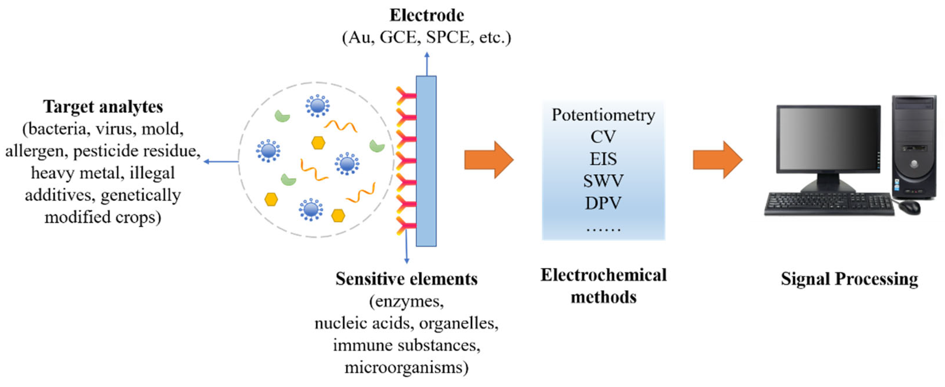
:1. Introduction
2. Biological Contamination
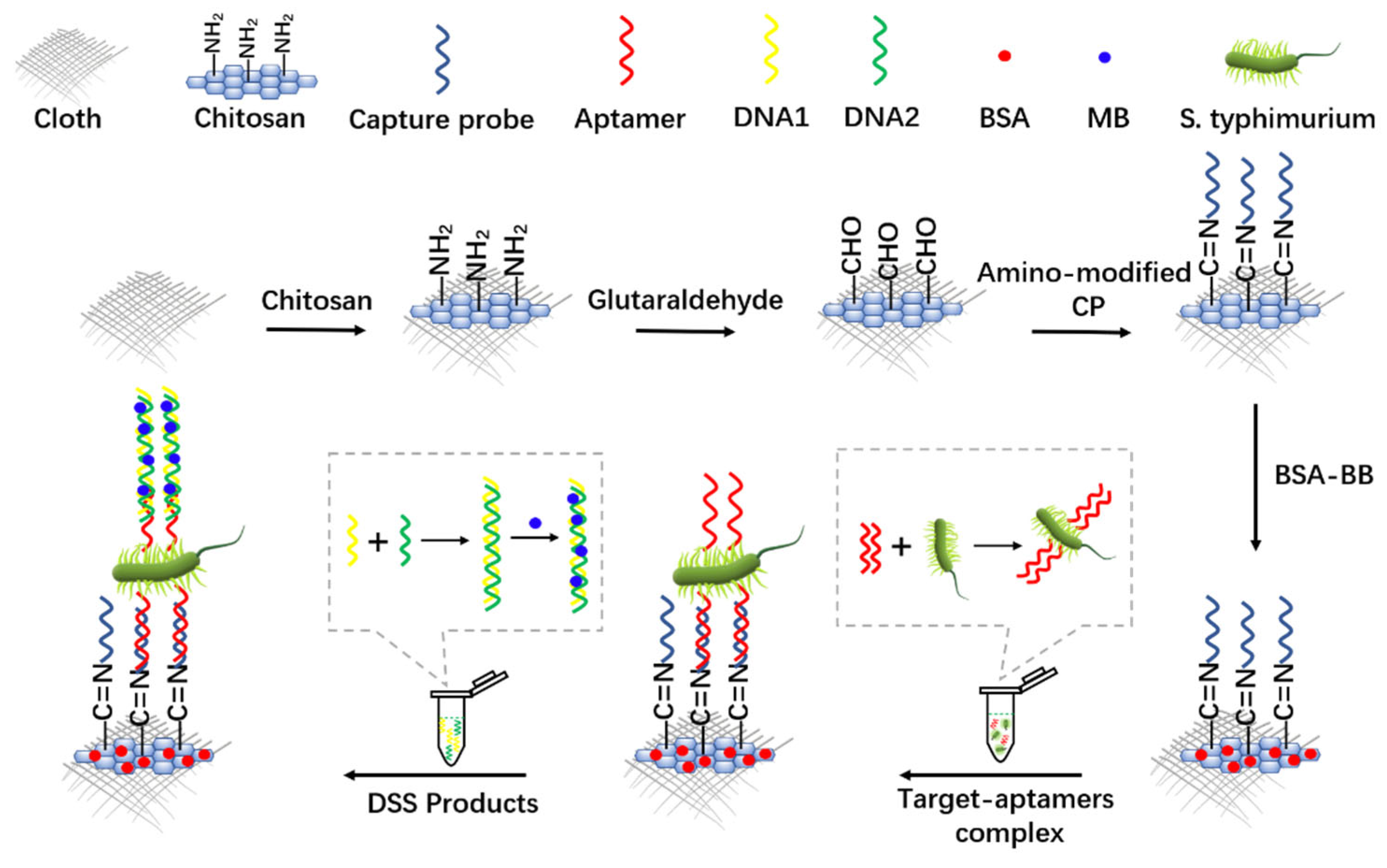
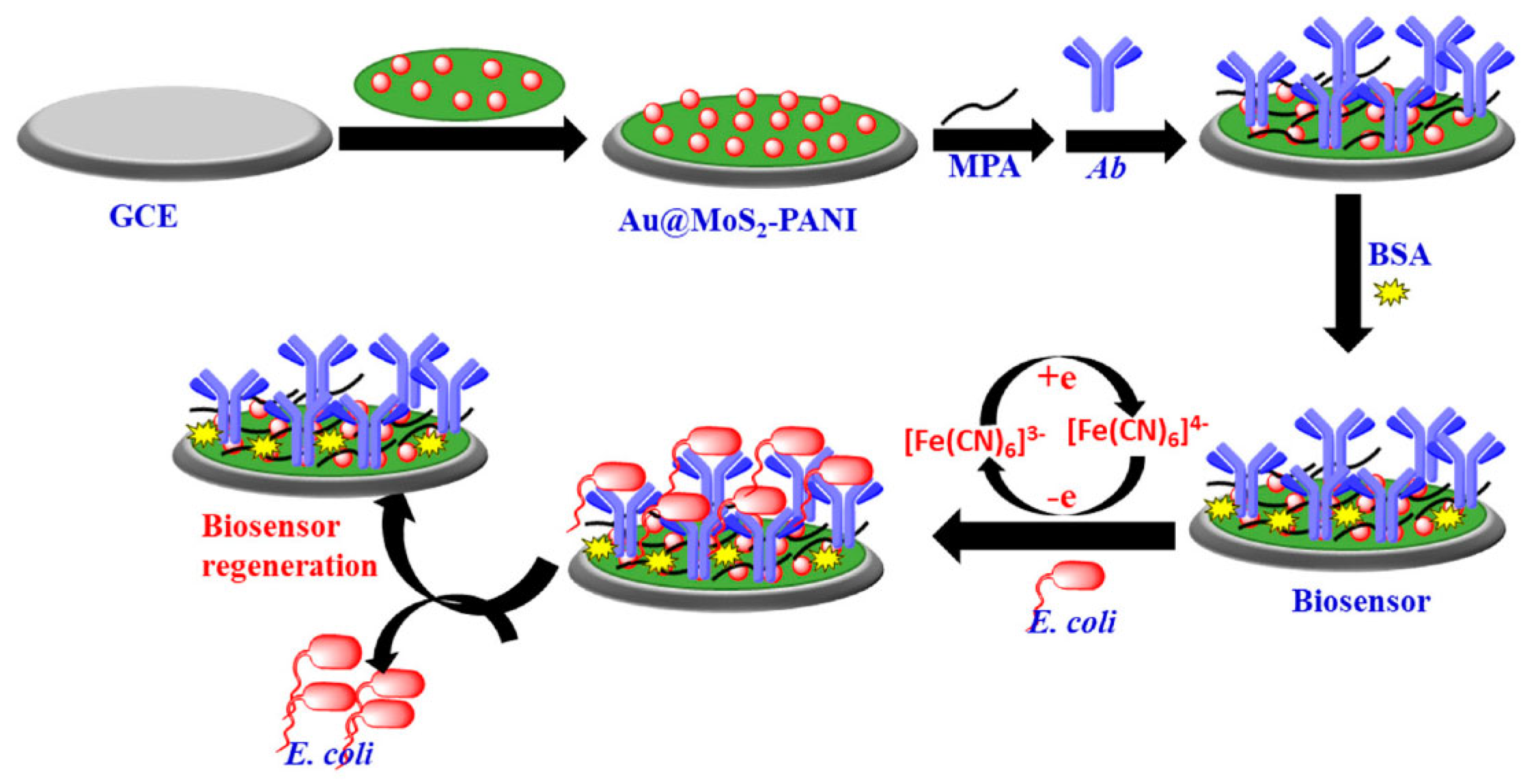
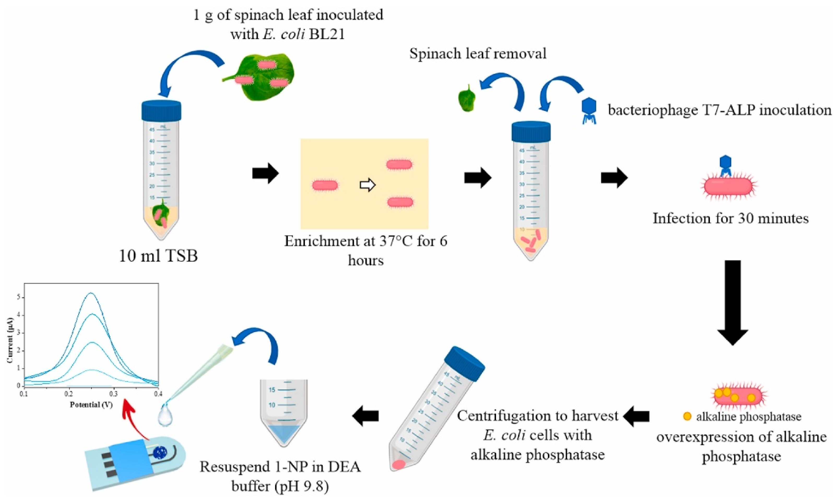
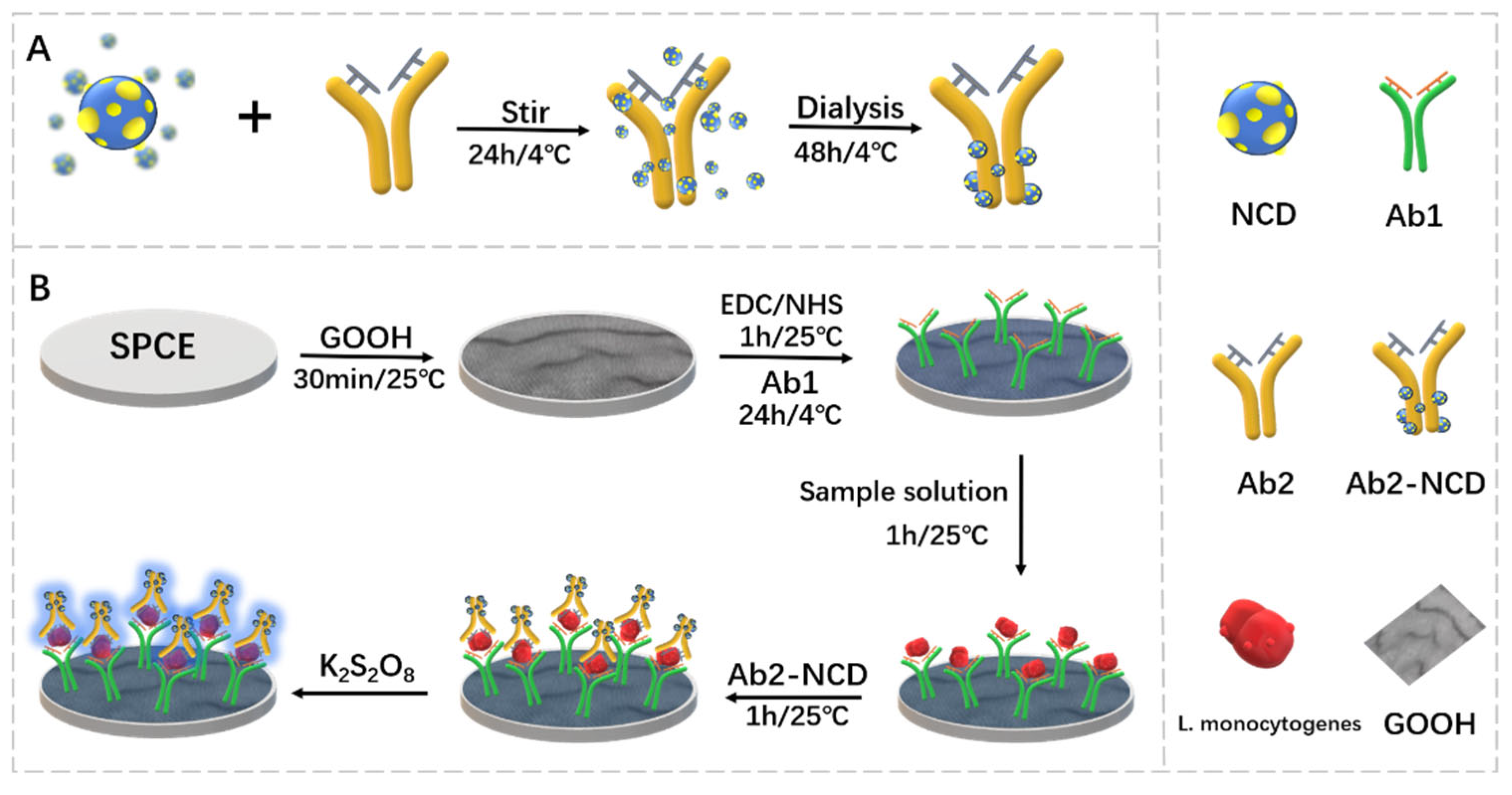
2.1. Bacteria

| Analyte | Electrode | Electrochemical Method | Linearity Range | LOD | Assay Time | Ref. |
|---|---|---|---|---|---|---|
| Salmonella | SPCIE | DPV | 102–108 CFU/mL | 16 CFU/mL | — | [14] |
| GCE | SWV | 30 fg/μL–30 ng/μL | 15.8 fg/μL | — | [15] | |
| E. coli | GCE | DPV | 10–107 CFU/mL | 10 CFU/mL | 30 min | [18] |
| SPE | DPV | 1–104 CFU/mL | 1 CFU/mL | 1 h | [19] | |
| Listeria | SPCE | CV | 2–1.0 × 10⁶ CFU/mL | 0.1 CFU/mL | — | [24] |
| SPPE | EIS | 101–108 CFU/mL | 10 CFU/mL | — | [25] |
2.2. Virus
2.3. Mold
2.4. Allergen
3. Chemical Contamination
3.1. Pesticide Residue
3.2. Heavy Metal
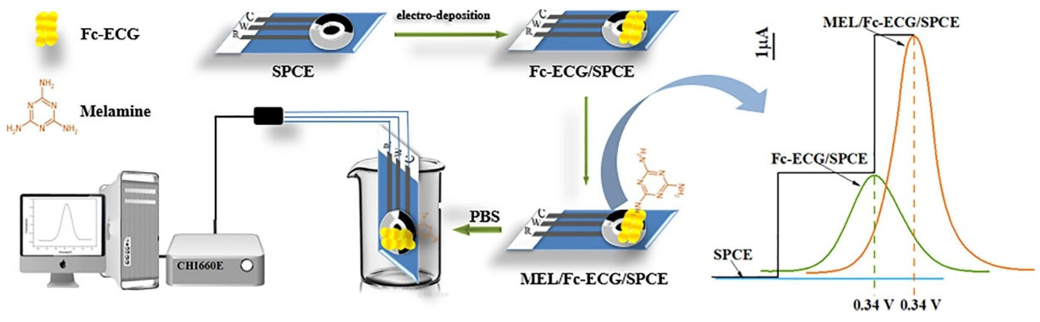
3.3. Illegal Food Additives
| Analyte | Electrode | Electrochemical Method | Linearity Range | LOD | Assay Time | Ref. |
|---|---|---|---|---|---|---|
| Melamine | GCE | I-V | 0.05 nM–0.5 mM | 14.0 ± 0.05 pM | — | [62] |
| SPCE | DPV | 0.20–2.00 μΜ 8.00–800 μM | 0.13 μM | — | [63] | |
| Sudan red | GCE | CV Amperometry | 10–260 µM | 4 nM | — | [65] |
| CPE | DPV | 0.01–20 mM | 0.0017 mM | — | [66] | |
| Clenbuterol | GCE | CV DPV | 1–128 μM | 120 nM | — | [68] |
| GCE | DPSV | 0.03–16.0 μM | 17 nM | — | [69] |
4. Genetically Modified Crops
5. Summary and Outlook
Author Contributions
Funding
Institutional Review Board Statement
Informed Consent Statement
Data Availability Statement
Conflicts of Interest
References
- Fukuda, K. Food safety in a globalized world. Bull. World Health Organ. 2015, 93, 212. [Google Scholar] [CrossRef]
- World Health Organization. Food Safety; WHO: Geneva, Switzerland, 2022. [Google Scholar]
- Jia, M.; Zhongbo, E.; Zhai, F.; Bing, X. Rapid Multi-Residue Detection Methods for Pesticides and Veterinary Drugs. Molecules 2020, 25, 3590. [Google Scholar] [CrossRef]
- Pang, G. Sequence “Food safety testing” album. J. Mass Spectrom. 2019, 40, 3–4. [Google Scholar]
- Lv, M.; Liu, Y.; Geng, J.H.; Kou, X.H.; Xin, Z.H.; Yang, D.Y. Engineering nanomaterials-based biosensors for food safety detection. Biosens. Bioelectron. 2018, 106, 122–128. [Google Scholar] [CrossRef]
- Düzgün, A.; Zelada-Guillén, G.A.; Crespo, G.A.; Macho, S.; Riu, J.; Rius, F.X. Nanostructured materials in potentiometry. Anal. Bioanal. Chem. 2011, 399, 171–181. [Google Scholar] [CrossRef]
- Pajkossy, T. Voltammetry coupled with impedance spectroscopy. J. Solid State Electrochem. 2020, 24, 2157–2159. [Google Scholar] [CrossRef]
- Mirceski, V.; Skrzypek, S.; Stojanov, L. Square-wave voltammetry. ChemTexts 2018, 4, 17. [Google Scholar] [CrossRef]
- Ortuño, J.A.; Serna, C.; Molina, A.; Gil, A. Differential Pulse Voltammetry and Additive Differential Pulse Voltammetry with Solvent Polymeric Membrane Ion Sensors. Anal. Chem. 2006, 78, 8129–8133. [Google Scholar] [CrossRef] [PubMed]
- Ortiz, D. Biological Contamination of Grains in Transportation-Farm to Fork. Cereal Foods World. 2020, 65, 1. [Google Scholar]
- Ferrari, A.G.M.; Crapnell, R.D.; Banks, C.E. Electroanalytical Overview: Electrochemical Sensing Platforms for Food and Drink Safety. Biosensors 2021, 11, 291. [Google Scholar] [CrossRef] [PubMed]
- Saravanan, A.; Kumar, P.S.; Hemavathy, R.V.; Jeevanantham; Kamalesh, R.; Sneha, S.; Yaashikaa, P.R. Methods of detection of food borne pathogens: A review. Environ. Chem. Lett. 2021, 19, 189–207. [Google Scholar] [CrossRef]
- Melo, A.M.A.; Alexandre, D.L.; Furtado, R.F.; Borges, M.F.; Figueiredo, E.A.T.; Biswas, A.; Cheng, H.N.; Alves, C.R. Electrochemical immunosensors for Salmonella detection in food. Appl. Microbiol. Biotechnol. 2016, 100, 5301–5312. [Google Scholar] [CrossRef] [PubMed]
- Li, J.; Jiang, J.; Su, Y.; Liang, Y.; Zhang, C. A novel cloth-based supersandwich electrochemical aptasensor for direct, sensitive detection of pathogens. Anal. Chim. Acta 2021, 1188, 339176. [Google Scholar] [CrossRef] [PubMed]
- Yu, H.; Yuan, N.; Zhang, Y.; Guo, W.; Lu, X.; Yang, Q.; Zhang, W. Saltatory Rolling Circle Amplifcation-Based Ratiometric Electrochemical Biosensor for Rapid Detection of Salmonella enterica serovar Typhimurium in Food. Food Anal. Methods 2022, 15, 820–832. [Google Scholar] [CrossRef]
- YHuang, i.; Su, Z.; Li, W.; Ren, J. Recent Progresses on Biosensors for Escherichia coli Detection. Food Anal. Methods 2022, 15, 338–366. [Google Scholar]
- Couto, R.A.S.; Chen, L.; Kuss, S.; Compton, R.G. Detection of Escherichia coli bacteria by impact electrochemistry. Analyst 2018, 143, 4840. [Google Scholar] [CrossRef]
- Raj, P.; Oh, M.H.; Han, K.; Lee, T.Y. Label-Free Electrochemical Biosensor Based on Au@MoS2–PANI for Escherichia coli Detection. Chemosensors 2021, 9, 49. [Google Scholar] [CrossRef]
- El-Moghazy, A.Y.; Wisuthiphaet, N.; Yang, X.; Sun, G.; Nitin, N. Electrochemical biosensor based on genetically engineered bacteriophage T7 for rapid detection of Escherichia coli on fresh produce. Food Control 2022, 135, 108811. [Google Scholar] [CrossRef]
- Blot, M.; Disson, O.; Leclercq, A.; Moura, A.; Bracq-Dieye, H.; Thouvenot, P.; Valès, G.; Burroni, B.; Lupo, A.; Lecuit, M. Listeria-Associated Lymphadenitis: A Series of 11 Consecutive Cases and Review of the Literature. Open Forum Infect. Dis. 2022, 9, ofab598. [Google Scholar] [CrossRef]
- Liu, R.; Zhang, Y.; Ali, S.; Haruna, S.A.; He, P.; Li, H.; Ouyang, Q.; Chen, Q. Development of a fluorescence aptasensor for rapid and sensitive detection of Listeria monocytogenes in food. Food Control 2021, 122, 107808. [Google Scholar] [CrossRef]
- Qia, X.; Wang, Z.; Luc, R.; Liua, J.; Lia, Y.; Chen, Y. One-step and DNA amplification-free detection of Listeria monocytogenes in ham samples: Combining magnetic relaxation switching and DNA hybridization reaction. Food Chem. 2021, 338, 127837. [Google Scholar] [CrossRef] [PubMed]
- Zhao, X.; Lin, C.; Wang, J.; Oh, D.H. Advances in Rapid Detection Methods for Foodborne Pathogens. J. Microbiol. Biotechnol. 2014, 24, 297–312. [Google Scholar] [CrossRef] [PubMed] [Green Version]
- Jampasa, S.; Ngamrojanavanich, N.; Rengpipat, S.; Chailapakul, O.; Kalcher, K.; Chaiyo, S. Ultrasensitive electrochemiluminescence sensor based on nitrogen-decorated carbon dots for Listeria monocytogenes determination using a screen-printed carbon electrode. Biosens. Bioelectron. 2021, 188, 113323. [Google Scholar] [CrossRef]
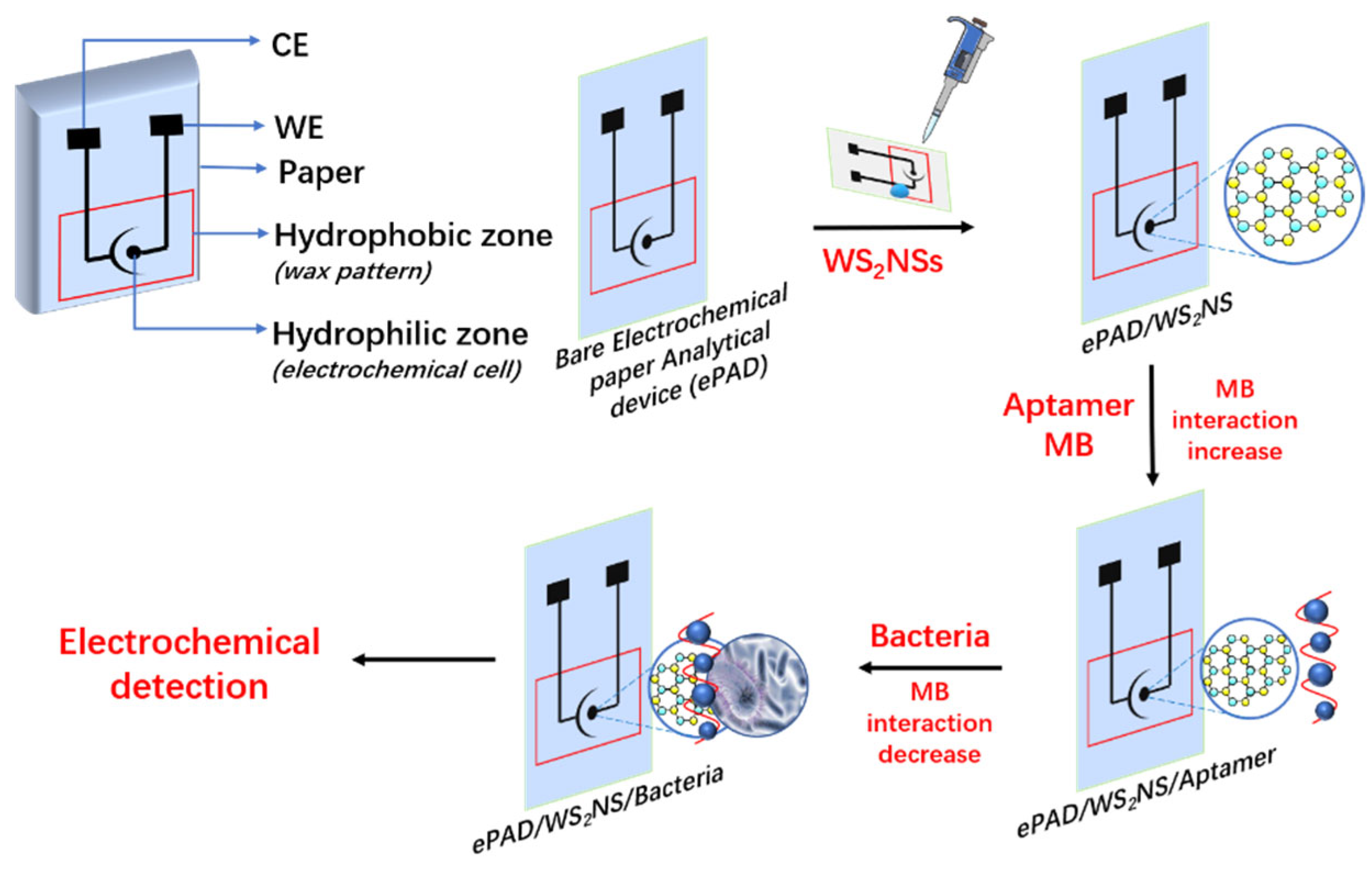
- Mishra, A.; Pilloton, R.; Jain, S.; Roy, S.; Khanuja, M.; Mathur, A.; Narang, J. Paper-Based Electrodes Conjugated with Tungsten Disulfide Nanostructure and Aptamer for Impedimetric Detection of Listeria monocytogenes. Biosensors 2022, 12, 88. [Google Scholar] [CrossRef] [PubMed]
- Kahyaoglu, L.N.; Irudayaraj, J. New approaches in microbial pathogen detection. Adv. Microb. Food Saf. 2013, 78, 202–226. [Google Scholar]
- Yee, M.Y.; Shamsuddin, S.; Nizam, Q.N.H.; Sidik, M.R.; Yusop, F.F.M.; Saeid, F.H.M.; Aziah, I. Detection methods of avian influenza—Current and novel approaches. Malays. J. Microbiol. 2019, 15, 492–504. [Google Scholar]
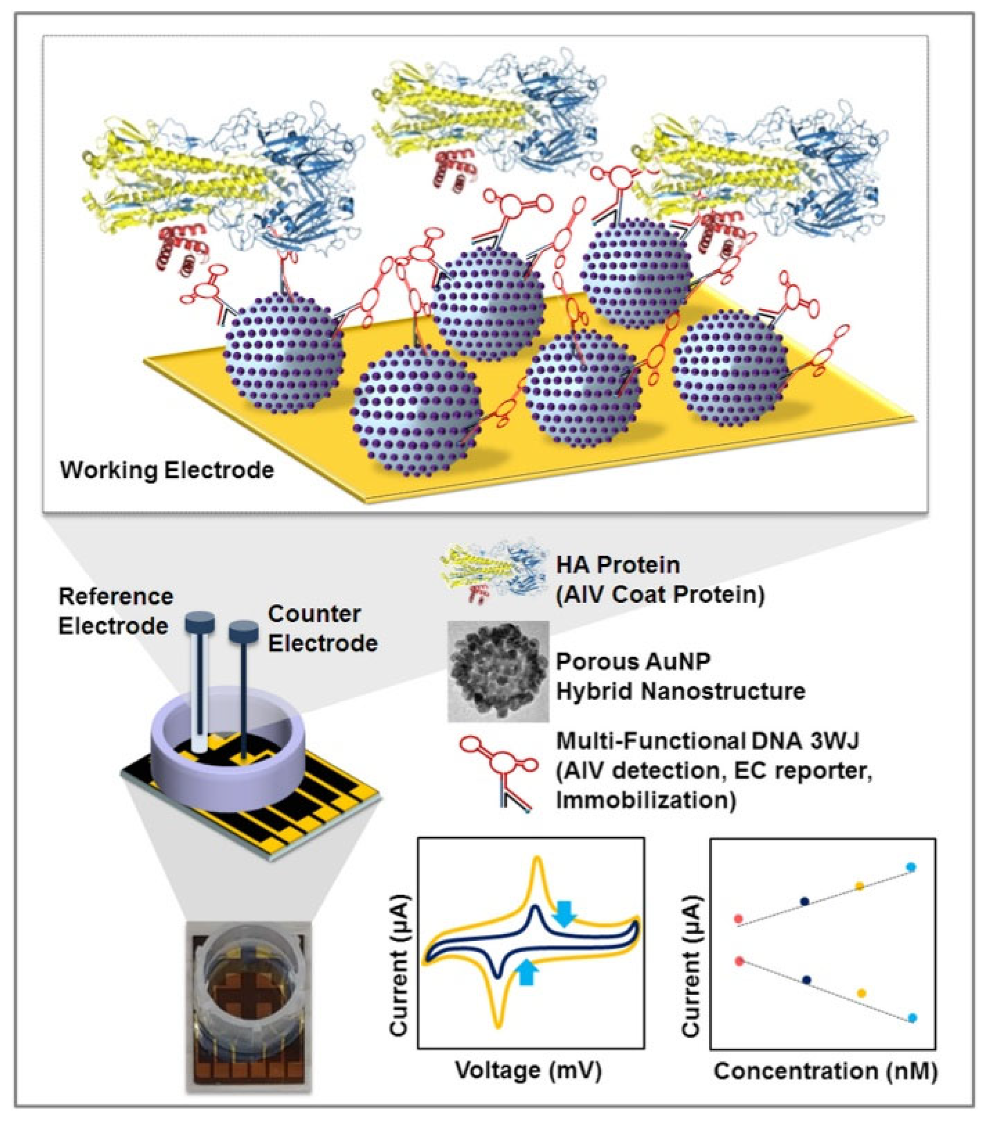
- Lee, T.; Park, S.Y.; Jang, H.; Kim, G.; Lee, Y.; Park, C.; Mohammadniaei, M.; Lee, M.; Min, J. Fabrication of electrochemical biosensor consisted of multi-functional DNA structure/porous au nanoparticle for avian influenza virus (H5N1) in chicken serum. Mater. Sci. Eng. C 2019, 99, 511–519. [Google Scholar] [CrossRef]
- Panigrahy, B.; Senne, D.A.; Pedersen, J.C.; Shafer, A.L.; Pearson, J.E. Susceptibility of pigeons to avian influenza. Avian Dis. 1996, 40, 600–604. [Google Scholar] [CrossRef]
- MaManzano, r.; Viezzi, S.; Mazerat, S.; Marks, R.S.; Vidic, J. Rapid and label-free electrochemical DNA biosensor for detecting hepatitis A virus. Biosens. Bioelectron. 2018, 100, 89–95. [Google Scholar] [CrossRef]
- Hwang, H.J.; Ryu, M.Y.; Park, C.Y.; Ahn, J.; Park, H.J.; Choi, C.; Ha, S.D.; Park, T.J.; Park, J.P. High sensitive and selective electrochemical biosensor: Label-free detection of human norovirus using affinity peptide as molecular binder. Biosens. Bioelectron. 2017, 87, 164–170. [Google Scholar] [CrossRef]
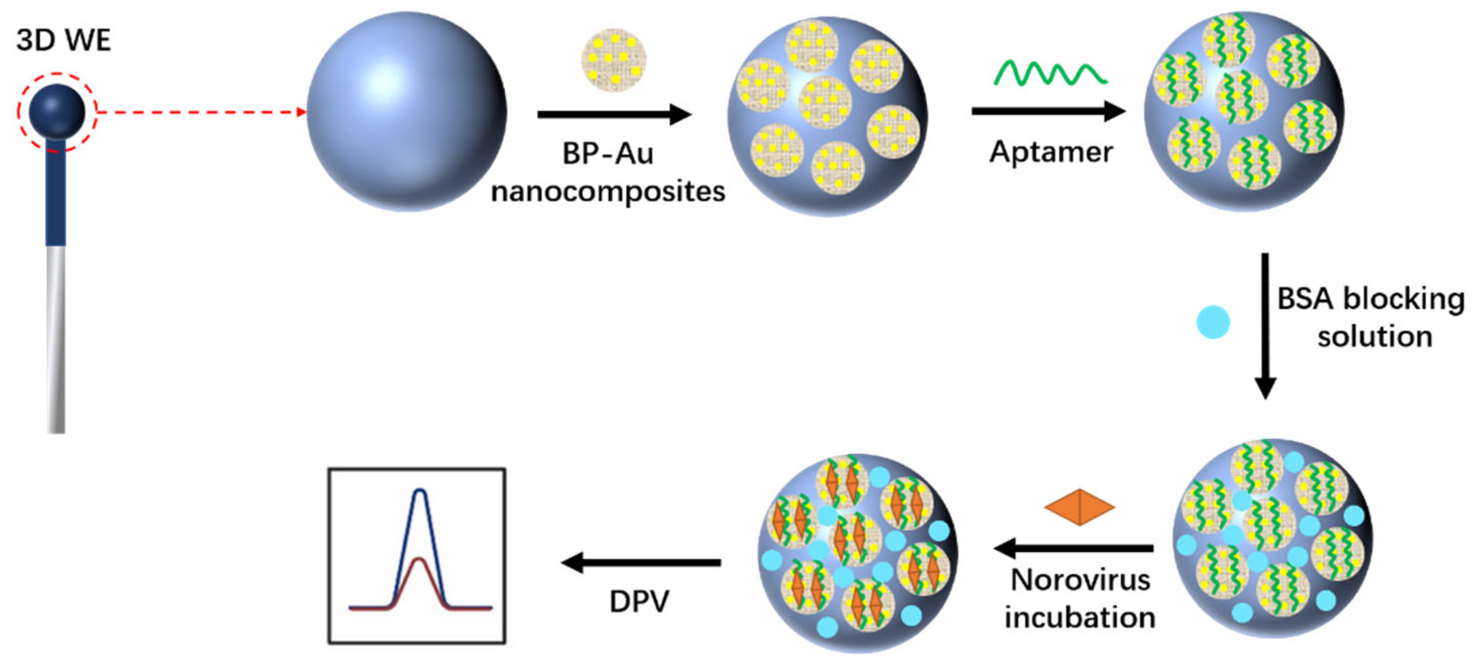
- Jiang, H.; Sun, Z.; Zhang, C.; Weng, X. 3D-architectured aptasensor for ultrasensitive electrochemical detection of norovirus based on phosphorene-gold nanocomposites. Sens. Actuators B Chem. 2022, 354, 131232. [Google Scholar] [CrossRef]
- Dong, S.; Yan, J.; Zhou, S.; Zhou, Q. Mycotoxins Detection Based on Electrochemical Approaches. Electroanalysis 2022, 34, 132–147. [Google Scholar] [CrossRef]
- Meng, D.; Gan, X.; Tian, T. An Electrochemical Sensing Method for Aflatoxin B1 Detection Based on Pt-coordinated Titanium-based Porphyrin. MOF 2022, 17, 220247. [Google Scholar] [CrossRef]
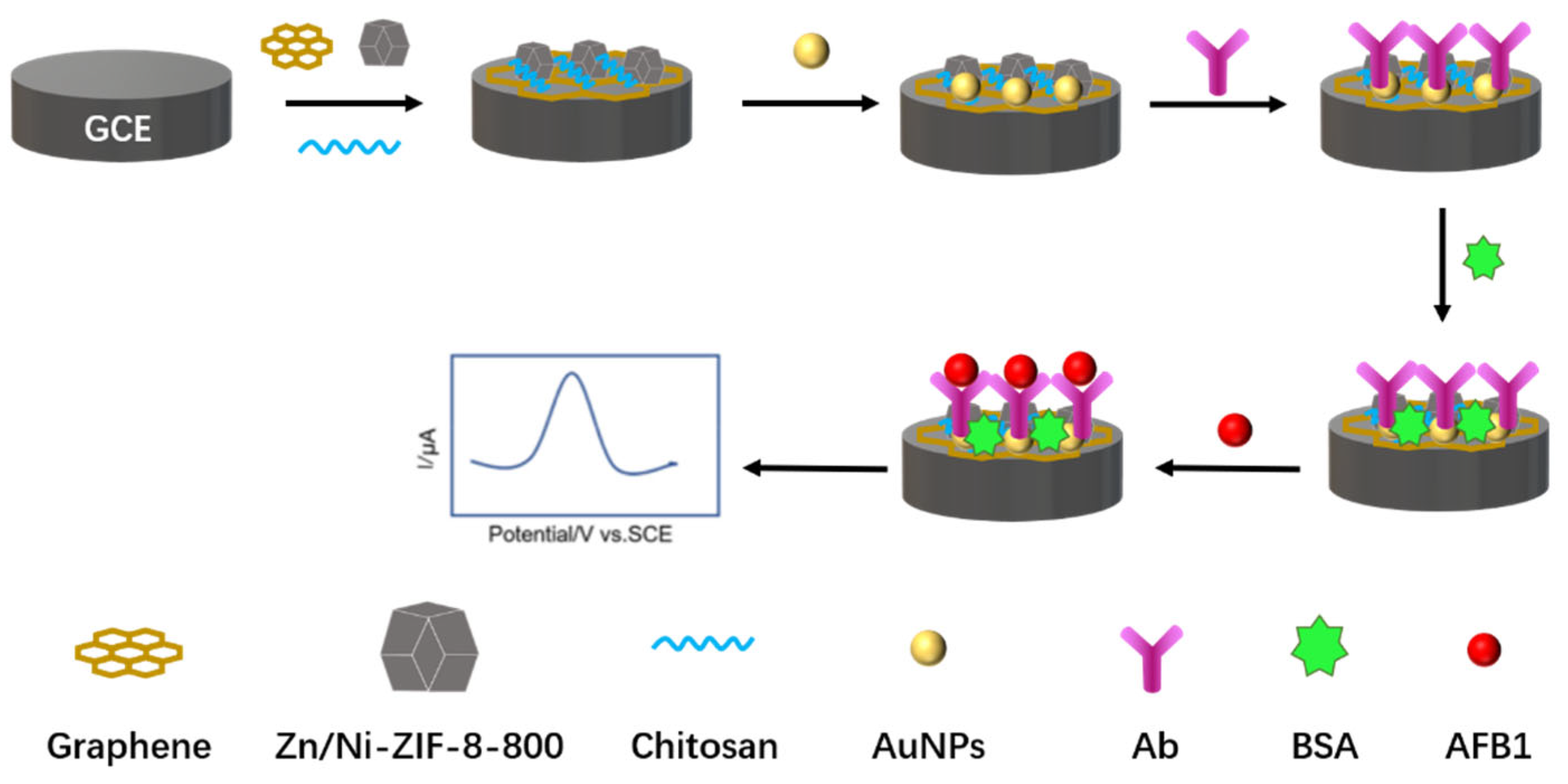
- Wang, N.; Liu, Q.; Hu, X.; Wang, F.; Hu, M.; Yu, Q.; Zhang, G. Electrochemical immunosensor based on AuNPs/Zn/Ni-ZIF-8-800@graphene for rapid detection of aflatoxin B1 in peanut oil. Anal. Biochem. 2022, 650, 114710. [Google Scholar] [CrossRef]
- Mazaafrianto, D.N.; Ishida, A.; Maeki, M.; Tani, H.; Tokeshi, M. An Electrochemical Sensor Based on Structure Switching of Dithiol-modified Aptamer for Simple Detection of Ochratoxin A. Anal. Sci. 2019, 35, 1221–1226. [Google Scholar] [CrossRef]
- Ji, Y.M.; Zhang, K.H.; Pan, Z.N.; Ju, J.Q.; Zhang, H.L.; Liu, J.C.; Wang, Y.; Sun, S.C. High-dose zearalenone exposure disturbs G2/M transition during mouse oocyte maturation. Reprod. Toxicol. 2022, 110, 172–179. [Google Scholar] [CrossRef]
- Radi, A.E.; Eissa, A.; Wahdan, T. Voltammetric behavior of mycotoxin zearalenone at a single walled carbon nanotube screen-printed electrode. Anal. Methods 2019, 11, 4494–4500. [Google Scholar] [CrossRef]
- Curulli, A. Recent Advances in Electrochemical Sensing Strategies for Food. Biosensors 2022, 12, 503. [Google Scholar] [CrossRef]
- Sundhoro, M.; Agnihotra, S.R.; Khan, N.D.; Barnes, A.; BelBruno, J.; Mendecki, L. Rapid and accurate electrochemical sensor for food allergen detection in complex foods. Sci. Rep. 2021, 11, 20831. [Google Scholar] [CrossRef]
- Sundhoro, M.; Agnihotra, S.R.; Amberger, B.; Augustus, K.; Khan, N.D.; Barnes, A.; BelBruno, J.; Mendecki, L. An electrochemical molecularly imprinted polymer sensor for rapid and selective food allergen detection. Food Chem. 2021, 344, 128648. [Google Scholar] [CrossRef]
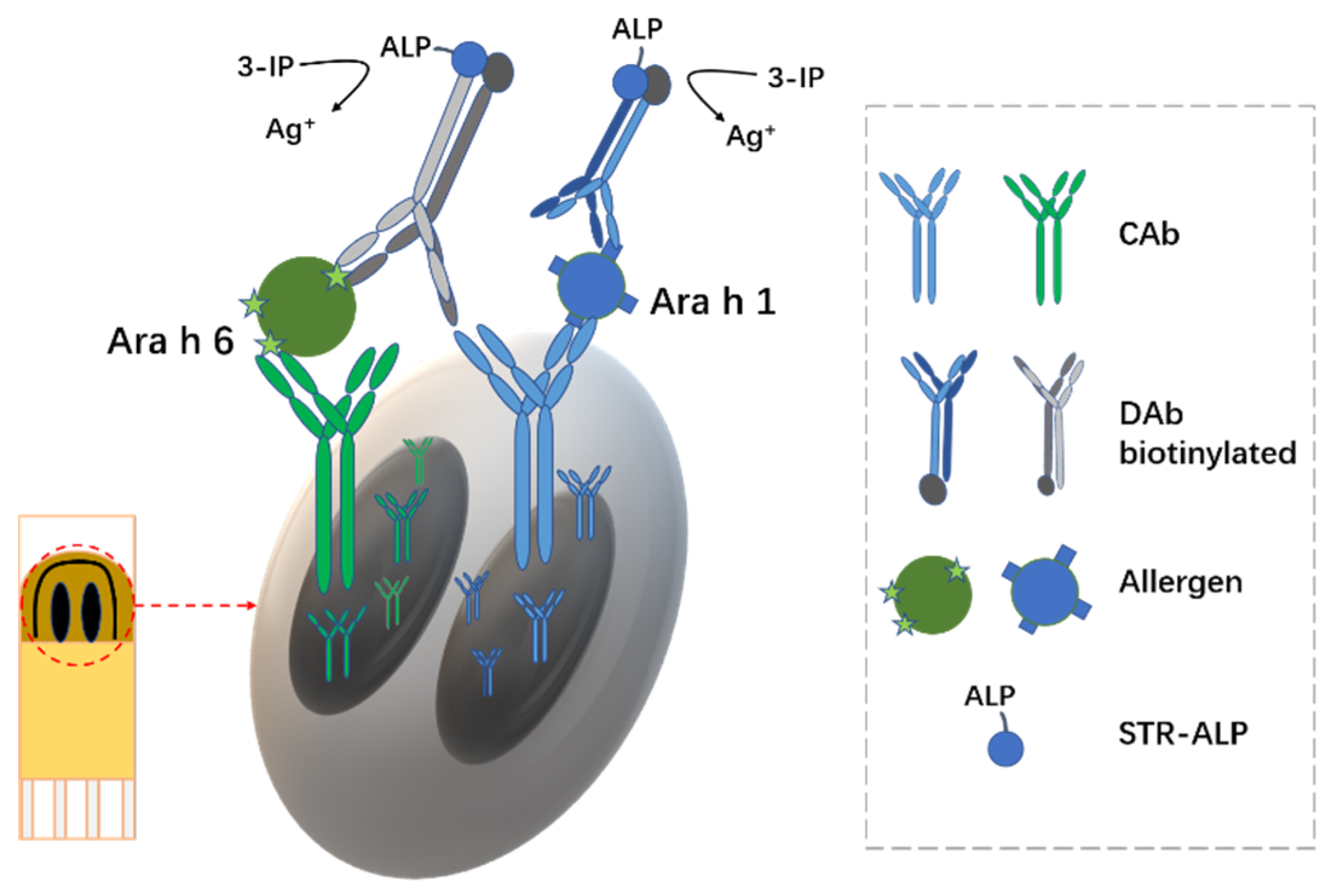
- Freitas, M.; Neves, M.M.P.S.; Nouws, H.P.A.; Delerue-Matos, C. Electrochemical Immunosensor for the Simultaneous Determination of Two Main Peanut Allergenic Proteins (Ara h 1 and Ara h 6) in Food Matrices. Foods 2021, 10, 1718. [Google Scholar] [CrossRef] [PubMed]
- Li, Y.; Su, R.; Li, H.; Guo, J.; Hildebrandt, N.; Sun, C. Fluorescent Aptasensors: Design Strategies and Applications in Analyzing Chemical Contamination of Food. Anal. Chem. 2022, 94, 193–224. [Google Scholar] [CrossRef] [PubMed]
- Hu, H.; Yang, L. Development of enzymatic electrochemical biosensors for organophosphorus pesticide detection. J. Environ. Sci. Health Part B 2021, 56, 168–180. [Google Scholar] [CrossRef] [PubMed]
- Maltzman, S.L.; Minteer, S.D. Mitochondrial-based voltammetric sensor for pesticides. Anal. Methods 2012, 4, 1202. [Google Scholar] [CrossRef]
- Nevin, T.; Selcan, K.; Cihat, T.; Gülsen, B. Highly sensitive and selective rGO based Non-Enzymatic electrochemical sensor for propamocarb fungicide pesticide detection. Food Chem. 2022, 372, 131267. [Google Scholar]
- Ma, Y.; Jiang, H.; Shen, C.; Hou, C.; Huo, D.; Wu, H.; Yang, M. Detection of Carbendazim Residues with a Colorimetric Sensor Based on Gold Nanoparticles. J. Appl. Spectrosc. 2017, 84, 460–465. [Google Scholar] [CrossRef]
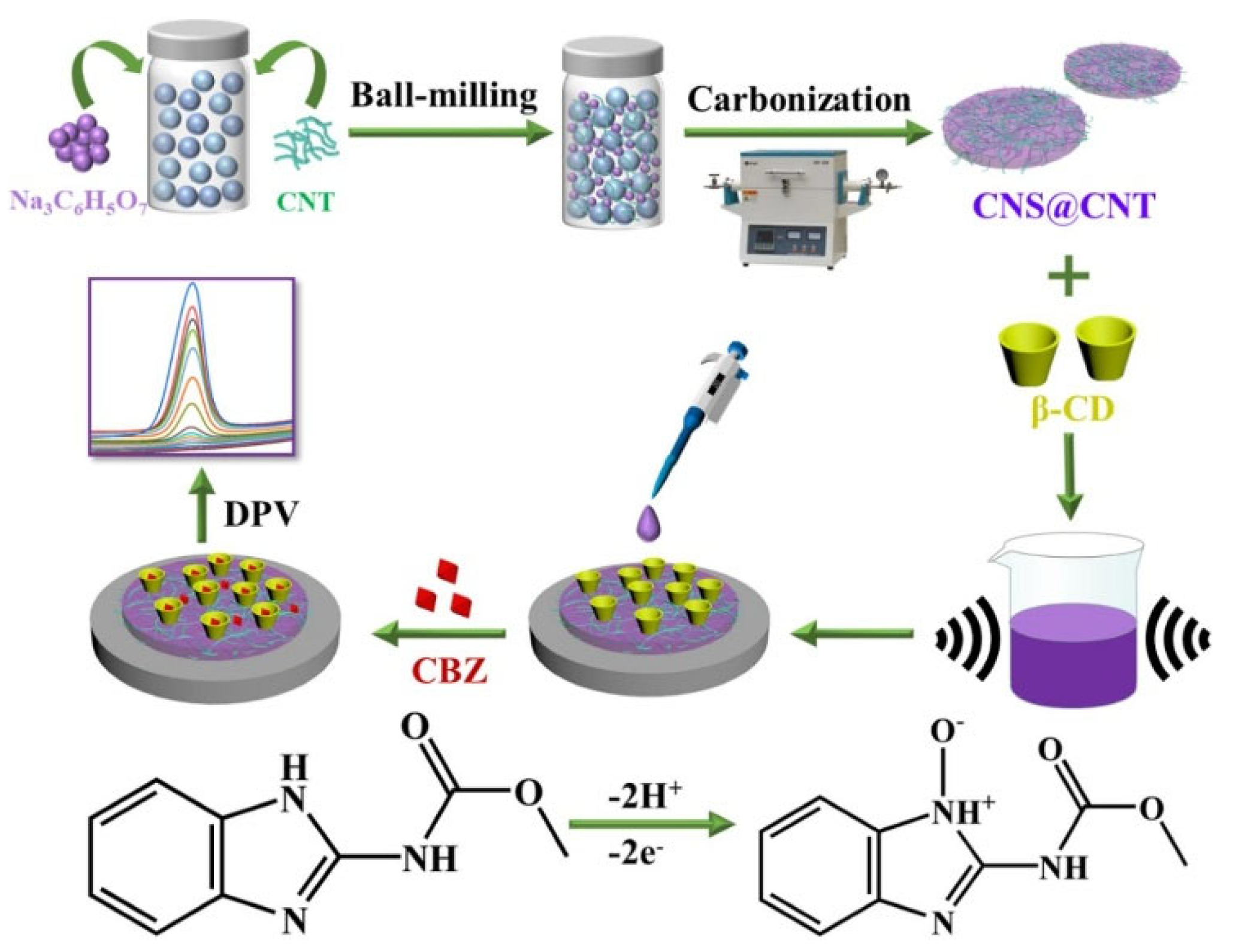
- Liu, R.; Li, B.; Li, F.; Dubovyk, V.; Chang, Y.; Li, D.; Ding, K.; Ran, Q.; Wang, G.; Zhao, H. A novel electrochemical sensor based on β-cyclodextrin functionalized carbon nanosheets@carbon nanotubes for sensitive detection of bactericide carbendazim in apple juice. Food Chem. 2022, 384, 132573. [Google Scholar] [CrossRef]
- Yang, J.; Qi, L.; Uqaili, J.A.; Shi, D.; Yin, L.; Liu, Z.; Tao, X.; Dai, L.; Lan, C. The terahertz metamaterial sensor for imidacloprid detection. Int. J. RF Microw. Comput. Aided Eng. 2021, 31, e22840. [Google Scholar] [CrossRef]
- Tang, F.; Hua, Q.; Wang, X.; Luan, F.; Wang, L.; Li, Y.; Zhuang, X.; Tian, C. A novel electrochemiluminescence sensor based on a molecular imprinting technique and UCNPs@ZIF-8 nanocomposites for sensitive determination of imidacloprid. Analyst 2022, 147, 3917. [Google Scholar] [CrossRef]
- Pham, D.S.; Nguyen, X.A.; Marsh, P.; Chu, S.S.; Lau, M.P.H.; Nguyen, A.H.; Cao, H. A Fluidics-Based Biosensor to Detect and Characterize Inhibition Patterns of Organophosphate to Acetylcholinesterase in Food Materials. Micromachines 2021, 12, 397. [Google Scholar] [CrossRef]
- Surucu, O. Trace determination of heavy metals and electrochemical removal of lead from drinking water. Chem. Pap. 2021, 75, 4227–4238. [Google Scholar] [CrossRef]
- Chailapakul, O.; Korsrisakul, S.; Siangproh, W.; Grudpan, K. Fast and simultaneous detection of heavy metals using a simple and reliable microchip-electrochemistry route: An alternative approach to food analysis. Talanta 2008, 74, 683–689. [Google Scholar] [CrossRef] [PubMed]
- Lahrich, S.; el Mhammedi, M.A. Review—Application of Deficient Apatites Materials in Electrochemical Detection of Heavy Metals: Case of Mercury (II) in Seawater and Fish Samples. J. Electrochem. Soc. 2019, 166, B1567–B1576. [Google Scholar] [CrossRef]
- Wisarut, K.; Phichanan, D.; Kriangsak, S.; Nuanlaorr, R.; Nunticha, L.; Piyada, J.; Thitirat, M.; Weena, S. An application of miniaturized electrochemical sensing for determination of arsenic in herbal medicines. Methods 2022, 14, 3087. [Google Scholar]
- Narouei, F.H.; Livernois, L.; Andreescu, D.; Andreescu, S. Highly sensitive mercury detection using electroactive gold-decorated polymer nanofibers. Sens. Actuators B Chem. 2021, 329, 129267. [Google Scholar] [CrossRef]
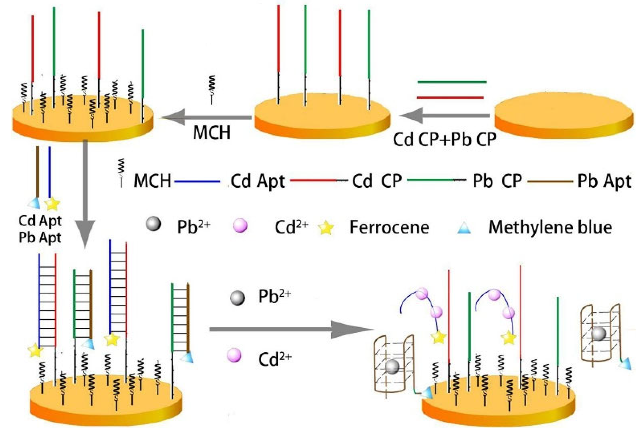
- Yuan, M.; Qian, S.; Cao, H.; Yu, J.; Ye, T.; Wu, X.; Chen, L.; Xu, F. An ultra-sensitive electrochemical aptasensor for simultaneous quantitative detection of Pb2+ and Cd2+ in fruit and vegetable. Food Chem. 2022, 382, 132173. [Google Scholar] [CrossRef]
- Tan, Z.; Wu, W.; Feng, C.; Wu, H.; Zhang, Z. Simultaneous determination of heavy metals by an electrochemical method based on a nanocomposite consisting of fluorinated graphene and gold nanocage. Microchim. Acta 2020, 187, 414. [Google Scholar] [CrossRef]
- Wu, L.; Zhang, C.; Long, Y.; Chen, Q.; Zhang, W.; Liu, G. Food additives: From functions to analytical Methods. Crit. Rev. Food Sci. Nutr. 2021, 61, 1–21. [Google Scholar] [CrossRef]
- Zhang, J.; Hu, S.; Du, Y.; Cao, D.; Wang, G.R.; Yuan, Z.Q. Improved food additive analysis by ever-increasing nanotechnology. J. Food Drug Anal. 2020, 28, 622–640. [Google Scholar] [CrossRef]
- Rao, H.; Chen, M.; Ge, H.; Lu, Z.; Liu, X.; Zou, P.; Wang, X.; He, H.; Zeng, X.; Wang, Y. A novel electrochemical sensor based on Au@PANI composites film modified glassy carbon electrode binding molecular imprinting technique for the determination of melamine. Biosens. Bioelectron. 2017, 87, 1029–1035. [Google Scholar] [CrossRef]
- Rahmana, M.M.; Ahmed, J. Cd-doped Sb2O4 nanostructures modified glassy carbon electrode for efficient detection of melamine by electrochemical approach. Biosens. Bioelectron. 2018, 102, 631–636. [Google Scholar] [CrossRef] [PubMed]
- An, Q.; Feng, X.; Zhou, Z.; Zhan, T.; Lian, S.; Zhu, J.; Han, G.; Chen, Z.; Kraatz, H. One step construction of an electrochemical sensor for melamine detection in milk towards an integrated portable system. Food Chem. 2022, 383, 132403. [Google Scholar] [CrossRef] [PubMed]
- Ensafi, A.A.; Rezaei, B.; Amini, M.; Heydari-Bafrooei, E. A novel sensitive DNA–biosensor for detection of a carcinogen, Sudan II, using electrochemically treated pencil graphite electrode by voltammetric methods. Talanta 2012, 88, 244–251. [Google Scholar] [CrossRef] [PubMed]
- Yang, L.; Wang, S.; Zhang, L. Electrochemical Sensor Based on MWCNTs/AuNPs/GCE for Sensitive Determination of Sudan I Content in Food Samples. ESG 2020, 15, 11168–11179. [Google Scholar] [CrossRef]
- Heydari, M.; Ghoreishi, S.M.; Khoobi, A. Novel electrochemical procedure for sensitive determination of Sudan II based on nanostructured modified electrode and multivariate optimization. Measurement 2019, 142, 105–112. [Google Scholar] [CrossRef]
- Shi, Z.; Tian, Y.; Wu, X.; Li, C.; Yu, L. A one-piece lateral flow impedimetric test strip for label-free clenbuterol detection. Anal. Methods 2015, 7, 4957. [Google Scholar] [CrossRef]
- Sun, Y.; Wang, T.; Chen, S.; Wang, X.; Reynoso, L.C. Highly Sensitive Electrochemical Sensor Based on rGO/Fe3O4 Composite as Electrocatalyst for Clenbuterol Detection in Doping Analysis. Int. J. Electrochem. Sci. 2022, 17, 220128. [Google Scholar] [CrossRef]
- Jing, H.; Ouyang, H.; Li, W.; Long, Y. Molten salt synthesis of BCNO nanosheets for the electrochemical detection of clenbuterol. Microchem. J. 2022, 178, 107359. [Google Scholar] [CrossRef]
- Zheng, Y.; Karimi-Maleh, H.; Fu, L. Advances in Electrochemical Techniques for the Detection and Analysis of Genetically Modified Organisms: An Analysis Based on Bibliometrics. Chemosensors 2022, 10, 194. [Google Scholar] [CrossRef]
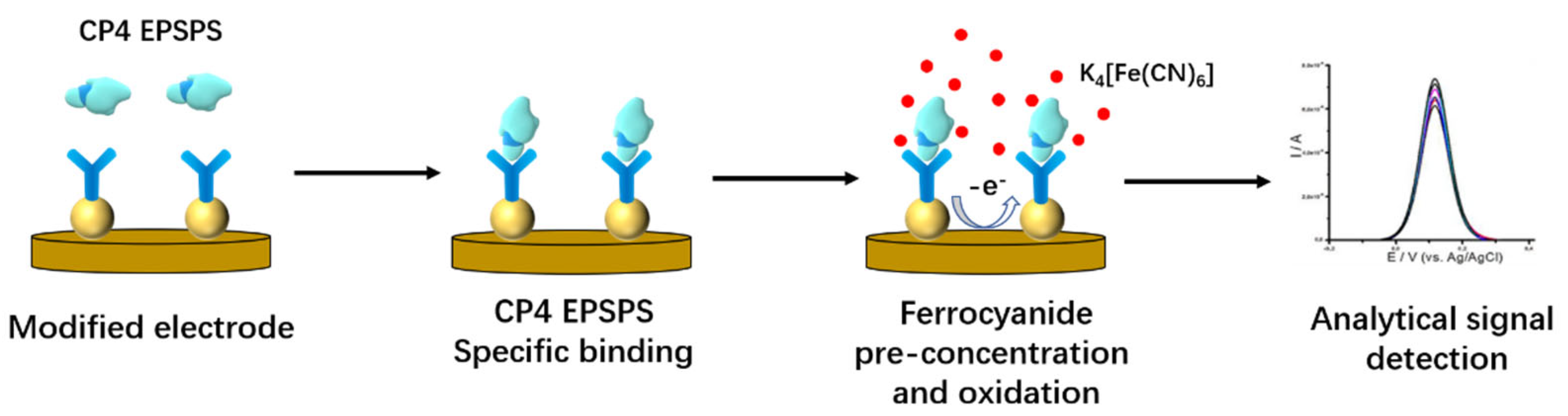
- Farías, M.E.; Correa, N.M.; Sosa, L.; Niebylski, A.M.; Molina, P.G. A simple electrochemical immunosensor for sensitive detection of transgenic soybean protein CP4-EPSPS in seeds. Talanta 2022, 237, 122910. [Google Scholar] [CrossRef]
- Gao, H.; Cui, D.; Zhai, S.; Yang, Y.; Wu, Y.; Yan, X.; Gang, W. A label-free electrochemical impedimetric DNA biosensor for genetically modifed soybean detection based on gold carbon dots. Microchim. Acta 2022, 189, 216. [Google Scholar] [CrossRef] [PubMed]
- Cui, D.; Zhai, S.; Yang, Y.; Wu, Y.; Li, J.; Yan, X.; Shen, P.; Gao, H.; Wu, G. A Label-Free Electrochemical Impedance Genosensor Coupled with Recombinase Polymerase Amplification for Genetically Modified Maize Detection. Agriculture 2022, 12, 454. [Google Scholar] [CrossRef]


| Analyte | Electrode | Electrochemical Method | Linearity Range | LOD | Assay Time | Ref. |
|---|---|---|---|---|---|---|
| Genistein | Carbon | DPV | 100 ppb–10 ppm | 100 ppb | — | [41] |
| Ara h 1 Ara h 6 | SPCE | LSV | 0–1000 ng/mL 0–1.0 ng/mL | 5.2 ng/mL 0.017 ng/mL | 2 h 20 min | [42] |
| Analyte | Electrode | Electrochemical Method | Linearity Range | LOD | Assay Time | Ref. |
|---|---|---|---|---|---|---|
| As | SPGE | SWASV | 0.1–3.0 ppm | 0.03 ppm | <3 min | [55] |
| Hg2+ | SPCE | SWASV | 0.8–12.0 nM | 0.23 nM | — | [56] |
| Cd2+ Pb2+ | Au | SWV | 0.1–1000 nmol/L | 89.31 pmol/L 16.44 pmol/L | 15 min | [57] |
| Hg2+ Cd2+ Pb2+ Cu2+ Zn2+ | GCE | SWASV | 6–7000, 4–6000, 6–5000, 4–4000, 6–5000 μg/L | 0.08, 0.09, 0.05, 0.19, 0.01 μg/L | — | [58] |
Publisher’s Note: MDPI stays neutral with regard to jurisdictional claims in published maps and institutional affiliations. |
© 2022 by the authors. Licensee MDPI, Basel, Switzerland. This article is an open access article distributed under the terms and conditions of the Creative Commons Attribution (CC BY) license (https://creativecommons.org/licenses/by/4.0/).
Share and Cite
Wang, K.; Lin, X.; Zhang, M.; Li, Y.; Luo, C.; Wu, J. Review of Electrochemical Biosensors for Food Safety Detection. Biosensors 2022, 12, 959. https://doi.org/10.3390/bios12110959
Wang K, Lin X, Zhang M, Li Y, Luo C, Wu J. Review of Electrochemical Biosensors for Food Safety Detection. Biosensors. 2022; 12(11):959. https://doi.org/10.3390/bios12110959
Chicago/Turabian StyleWang, Ke, Xiaogang Lin, Maoxiao Zhang, Yu Li, Chunfeng Luo, and Jayne Wu. 2022. "Review of Electrochemical Biosensors for Food Safety Detection" Biosensors 12, no. 11: 959. https://doi.org/10.3390/bios12110959