Article

CH3NH3PbBr3 Thin Film Served as Guided-Wave Layer for Enhancing the Angular Sensitivity of Plasmon Biosensor

1
Advanced Institute of Photonics Technology, School of Information Engineering, Guangdong University of Technology, Guangzhou 510006, China
2
Guangdong Provincial Key Laboratory of Information Photonics Technology, Guangdong University of Technology, Guangzhou 510006, China
3
School of Physics and Electronics, Hunan University, Changsha 410082, China
*
Authors to whom correspondence should be addressed.
Biosensors 2021, 11(11), 415; https://doi.org/10.3390/bios11110415
Submission received: 30 September 2021 / Revised: 19 October 2021 / Accepted: 22 October 2021 / Published: 23 October 2021
(This article belongs to the Special Issue Optical Biosensor with 2D Materials and Metamaterials)

Abstract

:
CH3NH3PbBr3 perovskite thin film is used as a guided-wave layer and coated on the surface of an Au film to form the Au-perovskite hybrid structure. Using the hybrid structure, a perovskite-based guided-wave surface plasmon resonance (GWSPR) biosensor is proposed with high angular sensitivity. First, it is found that the electric field at the sensing interface is improved by the CH3NH3PbBr3 perovskite thin film, thereby enhancing the sensitivity. The result demonstrates that the angular sensitivity of the Au-perovskite-based GWSPR biosensor is as high as 278.5°/RIU, which is 110.2% higher than that of a conventional Au-based surface plasmon resonance (SPR) biosensor. Second, the selection of the coupling prism in the configuration of the GWSPR biosensor is also analyzed, and it indicates that a low refractive index (RI) prism can generate greater sensitivity. Therefore, the low-RI BK7 prism is served as the coupling prism for the proposed GWSPR biosensor. Finally, the proposed GWSPR sensing structure can not only be used for liquid sensing, but also for gas sensing, and it has also been demonstrated that the GWSPR gas sensor is 2.8 times more sensitive than the Au-based SPR gas sensor.

1. Introduction

Surface plasmon resonance (SPR) is an optical phenomenon formed by the interaction between the incident TM-polarized light and the free electrons on metal surface. Using the Kretschmann–Raether (KR) configuration [1], the SPR phenomenon [2,3,4] can be excited with a coupling prism. When a light wave is incident from an optically dense medium into an optically thinner medium, the attenuated total reflection (ATR) will occur if the incident angle is greater than the critical angle, and an evanescent wave (EW) will be formed along the critical interface. Metals such as Au, Ag, Al, and Cu have a large number of free electrons on their surface. These free electrons can easily form a surface plasma wave (SPW) that oscillates perpendicular to the metal surface under the guidance of incident TM-polarized light. When the frequency, amplitude and phase of the EW and SPW are well matched, they will couple and resonate, thus forming a resonance dip in the reflectivity curve, that is, the SPR signal [4]. The SPR signal is sensitive to the changes of the surrounding environment, and is widely used as a detection signal for real-time sensory monitoring of the biological interaction [5,6,7] or chemical reaction [8,9,10] process in the sensing medium. There are many kinds of biochemical reactions in the sensing medium layer, such as human immunoglobulin G (IgG) detection [11,12,13], single-stranded DNA/RNA sensing [14,15,16], and the detection of harmful chemical substances [17,18,19]. The progress of these RI reactions will cause changes in the refractive index of the surrounding environment. Therefore, the monitoring of these biological or chemical processes can be transformed into the detection of changes in the RI of the sensing layer. The conventional SPR biosensor, which is made of 50 nm thick Au film coupled with a prism, has good stability, but the sensitivity is not high enough. Previous reports indicate that the sensitivity of the conventional SPR biosensor based on the BK7 prism is 137°/RIU [20], which results in it being difficult to detect more slight RI changes of sensing medium. Hence, it is necessary to explore other SPR biosensors with higher sensitivity.
In order to enhance the sensitivity of the SPR sensor, previous researchers have performed many investigations and proposed various novel sensor structures, such as long-range SPR (LRSPR) [21,22,23] and guided-wave surface plasmon resonance (GWSPR) [4,24,25]. In optical sensors, the sensitivity measurement methods are divided into four categories [4], including angular sensitivity, intensity sensitivity, spectral sensitivity, and phase sensitivity. For LRSPR sensors, the resonance dip is relatively narrow, which is suitable for measurement with intensity sensitivity. As for the GWSPR sensor, the resonance signal is wider, which is suitable for measuring with angular sensitivity. The so-called GWSPR configuration is the sandwiching of an additional nanodielectric layer between the metal layer and the sensing layer in conventional SPR sensor, and it has been experimentally confirmed by Abdulhalim et al. [25] in 2008. Their results demonstrated that by coating a dielectric thin film (10~15 nm) with a large real part of RI on the surface of a silver based SPR configuration, the sensitivity can be greatly improved due to the enhancement of the electric field intensity at the sensing interface. In this investigation, we proposed a new type of GWSPR configuration, based on an Au–perovskite hybrid structure, to enhance the angular sensitivity of the biosensor.
The perovskite is a general term, representing a structure which can be divided into an inorganic structure [26] and an organic-inorganic hybrid structure [27], which is widely used in solar cells [28,29], photodetectors [30,31], light-emitting diodes [32,33,34], photocatalysis [35,36], lasers [37,38] and other fields. Among these perovskites, CH3NH3PbBr3 [39,40] is a more representative one, which satisfies the requirements as a guided-wave layer (large RI with little absorption) in GWSPR sensors. The complex refractive index of CH3NH3PbBr3 is experimentally measured to be 2 + 0.003i at λ = 633 nm [41]. In the proposed new GWSPR configuration, the CH3NH3PbBr3 is used as a guided-wave layer to form a hybrid structure with an Au film. Our investigation demonstrates that the additional CH3NH3PbBr3 guided-wave layer can greatly improve the electric field intensity of the sensing interface, and thus improve the sensitivity of the proposed biosensor. In the case of the biosensor, the sensitivity of the proposed Au-CH3NH3PbBr3 hybrid structure-based GWSPR biosensor is as high as 278.5°/RIU, which is an improvement of 110.2%, compared with the conventional Au-based SPR sensor. Furthermore, if the Au-CH3NH3PbBr3-based GWSPR configuration is used as a gas sensor, the sensitivity can also be enhanced, by 2.8 times. This investigation shows that perovskite materials not only have an important application value in solar cells, photoelectric detection, and photocatalysis, but also have important application prospects in the field of sensing technology.

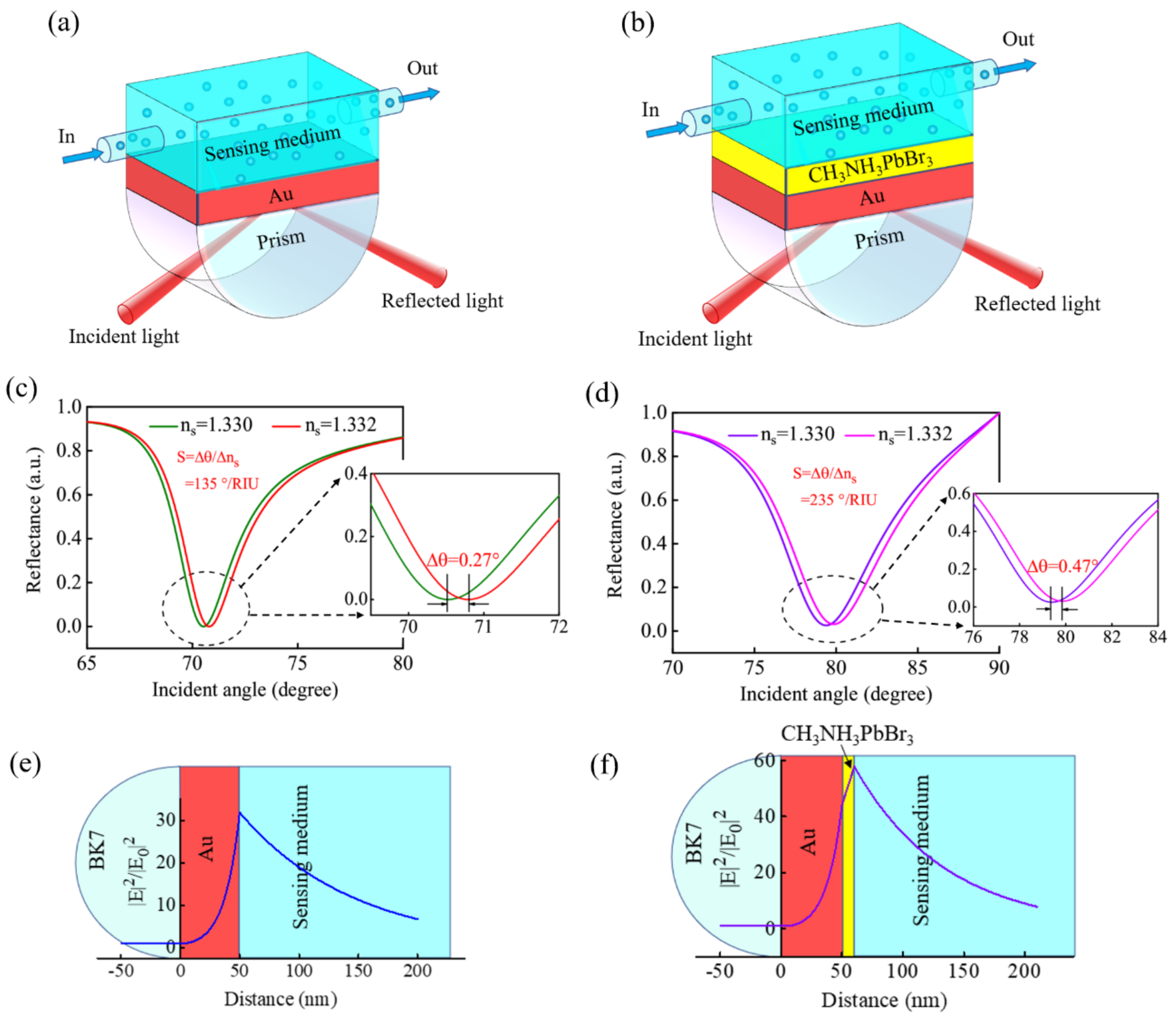
2. Design Consideration and Numerical Model

The proposed GWSPR biosensor is composed of five components (Figure 1), and these components are prism, Au film, waveguide layer, encapsulation layer and sensing layer. First, the BK7 glass, which has a low refractive index (nBK7), is used as the coupling prism in the proposed GWSPR biosensor. The nBK7 is defined as the relation [20,42]:
n B k 7 = 1.03961212 λ 2 λ 2 0.00600069867 + 0.231792344 λ 2 λ 2 0.0200179144 + 1.03961212 λ 2 λ 2 103.560653 + 1 1 / 2
where λ is the wavelength of incident light. In addition to BK7, other prisms such as SF10 and chalcogenide (2S2G: Ge20Ga5Sb10S65) have also been calculated and analyzed with the proposed GWSPR configuration. The nSF10 is defined as the following relation [43]:
n SF 10 = 1.62153902 λ 2 λ 2 0.0122241457 + 0.256287842 λ 2 λ 2 0.0595736775 + 1.64447552 λ 2 λ 2 147.468793 + 1 1 / 2
The n2S2G is defined as the relation [44]:
n 2 S 2 G = 2 . 24047 + 2.693 × 10 2 λ 2 + 8.08 × 10 3 λ 4
Second, the Au thin film with a thickness of 50 nm is used as the metal layer to excite the SPR signal, and the refractive index (nAu) is defined by the Drude–Lorentz model [20,45]:
n A u = 1 λ 2 λ C λ P 2 ( λ C + i λ ) 1 / 2
where λC = 8.9342 × 10⁻⁶ m and λP = 1.6826 × 10⁻⁷ m are the plasma and collision wavelengths for Au. Third, the guided-wave layer is CH3NH3PbBr3 perovskite, and its refractive index (nper) at λ = 633 nm is measured to be 2 + 0.003i [41]. Fourth, the cytop material, which has a low reflective index (nc = 1.34) close to water, is used as the encapsulation layer to protect the GWSPR biosensor, and its thickness is 2 nm. Finally, the sensing medium can be liquid analyte or gas analyte. For liquid analyte, the refractive index is defined as ns = 1.33 + Δns; for gas analyte, the refractive index is defined as ns = 1.00 + Δns, where Δns represents some biochemical reaction or concentration change. The change of the biosensing signal can be attributed to the change of the RI. In addition, biological signals can be reflected as changes in plasmon resonance signals, changes in fluorescence signals, and changes in interference signals. In this investigation, a GWSPR signal excited from the Au–perovskite hybrid structure is used to sense the change in the biological signal.
In the multilayer GWSPR structure, the transfer matrix method (TMM) [46] is used for numerical simulation, and the incident light is the He-Ne laser beam (λ = 633 nm) in TM-polarized mode. The characteristic transfer matrix (M) of the multilayer GWSPR structure is given as:
M = k = 2 N 1 M k = M 11 M 12 M 21 M 22 ,  
with
M k = = cos β k          i sin β k / q k i q k sin β k          cos β k
where
β k = 2 π d k λ ( ε k n 1 2 sin 2 θ ) 1 / 2 ,  
and
q k = ( ε k n 1 2 sin 2 θ ) 1 / 2 ε k ,  
where θ is the incident angle and n1 is the refractive index of prism (np). Then the total reflection coefficient rp is defined as:
r p = M 11 + M 12 q N q 1 M 21 + M 22 q N M 11 + M 12 q N q 1 + M 21 + M 22 q N .  
And the reflectance (Rp) is written as:
R = r p 2 .  
Finally, the sensitivity of the GWSPR biosensor is given by [4,47]:
S = Δ θ Δ n s .  
where Δθ is the variation of resonance angle caused by the Δns.

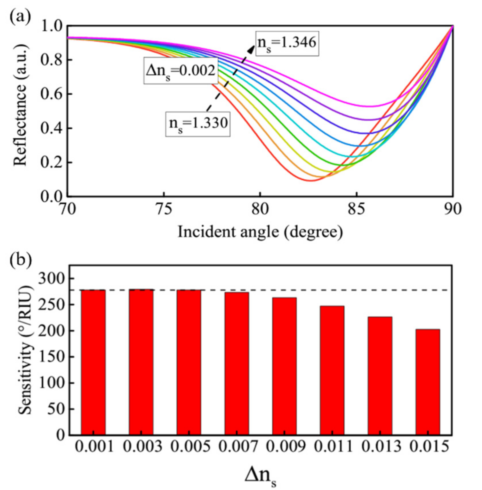
3. Results and Discussions

The conventional SPR biosensor is shown in Figure 2a, which uses a 50 nm thick Au thin film as the metal layer to excite the SPR. The reflectance curve of the conventional SPR biosensor is calculated by TMM, and it is found that the largest variation of the resonance angle caused by the Δns = 0.002 is Δθ = 0.27°, when the BK7 glass is used as the coupling prism (Figure 2c). Therefore, the sensitivity can be obtained to be S = 135°/RIU. To enhance the sensitivity, another CH3NH3PbBr3 perovskite guided-wave layer is coated on the Au surface to form an Au–perovskite hybrid structure (Figure 2b). When a 10 nm thick CH3NH3PbBr3 perovskite layer is coated on the Au surface, the Δθ will increase to 0.47°, and then the sensitivity is improved significantly to be 235°/RIU (Figure 2d). The enhancement mechanism is that the surface plasmon polaritons can spread along the guided-wave layer by adding a high refractive index perovskite layer. When the guided-wave layer is thin enough, it will result in a large part of the evanescent field being located in the analyte region. Therefore, the detection range is increased, and the sensitivity is improved [4,25]. The electric field distribution of the conventional SPR sensor and the GWSPR sensor are shown in Figure 2e,f for a comparison. The result demonstrates that the electric field at the sensing interface is greatly improved by the additional CH3NH3PbBr3 perovskite layer. Hence, the sensitivity is improved by the guided-wave layer. However, the CH3NH3PbBr3 perovskite is susceptible to pollution and damage, which will reduce the stability and sensitivity of the GWSPR biosensor. Therefore, we coated a cytop material (dc = 2 nm) as the encapsulation layer on the surface of the guided-wave layer (Figure 1). This encapsulation layer is made of low-refractive index materials with a refractive index close to that of the sensing medium, which can retain the high sensitivity of the GWSPR biosensor and play a protective role.
In the GWSPR biosensor, the thickness of the guided-wave layer is an important factor affecting the GWSPR signal and the sensitivity. When the thickness of the guided-wave layer changes from 8 nm to 15 nm, a significant change in the GWSPR signal can be observed (Figure 3a). Both the minimum R and the resonance angle are changed when there is an increase in dp (Figure 3b). When dp ≤ 12 nm, the minimum R (green line) maintains a small range of variation; however, when dp > 12 nm, the minimum R will change drastically. Similarly, the movement of the resonance angle (red line) is also greatly affected by the dp. With the increase in the dp, the resonance angle will move to a larger angle, and the largest resonance angle is 85.45° at dp = 14 nm. If the dp continues to increase (dp ≥ 14 nm), the movement of the resonance angle will be limited by the range of the incident angle (0°–90°). The relationship between the sensitivity and the dp is presented in Figure 3c. The sensitivity will firstly increase to 278.5°/RIU at dp = 12 nm, and then decrease rapidly due to the limitation of the angle range. After a calculation, the sensitivity enhancement is shown in Figure 3d, which demonstrates that the sensitivity of the proposed GWSPR biosensor is improved by 110.2%, compared with the conventional SPR sensor.
In the proposed prism-coupling technique-based GWSPR biosensor, the prism is a key component which can have a major impact on sensitivity. The above discussion is based on the BK7 prism. We will analyze and compare the influence of a variety of prisms with different refractive indices on sensitivity. Figure 4a–c shows the GWSPR signals obtained from the prisms of 2S2G, SF10, and BK7. The results demonstrate that the Δθ caused by Δns = 0.002 are 0.082°, 0.174°, and 0.557° for 2S2G, SF10, and BK7 prisms, respectively. The specific sensitivity change under different np is shown in Figure 4d, which indicates that a prism with a low refractive index can generate a higher sensitivity than that of a high refractive index. Therefore, the BK7 is used as the coupling prism in the new GWSPR biosensor.
The GWSPR signal will move with the change of ns, and its change process is similar to the effect of changing dp. As ns increases, the resonance signal will move toward a large angle and is limited by the range of incident angle (Figure 5a). However, in the biochemical reaction process, different reaction intensities will lead to changes in Δnsns is not a fixed value), which requires the GWSPR sensor to maintain a relatively stable sensitivity under different Δns to ensure the effectiveness of the detection process. Figure 5b shows the sensitivity changes corresponding to different values of Δns at dp = 12 nm, which indicates that the proposed GWSPR biosensor can maintain a relatively stable sensitivity (S ≈ 278.5°/RIU) when Δns < 0.005, and the corresponding detection range is (1.330, 1.335). In addition, if it is necessary to increase the detection range with stable sensitivity, it can be achieved by adjusting the thickness of dp. Figure 6a,b show the relationship curves of Δθ and Δns at dp = 10 nm and dp = 8 nm, respectively. The results demonstrate that the optimal detection ranges are (1.330, 1.3387) and (1.330, 1.3424) for dp = 10 nm and dp = 8 nm, respectively, and the sensitivity can keep in stable values of 243.7 °/RIU (dp = 10 nm) and 205.6 °/RIU (dp = 8 nm). The stable sensitivity is inversely proportional to the detection range. In practice, if the detection range needs to be increased, the sensitivity of the GWSPR sensor should be appropriately reduced by changing the thickness dp.
The proposed GWSPR configuration can also be developed as a gas sensor to detect changes in the concentration of harmful gases in the air, such as NO2 [48,49], NH3 [50,51], and CO [52,53]. The change of the concentration of harmful gases in the air will lead to the change of the air refractive index. Therefore, the harmful gas content in the air can be analyzed by detecting changes in the refractive index of the air. In the numerical simulation, the change of harmful gas in the air is simplified as the change of air refractive index. The refractive index of pure air is 1, which will change to 1 + Δns when mixed with harmful gases. Herein, the conventional SPR gas sensor is calculated to compare with the new Au-CH3NH3PbBr3-based GWSPR gas sensor. The SPR signal curves of the conventional SPR gas sensor at ns = 1 and 1.002 are shown in Figure 7a, and the sensitivity is calculated to be 58.5 °/RIU. Under the same Δns caused by the harmful gas, the sensitivity of the proposed GWSPR gas sensor at different thickness of dp is investigated in Figure 7b. The result shows that the highest sensitivity is 168.5°/RIU at dp = 41.5 nm, which is 2.8 times higher than the conventional SPR gas sensor. The detailed changes of GWSPR signal modulated by dp are shown in Figure 7c,d. Moreover, the electric field distributions of the conventional SPR gas sensor and the new Au-CH3NH3PbBr3-based GWSPR gas sensor are calculated for a comparison (Figure 7e,f). The result shows that a significant increase in the electric field at the sensing interface can be observed by coating the guided-wave layer, and then results in the enhancement of the sensitivity.

4. Conclusions

In this investigation, a new Au-CH3NH3PbBr3 hybrid structure-based GWSPR configuration is designed to be applied in RI biosensor and gas sensors. First, the CH3NH3PbBr3 thin film is used as a guided-wave layer to coat on the surface of the Au film in the GWSPR sensors, and the results demonstrate that the electric field at the sensing interface can be improved by the additional guided-wave layer. Hence, the sensitivity of the GWSPR biosensor is enhanced to be 278.5°/RIU, which is improved by 110.2% compared with the conventional SPR biosensor. Second, the selection of the prism in the GWSPR biosensor is discussed, and it is found that the prism with a low refractive index can possess a higher sensitivity. Therefore, a low refractive index glass of BK7 is used as a coupling prism in the GWSPR sensor. Third, the designed GWSPR configuration can also be applied in gas sensing, and the highest sensitivity is 168.5°/RIU.

Author Contributions

Conceptualization, Y.X. and Y.Q.; methodology, Y.Q. and L.W.; software, Y.X. and L.W.; validation, L.W., Y.X. and Y.Q.; investigation, L.W.; resources, Y.Q.; data curation, L.W.; writing—original draft preparation, L.W.; writing—review and editing, Y.X. and Y.Q.; supervision, Y.X.; project administration, Y.Q.; funding acquisition, Y.X. and Y.Q. All authors have read and agreed to the published version of the manuscript.

Funding

This research was funded by the National Key R&D Program of China, grant number 2018YFB1801001; the National Natural Science Foundation of China, grant number 62105069; the Postdoctoral Research Foundation of China, grant number 402210053; the Guangdong Introducing Innovative and Entrepreneurial Teams of “The Pearl River Talent Recruitment Program”, grant number 2019ZT08X340; the Research and Development Plan in Key Areas of Guangdong Province, grant number 2018B010114002; and the Guangdong Provincial Key Laboratory of Information Photonics Technology, grant number 2020B121201011.

Institutional Review Board Statement

Not applicable.

Informed Consent Statement

Not applicable.

Data Availability Statement

The data presented in this study are available on request from the corresponding author.

Conflicts of Interest

The authors declare no conflict of interest.

References

  1. Kretschmann, E.; Raether, H. Radiative decay of non-radiative surface plasmons excited by light. Z. Naturforsch. A 1968, 23, 2135–2136. [Google Scholar] [CrossRef]
  2. Zayats, A.V.; Smolyaninov, I.I.; Maradudin, A.A. Nano-optics of surface plasmon polaritons. Phys. Rep. 2005, 408, 131–314. [Google Scholar] [CrossRef]
  3. Pitarke, J.M.; Silkin, V.M.; Chulkov, E.V.; Echenique, P.M. Theory of surface plasmons and surface-plasmon polaritons. Rep. Prog. Phys. 2006, 70, 1–87. [Google Scholar] [CrossRef]
  4. Shalabney, A.; Abdulhalim, I. Sensitivity-enhancement methods for surface plasmon sensors. Laser Photonics Rev. 2011, 5, 571–606. [Google Scholar] [CrossRef]
  5. Yuk, J.S.; Hong, D.-G.; Jung, J.-W.; Jung, S.-H.; Kim, H.-S.; Han, J.-A.; Kim, Y.-M.; Ha, K.-S. Sensitivity enhancement of spectral surface plasmon resonance biosensors for the analysis of protein arrays. Eur. Biophys. J. 2006, 35, 469–476. [Google Scholar] [CrossRef]
  6. Xue, T.; Cui, X.; Guan, W.; Wang, Q.; Liu, C.; Wang, H.; Qi, K.; Singh, D.J.; Zheng, W. Surface plasmon resonance technique for directly probing the interaction of DNA and graphene oxide and ultra-sensitive biosensing. Biosens. Bioelectron. 2014, 58, 374–379. [Google Scholar] [CrossRef]
  7. Nanduri, V.; Bhunia, A.K.; Tu, S.-I.; Paoli, G.C.; Brewster, J.D. SPR biosensor for the detection of L. monocytogenes using phage-displayed antibody. Biosens. Bioelectron. 2007, 23, 248–252. [Google Scholar] [CrossRef]
  8. Kim, J.A.; Hwang, T.; Dugasani, S.R.; Amin, R.; Kulkarni, A.; Park, S.H.; Kim, T. Graphene based fiber optic surface plasmon resonance for bio-chemical sensor applications. Sens. Actuators B Chem. 2013, 187, 426–433. [Google Scholar] [CrossRef] [Green Version]
  9. Zhao, Y.; Deng, Z.; Li, J. Photonic crystal fiber based surface plasmon resonance chemical sensors. Sens. Actuators B Chem. 2014, 202, 557–567. [Google Scholar] [CrossRef]
  10. Homola, J. Surface Plasmon Resonance Sensors for Detection of Chemical and Biological Species. Chem. Rev. 2008, 108, 462–493. [Google Scholar] [CrossRef]
  11. Sai, V.V.R.; Kundu, T.; Mukherji, S. Novel U-bent fiber optic probe for localized surface plasmon resonance based biosensor. Biosens. Bioelectron. 2009, 24, 2804–2809. [Google Scholar] [CrossRef] [PubMed]
  12. Cao, J.; Tu, M.H.; Sun, T.; Grattan, K.T.V. Wavelength-based localized surface plasmon resonance optical fiber biosensor. Sens. Actuators B Chem. 2013, 181, 611–619. [Google Scholar] [CrossRef]
  13. Song, H.; Wang, Q.; Zhao, W.-M. A novel SPR sensor sensitivity-enhancing method for immunoassay by inserting MoS2 nanosheets between metal film and fiber. Opt. Laser Eng. 2020, 132, 106135. [Google Scholar] [CrossRef]
  14. Xue, T.; Liang, W.; Li, Y.; Sun, Y.; Xiang, Y.; Zhang, Y.; Dai, Z.; Duo, Y.; Wu, L.; Qi, K.; et al. Ultrasensitive detection of miRNA with an antimonene-based surface plasmon resonance sensor. Nat. Commun. 2019, 10, 28. [Google Scholar] [CrossRef] [PubMed]
  15. Huang, X.; Zeng, Z.; Fan, Z.; Liu, J.; Zhang, H. Graphene-Based Electrodes. Adv. Mater. 2012, 24, 5979–6004. [Google Scholar] [CrossRef]
  16. Xue, T.; Cui, X.; Chen, J.; Liu, C.; Wang, Q.; Wang, H.; Zheng, W. A Switch of the Oxidation State of Graphene Oxide on a Surface Plasmon Resonance Chip. ACS Appl. Mater. Interfaces 2013, 5, 2096–2103. [Google Scholar] [CrossRef]
  17. Xue, T.; Qi, K.; Hu, C. Novel SPR sensing platform based on superstructure MoS2 nanosheets for ultrasensitive detection of mercury ion. Sens. Actuators B Chem. 2019, 284, 589–594. [Google Scholar] [CrossRef]
  18. Yuting, Z.; Shuaiwen, G.; Leiming, W.; Jiaqi, Z.; Yuanjiang, X.; Xiaoyu, D. GeSe nanosheets modified surface plasmon resonance sensors for enhancing sensitivity. Nanophotonics 2020, 9, 327–336. [Google Scholar]
  19. Jiaqi, Z.; Yuxuan, K.; Jianfeng, D.; Qi, Y.; Leiming, W.; Jianqing, L.; Jun, G.; Yuanjiang, X.; Xiaoyu, D. Topological insulator overlayer to enhance the sensitivity and detection limit of surface plasmon resonance sensor. Nanophotonics 2020, 9, 1941–1951. [Google Scholar]
  20. Wu, L.; You, Q.; Shan, Y.; Gan, S.; Zhao, Y.; Dai, X.; Xiang, Y. Few-layer Ti3C2Tx MXene: A promising surface plasmon resonance biosensing material to enhance the sensitivity. Sens. Actuators B Chem. 2018, 277, 210–215. [Google Scholar] [CrossRef]
  21. Sarid, D. Long-Range Surface-Plasma Waves on Very Thin Metal Films. Phys. Rev. Lett. 1981, 47, 1927–1930. [Google Scholar] [CrossRef]
  22. Wu, L.; Guo, J.; Xu, H.; Dai, X.; Xiang, Y. Ultrasensitive biosensors based on long-range surface plasmon polariton and dielectric waveguide modes. Photonics Res. 2016, 4, 262–266. [Google Scholar] [CrossRef]
  23. Jing, J.-Y.; Wang, Q.; Zhao, W.-M.; Wang, B.-T. Long-range surface plasmon resonance and its sensing applications: A review. Opt. Laser Eng. 2019, 112, 103–118. [Google Scholar] [CrossRef]
  24. Shalabney, A.; Abdulhalim, I. Figure-of-merit enhancement of surface plasmon resonance sensors in the spectral interrogation. Opt. Lett. 2012, 37, 1175–1177. [Google Scholar] [CrossRef] [PubMed]
  25. Lahav, A.; Auslender, M.; Abdulhalim, I. Sensitivity enhancement of guided-wave surface-plasmon resonance sensors. Opt. Lett. 2008, 33, 2539–2541. [Google Scholar] [CrossRef] [PubMed]
  26. Wu, L.; Chen, K.; Huang, W.; Lin, Z.; Zhao, J.; Jiang, X.; Ge, Y.; Zhang, F.; Xiao, Q.; Guo, Z.; et al. Perovskite CsPbX3: A Promising Nonlinear Optical Material and Its Applications for Ambient All-Optical Switching with Enhanced Stability. Adv. Opt. Mater. 2018, 6, 1800400. [Google Scholar] [CrossRef]
  27. Jeon, N.J.; Noh, J.H.; Kim, Y.C.; Yang, W.S.; Ryu, S.; Seok, S.I. Solvent engineering for high-performance inorganic–organic hybrid perovskite solar cells. Nat. Mater. 2014, 13, 897–903. [Google Scholar] [CrossRef]
  28. Hao, F.; Stoumpos, C.C.; Cao, D.H.; Chang, R.P.H.; Kanatzidis, M.G. Lead-free solid-state organic–inorganic halide perovskite solar cells. Nat. Photonics 2014, 8, 489–494. [Google Scholar] [CrossRef]
  29. Heo, J.H.; Im, S.H.; Noh, J.H.; Mandal, T.N.; Lim, C.-S.; Chang, J.A.; Lee, Y.H.; Kim, H.-j.; Sarkar, A.; Nazeeruddin, M.K.; et al. Efficient inorganic–organic hybrid heterojunction solar cells containing perovskite compound and polymeric hole conductors. Nat. Photonics 2013, 7, 486–491. [Google Scholar] [CrossRef]
  30. Tian, W.; Zhou, H.; Li, L. Hybrid Organic–Inorganic Perovskite Photodetectors. Small 2017, 13, 1702107. [Google Scholar] [CrossRef]
  31. Ahmadi, M.; Wu, T.; Hu, B. A review on organic–inorganic halide perovskite photodetectors: Device engineering and fundamental physics. Adv. Mater. 2017, 29, 1605242. [Google Scholar] [CrossRef]
  32. Kim, Y.-H.; Cho, H.; Heo, J.H.; Kim, T.-S.; Myoung, N.; Lee, C.-L.; Im, S.H.; Lee, T.-W. Multicolored Organic/Inorganic Hybrid Perovskite Light-Emitting Diodes. Adv. Mater. 2015, 27, 1248–1254. [Google Scholar] [CrossRef] [PubMed]
  33. Zhang, X.; Liu, H.; Wang, W.; Zhang, J.; Xu, B.; Karen, K.L.; Zheng, Y.; Liu, S.; Chen, S.; Wang, K. Hybrid Perovskite Light-Emitting Diodes Based on Perovskite Nanocrystals with Organic–Inorganic Mixed Cations. Adv. Mater. 2017, 29, 1606405. [Google Scholar] [CrossRef] [PubMed]
  34. Cho, H.; Jeong, S.-H.; Park, M.-H.; Kim, Y.-H.; Wolf, C.; Lee, C.-L.; Heo, J.H.; Sadhanala, A.; Myoung, N.; Yoo, S.; et al. Overcoming the electroluminescence efficiency limitations of perovskite light-emitting diodes. Science 2015, 350, 1222. [Google Scholar] [CrossRef]
  35. Grabowska, E. Selected perovskite oxides: Characterization, preparation and photocatalytic properties—A review. Appl. Catal. B Environ. 2016, 186, 97–126. [Google Scholar] [CrossRef]
  36. Zhang, G.; Liu, G.; Wang, L.; Irvine, J.T.S. Inorganic perovskite photocatalysts for solar energy utilization. Chem. Soc. Rev. 2016, 45, 5951–5984. [Google Scholar] [CrossRef] [PubMed] [Green Version]
  37. Veldhuis, S.A.; Boix, P.P.; Yantara, N.; Li, M.; Sum, T.C.; Mathews, N.; Mhaisalkar, S.G. Perovskite Materials for Light-Emitting Diodes and Lasers. Adv. Mater. 2016, 28, 6804–6834. [Google Scholar] [CrossRef]
  38. Zhu, H.; Fu, Y.; Meng, F.; Wu, X.; Gong, Z.; Ding, Q.; Gustafsson, M.V.; Trinh, M.T.; Jin, S.; Zhu, X.Y. Lead halide perovskite nanowire lasers with low lasing thresholds and high quality factors. Nat. Mater. 2015, 14, 636–642. [Google Scholar] [CrossRef]
  39. Huang, H.; Susha, A.S.; Kershaw, S.V.; Hung, T.F.; Rogach, A.L. Control of Emission Color of High Quantum Yield CH3NH3PbBr3 Perovskite Quantum Dots by Precipitation Temperature. Adv. Sci. 2015, 2, 1500194. [Google Scholar] [CrossRef]
  40. Heo, J.H.; Song, D.H.; Im, S.H. Planar CH3NH3PbBr3 Hybrid Solar Cells with 10.4% Power Conversion Efficiency, Fabricated by Controlled Crystallization in the Spin-Coating Process. Adv. Mater. 2014, 26, 8179–8183. [Google Scholar] [CrossRef]
  41. Brittman, S.; Garnett, E.C. Measuring n and k at the Microscale in Single Crystals of CH3NH3PbBr3 Perovskite. J. Phys. Chem. C 2016, 120, 616–620. [Google Scholar] [CrossRef]
  42. Zeng, S.; Hu, S.; Xia, J.; Anderson, T.; Dinh, X.-Q.; Meng, X.-M.; Coquet, P.; Yong, K.-T. Graphene–MoS2 hybrid nanostructures enhanced surface plasmon resonance biosensors. Sens. Actuators B Chem. 2015, 207, 801–810. [Google Scholar] [CrossRef]
  43. Choi, S.H.; Kim, Y.L.; Byun, K.M. Graphene-on-silver substrates for sensitive surface plasmon resonance imaging biosensors. Opt. Express 2011, 19, 458–466. [Google Scholar] [CrossRef]
  44. Maharana, P.K.; Jha, R. Chalcogenide prism and graphene multilayer based surface plasmon resonance affinity biosensor for high performance. Sens. Actuators B Chem. 2012, 169, 161–166. [Google Scholar] [CrossRef]
  45. Gupta, B.D.; Sharma, A.K. Sensitivity evaluation of a multi-layered surface plasmon resonance-based fiber optic sensor: A theoretical study. Sens. Actuators B Chem. 2005, 107, 40–46. [Google Scholar] [CrossRef]
  46. Hansen, W.N. Electric Fields Produced by the Propagation of Plane Coherent Electromagnetic Radiation in a Stratified Medium. J. Opt. Soc. Am. 1968, 58, 380–390. [Google Scholar] [CrossRef]
  47. Wu, L.; Guo, J.; Wang, Q.; Lu, S.; Dai, X.; Xiang, Y.; Fan, D. Sensitivity enhancement by using few-layer black phosphorus-graphene/TMDCs heterostructure in surface plasmon resonance biochemical sensor. Sens. Actuators B Chem. 2017, 249, 542–548. [Google Scholar] [CrossRef]
  48. Paliwal, A.; Sharma, A.; Tomar, M.; Gupta, V. Room temperature detection of NO2 gas using optical sensor based on surface plasmon resonance technique. Sens. Actuators B Chem. 2015, 216, 497–503. [Google Scholar] [CrossRef]
  49. Jain, S.; Paliwal, A.; Gupta, V.; Tomar, M. Long Range Surface Plasmons assisted highly sensitive and room temperature operated NO2 gas sensor. Sens. Actuators B Chem. 2020, 311, 127897. [Google Scholar] [CrossRef]
  50. Wang, J.; Fan, S.; Xia, Y.; Yang, C.; Komarneni, S. Room-temperature gas sensors based on ZnO nanorod/Au hybrids: Visible-light-modulated dual selectivity to NO2 and NH3. J. Hazard. Mater. 2020, 381, 120919. [Google Scholar] [CrossRef] [PubMed]
  51. Maciak, E.; Pustelny, T. An optical ammonia (NH3) gas sensing by means of Pd/CuPc interferometric nanostructures based on white light interferometry. Sens. Actuators B Chem. 2013, 189, 230–239. [Google Scholar] [CrossRef]
  52. Paliwal, A.; Sharma, A.; Tomar, M.; Gupta, V. Carbon monoxide (CO) optical gas sensor based on ZnO thin films. Sens. Actuators B Chem. 2017, 250, 679–685. [Google Scholar] [CrossRef]
  53. Buso, D.; Post, M.; Cantalini, C.; Mulvaney, P.; Martucci, A. Gold Nanoparticle-Doped TiO2 Semiconductor Thin Films: Gas Sensing Properties. Adv. Funct. Mater. 2008, 18, 3843–3849. [Google Scholar] [CrossRef] [Green Version]
Figure 1. Schematic diagram of the guided-wave surface plasmon resonance (GWSPR) biosensor based on the Au–perovskite hybrid structure.
Biosensors 11 00415 g001
Figure 2. (a) Schematic diagram of the conventional Au-based SPR biosensor. (b) CH3NH3PbBr3 perovskite as a guided-wave layer to coat on the surface of Au-based SPR biosensor. (c) The sensitivity of the conventional Au-based SPR Biosensor at λ = 633 nm. (d) The sensitivity enhanced by a 10 nm thick CH3NH3PbBr3 perovskite thin film in Au-based SPR biosensor. (e) The electric field distribution of the conventional Au-based SPR biosensor. (f) The SPR sensor using the guided-wave layer of CH3NH3PbBr3 perovskite thin film to enhance the electric field at the sensing interface.
Biosensors 11 00415 g002
Figure 3. (a) The variation of the resonance signal when the thickness of the guided-wave layer changes from 8 nm to 15 nm. (b) The variation of minimum R and the resonance angle under different thicknesses of CH3NH3PbBr3 perovskite thin film (dp). (c) The sensitivity of the proposed GWSPR biosensor with different thicknesses of guided-wave layer to obtain the optimal thickness of CH3NH3PbBr3 perovskite thin film. (d) Sensitivity enhancement of the proposed GWSPR sensor compared with the conventional SPR sensor. The result demonstrates that the highest sensitivity is 278.5 °/RIU at dp = 12 nm, which is improved by 110.2% compared with the conventional SPR sensor.
Biosensors 11 00415 g003
Figure 4. (ac) The GWSPR signals excited from the prisms of 2S2G, SF10, and BK7 when Δns = 0.002. The sensitivity is calculated to be 41°/RIU, 87°/RIU, and 278.5°/RIU for the 2S2G, SF10, and BK7 prisms, respectively. (d) The specific change of sensitivity with respect to the np.
Biosensors 11 00415 g004
Figure 5. (a) The resonance signal obtained from the GWSPR biosensor at dp = 12 nm when ns ranges from 1.330 to 1.346. (b) The sensitivity obtained from different Δns. When Δns ≤ 0.005, the sensitivity can maintain a stable value.
Biosensors 11 00415 g005
Figure 6. Variation of the Δθ with respect to the different Δns at (a) dp = 10 nm and (b) dp = 8 nm. The results demonstrate that the optimal detection ranges are (1.330, 1.3387) and (1.330, 1.3424) at dp = 10 nm and dp = 8 nm, respectively. In these two ranges the sensitivity can keep in stable values of 243.7°/RIU (dp = 10 nm) and 205.6°/RIU (dp = 8 nm).
Biosensors 11 00415 g006
Figure 7. (a) SPR signal excited from the conventional SPR gas sensor, and the sensitivity is calculated to be 58.5°/RIU. (b) The sensitivity curve corresponding to different thicknesses of dp for the proposed GWSPR gas sensor, and the highest sensitivity (168.5°/RIU) is obtained at dp = 41.5 nm. (c) The movement of resonance angle corresponding to different thickness of dp. (d) The resonance signals excited from the proposed GWSPR gas sensor at different dp. (e,f) The electric field distributions of the conventional SPR gas sensor and the new Au-CH3NH3PbBr3-based GWSPR gas sensor.
Biosensors 11 00415 g007
Publisher’s Note: MDPI stays neutral with regard to jurisdictional claims in published maps and institutional affiliations.

