Next Issue
Volume 11, December
Previous Issue
Volume 11, October


Biosensors, Volume 11, Issue 11 (November 2021) – 65 articles

Cover Story (view full-size image): 
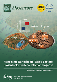
Bacterial infections in fish farms increase mass mortality and rapid detection of infection can help prevent its widespread. Lactate is an important biomarker for early diagnosis of bacterial infections in farmed olive flounder (Paralichthys olivaceus). To determine the lactate levels, we designed a disposable amperometric biosensor based on nanozyme and lactate oxidase (LOX) entrapped in copolymer-reduced graphene oxide (P-rGO) on screen-printed carbon electrodes. Because LOX is inherently unstable, P-rGO nanosheets were utilized as a base matrix to immobilize it and the fabricated biosensor was tested in olive flounder infected by Streptococcus parauberis against the uninfected control. Our results were validated using a standard colorimetric assay kit. View this paper
  • Issues are regarded as officially published after their release is announced to the table of contents alert mailing list.
  • You may sign up for e-mail alerts to receive table of contents of newly released issues.
  • PDF is the official format for papers published in both, html and pdf forms. To view the papers in pdf format, click on the "PDF Full-text" link, and use the free Adobe Reader to open them.
Order results
Result details
Section
Select all
Export citation of selected articles as:
29 pages, 8845 KiB  
Review
Recent Advances in Electrical Impedance Sensing Technology for Single-Cell Analysis
by Zhao Zhang, Xiaowen Huang, Ke Liu, Tiancong Lan, Zixin Wang and Zhen Zhu
Biosensors 2021, 11(11), 470; https://doi.org/10.3390/bios11110470 - 22 Nov 2021
Cited by 22 | Viewed by 5416
Abstract
Cellular heterogeneity is of significance in cell-based assays for life science, biomedicine and clinical diagnostics. Electrical impedance sensing technology has become a powerful tool, allowing for rapid, non-invasive, and label-free acquisition of electrical parameters of single cells. These electrical parameters, i.e., equivalent cell [...] Read more.
Cellular heterogeneity is of significance in cell-based assays for life science, biomedicine and clinical diagnostics. Electrical impedance sensing technology has become a powerful tool, allowing for rapid, non-invasive, and label-free acquisition of electrical parameters of single cells. These electrical parameters, i.e., equivalent cell resistance, membrane capacitance and cytoplasm conductivity, are closely related to cellular biophysical properties and dynamic activities, such as size, morphology, membrane intactness, growth state, and proliferation. This review summarizes basic principles, analytical models and design concepts of single-cell impedance sensing devices, including impedance flow cytometry (IFC) to detect flow-through single cells and electrical impedance spectroscopy (EIS) to monitor immobilized single cells. Then, recent advances of both electrical impedance sensing systems applied in cell recognition, cell counting, viability detection, phenotypic assay, cell screening, and other cell detection are presented. Finally, prospects of impedance sensing technology in single-cell analysis are discussed. Full article
(This article belongs to the Special Issue Microfluidics for Biomedical Applications)
Show Figures

Figure 1

14 pages, 3083 KiB  
Article
Accuracy of the Electronic Nose Breath Tests in Clinical Application: A Systematic Review and Meta-Analysis
by Hsiao-Yu Yang, Wan-Chin Chen and Rodger-Chen Tsai
Biosensors 2021, 11(11), 469; https://doi.org/10.3390/bios11110469 - 22 Nov 2021
Cited by 8 | Viewed by 2699
Abstract
(1) Background: An electronic nose applies a sensor array to detect volatile biomarkers in exhaled breath to diagnose diseases. The overall diagnostic accuracy remains unknown. The objective of this review was to provide an estimate of the diagnostic accuracy of sensor-based breath tests [...] Read more.
(1) Background: An electronic nose applies a sensor array to detect volatile biomarkers in exhaled breath to diagnose diseases. The overall diagnostic accuracy remains unknown. The objective of this review was to provide an estimate of the diagnostic accuracy of sensor-based breath tests for the diagnosis of diseases. (2) Methods: We searched the PubMed and Web of Science databases for studies published between 1 January 2010 and 14 October 2021. The search was limited to human studies published in the English language. Clinical trials were not included in this review. (3) Results: Of the 2418 records identified, 44 publications were eligible, and 5728 patients were included in the final analyses. The pooled sensitivity was 90.0% (95% CI, 86.3–92.8%, I2 = 47.7%), the specificity was 88.4% (95% CI, 87.1–89.5%, I2 = 81.4%), and the pooled area under the curve was 0.93 (95% CI 0.91–0.95). (4) Conclusion: The findings of our review suggest that a standardized report of diagnostic accuracy and a report of the accuracy in a test set are needed. Sensor array systems of electronic noses have the potential for noninvasiveness at the point-of-care in hospitals. Nevertheless, the procedure for reporting the accuracy of a diagnostic test must be standardized. Full article
(This article belongs to the Collection Novel Sensing System for Biomedical Applications)
Show Figures

Figure 1

12 pages, 4452 KiB  
Article
Hot-Band-Absorption-Induced Anti-Stokes Fluorescence of Aggregation-Induced Emission Dots and the Influence on the Nonlinear Optical Effect
by Yuhuang Zhang, Jing Zhou, Shiyi Peng, Wenbin Yu, Xiaoxiao Fan, Wen Liu, Zikang Ye, Ji Qi, Zhe Feng and Jun Qian
Biosensors 2021, 11(11), 468; https://doi.org/10.3390/bios11110468 - 22 Nov 2021
Cited by 4 | Viewed by 2413
Abstract
Hot-band absorption (HBA)-induced anti-Stokes fluorescence (ASF) with longer-wavelength excitation is one effective pathway to deep penetration and low autofluorescence in intravital fluorescence imaging, raising demands for fluorophores with broad spectra, high absorption, and strong emission. However, typical fluorescent dyes display some emission quenching [...] Read more.
Hot-band absorption (HBA)-induced anti-Stokes fluorescence (ASF) with longer-wavelength excitation is one effective pathway to deep penetration and low autofluorescence in intravital fluorescence imaging, raising demands for fluorophores with broad spectra, high absorption, and strong emission. However, typical fluorescent dyes display some emission quenching when their concentration is increased in order to obtain brighter fluorescence. In this work, the HBA-induced ASF of aggregation-induced emission (AIE) dots is reported. BPN-BBTD dots were synthesized and confirmed with a fluorescence enhancement and a considerable ASF intensity. In addition, the mechanism of ASF and the HBA process of BPN-BBTD dots were carefully validated and discussed. To obtain the full advantages of the long-wavelength excitation and the short fluorescence lifetime in deep-tissue bioimaging, a large-depth ASF confocal microscopic imaging of in vivo cerebral vasculature was conducted under the excitation of a 980 nm continuous wave laser after intravenous injection of BPN-BBTD dots. Meanwhile, the 3D structure of the cerebrovascular network was successfully reconstructed. Full article
Show Figures

Figure 1

12 pages, 2618 KiB  
Article
Label-free Surface Enhanced Raman Scattering (SERS) on Centrifugal Silver Plasmonic Paper (CSPP): A Novel Methodology for Unprocessed Biofluids Sampling and Analysis
by Alessandro Esposito, Alois Bonifacio, Valter Sergo and Stefano Fornasaro
Biosensors 2021, 11(11), 467; https://doi.org/10.3390/bios11110467 - 21 Nov 2021
Cited by 6 | Viewed by 2316
Abstract
Label-free SERS is a powerful bio-analytical technique in which molecular fingerprinting is combined with localized surface plasmons (LSPs) on metal surfaces to achieve high sensitivity. Silver and gold colloids are among the most common nanostructured substrates used in SERS, but since protein-rich samples [...] Read more.
Label-free SERS is a powerful bio-analytical technique in which molecular fingerprinting is combined with localized surface plasmons (LSPs) on metal surfaces to achieve high sensitivity. Silver and gold colloids are among the most common nanostructured substrates used in SERS, but since protein-rich samples such as serum or plasma can hinder the SERS effect due to protein–substrate interactions, they often require a deproteinization step. Moreover, SERS methods based on metal colloids often suffer from a poor reproducibility. Here, we propose a paper-based SERS sampling method in which unprocessed human serum samples are first soaked on paper strips (0.4 × 2 cm2), and then mixed with colloidal silver nanoparticles by centrifugation to obtain a Centrifugal Silver Plasmonic Paper (CSPP). The CSPP methodology has the potential to become a promising tool in bioanalytical SERS applications: it uses common colloidal substrates but without the need for sample deproteinization, while having a good reproducibility both in terms of overall spectral shape (r > 0.96) and absolute intensity (RSD < 10%). Moreover, this methodology allows SERS analysis more than one month after serum collection on the paper strip, facilitating storage and handling of clinical samples (including shipping from clinical sites to labs). Full article
(This article belongs to the Special Issue Biosensors for Body Fluid Analysis)
Show Figures

Figure 1

15 pages, 2715 KiB  
Article
Reagentless D-Tagatose Biosensors Based on the Oriented Immobilization of Fructose Dehydrogenase onto Coated Gold Nanoparticles- or Reduced Graphene Oxide-Modified Surfaces: Application in a Prototype Bioreactor
by Ieva Šakinytė, Marius Butkevičius, Vidutė Gurevičienė, Jonita Stankevičiūtė, Rolandas Meškys and Julija Razumienė
Biosensors 2021, 11(11), 466; https://doi.org/10.3390/bios11110466 - 19 Nov 2021
Cited by 2 | Viewed by 1844
Abstract
As electrode nanomaterials, thermally reduced graphene oxide (TRGO) and modified gold nanoparticles (AuNPs) were used to design bioelectrocatalytic systems for reliable D-tagatose monitoring in a long-acting bioreactor where the valuable sweetener D-tagatose was enzymatically produced from a dairy by-product D-galactose. For this goal [...] Read more.
As electrode nanomaterials, thermally reduced graphene oxide (TRGO) and modified gold nanoparticles (AuNPs) were used to design bioelectrocatalytic systems for reliable D-tagatose monitoring in a long-acting bioreactor where the valuable sweetener D-tagatose was enzymatically produced from a dairy by-product D-galactose. For this goal D-fructose dehydrogenase (FDH) from Gluconobacter industrius immobilized on these electrode nanomaterials by forming three amperometric biosensors: AuNPs coated with 4-mercaptobenzoic acid (AuNP/4-MBA/FDH) or AuNPs coated with 4-aminothiophenol (AuNP/PATP/FDH) monolayer, and a layer of TRGO on graphite (TRGO/FDH) were created. The immobilized FDH due to changes in conformation and spatial orientation onto proposed electrode surfaces catalyzes a direct D-tagatose oxidation reaction. The highest sensitivity for D-tagatose of 0.03 ± 0.002 μA mM−1cm−2 was achieved using TRGO/FDH. The TRGO/FDH was applied in a prototype bioreactor for the quantitative evaluation of bioconversion of D-galactose into D-tagatose by L-arabinose isomerase. The correlation coefficient between two independent analyses of the bioconversion mixture: spectrophotometric and by the biosensor was 0.9974. The investigation of selectivity showed that the biosensor was not active towards D-galactose as a substrate. Operational stability of the biosensor indicated that detection of D-tagatose could be performed during six hours without loss of sensitivity. Full article
(This article belongs to the Special Issue Electrochemistry and Spectroscopy-Based Biosensors)
Show Figures

Figure 1

10 pages, 7018 KiB  
Communication
ZnO Tips Dotted with Au Nanoparticles—Advanced SERS Determination of Trace Nicotine
by Jiaying Cao, Yan Zhai, Wanxin Tang, Xiaoyu Guo, Ying Wen and Haifeng Yang
Biosensors 2021, 11(11), 465; https://doi.org/10.3390/bios11110465 - 19 Nov 2021
Cited by 5 | Viewed by 2152
Abstract
Long-term exposure to nicotine causes a variety of human diseases, such as lung damage/adenocarcinoma, nausea and vomiting, headache, incontinence and heart failure. In this work, as a surface-enhanced Raman scattering (SERS) substrate, zinc oxide (ZnO) tips decorated with gold nanoparticles (AuNPs) are fabricated [...] Read more.
Long-term exposure to nicotine causes a variety of human diseases, such as lung damage/adenocarcinoma, nausea and vomiting, headache, incontinence and heart failure. In this work, as a surface-enhanced Raman scattering (SERS) substrate, zinc oxide (ZnO) tips decorated with gold nanoparticles (AuNPs) are fabricated and designated as ZnO/Au. Taking advantage of the synergistic effect of a ZnO semiconductor with morphology of tips and AuNPs, the ZnO/Au-based SERS assay for nicotine demonstrates high sensitivity and the limit of detection 8.9 × 10⁻¹² mol/L is reached, as well as the corresponding linear dynamic detection range of 10−10–10−6 mol/L. Additionally, the signal reproducibility offered by the SERS substrate could realize the reliable determination of trace nicotine in saliva. Full article
(This article belongs to the Special Issue Surface Enhanced Raman Spectroscopy Based Sensors and Biosensors)
Show Figures

Figure 1

29 pages, 5304 KiB  
Review
Progress of Microfluidic Continuous Separation Techniques for Micro-/Nanoscale Bioparticles
by Se-woon Choe, Bumjoo Kim and Minseok Kim
Biosensors 2021, 11(11), 464; https://doi.org/10.3390/bios11110464 - 18 Nov 2021
Cited by 13 | Viewed by 6485
Abstract
Separation of micro- and nano-sized biological particles, such as cells, proteins, and nucleotides, is at the heart of most biochemical sensing/analysis, including in vitro biosensing, diagnostics, drug development, proteomics, and genomics. However, most of the conventional particle separation techniques are based on membrane [...] Read more.
Separation of micro- and nano-sized biological particles, such as cells, proteins, and nucleotides, is at the heart of most biochemical sensing/analysis, including in vitro biosensing, diagnostics, drug development, proteomics, and genomics. However, most of the conventional particle separation techniques are based on membrane filtration techniques, whose efficiency is limited by membrane characteristics, such as pore size, porosity, surface charge density, or biocompatibility, which results in a reduction in the separation efficiency of bioparticles of various sizes and types. In addition, since other conventional separation methods, such as centrifugation, chromatography, and precipitation, are difficult to perform in a continuous manner, requiring multiple preparation steps with a relatively large minimum sample volume is necessary for stable bioprocessing. Recently, microfluidic engineering enables more efficient separation in a continuous flow with rapid processing of small volumes of rare biological samples, such as DNA, proteins, viruses, exosomes, and even cells. In this paper, we present a comprehensive review of the recent advances in microfluidic separation of micro-/nano-sized bioparticles by summarizing the physical principles behind the separation system and practical examples of biomedical applications. Full article
(This article belongs to the Special Issue Advances in Microfluidic Biosensing)
Show Figures

Figure 1

13 pages, 3040 KiB  
Communication
Superior Non-Invasive Glucose Sensor Using Bimetallic CuNi Nanospecies Coated Mesoporous Carbon
by Ahmed Bahgat Radwan, Sreedevi Paramparambath, John-John Cabibihan, Abdulaziz Khalid Al-Ali, Peter Kasak, Rana A. Shakoor, Rayaz A. Malik, Said A. Mansour and Kishor Kumar Sadasivuni
Biosensors 2021, 11(11), 463; https://doi.org/10.3390/bios11110463 - 18 Nov 2021
Cited by 9 | Viewed by 2899
Abstract
The assessment of blood glucose levels is necessary for the diagnosis and management of diabetes. The accurate quantification of serum or plasma glucose relies on enzymatic and nonenzymatic methods utilizing electrochemical biosensors. Current research efforts are focused on enhancing the non-invasive detection of [...] Read more.
The assessment of blood glucose levels is necessary for the diagnosis and management of diabetes. The accurate quantification of serum or plasma glucose relies on enzymatic and nonenzymatic methods utilizing electrochemical biosensors. Current research efforts are focused on enhancing the non-invasive detection of glucose in sweat with accuracy, high sensitivity, and stability. In this work, nanostructured mesoporous carbon coupled with glucose oxidase (GOx) increased the direct electron transfer to the electrode surface. A mixed alloy of CuNi nanoparticle-coated mesoporous carbon (CuNi-MC) was synthesized using a hydrothermal process followed by annealing at 700 °C under the flow of argon gas. The prepared catalyst’s crystal structure and morphology were explored using X-ray diffraction and high-resolution transmission electron microscopy. The electrocatalytic activity of the as-prepared catalyst was investigated using cyclic voltammetry (CV) and amperometry. The findings show an excellent response time of 4 s and linear range detection from 0.005 to 0.45 mM with a high electrode sensitivity of 11.7 ± 0.061 mA mM cm−2 in a selective medium. Full article
(This article belongs to the Special Issue Advances in Nanoporous Materials for Biosensing Applications)
Show Figures

Figure 1

15 pages, 5413 KiB  
Article
A Double-Deck Structure of Reduced Graphene Oxide Modified Porous Ti3C2Tx Electrode towards Ultrasensitive and Simultaneous Detection of Dopamine and Uric Acid
by Yangguang Zhu, Qichen Tian, Xiufen Li, Lidong Wu, Aimin Yu, Guosong Lai, Li Fu, Qiuping Wei, Dan Dai, Nan Jiang, He Li, Chen Ye and Cheng-Te Lin
Biosensors 2021, 11(11), 462; https://doi.org/10.3390/bios11110462 - 18 Nov 2021
Cited by 16 | Viewed by 2673
Abstract
Considering the vital physiological functions of dopamine (DA) and uric acid (UA) and their coexistence in the biological matrix, the development of biosensing techniques for their simultaneous and sensitive detection is highly desirable for diagnostic and analytical applications. Therefore, Ti3C2 [...] Read more.
Considering the vital physiological functions of dopamine (DA) and uric acid (UA) and their coexistence in the biological matrix, the development of biosensing techniques for their simultaneous and sensitive detection is highly desirable for diagnostic and analytical applications. Therefore, Ti3C2Tx/rGO heterostructure with a double-deck layer was fabricated through electrochemical reduction. The rGO was modified on a porous Ti3C2Tx electrode as the biosensor for the detection of DA and UA simultaneously. Debye length was regulated by the alteration of rGO mass on the surface of the Ti3C2Tx electrode. Debye length decreased with respect to the rGO electrode modified with further rGO mass, indicating that fewer DA molecules were capable of surpassing the equilibrium double layer and reaching the surface of rGO to achieve the voltammetric response of DA. Thus, the proposed Ti3C2Tx/rGO sensor presented an excellent performance in detecting DA and UA with a wide linear range of 0.1–100 μM and 1–1000 μM and a low detection limit of 9.5 nM and 0.3 μM, respectively. Additionally, the proposed Ti3C2Tx/rGO electrode displayed good repeatability, selectivity, and proved to be available for real sample analysis. Full article
(This article belongs to the Special Issue Biosensors for Monitoring of Biologically Relevant Molecules)
Show Figures

Figure 1

10 pages, 2173 KiB  
Article
Highly Sensitive Surface Plasmon Resonance Humidity Sensor Based on a Polyvinyl-Alcohol-Coated Polymer Optical Fiber
by Ying Wang, Jingru Wang, Yu Shao, Changrui Liao and Yiping Wang
Biosensors 2021, 11(11), 461; https://doi.org/10.3390/bios11110461 - 17 Nov 2021
Cited by 14 | Viewed by 2325
Abstract
A surface-plasmon-resonance-based fiber device is proposed for highly sensitive relative humidity (RH) sensing and human breath monitoring. The device is fabricated by using a polyvinyl alcohol (PVA) film and gold coating on the flat surface of a side-polished polymer optical fiber. The thickness [...] Read more.
A surface-plasmon-resonance-based fiber device is proposed for highly sensitive relative humidity (RH) sensing and human breath monitoring. The device is fabricated by using a polyvinyl alcohol (PVA) film and gold coating on the flat surface of a side-polished polymer optical fiber. The thickness and refractive index of the PVA coating are sensitive to environmental humidity, and thus the resonant wavelength of the proposed device exhibits a redshift as the RH increases. Experimental results demonstrate an average sensitivity of 4.98 nm/RH% across an ambient RH ranging from 40% to 90%. In particular, the sensor exhibits a linear response between 75% and 90% RH, with a sensitivity of 10.15 nm/RH%. The device is suitable for human breath tests and shows an average wavelength shift of up to 228.20 nm, which is 10 times larger than that of a silica-fiber-based humidity sensor. The corresponding response and recovery times are determined to be 0.44 s and 0.86 s, respectively. The proposed sensor has significant potential for a variety of practical applications, such as intensive care and human health analysis. Full article
(This article belongs to the Special Issue Optical Biosensor with 2D Materials and Metamaterials)
Show Figures

Figure 1

12 pages, 3052 KiB  
Article
Microfluidics: A Novel Approach for Dehydration Protein Droplets
by Van Nhat Pham, Dimitri Radajewski, Isaac Rodríguez-Ruiz and Sebastien Teychene
Biosensors 2021, 11(11), 460; https://doi.org/10.3390/bios11110460 - 16 Nov 2021
Cited by 1 | Viewed by 1808
Abstract
The equation of state of colloids plays an important role in the modelling and comprehension of industrial processes, defining the working conditions of processes such as drying, filtration, and mixing. The determination of the equation is based on the solvent equilibration, by dialysis, [...] Read more.
The equation of state of colloids plays an important role in the modelling and comprehension of industrial processes, defining the working conditions of processes such as drying, filtration, and mixing. The determination of the equation is based on the solvent equilibration, by dialysis, between the colloidal suspension and a reservoir with a known osmotic pressure. In this paper, we propose a novel microfluidic approach to determine the equation of state of a lysozyme solution. Monodispersed droplets of lysozyme were generated in the bulk of a continuous 1-decanol phase using a flow-focusing microfluidic geometry. In this multiphasic system and in the working operation conditions, the droplets can be considered to act as a permeable membrane system. A water mass transfer flow occurs by molecule continuous diffusion in the surrounding 1-decanol phase until a thermodynamic equilibrium is reached in a few seconds to minutes, in contrast with the standard osmotic pressure measurements. By changing the water saturation of the continuous phase, the equation of state of lysozyme in solution was determined through the relation of the osmotic pressure between protein molecules and the volume fraction of protein inside the droplets. The obtained equation shows good agreement with other standard approaches reported in the literature. Full article
(This article belongs to the Section Biosensor and Bioelectronic Devices)
Show Figures

Figure 1

20 pages, 5416 KiB  
Article
Electrochemical Biosensor for Markers of Neurological Esterase Inhibition
by Neda Rafat, Paul Satoh and Robert Mark Worden
Biosensors 2021, 11(11), 459; https://doi.org/10.3390/bios11110459 - 16 Nov 2021
Cited by 6 | Viewed by 2327
Abstract
A novel, integrated experimental and modeling framework was applied to an inhibition-based bi-enzyme (IBE) electrochemical biosensor to detect acetylcholinesterase (AChE) inhibitors that may trigger neurological diseases. The biosensor was fabricated by co-immobilizing AChE and tyrosinase (Tyr) on the gold working electrode of a [...] Read more.
A novel, integrated experimental and modeling framework was applied to an inhibition-based bi-enzyme (IBE) electrochemical biosensor to detect acetylcholinesterase (AChE) inhibitors that may trigger neurological diseases. The biosensor was fabricated by co-immobilizing AChE and tyrosinase (Tyr) on the gold working electrode of a screen-printed electrode (SPE) array. The reaction chemistry included a redox-recycle amplification mechanism to improve the biosensor’s current output and sensitivity. A mechanistic mathematical model of the biosensor was used to simulate key diffusion and reaction steps, including diffusion of AChE’s reactant (phenylacetate) and inhibitor, the reaction kinetics of the two enzymes, and electrochemical reaction kinetics at the SPE’s working electrode. The model was validated by showing that it could reproduce a steady-state biosensor current as a function of the inhibitor (PMSF) concentration and unsteady-state dynamics of the biosensor current following the addition of a reactant (phenylacetate) and inhibitor phenylmethylsulfonylfluoride). The model’s utility for characterizing and optimizing biosensor performance was then demonstrated. It was used to calculate the sensitivity of the biosensor’s current output and the redox-recycle amplification factor as a function of experimental variables. It was used to calculate dimensionless Damkohler numbers and current-control coefficients that indicated the degree to which individual diffusion and reaction steps limited the biosensor’s output current. Finally, the model’s utility in designing IBE biosensors and operating conditions that achieve specific performance criteria was discussed. Full article
Show Figures

Figure 1

18 pages, 8954 KiB  
Article
A New Covalent Organic Framework of Dicyandiamide-Benzaldehyde Nanocatalytic Amplification SERS/RRS Aptamer Assay for Ultratrace Oxytetracycline with the Nanogold Indicator Reaction of Polyethylene Glycol 600
by Aihui Liang, Shengfu Zhi, Qiwen Liu, Chongning Li and Zhiliang Jiang
Biosensors 2021, 11(11), 458; https://doi.org/10.3390/bios11110458 - 16 Nov 2021
Cited by 5 | Viewed by 2217
Abstract
In this paper, dicyandiamide (Dd) and p-benzaldehyde (Bd) were heated at 180 °C for 3 h to prepare a new type of stable covalent organic framework (COF) DdBd nanosol with high catalysis. It was characterized by molecular spectroscopy and electron microscopy. The study [...] Read more.
In this paper, dicyandiamide (Dd) and p-benzaldehyde (Bd) were heated at 180 °C for 3 h to prepare a new type of stable covalent organic framework (COF) DdBd nanosol with high catalysis. It was characterized by molecular spectroscopy and electron microscopy. The study found that DdBd had a strong catalytic effect on the new indicator reaction of polyethylene glycol 600 (PEG600)-chloroauric acid to form gold nanoparticles (AuNPs). AuNPs have strong resonance Rayleigh scattering (RRS) activity, and in the presence of Victoria Blue B (VBB) molecular probes, they also have a strong surface-enhanced Raman scattering (SERS) effect. Combined with a highly selective oxytetracycline (OTC) aptamer (Apt) reaction, new dual-mode scattering SERS/RRS methods were developed to quantitatively analyze ultratrace OTC. The linear range of RRS is 3.00 × 10⁻³ –6.00 × 10⁻² nmol/L, the detection limit is 1.1 × 10⁻³ nmol/L, the linear range of SERS is 3.00 × 10⁻³–7.00 × 10⁻² nmol/L, and the detection limit is 9.0 × 10⁻⁴ nmol/L. Using the SERS method to analyze OTC in soil samples, the relative standard deviation is 1.35–4.78%, and the recovery rate is 94.3–104.9%. Full article
(This article belongs to the Section Optical and Photonic Biosensors)
Show Figures

Figure 1

9 pages, 239 KiB  
Article
Real-World Outcomes of Glucose Sensor Use in Type 1 Diabetes—Findings from a Large UK Centre
by Kyuhan Lee, Shakthi Gunasinghe, Alyson Chapman, Lynne A. Findlow, Jody Hyland, Sheetal Ohol, Andrea Urwin, Martin K. Rutter, Jonathan Schofield, Hood Thabit and Lalantha Leelarathna
Biosensors 2021, 11(11), 457; https://doi.org/10.3390/bios11110457 - 15 Nov 2021
Cited by 6 | Viewed by 2887
Abstract
Flash glucose monitoring (FGM) and real-time continuous glucose monitoring (RT-CGM) are increasingly used in clinical practice, with improvements in HbA1c and time in range (TIR) reported in clinical studies. We aimed to evaluate the impact of FGM and RT-CGM use on glycaemic outcomes [...] Read more.
Flash glucose monitoring (FGM) and real-time continuous glucose monitoring (RT-CGM) are increasingly used in clinical practice, with improvements in HbA1c and time in range (TIR) reported in clinical studies. We aimed to evaluate the impact of FGM and RT-CGM use on glycaemic outcomes in adults with type 1 diabetes (T1DM) under routine clinical care. We performed a retrospective data analysis from electronic outpatient records and proprietary web-based glucose monitoring platforms. We measured HbA1c (pre-sensor vs. on-sensor data) and sensor-based outcomes from the previous three months as per the international consensus on RT-CGM reporting guidelines. Amongst the 789 adults with T1DM, HbA1c level decreased from 61.0 (54.0, 71.0) mmol/mol to 57 (49, 65.8) mmol/mol in 561 people using FGM, and from 60.0 (50.0, 70.0) mmol/mol to 58.8 (50.3, 66.8) mmol/mol in 198 using RT-CGM (p < 0.001 for both). We found that 23% of FGM users and 32% of RT-CGM users achieved a time-in-range (TIR) (3.9 to 10 mmol/L) of >70%. For time-below-range (TBR) < 4 mmol/L, 70% of RT-CGM users and 58% of FGM users met international recommendations of <4%. Our data add to the growing body of evidence supporting the use of FGM and RT-CGM in T1DM. Full article
(This article belongs to the Special Issue Glucose Sensors—an Essential Tool in Diabetes Management)
19 pages, 2330 KiB  
Review
Microfluidic-Chip-Integrated Biosensors for Lung Disease Models
by Shuang Ding, Haijun Zhang and Xuemei Wang
Biosensors 2021, 11(11), 456; https://doi.org/10.3390/bios11110456 - 15 Nov 2021
Cited by 18 | Viewed by 5251
Abstract
Lung diseases (e.g., infection, asthma, cancer, and pulmonary fibrosis) represent serious threats to human health all over the world. Conventional two-dimensional (2D) cell models and animal models cannot mimic the human-specific properties of the lungs. In the past decade, human organ-on-a-chip (OOC) platforms—including [...] Read more.
Lung diseases (e.g., infection, asthma, cancer, and pulmonary fibrosis) represent serious threats to human health all over the world. Conventional two-dimensional (2D) cell models and animal models cannot mimic the human-specific properties of the lungs. In the past decade, human organ-on-a-chip (OOC) platforms—including lung-on-a-chip (LOC)—have emerged rapidly, with the ability to reproduce the in vivo features of organs or tissues based on their three-dimensional (3D) structures. Furthermore, the integration of biosensors in the chip allows researchers to monitor various parameters related to disease development and drug efficacy. In this review, we illustrate the biosensor-based LOC modeling, further discussing the future challenges as well as perspectives in integrating biosensors in OOC platforms. Full article
(This article belongs to the Special Issue Biosensing and Bioimaging: Trends and Perspective)
Show Figures

Figure 1

8 pages, 2528 KiB  
Communication
Numerical Study of Graphene/Au/SiC Waveguide-Based Surface Plasmon Resonance Sensor
by Wei Du, Lucas Miller and Feng Zhao
Biosensors 2021, 11(11), 455; https://doi.org/10.3390/bios11110455 - 15 Nov 2021
Cited by 7 | Viewed by 2117
Abstract
A new waveguide-based surface plasmon resonance (SPR) sensor was proposed and investigated by numerical simulation. The sensor consists of a graphene cover layer, a gold (Au) thin film, and a silicon carbide (SiC) waveguide layer on a silicon dioxide/silicon (SiO2/Si) substrate. [...] Read more.
A new waveguide-based surface plasmon resonance (SPR) sensor was proposed and investigated by numerical simulation. The sensor consists of a graphene cover layer, a gold (Au) thin film, and a silicon carbide (SiC) waveguide layer on a silicon dioxide/silicon (SiO2/Si) substrate. The large bandgap energy of SiC allows the sensor to operate in the visible and near-infrared wavelength ranges, which effectively reduces the light absorption in water to improve the sensitivity. The sensor was characterized by comparing the shift of the resonance wavelength peak with change of the refractive index (RI), which mimics the change of analyte concentration in the sensing medium. The study showed that in the RI range of 1.33~1.36, the sensitivity was improved when the graphene layers were increased. With 10 graphene layers, a sensitivity of 2810 nm/RIU (refractive index unit) was achieved, corresponding to a 39.1% improvement in sensitivity compared to the Au/SiC sensor without graphene. These results demonstrate that the graphene/Au/SiC waveguide SPR sensor has a promising use in portable biosensors for chemical and biological sensing applications, such as detection of water contaminations (RI = 1.33~1.34), hepatitis B virus (HBV), and glucose (RI = 1.34~1.35), and plasma and white blood cells (RI = 1.35~1.36) for human health and disease diagnosis. Full article
Show Figures

Figure 1

12 pages, 4024 KiB  
Article
Non-Interventional and High-Precision Temperature Measurement Biochips for Long-Term Monitoring the Temperature Fluctuations of Individual Cells
by Danhong Han, Jingjing Xu, Han Wang, Zhenhai Wang, Nana Yang, Fan Yang, Qundong Shen and Shengyong Xu
Biosensors 2021, 11(11), 454; https://doi.org/10.3390/bios11110454 - 15 Nov 2021
Cited by 4 | Viewed by 1906
Abstract
Monitoring the thermal responses of individual cells to external stimuli is essential for studies of cell metabolism, organelle function, and drug screening. Fluorescent temperature probes are usually employed to measure the temperatures of individual cells; however, they have some unavoidable problems, such as, [...] Read more.
Monitoring the thermal responses of individual cells to external stimuli is essential for studies of cell metabolism, organelle function, and drug screening. Fluorescent temperature probes are usually employed to measure the temperatures of individual cells; however, they have some unavoidable problems, such as, poor stability caused by their sensitivity to the chemical composition of the solution and the limitation in their measurement time due to the short fluorescence lifetime. Here, we demonstrate a stable, non-interventional, and high-precision temperature-measurement chip that can monitor the temperature fluctuations of individual cells subject to external stimuli and over a normal cell life cycle as long as several days. To improve the temperature resolution, we designed temperature sensors made of Pd–Cr thin-film thermocouples, a freestanding Si3N4 platform, and a dual-temperature control system. Our experimental results confirm the feasibility of using this cellular temperature-measurement chip to detect local temperature fluctuations of individual cells that are 0.3–1.5 K higher than the ambient temperature for HeLa cells in different proliferation cycles. In the future, we plan to integrate this chip with other single-cell technologies and apply it to research related to cellular heat-stress response. Full article
(This article belongs to the Section Biosensor and Bioelectronic Devices)
Show Figures

Graphical abstract

17 pages, 1744 KiB  
Article
Automatic Multi-Label ECG Classification with Category Imbalance and Cost-Sensitive Thresholding
by Yang Liu, Qince Li, Kuanquan Wang, Jun Liu, Runnan He, Yongfeng Yuan and Henggui Zhang
Biosensors 2021, 11(11), 453; https://doi.org/10.3390/bios11110453 - 14 Nov 2021
Cited by 14 | Viewed by 2812
Abstract
Automatic electrocardiogram (ECG) classification is a promising technology for the early screening and follow-up management of cardiovascular diseases. It is, by nature, a multi-label classification task owing to the coexistence of different kinds of diseases, and is challenging due to the large number [...] Read more.
Automatic electrocardiogram (ECG) classification is a promising technology for the early screening and follow-up management of cardiovascular diseases. It is, by nature, a multi-label classification task owing to the coexistence of different kinds of diseases, and is challenging due to the large number of possible label combinations and the imbalance among categories. Furthermore, the task of multi-label ECG classification is cost-sensitive, a fact that has usually been ignored in previous studies on the development of the model. To address these problems, in this work, we propose a novel deep learning model–based learning framework and a thresholding method, namely category imbalance and cost-sensitive thresholding (CICST), to incorporate prior knowledge about classification costs and the characteristic of category imbalance in designing a multi-label ECG classifier. The learning framework combines a residual convolutional network with a class-wise attention mechanism. We evaluate our method with a cost-sensitive metric on multiple realistic datasets. The results show that CICST achieved a cost-sensitive metric score of 0.641 ± 0.009 in a 5-fold cross-validation, outperforming other commonly used thresholding methods, including rank-based thresholding, proportion-based thresholding, and fixed thresholding. This demonstrates that, by taking into account the category imbalance and predefined cost information, our approach is effective in improving the performance and practicability of multi-label ECG classification models. Full article
(This article belongs to the Section Intelligent Biosensors and Bio-Signal Processing)
Show Figures

Figure 1

12 pages, 4266 KiB  
Article
Fabrication of Co3O4/NiCo2O4 Nanocomposite for Detection of H2O2 and Dopamine
by Tianjiao Liu, Xiaoyuan Zhang, Kun Fu, Nan Zhou, Jinping Xiong and Zhiqiang Su
Biosensors 2021, 11(11), 452; https://doi.org/10.3390/bios11110452 - 13 Nov 2021
Cited by 15 | Viewed by 2786
Abstract
Herein, the Co3O4/NiCo2O4 nanocomposite has been prepared as a novel electrochemical sensor to accurately detect hydrogen peroxide (H2O2) and glucose. ZIF-67 is a metal-organic framework (MOF) with Co as the center metal [...] Read more.
Herein, the Co3O4/NiCo2O4 nanocomposite has been prepared as a novel electrochemical sensor to accurately detect hydrogen peroxide (H2O2) and glucose. ZIF-67 is a metal-organic framework (MOF) with Co as the center metal ion. Co3O4 can be obtained by calcination of ZIF-67 at 700 °C, which can retain the structure of ZIF-67. The hollow Co3O4 nanocrystal was synthesized based on a calcination process of ZIF-67. This open structure can promote the whole Co3O4/NiCo2O4 nanocomposite larger accessible surface area and reactive sites. Co3O4 has good electrocatalytic performance, which has been applied in many fields. Moreover, H2O2 and dopamine sensing tests indicate that the as-prepared non-enzymatic electrochemical biosensor has good detection properties. The testing results indicate the as-prepared biosensor has a wide detection range, low detection limit, high selectivity, and long-term stability. These testing results suggest the potential application in food security, biomedicine, environmental detection, and pharmaceutical analysis. Full article
(This article belongs to the Special Issue Micro/Nanomaterials for Diagnostic Biosensing Systems)
Show Figures

Figure 1

14 pages, 3153 KiB  
Article
A DNA Electrochemical Sensor via Terminal Protection of Small-Molecule-Linked DNA for Highly Sensitive Protein Detection
by Ping Ouyang, Chenxin Fang, Jialun Han, Jingjing Zhang, Yuxing Yang, Yang Qing, Yubing Chen, Wenyan Shang and Jie Du
Biosensors 2021, 11(11), 451; https://doi.org/10.3390/bios11110451 - 13 Nov 2021
Cited by 3 | Viewed by 2414
Abstract
The qualitative and quantitative determination of marker protein is of great significance in the life sciences and in medicine. Here, we developed an electrochemical DNA biosensor for protein detection based on DNA self-assembly and the terminal protecting effects of small-molecule-linked DNA. This strategy [...] Read more.
The qualitative and quantitative determination of marker protein is of great significance in the life sciences and in medicine. Here, we developed an electrochemical DNA biosensor for protein detection based on DNA self-assembly and the terminal protecting effects of small-molecule-linked DNA. This strategy is demonstrated using the small molecule biotin and its receptor protein streptavidin (SA). We immobilized DNA with a designed structure and sequence on the surface of the gold electrode, and we named it M1-Biotin DNA. M1-Biotin DNA selectively combines with SA to generate M1-Biotin-SA DNA and protects M1-Biotin DNA from digestion by EXO III; therefore, M1-Biotin DNA remains intact on the electrode surface. M1-Biotin-SA DNA was modified with methylene blue (MB); the MB reporter molecule is located near the surface of the gold electrode, which generates a substantial electrochemical signal during the detection of SA. Through this strategy, we can exploit the presence or absence of an electrochemical signal to provide qualitative target protein determination as well as the strength of the electrochemical signal to quantitatively analyze the target protein concentration. This strategy has been proven to be used for the quantitative analysis of the interaction between biotin and streptavidin (SA). Under optimal conditions, the detection limit of the proposed biosensor is as low as 18.8 pM, and the linear range is from 0.5 nM to 5 μM, showing high sensitivity. The detection ability of this DNA biosensor in complex serum samples has also been studied. At the same time, we detected the folate receptor (FR) to confirm that this strategy can be used to detect other proteins. Therefore, this electrochemical DNA biosensor provides a sensitive, low-cost, and fast target protein detection platform, which may provide a reliable and powerful tool for early disease diagnosis. Full article
(This article belongs to the Collection Novel Sensing System for Biomedical Applications)
Show Figures

Figure 1

13 pages, 2513 KiB  
Article
Real-Time Monitoring of Levetiracetam Effect on the Electrophysiology of an Heterogenous Human iPSC-Derived Neuronal Cell Culture Using Microelectrode Array Technology
by Andrea Di Credico, Giulia Gaggi, Pascal Izzicupo, Laura Ferri, Laura Bonanni, Giovanni Iannetti, Angela Di Baldassarre and Barbara Ghinassi
Biosensors 2021, 11(11), 450; https://doi.org/10.3390/bios11110450 - 12 Nov 2021
Cited by 7 | Viewed by 2264
Abstract
Levetiracetam (LEV) is a broad-spectrum and widely used antiepileptic drug that also has neuroprotective effects in different neurological conditions. Given its complex interaction with neuronal physiology, a better comprehension of LEV effects on neurons activity is needed. Microelectrode arrays (MEAs) represent an advanced [...] Read more.
Levetiracetam (LEV) is a broad-spectrum and widely used antiepileptic drug that also has neuroprotective effects in different neurological conditions. Given its complex interaction with neuronal physiology, a better comprehension of LEV effects on neurons activity is needed. Microelectrode arrays (MEAs) represent an advanced technology for the non-invasive study of electrophysiological activity of neuronal cell cultures. In this study, we exploited the Maestro Edge MEA system, a platform that allows a deep analysis of the electrical network behavior, to study the electrophysiological effect of LEV on a mixed population of human neurons (glutamatergic, GABAergic and dopaminergic neurons, and astrocytes). We found that LEV significantly affected different variables such as spiking, single-electrode bursting, and network bursting activity, with a pronounced effect after 15 min. Moreover, neuronal cell culture completely rescued its baseline activity after 24 h without LEV. In summary, MEA technology confirmed its high sensitivity in detecting drug-induced electrophysiological modifications. Moreover, our results allow one to extend the knowledge on the electrophysiological effects of LEV on the complex neuronal population that resembles the human cortex. Full article
(This article belongs to the Special Issue Biosensors and Systems for Stem-Cells Electrostimulation)
Show Figures

Figure 1

15 pages, 4831 KiB  
Review
Multiplexed Liquid Biopsy and Tumor Imaging Using Surface-Enhanced Raman Scattering
by Francesco Dell’Olio
Biosensors 2021, 11(11), 449; https://doi.org/10.3390/bios11110449 - 12 Nov 2021
Cited by 7 | Viewed by 2919
Abstract
The recent improvements in diagnosis enabled by advances in liquid biopsy and oncological imaging significantly better cancer care. Both these complementary approaches, which are used for early tumor detection, characterization, and monitoring, can benefit from applying techniques based on surface-enhanced Raman scattering (SERS). [...] Read more.
The recent improvements in diagnosis enabled by advances in liquid biopsy and oncological imaging significantly better cancer care. Both these complementary approaches, which are used for early tumor detection, characterization, and monitoring, can benefit from applying techniques based on surface-enhanced Raman scattering (SERS). With a detection sensitivity at the single-molecule level, SERS spectroscopy is widely used in cell and molecular biology, and its capability for the in vitro detection of several types of cancer biomarkers is well established. In the last few years, several intriguing SERS applications have emerged, including in vivo imaging for tumor targeting and the monitoring of drug release. In this paper, selected recent developments and trends in SERS applications in the field of liquid biopsy and tumor imaging are critically reviewed, with a special emphasis on results that demonstrate the clinical utility of SERS. Full article
(This article belongs to the Special Issue Feature Issue of Optical and Photonic Biosensors Section)
Show Figures

Figure 1

12 pages, 5097 KiB  
Article
Stable in Biocompatible Buffers Silver Nanoisland Films for SERS
by Alexey Skvortsov, Ekaterina Babich, Alexey Redkov, Andrey Lipovskii and Valentina Zhurikhina
Biosensors 2021, 11(11), 448; https://doi.org/10.3390/bios11110448 - 12 Nov 2021
Cited by 4 | Viewed by 1795
Abstract
We investigated the stability of silver nanoisland films, which were formed on glass surface by the method of out-diffusion, in biocompatible buffers and the applicability of the films in surface enhanced Raman scattering (SERS). We have shown that silver nanoisland films are stable [...] Read more.
We investigated the stability of silver nanoisland films, which were formed on glass surface by the method of out-diffusion, in biocompatible buffers and the applicability of the films in surface enhanced Raman scattering (SERS). We have shown that silver nanoisland films are stable in one of the most widespread in biological studies buffer—phosphate buffer saline (PBS), and in 1:100 water-diluted PBS, in the PBS-based buffer, in which NaCl is replaced by the same amount of NaClO4, and in acidic phosphate buffer. At the same time, the replacement of NaCl in PBS by N(CH3)4Cl leads to the degradation of the nanoislands. It was shown that after exposure to PBS the nanoisland films provided a good SERS signal from a monolayer of 1,2-di(4-pyridyl)ethylene (BPE), which makes silver nanoisland films promising for biosensor applications. Additionally, in our experiments, we registered for the first time that silver nanoparticles formed in the bulk of the samples dissolved after exposing to PBS, while nanoislands on the glass surface stayed unchanged. We associate this phenomenon with the interaction of ions contained in PBS solution with silver, which results in the shift of corresponding chemical equilibrium. Full article
(This article belongs to the Special Issue Emerging Nanoplasmonic Technologies in Biosensors)
Show Figures

Figure 1

17 pages, 26552 KiB  
Article
A Fluorescent Biosensor for Sensitive Detection of Salmonella Typhimurium Using Low-Gradient Magnetic Field and Deep Learning via Faster Region-Based Convolutional Neural Network
by Qiwei Hu, Siyuan Wang, Hong Duan and Yuanjie Liu
Biosensors 2021, 11(11), 447; https://doi.org/10.3390/bios11110447 - 11 Nov 2021
Cited by 11 | Viewed by 2238
Abstract
In this study, a fluorescent biosensor was developed for the sensitive detection of Salmonella typhimurium using a low-gradient magnetic field and deep learning via faster region-based convolutional neural networks (R-CNN) to recognize the fluorescent spots on the bacterial cells. First, magnetic nanobeads (MNBs) [...] Read more.
In this study, a fluorescent biosensor was developed for the sensitive detection of Salmonella typhimurium using a low-gradient magnetic field and deep learning via faster region-based convolutional neural networks (R-CNN) to recognize the fluorescent spots on the bacterial cells. First, magnetic nanobeads (MNBs) coated with capture antibodies were used to separate target bacteria from the sample background, resulting in the formation of magnetic bacteria. Then, fluorescein isothiocyanate fluorescent microspheres (FITC-FMs) modified with detection antibodies were used to label the magnetic bacteria, resulting in the formation of fluorescent bacteria. After the fluorescent bacteria were attracted against the bottom of an ELISA well using a low-gradient magnetic field, resulting in the conversion from a three-dimensional (spatial) distribution of the fluorescent bacteria to a two-dimensional (planar) distribution, the images of the fluorescent bacteria were finally collected using a high-resolution fluorescence microscope and processed using the faster R-CNN algorithm to calculate the number of the fluorescent spots for the determination of target bacteria. Under the optimal conditions, this biosensor was able to quantitatively detect Salmonella typhimurium from 6.9 × 101 to 1.1 × 10³ CFU/mL within 2.5 h with the lower detection limit of 55 CFU/mL. The fluorescent biosensor has the potential to simultaneously detect multiple types of foodborne bacteria using MNBs coated with their capture antibodies and different fluorescent microspheres modified with their detection antibodies. Full article
(This article belongs to the Special Issue Biosensors for Agriculture, Environment and Food)
Show Figures

Figure 1

19 pages, 4759 KiB  
Article
Fiber Optic Distributed Sensing Network for Shape Sensing-Assisted Epidural Needle Guidance
by Aida Amantayeva, Nargiz Adilzhanova, Aizhan Issatayeva, Wilfried Blanc, Carlo Molardi and Daniele Tosi
Biosensors 2021, 11(11), 446; https://doi.org/10.3390/bios11110446 - 11 Nov 2021
Cited by 11 | Viewed by 2266
Abstract
Epidural anesthesia is a pain management process that requires the insertion of a miniature needle through the epidural space located within lumbar vertebrae. The use of a guidance system for manual insertion can reduce failure rates and provide increased efficiency in the process. [...] Read more.
Epidural anesthesia is a pain management process that requires the insertion of a miniature needle through the epidural space located within lumbar vertebrae. The use of a guidance system for manual insertion can reduce failure rates and provide increased efficiency in the process. In this work, we present and experimentally assess a guidance system based on a network of fiber optic distributed sensors. The fibers are mounted externally to the needle, without blocking its inner channel, and through a strain-to-shape detection method reconstruct the silhouette of the epidural device in real time (1 s). We experimentally assessed the shape sensing methods over 25 experiments performed in a phantom, and we observed that the sensing system correctly identified bending patterns typical in epidural insertions, characterized by the different stiffness of the tissues. By studying metrics related to the curvatures and their temporal changes, we provide identifiers that can potentially serve for the (in)correct identification of the epidural space, and support the operator through the insertion process by recognizing the bending patterns. Full article
(This article belongs to the Special Issue Recent Progress of Optical Fiber Based Biosensors)
Show Figures

Graphical abstract

21 pages, 3088 KiB  
Review
Recent Advances in Multicellular Tumor Spheroid Generation for Drug Screening
by Kwang-Ho Lee and Tae-Hyung Kim
Biosensors 2021, 11(11), 445; https://doi.org/10.3390/bios11110445 - 11 Nov 2021
Cited by 36 | Viewed by 5567
Abstract
Multicellular tumor spheroids (MCTs) have been employed in biomedical fields owing to their advantage in designing a three-dimensional (3D) solid tumor model. For controlling multicellular cancer spheroids, mimicking the tumor extracellular matrix (ECM) microenvironment is important to understand cell–cell and cell–matrix interactions. In [...] Read more.
Multicellular tumor spheroids (MCTs) have been employed in biomedical fields owing to their advantage in designing a three-dimensional (3D) solid tumor model. For controlling multicellular cancer spheroids, mimicking the tumor extracellular matrix (ECM) microenvironment is important to understand cell–cell and cell–matrix interactions. In drug cytotoxicity assessments, MCTs provide better mimicry of conventional solid tumors that can precisely represent anticancer drug candidates’ effects. To generate incubate multicellular spheroids, researchers have developed several 3D multicellular spheroid culture technologies to establish a research background and a platform using tumor modelingvia advanced materials science, and biosensing techniques for drug-screening. In application, drug screening was performed in both invasive and non-invasive manners, according to their impact on the spheroids. Here, we review the trend of 3D spheroid culture technology and culture platforms, and their combination with various biosensing techniques for drug screening in the biomedical field. Full article
(This article belongs to the Special Issue Nano/Micro Biosensors for Biomedical Applications)
Show Figures

Figure 1

17 pages, 4981 KiB  
Article
Mechanism Study of Thermally Induced Anti-Tumor Drug Loading to Engineered Human Heavy-Chain Ferritin Nanocages Aided by Computational Analysis
by Shuang Yin, Yongdong Liu, Sheng Dai, Bingyang Zhang, Yiran Qu, Yao Zhang, Woo-Seok Choe and Jingxiu Bi
Biosensors 2021, 11(11), 444; https://doi.org/10.3390/bios11110444 - 11 Nov 2021
Cited by 3 | Viewed by 1872
Abstract
Diverse drug loading approaches for human heavy-chain ferritin (HFn), a promising drug nanocarrier, have been established. However, anti-tumor drug loading ratio and protein carrier recovery yield are bottlenecks for future clinical application. Mechanisms behind drug loading have not been elaborated. In this work, [...] Read more.
Diverse drug loading approaches for human heavy-chain ferritin (HFn), a promising drug nanocarrier, have been established. However, anti-tumor drug loading ratio and protein carrier recovery yield are bottlenecks for future clinical application. Mechanisms behind drug loading have not been elaborated. In this work, a thermally induced drug loading approach was introduced to load anti-tumor drug doxorubicin hydrochloride (DOX) into HFn, and 2 functionalized HFns, HFn-PAS-RGDK, and HFn-PAS. Optimal conditions were obtained through orthogonal tests. All 3 HFn-based proteins achieved high protein recovery yield and drug loading ratio. Size exclusion chromatography (SEC) and transmission electron microscopy (TEM) results showed the majority of DOX loaded protein (protein/DOX) remained its nanocage conformation. Computational analysis, molecular docking followed by molecular dynamic (MD) simulation, revealed mechanisms of DOX loading and formation of by-product by investigating non-covalent interactions between DOX with HFn subunit and possible binding modes of DOX and HFn after drug loading. In in vitro tests, DOX in protein/DOX entered tumor cell nucleus and inhibited tumor cell growth. Full article
(This article belongs to the Special Issue Nanoprobes for Tumor Theranostics)
Show Figures

Figure 1

11 pages, 25787 KiB  
Article
Paper-Based Multiplexed Colorimetric Device for the Simultaneous Detection of Salivary Biomarkers
by Tania Pomili, Paolo Donati and Pier Paolo Pompa
Biosensors 2021, 11(11), 443; https://doi.org/10.3390/bios11110443 - 10 Nov 2021
Cited by 18 | Viewed by 3165
Abstract
In this study, we describe a monolithic and fully integrated paper-based device for the simultaneous detection of three prognostic biomarkers in saliva. The pattern of the proposed multiplexed device is designed with a central sample deposition zone and three identical arms, each containing [...] Read more.
In this study, we describe a monolithic and fully integrated paper-based device for the simultaneous detection of three prognostic biomarkers in saliva. The pattern of the proposed multiplexed device is designed with a central sample deposition zone and three identical arms, each containing a pre-treatment and test zone. Its one-step fabrication is realized by CO2 laser cutting, providing remarkable parallelization and rapidity (ca. 5 s/device). The colorimetric detection is based on the sensitive and selective target-induced reshaping of plasmonic multibranched gold nanoparticles, which exhibit a clear spectral shift (and blue-to-pink color change) in case of non-physiological concentrations of the three salivary biomarkers. A rapid and multiplexed naked-eye or smartphone-based readout of the colorimetric response is achieved within 10 min. A prototype kit for POCT testing is also reported, providing robustness and easy handling of the device. Full article
(This article belongs to the Collection Novel Sensing System for Biomedical Applications)
Show Figures

Figure 1

14 pages, 2004 KiB  
Article
Silicon Nanowire Field-Effect Transistor as Label-Free Detection of Hepatitis B Virus Proteins with Opposite Net Charges
by Suh Kuan Yong, Shang-Kai Shen, Chia-Wei Chiang, Ying-Ya Weng, Ming-Pei Lu and Yuh-Shyong Yang
Biosensors 2021, 11(11), 442; https://doi.org/10.3390/bios11110442 - 10 Nov 2021
Cited by 4 | Viewed by 2308
Abstract
The prevalence of hepatitis B virus (HBV) is a global healthcare threat, particularly chronic hepatitis B (CHB) that might lead to hepatocellular carcinoma (HCC) should not be neglected. Although many types of HBV diagnosis detection methods are available, some technical challenges, such as [...] Read more.
The prevalence of hepatitis B virus (HBV) is a global healthcare threat, particularly chronic hepatitis B (CHB) that might lead to hepatocellular carcinoma (HCC) should not be neglected. Although many types of HBV diagnosis detection methods are available, some technical challenges, such as the high cost or lack of practical feasibility, need to be overcome. In this study, the polycrystalline silicon nanowire field-effect transistors (pSiNWFETs) were fabricated through commercial process technology and then chemically functionalized for sensing hepatitis B virus surface antigen (HBsAg) and hepatitis B virus X protein (HBx) at the femto-molar level. These two proteins have been suggested to be related to the HCC development, while the former is also the hallmark for HBV diagnosis, and the latter is an RNA-binding protein. Interestingly, these two proteins carried opposite net charges, which could serve as complementary candidates for evaluating the charge-based sensing mechanism in the pSiNWFET. The measurements on the threshold voltage shifts of pSiNWFETs showed a consistent correspondence to the polarity of the charges on the proteins studied. We believe that this report can pave the way towards developing an approachable tool for biomedical applications. Full article
(This article belongs to the Special Issue Immunosensors - Trends and Perspective)
Show Figures

Figure 1

10 pages, 2791 KiB  
Article
Combination of Porous Silk Fibroin Substrate and Gold Nanocracks as a Novel SERS Platform for a High-Sensitivity Biosensor
by Ji Hyeon Choi, Munsik Choi, Taeyoung Kang, Tien Son Ho, Seung Ho Choi and Kyung Min Byun
Biosensors 2021, 11(11), 441; https://doi.org/10.3390/bios11110441 - 06 Nov 2021
Cited by 8 | Viewed by 2622
Abstract
Novel concepts for developing a surface-enhanced Raman scattering (SERS) sensor based on biocompatible materials offer great potential in versatile applications, including wearable and in vivo monitoring of target analytes. Here, we report a highly sensitive SERS sensor consisting of a biocompatible silk fibroin [...] Read more.
Novel concepts for developing a surface-enhanced Raman scattering (SERS) sensor based on biocompatible materials offer great potential in versatile applications, including wearable and in vivo monitoring of target analytes. Here, we report a highly sensitive SERS sensor consisting of a biocompatible silk fibroin substrate with a high porosity and gold nanocracks. Our silk-based SERS detection takes advantage of strong local field enhancement in the nanoscale crack regions induced by gold nanostructures evaporated on a porous silk substrate. The SERS performance of the proposed sensor is evaluated in terms of detection limit, sensitivity, and linearity. Compared to the performance of a counterpart SERS sensor with a thin gold film, SERS results using 4-ABT analytes present that a significant improvement in the detection limit and sensitivity by more than 4 times, and a good linearity and a wide dynamic range is achieved. More interestingly, overlap is integral, and a quantitative measure of the local field enhancement is highly consistent with the experimental SERS enhancement. Full article
(This article belongs to the Section Biosensor and Bioelectronic Devices)
Show Figures

Figure 1

Previous Issue
Next Issue
Back to TopTop